Endothelin-1 Inhibits Prolyl Hydroxylase Domain 2 to Activate Hypoxia-Inducible Factor-1a in Melanoma Cells Francesca Spinella 1 , Laura Rosano ` 1 , Martina Del Duca 1 , Valeriana Di Castro 1 , Maria Rita Nicotra 2 , Pier Giorgio Natali 1 , Anna Bagnato 1 * 1 Laboratory of Molecular Pathology, Regina Elena National Cancer Institute, Rome, Italy, 2 Molecular Biology and Pathology Institute, National Research Council, Rome, Italy Abstract Background: The endothelin B receptor (ET B R) promotes tumorigenesis and melanoma progression through activation by endothelin (ET)-1, thus representing a promising therapeutic target. The stability of hypoxia-inducible factor (HIF)-1a is essential for melanomagenesis and progression, and is controlled by site-specific hydroxylation carried out by HIF-prolyl hydroxylase domain (PHD) and subsequent proteosomal degradation. Principal Findings: Here we found that in melanoma cells ET-1, ET-2, and ET-3 through ET B R, enhance the expression and activity of HIF-1a and HIF-2a that in turn regulate the expression of vascular endothelial growth factor (VEGF) in response to ETs or hypoxia. Under normoxic conditions, ET-1 controls HIF-a stability by inhibiting its degradation, as determined by impaired degradation of a reporter gene containing the HIF-1a oxygen-dependent degradation domain encompassing the PHD-targeted prolines. In particular, ETs through ET B R markedly decrease PHD2 mRNA and protein levels and promoter activity. In addition, activation of phosphatidylinositol 3-kinase (PI3K)-dependent integrin linked kinase (ILK)-AKT- mammalian target of rapamycin (mTOR) pathway is required for ET B R-mediated PHD2 inhibition, HIF-1a, HIF-2a, and VEGF expression. At functional level, PHD2 knockdown does not further increase ETs-induced in vitro tube formation of endothelial cells and melanoma cell invasiveness, demonstrating that these processes are regulated in a PHD2-dependent manner. In human primary and metastatic melanoma tissues as well as in cell lines, that express high levels of HIF-1a, ET B R expression is associated with low PHD2 levels. In melanoma xenografts, ET B R blockade by ET B R antagonist results in a concomitant reduction of tumor growth, angiogenesis, HIF-1a, and HIF-2a expression, and an increase in PHD2 levels. Conclusions: In this study we identified the underlying mechanism by which ET-1, through the regulation of PHD2, controls HIF-1a stability and thereby regulates angiogenesis and melanoma cell invasion. These results further indicate that targeting ET B R may represent a potential therapeutic treatment of melanoma by impairing HIF-1a stability. Citation: Spinella F, Rosano ` L, Del Duca M, Di Castro V, Nicotra MR, et al. (2010) Endothelin-1 Inhibits Prolyl Hydroxylase Domain 2 to Activate Hypoxia-Inducible Factor-1a in Melanoma Cells. PLoS ONE 5(6): e11241. doi:10.1371/journal.pone.0011241 Editor: Mikhail V. Blagosklonny, Roswell Park Cancer Institute, United States of America Received March 25, 2010; Accepted May 28, 2010; Published June 21, 2010 Copyright: ß 2010 Spinella et al. This is an open-access article distributed under the terms of the Creative Commons Attribution License, which permits unrestricted use, distribution, and reproduction in any medium, provided the original author and source are credited. Funding: Associazione Italiana Ricerca sul Cancro and Ministero della Salute. The funders had no role in study design, data collection and analysis, decision to publish, or preparation of the manuscript. Competing Interests: The authors have declared that no competing interests exist. * E-mail: [email protected] Introduction In melanoma hypoxic setting, the upregulation of hypoxia- inducible factor (HIF)-1a, the main transcriptional factor that allows cellular adaptation to hypoxia, is associated with vascular endothelial growth factor (VEGF) expression, neovascularization, poor prognosis, and resistance to therapy [1–4]. Moreover, it has been demonstrated that HIF-1a stabilization is essential for oncogene-driven melanocyte transformation and early stages of melanoma progression [5]. The HIF transcriptional activity is mediated by two distinct heterodimeric complexes composed by a constitutively expressed HIF-b subunit bound to either HIF-1a or HIF-2a [6–9]. HIF-a subunit is constantly transcribed and translated, but under normal oxygen conditions, undergoes hydroxylation at two prolyl residues located in the oxygen- dependent degradation domain (ODDD). The hydroxylation allows interaction of HIF-a with the E3-ubiquitin ligase, containing the von Hippen-Lindau protein (pVHL), and subsequently polyubiqui- tinated, leading to destruction by the proteasome [10,11]. The increase of HIF-1a subunit is critically dependent on the three prolyl hydroxylase domain proteins termed PHD1, PHD2, and PHD3, that hydroxylate prolines Pro402 and Pro564 in the ODDD of HIF-1a [10–13]. Experimental evidences indicate that PHD2 is the major PHD isoform controlling HIF-1a protein stability [14]. In response to hypoxia, HIF-1 binds a conserved DNA consensus sequence known as the hypoxia-responsive element (HRE) on promoters of genes encoding molecules controlling tumor angiogenesis, such as endothelin-1 (ET-1), VEGF, and erythropoietin, in different tumor cells [6,15,16]. Recent studies have demonstrated that endothelins (ETs) and endothelin B receptor (ET B R) pathway plays a relevant role in melanocyte transformation and melanoma progression [17,19]. The ET family consists of three isopeptides, ET-1, ET-2, and ET- 3, which bind to two distinct subtypes, ET A R and ET B R, of G PLoS ONE | www.plosone.org 1 June 2010 | Volume 5 | Issue 6 | e11241

Welcome message from author
This document is posted to help you gain knowledge. Please leave a comment to let me know what you think about it! Share it to your friends and learn new things together.
Transcript
Endothelin-1 Inhibits Prolyl Hydroxylase Domain 2 toActivate Hypoxia-Inducible Factor-1a in Melanoma CellsFrancesca Spinella1, Laura Rosano1, Martina Del Duca1, Valeriana Di Castro1, Maria Rita Nicotra2, Pier
Giorgio Natali1, Anna Bagnato1*
1 Laboratory of Molecular Pathology, Regina Elena National Cancer Institute, Rome, Italy, 2 Molecular Biology and Pathology Institute, National Research Council, Rome,
Italy
Abstract
Background: The endothelin B receptor (ETBR) promotes tumorigenesis and melanoma progression through activation byendothelin (ET)-1, thus representing a promising therapeutic target. The stability of hypoxia-inducible factor (HIF)-1a isessential for melanomagenesis and progression, and is controlled by site-specific hydroxylation carried out by HIF-prolylhydroxylase domain (PHD) and subsequent proteosomal degradation.
Principal Findings: Here we found that in melanoma cells ET-1, ET-2, and ET-3 through ETBR, enhance the expression andactivity of HIF-1a and HIF-2a that in turn regulate the expression of vascular endothelial growth factor (VEGF) in response toETs or hypoxia. Under normoxic conditions, ET-1 controls HIF-a stability by inhibiting its degradation, as determined byimpaired degradation of a reporter gene containing the HIF-1a oxygen-dependent degradation domain encompassing thePHD-targeted prolines. In particular, ETs through ETBR markedly decrease PHD2 mRNA and protein levels and promoteractivity. In addition, activation of phosphatidylinositol 3-kinase (PI3K)-dependent integrin linked kinase (ILK)-AKT-mammalian target of rapamycin (mTOR) pathway is required for ETBR-mediated PHD2 inhibition, HIF-1a, HIF-2a, and VEGFexpression. At functional level, PHD2 knockdown does not further increase ETs-induced in vitro tube formation ofendothelial cells and melanoma cell invasiveness, demonstrating that these processes are regulated in a PHD2-dependentmanner. In human primary and metastatic melanoma tissues as well as in cell lines, that express high levels of HIF-1a, ETBRexpression is associated with low PHD2 levels. In melanoma xenografts, ETBR blockade by ETBR antagonist results in aconcomitant reduction of tumor growth, angiogenesis, HIF-1a, and HIF-2a expression, and an increase in PHD2 levels.
Conclusions: In this study we identified the underlying mechanism by which ET-1, through the regulation of PHD2, controlsHIF-1a stability and thereby regulates angiogenesis and melanoma cell invasion. These results further indicate thattargeting ETBR may represent a potential therapeutic treatment of melanoma by impairing HIF-1a stability.
Citation: Spinella F, Rosano L, Del Duca M, Di Castro V, Nicotra MR, et al. (2010) Endothelin-1 Inhibits Prolyl Hydroxylase Domain 2 to Activate Hypoxia-InducibleFactor-1a in Melanoma Cells. PLoS ONE 5(6): e11241. doi:10.1371/journal.pone.0011241
Editor: Mikhail V. Blagosklonny, Roswell Park Cancer Institute, United States of America
Received March 25, 2010; Accepted May 28, 2010; Published June 21, 2010
Copyright: � 2010 Spinella et al. This is an open-access article distributed under the terms of the Creative Commons Attribution License, which permitsunrestricted use, distribution, and reproduction in any medium, provided the original author and source are credited.
Funding: Associazione Italiana Ricerca sul Cancro and Ministero della Salute. The funders had no role in study design, data collection and analysis, decision topublish, or preparation of the manuscript.
Competing Interests: The authors have declared that no competing interests exist.
* E-mail: [email protected]
Introduction
In melanoma hypoxic setting, the upregulation of hypoxia-
inducible factor (HIF)-1a, the main transcriptional factor that allows
cellular adaptation to hypoxia, is associated with vascular
endothelial growth factor (VEGF) expression, neovascularization,
poor prognosis, and resistance to therapy [1–4]. Moreover, it has
been demonstrated that HIF-1a stabilization is essential for
oncogene-driven melanocyte transformation and early stages of
melanoma progression [5]. The HIF transcriptional activity is
mediated by two distinct heterodimeric complexes composed by a
constitutively expressed HIF-b subunit bound to either HIF-1a or
HIF-2a [6–9]. HIF-a subunit is constantly transcribed and
translated, but under normal oxygen conditions, undergoes
hydroxylation at two prolyl residues located in the oxygen-
dependent degradation domain (ODDD). The hydroxylation allows
interaction of HIF-a with the E3-ubiquitin ligase, containing the
von Hippen-Lindau protein (pVHL), and subsequently polyubiqui-
tinated, leading to destruction by the proteasome [10,11].
The increase of HIF-1a subunit is critically dependent on the
three prolyl hydroxylase domain proteins termed PHD1, PHD2,
and PHD3, that hydroxylate prolines Pro402 and Pro564 in the
ODDD of HIF-1a [10–13]. Experimental evidences indicate that
PHD2 is the major PHD isoform controlling HIF-1a protein
stability [14]. In response to hypoxia, HIF-1 binds a conserved
DNA consensus sequence known as the hypoxia-responsive
element (HRE) on promoters of genes encoding molecules
controlling tumor angiogenesis, such as endothelin-1 (ET-1), VEGF,
and erythropoietin, in different tumor cells [6,15,16].
Recent studies have demonstrated that endothelins (ETs) and
endothelin B receptor (ETBR) pathway plays a relevant role in
melanocyte transformation and melanoma progression [17,19].
The ET family consists of three isopeptides, ET-1, ET-2, and ET-
3, which bind to two distinct subtypes, ETAR and ETBR, of G
PLoS ONE | www.plosone.org 1 June 2010 | Volume 5 | Issue 6 | e11241
protein-coupled receptors [20]. Gene expression profiling of
human melanoma biopsies and cell lines indicated ETBR as a
tumor progression marker associated with an aggressive phenotype
[21,22]. Activation of ETBR occurs since the early stages of
melanoma progression allowing tumor cells to escape growth
control, and to invade indicating that ETBR may represent a
potential therapeutic target for melanoma [23–25]. Among
emerging evidences underlining the contribution of ET-1 axis to
tumor progression is the finding that ET-1 can influence the
accumulation of HIF-1a in different cell types, including
melanoma, ovarian and breast cancer and lymphatic endothelial
cells [16,25–28]. However the detailed molecular mechanism
responsible for the HIF-1a increase remains unknown.
Here we demonstrate that in melanoma cells in normoxic
conditions ETBR activation induces HIF-1a and HIF-2a accu-
mulation, activity, and target gene expression by inhibiting HIF-adegradation. These effects are accompanied by inhibition of
PHD2 protein levels and promoter activity, associated with
increased angiogenic effects and melanoma cell invasion. Finally,
we demonstrated that in vivo the inhibition of tumor growth and
neovascularization by treatment with a selective ETBR antagonist
is associated with an increase in PHD2 protein levels. Therefore,
our findings identify the molecular mechanism by which ET-1 axis
controls HIF-1a stabilization through the involvement of PHD2
degradation pathway, providing further support to the notion that
ETBR blockade may offer a potential tool for melanoma
treatment.
Results
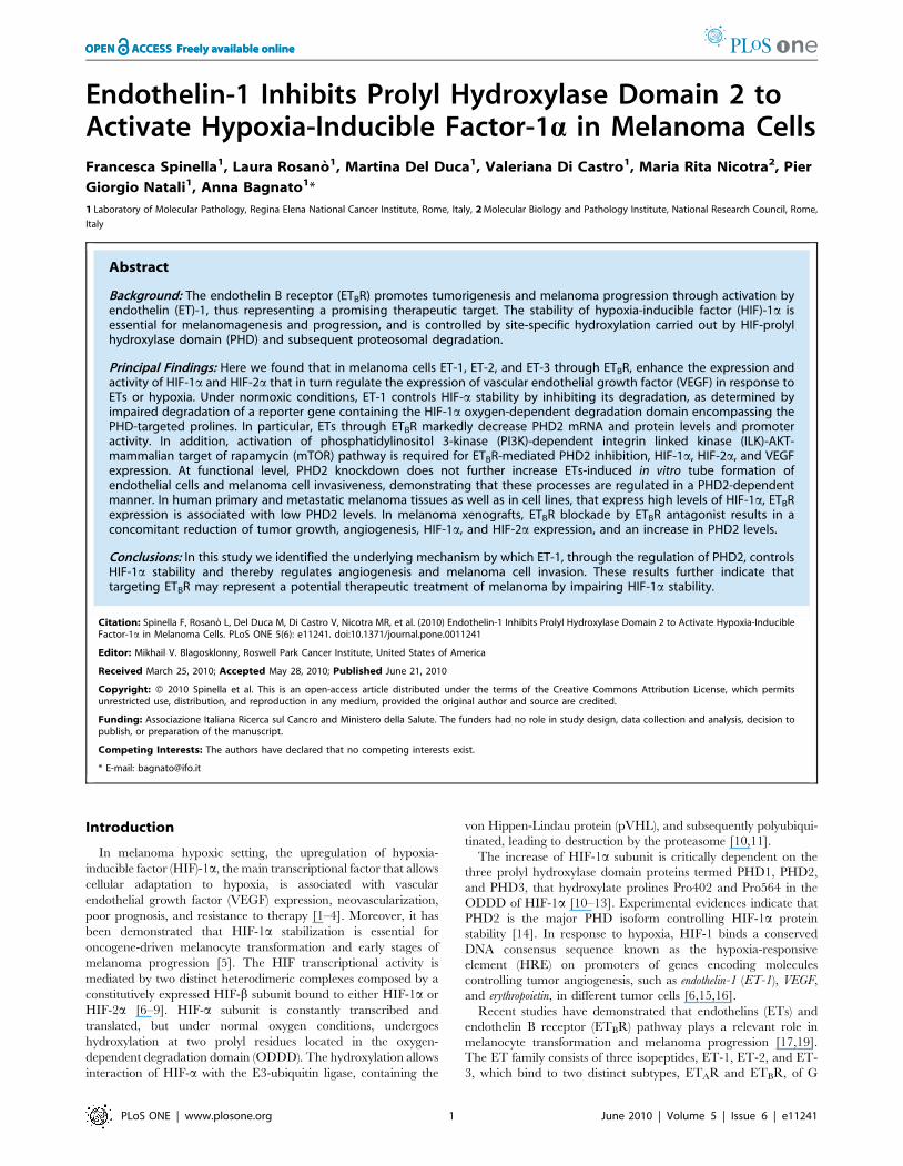
ETs induce HIF-1a and HIF-2a accumulation and activitythrough ETBR
HIF-1a and HIF-2a have been proposed to function as key
factors in angiogenesis and their expression has been associated
with VEGF expression in human melanoma [4]. In this study we
investigated the role of ET-1 axis on both HIF-1a and HIF-2ainduction and transcriptional activity in melanoma cells. In
primary (1007) and metastatic (SKMel28, M10, Mel120, M14)
melanoma cell lines cultured in normoxic conditions ET-1 or
ET-3 markedly increased HIF-2a protein levels, that paralleled
HIF-1a accumulation, in all cell lines (Figure 1A). Moreover ET-
2, similarly to ET-1 and ET-3, was able to induce HIF-1a and
HIF-2a protein accumulation (Figure 1B). The inhibitory effect
produced by two different ETBR pharmacological inhibitors,
BQ788, a peptide antagonist, and A-192621, a nonpeptide
ETBR antagonist, as well as by ETBR silencing by specific
siRNA showed that ETBR is the relevant receptor that controls
HIF-1a and HIF-2a protein accumulation (Figure 1B and Figure
S1A). In melanoma cells, ET-1 induced a dose- and time-
dependent induction of HIF-1a and HIF-2a reaching the
maximum at 100 nM following 16–24 h stimulation (Figure
S1B). Similarly, ET-3 stimulated a dose- and time-dependent
HIF-1a accumulation, whereas an unrelated peptide not
implicated in angiogenesis [29] was unable to induce it (Figure
S1C). To determine whether ETs-induced HIF-1a is transcrip-
tionally active, we transfected melanoma cells with a luciferase
reporter gene driven by three specific HRE. ET-1 or ET-3
treatment resulted in a significant increase (p,0.005) in HIF-1a-
induced luciferase reporter activity, that was blocked by BQ788,
as well as by ETBR siRNA (Figure 1C). The ET-1-induced HIF-
1a transcriptional activation was further investigated by
analyzing the effect of ET-1 or ET-3 on VEGF. The increase
in HIF-1a and HIF-2a protein levels in the presence of ET-1 or
ET-3 or hypoxia paralleled those of VEGF (Figure 1D). When
HIF-1a or HIF-2a were silenced by specific siRNA, ETs- or
hypoxia-induced VEGF expression was inhibited (Figure 1D),
indicating that either HIF-1a or HIF-2a can regulate target
genes, such as VEGF, in melanoma cells.
Figure 1. ETs induce HIF-1a and HIF-2a accumulation and activation through ETBR. HIF-1a or HIF-2a protein expression was analysed incell lysates from: A. Primary 1007, and metastatic, SKMel28, M10, Mel120, and M14 melanoma cells treated with ET-1 or ET-3; B. 1007 cells treatedwith ET-1, ET-2 or ET-3 or with BQ788 or A-192621, in combination with ET-1, or transfected with scRNA or ETBR siRNA and treated with ET-1 for 16 h.C. 1007 cells were transiently transfected with HRE-luciferase promoter construct in the presence of either ET-1 or ET-3 or in combination with BQ788,or transfected with ETBR siRNA for 16 h. Luciferase activity was measured and expressed as fold-increase, Bars, 6 SD. *, p,0.005 compared to control;**, p,0.001 compared to ET-1 or ET-3. D. 1007 cells transfected with scRNA or with HIF-1a siRNA or HIF-2a siRNA were stimulated with either ET-1 orET-3 or hypoxia (H) for 16 h, and cell lysates were analyzed for protein expression.doi:10.1371/journal.pone.0011241.g001
Endothelin-1 Stabilizes HIF-1a
PLoS ONE | www.plosone.org 2 June 2010 | Volume 5 | Issue 6 | e11241
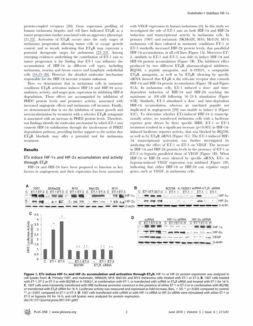
ETs induce HIF-1a stability by impairing HIF-1ahydroxylation
To asses whether ET-1 axis stabilizes HIF-1a protein, we
monitored the decay of HIF-1a after blockade of protein synthesis
with cyclohexamide (CHX). Melanoma cells were stimulated for
24 h either with hypoxia, or with ET-1 and then treated with CHX
under normoxic conditions for the indicated times. In these
conditions the decay of HIF-1a protein was observed within
120 min and was completely undetectable by the end of 240 min
(Figure 2A). When the cells were treated for 24 h with ET-1 and
then with CHX and ET-1, the increased levels of HIF-1a remained
constant up to 240 min, demonstrating that ET-1 is able to
maintain stability of HIF-1a in normoxia by slowing down its
degradation. The proteosome inhibitor MG132 protected the HIF-
1a subunit from proteosome degradation and this effect was further
increased in the presence of ET-1, indicating that ET-1, similarly to
MG132, inhibits HIF-1a degradation (Figure 2B). Because
hydroxylation at the 4-position of Pro402 and Pro564 within the
ODDD of HIF-1a is responsible for its degradation under normoxia
[10], we further investigated the role of ET-1 on the stability of HIF-
1a by transfecting melanoma cells with a reporter plasmid
expressing HIF-1a ODDD fused with luciferase (CMV-Luc-
ODDD). Following the transfection, cells were stimulated for
different times with ET-1 or cultured under hypoxia. As shown in
Figure 2C, luciferase-ODDD stabilization increased in a time-
dependent manner after stimulation with ET-1 or hypoxia, with
maximal levels attained at 16h. Dose-response analysis showed that
CMV-Luc-ODDD stability increased progressively reaching 3,5
fold induction compared to control at 100 nM ET-1 (Figure S2).
ET-1 or ET-3-induced effect on HIF-1a stability was mediated by
ETBR, as demonstrated by the inhibitory effect of BQ788
(Figure 2D). Altogether these results indicate that ET-1 axis
increases HIF-1a protein stabilization by impairing HIF-1ahydroxylation.
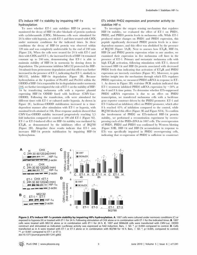
ETs inhibit PHD2 expression and promoter activity tostabilize HIF-a
To investigate the oxygen sensing mechanism that regulates
HIF-1a stability, we evaluated the effect of ET-1 on PHD1,
PHD2, and PHD3 protein levels in melanoma cells. While ET-1
produced minor changes on PHD1 and PHD3 expression, this
peptide significantly decreased PHD2 protein levels in a time-
dependent manner, and this effect was abolished by the presence
of BQ788 (Figure 3A,B). Next to assesses how ETBR, HIF-1a,
HIF-2a and PHD2 protein expression relate to one another, we
examined their expression in five melanoma cell lines in the
presence of ET-1. Primary and metastatic melanoma cells with
high ETBR activation, following stimulation with ET-1, showed
increased HIF-1a and HIF-2a protein associated with decreased
PHD2 levels thus indicating that activation of ETBR and PHD2
expression are inversely correlates (Figure 3C). Moreover, to gain
further insight into the mechanism through which ETs regulates
PHD2 expression, we measured PHD2 mRNA in response to ET-
1. As shown in Figure 3D, real-time PCR analysis indicated that
ET-1 treatment inhibited PHD2 mRNA expression by ,50% at
the 6 and 8 h time points. To determine whether ETs-suppressed
PHD2 mRNA expression is due to an effect on PHD2
transcription, we transfected melanoma cells with a luciferase
gene reporter construct driven by the PHD2 promoter. ET-1 and
ET-3 induced an inhibitory effect on PHD2 promoter, which after
8 h reached 45% of inhibition compared to the control, while
BQ788 blocked this effect (Figure 3E and Figure S3A). To confirm
the involvement of PHD2 on ETs-induced HIF-1a protein
stability, we performed a reconstitution experiment by overex-
pressing each of the PHD-cDNA in 1007 cells. The overexpression
of PHD1, PHD2 and PHD3 was confirmed by Western blotting
(Figure S3B). HIF-1a and HIF-2a accumulation in response to
ETs was specifically impaired in PHD2 overexpressing cells,
indicating that re-expression of PHD2 is sufficient to counteract
Figure 2. ETs induce HIF-1a protein stability by impairing HIFa hydroxylation. A. 1007 cells were cultured under normoxic conditions (C) orexposed to hypoxia (H) or treated with ET-1 for 24 h. Following stimulation of CHX alone or in combination with ET-1 for the indicated times. B. 1007cells were treated with MG132 alone or in combination with ET-1 for 24 h. C. 1007 and SKMel28 cells were transfected with CMV-Luc- ODDDconstruct and stimulated as indicated. Luciferase activity was expressed as fold induction. Bars, 6 SD. *, p,0.004 compared to control. D. Cellstransfected as in A were treated with ET-1 or ET-3 alone or in combination with BQ788 for 16 h. Bars, 6 SD. *, p,0.005, compared to control;**, p,0.001 compared to ET-1 or ET-3.doi:10.1371/journal.pone.0011241.g002
Endothelin-1 Stabilizes HIF-1a
PLoS ONE | www.plosone.org 3 June 2010 | Volume 5 | Issue 6 | e11241
the ET-1- or ET-3-induced HIF-a expression (Figure 3F). These
results identify the inhibition of PHD2 expression as the
mechanism underlying ETs-induced HIF-a stabilization. Con-
comitantly to the block of HIF-a accumulation, the exogenous
expression of PHD2 makes unable ET-1 and ET-3 to increase
VEGF protein levels demonstrating a tight link between PHD2/
HIF-a and ET-1-dependent VEGF expression (Figure 3F).
Moreover, knockdown of PHD2 by inhibiting the prolyl
hydroxylases with deferoxamine mesylate (DFO) resulted in a
strong induction of HIF-a and VEGF expression. The addition of
ET-1 to DFO did not induce a further increase in HIF-a, and
VEGF protein, implying that ET-1 primarily regulates HIF-aprotein accumulation through inhibition of PHD2 (Figure 3F).
Furthermore, the luciferase activity of CMV-Luc-ODDD in-
Figure 3. ETs decrease PHD2 expression and promoter activity. A. PHD1, PHD2 and PHD3 expression was analyzed in melanoma cellsunstimulated (C) or stimulated with ET-1 for the indicated times. B. PHD2 protein expression was analyzed in cells stimulated as indicated for 24 h. C.Melanoma cells were treated with ET-1 and protein expression was analysed. D. 1007 cells were stimulated as indicated. Results are expressed as copynumbers of PHD2 transcripts over cyclophilin-A. Bars, 6 SD. *, p,0.05 compared to the control. Inset shows PCR products for PHD2 and cyclophilin-A(CypA) E. Cells were transfected with the PHD2 promoter construct and stimulated as indicated for 8 h. Luciferase activity was expressed as foldinduction. Bars, 6 SD. *, p,0.006 compared to control; **, p,0.004 compared to ET-1. F. MOCK- and PHD1-, PHD2-, or PHD3-cDNA-transfected 1007cells were stimulated with ET-1 or ET-3 for 16 h. Cells were treated with DFO alone or in combination with ET-1 and lysates were analysed for proteinexpression. G. 1007 cells were cotransfected with the CMV-Luc-ODDD construct and with the construct indicated in F, and stimulated with ET-1 or ET-3 for 16 h. Luciferase activity was expressed as fold induction. Bars, 6 SD. *, p,0.001 compared to the control; **, p,0.005 compared to MOCK-transfected cells treated with ET-1 or ET-3.doi:10.1371/journal.pone.0011241.g003
Endothelin-1 Stabilizes HIF-1a
PLoS ONE | www.plosone.org 4 June 2010 | Volume 5 | Issue 6 | e11241
creased by ET-1 or ET-3 was impaired only in cells overexpressing
PHD2 (Figure 3G), demonstrating that the re-expression of PHD2
antagonizes the effect of ET-1 and ET-3 on HIF-a degradation.
These results further support the role of PHD2 on ETs-induced
HIF-1a stability and angiogenic-related factor expression.
ETs signal through a PI3K-dependent ILK-AKT-mTORpathway to induce HIF-1a stability and PHD2 inhibition
It has been reported that ILK, AKT and mTOR signalling are
the main pathways controlling HIF-1a expression [6,30,31]. ILK
is a serine/threonine kinase that plays an important role in linking
extracellular signalling to the regulation of melanoma tumor
growth and progression [30–33]. Therefore we analyzed the
signalling pathways involved in ET-1-induced HIF-1a stability. In
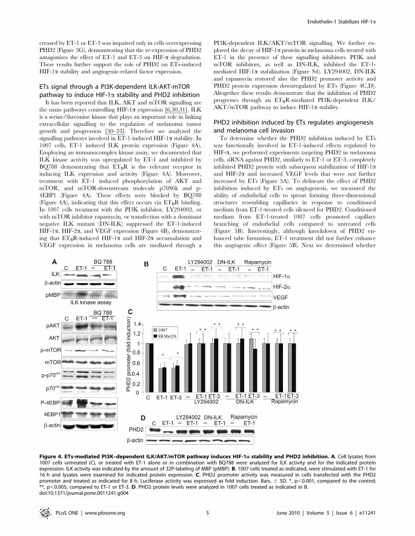
1007 cells, ET-1 induced ILK protein expression (Figure 4A).
Employing an immunocomplex kinase assay, we documented that
ILK kinase activity was upregulated by ET-1 and inhibited by
BQ788 demonstrating that ETBR is the relevant receptor in
inducing ILK expression and activity (Figure 4A). Moreover,
treatment with ET-1 induced phosphorylation of AKT and
mTOR, and mTOR-downstream molecule p70S6k and p-
4EBP1 (Figure 4A). These effects were blocked by BQ788
(Figure 4A), indicating that this effect occurs via ETBR binding.
In 1007 cells treatment with the PI3K inhibitor, LY294002, or
with mTOR inhibitor rapamycin, or transfection with a dominant
negative ILK mutant (DN-ILK) suppressed the ET-1-induced
HIF-1a, HIF-2a, and VEGF expression (Figure 4B), demonstrat-
ing that ETBR-induced HIF-1a and HIF-2a accumulation and
VEGF expression in melanoma cells are mediated through a
PI3K-dependent ILK/AKT/mTOR signalling. We further ex-
plored the decay of HIF-1a protein in melanoma cells treated with
ET-1 in the presence of these signalling inhibitors. PI3K and
mTOR inhibitors, as well as DN-ILK, inhibited the ET-1-
mediated HIF-1a stabilization (Figure S4). LY294002, DN-ILK
and rapamycin restored also the PHD2 promoter activity and
PHD2 protein expression downregulated by ETs (Figure 4C,D).
Altogether these results demonstrate that the inhibition of PHD2
progresses through an ETBR-mediated PI3K-dependent ILK/
AKT/mTOR pathway to induce HIF-1a stability.
PHD2 inhibition induced by ETs regulates angiogenesisand melanoma cell invasion
To determine whether the PHD2 inhibition induced by ETs
was functionally involved in ET-1-induced effects regulated by
HIF-a, we performed experiments targeting PHD2 in melanoma
cells. siRNA against PHD2, similarly to ET-1 or ET-3, completely
inhibited PHD2 protein with subsequent stabilization of HIF-1aand HIF-2a and increased VEGF levels that were not further
increased by ETs (Figure 5A). To delineate the effect of PHD2
inhibition induced by ETs on angiogenesis, we measured the
ability of endothelial cells to sprout forming three-dimensional
structures resembling capillaries in response to conditioned
medium from ET-1-treated cells silenced for PHD2. Conditioned
medium from ET-1-treated 1007 cells promoted capillary
branching of endothelial cells compared to untreated cells
(Figure 5B). Interestingly, although knockdown of PHD2 en-
hanced tube formation, ET-1 treatment did not further enhance
this angiogenic effect (Figure 5B). Next we determined whether
Figure 4. ETs-mediated PI3K–dependent ILK/AKT/mTOR pathway induces HIF-1a stability and PHD2 inhibition. A. Cell lysates from1007 cells untreated (C), or treated with ET-1 alone or in combination with BQ788 were analyzed for ILK activity and for the indicated proteinexpression. ILK activity was indicated by the amount of 32P-labeling of MBP (pMBP). B. 1007 cells treated as indicated, were stimulated with ET-1 for16 h and lysates were examined for indicated protein expression. C. PHD2 promoter activity was measured in cells transfected with the PHD2promoter and treated as indicated for 8 h. Luciferase activity was expressed as fold induction. Bars, 6 SD. *, p,0.001, compared to the control;**, p,0.005, compared to ET-1 or ET-3. D. PHD2 protein levels were analyzed in 1007 cells treated as indicated in B.doi:10.1371/journal.pone.0011241.g004
Endothelin-1 Stabilizes HIF-1a
PLoS ONE | www.plosone.org 5 June 2010 | Volume 5 | Issue 6 | e11241
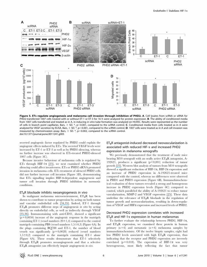
secreted angiogenic factor regulated by PHD2 could explain the
angiogenic effects induced by ETs. The secreted VEGF levels were
increased by ET-1 or ET-3 as well as by PHD2 silencing, whereas
no further increase was observed in ETs-treated PHD2-silenced
1007 cells (Figure 5C).
Because invasive behaviour of melanoma cells is regulated by
ETs through HIF-1a [25], we next examined whether PHD2
silencing could affect invasiveness. ETs or PHD2 siRNA promoted
invasion in melanoma cells. ETs treatment of silenced PHD2 cells
did not further increase cell invasion (Figure 5D), demonstrating
that ETs signalling implies HIF-a-dependent angiogenesis and
tumor cell invasion through PHD2 inhibition in normoxic
conditions.
ETBR blockade inhibits neoangiogenesis in vivoIn malignant melanoma microenvironment, ETBR has been
shown to contribute to tumor progression by acting on both tumor
and vascular endothelial cells [34,35]. Indeed, ET-1 through
ETBR promotes different steps of angiogenesis in vitro by acting
directly on endothelial cells, as well as indirectly through VEGF
[35,36]. Immunostaining with anti-CD31, showed a significant
(p = 0,0056) increase of the angiogenic response in the matrigels
containing ET-1 (vessel numbers 2061,4) compared to the control
matrigels containing PBS (vessel numbers 1,560,3) (Figure 6A). In
the plugs containing BQ788 and ET-1, the number of blood
vessels was significantly (p = 0,0028) reduced (vessel numbers
1,560,2) compared to the matrigels containing ET-1 alone
(Figure 6A). These results demonstrate that ET-1 selectively
through ETBR promotes neoangiogenesis and that a selective
ETBR antagonist can effectively impair angiogenesis in vivo.
ETBR antagonist-induced decreased neovascularization isassociated with reduced HIF-a and increased PHD2expression in melanoma xenografts
We previously demonstrated that the treatment of nude mice
bearing M10 xenograft with an orally active ETBR antagonist, A-
192621, produces a significant (p,0,001) reduction of tumor
growth [25]. Western blot analysis of tumors from M10 xenografts
showed a significant reduction of HIF-1a, HIF-2a expression and
an increase of PHD2 expression in A-192621-treated mice
compared with the control, whereas no differences were observed
in PHD1 and PHD3 expression (Figure 6B). Immunohistochem-
ical evaluation of these tumors revealed a strong and homogenous
increase in PHD2 expression levels (Figure 6C) compared to
control, which paralleled the ability of A-192621 to reduce tumor
vascularization, MMP-2 and VEGF expression [25]. These data
underline the relevance of ETBR blockade in the regulation of
tumor growth and neovascularization, resulting in down-regula-
tion of VEGF and HIFa expression and increased levels of PHD2.
Decreased PHD2 expression correlates with increasedETBR and HIF-1a expression in human melanomas
To further evaluate the relationship between PHD2, HIF-1a,
and ETBR expression, we examined these protein in human
primary (n = 6) and metastatic (n = 6) melanoma samples by
immunohistochemistry. Of the twelve bioptic samples, eight had
low PHD2 levels associated with high ETBR expression, thus
indicating that the receptor and PHD2 expression are inversely
correlated (p = 0.018). The expression of HIF-1a was very
heterogeneous, most likely reflecting the fact that tumor
Figure 5. ETs regulate angiogenesis and melanoma cell invasion through inhibition of PHD2. A. Cell lysates from scRNA or siRNA forPHD2-transfected 1007 cells treated with or without ET-1 or ET-3 for 16 h were analyzed for protein expression. B. The ability of conditioned mediafrom 1007 cells transfected and treated as in A, in inducing in vitro tube formation was analyzed on HUVEC. Results were represented as the numberof cells in branch point capillaries. Bars, 6 SD. *, p,0.001, compared to the scRNA control. C. Conditioned media from cells treated as in A wereanalyzed for VEGF secretion by ELISA. Bars, 6 SD. *, p,0.001, compared to the scRNA control. D. 1007 cells were treated as in A and cell invasion wasmeasured by chemoinvasion assay. Bars, 6 SD. *, p,0.002, compared to the scRNA control.doi:10.1371/journal.pone.0011241.g005
Endothelin-1 Stabilizes HIF-1a
PLoS ONE | www.plosone.org 6 June 2010 | Volume 5 | Issue 6 | e11241
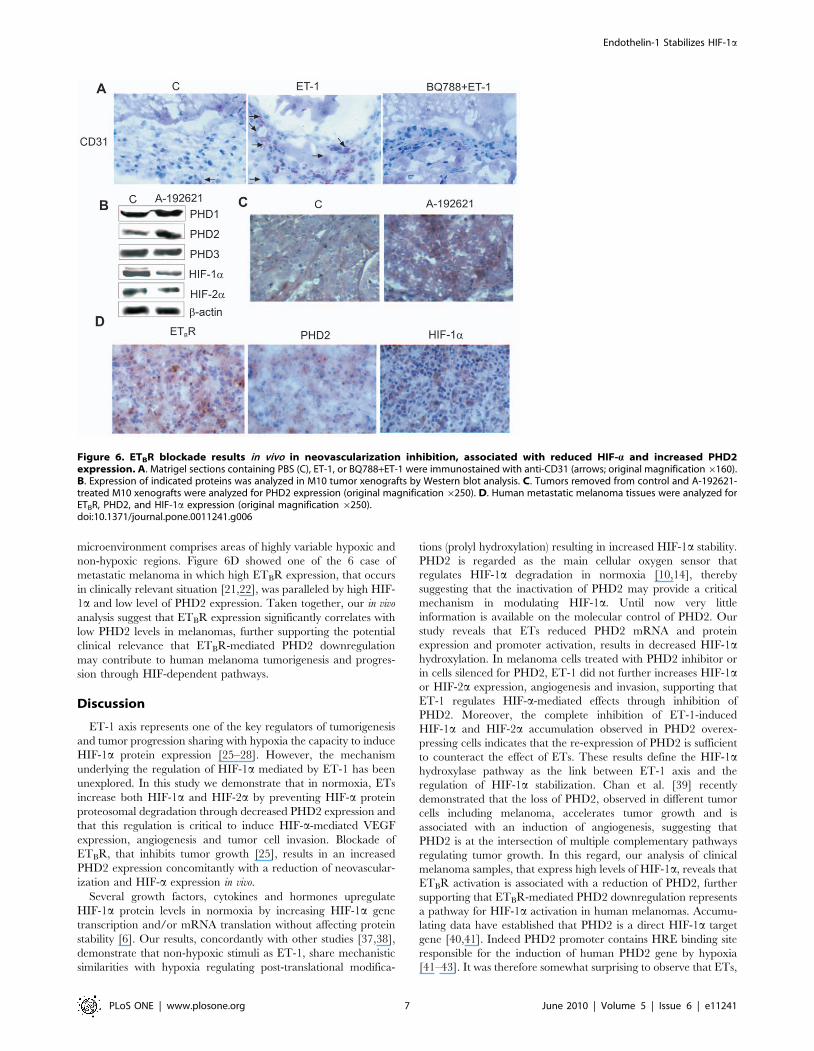
microenvironment comprises areas of highly variable hypoxic and
non-hypoxic regions. Figure 6D showed one of the 6 case of
metastatic melanoma in which high ETBR expression, that occurs
in clinically relevant situation [21,22], was paralleled by high HIF-
1a and low level of PHD2 expression. Taken together, our in vivo
analysis suggest that ETBR expression significantly correlates with
low PHD2 levels in melanomas, further supporting the potential
clinical relevance that ETBR-mediated PHD2 downregulation
may contribute to human melanoma tumorigenesis and progres-
sion through HIF-dependent pathways.
Discussion
ET-1 axis represents one of the key regulators of tumorigenesis
and tumor progression sharing with hypoxia the capacity to induce
HIF-1a protein expression [25–28]. However, the mechanism
underlying the regulation of HIF-1a mediated by ET-1 has been
unexplored. In this study we demonstrate that in normoxia, ETs
increase both HIF-1a and HIF-2a by preventing HIF-a protein
proteosomal degradation through decreased PHD2 expression and
that this regulation is critical to induce HIF-a-mediated VEGF
expression, angiogenesis and tumor cell invasion. Blockade of
ETBR, that inhibits tumor growth [25], results in an increased
PHD2 expression concomitantly with a reduction of neovascular-
ization and HIF-a expression in vivo.
Several growth factors, cytokines and hormones upregulate
HIF-1a protein levels in normoxia by increasing HIF-1a gene
transcription and/or mRNA translation without affecting protein
stability [6]. Our results, concordantly with other studies [37,38],
demonstrate that non-hypoxic stimuli as ET-1, share mechanistic
similarities with hypoxia regulating post-translational modifica-
tions (prolyl hydroxylation) resulting in increased HIF-1a stability.
PHD2 is regarded as the main cellular oxygen sensor that
regulates HIF-1a degradation in normoxia [10,14], thereby
suggesting that the inactivation of PHD2 may provide a critical
mechanism in modulating HIF-1a. Until now very little
information is available on the molecular control of PHD2. Our
study reveals that ETs reduced PHD2 mRNA and protein
expression and promoter activation, results in decreased HIF-1ahydroxylation. In melanoma cells treated with PHD2 inhibitor or
in cells silenced for PHD2, ET-1 did not further increases HIF-1aor HIF-2a expression, angiogenesis and invasion, supporting that
ET-1 regulates HIF-a-mediated effects through inhibition of
PHD2. Moreover, the complete inhibition of ET-1-induced
HIF-1a and HIF-2a accumulation observed in PHD2 overex-
pressing cells indicates that the re-expression of PHD2 is sufficient
to counteract the effect of ETs. These results define the HIF-1ahydroxylase pathway as the link between ET-1 axis and the
regulation of HIF-1a stabilization. Chan et al. [39] recently
demonstrated that the loss of PHD2, observed in different tumor
cells including melanoma, accelerates tumor growth and is
associated with an induction of angiogenesis, suggesting that
PHD2 is at the intersection of multiple complementary pathways
regulating tumor growth. In this regard, our analysis of clinical
melanoma samples, that express high levels of HIF-1a, reveals that
ETBR activation is associated with a reduction of PHD2, further
supporting that ETBR-mediated PHD2 downregulation represents
a pathway for HIF-1a activation in human melanomas. Accumu-
lating data have established that PHD2 is a direct HIF-1a target
gene [40,41]. Indeed PHD2 promoter contains HRE binding site
responsible for the induction of human PHD2 gene by hypoxia
[41–43]. It was therefore somewhat surprising to observe that ETs,
Figure 6. ETBR blockade results in vivo in neovascularization inhibition, associated with reduced HIF-a and increased PHD2expression. A. Matrigel sections containing PBS (C), ET-1, or BQ788+ET-1 were immunostained with anti-CD31 (arrows; original magnification6160).B. Expression of indicated proteins was analyzed in M10 tumor xenografts by Western blot analysis. C. Tumors removed from control and A-192621-treated M10 xenografts were analyzed for PHD2 expression (original magnification 6250). D. Human metastatic melanoma tissues were analyzed forETBR, PHD2, and HIF-1a expression (original magnification 6250).doi:10.1371/journal.pone.0011241.g006
Endothelin-1 Stabilizes HIF-1a
PLoS ONE | www.plosone.org 7 June 2010 | Volume 5 | Issue 6 | e11241
which rapidly increased HIF-1a levels, inhibited PHD2 protein
expression. This could be explained by recent results indicating
that PHD2 induction generates an autoregulatory loop controlling
HIF-1a stability [43–45]. Therefore our hypothesis supports the
notion that ET-1 axis, similarly to hypoxia, modulates the
autoregulatory loop of HIF-1a-PHD2 in melanoma cells through
a balance between the inhibitory ET-1 and the stimulatory HIF-
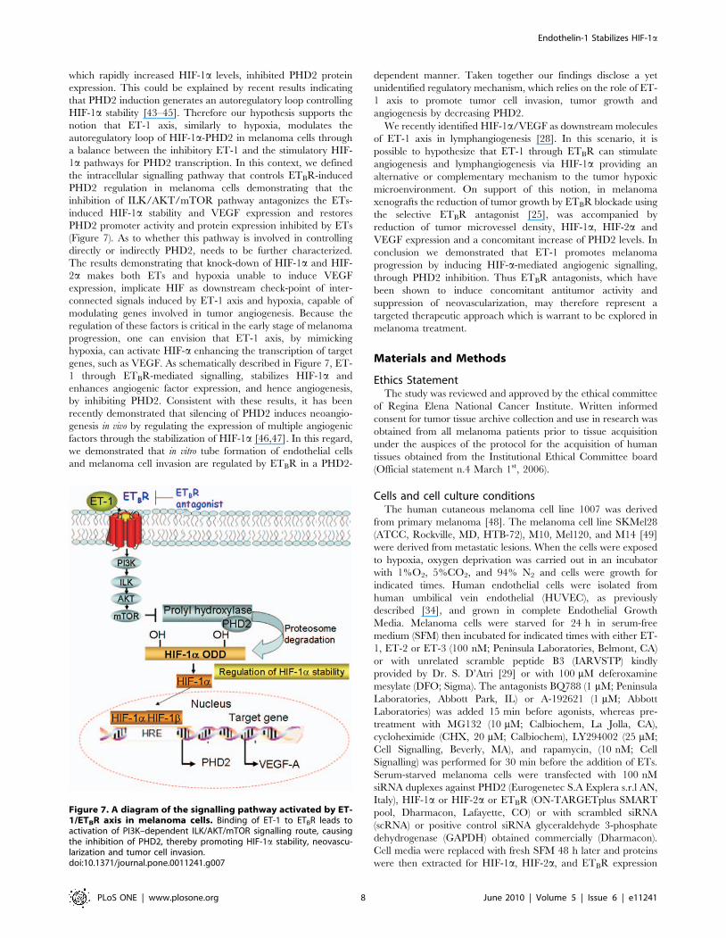
1a pathways for PHD2 transcription. In this context, we defined
the intracellular signalling pathway that controls ETBR-induced
PHD2 regulation in melanoma cells demonstrating that the
inhibition of ILK/AKT/mTOR pathway antagonizes the ETs-
induced HIF-1a stability and VEGF expression and restores
PHD2 promoter activity and protein expression inhibited by ETs
(Figure 7). As to whether this pathway is involved in controlling
directly or indirectly PHD2, needs to be further characterized.
The results demonstrating that knock-down of HIF-1a and HIF-
2a makes both ETs and hypoxia unable to induce VEGF
expression, implicate HIF as downstream check-point of inter-
connected signals induced by ET-1 axis and hypoxia, capable of
modulating genes involved in tumor angiogenesis. Because the
regulation of these factors is critical in the early stage of melanoma
progression, one can envision that ET-1 axis, by mimicking
hypoxia, can activate HIF-a enhancing the transcription of target
genes, such as VEGF. As schematically described in Figure 7, ET-
1 through ETBR-mediated signalling, stabilizes HIF-1a and
enhances angiogenic factor expression, and hence angiogenesis,
by inhibiting PHD2. Consistent with these results, it has been
recently demonstrated that silencing of PHD2 induces neoangio-
genesis in vivo by regulating the expression of multiple angiogenic
factors through the stabilization of HIF-1a [46,47]. In this regard,
we demonstrated that in vitro tube formation of endothelial cells
and melanoma cell invasion are regulated by ETBR in a PHD2-
dependent manner. Taken together our findings disclose a yet
unidentified regulatory mechanism, which relies on the role of ET-
1 axis to promote tumor cell invasion, tumor growth and
angiogenesis by decreasing PHD2.
We recently identified HIF-1a/VEGF as downstream molecules
of ET-1 axis in lymphangiogenesis [28]. In this scenario, it is
possible to hypothesize that ET-1 through ETBR can stimulate
angiogenesis and lymphangiogenesis via HIF-1a providing an
alternative or complementary mechanism to the tumor hypoxic
microenvironment. On support of this notion, in melanoma
xenografts the reduction of tumor growth by ETBR blockade using
the selective ETBR antagonist [25], was accompanied by
reduction of tumor microvessel density, HIF-1a, HIF-2a and
VEGF expression and a concomitant increase of PHD2 levels. In
conclusion we demonstrated that ET-1 promotes melanoma
progression by inducing HIF-a-mediated angiogenic signalling,
through PHD2 inhibition. Thus ETBR antagonists, which have
been shown to induce concomitant antitumor activity and
suppression of neovascularization, may therefore represent a
targeted therapeutic approach which is warrant to be explored in
melanoma treatment.
Materials and Methods
Ethics StatementThe study was reviewed and approved by the ethical committee
of Regina Elena National Cancer Institute. Written informed
consent for tumor tissue archive collection and use in research was
obtained from all melanoma patients prior to tissue acquisition
under the auspices of the protocol for the acquisition of human
tissues obtained from the Institutional Ethical Committee board
(Official statement n.4 March 1st, 2006).
Cells and cell culture conditionsThe human cutaneous melanoma cell line 1007 was derived
from primary melanoma [48]. The melanoma cell line SKMel28
(ATCC, Rockville, MD, HTB-72), M10, Mel120, and M14 [49]
were derived from metastatic lesions. When the cells were exposed
to hypoxia, oxygen deprivation was carried out in an incubator
with 1%O2, 5%CO2, and 94% N2 and cells were growth for
indicated times. Human endothelial cells were isolated from
human umbilical vein endothelial (HUVEC), as previously
described [34], and grown in complete Endothelial Growth
Media. Melanoma cells were starved for 24 h in serum-free
medium (SFM) then incubated for indicated times with either ET-
1, ET-2 or ET-3 (100 nM; Peninsula Laboratories, Belmont, CA)
or with unrelated scramble peptide B3 (IARVSTP) kindly
provided by Dr. S. D’Atri [29] or with 100 mM deferoxamine
mesylate (DFO; Sigma). The antagonists BQ788 (1 mM; Peninsula
Laboratories, Abbott Park, IL) or A-192621 (1 mM; Abbott
Laboratories) was added 15 min before agonists, whereas pre-
treatment with MG132 (10 mM; Calbiochem, La Jolla, CA),
cycloheximide (CHX, 20 mM; Calbiochem), LY294002 (25 mM;
Cell Signalling, Beverly, MA), and rapamycin, (10 nM; Cell
Signalling) was performed for 30 min before the addition of ETs.
Serum-starved melanoma cells were transfected with 100 nM
siRNA duplexes against PHD2 (Eurogenetec S.A Explera s.r.l AN,
Italy), HIF-1a or HIF-2a or ETBR (ON-TARGETplus SMART
pool, Dharmacon, Lafayette, CO) or with scrambled siRNA
(scRNA) or positive control siRNA glyceraldehyde 3-phosphate
dehydrogenase (GAPDH) obtained commercially (Dharmacon).
Cell media were replaced with fresh SFM 48 h later and proteins
were then extracted for HIF-1a, HIF-2a, and ETBR expression
Figure 7. A diagram of the signalling pathway activated by ET-1/ETBR axis in melanoma cells. Binding of ET-1 to ETBR leads toactivation of PI3K–dependent ILK/AKT/mTOR signalling route, causingthe inhibition of PHD2, thereby promoting HIF-1a stability, neovascu-larization and tumor cell invasion.doi:10.1371/journal.pone.0011241.g007
Endothelin-1 Stabilizes HIF-1a
PLoS ONE | www.plosone.org 8 June 2010 | Volume 5 | Issue 6 | e11241
analysis. Conditioned cell medium containing secreted proteins
was collected, centrifuged, filtered and concentrated.
Western blot analysisWhole cell lysates or homogenized M10 tumor specimens were
subjected to SDS-PAGE and analyzed by Western blotting. Blots
were developed with the enhanced chemiluminescence detection
system (ECL; Amersham Pharmacia Biotech, Buckinghamshire,
UK). Antibody against HIF-1a was from Transduction Labora-
tory (Lexington, KY). HIF-2a, PHD1, PHD2 and PHD3
antibodies were from Novus Biologicals (Littleton, CO), VEGF
was from Santa Cruz Biotechnology (Santa Cruz, CA), ETBR was
from Abcam plc (Cambridge, UK), GAPDH and b-actin, used as
loading control, were from Oncogene (CN Biosciences, Inc.,
Darmastadt, Germany).
Real-time PCR. Total RNA was isolated using the Trizol
(Invitrogen, Carlsbad, CA) according to the manufacturer’s
protocol. 5 mg of RNA was reversed transcribed using
SuperScriptH VILOTM cDNA synthesis kit (Invitrogen).
Quantitative real-time-PCR was performed by using LightCycler
rapid thermal cycler system (Roche Diagnostics, Indianapolis IN)
according to the manufacturer’s instructions. Reaction was
performed in 20 ml volume with 0,3 mM primers, by using
LightCycler-FastStart DNA Master Plus SYBR Green mix
(Roche Diagnostics) from 1 ml cDNA. Primers used were as
follow: PHD2, (forward) 59-GCACGACACCGGGAAGTT-39,
(reverse) 59-CCAGCTTCCCGTTACAGT-39, Cyclophilin-A,
(forward) 59-TTCATCTGCACTGCCAAGAC -39, (reverse) 59–
TGGAGTTGTCCACAGTCAGC-39. The number of each gene-
amplified product was normalized to the number of cyclophilin-A
amplified product and expressed as copy numbers of PHD2
transcripts over cyclophilin-A (61023).
Transfectiona and luciferase assay. Transfection
experiments employed the LipofectAMINE reagent (Invitrogen)
according to the manufacturer’s protocol. Plasmid for transfections
were used as follow: 1 mg of ILK cDNA (kinase dead, DN-ILK) in
pUSEamp (E359K mutant) (Millipore, Billerica, MA) or with
pcDNA3-PHD1, pcDNA3-PHD2, or pcDNA3-PHD3 vectors
(Dr. J. Geadle, The Henry Wellcome Trust Centre for Human
Genetics, Oxford, UK) or empty vector pcDNA3 (MOCK)
(Promega Corporation, Madison, WI) as control. 1007, and
SKMel28 cells were transfected with different luciferase reporter
constructs, including a plasmid encoding CMV-Luc- ODDD (Dr.
R. K. Bruick, University of Texas Southwestern Medical Center,
TX), or the previously described HRE-Luc construct (Dr. A.
Giaccia, Stanford University School of Medicine, Stanford, CA),
as well as the human PHD2 proximal promoter construct pGL3b
(1454/3172) P2PWT (Dr. E. Metzen, University of Luebeck,
Luebeck, Germany). The pCMV-b-galactosidase plasmid
(Promega) was used as control for transfection efficiency. The
cells were lysed and their luciferase activities were measured
(Luciferase assay system, Promega).
ILK Immune Complex Kinase Assay. Integrin linked kinase
(ILK) activity was measured as previously described [50]. Briefly cell
lysates were immunoprecipitated with anti-ILK (Millipore). Assays
were done directly on the protein A-Sepharose (Sigma) beads in the
presence of 5 mCi of c-32P (Amersham Pharmacia Biotech) and
2.5 mg of myelin basic protein (MBP) was used as substrate
(Millipore). Phosphorylated MBP bands were visualized by
autoradiography of dried SDS-10% PAGE gels.
ELISA. The VEGF protein levels in the conditioned medium
were determined in triplicate by ELISA using the Quantikine
Human VEGF immunoassay kit (R&D Systems, Minneapolis,
MN).
In vitro Angiogenesis Assay. HUVEC were plated on basal
membrane extract (10 mg/ml, Cultrex BME; Trevigen Inc.
Helgerman, CT) in the presence of conditioned media from
scRNA- or siPHD2-transfected 1007 cells. After 24 h, cells were
visualized by light microscopy. The amount of angiogenesis was
quantified by counting the number of cells in branch point
capillaries ($3 cells per branch) in five random fields per replicate.
Chemoinvasion assay. Chemoinvasion was assessed using a
48-well–modified Boyden’s chamber (Neuro Probe Inc.
Gaithersburg, MD) and 8 mm pore polyvinyl pyrrolidone–free
polycarbonate Nucleopore filters (Costar, New York, NY) as
previously described [25]. The filters were coated with an even
layer of 0.5 mg/ml Matrigel (Becton Dickinson, Franklin Lakes,
NJ). The lower compartment of chamber was filled with
chemoattractant (ET-1 or ET-3). 1007 cells (16106 cells/ml)
were harvested and placed in the upper compartment (55 ml per
well). After 6 h of incubation at 37uC, the filters were removed,
stained with Diff-Quick (Merz-Dade, Dudingen, Switzerland), and
the migrated cells in 10 high-power fields were counted. Each
experimental point was analyzed in triplicate.
M10 melanoma xenografts. Female athymic (nu+/nu+)
mice, 4 to 6 weeks of age (Charles River Laboratories, Milan,
Italy), were handled according to the Institutional guidelines under
the control of the Italian Ministry of Health (DL 116/92),
following detailed internal rules according to: Workamn P., et al.
(1998) United Kingdom Coordinating Committee on Cancer
Research (Guidelines for the welfare of animals in experimental
neoplasia. Br. J. Cancer 77: 1–10). Mice were injected s.c. on one
flank with 1.56106 viable M10 cells expressing ETBR. The mice
were randomized in groups (n = 10) to receive treatment i.p. for 21
days with A-192621 (10 mg/kg/d), and controls were injected
with 200 ml drug vehicle (0.25 N NaHCO3). The treatments were
started 7 days after the xenografts, when the tumor was palpable
[25]. Each experiment was repeated thrice, with a total of 20 mice
for each experiment. All tumors for each group for each
experiment were harvested from M10 xenografts for Western
Blot analysis. Immunohistochemical analysis was performed in six
samples of each group of the tumors previously analyzed by
Western blot.
Matrigel plug assay. Male C57BL/6 mice (Charles River
Laboratories) were handled according to the institutional
guidelines under the control of the Italian Ministry of Health
(DL 116/92), Mice were subcutaneously injected with 0.5 ml
matrigel containing PBS (control), 0.8 mM ET-1 alone or in
combination with 8mM BQ788, as previously described [34]. The
matrigels surrounded by murine tissue were removed 10 days after
implantation, and snap frozen in liquid nitrogen for
immunohistochemical analysis.
ImmunohistochemistryIndirect immunoperoxidase staining was carried out on acetone-
fixed 4 mm tissue sections. The avidin biotin assays were performed
using the Vectastatin Elite kit (for nonmurine primary antibodies)
and the Vector MOM immunodetection kit (for murine primary
antibodies) obtained from Vector Laboratories (Burlingame, CA) on
size-matchable tumor tissues from control and A-192621 treated
M10 xenografts [25] and on human melanoma samples. Sections
incubated with isotype-matched immunoglobulins or normal
immunoglobulins served as negative control.
Statistical analysisResults are representative of at least three independent
experiments each performed in triplicate. Statistical analysis was
done using the Student t test, Fisher’s exact test, as appropriated.
Endothelin-1 Stabilizes HIF-1a
PLoS ONE | www.plosone.org 9 June 2010 | Volume 5 | Issue 6 | e11241
All analyses were performed using the SPSS 11 software (SPSS,
Inc., Chicago, IL). All statistical tests were two-sided. p,0.05 was
considered statistically significant.
Supporting Information
Figure S1 ETs induce HIF-1a and HIF-2a expression in
melanoma cells. A. 1007 cells were transfected for 48 h with
scRNA or siRNA for ETB R or siRNA for GAPDH, and ETBR or
GAPDH protein expression was analyzed by Western blotting. B.
Western blotting analysis of HIF-1a and HIF-2a expression was
performed in whole cell lysates from 1007 and SKMel28 cells
treated with increased concentrations of ET-1 for 16 h or with
100 nM ET-1 for the indicated times. C. Western blotting analysis
of HIF-1a expression was performed in whole cell lysates from
1007 cells were treated with increased concentration ET-3 or with
unrelated peptide scramble B3 (B3; 30 mM), for 16 h, or with
100 nM ET-3 for the indicated times. Anti-b-actin was used as
loading control.
Found at: doi:10.1371/journal.pone.0011241.s001 (0.24 MB TIF)
Figure S2 ET-1 impairs HIF-1a hydroxylation. 1007 and
SKMel28 cells transfected with CMV-Luc-ODDD were treated
with the indicated concentrations of ET-1 for 16 h. Luciferase
activity was expressed as fold induction. Bars, 6 SD. *, p,0.004,
compared to control.
Found at: doi:10.1371/journal.pone.0011241.s002 (0.05 MB TIF)
Figure S3 ET-3 decreases PHD2 promoter activity. A. 1007 and
SKMel28 cells were transfected with the construct containing the
PHD2 promoter and treated with 100 nM ET-3 alone or in
combination with 1mM BQ788 for 8h. Luciferase activity was
expressed as fold induction. Bars, 6 SD. *, p,0.006 compared to
control, **, p,0.005 compared to ET-1. B. 1007 cells were
transfected with each of the pcDNA3-PHDs vectors or with
pcDNA3 (empty vector, C). The expression of PHD isoforms was
analyzed by Western blotting. Anti-b-actin was used as loading
control.
Found at: doi:10.1371/journal.pone.0011241.s003 (0.14 MB TIF)
Figure S4 ET-1-mediated PI3K-dependent ILK/AKT/mTOR
pathway induces HIF-1a stability. 1007 or DN-ILK-transfected
cells were stimulated with ET-1. Following 24 h, cells were
stimulated with CHX for the indicated times with ET-1 alone or
in combination with signalling inhibitors and analyzed for protein
expression.
Found at: doi:10.1371/journal.pone.0011241.s004 (0.12 MB TIF)
Acknowledgments
We gratefully acknowledge Valentina Caprara, Danilo Giaccari, Stefano
Masi and Aldo Lupo for excellent technical assistance and Maria Vincenza
Sarcone for secretarial support. We also thank Dr. J. Geadle, The Henry
Wellcome Trust Centre for Human Genetics, Oxford, UK for pcDNA3-
PHDs vectors, Dr. R.K. Bruick, University of Texas Southwestern Medical
Center, TX, for the plasmid encoding CMV-Luc-HIF-1a ODDD, Dr. A.
Giaccia, Stanford University School of Medicine, Stanford, CA for HRE-
Luc construct, and Dr. E. Metzen, University of Luebeck, Luebeck,
Germany for the human PHD2 promoter construct.
Author Contributions
Conceived and designed the experiments: FS AB. Performed the
experiments: FS LR MDD VDC MRN. Analyzed the data: FS LR PGN
AB. Contributed reagents/materials/analysis tools: FS. Wrote the paper:
FS AB.
References
1. Chudnovsky Y, Khavari PA, Adams AE (2005) Melanoma genetics and the
development of rational therapeutics. J Clin Invest 115: 813–824.
2. Pouyssegur J, Dayan F, Mazure NM (2006) Hypoxia signalling in cancer and
approaches to enforce tumour regression. Nature 441: 437–443.
3. Postovit LM, Seftor EA, Seftor RE, Hendrix MJ (2006) Influence of themicroenvironment on melanoma cell fate determination and phenotype. Cancer
Res 66: 7833–7836.
4. Giatromanolaki A, Sivridis E, Kouskoukis C, Gatter KC, Harris AL, et al. (2003)Hypoxia-inducible factors 1alpha and 2alpha are related to vascular endothelial
growth factor expression and a poorer prognosis in nodular malignantmelanomas of the skin. Melanoma Res 13: 493–501.
5. Bedogni B, Welford SM, Cassarino DS, Nickoloff BJ, Giaccia AJ, et al. (2005)
The hypoxic microenvironment of the skin contributes to Akt-mediated
melanocyte transformation. Cancer Cell 8: 443–454.
6. Semenza GL (2003) Targeting HIF-1 for cancer therapy. Nat Rev Cancer 3:
721–732.
7. Giaccia A, Siim BG, Johnson RS (2003) HIF-1 as a target for drug development.
Nat Rev Drug Discov 2: 803–811.
8. Hu CJ, Wang LY, Chodosh LA, Keith B, Simon MC (2003) Differential roles ofhypoxiainducible factor 1alpha (HIF-1alpha) and HIF-2alpha in hypoxic gene
regulation. Mol Cell Biol 23: 9361–9374.
9. Raval RR, Lau KW, Tran MG, Sowter HM, Mandriota SJ, et al. (2005)Contrasting properties of hypoxia-inducible factor 1 (HIF-1) and HIF-2 in
von Hippel-Lindau-associated renal cell carcinoma. Mol Cell Biol 25: 5675–5686.
10. Kaelin WG, Jr., Ratcliffe PJ (2008) Oxygen sensing by metazoans: the central
role of the HIF hydroxylase pathway. Mol Cell 30: 393–402.
11. Ivan M, Kondo K, Yang H, et al. (2001) HIFa targeted for VHL-mediated
destruction by proline hydroxylation: implications for O2 sensing. Science 292:464–468.
12. Bruick RK, McKnight SL (2001) A conserved family of prolyl-4-hydroxylases
that modify HIF. Science 294: 1337–1340.
13. Hewitson KS, McNeill LA, Riordan MV, Tian YM, Bullock AN, et al. (2002)Hypoxia-inducible factor (HIF) asparagine hydroxylase is identical to factor
inhibiting HIF (FIH) and is related to the cupin structural family. J Biol Chem277: 26351–26355.
14. Berra E, Benizri E, Ginouves A, Volmat V, Roux D, et al. (2003) HIF prolyl-
hydroxylase 2 is the key oxygen sensor setting low steady-state levels of HIF-1ain normoxia. EMBO J 22: 4082–4090.
15. Patel N, Gonsalves CS, Malik P, Kalra VK (2008) Placenta growth factor
augments endothelin-1 and endothelin-B receptor expression via hypoxia-
inducible factor-1 alpha. Blood 112: 856–865.
16. Grimshaw MJ (2007) Endothelins and hypoxia-inducible factor in cancer.Endocr Relat Cancer 14: 233–244.
17. Berking C, Takemoto R, Satyamoorthy K, Shirakawa T, Eskandarpour M, et al.
(2004) Induction of melanoma phenotypes in human skin by growth factors andultraviolet B. Cancer Res 64: 807–811.
18. Lahav R, Suva ML, Rimoldi D, Patterson PH, Stamenkovic I (2004) Endothelin
receptor B inhibition triggers apoptosis and enhances angiogenesis inmelanomas. Cancer Res 64: 8945–8953.
19. Lahav R (2005) Endothelin receptor B is required for the expansion of
melanocyte precursors and malignant melanoma. Int J Dev Biol 49: 173–180.
20. Levin ER (1995) Endothelins. N Engl J Med 333: 356–363.
21. Bittner M, Meltzer P, Chen Y, Jiang Y, Seftor E, et al. (2000) Molecularclassification of cutaneous malignant melanoma by gene expression profiling.
Nature 406: 536–540.
22. Demunter A, De Wolf-Peeters C, Degreef H, Stas M, van den Oord JJ (2001)
Expression of the endothelin-B receptor in pigment cell lesions of the skin.Evidence for its role as tumor progression marker in malignant melanoma.
Virchows Arch 438: 485–491.
23. Bagnato A, Rosano L, Spinella F, Di Castro V, Tecce R, et al. (2004) EndothelinB receptor blockade inhibits dynamics of cell interactions and communications
in melanoma cell progression. Cancer Res 64: 1436–1443.
24. Lahav R, Heffner G, Patterson PH (1999) An endothelin receptor B antagonistinhibits growth and induces cell death in human melanoma cells in vitro and in
vivo. Proc Natl Acad Sci U S A 96: 11496–11500.
25. Spinella F, Rosano L, Di Castro V, Decandia S, Nicotra MR, et al. (2007)
Endothelin-1 and endothelin-3 promote invasive behavior via hypoxia-induciblefactor-1alpha in human melanoma cells. Cancer Res 67: 1725–1734.
26. Spinella F, Rosano L, Di Castro V, Natali PG, Bagnato A (2002) Endothelin-1
induces vascular endothelial growth factor by increasing hypoxia-induciblefactor-1alpha in ovarian carcinoma cells. J Biol Chem 277: 27850–27855.
27. Wilson JL, Burchell J, Grimshaw MJ (2006) Endothelins induce CCR7
expression by breast tumor cells via endothelin receptor A and hypoxia-inducible factor-1. Cancer Res 66: 11802–11807.
28. Spinella F, Garrafa E, Di Castro V, Rosano L, Nicotra MR, et al. (2009)
Endothelin-1 stimulates lymphatic endothelial cells and lymphatic vessels togrow and invade. Cancer Res 69: 2224–2233.
Endothelin-1 Stabilizes HIF-1a
PLoS ONE | www.plosone.org 10 June 2010 | Volume 5 | Issue 6 | e11241
29. Lacal PM, Morea V, Ruffini F, Orecchia A, Dorio AS, et al. (2008) Inhibition of
endothelial cell migration and angiogenesis by a vascular endothelial growth
factor receptor-1 derived peptide. Eur J Cancer 44: 1914–1921.
30. Tan C, Cruet-Hennequart S, Troussard A, Fazli L, Costello P, et al. (2004)
Regulation of tumor angiogenesis by integrin-linked kinase (ILK). Cancer Cell 5:
79–90.
31. Tang CH, Lu DY, Tan TW, Fu WM, Yang RS (2007) Ultrasound induces
hypoxia-inducible factor-1 activation and inducible nitric-oxide synthase
expression through the integrin/integrin-linked kinase/Akt/mammalian target
of rapamycin pathway in osteoblasts. J Biol Chem 282: 25406–25415.
32. Dai DL, Makretsov N, Campos EI, Huang C, Zhou Y, et al. (2003) Increased
expression of integrin-linked kinase is correlated with melanoma progression and
poor patient survival. Clin Cancer Res 9: 4409–4414.
33. Troussard AA, Mawji NM, Ong C, Mui A, St-Arnaud R, et al. (2003)
Conditional knock-out of integrin-linked kinase demonstrates an essential role in
protein kinase B/Akt activation. J Biol Chem 278: 22374–22378.
34. Bagnato A, Spinella F (2003) Emerging role of endothelin-1 in tumor
angiogenesis. Trends Endocrinol Metab 14: 44–50.
35. Salani D, Taraboletti G, Rosano L, Di Castro V, Borsotti P, et al. (2000)
Endothelin-1 induces an angiogenic phenotype in cultured endothelial cells and
stimulates neovascularization in vivo. Am J Pathol 157: 1703–1711.
36. Knowles J, Loizidou M, Taylor I (2005) Endothelin-1 and angiogenesis in
cancer. Curr Vasc Pharmacol 3: 309–314.
37. McMahon S, Charbonneau M, Grandmont S, Richard DE, Dubois CM (2006)
Transforming growth factor beta1 induces hypoxia-inducible factor-1 stabiliza-
tion through selective inhibition of PHD2 expression. J Biol Chem 281:
24171–24181.
38. Berchner-Pfannschmidt U, Yamac H, Trinidad B, Fandrey J (2007) Nitric oxide
modulates oxygen sensing by hypoxia-inducible factor 1-dependent induction of
prolyl hydroxylase 2. J Biol Chem 282: 1788–1796.
39. Chan DA, Kawahara TL, Sutphin PD, Chang HY, Chi JT (2009) Tumor
vasculature is regulated by PHD2-mediated angiogenesis and bone marrow-
derived cell recruitment. Cancer Cell 15: 527–38.
40. Metzen E, Stiehl DP, Doege K, Marxsen JH, Hellwig-Burgel T, et al. (2005)
Regulation of the prolyl hydroxylase domain protein 2 (phd2/egln-1) gene:identification of a functional hypoxia-responsive element. Biochem J 387:
711–717.
41. Aprelikova O, Chandramouli GV, Wood M, Vasselli JR, Riss J, et al. (2004)Regulation of HIF prolyl hydroxylases by hypoxia-inducible factors. J Cell
Biochem 92: 491–501.42. Kaelin WG (2005) Proline hydroxylation and gene expression. Annu Rev
Biochem 74: 115–128.
43. D’Angelo G, Duplan E, Boyer N, Vigne P, Frelin C (2003) Hypoxia up-regulatesprolyl hydroxylase activity: a feedback mechanism that limits HIF-1 responses
during reoxygenation. J Biol Chem 278: 38183–38187.44. Ginouves A, Ilc K, Macıas N, Pouyssegur J, Berra E (2008) PHDs overactivation
during chronic hypoxia ‘‘desensitizes’’ HIFalpha and protects cells from necrosis.Proc Natl Acad Sci U S A 105: 4745–4750.
45. Semenza GL (2004) O2-regulated gene expression: transcriptional control of
cardiorespiratory physiology by HIF-1. J Appl Physiol 96: 1173–1177.46. Wu S, Nishiyama N, Kano MR, Morishita Y, Miyazono K, et al. (2008)
Enhancement of angiogenesis through stabilization of hypoxia-inducible factor-1by silencing prolyl hydroxylase domain-2 gene. Mol Ther 16: 1227–1234.
47. Knowles HJ, Tian YM, Mole DR, Harris AL (2004) Novel mechanism of action
for hydralazine: induction of hypoxia-inducible factor-1alpha, vascular endo-thelial growth factor, and angiogenesis by inhibition of prolyl hydroxylases. Circ
Res 95: 162–169.48. Daniotti M, Oggionni M, Ranzani T, Vallacchi V, Campi V, et al. (2004) BRAF
alterations are associated with complex mutational profiles in malignantmelanoma. Oncogene 23: 5968–5977.
49. Golub SH, Hanson DC, Sulit HL, Morton DL, Pellegrino MA, et al. (1976)
Comparison of histocompatibility antigens on cultured human tumor cells andfibroblasts by quantitative antibody absorption and sensitivity to cell-mediated
cytotoxicity. J Natl Cancer Inst 56: 167–170.50. Rosano L, Spinella F, Di Castro V, Dedhar S, Nicotra MR, et al. (2006)
Integrin-linked kinase functions as a downstream mediator of endothelin-1 to
promote invasive behavior in ovarian carcinoma. Mol Cancer Ther 5: 833–842.
Endothelin-1 Stabilizes HIF-1a
PLoS ONE | www.plosone.org 11 June 2010 | Volume 5 | Issue 6 | e11241
Related Documents