Endothelial Dysfunction in Tristetraprolin-deficient Mice Is Not Caused by Enhanced Tumor Necrosis Factor- Expression * Received for publication, March 24, 2014, and in revised form, April 9, 2014 Published, JBC Papers in Press, April 11, 2014, DOI 10.1074/jbc.M114.566984 Franziska Bollmann ‡§ , Zhixiong Wu ‡ , Matthias Oelze ¶ , Daniel Siuda ‡§ , Ning Xia ‡ , Jenny Henke ‡ , Andreas Daiber ¶ , Huige Li ‡ , Deborah J. Stumpo , Perry J. Blackshear , Hartmut Kleinert ‡1 , and Andrea Pautz ‡2 From the ‡ Department of Pharmacology, § Center for Thrombosis and Hemostasis, and ¶ 2nd Medical Clinic, Molecular Cardiology, University Medical Center of the Johannes Gutenberg University, 55131 Mainz, Germany and the Laboratory of Signal Transduction, National Institute of Environmental Health Sciences, Research Triangle Park, North Carolina 27709 Background: Chronic inflammatory diseases are associated with increased cardiovascular mortality due to accelerated atherosclerosis. Results: Chronic inflammation in tristetraprolin (TTP)-deficient mice leads to endothelial dysfunction, which is related to enhanced Nox2-dependent reactive oxygen species production but independent from TNF-. Conclusion: Inflammation-related oxidative stress is an important mediator in inflammation-driven atherogenesis. Significance: In inflammatory diseases oxidative stress seems to be a major cause of cardiovascular events. Cardiovascular events are important co-morbidities in patients with chronic inflammatory diseases like rheumatoid arthritis. Tris- tetraprolin (TTP) regulates pro-inflammatory processes through mRNA destabilization and therefore TTP-deficient mice (TTP / mice) develop a chronic inflammation resembling human rheuma- toid arthritis. We used this mouse model to evaluate molecular signaling pathways contributing to the enhanced atherosclerotic risk in chronic inflammatory diseases. In the aorta of TTP / mice we observed elevated mRNA expression of known TTP targets like tumor necrosis factor- (TNF-) and macrophage inflammatory protein-1, as well as of other pro-atherosclerotic mediators, like Calgranulin A, Cathepsin S, and Osteopontin. Independent of cho- lesterol levels TTP / mice showed a significant reduction of ace- tylcholine-induced, nitric oxide-mediated vasorelaxation. The endothelial dysfunction in TTP / mice was associated with increased levels of reactive oxygen and nitrogen species (RONS), indicating an enhanced nitric oxide inactivation by RONS in the TTP / animals. The altered RONS generation correlates with increased expression of NADPH oxidase 2 (Nox2) resulting from enhanced Nox2 mRNA stability. Although TNF- is believed to be a central mediator of inflammation-driven atherosclerosis, genetic inactivation of TNF- neither improved endothelial function nor normalized Nox2 expression or RONS production in TTP / ani- mals. Systemic inflammation caused by TTP deficiency leads to endothelial dysfunction. This process is independent of cholesterol and not mediated by TNF- solely. Thus, other mediators, which need to be identified, contribute to enhanced cardiovascular risk in chronic inflammatory diseases. Atherosclerosis (AS) 3 is a disorder of the medium-sized and large arteries with thrombotic events as its clinical manifesta- tion (1). Alterations in the endothelial function are one of the earliest measurable markers in atherogenesis, which precede the development of morphologic atherosclerotic changes and are predictors of cardiovascular events (2, 3). Endothelial dys- function is a common feature of patients with different risk factors of atherosclerosis, such as hypercholesterolemia, diabe- tes, hypertension, and smoking (4). Today the key role of inflammation during atherogenesis is well accepted (5). Rheumatoid arthritis (RA) is a chronic inflammatory disease associated with enhanced cardiovascular mortality (6 – 8). The elevated risk for cardiovascular events in RA patients is independent of age, sex, smoking, hypercholes- terolemia, or body mass index (9, 10). Formation of atherosclerotic plaques involves the internal- ization of lipids into the intima of the vessel wall and endothelial cell activation. This results in enhanced chemokine/cytokine expression leading to increased expression of vascular cell adhesion molecule 1 (VCAM-1) on the surface of endothelial cells (11). The adhesion and infiltration of leukocytes and monocytes into the arterial wall contribute to plaque progres- sion. Activation of the endothelium also leads to an imbalanced secretion of endothelium-derived relaxing and contracting fac- tors and to the development of an endothelial dysfunction. In patients suffering from AS the endothelium-dependent relax- ation is impaired and the endothelial dysfunction is accepted as an early atherosclerotic marker (12). Enhanced formation of * This work was supported by Federal Ministry of Education and Research Grant BMBF 01EO1003, Innovation Foundation of the State of Rhineland- Palatinate Grant 961-386261/917K, and Deutsche Forschungsgemein- schaft Grant LI 1759/1-1 (to H. K.). 1 To whom correspondence may be addressed. E-mail: kleinert@mail. uni-mainz.de. 2 To whom correspondence may be addressed: Obere Zahlbacher Str. 67, 55101 Mainz, Germany. Tel.: 49-6131-17-9276; Fax: 49-6131-17-9042; E-mail: [email protected]. 3 The abbreviations used are: AS, atherosclerosis; CD68, cluster of differentia- tion 68; CIA, collagen-induced arthritis; CTSS, cathepsin S; DRB, 6-dichloro- 1-ribofuranosylbenzimidazole; eNOS, endothelial nitric-oxide synthase; ICAM-1, intercellular adhesion molecule 1; Ier3, immediate early response 3; iNOS, inducible nitric-oxide synthase; Mip-1 (CCL3), macrophage inflammatory protein-1; Nox, NADPH oxidase; RA, rheumatoid arthritis; RONS, reactive oxygen and nitrogen species; S100A8, S100 calcium-bind- ing protein A8; SPP1, osteopontin; TTP, tristetraprolin; VASP, vasodilator- stimulated phosphoprotein; VCAM-1, vascular cell adhesion molecule 1; qRT, quantitative RT; PDBu, phorbol 12,13-dibutyrate; ANOVA, analysis of variance. THE JOURNAL OF BIOLOGICAL CHEMISTRY VOL. 289, NO. 22, pp. 15653–15665, May 30, 2014 Published in the U.S.A. MAY 30, 2014 • VOLUME 289 • NUMBER 22 JOURNAL OF BIOLOGICAL CHEMISTRY 15653 by guest on February 13, 2018 http://www.jbc.org/ Downloaded from

Welcome message from author
This document is posted to help you gain knowledge. Please leave a comment to let me know what you think about it! Share it to your friends and learn new things together.
Transcript
Endothelial Dysfunction in Tristetraprolin-deficient Mice IsNot Caused by Enhanced Tumor Necrosis Factor-�Expression*
Received for publication, March 24, 2014, and in revised form, April 9, 2014 Published, JBC Papers in Press, April 11, 2014, DOI 10.1074/jbc.M114.566984
Franziska Bollmann‡§, Zhixiong Wu‡, Matthias Oelze¶, Daniel Siuda‡§, Ning Xia‡, Jenny Henke‡, Andreas Daiber¶,Huige Li‡, Deborah J. Stumpo�, Perry J. Blackshear�, Hartmut Kleinert‡1, and Andrea Pautz‡2
From the ‡Department of Pharmacology, §Center for Thrombosis and Hemostasis, and ¶2nd Medical Clinic, Molecular Cardiology,University Medical Center of the Johannes Gutenberg University, 55131 Mainz, Germany and the �Laboratory of SignalTransduction, National Institute of Environmental Health Sciences, Research Triangle Park, North Carolina 27709
Background: Chronic inflammatorydiseasesareassociatedwithincreasedcardiovascularmortalityduetoacceleratedatherosclerosis.Results: Chronic inflammation in tristetraprolin (TTP)-deficient mice leads to endothelial dysfunction, which is related toenhanced Nox2-dependent reactive oxygen species production but independent from TNF-�.Conclusion: Inflammation-related oxidative stress is an important mediator in inflammation-driven atherogenesis.Significance: In inflammatory diseases oxidative stress seems to be a major cause of cardiovascular events.
Cardiovascular events are important co-morbidities in patientswith chronic inflammatory diseases like rheumatoid arthritis. Tris-tetraprolin (TTP) regulates pro-inflammatory processes throughmRNA destabilization and therefore TTP-deficient mice (TTP�/�
mice) develop a chronic inflammation resembling human rheuma-toid arthritis. We used this mouse model to evaluate molecularsignaling pathways contributing to the enhanced atheroscleroticrisk in chronic inflammatory diseases. In the aorta of TTP�/� micewe observed elevated mRNA expression of known TTP targets liketumor necrosis factor-� (TNF-�) and macrophage inflammatoryprotein-1�, as well as of other pro-atherosclerotic mediators, likeCalgranulin A, Cathepsin S, and Osteopontin. Independent of cho-lesterol levels TTP�/� mice showed a significant reduction of ace-tylcholine-induced, nitric oxide-mediated vasorelaxation. Theendothelial dysfunction in TTP�/� mice was associated withincreased levels of reactive oxygen and nitrogen species (RONS),indicating an enhanced nitric oxide inactivation by RONS in theTTP�/� animals. The altered RONS generation correlates withincreased expression of NADPH oxidase 2 (Nox2) resulting fromenhanced Nox2 mRNA stability. Although TNF-� is believed to bea central mediator of inflammation-driven atherosclerosis, geneticinactivation of TNF-� neither improved endothelial function nornormalized Nox2 expression or RONS production in TTP�/� ani-mals. Systemic inflammation caused by TTP deficiency leads toendothelial dysfunction. This process is independent of cholesteroland not mediated by TNF-� solely. Thus, other mediators, whichneed to be identified, contribute to enhanced cardiovascular risk inchronic inflammatory diseases.
Atherosclerosis (AS)3 is a disorder of the medium-sized andlarge arteries with thrombotic events as its clinical manifesta-tion (1). Alterations in the endothelial function are one of theearliest measurable markers in atherogenesis, which precedethe development of morphologic atherosclerotic changes andare predictors of cardiovascular events (2, 3). Endothelial dys-function is a common feature of patients with different riskfactors of atherosclerosis, such as hypercholesterolemia, diabe-tes, hypertension, and smoking (4).
Today the key role of inflammation during atherogenesis iswell accepted (5). Rheumatoid arthritis (RA) is a chronicinflammatory disease associated with enhanced cardiovascularmortality (6 – 8). The elevated risk for cardiovascular events inRA patients is independent of age, sex, smoking, hypercholes-terolemia, or body mass index (9, 10).
Formation of atherosclerotic plaques involves the internal-ization of lipids into the intima of the vessel wall and endothelialcell activation. This results in enhanced chemokine/cytokineexpression leading to increased expression of vascular celladhesion molecule 1 (VCAM-1) on the surface of endothelialcells (11). The adhesion and infiltration of leukocytes andmonocytes into the arterial wall contribute to plaque progres-sion. Activation of the endothelium also leads to an imbalancedsecretion of endothelium-derived relaxing and contracting fac-tors and to the development of an endothelial dysfunction. Inpatients suffering from AS the endothelium-dependent relax-ation is impaired and the endothelial dysfunction is accepted asan early atherosclerotic marker (12). Enhanced formation of
* This work was supported by Federal Ministry of Education and ResearchGrant BMBF 01EO1003, Innovation Foundation of the State of Rhineland-Palatinate Grant 961-386261/917K, and Deutsche Forschungsgemein-schaft Grant LI 1759/1-1 (to H. K.).
1 To whom correspondence may be addressed. E-mail: [email protected].
2 To whom correspondence may be addressed: Obere Zahlbacher Str. 67,55101 Mainz, Germany. Tel.: 49-6131-17-9276; Fax: 49-6131-17-9042;E-mail: [email protected].
3 The abbreviations used are: AS, atherosclerosis; CD68, cluster of differentia-tion 68; CIA, collagen-induced arthritis; CTSS, cathepsin S; DRB, 6-dichloro-1-ribofuranosylbenzimidazole; eNOS, endothelial nitric-oxide synthase;ICAM-1, intercellular adhesion molecule 1; Ier3, immediate early response3; iNOS, inducible nitric-oxide synthase; Mip-1� (CCL3), macrophageinflammatory protein-1�; Nox, NADPH oxidase; RA, rheumatoid arthritis;RONS, reactive oxygen and nitrogen species; S100A8, S100 calcium-bind-ing protein A8; SPP1, osteopontin; TTP, tristetraprolin; VASP, vasodilator-stimulated phosphoprotein; VCAM-1, vascular cell adhesion molecule 1;qRT, quantitative RT; PDBu, phorbol 12,13-dibutyrate; ANOVA, analysis ofvariance.
THE JOURNAL OF BIOLOGICAL CHEMISTRY VOL. 289, NO. 22, pp. 15653–15665, May 30, 2014Published in the U.S.A.
MAY 30, 2014 • VOLUME 289 • NUMBER 22 JOURNAL OF BIOLOGICAL CHEMISTRY 15653
by guest on February 13, 2018http://w
ww
.jbc.org/D
ownloaded from
reactive oxygen and nitrogen species (RONS) is an importantfactor in this process (13). RONS are able to influence the endo-thelium via different pathways. Superoxide can directly inhibitsoluble guanylyl cyclase, the main target of nitric oxide (NO),and reduce the bioavailability of NO by reaction to peroxyni-trite (14). Furthermore, RONS can enhance oxidative stress bythe inhibition of superoxide dismutases (14) and uncoupling ofNO synthases (15).
In RA patients the molecular mechanisms leading to AS arenot well understood, despite the knowledge that both RA andAS are inflammatory diseases. There are several pro-inflamma-tory mediators known to be elevated in RA patients that are alsoresponsible for the progression of inflammation-induced AS:interleukin-1� and tumor necrosis factor-� (TNF-�) caninduce the expression of VCAM-1 and therefore elicit themigration of leukocytes into the intima (16).
The expression of most pro-inflammatory mediators is reg-ulated both transcriptionally and post-transcriptionally. RNA-binding proteins are important for the post-transcriptional reg-ulation of mRNA stability. They bind their target mRNAspredominantly via AU-rich elements (ARE) and lead to eitheractivation or inhibition of ARE-mediated mRNA decay (17).Tristetraprolin (TTP) is an ARE-binding protein and stimulatesthe deadenylation and breakdown of ARE-containing mRNAs(18). Several genes, for example TNF-� (19) or the macrophageinflammatory protein-1� (MIP-1�) (20), are regulated by TTP.
Mice lacking TTP (TTP�/�) are characterized by a short-ened life span due to a complex phenotype of inflammatoryarthritis, cachexia, left-sided cardiac valvulitis, and blood cellhyperplasia (21, 22). They have higher levels of TNF-� and theirsevere phenotype can be improved by an anti-TNF-� therapy(23). As it is known that RA patients also have increasedamounts of TNF-� (24), TTP-deficient mice seem to be a suit-able model for investigating the relationship between RA andAS mechanistically. Our objective was to identify the mediatorsfor the initiation of AS (endothelial dysfunction) in this chronicinflammatory mouse model, as well as to characterize complexsignaling pathways leading from RA to AS.
EXPERIMENTAL PROCEDURES
Animals—All mice were housed in accordance with standardanimal care requirements and maintained under specifiedpathogen-free conditions on a 12/12-h light/dark circle. Waterand food were given ad libitum. TTP�/� mice (obtained fromthe laboratory of Dr. Blackshear) and TNF-��/� mice (stock003008; The Jackson Laboratory) had a C57BL/6 background.We further cross-bred TNF-��/� and TTP�/� animals to obtainTTP�/�/TNF-��/� mice. Experimental TTP�/�, TTP�/�, andTTP�/� animals were obtained by mating TTP�/� animals.Experimental TTP�/�/TNF-��/� animals were obtained by mat-ing TTP�/�/TNF-��/� animals. Genotyping of the animals wasperformed by polymerase chain reaction, using primers that spanthe regions of the wild type genes disrupted by the targeting vec-tors. The following oligonucleotides (obtained from Sigma) wereused for genotyping the Ttp-locus: TTP-wt/ko-for, GAGGGCC-GAAGCTGCGGTGGGT; TTP-wt-rev, GGCTGGCCAGGGA-GAGCTAGGTC; and TTP-ko-rev, CTGTTGTGCCCAGTCA-TAGCCG. For genotyping the Tnf-� locus the following
oligonucleotides were used: TNF-�-wt-for, GCACAGAAAG-CATGATCCG; TNF-�-wt-rev, TCCTTATCTCTCATGCCT-CTCTC; TNF-�-ko-for, CTTGGGTGGAGAGGCTATTC;and TNF-�-ko-rev, AGGTGAGATGACAGGAGATC. For theanalysis of TTP and Nox2 mRNA expression in the model ofcollagen-induced arthritis (CIA) DBA/1 mice, expressing atransgenic T cell receptor �-chain (V�12) obtained from a coll-agen type II (CII)-specific T cell clone (25) were used.
The animal studies were approved by the ethical board andwere performed in accordance with German animal protectionlaw and the guidelines for the use of experimental animals asstipulated by the Guide of Care and Use of Laboratory Animalsof the National Institutes of Health. Mice were euthanized byintraperitoneal injection of 700 �l of pentobarbital solution (1%pentobarbital in PBS).
Induction of CIA and Measurement of mRNA Expression inPaws—CIA induction in V�12-DBA/1 mice, RNA isolation,and mRNA expression analyses were performed as described(26). mRNA expression of TTP and Nox2 in paws of PBS- orchicken CII-treated animals was analyzed at day 33 after thefirst CII treatment.
Analysis of Nox2 mRNA Stability in Peritoneal Cells of TTP�/�
or TTP�/� Animals—To analyze Nox2 mRNA stability in pri-mary cells of TTP�/� or TTP�/� animals we isolated perito-neal cells as described by Ray and Dittle (27). Adherent cells(mostly monocytes/macrophages) were incubated with LPS (2�g/ml) and IFN-� (100 units/ml) to induce Nox2 mRNAexpression. After 4 h 6-dichloro-1-ribofuranosylbenzimidazole(DRB) (25 �g/ml) was added to stop RNA Polymerase II-depen-dent transcription. After 15, 30, 60, 120, and 240 min theexpression of Nox2 and GAPDH mRNA were measured. Nox2mRNA expression was normalized to GAPDH mRNA expres-sion. The relative amount of Nox2 mRNA at 0 h DRB was set to100%. Curve fittings of the resulting DRB time curves were per-formed by non-linear regression using Prism 6.0 (GraphPadSoftware, San Diego, CA).
Organ Bath Experiments—Aortas were isolated from TTP�/�,TTP�/�, or TTP�/�/TNF-��/� animals, cut into 3-mm rings,and set up in organ bath chambers. After pre-contracting theaortic rings with 100 nmol/liter of norepinephrine, the endo-thelium-dependent vasodilatation was measured in response toacetylcholine or sodium nitroprusside in the presence orabsence of L-NAME (1 mmol/liter) (28).
Cholesterol Measurements—Total and HDL cholesterol aswell as triglycerides and blood glucose were measured in mouseserum using the Alere Cholestech LDX� Analyzer and Cho-lestech LDX� Test Cassettes as described by the manufacturer(Alere, Köln, Germany). The LDL cholesterol was calculatedusing the method published by Friedewald et al. (29).
Blood Cells Counts—Blood cell counts (leukocytes, erythrocytesand thrombocytes) were performed using the HEMAVET� 950system as described by the manufacturer.
Blood Pressure Measurement—Blood pressure and heart beatrate were measured by a non-invasive tail-cuff method. A pres-sure signal from the tail artery was detected by a pulse trans-ducer relayed via a NIBP controller and a Powerlab, andrecorded by Chart software (all from AD Instruments, Sydney,
TTP Deficiency Causes TNF-�-independent Endothelial Dysfunction
15654 JOURNAL OF BIOLOGICAL CHEMISTRY VOLUME 289 • NUMBER 22 • MAY 30, 2014
by guest on February 13, 2018http://w
ww
.jbc.org/D
ownloaded from
Australia). Pressure measurements were performed five timesfor each mouse to obtain an average value.
RNA Isolation—The organs of TTP�/�, TTP�/�, or TTP�/�/TNF-��/� animals were homogenized in guanidinium thiocya-nate buffer and total RNA was isolated by guanidinium thiocya-nate/phenol/chloroform extraction as previously described (30).
Real-time Reverse Transcription Polymerase Chain ReactionAnalysis—To analyze the gene expression in mouse samples,two-step real-time RT-PCRs (qRT-PCR) were performed. 500ng of total RNA was reverse transcribed using the High Capac-ity cDNA Reverse Transcription Kit (Applied Biosystems,Darmstadt, Germany) following the manufacturer’s recom-mendations. Subsequently qRT-PCR (TaqMan or SYBR Green)was performed as described (31, 32) with the following oligo-nucleotides (obtained from Sigma) as sense and antisense prim-ers, as well as TaqMan hybridization probes (Table 1).
mRNA expression data were normalized to GAPDH mRNAexpression. To calculate the relative mRNA expressions, the2(���Ct) method (33) was used.
Immunoblot and Dot Blot Experiments—For protein expres-sion analysis, 50 –100 �g of protein was separated on SDS-poly-acrylamide gels and transferred to nitrocellulose membrane bysemi-dry electroblotting. The antibodies for the detection of�-Actinin, �-Actin (both Sigma), CD68, P-VASP (both MerckMillipore), eNOS, iNOS, and Nox2 (all BD Bioscience) wereused following the manufacturer’s recommendations. Second-ary anti-mouse and anti-rabbit HRP antibodies were obtainedfrom Vector Laboratories/Biozol (Eching, Germany). For thedetermination of peroxynitrite, 20 �g of protein of TTP�/� andTTP�/� hearts was transferred to nitrocellulose membrane,detected with an anti-nitrotyrosine antibody (Upstate/Milli-pore) and an anti-mouse antibody (Vector Laboratories/Biozol,Eching, Germany). The immunoreactive proteins on the blotswere visualized by the enhanced chemiluminescence detectionsystem (ECL, Thermo Scientific, Braunschweig, Germany).
Whole Blood RONS Formation—The oxidative burst of wholeblood samples was measured by L-012 (100 �mol/liter) ECL underPDBu (1 �mol/liter)-stimulated conditions as described (34).Briefly, blood was freshly collected in citrate monovettes anddiluted in L-012 containing PBS. PDBu-stimulated RONS forma-tion was determined using a Centro chemiluminescence platereader from Berthold Technologies (Bad Wildbach, Germany).The ECL signal was expressed as counts (photons) per s.
Membranous Superoxide Formation (NADPH Oxidase Activity)—The NADPH oxidase activity in cardiac membrane fractionswas determined by Amplex Red (100 �mol/liter)/horseradishperoxidase (0.1 �mol/liter) ECL in the presence of NADPH(200 �mol/liter) using a Twinkle fluorescence plate reader asdescribed (34, 35). Membranous fractions were generated fromglass/glass-homogenized heart tissue in DTT (5 mmol/liter)-containing Tris buffer by differential centrifugation at 2,000,20,000, and 100,000 � g. The protein amount was determined byLowry analysis and the final protein content was adjusted to 0.2mg/ml. NADPH oxidase-derived superoxide formation wasinduced by addition of NADPH and lucigenin-derived enhancedchemiluminescence was detected using a Lumat LB9507 singlevial chemiluminometer from Berthold Technologies. The ECLsignal was expressed as counts (photons) per 30 s.
Determination of Vascular RONS Formation—Vascular RONSformation was determined by dihydroethidine (1 �mol/liter)-dependent fluorescence microtopography in aortic cryosec-tions (36, 37).
Statistics—Data represent mean � S.E. Statistical differenceswere determined by factorial analysis of variance followed by one-way or two-way ANOVA multiple comparison test. In the case oftwo means classical t test analyses were used. All statistical analyseswere performed using GraphPad Prism 6.0.
RESULTS
Tristetraprolin Deficiency Causes an Increase in Pro-inflam-matory and -Atherosclerotic Marker Gene Expression—In 3– 4-month-old TTP�/� mice aortas we observed a significantincrease in the mRNA expression of known TTP target genes(e.g. Tnf-� and Mip-1�, Fig. 1A). These results indicate a highlyinflamed status in the aorta of those mice. In addition wedetected increased mRNA expression of other pro-atheroscle-rotic mediators (Fig. 1A) in the aorta of TTP�/� mice: Cal-
TABLE 1Oligonucleotides as sense and antisense primers and TaqMan hybrid-ization probes
Sequence
CTSSSense CATGGTGTTCTTGTGGTTGGAntisense CAATAACTAGCAATTCCGCAGTGProbe TGGCAAGCACGACTTCCGGGTG
eNOSSense CCTTCCGCTACCAGCCAGAAntisense CAGAGATCTTCACTGCATTGGCTA
GAPDHSense TTCACCACCATGGAGAAGGCAntisense GGCATGGACTGTGGTCATGAProbe TGCATCCTGCACCACCAACTGCTTAG
ICAMSense ACCCCGCAGGTCCAATTCAntisense CCAGAGCGGCAGAGCAAA
iNOSSense CAGCTGGGCTGTACAAACCTTAntisense CATTGGAAGTGAAGCGTTTCGProbe CGGGCAGCCTGTGAGACCTTTGA
Mip-1�Sense CTGCAACCAAGTCTTCTCAGCAntisense CTGCCTCCAAGACTCTCAGGProbe ACTGCCTGCTGCTTCTCCTACAGCC
Nox1Sense GGAGGAATTAGGCAAAATGGATTAntisense GCTGCATGACCAGCAATGTT
Nox2Sense CCAACTGGGATAACGAGTTCAAntisense GAGAGTTTCAGCCAAGGCTTC
Nox 4Sense TGTAACAGAGGGAAAACAGTTGGAAntisense GTTCCGGTTACTCAAACTATGAAGAGT
S100A8Sense CTCCGTCTTCAAGACATCGTTTGAntisense TCATTCTTGTAGAGGGCATGGTGProbe CAATGCCGTCTGAACTGGAGAAGGCC
SPP1Sense GCTTGGCTTATGGACTGAGGAntisense CCTCATCTGTGGCATCAGGProbe TCAAAGTCTAGGAGTTTCCAGGTTTCTGATGA
TNF-�Sense CATCTTCTCAAAATTCGAGTGACAAntisense TGGGAGTAGACAAGGTACAACCCProbe CACGTCGTAGCAAACCACCAAGTGG
TTPSense CCATGGATCTCTCTGCCATCAntisense CAGTCAGGCGAGAGGTGACProbe CGGAGGACTTTGGAACATAAACTCGGAC
VCAM-1Sense GACTCCATGGCCCTCACTTGAntisense CGCGTTTAGTGGGCTGTCTATC
TTP Deficiency Causes TNF-�-independent Endothelial Dysfunction
MAY 30, 2014 • VOLUME 289 • NUMBER 22 JOURNAL OF BIOLOGICAL CHEMISTRY 15655
by guest on February 13, 2018http://w
ww
.jbc.org/D
ownloaded from
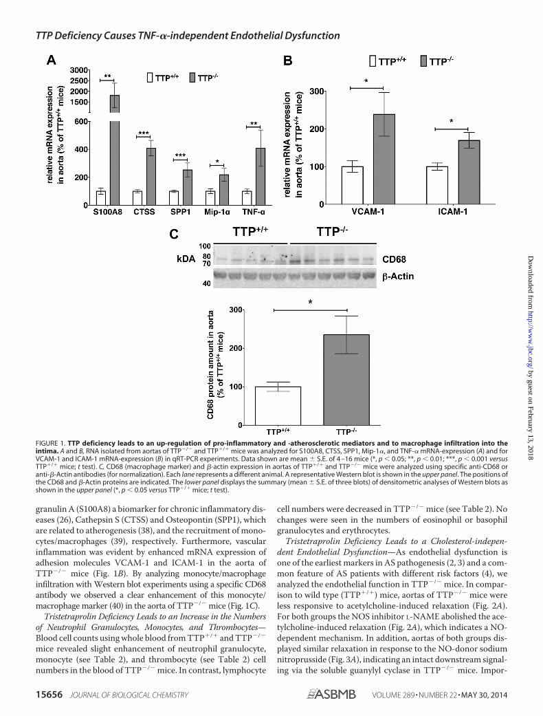
granulin A (S100A8) a biomarker for chronic inflammatory dis-eases (26), Cathepsin S (CTSS) and Osteopontin (SPP1), whichare related to atherogenesis (38), and the recruitment of mono-cytes/macrophages (39), respectively. Furthermore, vascularinflammation was evident by enhanced mRNA expression ofadhesion molecules VCAM-1 and ICAM-1 in the aorta ofTTP�/� mice (Fig. 1B). By analyzing monocyte/macrophageinfiltration with Western blot experiments using a specific CD68antibody we observed a clear enhancement of this monocyte/macrophage marker (40) in the aorta of TTP�/� mice (Fig. 1C).
Tristetraprolin Deficiency Leads to an Increase in the Numbersof Neutrophil Granulocytes, Monocytes, and Thrombocytes—Blood cell counts using whole blood from TTP�/� and TTP�/�
mice revealed slight enhancement of neutrophil granulocyte,monocyte (see Table 2), and thrombocyte (see Table 2) cellnumbers in the blood of TTP�/� mice. In contrast, lymphocyte
cell numbers were decreased in TTP�/� mice (see Table 2). Nochanges were seen in the numbers of eosinophil or basophilgranulocytes and erythrocytes.
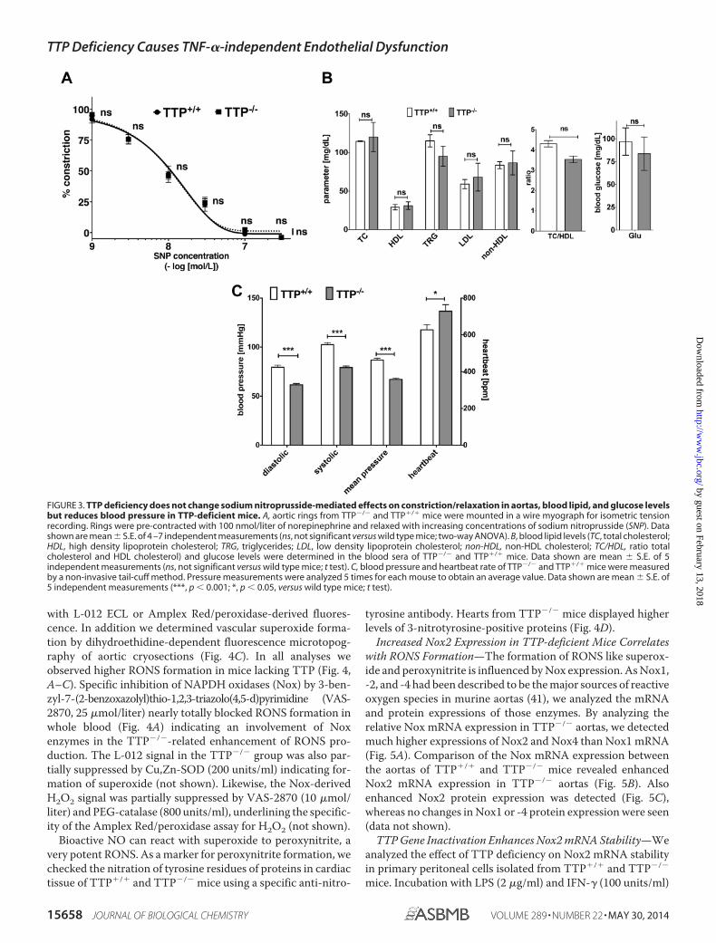
Tristetraprolin Deficiency Leads to a Cholesterol-indepen-dent Endothelial Dysfunction—As endothelial dysfunction isone of the earliest markers in AS pathogenesis (2, 3) and a com-mon feature of AS patients with different risk factors (4), weanalyzed the endothelial function in TTP�/� mice. In compar-ison to wild type (TTP�/�) mice, aortas of TTP�/� mice wereless responsive to acetylcholine-induced relaxation (Fig. 2A).For both groups the NOS inhibitor L-NAME abolished the ace-tylcholine-induced relaxation (Fig. 2A), which indicates a NO-dependent mechanism. In addition, aortas of both groups dis-played similar relaxation in response to the NO-donor sodiumnitroprusside (Fig. 3A), indicating an intact downstream signal-ing via the soluble guanylyl cyclase in TTP�/� mice. Impor-
FIGURE 1. TTP deficiency leads to an up-regulation of pro-inflammatory and -atherosclerotic mediators and to macrophage infiltration into theintima. A and B, RNA isolated from aortas of TTP�/� and TTP�/� mice was analyzed for S100A8, CTSS, SPP1, Mip-1�, and TNF-� mRNA-expression (A) and forVCAM-1 and ICAM-1 mRNA-expression (B) in qRT-PCR experiments. Data shown are mean � S.E. of 4 –16 mice (*, p � 0.05; **, p � 0.01; ***, p � 0.001 versusTTP�/� mice; t test). C, CD68 (macrophage marker) and �-actin expression in aortas of TTP�/� and TTP�/� mice were analyzed using specific anti-CD68 oranti-�-Actin antibodies (for normalization). Each lane represents a different animal. A representative Western blot is shown in the upper panel. The positions ofthe CD68 and �-Actin proteins are indicated. The lower panel displays the summary (mean � S.E. of three blots) of densitometric analyses of Western blots asshown in the upper panel (*, p � 0.05 versus TTP�/� mice; t test).
TTP Deficiency Causes TNF-�-independent Endothelial Dysfunction
15656 JOURNAL OF BIOLOGICAL CHEMISTRY VOLUME 289 • NUMBER 22 • MAY 30, 2014
by guest on February 13, 2018http://w
ww
.jbc.org/D
ownloaded from

tantly the development of this endothelial dysfunction is notcholesterol-dependent, as TTP�/� and TTP�/� mice showedno significant differences in blood cholesterol and triglyceridelevels (Fig. 3B). In addition TTP�/� mice displayed normal
blood glucose levels (Fig. 3B) and reduced blood pressure (Fig.3C).
An accepted marker for NO bioavailability and its down-stream signaling is phosphorylation of Ser-239 of the vasodila-tor-stimulated phosphoprotein (VASP). By analyzing P(Ser-239)-VASP expression in the aortas, we found a 25% decrease inNO/cGMP signaling in TTP�/� mice (Fig. 2B). Again, thisresult indicates a reduced NO bioavailability.
A reduced NO bioavailability can result from a reduction ofNO production by NO synthases or by enhanced inactivation ofNO by superoxide. In qRT-PCR (RNA) and Western blot (pro-tein) analyses we observed no reduction in the expression ofeNOS or iNOS, the main producers of NO in the vascular sys-tem (Fig. 2, C and D). Therefore, the reduced NO bioavailabilitywas more likely the result of enhanced NO breakdown by oxi-dative stress.
Tristetraprolin Deficiency Leads to Enhanced Burden of Oxi-dative Stress—We measured RONS formation either underPDBu-stimulated conditions in whole blood (Fig. 4A) or in thepresence of NADPH in cardiac membrane fractions (Fig. 4B)
FIGURE 2. TTP deficiency leads to endothelial dysfunction with unchanged eNOS or iNOS expression in TTP-deficient mice. A, aortic rings from TTP�/�,TTP�/�, and TTP�/�/TNF-��/� mice were mounted in a wire myograph for isometric tension recording. Rings were pre-contracted with 100 nmol/liter ofnorepinephrine and relaxed with increasing concentrations of acetylcholine in the presence or absence of the NOS inhibitor L-NAME (L-N, 1 mmol/liter; onlyTTP�/� or TTP�/� mice). Data shown are mean � S.E. of 4 –7 independent measurements (***, p � 0.001; ns, not significant versus wild type mice; ##, p � 0.01;#, p � 0.05; ns, not significant versus TTP�/� aortas; ns§, not significant versus L-NAME treated TTP�/� aortas; two-way ANOVA). B, the NO/cGMP signaling in theaorta of TTP�/� mice in comparison to wild type mice was measured by immunoblot analyses with a specific P(Ser-239)-VASP antibody. The figure displays thesummary (mean � S.E.) of densitometric analysis of Western blots using proteins from 11 mice (**, p � 0.01 versus TTP�/� mice; t test). C, RNA from aortas ofTTP�/� or TTP�/� mice was analyzed for iNOS and eNOS mRNA expression by qRT-PCR (ns, not significant versus TTP�/� mice; t test). D, eNOS or iNOS proteinexpression in aortas from TTP�/� or TTP�/� mice was analyzed in immunoblots using specific anti-eNOS or -iNOS antibodies. The figure is the summary(mean � S.E.) of densitometric analyses of Western blots using protein extracts from 10 to 11 mice (ns, not significant versus TTP�/� mice; t test).
TABLE 2TTP deficiency leads to an enhancement in the numbers of neutrophilgranulocytes, monocytes, and thrombocytes, whereas lymphocytenumbers were decreasedBlood cell counts of whole blood (20 �l) (leucocytes, thrombocytes, and erythro-cytes) from TTP �/� and TTP�/� mice were performed using the HEMAVET� 950system as described by the manufacturer. Data shown (mean � S.E.) are from 8 to 10animals).
TTP �/� TTP �/�
Cell type Mean S.E. Mean S.E. p value
% of white blood cellsNeutrophils 2.34 0.54 5.18 0.85 0.0384Eosinophils 0.19 0.08 0.17 0.06 �0.05Basophils 0.00 0.00 0.02 0.01 �0.05Monocytes 3.50 1.07 9.56 0.76 0.0014Lymphocytes 93.97 1.36 85.07 1.45 0.0029
Cell counts (�103 cells/�l)Thrombocytes 657,75 50,18 939,83 97,74 0.0297Erythrocytes 7.10 0.27 6.61 0.27 �0.05
TTP Deficiency Causes TNF-�-independent Endothelial Dysfunction
MAY 30, 2014 • VOLUME 289 • NUMBER 22 JOURNAL OF BIOLOGICAL CHEMISTRY 15657
by guest on February 13, 2018http://w
ww
.jbc.org/D
ownloaded from
with L-012 ECL or Amplex Red/peroxidase-derived fluores-cence. In addition we determined vascular superoxide forma-tion by dihydroethidine-dependent fluorescence microtopog-raphy of aortic cryosections (Fig. 4C). In all analyses weobserved higher RONS formation in mice lacking TTP (Fig. 4,A–C). Specific inhibition of NAPDH oxidases (Nox) by 3-ben-zyl-7-(2-benzoxazolyl)thio-1,2,3-triazolo(4,5-d)pyrimidine (VAS-2870, 25 �mol/liter) nearly totally blocked RONS formation inwhole blood (Fig. 4A) indicating an involvement of Noxenzymes in the TTP�/�-related enhancement of RONS pro-duction. The L-012 signal in the TTP�/� group was also par-tially suppressed by Cu,Zn-SOD (200 units/ml) indicating for-mation of superoxide (not shown). Likewise, the Nox-derivedH2O2 signal was partially suppressed by VAS-2870 (10 �mol/liter) and PEG-catalase (800 units/ml), underlining the specific-ity of the Amplex Red/peroxidase assay for H2O2 (not shown).
Bioactive NO can react with superoxide to peroxynitrite, avery potent RONS. As a marker for peroxynitrite formation, wechecked the nitration of tyrosine residues of proteins in cardiactissue of TTP�/� and TTP�/� mice using a specific anti-nitro-
tyrosine antibody. Hearts from TTP�/� mice displayed higherlevels of 3-nitrotyrosine-positive proteins (Fig. 4D).
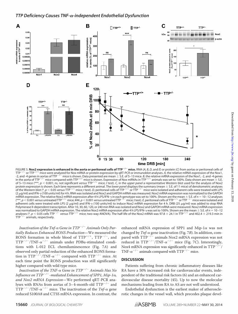
Increased Nox2 Expression in TTP-deficient Mice Correlateswith RONS Formation—The formation of RONS like superox-ide and peroxynitrite is influenced by Nox expression. As Nox1,-2, and -4 had been described to be the major sources of reactiveoxygen species in murine aortas (41), we analyzed the mRNAand protein expressions of those enzymes. By analyzing therelative Nox mRNA expression in TTP�/� aortas, we detectedmuch higher expressions of Nox2 and Nox4 than Nox1 mRNA(Fig. 5A). Comparison of the Nox mRNA expression betweenthe aortas of TTP�/� and TTP�/� mice revealed enhancedNox2 mRNA expression in TTP�/� aortas (Fig. 5B). Alsoenhanced Nox2 protein expression was detected (Fig. 5C),whereas no changes in Nox1 or -4 protein expression were seen(data not shown).
TTP Gene Inactivation Enhances Nox2 mRNA Stability—Weanalyzed the effect of TTP deficiency on Nox2 mRNA stabilityin primary peritoneal cells isolated from TTP�/� and TTP�/�
mice. Incubation with LPS (2 �g/ml) and IFN-� (100 units/ml)
FIGURE 3. TTP deficiency does not change sodium nitroprusside-mediated effects on constriction/relaxation in aortas, blood lipid, and glucose levelsbut reduces blood pressure in TTP-deficient mice. A, aortic rings from TTP�/� and TTP�/� mice were mounted in a wire myograph for isometric tensionrecording. Rings were pre-contracted with 100 nmol/liter of norepinephrine and relaxed with increasing concentrations of sodium nitroprusside (SNP). Datashown are mean � S.E. of 4 –7 independent measurements (ns, not significant versus wild type mice; two-way ANOVA). B, blood lipid levels (TC, total cholesterol;HDL, high density lipoprotein cholesterol; TRG, triglycerides; LDL, low density lipoprotein cholesterol; non-HDL, non-HDL cholesterol; TC/HDL, ratio totalcholesterol and HDL cholesterol) and glucose levels were determined in the blood sera of TTP�/� and TTP�/� mice. Data shown are mean � S.E. of 5independent measurements (ns, not significant versus wild type mice; t test). C, blood pressure and heartbeat rate of TTP�/� and TTP�/� mice were measuredby a non-invasive tail-cuff method. Pressure measurements were analyzed 5 times for each mouse to obtain an average value. Data shown are mean � S.E. of5 independent measurements (***, p � 0.001; *, p � 0.05, versus wild type mice; t test).
TTP Deficiency Causes TNF-�-independent Endothelial Dysfunction
15658 JOURNAL OF BIOLOGICAL CHEMISTRY VOLUME 289 • NUMBER 22 • MAY 30, 2014
by guest on February 13, 2018http://w
ww
.jbc.org/D
ownloaded from

for 4 h induced Nox2 mRNA expression in peritoneal cells ofboth genotypes nearly 5-fold (see Fig. 5D). As shown in Fig. 5Einactivation of the Ttp gene resulted in markedly enhancedNox2 mRNA stability (TTP�/� t1⁄2 97.8 � 24.1 min; TTP�/�
t1⁄2 402.4 � 219.3 min).Collagen-induced Chronic Inflammation Results in Reduced
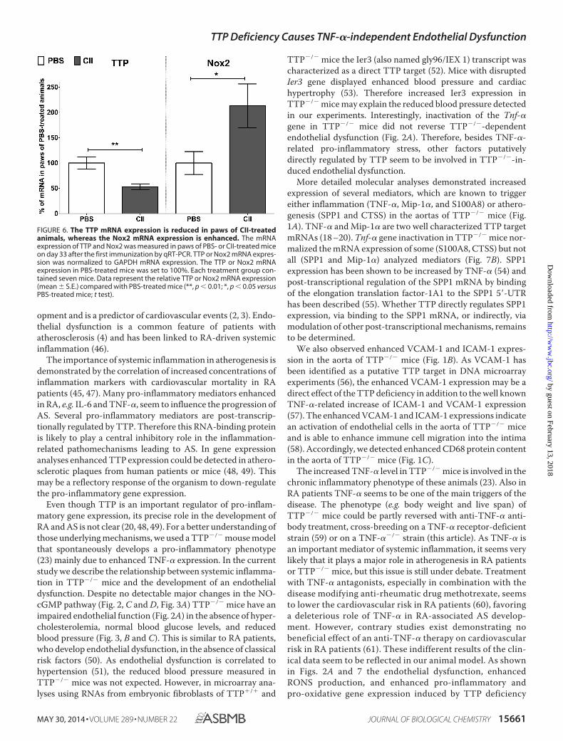
TTP and Enhanced Nox2 mRNA Expression—To analyze thecorrelation of TTP and Nox2 expression in another model ofchronic inflammation we performed analyses in mice treatedwith collagen type II (CII, in CIA) (26). Compared with PBS-treated mice we observed reduced TTP and enhanced Nox2mRNA expression in the paws of CII-treated mice (Fig. 6).
Inactivation of the Tnf-� Gene in TTP�/� Animals Does NotReverse Endothelial Dysfunction—TTP has been shown to be amajor post-transcriptional regulator of TNF-� expression (21)and the pro-inflammatory phenotype of TTP�/� mice can bereversed by TNF-� blockade (23). TNF-� has been described tobe an important mediator of atherogenesis (42), as well as toregulate Nox expression in cell culture (43) and mouse models(44). Therefore we expected that genetic inactivation of theTnf-� gene in TTP�/� mice normalizes the atherogenic phe-notype. Surprisingly, inactivation of the Tnf-� gene in TTP�/�
mice did not improve the endothelial dysfunction caused byTTP deficiency (Fig. 2A).
FIGURE 4. TTP deficiency leads to enhanced formation of RONS. A, formation of RONS in the blood of TTP�/� and TTP�/� mice was measured under PDBu(1 �mol/liter)-stimulated conditions in the presence or absence of the NADPH oxidase inhibitor 3-benzyl-7-(2-benzoxazolyl)thio-1,2,3-triazolo(4,5-d)pyrimi-dine (VAS-2870, 25 �mol/liter) with L-012 (100 �mol/liter) ECL. Data shown are mean � S.E. of 12 mice (***, p � 0.001; ns, not significant versus wild type; ###,p � 0.001 versus TTP�/� mice; two-way ANOVA). B, formation of RONS in cardiac membrane fractions was measured with Amplex Red/peroxidase-derivedfluorescence. Data shown are mean � S.E. of 16 mice (*, p � 0.05,versus TTP�/� mice; t test). C, vascular superoxide formation was determined by dihydro-ethidine-dependent fluorescence microtopography of aortic cryosections. In the upper panel representative dihydroethidine (DHE) stainings with superoxideproducing cells are shown. The locations of the endothelium (E), media (M), and adventitia (A) are indicated. In the lower panel a quantitative analysis of severalsuch assays is shown. Data shown are mean � S.E. of 5–7 mice (***, p � 0.001 versus TTP�/� mice; t test). D, peroxynitrite formation in the hearts of TTP�/� andTTP�/� mice was measured by a dot blot using specific anti-nitrotyrosine antibody. Protein expression was analyzed by densitometric analysis of the dot blotmembrane. Data shown are mean � S.E. of 13 mice (**, p � 0.01 versus TTP�/� mice; t test).
TTP Deficiency Causes TNF-�-independent Endothelial Dysfunction
MAY 30, 2014 • VOLUME 289 • NUMBER 22 JOURNAL OF BIOLOGICAL CHEMISTRY 15659
by guest on February 13, 2018http://w
ww
.jbc.org/D
ownloaded from
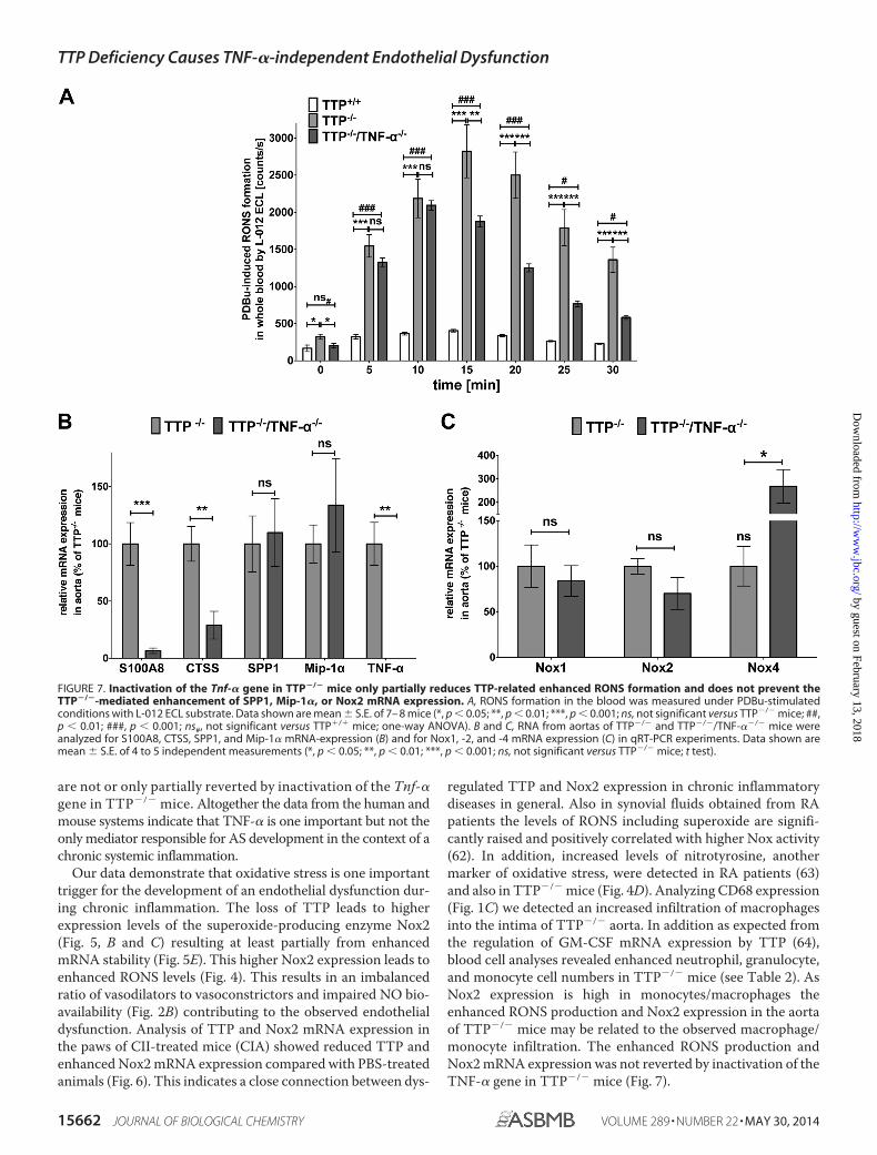
Inactivation of the Tnf-� Gene in TTP�/� Animals Only Par-tially Reduces Enhanced RONS Production—We measured theRONS formation in whole blood of TTP�/�, TTP�/�, andTTP�/�/TNF-��/� animals under PDBu-stimulated condi-tions with L-012 ECL chemiluminescence (Fig. 7A) andobserved only partial reduction of the enhanced RONS forma-tion in TTP�/�/TNF-��/� compared with TTP�/� mice. Ateach time point the RONS production was still significantlyhigher compared with wild type mice.
Inactivation of the TNF-� Gene in TTP�/� Animals Has NoInfluence on TTP�/�-mediated Enhancement of SPP1, Mip-1�,and Nox2 mRNA Expression—We performed qRT-PCR ana-lyses with RNAs from aortas of 3– 4-month-old TTP�/� andTTP�/�/TNF-��/� mice. The inactivation of the Tnf-� genereduced S100A8 and CTSS mRNA expression. In contrast, the
enhanced mRNA expression of SPP1 and Mip-1� was notchanged by Tnf-� gene inactivation (Fig. 7B). In addition, com-pared with TTP�/� animals Nox2 mRNA expression was notreduced in TTP�/�/TNF-��/� mice (Fig. 7C). Interestingly,Nox4 mRNA expression was significantly enhanced in TTP�/�/TNF-��/� animals compared with TTP�/� mice.
DISCUSSION
Patients suffering from chronic inflammatory diseases likeRA have a 50% increased risk for cardiovascular events, inde-pendent of the traditional risk factors (6) and an enhanced car-diovascular disease mortality (45). Up to now the molecularmechanisms leading from RA to AS are not well understood.
Endothelial dysfunction is the earliest maker of atheroscle-rotic changes in the vessel wall, which precedes plaque devel-
FIGURE 5. Nox2 expression is enhanced in the aorta or peritoneal cells of TTP�/� mice. RNA (A, B, D, and E) or protein (C) from aortas or peritoneal cells ofTTP�/� or TTP�/� mice were analyzed for Nox mRNA or protein expression by qRT-PCR or immunoblot analyses. A, the relative mRNA expression of the Nox1,-2, and -4 genes in aortas of TTP�/� mice is shown. Data presented are mean � S.E. of 5–13 mice. B, the relative mRNA expression of the Nox1, -2, and -4 genesin the aorta of TTP�/� mice compared with TTP�/� mice is shown. Expression of Nox mRNAs in TTP�/� animals was set to 100%. Data shown are mean � S.E.of 5–13 mice (***, p � 0.001; ns, not significant versus TTP�/� mice; t test). C, in the upper panel a representative Western blot used for the analysis of Nox2protein expression is shown. Each lane represents a different animal. The lower panel displays the summary (mean � S.E. of 11 mice) of densitometric analysesof the Western blot (*, p � 0.05 versus TTP�/� mice; t test). D, peritoneal cells of TTP�/� or TTP�/� mice were isolated and adherent cells were treated with LPS(2 �g/ml) and IFN-� (100 units/ml) for 4 h. RNA was isolated and Nox2 and GAPDH mRNA was measured. Nox2 mRNA expression was normalized to the GAPDHmRNA expression. The relative Nox2 mRNA expression after 4 h LPS/IFN-� in each genotype was set to 100%. Shown are the mean � S.E. of n 10 –12 analyses(***, p � 0.001 versus untreated TTP�/� mice; ###, p � 0.001 versus untreated TTP�/� mice; t test). E, peritoneal cells of TTP�/� or TTP�/� mice were isolated andadherent cells were treated with LPS (2 �g/ml) and IFN-� (100 units/ml) to induce Nox2 mRNA expression for 4 h. DRB (25 �g/ml) was added to stop RNAPolymerase II-dependent transcription. After 10, 30, 60, 120, or 240 min RNA was isolated and Nox2 and GAPDH mRNA were measured. Nox2 mRNA expressionwas normalized to GAPDH mRNA expression. The relative Nox2 mRNA expression after 4 h LPS/IFN-� was set to 100%. Shown are the mean � S.E. of n 10 –12analyses (*, p � 0.05 cells TTP�/� versus TTP�/� mice; two-way ANOVA). The half-life of the Nox2 mRNA was 97.8 � 24.1 in TTP�/� and 402.4 � 219.3 min inTTP�/� animals, respectively.
TTP Deficiency Causes TNF-�-independent Endothelial Dysfunction
15660 JOURNAL OF BIOLOGICAL CHEMISTRY VOLUME 289 • NUMBER 22 • MAY 30, 2014
by guest on February 13, 2018http://w
ww
.jbc.org/D
ownloaded from
opment and is a predictor of cardiovascular events (2, 3). Endo-thelial dysfunction is a common feature of patients withatherosclerosis (4) and has been linked to RA-driven systemicinflammation (46).
The importance of systemic inflammation in atherogenesis isdemonstrated by the correlation of increased concentrations ofinflammation markers with cardiovascular mortality in RApatients (45, 47). Many pro-inflammatory mediators enhancedin RA, e.g. IL-6 and TNF-�, seem to influence the progression ofAS. Several pro-inflammatory mediators are post-transcrip-tionally regulated by TTP. Therefore this RNA-binding proteinis likely to play a central inhibitory role in the inflammation-related pathomechanisms leading to AS. In gene expressionanalyses enhanced TTP expression could be detected in athero-sclerotic plaques from human patients or mice (48, 49). Thismay be a reflectory response of the organism to down-regulatethe pro-inflammatory gene expression.
Even though TTP is an important regulator of pro-inflam-matory gene expression, its precise role in the development ofRA and AS is not clear (20, 48, 49). For a better understanding ofthose underlying mechanisms, we used a TTP�/� mouse modelthat spontaneously develops a pro-inflammatory phenotype(23) mainly due to enhanced TNF-� expression. In the currentstudy we describe the relationship between systemic inflamma-tion in TTP�/� mice and the development of an endothelialdysfunction. Despite no detectable major changes in the NO-cGMP pathway (Fig. 2, C and D, Fig. 3A) TTP�/� mice have animpaired endothelial function (Fig. 2A) in the absence of hyper-cholesterolemia, normal blood glucose levels, and reducedblood pressure (Fig. 3, B and C). This is similar to RA patients,who develop endothelial dysfunction, in the absence of classicalrisk factors (50). As endothelial dysfunction is correlated tohypertension (51), the reduced blood pressure measured inTTP�/� mice was not expected. However, in microarray ana-lyses using RNAs from embryonic fibroblasts of TTP�/� and
TTP�/� mice the Ier3 (also named gly96/IEX 1) transcript wascharacterized as a direct TTP target (52). Mice with disruptedIer3 gene displayed enhanced blood pressure and cardiachypertrophy (53). Therefore increased Ier3 expression inTTP�/� mice may explain the reduced blood pressure detectedin our experiments. Interestingly, inactivation of the Tnf-�gene in TTP�/� mice did not reverse TTP�/�-dependentendothelial dysfunction (Fig. 2A). Therefore, besides TNF-�-related pro-inflammatory stress, other factors putativelydirectly regulated by TTP seem to be involved in TTP�/�-in-duced endothelial dysfunction.
More detailed molecular analyses demonstrated increasedexpression of several mediators, which are known to triggereither inflammation (TNF-�, Mip-1�, and S100A8) or athero-genesis (SPP1 and CTSS) in the aortas of TTP�/� mice (Fig.1A). TNF-� and Mip-1� are two well characterized TTP targetmRNAs (18 –20). Tnf-� gene inactivation in TTP�/� mice nor-malized the mRNA expression of some (S100A8, CTSS) but notall (SPP1 and Mip-1�) analyzed mediators (Fig. 7B). SPP1expression has been shown to be increased by TNF-� (54) andpost-transcriptional regulation of the SPP1 mRNA by bindingof the elongation translation factor-1A1 to the SPP1 5-UTRhas been described (55). Whether TTP directly regulates SPP1expression, via binding to the SPP1 mRNA, or indirectly, viamodulation of other post-transcriptional mechanisms, remainsto be determined.
We also observed enhanced VCAM-1 and ICAM-1 expres-sion in the aorta of TTP�/� mice (Fig. 1B). As VCAM-1 hasbeen identified as a putative TTP target in DNA microarrayexperiments (56), the enhanced VCAM-1 expression may be adirect effect of the TTP deficiency in addition to the well knownTNF-�-related increase of ICAM-1 and VCAM-1 expression(57). The enhanced VCAM-1 and ICAM-1 expressions indicatean activation of endothelial cells in the aorta of TTP�/� miceand is able to enhance immune cell migration into the intima(58). Accordingly, we detected enhanced CD68 protein contentin the aorta of TTP�/� mice (Fig. 1C).
The increased TNF-� level in TTP�/� mice is involved in thechronic inflammatory phenotype of these animals (23). Also inRA patients TNF-� seems to be one of the main triggers of thedisease. The phenotype (e.g. body weight and live span) ofTTP�/� mice could be partly reversed with anti-TNF-� anti-body treatment, cross-breeding on a TNF-� receptor-deficientstrain (59) or on a TNF-��/� strain (this article). As TNF-� isan important mediator of systemic inflammation, it seems verylikely that it plays a major role in atherogenesis in RA patientsor TTP�/� mice, but this issue is still under debate. Treatmentwith TNF-� antagonists, especially in combination with thedisease modifying anti-rheumatic drug methotrexate, seemsto lower the cardiovascular risk in RA patients (60), favoringa deleterious role of TNF-� in RA-associated AS develop-ment. However, contrary studies exist demonstrating nobeneficial effect of an anti-TNF-� therapy on cardiovascularrisk in RA patients (61). These indifferent results of the clin-ical data seem to be reflected in our animal model. As shownin Figs. 2A and 7 the endothelial dysfunction, enhancedRONS production, and enhanced pro-inflammatory andpro-oxidative gene expression induced by TTP deficiency
FIGURE 6. The TTP mRNA expression is reduced in paws of CII-treatedanimals, whereas the Nox2 mRNA expression is enhanced. The mRNAexpression of TTP and Nox2 was measured in paws of PBS- or CII-treated miceon day 33 after the first immunization by qRT-PCR. TTP or Nox2 mRNA expres-sion was normalized to GAPDH mRNA expression. The TTP or Nox2 mRNAexpression in PBS-treated mice was set to 100%. Each treatment group con-tained seven mice. Data represent the relative TTP or Nox2 mRNA expression(mean � S.E.) compared with PBS-treated mice (**, p � 0.01; *, p � 0.05 versusPBS-treated mice; t test).
TTP Deficiency Causes TNF-�-independent Endothelial Dysfunction
MAY 30, 2014 • VOLUME 289 • NUMBER 22 JOURNAL OF BIOLOGICAL CHEMISTRY 15661
by guest on February 13, 2018http://w
ww
.jbc.org/D
ownloaded from
are not or only partially reverted by inactivation of the Tnf-�gene in TTP�/� mice. Altogether the data from the human andmouse systems indicate that TNF-� is one important but not theonly mediator responsible for AS development in the context of achronic systemic inflammation.
Our data demonstrate that oxidative stress is one importanttrigger for the development of an endothelial dysfunction dur-ing chronic inflammation. The loss of TTP leads to higherexpression levels of the superoxide-producing enzyme Nox2(Fig. 5, B and C) resulting at least partially from enhancedmRNA stability (Fig. 5E). This higher Nox2 expression leads toenhanced RONS levels (Fig. 4). This results in an imbalancedratio of vasodilators to vasoconstrictors and impaired NO bio-availability (Fig. 2B) contributing to the observed endothelialdysfunction. Analysis of TTP and Nox2 mRNA expression inthe paws of CII-treated mice (CIA) showed reduced TTP andenhanced Nox2 mRNA expression compared with PBS-treatedanimals (Fig. 6). This indicates a close connection between dys-
regulated TTP and Nox2 expression in chronic inflammatorydiseases in general. Also in synovial fluids obtained from RApatients the levels of RONS including superoxide are signifi-cantly raised and positively correlated with higher Nox activity(62). In addition, increased levels of nitrotyrosine, anothermarker of oxidative stress, were detected in RA patients (63)and also in TTP�/� mice (Fig. 4D). Analyzing CD68 expression(Fig. 1C) we detected an increased infiltration of macrophagesinto the intima of TTP�/� aorta. In addition as expected fromthe regulation of GM-CSF mRNA expression by TTP (64),blood cell analyses revealed enhanced neutrophil, granulocyte,and monocyte cell numbers in TTP�/� mice (see Table 2). AsNox2 expression is high in monocytes/macrophages theenhanced RONS production and Nox2 expression in the aortaof TTP�/� mice may be related to the observed macrophage/monocyte infiltration. The enhanced RONS production andNox2 mRNA expression was not reverted by inactivation of theTNF-� gene in TTP�/� mice (Fig. 7).
FIGURE 7. Inactivation of the Tnf-� gene in TTP�/� mice only partially reduces TTP-related enhanced RONS formation and does not prevent theTTP�/�-mediated enhancement of SPP1, Mip-1�, or Nox2 mRNA expression. A, RONS formation in the blood was measured under PDBu-stimulatedconditions with L-012 ECL substrate. Data shown are mean � S.E. of 7– 8 mice (*, p � 0.05; **, p � 0.01; ***, p � 0.001; ns, not significant versus TTP�/� mice; ##,p � 0.01; ###, p � 0.001; ns#, not significant versus TTP�/� mice; one-way ANOVA). B and C, RNA from aortas of TTP�/� and TTP�/�/TNF-��/� mice wereanalyzed for S100A8, CTSS, SPP1, and Mip-1� mRNA-expression (B) and for Nox1, -2, and -4 mRNA expression (C) in qRT-PCR experiments. Data shown aremean � S.E. of 4 to 5 independent measurements (*, p � 0.05; **, p � 0.01; ***, p � 0.001; ns, not significant versus TTP�/� mice; t test).
TTP Deficiency Causes TNF-�-independent Endothelial Dysfunction
15662 JOURNAL OF BIOLOGICAL CHEMISTRY VOLUME 289 • NUMBER 22 • MAY 30, 2014
by guest on February 13, 2018http://w
ww
.jbc.org/D
ownloaded from
The enhanced expression of Nox2 detected in TTP�/� andTTP�/�/TNF-��/� mice may be a direct consequence of TTPdeficiency or an indirect but TNF-�-independent effect, due tosystemic inflammatory processes. Reduction of Nox2 expres-sion/activity by p38 MAPK inhibitors has been described (65,66). p38 MAPK is a major regulator of TTP expression andactivity (67). In addition the 3-UTR of the Nox2 gene containsseveral AREs, which may be TTP binding sites. In the currentstudy we detected marked enhancement of Nox2 mRNA stabil-ity in peritoneal cells isolated from TTP�/� animals (Fig. 5D).Therefore it seems very likely that TTP mediates the decay ofNox2 mRNA under physiological conditions. Because notmuch is known about post-transcriptional regulation of Noxexpression (68), this hypothesis has to be verified in furtherexperiments. According to our results we postulate that duringsystemic inflammation molecular pathways leading to thedevelopment of atherogenesis through induction of oxidativestress are only partially activated by TNF-�.
Altogether we have demonstrated that oxidative stress, inparticular increased Nox2 activity, promotes the developmentof atherosclerosis in models of systemic inflammatory diseasesas RA. Moreover we provide evidence that TNF-� alone is notthe driving force in atherogenic processes. As the standardtherapeutics in RA, TNF-� antagonists, and disease modifyinganti-rheumatic drugs have only a limited success in preventionof cardiovascular events, the additional use of antioxidativedrugs could be an alternative. In numerous clinical trials theusage of nutritional supplements (such as �-carotene, sele-nium, vitamin C, and vitamin E) as antioxidative substances inthe prevention of coronary heart disease and stroke resulted inconflicting data (positive, neutral, and negative) (69, 70). There-fore more specific drugs acting via down-regulation of Noxcould be promising.
REFERENCES1. Hansson, G. K., Robertson, A. K., and Söderberg-Nauclér, C. (2006) In-
flammation and atherosclerosis. Annu. Rev. Pathol. 1, 297–3292. Dai, X. Y., Cai, Y., Sun, W., Ding, Y., Wang, W., Kong, W., Tang, C., Zhu,
Y., Xu, M. J., and Wang, X. (2014) Intermedin inhibits macrophage foam-cell formation via tristetraprolin-mediated decay of CD36 mRNA. Cardio-vasc. Res. 101, 297–305
3. Jamal Uddin, M., Joe, Y., Zheng, M., Blackshear, P. J., Ryter, S. W., Park,J. W., and Chung, H. T. (2013) A functional link between heme oxyge-nase-1 and tristetraprolin in the anti-inflammatory effects of nicotine.Free Radic. Biol. Med. 65, 1331–1339
4. Cao, H., Cao, F., Roussel, A. M., and Anderson, R. A. (2013) QuantitativePCR for glucose transporter and tristetraprolin family gene expression incultured mouse adipocytes and macrophages. In Vitro Cell. Dev. Biol.Anim. 49, 759 –770
5. Galkina, E., and Ley, K. (2009) Immune and inflammatory mechanisms ofatherosclerosis (*). Annu. Rev. Immunol. 27, 165–197
6. Aviña-Zubieta, J. A., Choi, H. K., Sadatsafavi, M., Etminan, M., Esdaile,J. M., and Lacaille, D. (2008) Risk of cardiovascular mortality in patientswith rheumatoid arthritis: a meta-analysis of observational studies. Arthri-tis Rheum. 59, 1690 –1697
7. Roman, M. J., Moeller, E., Davis, A., Paget, S. A., Crow, M. K., Lockshin,M. D., Sammaritano, L., Devereux, R. B., Schwartz, J. E., Levine, D. M., andSalmon, J. E. (2006) Preclinical carotid atherosclerosis in patients withrheumatoid arthritis. Ann. Intern. Med. 144, 249 –256
8. Maradit-Kremers, H., Nicola, P. J., Crowson, C. S., Ballman, K. V., andGabriel, S. E. (2005) Cardiovascular death in rheumatoid arthritis: a pop-ulation-based study. Arthritis Rheum. 52, 722–732
9. del Rincón, I. D., Williams, K., Stern, M. P., Freeman, G. L., and Escalante,A. (2001) High incidence of cardiovascular events in a rheumatoid arthri-tis cohort not explained by traditional cardiac risk factors. ArthritisRheum. 44, 2737–2745
10. Gabriel, S. E. (2008) Cardiovascular morbidity and mortality in rheuma-toid arthritis. Am. J. Med. 121, S9 –14
11. Iademarco, M. F., Barks, J. L., and Dean, D. C. (1995) Regulation of vascularcell adhesion molecule-1 expression by IL-4 and TNF-� in cultured en-dothelial cells. J. Clin. Invest. 95, 264 –271
12. Jayakody, L., Kappagoda, T., Senaratne, M. P., and Thomson, A. B. (1988)Impairment of endothelium-dependent relaxation: an early marker foratherosclerosis in the rabbit. Br. J. Pharmacol. 94, 335–346
13. Griendling, K. K., and FitzGerald, G. A. (2003) Oxidative stress and car-diovascular injury: part II: animal and human studies. Circulation 108,2034 –2040
14. Münzel, T., Daiber, A., Ullrich, V., and Mülsch, A. (2005) Vascular conse-quences of endothelial nitric oxide synthase uncoupling for the activityand expression of the soluble guanylyl cyclase and the cGMP-dependentprotein kinase. Arterioscler. Thromb. Vasc. Biol. 25, 1551–1557
15. Förstermann, U., and Münzel, T. (2006) Endothelial nitric-oxide synthasein vascular disease: from marvel to menace. Circulation 113, 1708 –1714
16. Bosello, S., Santoliquido, A., Zoli, A., Di Campli, C., Flore, R., Tondi, P.,and Ferraccioli, G. (2008) TNF-alpha blockade induces a reversible buttransient effect on endothelial dysfunction in patients with long-standingsevere rheumatoid arthritis. Clin. Rheumatol. 27, 833– 839
17. Chen, C. Y., and Shyu, A. B. (1995) AU-rich elements: characterizationand importance in mRNA degradation. Trends Biochem. Sci. 20, 465– 470
18. Brooks, S. A., and Blackshear, P. J. (2013) Tristetraprolin (TTP): interac-tions with mRNA and proteins, and current thoughts on mechanisms ofaction. Biochim. Biophys. Acta 1829, 666 – 679
19. Carballo, E., Lai, W. S., and Blackshear, P. J. (1998) Feedback inhibition ofmacrophage tumor necrosis factor-� production by tristetraprolin. Sci-ence 281, 1001–1005
20. Kang, J. G., Amar, M. J., Remaley, A. T., Kwon, J., Blackshear, P. J., Wang,P. Y., and Hwang, P. M. (2011) Zinc finger protein tristetraprolin interactswith CCL3 mRNA and regulates tissue inflammation. J. Immunol. 187,2696 –2701
21. Blackshear, P. J. (2002) Tristetraprolin and other CCCH tandem zinc-finger proteins in the regulation of mRNA turnover. Biochem. Soc. Trans.30, 945–952
22. Ghosh, S., Hoenerhoff, M. J., Clayton, N., Myers, P., Stumpo, D. J., Maron-pot, R. R., and Blackshear, P. J. (2010) Left-sided cardiac valvulitis in tris-tetraprolin-deficient mice: the role of tumor necrosis factor �. Am. J.Pathol. 176, 1484 –1493
23. Taylor, G. A., Carballo, E., Lee, D. M., Lai, W. S., Thompson, M. J., Patel,D. D., Schenkman, D. I., Gilkeson, G. S., Broxmeyer, H. E., Haynes, B. F.,and Blackshear, P. J. (1996) A pathogenetic role for TNF � in the syndromeof cachexia, arthritis, and autoimmunity resulting from tristetraprolin(TTP) deficiency. Immunity 4, 445– 454
24. Ridker, P. M., Rifai, N., Pfeffer, M., Sacks, F., Lepage, S., and Braunwald, E.(2000) Elevation of tumor necrosis factor-� and increased risk of recur-rent coronary events after myocardial infarction. Circulation 101,2149 –2153
25. Mori, L., Loetscher, H., Kakimoto, K., Bluethmann, H., and Steinmetz, M.(1992) Expression of a transgenic T cell receptor � chain enhances colla-gen-induced arthritis. J. Exp. Med. 176, 381–388
26. Schmidt, N., Art, J., Forsch, I., Werner, A., Erkel, G., Jung, M., Horke, S.,Kleinert, H., and Pautz, A. (2012) The anti-inflammatory fungal com-pound (S)-curvularin reduces proinflammatory gene expression in an invivo model of rheumatoid arthritis. J. Pharmacol. Exp. Ther. 343, 106 –114
27. Ray, A., and Dittel, B. N. (2010) Isolation of mouse peritoneal cavity cells.J. Vis. Exp. 35, e1488
28. Li, H., Hergert, S. M., Schäfer, S. C., Brausch, I., Yao, Y., Huang, Q., Mang,C., Lehr, H. A., and Förstermann, U. (2005) Midostaurin up-regulateseNOS gene expression and preserves eNOS function in the microcircula-tion of the mouse. Nitric Oxide 12, 231–236
29. Friedewald, W. T., Levy, R. I., and Fredrickson, D. S. (1972) Estimation ofthe concentration of low-density lipoprotein cholesterol in plasma, with-
TTP Deficiency Causes TNF-�-independent Endothelial Dysfunction
MAY 30, 2014 • VOLUME 289 • NUMBER 22 JOURNAL OF BIOLOGICAL CHEMISTRY 15663
by guest on February 13, 2018http://w
ww
.jbc.org/D
ownloaded from
out use of the preparative ultracentrifuge. Clin. Chem. 18, 499 –50230. Chomczynski, P., and Sacchi, N. (1987) Single-step method of RNA isola-
tion by acid guanidinium thiocyanate-phenol-chloroform extraction.Anal. Biochem. 162, 156 –159
31. Schmidt, N., Pautz, A., Art, J., Rauschkolb, P., Jung, M., Erkel, G., Goldring,M. B., and Kleinert, H. (2010) Transcriptional and post-transcriptionalregulation of iNOS expression in human chondrocytes. Biochem. Phar-macol. 79, 722–732
32. Siuda, D., Zechner, U., El Hajj, N., Prawitt, D., Langer, D., Xia, N., Horke,S., Pautz, A., Kleinert, H., Förstermann, U., and Li, H. (2012) Transcrip-tional regulation of Nox4 by histone deacetylases in human endothelialcells. Basic Res. Cardiol. 107, 283
33. Livak, K. J., and Schmittgen, T. D. (2001) Analysis of relative gene expres-sion data using real-time quantitative PCR and the 2(�[/Delta][/Delta]C(T)) method. Methods 25, 402– 408
34. Daiber, A., August, M., Baldus, S., Wendt, M., Oelze, M., Sydow, K., Kle-schyov, A. L., and Munzel, T. (2004) Measurement of NAD(P)H oxidase-derived superoxide with the luminol analogue L-012. Free Radic. Biol.Med. 36, 101–111
35. Oelze, M., Daiber, A., Brandes, R. P., Hortmann, M., Wenzel, P., Hink, U.,Schulz, E., Mollnau, H., von Sandersleben, A., Kleschyov, A. L., Mülsch, A.,Li, H., Förstermann, U., and Münzel, T. (2006) Nebivolol inhibits super-oxide formation by NADPH oxidase and endothelial dysfunction in ang-iotensin II-treated rats. Hypertension 48, 677– 684
36. Schuhmacher, S., Wenzel, P., Schulz, E., Oelze, M., Mang, C., Kamuf, J.,Gori, T., Jansen, T., Knorr, M., Karbach, S., Hortmann, M., Mäthner, F.,Bhatnagar, A., Förstermann, U., Li, H., Münzel, T., and Daiber, A. (2010)Pentaerythritol tetranitrate improves angiotensin II-induced vasculardysfunction via induction of heme oxygenase-1. Hypertension 55,897–904
37. Knorr, M., Hausding, M., Kröller-Schuhmacher, S., Steven, S., Oelze, M.,Heeren, T., Scholz, A., Gori, T., Wenzel, P., Schulz, E., Daiber, A., andMünzel, T. (2011) Nitroglycerin-induced endothelial dysfunction and tol-erance involve adverse phosphorylation and S-glutathionylation of endo-thelial nitric oxide synthase: beneficial effects of therapy with the AT1receptor blocker telmisartan. Arterioscler. Thromb. Vasc. Biol. 31,2223–2231
38. Sukhova, G. K., Zhang, Y., Pan, J. H., Wada, Y., Yamamoto, T., Naito, M.,Kodama, T., Tsimikas, S., Witztum, J. L., Lu, M. L., Sakara, Y., Chin, M. T.,Libby, P., and Shi, G. P. (2003) Deficiency of cathepsin S reduces athero-sclerosis in LDL receptor-deficient mice. J. Clin. Invest. 111, 897–906
39. Scatena, M., Liaw, L., and Giachelli, C. M. (2007) Osteopontin: a multi-functional molecule regulating chronic inflammation and vascular dis-ease. Arterioscler. Thromb. Vasc. Biol. 27, 2302–2309
40. Bobryshev, Y. V., and Lord, R. S. (2002) Expression of heat shock pro-tein-70 by dendritic cells in the arterial intima and its potential signifi-cance in atherogenesis. J. Vasc. Surg. 35, 368 –375
41. Pendyala, S., Gorshkova, I. A., Usatyuk, P. V., He, D., Pennathur, A., Lam-beth, J. D., Thannickal, V. J., and Natarajan, V. (2009) Role of Nox4 andNox2 in hyperoxia-induced reactive oxygen species generation and mi-gration of human lung endothelial cells. Antioxid. Redox Signal. 11,747–764
42. Brånén, L., Hovgaard, L., Nitulescu, M., Bengtsson, E., Nilsson, J., andJovinge, S. (2004) Inhibition of tumor necrosis factor-� reduces athero-sclerosis in apolipoprotein E knockout mice. Arterioscler. Thromb. Vasc.Biol. 24, 2137–2142
43. Yoshida, L. S., and Tsunawaki, S. (2008) Expression of NADPH oxidasesand enhanced H2O2-generating activity in human coronary artery endo-thelial cells upon induction with tumor necrosis factor-�. Int. Immunop-harmacol. 8, 1377–1385
44. Moe, K. T., Yin, N. O., Naylynn, T. M., Khairunnisa, K., Wutyi, M. A., Gu,Y., Atan, M. S., Wong, M. C., Koh, T. H., and Wong, P. (2011) Nox2 andNox4 mediate tumour necrosis factor-�-induced ventricular remodellingin mice. J. Cell. Mol. Med. 15, 2601–2613
45. Goodson, N., Marks, J., Lunt, M., and Symmons, D. (2005) Cardiovascularadmissions and mortality in an inception cohort of patients with rheuma-toid arthritis with onset in the 1980s and 1990s. Ann. Rheum. Dis. 64,1595–1601
46. Lee, H. H., Yang, S. S., Vo, M. T., Cho, W. J., Lee, B. J., Leem, S. H., Lee,S. H., Cha, H. J., and Park, J. W. (2013) Tristetraprolin down-regulatesIL-23 expression in colon cancer cells. Mol. Cells 36, 571–576
47. Gonzalez-Gay, M. A., Gonzalez-Juanatey, C., Lopez-Diaz, M. J., Piñeiro,A., Garcia-Porrua, C., Miranda-Filloy, J. A., Ollier, W. E., Martin, J., andLlorca, J. (2007) HLA-DRB1 and persistent chronic inflammation contrib-ute to cardiovascular events and cardiovascular mortality in patients withrheumatoid arthritis. Arthritis Rheum. 57, 125–132
48. Patino, W. D., Kang, J. G., Matoba, S., Mian, O. Y., Gochuico, B. R., andHwang, P. M. (2006) Atherosclerotic plaque macrophage transcriptionalregulators are expressed in blood and modulated by tristetraprolin. Circ.Res. 98, 1282–1289
49. Zhang, H., Taylor, W. R., Joseph, G., Caracciolo, V., Gonzales, D. M.,Sidell, N., Seli, E., Blackshear, P. J., and Kallen, C. B. (2013) mRNA-bindingprotein ZFP36 is expressed in atherosclerotic lesions and reduces inflam-mation in aortic endothelial cells. Arterioscler. Thromb. Vasc. Biol. 33,1212–1220
50. Kerekes, G., Szekanecz, Z., Dér, H., Sándor, Z., Lakos, G., Muszbek, L.,Csipö, I., Sipka, S., Seres, I., Paragh, G., Kappelmayer, J., Szomják, E., Veres,K., Szegedi, G., Shoenfeld, Y., and Soltész, P. (2008) Endothelial dysfunc-tion and atherosclerosis in rheumatoid arthritis: a multiparametric anal-ysis using imaging techniques and laboratory markers of inflammationand autoimmunity. J. Rheumatol. 35, 398 – 406
51. Dharmashankar, K., and Widlansky, M. E. (2010) Vascular endothelialfunction and hypertension: insights and directions. Curr. Hypertens. Rep.12, 448 – 455
52. Lai, W. S., Parker, J. S., Grissom, S. F., Stumpo, D. J., and Blackshear, P. J.(2006) Novel mRNA targets for tristetraprolin (TTP) identified by globalanalysis of stabilized transcripts in TTP-deficient fibroblasts. Mol. Cell.Biol. 26, 9196 –9208
53. Sommer, S. L., Berndt, T. J., Frank, E., Patel, J. B., Redfield, M. M., Dong, X.,Griffin, M. D., Grande, J. P., van Deursen, J. M., Sieck, G. C., Romero, J. C.,and Kumar, R. (2006) Elevated blood pressure and cardiac hypertrophyafter ablation of the gly96/IEX-1 gene. J. Appl. Physiol. 100, 707–716
54. Mangan, S. H., Van Campenhout, A., Rush, C., and Golledge, J. (2007)Osteoprotegerin up-regulates endothelial cell adhesion molecule re-sponse to tumor necrosis factor-� associated with induction of angiopoi-etin-2. Cardiovasc. Res. 76, 494 –505
55. Zhang, J., Guo, H., Mi, Z., Gao, C., Bhattacharya, S., Li, J., and Kuo, P. C.(2009) EF1A1-actin interactions alter mRNA stability to determine differ-ential osteopontin expression in HepG2 and Hep3B cells. Exp. Cell Res.315, 304 –312
56. Ishmael, F. T., Fang, X., Galdiero, M. R., Atasoy, U., Rigby, W. F., Gorospe,M., Cheadle, C., and Stellato, C. (2008) Role of the RNA-binding proteintristetraprolin in glucocorticoid-mediated gene regulation. J. Immunol.180, 8342– 8353
57. McMurray, R. W. (1996) Adhesion molecules in autoimmune disease.Semin. Arthritis Rheum. 25, 215–233
58. Crook, M. F., Southgate, K. M., and Newby, A. C. (2002) Both ICAM-1-and VCAM-1-integrin interactions are important in mediating monocyteadhesion to human saphenous vein. J. Vasc. Res. 39, 221–229
59. Carballo, E., and Blackshear, P. J. (2001) Roles of tumor necrosis factor-�receptor subtypes in the pathogenesis of the tristetraprolin-deficiencysyndrome. Blood 98, 2389 –2395
60. Greenberg, J. D., Kremer, J. M., Curtis, J. R., Hochberg, M. C., Reed, G.,Tsao, P., Farkouh, M. E., Nasir, A., Setoguchi, S., Solomon, D. H., andCORRONA Investigators (2011) Tumour necrosis factor antagonist useand associated risk reduction of cardiovascular events among patientswith rheumatoid arthritis. Ann. Rheum. Dis. 70, 576 –582
61. Al-Aly, Z., Pan, H., Zeringue, A., Xian, H., McDonald, J. R., El-Achkar,T. M., and Eisen, S. (2011) Tumor necrosis factor-� blockade, cardiovas-cular outcomes, and survival in rheumatoid arthritis. Transl. Res. 157,10 –18
62. Kundu, S., Ghosh, P., Datta, S., Ghosh, A., Chattopadhyay, S., and Chat-terjee, M. (2012) Oxidative stress as a potential biomarker for determiningdisease activity in patients with rheumatoid arthritis. Free Radic. Res. 46,1482–1489
63. Ikonomidis, I., Lekakis, J. P., Nikolaou, M., Paraskevaidis, I., Andreadou, I.,
TTP Deficiency Causes TNF-�-independent Endothelial Dysfunction
15664 JOURNAL OF BIOLOGICAL CHEMISTRY VOLUME 289 • NUMBER 22 • MAY 30, 2014
by guest on February 13, 2018http://w
ww
.jbc.org/D
ownloaded from
Kaplanoglou, T., Katsimbri, P., Skarantavos, G., Soucacos, P. N., and Kre-mastinos, D. T. (2008) Inhibition of interleukin-1 by anakinra improvesvascular and left ventricular function in patients with rheumatoid arthri-tis. Circulation 117, 2662–2669
64. Carrick, D. M., Lai, W. S., and Blackshear, P. J. (2004) The tandem CCCHzinc finger protein tristetraprolin and its relevance to cytokine mRNAturnover and arthritis. Arthritis Res. Ther. 6, 248 –264
65. Huo, Y., Rangarajan, P., Ling, E. A., and Dheen, S. T. (2011) Dexametha-sone inhibits the Nox-dependent ROS production via suppression ofMKP-1-dependent MAPK pathways in activated microglia. BMC Neuro-sci. 12, 49
66. Lim, H., Kim, D., and Lee, S. J. (2013) Toll-like receptor 2 mediates periph-
eral nerve injury-induced NADPH oxidase 2 expression in spinal cordmicroglia. J. Biol. Chem. 288, 7572–7579
67. Sandler, H., and Stoecklin, G. (2008) Control of mRNA decay by phosphor-ylation of tristetraprolin. Biochem. Soc. Trans. 36, 491– 496
68. Lambeth, J. D., Kawahara, T., and Diebold, B. (2007) Regulation of Noxand Duox enzymatic activity and expression. Free Radic. Biol. Med. 43,319 –331
69. Badimon, L., Vilahur, G., and Padro, T. (2010) Nutraceuticals and ather-osclerosis: human trials. Cardiovasc. Therap. 28, 202–215
70. Schramm, A., Matusik, P., Osmenda, G., and Guzik, T. J. (2012) TargetingNADPH oxidases in vascular pharmacology. Vascul. Pharmacol. 56,216 –231
TTP Deficiency Causes TNF-�-independent Endothelial Dysfunction
MAY 30, 2014 • VOLUME 289 • NUMBER 22 JOURNAL OF BIOLOGICAL CHEMISTRY 15665
by guest on February 13, 2018http://w
ww
.jbc.org/D
ownloaded from

Kleinert and Andrea PautzHenke, Andreas Daiber, Huige Li, Deborah J. Stumpo, Perry J. Blackshear, Hartmut Franziska Bollmann, Zhixiong Wu, Matthias Oelze, Daniel Siuda, Ning Xia, Jenny
ExpressionαEnhanced Tumor Necrosis Factor-Endothelial Dysfunction in Tristetraprolin-deficient Mice Is Not Caused by
doi: 10.1074/jbc.M114.566984 originally published online April 11, 20142014, 289:15653-15665.J. Biol. Chem.
10.1074/jbc.M114.566984Access the most updated version of this article at doi:
Alerts:
When a correction for this article is posted•
When this article is cited•
to choose from all of JBC's e-mail alertsClick here
http://www.jbc.org/content/289/22/15653.full.html#ref-list-1
This article cites 70 references, 22 of which can be accessed free at
by guest on February 13, 2018http://w
ww
.jbc.org/D
ownloaded from
Related Documents