Endothelial dysfunction in the early- and late-stage type-2 diabetic Goto-Kakizaki rat aorta Emi Kazuyama, Motoaki Saito, Yukako Kinoshita, Itaru Satoh, Fotios Dimitriadis Division of Molecular Pharmacology, Tottori University Faculty of Medicine, 86 Nishimachi, Yonago, 683-8503, Japan and Keisuke Satoh Key Words: diabetes; Goto-Kakizaki (GK) rat; aorta; nitric oxide synthetase; aging Running head: GK diabetic rat aorta Correspondence: Motoaki Saito, MD, PhD Department of Pathophysiological and Therapeutic Science, Division of Molecular Pharmacology, Tottori University Faculty of Medicine, 86 Nishimachi, Yonago, 683-8503, Japan Telephone: +81-859-38-6162 FAX: +81-859-38-6160 e-mail address: [email protected]

Welcome message from author
This document is posted to help you gain knowledge. Please leave a comment to let me know what you think about it! Share it to your friends and learn new things together.
Transcript
Endothelial dysfunction in the early- and late-stage type-2
diabetic Goto-Kakizaki rat aorta
Emi Kazuyama, Motoaki Saito, Yukako Kinoshita, Itaru Satoh, Fotios
Dimitr iadis
Division of Molecular Pharmacology, Tottori University Faculty of Medicine, 86
Nishimachi, Yonago, 683-8503, Japan
and Keisuke Satoh
Key Words: diabetes; Goto-Kakizaki (GK) rat; aorta; nitric oxide synthetase; aging
Running head: GK diabetic rat aorta
Correspondence:
Motoaki Saito, MD, PhD
Department of Pathophysiological and Therapeutic Science, Division of Molecular
Pharmacology, Tottori University Faculty of Medicine, 86 Nishimachi, Yonago,
683-8503, Japan
Telephone: +81-859-38-6162
FAX: +81-859-38-6160
e-mail address: [email protected]
Abstract
As there are increasing evidences that human diabetes induces cardiovascular
dysfunction, we investigated the type 2 diabetes-induced endothelial dysfunction in the
early and late-stage Goto-Kakizaki (GK) rat aorta. We performed organ bath studies,
and examined the changes in expression levels of muscarinic M3 receptor, endothelial,
inducible and neuronal nitric oxide synthetase (eNOS, iNOS and nNOS, respectively)
mRNAs in the rat aorta utilizing real-time polymerase chain reaction in 12-weeks-old
and 70-week-old GK rats as well as in age-matched Wistar rats. In the 12-week-old GK
rat aorta, a significant increase in norepinephrine-induced contraction and a significant
decrease in acetylcholine-induced relaxation as well as significant increases in
expression levels of muscarinic M3 receptor and eNOS and a significant decease in
nNOS mRNAs were observed compared to age-matched controls. In the older GK rat
aorta, significant decreases in acetylcholine- and nitroglycerine-induced relaxations as
well as significant decreases in the expression levels of muscarinic M3 receptor, eNOS,
iNOS and nNOS mRNAs were observed compared to those in the younger GK rats. In
contrast, although significant decreases in acetylcholine and nitroglycerine-induced
relaxations were observed, the expression levels of muscarinic M3 receptor, eNOS,
iNOS and nNOS mRNAs in the older Wistar rats aorta were unchanged, increased,
increased and decreased, respectively, compared to the younger Wistar rat aorta. These
results indicate that endothelial dysfunction in the rat aorta progresses with age and
development of diabetes condition, and that decreased relaxations in the late-stage rat
aorta may be due to these alterations.
Introduction
It has been estimated that there will be 200-300 million cases of metabolic syndrome
including type 2 diabetes in Western society by 2010. Moreover, all characteristics of
the metabolic syndrome are known to be significant risk factors for cardiovascular
diseases [1]. Although several studies have examined vascular dysfunction in patients
with diabetes or in animal models of diabetes, there are only limited data describing
vascular dysfunction with age of type 2 diabetes animals [2]. Endothelial cells in the
aorta play an important roll in the control of vascular smooth muscle and
auto-regulation of vascular homeostasis through the release of a variety of
vasconstricting and vasodilating autacoids, including thromboxan, prostacyclin, nitric
oxide (NO) [3,4]. In 1980, Furchgott and Zwadaki reported that the relaxation of
isolated rabbit aorta and other arteries induced by acetylcholine (ACh) and other
agonists for muscarinic receptors depended on the presence of endothelial cells in the
preparation [5]. In the diabetic rat aorta, endothelium-dependent vasodilatation is
impaired, largely due to the production of vasoconstrictor prostanoids and/or
oxygen-derived free radicals [6]. It is also thought that hyperglycemia, oxidation of
low-density lipoprotein, hypertension, hypertriglyceridemia and autonomic neuropathy
promote vascular dysfunction in diabetes patients [6].
It has been reported that NO derived from endothelial cells is an important mediator of
vasodilatation [7]. Abnormal release of, or response to, NO has been proposed to
contribute to vascular and endothelial dysfunction in the diabetic state [8]. Furthermore,
it has been reported that the muscarinic M3
The mildly diabetic Goto-Kakizaki (GK) rat, which is obtained by selective breeding
receptor mainly mediates vasodilatation via
the actions of eNOS in the rat aorta [9,10].
of individuals with glucose intolerance from a non-diabetic Wistar rat colony, is a
widely accepted, genetically determined rodent model for human type 2 diabetes [11].
Impaired response to glucose both in vivo and in isolated islets of the young GK rat [12]
and raised plasma levels of glucagons in GK rats are common features of human type 2
diabetes and contribute to its development [13]. GK rats are characterized by early mild
insulin resistance, followed by abnormalities in insulin secretion and modest
hyperglycemia [14], and this model has been shown to be useful for studying type 2
diabetic complications [15].
Because information concerning age-related endothelium dysfunction in diabetic type
2 animal models is limited, we conducted a longitudinal examination using this model.
In order to clarify the long-term influence of diabetes type 2-induced vascular
dysfunction and reveal its molecular mechanisms, we investigated aortic relaxation and
contraction as well as the aortic expression levels of the mRNAs for muscarinic M3
receptor and NO synthase (NOS) of three isotypes (eNOS, iNOS, nNOS) in the early-
and late-stage GK rat.
Materials and methods
Animals
All animal experiments were performed in accordance with the guidelines
established by the Tottori University Committee for Animal Experimentation.
Six-week-old male GK and Wistar rats were purchased from SLC (SLC, Shizuoka,
Japan). All groups were kept under identical conditions, with free access to food and
drinking water. The rats were divided randomly into 4 groups (n=6–8 each): groups A
and B consisted of 12-week-old Wistar and GK rats respectively, and groups C and D
consisted of 70-week-old Wistar and GK rats, respectively. Upon reaching 12 or 70
weeks of age, the rats were sacrificed with an overdose of pentobarbital (60 mg/animal,
i.p.). Blood samples were collected from the vena cava. The aorta was removed from
each animal and immediately used for functional studies or frozen at -80°C until used to
measure the expression levels of muscarinic M3
receptor and NOS mRNAs.
Serum glucose and insulin measurement
Serum glucose concentrations in the experimental rats were measured by the
hexokinase method using a Glucose CII kit (Wako Pure Chemical, Osaka, Japan)
according to the manufacturer's instructions. Insulin concentrations were measured by
ELISA using a Rat Insulin ELISA kit according to the manufacturer's instructions
(Mercodia AB, Uppsala, Sweden).
In vitro organ bath experiments
The functional studies were performed according to our previous report [16].
The thoracic aortas were cut into approximately 3-mm-long ring segments. Each ring
was suspended on a wire hook in an organ bath (25 ml) containing Krebs–Henseleit
solution, and bubbled with 5% CO2 and 95% O2 (37°C). One hook was suspended from
a transducer (Mitutoyo, Tokyo, Japan). Each ring was equilibrated unstretched for 30
min. A load of 0.5 g was applied to each ring by micrometer adjustment, and the load
was readjusted to this level 30 min later. Changes in the tone were recorded by a force
transducer on a personal computer (Macintosh G3; Apple Computer, Cupertino, CA) by
use of Chart v 3.6.9 software and a PowerLab/16sp data acquisition system (AD
Instruments, Castle Hill, Australia). Following a 30-min period of equilibration, the
rings were exposed to 100 mM KCl. In the aorta rings, the contractile response to
norepinephrine (1×10⁻⁹– 3×10⁻⁶ M) was determined cumulatively. After a 30-min
washout period, propranolol (1×10⁻⁶ M) was added to prevent the involvement of
β-adrenoceptors. Endothelium-mediated relaxation was measured as a
concentration–response curve to acetylcholine (1×10⁻⁸–3×10⁻⁵ M) in rings contracted
with the submaximal dose of norepinephrine (3×10⁻⁷ M). Endothelium-independent
aortic relaxation in response to nitroglycerin (NTG) (1×10⁻⁶
M) was also measured in
the rings.
Real-time polymerase chain reaction (quantification of muscarinic M3
Expressions of muscarinic M
receptor,
eNOS, iNOS and nNOS mRNAs)
3 receptor, eNOS and iNOS and nNOS mRNAs in
the experimental aorta were measured by real-time polymerase chain reaction (PCR)
method. The mRNAs were purified using an RNeasy Mini Kit (Quiagen, Valencia, CA)
according to the manufacturer's instructions. A reverse transcriptase mixture (28 μl)
containing 2 μg of total RNA was made and incubated at 37°C for 60 min. Fifteen μl of
the master mix was used for the real-time PCR, which was carried out using a
LightCycler thermal cycler system with a LightCycler-FastStart Hybridization Probe kit
according to the manufacturer's instructions (Roche Diagnostics, Tokyo, Japan). The
muscarinic M3
receptor (GeneBank Accession: NM_012527) primers and probe
sequences were as follows: primer forward (1227–1245: 5′-GGACTGTGGATGT
GGAGAG-3′), primer reverse (1358–1375: 5′-CGAGGAGTT GGTGTCAGA-3′); and
probe forward (1267–1284: 5′-CCA GAAGAGCATGGGTGATGGTGACAACT-3′),
probe reverse (1286–1325: 5′-XGTCAGAAGGATTTCACCAAGC-TTC
CCATCCT-3′). The primer and probe of the β-actin (GeneBank Accession:
NM_031144), eNOS (GeneBank Accession: AJ011116), iNOS (GeneBank Accession:
D44591) and nNOS (GeneBank Accession: NM_052799) were from a
LightCycler-Primer/Probe Set (rat) (Roche Diagnostics, Tokyo, Japan). A total of 5 μl
of template was used for the sample. The specificity of the reaction was confirmed by
2% agarose gel electrophoresis. The primers for the β-actin gene were used as the
internal standard and gene levels were analyzed by real-time PCR using the same
reverse transcriptase mixture.
Measurement of NO 2-NO 3 concentrations in the aortas
As the final products of NO in vivo are NO2-NO3, NO2-NO3 concentrations in
the aortas of 70-week-old GK and Wistar rats were measured by means of the Griess
method. In brief, the tissue was homogenized with a Multi-beads Shocker®, and then
centrifuged at 10,000 ×g for 20 min. The supernatant solution was centrifuged again at
100,000 ×g for 30 min and then was used for the NO2-NO3 assay.
NO2-NO3 concentrations were measured by colorimetric assay according to the
manufacturer’s instructions (Nitrate/ Nitrite Colorimetric Assay Kit; Cayman Chemical
Co., Ann Arbor, MI). The absorbance was measured at 540 nm. The concentrations were
standardized per amount of protein in the tissue. The protein was measured using a
commercial kit (Protein Assay Rapid Kit Wako; Wako Pure Chemical, Osaka, Japan).
Data analysis
The data for the contractions induced by norepinephrine were normalized by
the contractions induced by 100 mM KCl. The relaxation responses with submaximal
contraction caused by 3×10⁻⁷ M norepinephrine were expressed as percentages of its
contraction. The EC50 and Emax values were obtained using a Macintosh computer (G3)
loaded with Chart v3.6.9 software and a PowerLab/16sp data acquisition system. The
EC50 values were calculated as geometric means, whereas the Emax values were
calculated as arithmetic means. The expressions of muscarinic M3
receptor, eNOS,
iNOS and nNOS mRNAs were quantified according to the expression of β-actin mRNA
in the experimental rat aorta. A statistical comparison of differences between groups
was performed using analysis of variance and Fisher's multiple comparison tests.
P<0.05 was regarded as the level of significance.
Drugs and chemicals
Propranolol and norepinephrine were purchased from Sigma (St. Louis, MO),
and NTG (millisrol®) was purchased from Nihonkayaku Co., Ltd. (Tokyo, Japan). All
other chemicals were available commercially and of reagent grade.
Results
Animals
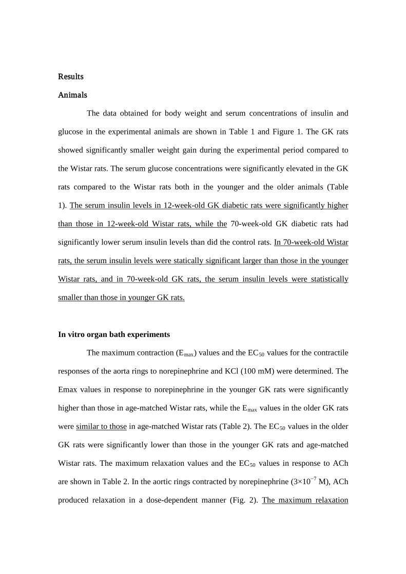
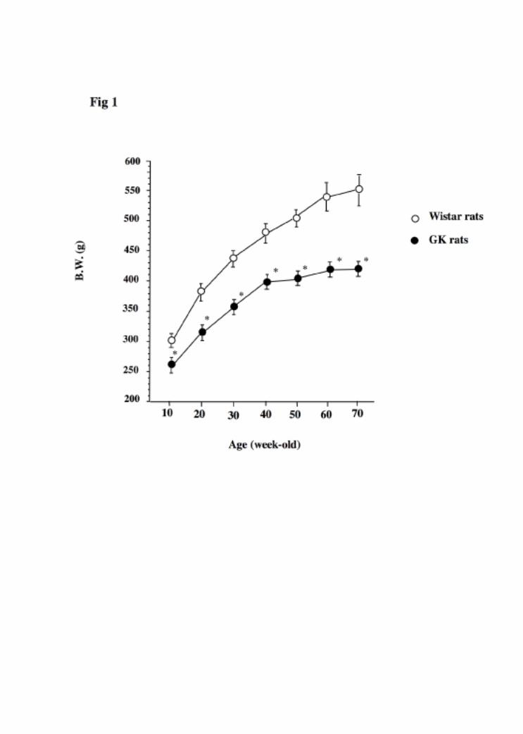
The data obtained for body weight and serum concentrations of insulin and
glucose in the experimental animals are shown in Table 1 and Figure 1. The GK rats
showed significantly smaller weight gain during the experimental period compared to
the Wistar rats. The serum glucose concentrations were significantly elevated in the GK
rats compared to the Wistar rats both in the younger and the older animals (Table
1). The serum insulin levels in 12-week-old GK diabetic rats were significantly higher
than those in 12-week-old Wistar rats, while the 70-week-old GK diabetic rats had
significantly lower serum insulin levels than did the control rats.
In 70-week-old Wistar
rats, the serum insulin levels were statically significant larger than those in the younger
Wistar rats, and in 70-week-old GK rats, the serum insulin levels were statistically
smaller than those in younger GK rats.
In vitro organ bath experiments
The maximum contraction (Emax) values and the EC50 values for the contractile
responses of the aorta rings to norepinephrine and KCl (100 mM) were determined. The
Emax values in response to norepinephrine in the younger GK rats were significantly
higher than those in age-matched Wistar rats, while the Emax values in the older GK rats
were similar to those in age-matched Wistar rats (Table 2). The EC50 values in the older
GK rats were significantly lower than those in the younger GK rats and age-matched
Wistar rats. The maximum relaxation values and the EC50 values in response to ACh
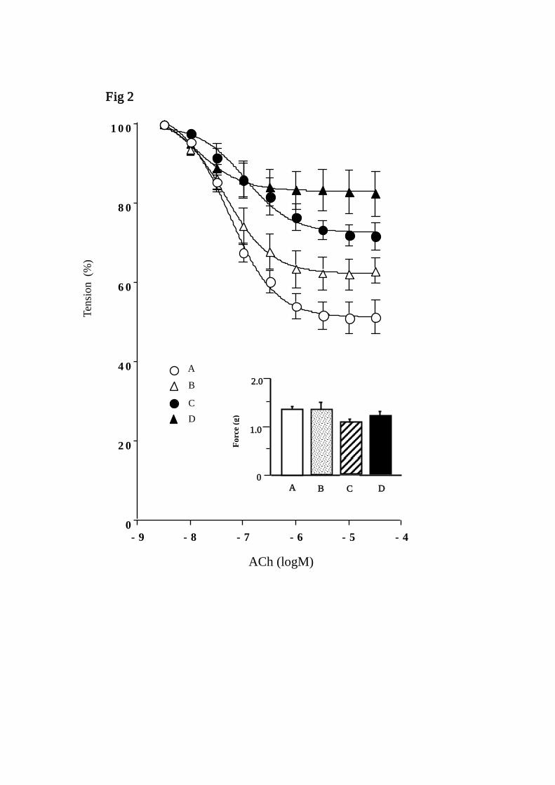
are shown in Table 2. In the aortic rings contracted by norepinephrine (3×10⁻⁷ M), ACh
produced relaxation in a dose-dependent manner (Fig. 2). The maximum relaxation
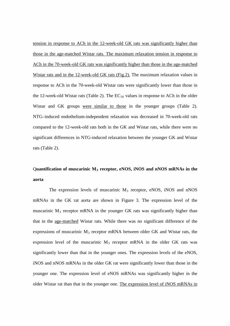
tension in response to ACh in the 12-week-old GK rats was significantly higher than
those in the age-matched Wistar rats. The maximum relaxation tension in response to
ACh in the 70-week-old GK rats was significantly higher than those in the age-matched
Wistar rats and in the 12-week-old GK rats (Fig.2). The maximum relaxation values in
response to ACh in the 70-week-old Wistar rats were significantly lower than those in
the 12-week-old Wistar rats (Table 2). The EC50 values in response to ACh in the older
Wistar and GK groups were similar to those
in the younger groups (Table 2).
NTG–induced endothelium-independent relaxation was decreased in 70-week-old rats
compared to the 12-week-old rats both in the GK and Wistar rats, while there were no
significant differences in NTG-induced relaxation between the younger GK and Wistar
rats (Table 2).
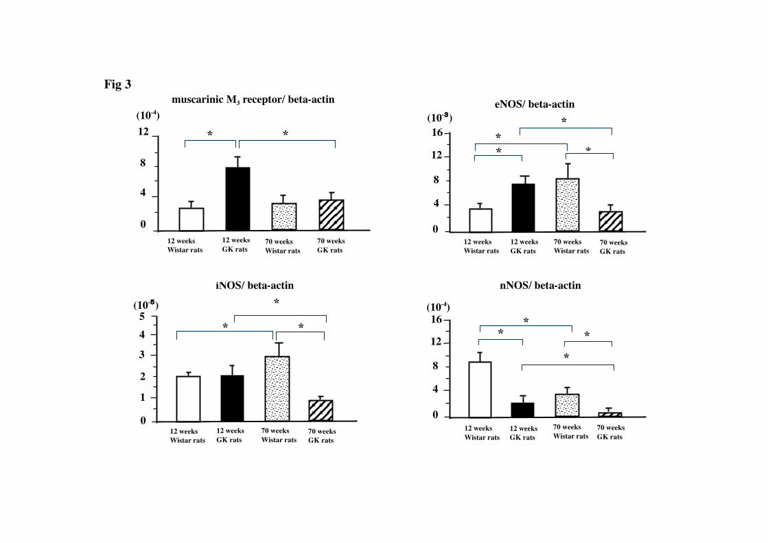
Quantification of muscarinic M3
The expression levels of muscarinic M
receptor, eNOS, iNOS and nNOS mRNAs in the
aorta
3 receptor, eNOS, iNOS and nNOS
mRNAs in the GK rat aorta are shown in Figure 3. The expression level of the
muscarinic M3 receptor mRNA in the younger GK rats was significantly higher than
that in the age-matched Wistar rats. While there was no significant difference of the
expressions of muscarinic M3 receptor mRNA between older GK and Wistar rats, the
expression level of the muscarinic M3 receptor mRNA in the older GK rats was
significantly lower than that in the younger ones. The expression levels of the eNOS,
iNOS and nNOS mRNAs in the older GK rat were significantly lower than those in the
younger one. The expression level of eNOS mRNAs was significantly higher in the
older Wistar rat than that in the younger one. The expression level of iNOS mRNAs in
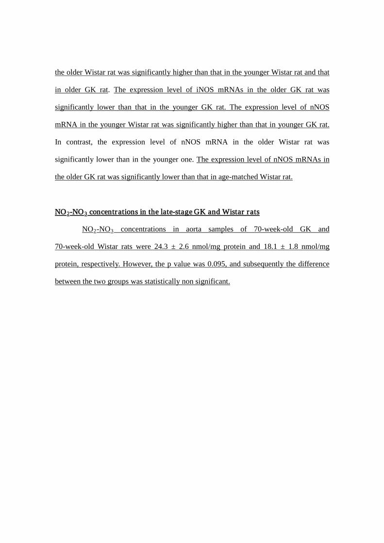
the older Wistar rat was significantly higher than that in the younger Wistar rat and that
in older GK rat. The expression level of iNOS mRNAs in the older GK rat was
significantly lower than that in the younger GK rat. The expression level of nNOS
mRNA in the younger Wistar rat was significantly higher than that in younger GK rat.
In contrast, the expression level of nNOS mRNA in the older Wistar rat was
significantly lower than in the younger one.
The expression level of nNOS mRNAs in
the older GK rat was significantly lower than that in age-matched Wistar rat.
NO 2-NO 3 concentrations in the late-stage GK and Wistar rats
NO2-NO3
concentrations in aorta samples of 70-week-old GK and
70-week-old Wistar rats were 24.3 ± 2.6 nmol/mg protein and 18.1 ± 1.8 nmol/mg
protein, respectively. However, the p value was 0.095, and subsequently the difference
between the two groups was statistically non significant.
Discussion
The main findings of the present study are that in the late stage (70-week-old)
GK rats, endothelium-independent relaxations were impaired compared to those in the
younger (12-week-old) GK or age-matched Wistar rats, and that reduced expressions of
eNOS, iNOS and nNOS mRNAs were observed in the old GK rats compared to the
age-matched Wistar rats, resulting in a decline of relaxation directly via smooth
muscle.
Vascular dysfunction and cardiovascular diseases are major complications in
diabetes mellitus, and are principally caused by endothelial dysfunction [18-20].
Although a number of studies have examined endothelial dysfunction in the metabolic
syndrome and/or diabetes, few studies have examined the progression of vascular
dysfunction in a long term fashion as these diseases develop [2, 21]. Long-term
examination of endothelial dysfunction is necessary, because development of the
metabolic syndrome and/or type 2 diabetes occur over months or years as they are
Furthermore, in this study, we confirmed that 12-week-old GK rats were mild
hyperinsulinemic and hyperglycemic, and that 70-week-old GK rats were modest
hypoinsulinemic and hyperglycemic. Portha et al. (2009) proposed that the defective
beta-cell mass and function in the GK model reflect the complex interactions of
multiple pathogenic players: (i) several independent loci containing genes responsible
for some diabetic traits (but not decreased beta-cell mass); (ii) gestational metabolic
impairment inducing an epigenetic programming of the pancreas (decreased beta-cell
neogenesis and/or proliferation) which is transmitted to the next generation; and (iii)
loss of beta-cell differentiation due to chronic exposure to
hyperglycemia/hyperlipidemia, inflammatory mediators, oxidative stress and to
perturbed islet microarchitecture [17].
progressive diseases.
In the present study, aortas from 70-week-old GK rats exhibited decreased
endothelium-dependent relaxation in response to ACh in comparison with both
age-matched Wistar rats and younger GK rats. These results are in agreement with
previous studies conducted on the aorta of Zucker diabetic fatty (ZDF) rats. Oltman et al.
(2006) showed that ACh-induced relaxation was attenuated in the aortas of 28- to
40-week-old ZDF diabetic rats in comparison with the aortas of 8- to 12-week-old ZDF
rats [2]. Additionally, Zhang et al. (2004) reported decreasing endothelial function over
time (3 to 9 months of age) in the ZDF rat aorta [22]. In the present study, although
there were no significant differences between 12-week-old GK and age-matched Wistar
rats in regard to the maximum relaxations in response to NTG, significant decreases in
the maximum relaxations produced by NTG in both 70-week-old GK and 70-week-old
Wistar rats compared to the younger groups were observed. This finding might be
attributable to the fact that diabetes impairs the endothelium relatively early in its
progression, and affects the aortic smooth muscle later. Bitar et al. (2005) reported that
there were no significant differences in the maximum relaxation in response to Ach
between aortic rings from one-month-old GK rats and age-matched Wistar rats [23].
Both Sandu et al. (2000) and Witte et al. (2002) reported that aortas from rats with
long-term GK diabetes show an impaired endothelium-independent relaxation [24, 25].
Our data are consistent with these previous reports. Hyperreactivity to endothelin
(ET)-1 is an important issue in diabetic vascular dysfunction. Insulin resistance, an
important component of type 2 diabetes, contributes to vascular responsiveness to
vasoactive factors. The potent vasoconstrictor ET-1 is chronically up-regulated in
diabetes [26]. Elgebaly et al. (2008) reported that vascular insulin resistance occurs in a
nonobese model of diabetic GK rats and that both ETA and ETB receptor subtypes are
involved in vascular relaxation to insulin [27]. The key factor in endothelial function is
NO, which is generated via the constitutive eNOS. There are three distinct isoforms of
NOS, and they have been shown to have different localizations and catalytic properties
[28, 29]. The eNOS and nNOS enzymes are suggested to be important regulators of
glucagon release [30]. nNOS functionally controls vascular homeostasis [28, 31, 32].
The expression of iNOS is only promoted when endothelial dysfunction occurs [28,
31-33]. eNOS is constitutively expressed and essential for the control of vascular tone
[34]. Thus, impaired NO synthesis and/or availability may result in endothelial and
vascular wall dysfunction. In this study, we demonstrated that NO2-NO3 levels in the
aortas of 70-week-old GK (group C) and age-matched Wistar rats (group D) were
similar. Bitar et al (2005) suggested a possible mechanism of endothelial dysfunction in
the GK diabetic rat. An enhancement in superoxide production and a diminution in NO
bioavailability are evident in the GK rat. Diabetes produces a cascade of events
involving production of reactive oxygen species from NADPH oxidase leading to
oxidation of BH4
In our study, endothelium-dependent relaxation in 12-week-old GK rats was
significantly decreased in comparison with the age-matched Wistar rats. In addition, the
level of eNOS mRNA expression in 12-week-old GK rats was elevated compared to that
in age-matched Wistar rats. Several other investigators have reported similar findings in
GK rats. Bitar et al.
and uncoupling of NOS. This may promote the oxidative inactivation
of NO with subsequent formation of peroxynitrate. An alteration in the balance of these
bioactive radicals in concert play a potentially role in pathogenesis of diabetic
endothelial dysfunction [23].
(2005) demonstrated that ACh-induced relaxation was markedly
reduced and eNOS protein was significantly increased in GK rats compared to controls
[23].
There have also been reports indicating that eNOS mRNA and protein levels in
the aortic wall are increased in diabetic rats [19, 31, 37]. Cai et al.
Lee et al. (2009) reported the presence of Akt-independent iNOS expression in the
GK diabetic model. They noticed that the defective insulin-induced vasodilation
observed in the diabetic vasculature can be restored by the overexpression of active Akt,
which advocates a potential novel therapeutic strategy for treating diabetes [35].
Matsumoto et al. (2009) reported that up-regulation of ETA, a defect in ETB-mediated
NO signaling, and activation of the MEK/ERK pathway together represent a likely
mechanism mediating the hyperreactivity to ET-1 examined in GK diabetic rat
mesenteric artery [36].
(2005) recently
reported that, although total eNOS levels were increased, NO levels were significantly
decreased in the aortas of diabetic mice [38]. These data suggest that diabetes may
increase eNOS expression and reduce NO bioavailability in the vascular wall [34].
Furthermore, in the present study, we reported for the first time that eNOS mRNA
levels were lower in older GK rats than in younger ones. Moreover, our report suggests
that the expression levels of iNOS mRNA in 70-week-old Wistar rats were significantly
increased compared to those in the younger Wistar rats. In contrast, expression levels of
iNOS mRNA in GK rats in the late stage were significantly decreased compared to
those in the younger GK rats. It is well known that iNOS is induced by inflammation
and/or infiltration of neutrophils and that iNOS-derived NO exhibits a powerful
protective activity under a cellular stress condition [38]. In various pathologic
conditions, including diabetes mellitus, iNOS is expressed in atherosclerotic plaques
and produces a large amount of NO and O2- locally, which results in production of
oxidative products [38]. Recently, Ji et al. (2008)
In the 12-week-old GK rats, the expression levels of muscarinic M
reported that increased levels of free
radicals with a decrease of vascular endothelial growth factor and iNOS contribute to
the progression of the pressure-overload hypertrophied heart to the failing heart [39].
3 receptor
mRNAs were significantly higher than those in the age-matched Wistar rats. We
previously reported a similar pattern in the expression levels of muscarinic M3 receptor
mRNAs in streptozotocin-induced diabetic rat aortas [16]. This up-regulation of
muscarinic M3 receptor mRNAs may be related to a decrease in Ach in the early phase
of diabetes. In the later stage of diabetes, the muscarinic M3
In conclusion, this study has demonstrated diabetes-related and age-related
alterations in endothelial function, and expressions of muscarinic M
receptor mRNA levels in
GK rats were returned to similar levels with those of the age-matched Wistar rats with
decompensation.
3
receptor, eNOS,
iNOS and nNOS mRNAs in the aortas of GK rats and Wistar rats.
References
1. Amos AF, McCarty DJ, Zimmet P (1997) The rising global burden of diabetes and its
complications: estimates and projections to the year 2010. Diabet Med 14: S1-S85.
2. Oltman CL, Richou LL, Davidson EP, Coppey LJ, Lund DD, Yorek MA (2006)
Progression of coronary and mesenteric vascular dysfunction in Zucker obese and
Zucker diabetic fatty rats. Am J Physiol Heart Circ Physiol 291: H1780-1787.
3. Furchgott RF, Vanhoutte PM (1989) Endothelium-derived relaxing and contracting
factors. FASEB J 3: 2007-2018.
4. Furchgott RF (1999) Endothelium-derived relaxing factor: discovery, early studies,
and identification as nitric oxide. Biosci Report 1: 235-251.
5. Furchgott RF, Zawadzki JV (1980) The obligatory role of endothelial cells in the
relaxation of arterial smooth muscle by acetylcholine. Nature 288: 373-376.
6. De Vriese AS, Verbeuren TJ, Van de Voorde J, Lameire NH, Vanhoutte PM (2000)
Endothelial dysfunction in diabetes. Br J Pharmacol 130: 963-974.
7. Feletou M, Vanhoutte PM (1999) The third pathway: endothelium-dependent
hyperpolarization. J Physiol Pharmacol 50: 525-534.
8. Huszka M, Kaplar M, Rejto L, Tornai I, Palatka K, Laszlo P, Udvardy M (1997) The
association of reduced endothelium derived relaxing factor-NO production with
endothelial damage and increased in vivo platelet activation in patients with diabetes
mellitus. Thromb Res 86: 173-180.
9. Boulanger CM, Morrison KJ, Vanhoutte PM (1994) Mediation by M3-muscarinic
receptors of both endothelium-dependent contraction and relaxation to acetylcholine in
the aorta of the spontaneously hypertensive rat. Br J Pharmacol 112: 519–524.
10. Khurana S, Chacon I, Xie G, Yamada M, Wess J, Raufman JP, Kennedy RH (2004)
Vasodilatory effects of cholinergic agonists are greatly diminished in aorta from
M3R−/− mice. Eur J Pharmacol 493: 127-132.
11. Goto Y, Kakizaki M, Masaki N (1976) Production of spontaneous diabetic rats by
repetition of selective breeding. Tohoku J Exp Med 119: 85-90.
12. Salehi A, Carlberg M, Henningson R, Lundquist I (1996) Islet constitutive nitric
oxide synthase: biochemical determination and regulatory function. Am J Physiol 270:
C1634–1641.
13. Unger RH (1976) The Banting Memorial Lecture 1975. Diabetes and the alpha cell.
Diabetes 25: 136–151.
14. Murakawa Y, Zhang W, Pierson CR, Brismar T, Östenson CG, Efendic S, Sima AA
(2002) Impaired glucose tolerance and insulinopenia in the GK-rat causes peripheral
neuropathy. Diabetes Metab Res Rev 18: 473-483.
15. Yagihashi S, Tnosaki A, Yamada K, Kakizaki M, Goto Y (1982) Peripheral
neuropathy in selectively-inbred spontaneously diabetic rats: Electrophysiological,
morphometrical and freeze-replica studies. Tohoku J Exp Med 138: 39-48.
16. Shinbori C, Saito M, Kinoshita Y, Satoh I, Kono T, Hanada T, Nanba E, Adachi K,
Suzuki H, Yamada M, Satoh K (2007) Cyclohexenonic long-chain fatty alcohol has
therapeutic effects on diabetes-induced angiopathy in the rat aorta. Eur J Pharmacol 56:
139-144.
17. Portha B, Lacraz G, Kergoat M, Homo-Delarche F, Giroix MH, Bailbé D,
Gangnerau MN, Dolz M, Tourrel-Cuzin C, Movassat J (2009) The GK rat beta-cell: a
prototype for the diseased human beta-cell in type 2 diabetes? Mol Cell Endocrinol.
297: 73-85.
18. Guerci B, Bohme P, Kearney-Schwartz A, Zannad F, Drouin P (2001) Endothelial
dysfunction and type 2 diabetes. Part 2: altered endothelial function and the effects of
treatments in type 2 diabetes mellitus. Diabet Metabol 27: 436-447.
19. Hink U, Li H, Mollnau H, Oelze M, Matheis E, Hartmann M, Skatchkov M, Thaiss
F, Stahl RA, Warnholtz A, Meinertz T, Griendling K, Harrison DG, Forstermann U,
Munzel T (2001) Mechanisms underlying endothelial dysfunction in diabetes mellitus.
Circ Res 88, E14-22.
20. Pannirselvam M, Verma S, Anderson TJ, Triggle CR (2002) Cellular basis of
endothelial dysfunction in small mesenteric arteries from spontaneously diabetic (db/db
-/-) mice: role of decreased tetrahydrobiopterin bioavailability. Br J Pharmacol 136:
255–263.
21. Kobayashi T, Matsumoto T, Ooishi K, Kamata K (2004) Differential expression of
alpha2D-adrenoceptor and eNOS in aortas from early and later stages of diabetes in
Goto-Kakizaki rats. Am J Physiol Heart Circ Physiol. 287: H135-143.
22. Zhang C, Yang J, Jennings LK (2004) Leukocyte-derived myeloperoxidase
amplifies high-glucose-induced endothelial dysfunction through interaction with
high-glucose-stimulated, vascular non-leukocyte-derived reactive oxygen species.
Diabetes 53: 2950-2959.
23. Bitar MS. Wahid S, Mustafa S, AI-Saleh E, Dhaunsi GS, AI-Mulla F (2005) Nitric
oxide dynamics and endothelial dysfunction on type II model of genetic diabetes. Eur J
Pharmacol 511: 53-64.
24. Sandu OA, Ragolia L, Begum N (2000) Diabetes in the Goto-Kakizaki rat is
accompanied by impaired insulin-mediated myosin-bound phosphatase activation and
vascular smooth muscle cell relaxation. Diabetes 49: 2178–2189.
25. Witte K, Jacke K, Stahrenberg R, Arlt G, Reitenbach I, Schilling L, Lemmer B
(2002) Dysfunction of soluble guanylyl cyclase in aorta and kidney of Goto-Kakizaki
rats: influence of age and diabetic state. Nitric oxide 6: 85–95.
26. Chen S, Mukherjee S, Chakraborty C, Chakrabarti, (2003) High glucose-induced,
endothelin-dependent fibronectin synthesis is mediated via NF-kappa B and AP-1. Am.
J. Physiol. Cell Physiol. 284: C263–C272.
27. Elgebaly MM, Kelly A, Harris AK, Elewa H, Portik-Dobos V, Ketsawatsomkron P,
Marrero M, Ergul A (2008) Impaired insulin-mediated vasorelaxation in a nonobese
model of type 2 diabetes: role of endothelin-1. Can J Physiol Pharmacol. 2008 86:
358-64.
28. Alderton WK, Cooper CE, Knowles RG (2001) Nitric oxide synthases: structure,
function and inhibition. Biochem J 357: 593–615.
29. Calles-Escandon J, Cipolla M (2001) Diabetes and endothelial dysfunction: a
clinical perspective. Endocr Rev 22: 36–52.
30. Henningsson R, Alm P, Lindstrom E, Lundquist I (2000) Chronic blockade of NO
synthase paradoxically increases islet NO production and modulates islet hormone
release. Am J Physiol. Endocri Metabol 279: E95–E107.
31. Papapetropoulos A, Rudic RD, Sessa WC (1999) Molecular control of nitric oxide
synthases in the cardiovascular system. Cardiovasc Res 43: 509–520.
32. Li H, Forstermann U (2000) Nitric oxide in the pathogenesis of vascular disease. J
Pathol 190: 244–254.
33. Kibbe M, Billiar T, Tzeng E (1999) Inducible nitric oxide synthase and vascular
injury. Cardiovas Res 43: 650–657.
34. Pandolfi A, De Filippis EA (2007) Chronic hyperglycemia and nitric oxide
bioavailability play a pivotal role in pro-atherogenic vascular modifications. Genes and
nutrition 2, 195–208.
35. Lee JH, Palasia T, Ragolia L (2009) Impaired insulin-mediated vasorelaxation in
diabetic Goto-Kakizaki rats is caused by impaired Akt phosphorylation. Am J Physiol
Cell Physiol. 296: C327-38.
36. Matsumoto T, Ishida K, Nakayama N, Kobayashi T, Kamata K (2009) Involvement
of NO and MEK/ERK pathway in enhancement of endothelin-1-induced mesenteric
artery contraction in later-stage type 2 diabetic Goto-Kakizaki rat. Am J Physiol Heart
Circ Physiol. 296: H1388-97.
37. Oyadomari S, Gotoh T, Aoyagi K, Araki E, Shichiri M, Mori M (2001) Coinduction
of endothelial nitric oxide synthase and arginine recycling enzymes in aorta of diabetic
rats. Nitric oxide 5, 252–260.
38. Cai S, Khoo J. Mussa S, Alp NJ, Channon KM (2005) Endothelial nitric oxide
synthase dysfunction in diabetic mice: importance of tetrahydrobiopterin in eNOS
dimerisation. Diabetologia 48: 1933–1940.
39. Ji K, Minakawa M, Fukui K, Suzuki Y, Fukuda I (2008) Increased superoxide
radical with a decrease in vascular endothelial growth factor and inducible nitric oxide
synthase level leads to the progression of left ventricular hypertrophy in a
pressure-overload rat heart model. Ann Thorac Cardiovasc Surg 14: 210-217.
Figure legends
Fig. 1: Changes of body weight during the experimental period.
Data are shown as the means ±SEM for 6-8 animals in each group. *Significantly
different from age-matched Wistar rats.
Fig. 2: Endothelium-dependent relaxation of rat aortic rings in response to Ach (large
panel). Inset: Contractions (means ± SEM) produced by norepinephrine (3×10⁻⁷ M) in
each group (small panel). *) significantly different from age-matched Wistar group
(group A vs group B, group C vs group D). †
) significantly different from younger group
(group A vs group C, group B vs group D). (p<0.05). Data are shown as the means ±
SEM for 6-8 animals in each group.
Fig. 3: The expressions of muscarinic M3
*) significantly different between group (p<0.05).
receptor, eNOS, iNOS, and nNOS mRNAs
normalized by β-actin mRNA in the aortas of GK rats
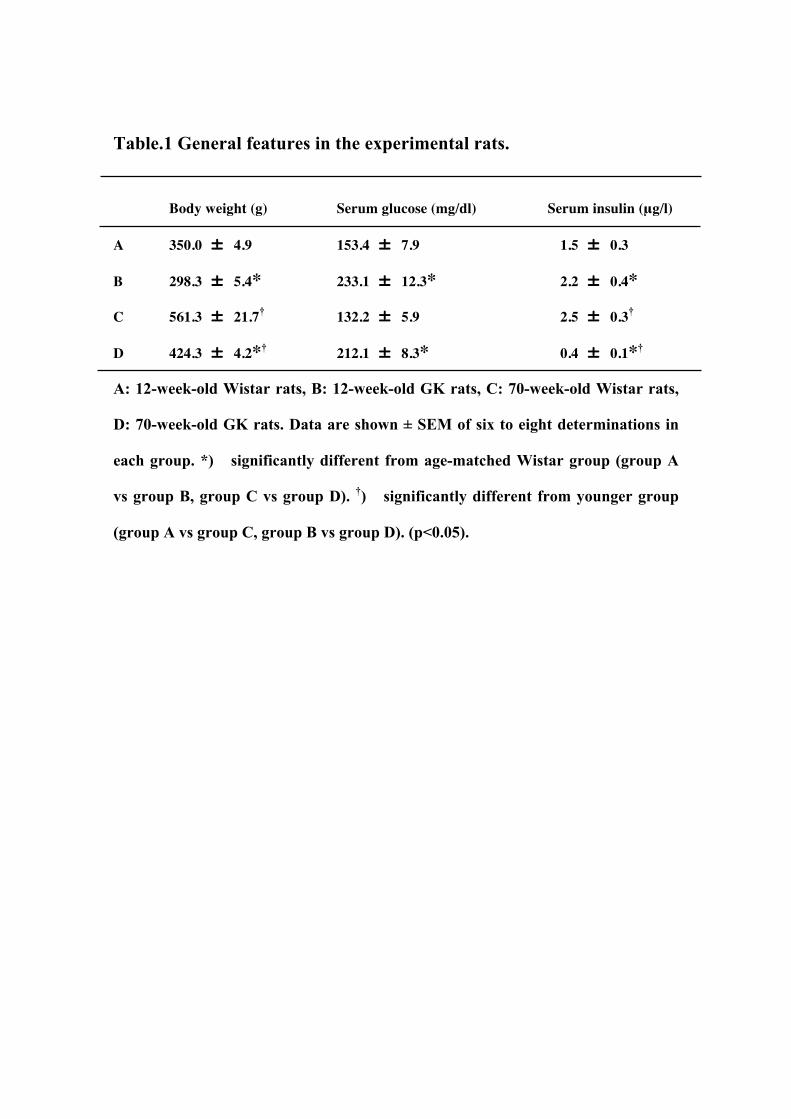
Table.1 General features in the experimental rats.
Body weight (g) Serum glucose (mg/dl) Serum insulin (μg/l)
A 350.0 ± 4.9 153.4 ± 7.9 1.5 ± 0.3
B 298.3 ± 5.4* 233.1 ± 12.3* 2.2 ± 0.4*
C 561.3 ± 21.7† 132.2 ± 5.9 2.5 ± 0.3†
D 424.3 ± 4.2*† 212.1 ± 8.3* 0.4 ± 0.1*†
A: 12-week-old Wistar rats, B: 12-week-old GK rats, C: 70-week-old Wistar rats,
D: 70-week-old GK rats. Data are shown ± SEM of six to eight determinations in
each group. *) significantly different from age-matched Wistar group (group A
vs group B, group C vs group D). †) significantly different from younger group
(group A vs group C, group B vs group D). (p<0.05).
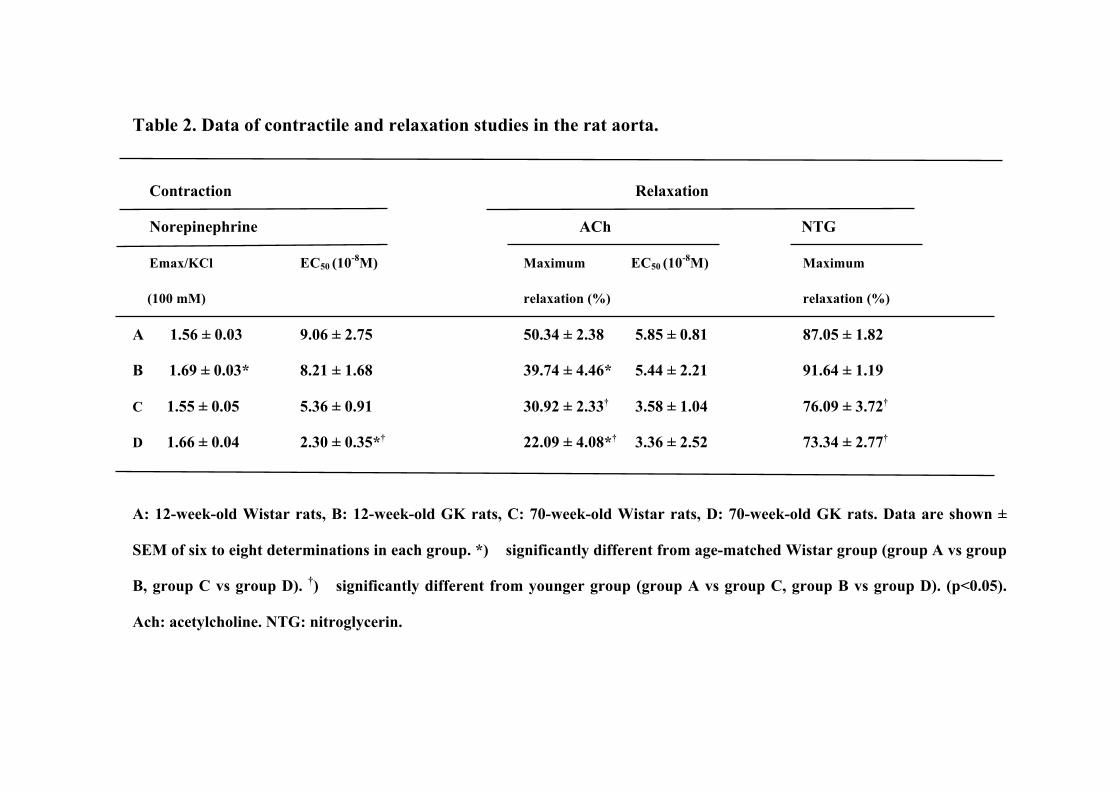
Table 2. Data of contractile and relaxation studies in the rat aorta.
Contraction Relaxation
Norepinephrine ACh NTG
Emax/KCl EC50 (10-8M) Maximum EC50 (10-8M) Maximum
(100 mM) relaxation (%) relaxation (%)
A 1.56 ± 0.03 9.06 ± 2.75 50.34 ± 2.38 5.85 ± 0.81 87.05 ± 1.82
B 1.69 ± 0.03* 8.21 ± 1.68 39.74 ± 4.46* 5.44 ± 2.21 91.64 ± 1.19
C 1.55 ± 0.05 5.36 ± 0.91 30.92 ± 2.33† 3.58 ± 1.04 76.09 ± 3.72†
D 1.66 ± 0.04 2.30 ± 0.35*† 22.09 ± 4.08*† 3.36 ± 2.52 73.34 ± 2.77†
A: 12-week-old Wistar rats, B: 12-week-old GK rats, C: 70-week-old Wistar rats, D: 70-week-old GK rats. Data are shown ±
SEM of six to eight determinations in each group. *) significantly different from age-matched Wistar group (group A vs group
B, group C vs group D). †) significantly different from younger group (group A vs group C, group B vs group D). (p<0.05).
Ach: acetylcholine. NTG: nitroglycerin.
- 9 - 8 - 7 - 6 - 5 - 4 0
2 0
4 0
6 0
8 0
1 0 0
ACh (logM)
A B D
1.0
2.0
0 C
Forc
e (g
)
B
A
C D
Tens
ion
(%)
Fig 2
muscarinic M3 receptor/ beta-actin eNOS/ beta-actin
Fig 3 Fig 3
(10-4) (10-3)
1
2
3
4
0 12 weeks Wistar rats
70 weeks Wistar rats
12 weeks GK rats
70 weeks GK rats
5
iNOS/ beta-actin
(10-5) (10-4)
nNOS/ beta-actin
4
8
12
0 12 weeks Wistar rats
70 weeks Wistar rats
*
12 weeks GK rats
70 weeks GK rats
*
4
8
12
16
0 12 weeks Wistar rats
70 weeks Wistar rats
*
12 weeks GK rats
70 weeks GK rats
* *
*
4
8
12
16
0 12 weeks Wistar rats
70 weeks Wistar rats
*
12 weeks GK rats
70 weeks GK rats
* *
*
*
*
*
Related Documents