Endosulfan induces autophagy and endothelial dysfunction via the AMPK/mTOR signaling pathway triggered by oxidative stress * Lianshuang Zhang a, b, c , Jialiu Wei a, b , Lihua Ren a, b , Jin Zhang a, b , Ji Wang a, b , Li Jing a, b , Man Yang a, b , Yang Yu a, b , Zhiwei Sun a, b , Xianqing Zhou a, b, * a Department of Toxicology and Hygienic Chemistry, School of Public Health, Capital Medical University, Beijing 100069, China b Beijing Key Laboratory of Environmental Toxicology, Capital Medical University, Beijing 100069, China c Department of Histology and Embryology, Bin Zhou Medical College, Yan Tai 264003, China article info Article history: Received 16 August 2016 Received in revised form 19 October 2016 Accepted 23 October 2016 Available online 1 November 2016 abstract Cardiovascular diseases is related to environmental pollution. Endosulfan is an organochlorine pesticide and its toxicity has been reported. However, the relationship between oxidative stress and autophagy induced by endosulfan and its underlying mechanism remain confusing. In this study, human umbilical vein endothelial cells (HUVECs) were chosen to explore the toxicity mechanism and were treated with 0, 1, 6, 12 mg/mL 1 endosulfan for 24 h, respectively. The present results showed that autophagy could be induced by endosulfan, which was verified by the monodansylcadaverine staining, autophagic ultra- structural observation, and LC3-I/LC3-II conversion. In addition, the levels of adenosine triphosphate (ATP), the mitochondria membrane potential (MMP) were significantly decreased in a dose-dependent way. The expression of proinflammatory cytokines (tumor necrosis factor a, interleukin-1b, and interleukin-6) were significantly elevated, and the index of endothelial function such as monocyte chemotactic protein 1 (MCP-1), intercellular cell adhesion molecule-1 (ICAM-1) increased. Moreover, endosulfan had an activation effect on the 5 0 AMP-activated protein kinase (AMPK)/rapamycin (mTOR) signaling pathway. Our findings demonstrated that endosulfan could induce oxidative stress and mito- chondria injury, activate autophagy, induce inflammatory response, and eventually lead to endothelial dysfunction via the AMPK/mTOR pathway. This indicates that exposure to endosulfan is a potential risk factor for cardiovascular diseases. © 2016 Elsevier Ltd. All rights reserved. 1. Introduction Endosulfan (6,7,8,9,10,10-hexachloro-1,5,5a, 6,9,9a-hexahydro- 6,9-methano-2,4,3-benzodioxathiepine-3-oxide) is a cyclodiene broad organochlorine pesticide, which is. widely used on wide variety of crops in many parts of the world (Mersie et al., 2003). As a result of its widespread use, it is an environmental contaminant and a public health hazard (Jaiswal et al., 2005). Endosulfan is hazardous to various organs including testes, kidney and immune (Ozmen and Mor, 2015; Choudhary and Joshi, 2003; Jamil et al., 2004; Aggarwal et al., 2008). However, there is still a lack of evaluation regarding the toxicity of endosulfan on cardiovascular system. A growing body of literature suggests that apoptosis may contribute to the toxicity of POPs (Quan et al., 2014; Gregoraszczuk et al., 2008). But few study pay attention to the relationship be- tween autophagy and POPs. Autophagy is defined as a protective mechanism in cells that can degrade proteins and/or damaged or- ganelles to maintain cellular homeostasis (Teng et al., 2012). Nevertheless, the process of autophagy can be stimulated by oxidative stress (Liu et al., 2015; Li et al., 2015). Our previous study showed that endosulfan could activate extrinsic coagulation pathway by inducing endothelial cell injury via oxidative stress (Zhang et al., 2015). Whether endosulfan can induce autophagy and influence endothelial cell function is a crucial issue in human health and safety. Endothelial dysfunction, first described by Panza et al. (Panza et al., 1990) Endemann and Schiffrin (Endemann and Schiffrin, 2004) is defined as a series of events, mainly including vasodila- tion reduction, proinflammatory response, and reducing of mono- cyte chemotactic protein 1 (MCP-1) and intercellular cell adhesion * This paper has been recommended for acceptance by David Carpenter. * Corresponding author. Department of Toxicology and Hygienic Chemistry, School of Public Health, Capital Medical University, Beijing 100069, China. E-mail addresses: [email protected], [email protected] (X. Zhou). Contents lists available at ScienceDirect Environmental Pollution journal homepage: www.elsevier.com/locate/envpol http://dx.doi.org/10.1016/j.envpol.2016.10.067 0269-7491/© 2016 Elsevier Ltd. All rights reserved. Environmental Pollution 220 (2017) 843e852

Welcome message from author
This document is posted to help you gain knowledge. Please leave a comment to let me know what you think about it! Share it to your friends and learn new things together.
Transcript
lable at ScienceDirect
Environmental Pollution 220 (2017) 843e852
Contents lists avai
Environmental Pollution
journal homepage: www.elsevier .com/locate/envpol
Endosulfan induces autophagy and endothelial dysfunction viathe AMPK/mTOR signaling pathway triggered by oxidative stress*
Lianshuang Zhang a, b, c, Jialiu Wei a, b, Lihua Ren a, b, Jin Zhang a, b, Ji Wang a, b, Li Jing a, b,Man Yang a, b, Yang Yu a, b, Zhiwei Sun a, b, Xianqing Zhou a, b, *
a Department of Toxicology and Hygienic Chemistry, School of Public Health, Capital Medical University, Beijing 100069, Chinab Beijing Key Laboratory of Environmental Toxicology, Capital Medical University, Beijing 100069, Chinac Department of Histology and Embryology, Bin Zhou Medical College, Yan Tai 264003, China
a r t i c l e i n f o
Article history:Received 16 August 2016Received in revised form19 October 2016Accepted 23 October 2016Available online 1 November 2016
* This paper has been recommended for acceptanc* Corresponding author. Department of Toxicolo
School of Public Health, Capital Medical University, BE-mail addresses: [email protected], xianqingz
http://dx.doi.org/10.1016/j.envpol.2016.10.0670269-7491/© 2016 Elsevier Ltd. All rights reserved.
a b s t r a c t
Cardiovascular diseases is related to environmental pollution. Endosulfan is an organochlorine pesticideand its toxicity has been reported. However, the relationship between oxidative stress and autophagyinduced by endosulfan and its underlying mechanism remain confusing. In this study, human umbilicalvein endothelial cells (HUVECs) were chosen to explore the toxicity mechanism and were treated with 0,1, 6, 12 mg/mL�1 endosulfan for 24 h, respectively. The present results showed that autophagy could beinduced by endosulfan, which was verified by the monodansylcadaverine staining, autophagic ultra-structural observation, and LC3-I/LC3-II conversion. In addition, the levels of adenosine triphosphate(ATP), the mitochondria membrane potential (MMP) were significantly decreased in a dose-dependentway. The expression of proinflammatory cytokines (tumor necrosis factor a, interleukin-1b, andinterleukin-6) were significantly elevated, and the index of endothelial function such as monocytechemotactic protein 1 (MCP-1), intercellular cell adhesion molecule-1 (ICAM-1) increased. Moreover,endosulfan had an activation effect on the 50AMP-activated protein kinase (AMPK)/rapamycin (mTOR)signaling pathway. Our findings demonstrated that endosulfan could induce oxidative stress and mito-chondria injury, activate autophagy, induce inflammatory response, and eventually lead to endothelialdysfunction via the AMPK/mTOR pathway. This indicates that exposure to endosulfan is a potential riskfactor for cardiovascular diseases.
© 2016 Elsevier Ltd. All rights reserved.
1. Introduction
Endosulfan (6,7,8,9,10,10-hexachloro-1,5,5a, 6,9,9a-hexahydro-6,9-methano-2,4,3-benzodioxathiepine-3-oxide) is a cyclodienebroad organochlorine pesticide, which is.
widely used on wide variety of crops in many parts of the world(Mersie et al., 2003). As a result of its widespread use, it is anenvironmental contaminant and a public health hazard (Jaiswalet al., 2005). Endosulfan is hazardous to various organs includingtestes, kidney and immune (Ozmen and Mor, 2015; Choudhary andJoshi, 2003; Jamil et al., 2004; Aggarwal et al., 2008). However,there is still a lack of evaluation regarding the toxicity of endosulfanon cardiovascular system.
e by David Carpenter.gy and Hygienic Chemistry,eijing 100069, [email protected] (X. Zhou).
A growing body of literature suggests that apoptosis maycontribute to the toxicity of POPs (Quan et al., 2014; Gregoraszczuket al., 2008). But few study pay attention to the relationship be-tween autophagy and POPs. Autophagy is defined as a protectivemechanism in cells that can degrade proteins and/or damaged or-ganelles to maintain cellular homeostasis (Teng et al., 2012).Nevertheless, the process of autophagy can be stimulated byoxidative stress (Liu et al., 2015; Li et al., 2015). Our previous studyshowed that endosulfan could activate extrinsic coagulationpathway by inducing endothelial cell injury via oxidative stress(Zhang et al., 2015). Whether endosulfan can induce autophagy andinfluence endothelial cell function is a crucial issue in human healthand safety.
Endothelial dysfunction, first described by Panza et al. (Panzaet al., 1990) Endemann and Schiffrin (Endemann and Schiffrin,2004) is defined as a series of events, mainly including vasodila-tion reduction, proinflammatory response, and reducing of mono-cyte chemotactic protein 1 (MCP-1) and intercellular cell adhesion
L. Zhang et al. / Environmental Pollution 220 (2017) 843e852844
molecule-1 (ICAM-1) (Catal�an et al., 2015). Yet, the biologicalbehavior and toxic effects of endosulfan on the vasculature is stillpoorly understood. In this study, we confirmed the associationbetween endosulfan-induced autophagic activity and endothelialdysfunction by conducting a series of assessments in the humanumbilical vein endothelial cells (HUVECs), such as oxidative stress,autophagy, release of cell adhesion cytokines and proinflammatorycytokine, we also examined the AMPK/rapamycin (mTOR) signalingpathway to explore the possible mechanism of the toxicity inducedby endosulfan. These findings provide persuasive evidence for thetoxic effects of endosulfan on the cardiovascular system.
2. Materials and methods
2.1. Cell culture and co-incubation with endosulfan
HUVECs were purchased from ATCC Manassas (No. CRL-1730),the cells were cultured in a humidified environment (37 �C; 5%CO2), maintained in Dulbecco's Modified Eagle's Medium (DMEM)(Hecolon; Thermo Fisher Scientific, Waltham, MA, USA) with 10%fetal bovine serum (Gibco; Thermo Fisher Scientific), 100 U/mL ofpenicillin and 100 mg/mL of streptomycin. For all tests, the HUVECswere seeded in six-well plates (1 � 105 cells/mL) and allowed toattach for 24 h; they were then exposed to different concentrations(1, 6 and 12 mg/mL) of endosulfan for another 24 h. N-acetylcysteine (NAC) (Sigma, USA) was chosen as the antioxidant andDorsomorphin (Dor) as the inhibitor of AMPK respectively.Considering the possible reaction between endosulfan and NAC orDor, cells were pretreatedwith 3mM/ml NAC and 20 mM/ml Dor for2 h before exposure to endosulfan. The equivalent volume of cul-ture mediumwithout endosulfan was set as the control group. Fivereplicate wells were used in each treatment group.
2.2. Detection of mitochondria membrane potential (MMP)
MMP was detected by using the fluorescent probe JC-1 (Jian-cheng, China). This probe can selectively enter into mitochondriaand reversibly change color from red to green as the membranepotential decreased. The ratio of red to green expresses the changeof MMP. Cells were treated with endosulfan for 24 h, after washingwith PBS, the cells were incubated with 10 mg/mL working solutionof JC-1 for 15 min at room temperature. Then the cells werecollected and washed with PBS twice followed by flow cytometric(FCM) (Becton-Dickison, USA) analysis. The green fluorescence in-tensity was determined at an excitation wavelength of 488 nm andan emission wavelength of 525 nm, whereas the red fluorescenceintensity was determined at an excitation wavelength of 488 nmand an emission wavelength of 590 nm. For each sample, at least1 � 104 cells were collected.
2.3. ATP assay
The levels of ATP in the cells were measured using a fireflyluciferase-based ATP assay kit (Jiancheng, nanjing, China), accord-ing to the manufacturer's instructions. After rinsing withphosphate-buffered saline (PBS), the cells were disrupted in 200 mllysis buffer, and centrifuged at 12,000 rpm at 4 �C for 5 min, fromwhich the supernatant was collected. In a 1.5 ml tube, a 100 mlsample of the supernatant was mixed with 100 ml ATP detectionworking solution. The luminance (RLU) was immediately measuredusing a Turner BioSystems luminometer (Promega Corporation,Madison, WI, USA). Standard curves for quantification weregenerated using known amounts of an ATP standard, and the pro-tein concentration of each treatment groupwas determined using abicinchoninic acid protein assay kit (Beyotime Institute of
Biotechnology). Total ATP levels were expressed as mmol/g prot (Liet al., 2012).
2.4. Measurement of ROS and MDA
The ROS generation was measured through fluorescence in-tensity of dichlorofluorescein (DCF) by flow cytometry. Afterexposure to endosulfan for 24 h, cells were incubated in dark at37�Cwith 2,7-dichlorofluorescein-diacetate (DCFH-DA, 100 ng/mL,Jiancheng, China) for 30min as previously described (Wang et al.,2009). Then the cells were washed twice with cold PBS andresuspended in PBS for further analysis. At last, the fluorescenceintensity of 1 � 104 cells for each sample was measured by flowcytometry (Becton-Dickinson, USA).
The concentration of Malondialdehyde (MDA) as a marker oflipid peroxidation (LPO) was determined using an MDA kit. Theassaymixture, including protein of cells, distilled water and sodiumdodecyl sulfate (SDS) was incubated in a test tube at room tem-perature for 10 min, followed by the addition of a 20% acetic acidsolution (1.0 ml, pH 3.5). After incubation for 10 min at 37 �C, 0.8%thiobarbituric acid (TBA) (1.0 ml) was added to the mixture. Thereaction mixture was placed over a boiling water bath at 100 �C for1 h. After n-butanol (4.0 ml) was added to the mixture, the organiclayer was separated by centrifugation at 3000 rpm for 10 min atroom temperature. The supernatant liquid was transferred to afresh tube. The absorbance was measured at 532 nm with amicroplate reader (DNM-9602G, China) using n-butanol as astandard. The data are shown as nmoles of MDA formed h-1 mg-1protein.
2.5. Observation of mitochondria and autophagy by TEM
The cell samples were fixed overnight in 2.5% glutaraldehyde.Then, the samples were washed by 0.1 M PB for three times andpostfixed with 1% osmic acid for 2 h. Then, a series of dehydrationprocesses (50%, 70%, 80%, 90%, and 100% alcohol, and 100% acetone)was performed. After that, all the cell samples were embedded inepoxy resin. The thickness of the ultrathin sections was approxi-mately 50 nm, which were made by an ultramicrotome (UltracutUCT; Leica Microsystems). After being stained by lead citrate anduranyl acetate, these samples were observed under a TEM (JEM-2100; JEOL).
2.6. MDC staining
The fluorescent dye, monodansylcadaverine (MDC), is a specialmarker for autophagic vacuoles. HUVECs were stained with 0.1 mMofMDC (Jiancheng, China) for 15min in the dark after the cells wereexposed to endosulfan for 24 h. Then, the cells were washed withPBS three times. The visualization of MDC staining was detected byLSCM (Leica TCS SP5; Leica Microsystems). And the fluorescenceintensity of 1 � 104 cells for each sample was measured by flowcytometry (Becton-Dickinson, USA).
2.7. Measurement of ICAM-1 and MCP-1
The supernatants were collected after the HUVECs wereexposed to endosulfan for 24 h. The levels of MCP-1 and ICAM-1were measured by enzyme-linked immunosorbent assay kits(Yuchen Biotechnology Company, Shanghai) according to themanufacturer's protocols. Briefly, 50 ml of supernatants were addedin each well and incubated for 2 h at room temperature. Then,biotin antibody was added to each well and incubated for 1 h. Afterthat, streptavidin solution was added and incubated for 30 min.Next, the substrate reagent was added and incubated for 30 min.
L. Zhang et al. / Environmental Pollution 220 (2017) 843e852 845
After the stop solution was added to each well, the absorbance at450 nm was detected immediately using a microplate reader(Thermo Multiskan™ MK3; Thermo Fisher Scientific).
2.8. Measurement of proinflammatory cytokine
The supernatants were collected after the HUVECs wereexposed to endosulfan for 24 h. The levels of tumor necrosis factor a(TNF-a), interleukin-1b (IL-1b), and interleukin-6(IL-6) weremeasured by enzyme-linked immunosorbent assay kits (YuchenBiotechnology Company, Shanghai) according to the manufac-turer's protocols. Briefly, 50 mL of supernatants were added in eachwell and incubated for 2 h at room temperature. Then, biotinantibody was added to each well and incubated for 1 h. After that,streptavidin solutionwas added and incubated for 30min. Next, thesubstrate reagent was added and incubated for 30 min. After thestop solution was added to each well, the absorbance at 450 nmwas detected immediately using a microplate reader (ThermoMultiskan™ MK3; Thermo Fisher Scientific).
2.9. Western blot analysis
Equal amounts of 20 mg of lysate proteins were loaded onto 12%sodium dodecyl sulfate-polyacrylamide gels and electrophoreti-cally transferred to polyvinylidene fluoride (PVDF) membranes(EMDMillipore, Billerica, MA, USA). After blocking with nonfat milk(5%) in Tris-buffered saline (TBS) for 1 h, the PVDF membrane wasincubated, respectively, with AMPK-a(Santa Cruz Biothecnology,USA) (1:1000, rabbit antibodies), LC3, p-AMPK-a,beclin-1, mTOR,and p-mTOR (CST, USA) (1:1000, rabbit antibodies) at 4 �C over-night. Then, the PVDF membrane was rinsed with TBS and Tween20 (TBST) and incubated with antirabbit immunoglobulin G sec-ondary antibody (CST) for 1 h. After rinsing with TBST for a total ofthree times, the proteins bound with the antibody were measuredby the enhanced chemiluminescence reagent (Thermo Fisher Sci-entific). Using the Image Lab™ Software (Bio-Rad Laboratories Inc.,Hercules, CA, USA), the densitometric analysis of the Western blotresults was performed.
2.10. Statistical analysis
The statistical analysis was performed using the SPSS 16.0software. Five replicate wells were used in each treatment group,and the mean was calculated as the sample size for one indepen-dent experiment. The results were analyzed by one-way analysis ofvariance followed by the least significant difference method formultiple comparisons. Datawere expressed as themean ± standard
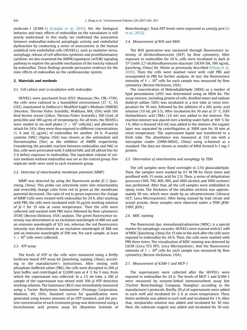
Fig. 1. Effects of endosulfan on the production of cytokines in HUVECs. The expressionsData are expressed as the means ± SD from three independent experiments (*P < 0.05 ver
deviation from three or five independent experiments. If the Pvalue was less than 0.05, differences were considered statisticallysignificant.
3. Results
3.1. Effect of endosulfan on cytokines expression in HUVECs
The expressions of ICAM-1 and MCP- 1 in supernatant weremeasured by ELISA. As shown in Fig. 1, the production of ICAM-1and MCP-1 were significantly increased in 6 and 12 mg/ml groupswhen compared to that of the control. The inhibitor Dor couldreduce the levels of proinflammatory factor levels when comparedto 12 mg/ml endosulfan treated group (P < 0.05).
3.2. Effect of endosulfan on proinflammatory factor expression inHUVECs
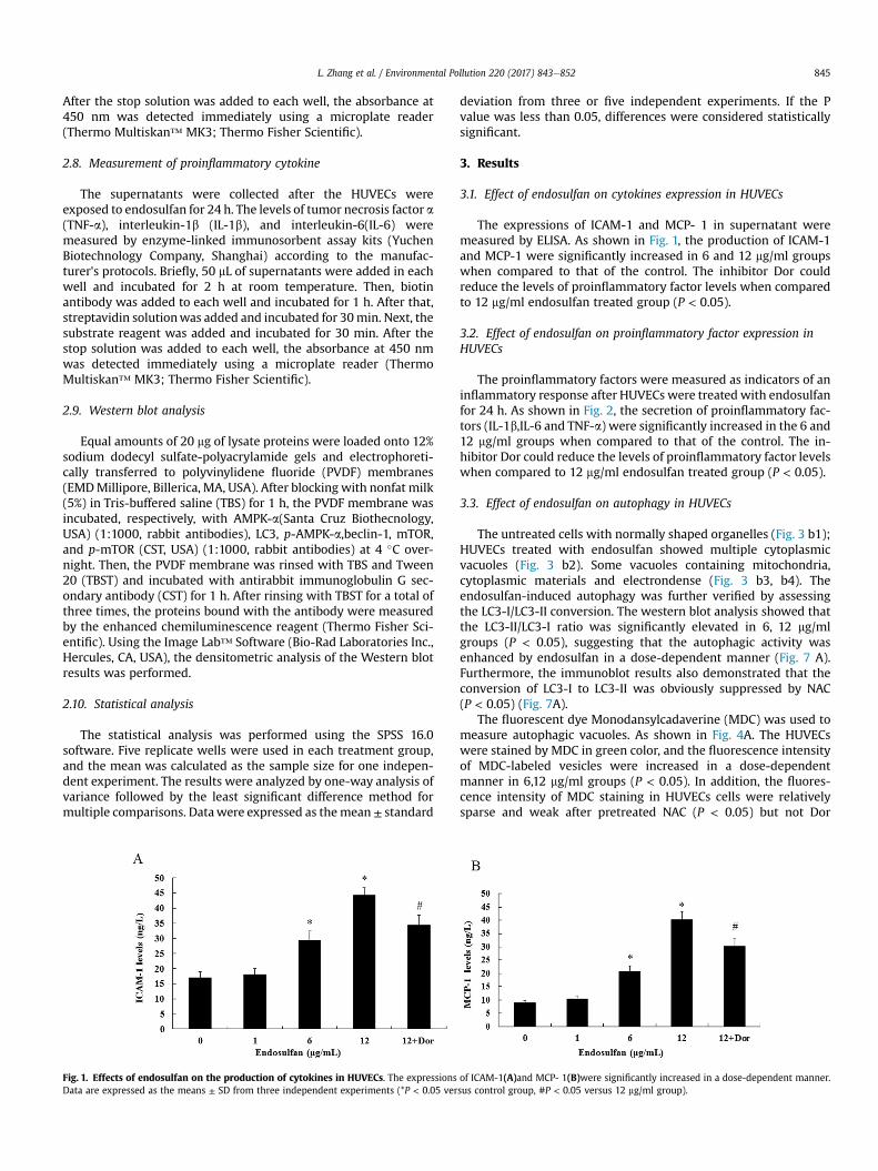
The proinflammatory factors were measured as indicators of aninflammatory response after HUVECs were treated with endosulfanfor 24 h. As shown in Fig. 2, the secretion of proinflammatory fac-tors (IL-1b,IL-6 and TNF-a) were significantly increased in the 6 and12 mg/ml groups when compared to that of the control. The in-hibitor Dor could reduce the levels of proinflammatory factor levelswhen compared to 12 mg/ml endosulfan treated group (P < 0.05).
3.3. Effect of endosulfan on autophagy in HUVECs
The untreated cells with normally shaped organelles (Fig. 3 b1);HUVECs treated with endosulfan showed multiple cytoplasmicvacuoles (Fig. 3 b2). Some vacuoles containing mitochondria,cytoplasmic materials and electrondense (Fig. 3 b3, b4). Theendosulfan-induced autophagy was further verified by assessingthe LC3-I/LC3-II conversion. The western blot analysis showed thatthe LC3-II/LC3-I ratio was significantly elevated in 6, 12 mg/mlgroups (P < 0.05), suggesting that the autophagic activity wasenhanced by endosulfan in a dose-dependent manner (Fig. 7 A).Furthermore, the immunoblot results also demonstrated that theconversion of LC3-I to LC3-II was obviously suppressed by NAC(P < 0.05) (Fig. 7A).
The fluorescent dye Monodansylcadaverine (MDC) was used tomeasure autophagic vacuoles. As shown in Fig. 4A. The HUVECswere stained by MDC in green color, and the fluorescence intensityof MDC-labeled vesicles were increased in a dose-dependentmanner in 6,12 mg/ml groups (P < 0.05). In addition, the fluores-cence intensity of MDC staining in HUVECs cells were relativelysparse and weak after pretreated NAC (P < 0.05) but not Dor
of ICAM-1(A)and MCP- 1(B)were significantly increased in a dose-dependent manner.sus control group, #P < 0.05 versus 12 mg/ml group).
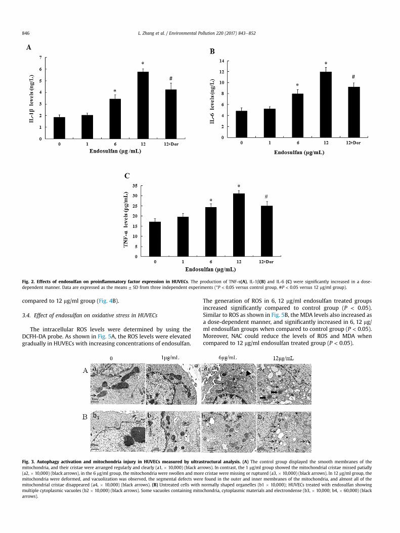
Fig. 2. Effects of endosulfan on proinflammatory factor expression in HUVECs. The production of TNF-a(A), IL-1b(B) and IL-6 (C) were significantly increased in a dose-dependent manner. Data are expressed as the means ± SD from three independent experiments (*P < 0.05 versus control group, #P < 0.05 versus 12 mg/ml group).
L. Zhang et al. / Environmental Pollution 220 (2017) 843e852846
compared to 12 mg/ml group (Fig. 4B).
3.4. Effect of endosulfan on oxidative stress in HUVECs
The intracellular ROS levels were determined by using theDCFH-DA probe. As shown in Fig. 5A, the ROS levels were elevatedgradually in HUVECs with increasing concentrations of endosulfan.
Fig. 3. Autophagy activation and mitochondria injury in HUVECs measured by ultrasmitochondria, and their cristae were arranged regularly and clearly (a1, � 10,000) (black ar(a2, � 10,000) (black arrows), in the 6 mg/ml group, the mitochondria were swollen and mormitochondria were deformed, and vacuolization was observed, the segmental defects weremitochondrial cristae disappeared (a4, � 10,000) (black arrows). (B) Untreated cells withmultiple cytoplasmic vacuoles (b2 � 10,000) (black arrows). Some vacuoles containing mitoarrows).
The generation of ROS in 6, 12 mg/ml endosulfan treated groupsincreased significantly compared to control group (P < 0.05).Similar to ROS as shown in Fig. 5B, the MDA levels also increased asa dose-dependent manner, and significantly increased in 6, 12 mg/ml endosulfan groups when compared to control group (P < 0.05).Moreover, NAC could reduce the levels of ROS and MDA whencompared to 12 mg/ml endosulfan treated group (P < 0.05).
tructural analysis. (A) The control group displayed the smooth membranes of therows). In contrast, the 1 mg/ml group showed the mitochondrial cristae missed patiallye cristae were missing or ruptured (a3, � 10,000) (black arrows). In 12 mg/ml group, thefound in the outer and inner membranes of the mitochondria, and almost all of the
normally shaped organelles (b1 � 10,000); HUVECs treated with endosulfan showingchondria, cytoplasmic materials and electrondense (b3, � 10,000; b4, � 60,000) (black
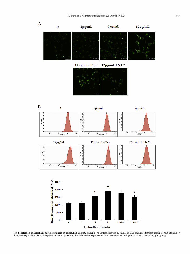
Fig. 4. Detection of autophagic vacuoles induced by endosulfan via MDC staining. (A) Confocal microscopy images of MDC staining. (B) Quantification of MDC staining byflowcytometry analysis. Data are expressed as means ± SD from five independent experiments (*P < 0.05 versus control group, #P < 0.05 versus 12 mg/ml group).
L. Zhang et al. / Environmental Pollution 220 (2017) 843e852 847
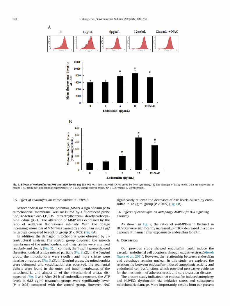
Fig. 5. Effects of endosulfan on ROS and MDA levels. (A) The ROS was detected with DCFH probe by flow cytometry. (B) The changes of MDA levels. Data are expressed asmeans ± SD from five independent experiments (*P < 0.05 versus control group, #P < 0.05 versus 12 mg/ml group).
L. Zhang et al. / Environmental Pollution 220 (2017) 843e852848
3.5. Effect of endosulfan on mitochondrial in HUVECs
Mitochondrial membrane potential (MMP), a sign of damage tomitochondrial membrane, was measured by a fluorescent probe5,50,6,6’-tetrachloro-1,10,3,3’- tetraethylbenzimi dazolylcarbocya-nide iodine (JC-1). The alteration of MMP was expressed by theratio of red/green fluorescence intensity. With the dosageincreasing, more loss of MMP was caused by endosulfan in 6,12 mg/ml groups compared to control group (P < 0.05) (Fig. 6A).
In addition, the damaged mitochondria were observed by ul-trastructural analysis. The control group displayed the smoothmembranes of the mitochondria, and their cristae were arrangedregularly and clearly (Fig. 3). In contrast, the 1 mg/ml group showedthe mitochondrial cristae missed partially (Fig. 3 a2), in the 6 mg/mlgroup, the mitochondria were swollen and more cristae weremissing or ruptured (Fig. 3 a3). In 12 mg/ml group, the mitochondriawere deformed, and vacuolization was observed, the segmentaldefects were found in the outer and inner membranes of themitochondria, and almost all of the mitochondrial cristae dis-appeared (Fig. 3 a4). After 24 h of endosulfan exposure, the ATPlevels in 6,12 mg/ml treatment groups were significantly lower(P < 0.05) compared with the control group, However, NAC
significantly relieved the decreases of ATP levels caused by endo-sulfan in 12 mg/ml group (P < 0.05) (Fig. 6B).
3.6. Effects of endosulfan on autophagy AMPK-a/mTOR signalingpathway
As shown in Fig. 7, the ratios of p-AMPK-aand Beclin-1 inHUVECs were significantly increased, p-mTOR decreased in a dose-dependent manner after exposure to endosulfan for 24 h.
4. Discussion
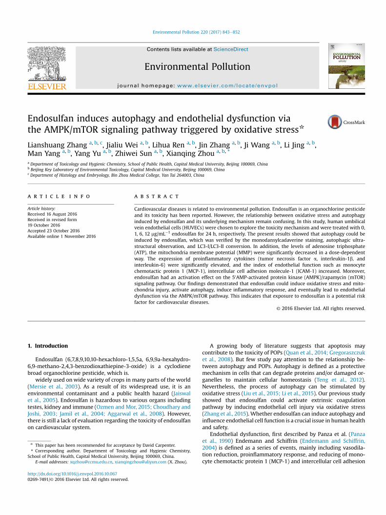
Our previous study showed endosulfan could induce thevascular endothelial cell apoptosis through oxidative stress(AfesehNgwa et al., 2011). However, the relationship between endosulfanand autophagy remains unclear. In this study, we explored therelationship between endosulfan-induced autophagic activity andendothelial cell dysfunction, which provided persuasive evidencefor the mechanism of atherosclerosis and cardiovascular disease.
The present study indicated that endosulfan induced autophagyand HUVECs dysfunction via oxidative stress and subsequentmitochondria damage. More importantly, results from our present
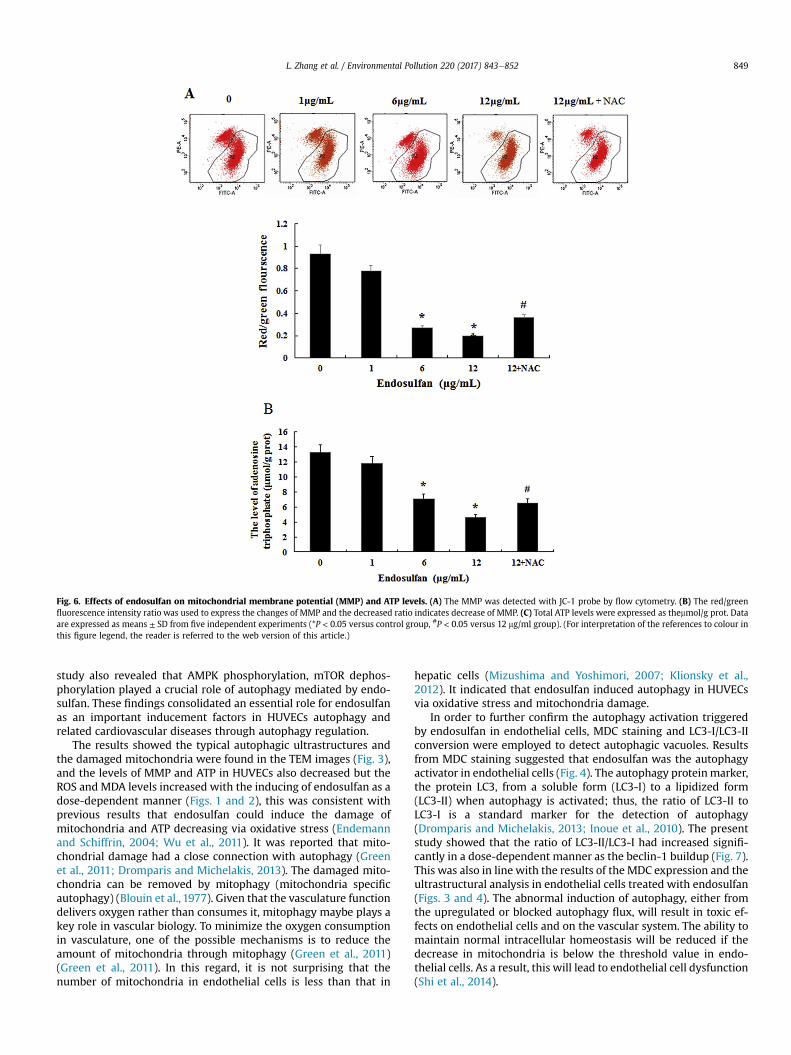
Fig. 6. Effects of endosulfan on mitochondrial membrane potential (MMP) and ATP levels. (A) The MMP was detected with JC-1 probe by flow cytometry. (B) The red/greenfluorescence intensity ratio was used to express the changes of MMP and the decreased ratio indicates decrease of MMP. (C) Total ATP levels were expressed as themmol/g prot. Dataare expressed as means ± SD from five independent experiments (*P < 0.05 versus control group, #P < 0.05 versus 12 mg/ml group). (For interpretation of the references to colour inthis figure legend, the reader is referred to the web version of this article.)
L. Zhang et al. / Environmental Pollution 220 (2017) 843e852 849
study also revealed that AMPK phosphorylation, mTOR dephos-phorylation played a crucial role of autophagy mediated by endo-sulfan. These findings consolidated an essential role for endosulfanas an important inducement factors in HUVECs autophagy andrelated cardiovascular diseases through autophagy regulation.
The results showed the typical autophagic ultrastructures andthe damaged mitochondria were found in the TEM images (Fig. 3),and the levels of MMP and ATP in HUVECs also decreased but theROS and MDA levels increased with the inducing of endosulfan as adose-dependent manner (Figs. 1 and 2), this was consistent withprevious results that endosulfan could induce the damage ofmitochondria and ATP decreasing via oxidative stress (Endemannand Schiffrin, 2004; Wu et al., 2011). It was reported that mito-chondrial damage had a close connection with autophagy (Greenet al., 2011; Dromparis and Michelakis, 2013). The damaged mito-chondria can be removed by mitophagy (mitochondria specificautophagy) (Blouin et al., 1977). Given that the vasculature functiondelivers oxygen rather than consumes it, mitophagy maybe plays akey role in vascular biology. To minimize the oxygen consumptionin vasculature, one of the possible mechanisms is to reduce theamount of mitochondria through mitophagy (Green et al., 2011)(Green et al., 2011). In this regard, it is not surprising that thenumber of mitochondria in endothelial cells is less than that in
hepatic cells (Mizushima and Yoshimori, 2007; Klionsky et al.,2012). It indicated that endosulfan induced autophagy in HUVECsvia oxidative stress and mitochondria damage.
In order to further confirm the autophagy activation triggeredby endosulfan in endothelial cells, MDC staining and LC3-I/LC3-IIconversion were employed to detect autophagic vacuoles. Resultsfrom MDC staining suggested that endosulfan was the autophagyactivator in endothelial cells (Fig. 4). The autophagy proteinmarker,the protein LC3, from a soluble form (LC3-I) to a lipidized form(LC3-II) when autophagy is activated; thus, the ratio of LC3-II toLC3-I is a standard marker for the detection of autophagy(Dromparis and Michelakis, 2013; Inoue et al., 2010). The presentstudy showed that the ratio of LC3-II/LC3-I had increased signifi-cantly in a dose-dependent manner as the beclin-1 buildup (Fig. 7).This was also in line with the results of the MDC expression and theultrastructural analysis in endothelial cells treated with endosulfan(Figs. 3 and 4). The abnormal induction of autophagy, either fromthe upregulated or blocked autophagy flux, will result in toxic ef-fects on endothelial cells and on the vascular system. The ability tomaintain normal intracellular homeostasis will be reduced if thedecrease in mitochondria is below the threshold value in endo-thelial cells. As a result, this will lead to endothelial cell dysfunction(Shi et al., 2014).
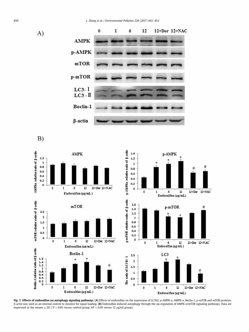
Fig. 7. Effects of endosulfan on autophagy signaling pathways. (A) Effects of endosulfan on the expression of LC3Ⅱ/Ⅰ, p-AMPK-a, AMPK-a, Beclin-1, p-mTOR and mTOR proteins.b-actin was used as an internal control to monitor for equal loading. (B) Endosulfan induced autophagy through the up-regulation of AMPK-a/mTOR signaling pathways; Data areexpressed as the means ± SD (*P < 0.05 versus control group, #P < 0.05 versus 12 mg/ml group).
L. Zhang et al. / Environmental Pollution 220 (2017) 843e852850
L. Zhang et al. / Environmental Pollution 220 (2017) 843e852 851
Endothelial cell dysfunction has a potential role in atheroscle-rosis which partially characterized by chronic inflammation and byan increased expression level of protein biomarkers, such asintercellular adhesion molecule-1 (ICAM-1) and monocytechemotactic protein-1 (MCP-1) in the activated endothelial cells(Kumar et al., 2014). Given that the MCP-1, ICAM-1 is directlyrelated to endothelial cell functions (Yin et al., 2015), we measuredthe levels of MCP-1and ICAM-1, in this study, the results showedthat endosulfan increased the production of MCP-1, ICAM-1levelswith the development of autophagy. Meanwhile, recent studieshave identified that PCB was significantly associated with levels ofICAM-1in people blood (Harris, 2011) and there was a link betweenMCP-1, ICAM-1 and autophagy (Habib, 2011). Moreover, it shouldbe noted that there is crosstalk between autophagy and cytokinesin regard to the inflammatory response (Kanamori et al., 2011). Thecytokines (TNF-a, IL-1b, and IL-6) have been shown to induceautophagic activity; in contrast, autophagy can regulate the pro-duction of proinflammatory cytokines (Kanamori et al., 2011). Inthe present study, our data demonstrated that endosulfan couldenhance the generation of proinflammatory cytokines (TNF-a, IL-1b, and IL-6) (Fig. 7). Similar to our previous findings, exposure toendosulfan could lead to a proinflammatory response in vascularendothelial cells (Endemann and Schiffrin, 2004).
To gain insight into the mechanism of endosulfan-inducedautopagic activity and endothelial dysfunction, we examined theAMPK/mTOR signaling pathway by western blot assay (Fig. 7). Theserine/threonine kinase mTOR, is inhibited by AMPK, which regu-lates intracellular energy status by sensing the AMP/ATP ratio. Ourresults revealed that endosulfan treatment decreased ATP levels,increased the phosphorylation of AMPK and mTOR dephosphory-lation, respectively. These findings are consistent with previousfindings where rapamycin directly stimulates AMPK and inhibitsMtor (Hahn-Windgassen et al., 2005; Hardie, 2007). Consistent
Fig. 8. The schematic diagram of autophagy induced by endosulfan via AMPK/mTOR pathway through oxidative stress. Oxidative stress was induced by endo-sulfan, and then mitochondria was damaged and AMPK was activated, resulted inautophagy and dysfunction of HUVECs.
with AMPK phosphorylation and mTOR dephosphorylation, rapa-mycin promoted autophagy as evidenced by increased LC3II andLC3II-to-LC3I ratio, in agreement with the previous findings (Hahn-Windgassen et al., 2005; Hardie, 2007; Hay and Sonenberg, 2004;Wu and Schwartzman, 2011).
In conclusion, our results confirmed that endosulfan inducedHUVECs dysfunction, as well as activated autophagy (Fig. 8). Theendothelial cell function is of great importance in maintainingvascular homeostasis. Endothelial dysfunction is an initial eventthat has been implicated in several cardiovascular diseases (Hayand Sonenberg, 2004). Thus, more investigations are required toexplore the biological mechanisms of the interaction betweenautophagy, endothelial dysfunction, and cardiovascular diseasestriggered by endosulfan.
Acknowledgments
This research was supported by the National Natural ScienceFoundation of China (No. 31370430).
References
Afeseh Ngwa, H., Kanthasamy, A., Gu, Y., Fang, N., Anantharam, V., Kanthasamy, A.G.,2011. Manganese nanoparticle activates mitochondrial dependent apoptoticsignaling and autophagy in dopaminergic neuronal cells. Toxicol. Appl. Phar-macol. 256, 227e240.
Aggarwal, M., Naraharisetti, S.B., Dandapat, S., Degen, G.H., Malik, J.K., 2008. Per-turbations in immune responses induced by concurrent subchronic exposure toarsenic and endosulfan. Toxicology 251, 51e60.
Blouin, A., Bolender, R.P., Weibel, E.R., 1977. Distribution of organelles and mem-branes between hepatocytes and nonhepatocytes in the rat liver parenchyma. Astereological study. J.Cell. Biol. 72, 441e455.
Catal�an, Ú., de Las, L�opez, Hazas, M.C., Rubi�o, L., Fern�andez-Castillejo, S., Pedret, A.,de la Torre, R., Motilva, M.J., Sol�a, R., 2015. Protective effect of hydroxytyrosoland its predominant plasmatic human metabolites against endothelialdysfunction in human aortic endothelial cells. Mol. Nutr. Food. Res. 59,2523e2536.
Choudhary, N., Joshi, S.C., 2003. Reproductive toxicity of endosulfan in male albinorats. Bull. Environ. Contam. Toxicol. 70, 285e289.
Dromparis, P., Michelakis, E.D., 2013. Mitochondria in vascular health and disease.Annu. Rev. Physiol. 75, 95e126.
Endemann, D.H., Schiffrin, E.L., 2004. Endothelial dysfunction. J. Am. Soc. Nephrol.15, 1983e1992.
Green, D.R., Galluzzi, L., Kroemer, G., 2011. Mitochondria and the autophagyinflammation-cell death axis in organismal aging. Science 333, 1109e1112.
Gregoraszczuk, E.L., Ptak, A., Karniewska, M., Ropstad, E., 2008. Action of definedmixtures of PCBs, p,p'-DDT and its metabolite p,p'-DDE, on co-culture of porcinetheca and granulosa cells: steroid secretion, cell proliferation and apoptosis.Reprod. Toxicol. 26, 170e174.
Habib, S.L., 2011. Mechanism of activation of AMPK and upregulation of OGG1byrapamycin in cancer cells. Oncotarget 2, 958e959.
Hahn-Windgassen, A., Nogueira, V., Chen, C.C., Skeen, J.E., Sonenberg, N., Hay, N.,2005. Akt activates the mammalian target of rapamycin by regulating cellularATP level and AMPK activity. J. Biol. Chem. 280, 32081e32089.
Hardie, D.G., 2007. AMP-activated/SNF1 protein kinases: conserved guardians ofcellular energy. Nat. Rev. Mol. Cell. Biol. 8, 774e785.
Harris, J., 2011. Autophagy and cytokines. Cytokine 56, 140e144.Hay, N., Sonenberg, N., 2004. Upstream and downstream of mTOR. Genes. Dev. 18,
1926e1945.Inoue, M., Ishida, T., Yasuda, T., Toh, R., Hara, T., Cangara, H.M., Rikitake, Y., Taira, K.,
Sun, L., Kundu, R.K., Quertermous, T., Hirata, K., 2010. Endothelial cell-selectiveadhesion molecule modulates atherosclerosis through plaque angiogenesis andmonocyte-endothelial interaction. Microvasc. Res. 80, 179e187.
Jaiswal, A., Parihar, V.K., Sudheer Kumar, M., Manjula, S.D., Krishnanand, B.R.,Shanbhag, R., Unnikrishnan, M.K., 2005. 5-Aminosalicylic acid reversesendosulfan-induced testicular toxicity in male rats. Mutat. Res. 585, 50e59.
Jamil, K., Shaik, A.P., Mahboob, M., Krishna, D., 2004. Effect of organophosphorusand organochlorine pesticides (monochrotophos, chlorpyriphos, dimethoate,and endosulfan) on human lymphocytes in-vitro. Drug. Chem. Toxicol. 27,133e144.
Kanamori, H., Takemura, G., Goto, K., Maruyama, R., Tsujimoto, A., Ogino, A.,Takeyama, T., Kawaguchi, T., Watanabe, T., Fujiwara, T., Fujiwara, H.,Seishima, M., Minatoguchi, S., 2011. The role of autophagy emerging in post-infarction cardiac remodelling. Cardiovasc. Res. 91, 330e339.
Klionsky, D.J., Abdalla, F.C., Abeliovich, H., Abraham, R.T., Acevedo-Arozena, A.,Adeli, K., et al., 2012. Guidelines for the use and interpretation of assays formonitoring autophagy. Autophagy 8, 445e544.
Kumar, J., Lind, P.M., Salihovic, S., van Bavel, B., Ingelsson, E., Lind, L., 2014. Persistent
L. Zhang et al. / Environmental Pollution 220 (2017) 843e852852
organic pollutants and inflammatory markers in a cross-sectional study ofelderly swedish people: the PIVUS cohort. Environ. Health.Perspect 122,977e983.
Li, X.Y., Jing, C.Q., Zang, X.Y., Yang, S., Wang, J.J., 2012. Toxic cytological alteration andmitochondrial dysfunction in PC12 cells induced by 1-octyl-3-methylimidazolium chloride. Toxicol. Vitro 26, 1087e1092.
Li, Y., Zhu, H., Wang, S., Qian, X., Fan, J., Wang, Z., Song, P., Zhang, X., Lu, W., Ju, D.,2015. Interplay of oxidative stress and autophagy in PAMAM dendrimers-induced neuronal cell death. Theranostics 5, 1363e1377.
Liu, G.Y., Jiang, X.X., Zhu, X., He, W.Y., Kuang, Y.L., Ren, K., Lin, Y., Gou, X., 2015. ROSactivates JNK-mediated autophagy to counteract apoptosis in mouse mesen-chymal stem cells in vitro. Acta. Pharmacol. Sin. 36, 1473e1479.
Mersie, W., Seybold, C.A., Mcnamee, C., et al., 2003. A bating endosulfan from runoffusing vegetative filter strips: the importance of plant species and flow rate.Agric. Ecosyst. Environ 97, 215e223.
Mizushima, N., Yoshimori, T., 2007. How to interpret LC3 immunoblotting. Auto-phagy 3, 542e545.
Ozmen, O., Mor, F., 2015. Effects of vitamin C on pathology and caspase-3 activity ofkidneys with subacute endosulfan toxicity. Biotech. Histochem 90, 25e30.
Panza, J.A., Quyyumi, A.A., Brush, J.E., Epstein, S.E., 1990. Abnormal endothelium-dependent vascular relaxation in patients with essential hypertension.N. Engl. J. Med. 323, 22e27.
Quan, C., Shi, Y., Wang, C., Wang, C., Yang, K., 2014. p,p'-DDE damages spermato-genesis via phospholipid hydroperoxide glutathione peroxidase depletion and
mitochondria apoptosis pathway. Environ. Toxicol. http://dx.doi.org/10.1002/tox.22072.
Shi, J., Sun, X., Lin, Y., Zou, X., Li, Z., Liao, Y., Du, M., Zhang, H., 2014. Endothelial cellinjury and dysfunction induced by silver nanoparticles through oxidative stressvia IKK/NF-kB pathways. Biomaterials 35, 6657e6666.
Teng, R.J., Du, J., Welak, S., Guan, T., Eis, A., Shi, Y., Konduri, G.G., 2012. Cross talkbetween NADPH oxidase and autophagy in pulmonary artery endothelial cellswith intrauterine persistent pulmonary hypertension. Am. J. Physiol. Lung. Cell.Mol. Physiol. 302, L651eL663.
Wang, X., Liu, Q., Wang, Z., Wang, P., Hao, Q., Li, C., 2009. Bioeffects of low-energycontinuous ultrasound on isolated sarcoma 180 cells. Chemotherapy 55,253e261.
Wu, C.C., Schwartzman, M.L., 2011. The role of 20-HETE in androgen-mediatedhypertension. Prosta. Gl. Lipid. Mediat 96, 45e53.
Wu, Y.N., Yang, L.X., Shi, X.Y., Li, I.C., Biazik, J.M., Ratinac, K.R., Chen, D.H.,Thordarson, P., Shieh, D.B., Braet, F., 2011. The selective growth inhibition of oralcancer by iron core-gold shell nanoparticles through mitochondria mediatedautophagy. Biomaterials 32, 4565e4573.
Yin, J., Wang, Y., Gu, L., Fan, N., Ma, Y., Peng, Y., 2015. Palmitate induces endoplasmicreticulum stress and autophagy in mature adipocytes: implications forapoptosis and inflammation. Int. J. Mol. Med. 35, 932e940.
Zhang, L., Wei, J., Guo, F., Duan, J., Li, Y., Shi, Z., Yang, Y., Zhou, X., Sun, Z., 2015.Endosulfan activates the extrinsic coagulation pathway by inducing endothelialcell injury in rats. Environ. Sci. Pollut. Res. 22, 15722e15730.
Related Documents