Endoscopic Palliation of Pancreatic Cancer Simon K. Lo, MD a,b, * KEYWORDS • Pancreatic adenocarcinoma • Palliation • Biliary decompression • Intestinal stenting • Celiac plexus neurolysis • Fiducial placement Pancreatic adenocarcinoma is the fourth leading cause of cancer death in the United States, with a 5-year survival rate of 5.6%. 1 One third of pancreatic cancer patients have local or regional disease at the time the diagnosis is made. 2 The majority of patients are treated with palliation in mind. With the rapid expansion of endoscopic technology, our ability to palliate symptoms of pancreatic cancer has improved significantly over the years. More than 90% of cancers of the pancreas are ductal adenocarcinomas. Neuroen- docrine and intraductal papillary mucinous neoplasm (IPMN) cancers make up the remainder of the malignant diseases of the pancreas. 3 Because of the biological differences between these cancers, their symptoms and presentations may vary significantly. Adenocarcinomas may present early by obstructing the common bile duct, but at the late stages they cause pain and duodenal obstruction. IPMN cancers may cause gastric, duodenal, or biliary fistulae or obstruction of the adjacent organs. Neuroendocrine cancers enlarge slowly as a locally confined disease but may cause jaundice due to extensive liver metastasis or compression of the common hepatic duct with lymph node metastasis. As a result, the modes of palliation differ greatly depending on the nature of these cancers. For instance, the short life spans of patients with adenocarcinoma make endoscopic biliary stenting an appropriate palliative option for obstructive jaundice. On the other hand, a neuroendocrine cancer of the head of the pancreas in a young patient is better palliated by a Whipple resection or surgical biliary bypass. Although a 10-mm-caliber biliary stent is the standard for palliation of adenocarcinoma-related biliary obstruction, it is frequently too small to remain patent or too easy to dislodge within a very large bile duct that is obstructed by the copious, thick mucin of an IPMN cancer. In this case a large-caliber esophageal stent or multiple 10-mm biliary metal stents placed side by side may The author has nothing to disclose. a Cedars-Sinai Medical Center, 8700 Beverly Boulevard, Room 7511, Los Angeles, CA 90048, USA b David Geffen School of Medicine at UCLA, Los Angeles, CA 90095, USA * Cedars-Sinai Medical Center, 8700 Beverly Boulevard, Room 7511, Los Angeles, CA 90048. E-mail address: [email protected] Gastroenterol Clin N Am 41 (2012) 237–253 doi:10.1016/j.gtc.2011.12.005 gastro.theclinics.com 0889-8553/12/$ – see front matter © 2012 Elsevier Inc. All rights reserved.

Welcome message from author
This document is posted to help you gain knowledge. Please leave a comment to let me know what you think about it! Share it to your friends and learn new things together.
Transcript
h
dsdmNjddpporstoe
Endoscopic Palliation ofPancreatic Cancer
Simon K. Lo, MDa,b,*
KEYWORDS
• Pancreatic adenocarcinoma • Palliation• Biliary decompression • Intestinal stenting• Celiac plexus neurolysis • Fiducial placement
Pancreatic adenocarcinoma is the fourth leading cause of cancer death in the UnitedStates, with a 5-year survival rate of 5.6%.1 One third of pancreatic cancer patientsave local or regional disease at the time the diagnosis is made.2 The majority of
patients are treated with palliation in mind. With the rapid expansion of endoscopictechnology, our ability to palliate symptoms of pancreatic cancer has improvedsignificantly over the years.
More than 90% of cancers of the pancreas are ductal adenocarcinomas. Neuroen-docrine and intraductal papillary mucinous neoplasm (IPMN) cancers make up theremainder of the malignant diseases of the pancreas.3 Because of the biological
ifferences between these cancers, their symptoms and presentations may varyignificantly. Adenocarcinomas may present early by obstructing the common bileuct, but at the late stages they cause pain and duodenal obstruction. IPMN cancersay cause gastric, duodenal, or biliary fistulae or obstruction of the adjacent organs.euroendocrine cancers enlarge slowly as a locally confined disease but may cause
aundice due to extensive liver metastasis or compression of the common hepaticuct with lymph node metastasis. As a result, the modes of palliation differ greatlyepending on the nature of these cancers. For instance, the short life spans ofatients with adenocarcinoma make endoscopic biliary stenting an appropriatealliative option for obstructive jaundice. On the other hand, a neuroendocrine cancerf the head of the pancreas in a young patient is better palliated by a Whippleesection or surgical biliary bypass. Although a 10-mm-caliber biliary stent is thetandard for palliation of adenocarcinoma-related biliary obstruction, it is frequentlyoo small to remain patent or too easy to dislodge within a very large bile duct that isbstructed by the copious, thick mucin of an IPMN cancer. In this case a large-calibersophageal stent or multiple 10-mm biliary metal stents placed side by side may
The author has nothing to disclose.a Cedars-Sinai Medical Center, 8700 Beverly Boulevard, Room 7511, Los Angeles, CA 90048, USAb David Geffen School of Medicine at UCLA, Los Angeles, CA 90095, USA* Cedars-Sinai Medical Center, 8700 Beverly Boulevard, Room 7511, Los Angeles, CA 90048.E-mail address: [email protected]
Gastroenterol Clin N Am 41 (2012) 237–253doi:10.1016/j.gtc.2011.12.005 gastro.theclinics.com
0889-8553/12/$ – see front matter © 2012 Elsevier Inc. All rights reserved.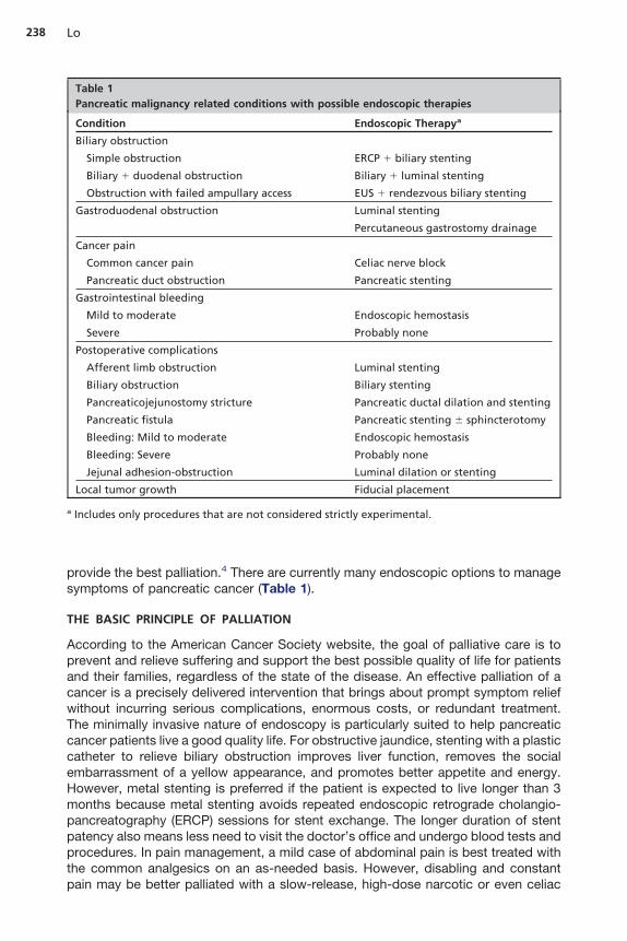
s
238 Lo
provide the best palliation.4 There are currently many endoscopic options to manageymptoms of pancreatic cancer (Table 1).
THE BASIC PRINCIPLE OF PALLIATION
According to the American Cancer Society website, the goal of palliative care is toprevent and relieve suffering and support the best possible quality of life for patientsand their families, regardless of the state of the disease. An effective palliation of acancer is a precisely delivered intervention that brings about prompt symptom reliefwithout incurring serious complications, enormous costs, or redundant treatment.The minimally invasive nature of endoscopy is particularly suited to help pancreaticcancer patients live a good quality life. For obstructive jaundice, stenting with a plasticcatheter to relieve biliary obstruction improves liver function, removes the socialembarrassment of a yellow appearance, and promotes better appetite and energy.However, metal stenting is preferred if the patient is expected to live longer than 3months because metal stenting avoids repeated endoscopic retrograde cholangio-pancreatography (ERCP) sessions for stent exchange. The longer duration of stentpatency also means less need to visit the doctor’s office and undergo blood tests andprocedures. In pain management, a mild case of abdominal pain is best treated withthe common analgesics on an as-needed basis. However, disabling and constant
Table 1Pancreatic malignancy related conditions with possible endoscopic therapies
Condition Endoscopic Therapya
Biliary obstruction
Simple obstruction ERCP � biliary stenting
Biliary � duodenal obstruction Biliary � luminal stenting
Obstruction with failed ampullary access EUS � rendezvous biliary stenting
Gastroduodenal obstruction Luminal stenting
Percutaneous gastrostomy drainage
Cancer pain
Common cancer pain Celiac nerve block
Pancreatic duct obstruction Pancreatic stenting
Gastrointestinal bleeding
Mild to moderate Endoscopic hemostasis
Severe Probably none
Postoperative complications
Afferent limb obstruction Luminal stenting
Biliary obstruction Biliary stenting
Pancreaticojejunostomy stricture Pancreatic ductal dilation and stenting
Pancreatic fistula Pancreatic stenting � sphincterotomy
Bleeding: Mild to moderate Endoscopic hemostasis
Bleeding: Severe Probably none
Jejunal adhesion-obstruction Luminal dilation or stenting
Local tumor growth Fiducial placement
a Includes only procedures that are not considered strictly experimental.
pain may be better palliated with a slow-release, high-dose narcotic or even celiac
qsp
239Endoscopic Palliation of Pancreatic Cancer
neurolysis. As treating physicians, we should constantly assess what is the best wayto palliate patients based on proven outcomes, the patient’s wish, and our technicalability.
BILIARY OBSTRUCTION
Approximately 70% of pancreatic adenocarcinomas are located in the head region.Some degree of biliary compression may eventually occur in most patients withpancreatic adenocarcinoma; therefore, biliary decompression is the best-known formof pancreatic cancer palliation. At one time, surgical biliary bypass, such as chole-cystojejunostomy or hepaticojejunostomy, was the treatment standard for obstructivejaundice; however, it has largely been replaced by endoscopic stenting because ofcomparable survivals and effectiveness in relieving jaundice.5 Procedure-relatedmorbidities are either the same or in favor of endoscopic stenting.6–8
Types of Plastic Stents
Most of today’s plastic stents are made of a polyethylene material. These stents comein external diameters that range from 5 french to 11.5 french. The most popular stentcaliber for distal biliary decompression is 10 french and the most desirable lengths are7 cm and 10 cm. Both the straight Amsterdam style and double pigtail stents arecommercially available for biliary drainage, but virtually all endoscopists choose thestraight stents because of their better patency performance relative to the pigtailstents. A single flap near each end of the straight stents provides the anchoragenecessary to hold them in place. Unsatisfied with the typical 3-month patency of thepolyethylene stents, the Hamburg group introduced a Teflon stent to take advantageof its lower coefficient of friction, compared to common plastic stents, and smallerchance of clogging without any sideholes. This new product was thought to be theideal plastic stent and the early results showed it to be patent for a median durationof 64 weeks.9 However, the enthusiasm toward this specially designed stent waned
uickly as a randomized prospective study that compared the Tannenbaum Teflontent to conventional polyethylene stents showed no difference in mean 90-day stentatency.10
Plastic Stenting Technique
Plastic stenting begins with guidewire passage through the stricture. A biliarysphincterotomy is not typically required for insertion of a single plastic stent. Likewise,stricture dilation is not usually required for placement of a single plastic stent exceptwhen there is an extremely tight blockage; in that case a 4-mm or 6-mm over-the-wireballoon dilator may be used. The length of the stent is selected by estimating thedistance between the top of the stricture and the papilla against a known dimension,such as the width of the duodenoscope, based on fluoroscopic assessment. Analternative is to measure the distance of a catheter that has been pulled back from thetop of the stricture to the papilla. The plastic stent is then loaded onto the tip of adelivery device. There are three types of stent delivery devices, but they all have aninner guiding catheter and a shorter external pusher catheter. Once the inner catheterhas securely passed the stricture, the pusher is advanced forward to push the stentup the bile duct until it reaches the desired location. The guiding catheter and theguidewire are then removed, leaving the stent in place. There are two commerciallyavailable modified stenting devices that allow simultaneous removal of the stent anddevice while leaving the guidewire in place, in case a decision is made to retrieve the
stent.
mt
E
240 Lo
Metallic Stenting
The first commercially available metal biliary stent was introduced in 1989. It wasmade of individual stainless steel wires that were twined together. One of the firststudies reported effectiveness of the 10-mm caliber metal stents without significantproblems.11 A comparison between metallic and plastic stents showed that the
edian duration of patency (273 days) of the metal stent was significantly longer thanhat of a polyethylene stent (126 days).12 However, the overall median survival was
149 days and did not differ significantly between the plastic and metal stent groups.Not only were metallic stents more patent over time than plastic stents, but they werealso associated with less cholangitis, shorter duration of hospitalization, and lessoverall costs than plastic stents.13 A meta-analysis was performed to analyze sevenrandomized controlled trials that aimed to compare plastic and metal stents. In total,724 patients were randomized into one of the treatment arms. The meta-analysisshowed that metallic stents were associated with a significantly reduced risk of stentocclusion at 4 months and a lower overall risk of recurrent biliary obstruction.14 Thisanalysis also showed that the metal stents were more cost effective, as the additionalERCP accounted for substantial cost. But a recent Korean study demonstrated thateven in countries where ERCP costs were lower than those of metal stents, metalbiliary stents were still the first-line treatment because they offered better palliationwithout adding a significant cost in palliating the malignant biliary obstruction.15
Hence, these studies provided strong support for the practice of metal stenting as thefirst-line palliative modality for malignant biliary obstruction that is typically caused bypancreatic carcinoma. However, these stents were appropriate only when treatingdefinitively unresectable cancers because the strong adherence of these open meshstents to the biliary tissue made them difficult to remove from the bile duct duringdissection and creation of surgical anastomoses.
Early self-expanding metal stents had no covers to prevent tissue penetrationthrough the wire mesh. Indeed, stent failures were noted to be caused by tumoringrowth across the metal wires.12 Reactive hyperplastic reactions may also occur ateither end of the stent where it is in close contact with the biliary or duodenal tissue.16
Creating a barrier to tissue ingrowth and, in turn, prolonging stent patency was thedriving force that led to the development of partially covered stents.17 Today, severalpartially covered metal stents are available. The short, uncovered ends of these stentsare designed to provide adherence to the biliary tissue whereas the large, coveredportion provides protection from tissue penetration into the stent lumen.
Even though the partially covered stents were designed to prevent tumor ingrowth,occasionally they are still difficult to remove. As neoadjuvant therapy for tumordownstaging and surgical resection becomes an increasingly common practice, theease of removing these stents is an important consideration in the selection of a metalstent. Fully covered stents became available a few years ago and their removal at thetime of a pancreaticoduodenectomy became feasible in virtually all patients.18
Multiple studies have compared covered to uncovered metal stents in palliatingmalignant biliary obstruction. Yoon and colleagues found no significant differences instent patency at 100, 200, 300, and 400 days between the two groups of metalstents.19 A randomized trial showed that the median time to recurrent biliaryobstruction was 711 days for the uncovered and 357 days for the partially coveredself-expanding metal stents.20 In addition, serious adverse events occurred signifi-cantly more frequently with the partially covered stents than with the uncovered stents(62% vs 44%, P�.05), mainly due to stent migration (12% vs 0, P�.01). Further, a
uropean study that enrolled 400 patients and randomized them to covered or
bthcdin
l
241Endoscopic Palliation of Pancreatic Cancer
uncovered stents showed no significant differences in stent patency, patient survival,or complication rates between the two groups.21 The metal stents used in this studywere made by a different manufacturer from the metal stents used in the other trials,but the conclusions of these independent studies were identical, disputing anypotential advantage of the covered metal stents over their uncovered counterparts.Even though there are the suspicions that the stent cover would contribute tocholecystitis and pancreatitis, there is no evidence that it is the case.17
Endoscopic Ultrasound-Guided Bile Duct Stenting
When the bile duct cannot be accessed through the papilla, or when the duodenumis completely obstructed by a large pancreatic tumor, an interventional radiologist ora surgeon is enlisted to perform a drainage or bypass procedure. However, endo-scopic ultrasound (EUS) allows gastrointestinal interventionists to puncture the bileduct directly from the duodenal bulb or stomach. This allows the possibility to inserta guidewire pass the stricture and the major papilla to perform biliary stenting and thistechnique is commonly referred to as the endoscopic or EUS rendezvous proce-dure.22,23 Roughly two-thirds of the cases attempted in this manner were reported to
e successful in draining the obstructed bile duct, ending with biliary stents crossinghe major papillae. EUS-guided choledochoduodenostomy24–26 and EUS-guidedepatogastrostomy27,28 have been performed when the duodenum or major papillaannot be accessed. Plastic or metal stents were inserted directly across theuodenum or stomach into the extrahepatic or intrahepatic bile duct. These highly
nvasive procedures demand great technical skills and clinical experience and shouldot be regarded as standard treatment options at the present time.
Drug-Eluting Biliary Stents
Taking biliary stenting one step further, experimental treatment of malignant stricturewith drug-eluting metallic stents has been proposed to extend the duration of biliarypatency. Paclitaxel, a chemotherapeutic agent, was mixed in a liquid form withpolyurethane and tetrahydrofuran to create a stent membrane that slowly releasedpaclitaxel.29 A low serum level of paclitaxel could be detected in patients stented foronger than 50 days.29 The mean patency of these covered, paclitaxel-eluting stentswas 429 days in 21 patients with unresectable malignant biliary obstruction. Thisseemingly prolonged patency may serve as supporting evidence to further developdrug-eluting stents in the future.
Metal Stenting Technique
The technique of metal stent placement is similar to that for plastic stenting, althoughthere are some key differences. The first step is to pass a wire across the stricture. Aswith plastic stenting, dilating a distal bile duct stricture is rarely necessary. Mostcommercially available metal stents have markings on the proximal and distal ends toguide deployment. Some even have a “point of no return” radio-opaque mark thatdenotes the position of stent release beyond which the stent cannot be recaptured incase it is necessary to adjust the stenting position. The stainless steel alloy materialof some stents is radio-opaque and all wire elements can be seen on fluoroscopy.However, nitinol-based wires are not radio-dense and therefore are marked withradio-opaque spots that are typically placed on both ends of the stents to guide stentrelease. Virtually all these stents have the tendency to propel forward duringdeployment; therefore, it is crucial to open up the stent very slowly to avoid
inadvertent placement of the entire stent above the stricture. Gradual pullback of the
242 Lo
stent is typically needed to compensate for the upward thrust of these stents duringdeployment. Shortening of the stents is also common as they are allowed to expand.Virtually all metal stents show a temporary waist at the point of maximal obstructionthat disappears gradually over the next few days as the stents expand to their fullcalibers. Although there is no scientific evidence of how the covered metal stentsspontaneously migrate, it is logical to assume that the unidirectional expansive forcecontributes to stent movement. Therefore, if one side of the waist is larger and longerthan the other side, there will be the tendency for the stent to shift toward the side thatis already more expanded. If a partially or fully covered stent is used, it is best to placethe stent such that the waist is located in the center of the stent.
GASTRODUODENAL OBSTRUCTION
Duodenal obstruction eventually occurs in 10% to 20% of patients with pancreatichead cancer.30 Ampullary cancer and neuroendocrine cancers also tend to involve theduodenum. Because the duodenum is rarely compressed in the initial presentation,most patients with duodenal obstruction are not candidates for curative surgery. Asin most malignant obstructions of the gastrointestinal tract, balloon dilation does notproduce any lasting improvement of the obstructive symptoms.
Metallic stenting is currently the endoscopic palliation modality of choice. Thesestents are constructed very similarly to those for the biliary tract, except that they aretypically longer (6–12 cm) and of larger calibers (18–23 mm). To prevent spontaneousmigration and to avoid compression of the ampullary opening, the stents areuncovered. Duodenal stenting requires some experience and technical skill, but isgenerally easy to complete. In a four-center study that involved 176 patients withmostly pancreatic cancer obstruction of the gastric outlet and duodenum, stentingwas feasible in all patients.31 Eighty-four percent of these patients were able toresume oral intake for a median of 146 days. The value of duodenal stenting is bestdemonstrated in a study in which 17 of 25 patients with pancreatic cancer and outletobstruction were treated with surgical gastrojejunostomy.32 This group of patientssurvived for a median duration of 64 days, but their postoperative stay was 15 days.In contrast, six patients received palliation with the Wallstents; they survived 111 daysand their postprocedure hospital stay was 4 days. One key advantage of stenting oversurgical bypass is avoiding postoperative delayed gastric emptying, which occurredin 59% of the surgical patients. Although other authors did not report such a high rateof delayed gastric emptying, a significant delay in the resumption of oral intake andprolonged hospital stays are still common.33,34
It has been suggested that inserting a large-bore gastrostomy tube may be aneffective alternative to duodenal stenting when placing a stent is not feasible.35
Although effective in preventing gastric distention and vomiting, gastrostomy tubedecompression is a poor form of palliation of gastric outlet obstruction because of theinability to eat and the required separate feeding jejunostomy or total parenteralnutrition. In the hands of an experienced endoscopist, ineffective duodenal stentdecompression may arise only in patients with carcinomatosis, underlying gastricdysmotility, or additional downstream intestinal blockages.
Intestinal Stenting Technique
The technique of intestinal stenting is very similar to biliary stenting, as most of theplacement requires endoscopic and fluoroscopic guidance. The procedure must beperformed with a large-channeled endoscope, such as a therapeutic upper endo-scope or a therapeutic duodenoscope, to allow for passing the stenting device
through the instrument channel. On rare occasions, the endoscope can be advanced
caaac
243Endoscopic Palliation of Pancreatic Cancer
across the stricture. This allows the inspection of the duodenal wall and placement ofa stiff guidewire into the proximal jejunum. More commonly, the endoscope is toolarge to get through the stricture. In that case, a guidewire in combination with anERCP catheter allows for injection of contrast to estimate the length of the stricture,while at the same time placing the guidewire. It is preferred that the wire is securelyinserted into the proximal jejunum because of the long, stiff leading tip of the stentingdevice. It is also important to determine if the major papilla is affected by the stent, aspostprocedure cholangitis has been noted in 6% of patients.31 It is always preferredto place the stent clear of the major papilla, but it is not always possible. If the patientalso presents with jaundice, a metallic biliary stent placement should be attemptedbefore placement of the duodenal stent. Combined duodenal and biliary stenting hasbeen reported to be successful in 91% of the cases.36 Very gentle manipulations andexcellent ERCP skill are needed in this situation, as it is difficult to approach the majorpapilla within a tight space. Duodenal perforation is a serious concern because of thepossible need to dilate the stricture and then negotiate through it with a side-viewingendoscope. If the stricture is very tight, it may be best to first place the duodenal stentand then perform a transhepatic procedure to insert the biliary stent.
It is also important to avoid resting the ends of the duodenal stent at the corners,such as the duodenal bulb, junction between the second and third portions of theduodenum, and the ligament of Treitz. The duodenal folds in these corners can coverthe stent lumen and render the stent ineffective in palliating obstructive symptoms.Mechanical irritation of the intestinal mucosa at these locations may eventually causebleeding or perforation. Using a stent with smooth or soft ends may also preventthese delayed complications. In deploying the stent, it is important to know the extentof stent shortening during and after its release from the device. Fluoroscopic andendoscopic monitoring is important in achieving optimal stent placement.
POSTOPERATIVE INTESTINAL OBSTRUCTION
Afferent limb obstruction after a Whipple procedure may present as cholangitis,jaundice, pain, or nonspecific discomfort. The median time from pancreaticoduode-nectomy to presentation may be as long as 1.2 years.37 Most of these cases areaused by cancer recurrence, although radiation stricture or kinks caused bydhesions may be the cause as well. Patients with this condition usually havedvanced disease and wish to receive a minimal treatment. Endoscopic stenting withlarge-caliber (18–22 mm diameter) intestinal stent is a simple way to palliate this
ondition.38 However, a variety of treatment modalities have been used, includingballoon dilation and plastic or metallic biliary stent placement, alone or in combina-tion. The results of such palliation are quite variable, ranging from immediate relief topersistent symptoms until death.37
DISABLING ABDOMINAL AND BACK PAIN
Pain is typically a late symptom of pancreatic cancer but can be a very difficultproblem to manage. Even the most intensive narcotic analgesics may fail to controlthe discomfort. When conventional analgesic therapy becomes ineffective, or when apatient cannot tolerate the side effects of heavy doses of narcotics, an alternativetreatment modality is typically desperately needed. As pancreatic stenting becametechnically feasible, endoscopists began to report that pancreatic ductal stentingmight improve pancreatic cancer pain due to ductal obstruction.39 It has beensuggested that pancreatic duct stenting plays a role in 15% of pancreatic cancer
pain.40 Carr-Locke and colleagues proposed that pain brought on by oral intake might
pp
t
nhwccoel
oagraawncspnt
stsig
244 Lo
be best treated in this manner.41 The plastic stents used for ductal decompression forain control ranged from 7 french to 10 french in caliber.40 Some authors evenroposed using metallic stents for pain control.41,42 The risk of acute and chronic
pancreatitis is a genuine concern in treating patients who are already suffering a greatdeal from their incurable cancer. Limited available data show that about 60% of carefullychosen patients with successful pancreatic stent placement will experience significantresolution of pain and that another 20% to 25% of patients will be able to decrease theiruse of narcotics.40 But the series that reported success of stenting are few and small, andhe limited evidence cannot be used to support its widespread use.
A more proven and less invasive procedure to control pancreatic cancer pain whenarcotic analgesics fail is neurolytic celiac nerve blocks. Patients with cancer in theead of pancreas may experience significantly more benefit in pain control than thoseith cancer in the body or tail of the pancreas. In one study using fluoroscopy oromputed tomography (CT)-guided percutaneous neurolysis, 92% of 36 patients withancer in the head of pancreas achieved improvement of pain, with a mean durationf relief of 119 days.43 In contrast, only 29% of 14 patients with body and tail lesionsxperienced relief for a duration of 65 days. As expected, patients with a large tumor
oad do not have satisfactory pain control.There are at least three routes of delivering celiac block or neurolysis: percutane-
us, operative, and endoscopic. When performed endoscopically, ultrasound guid-nce is used. For cancer pain control, the objective is permanent damage of the nerveanglia; therefore, alcohol is used to achieve neurolysis. By far, the most commonoute of treatment today is the percutaneous method. There are few studies thatddress the superiority of one method over the other. Gress and colleagues publishedsmall randomized, prospective series on the treatment of chronic pancreatitis painith celiac nerve block using bupivacaine and triamcinolone comparing the effective-ess of pain control between the CT and EUS-guided blocks.44 While pain control ofhronic pancreatitis cannot be regarded as the same as that for cancer pain, thistudy showed that the EUS-guided method provided significantly more persistentain relief than the CT technique. A meta-analysis evaluated the results of percuta-eous celiac neurolysis for pancreatic cancer based on five randomized controlledrials.45 A total of 302 patients were evaluated. Compared with control patients who
were treated with nonsteroidal anti-inflammatory drugs (NSAIDs) and narcotics,patients treated with neurolysis had lower pain scores and opioid usage at 2, 4, and8 weeks. The patients in the neurolysis arm also reported a reduction in constipation.While most studies reported improvement of pain control with neurolysis, someactually dispute the efficacy of either celiac block or splanchnicectomy.46 It appearsthat celiac neurolysis may bring about only mild to moderate pain relief in patientswith pancreatic pain. As noted elsewhere, this form of pain therapy should beconsidered an adjunct to opioid use and an adjuvant therapy in patients withpancreatic cancer pain.47
Most recent publications on celiac neurolysis for pancreatic cancer have evolvedaround EUS-guided neurolysis, which may be a reflection of the development oftechnology, enthusiasm of endoscopists, or advantage of EUS over the conventionalpercutanueous modality. A meta-analysis showed that EUS neurolysis had providedpain relief in 80.1% of patients with pancreatic cancer pain.48 The review furtherhowed that the technique of bilateral injections provided superior pain relief relativeo the unilateral injection technique (85% vs 46%). A more recent randomized,ingle-blind study showed no significant difference in pain relief (81% for twonjections vs 69% for one injection), median onset of pain relief (1 day for both
roups), median duration of pain relief (14 weeks for two injections vs 11 weeks for
db
dbsmadngoirr
mtatf
245Endoscopic Palliation of Pancreatic Cancer
one injection) or patients with complete pain relief (8% both groups).49 Until moreefinitive studies become available, the single and double injection methods shoulde considered interchangeable.
EUS Neurolysis Technique
The basic technique of EUS neurolysis has been described elsewhere.50 Preproce-ure intravenous hydration and antibiotic are usually given. The procedure itselfegins with passage of a curved linear-array echoendoscope into the proximaltomach. Once the celiac artery is identified, neurolysis is performed with the singleidline or bilateral approach. The midline method targets a location slightly anterior
nd cephalad to the origin of the celiac artery. The bilateral approach uses equal, splitoses to inject both sides of the origin of the celiac artery. Because the individualerve ganglia can be visualized on EUS, some endoscopists have advocated directanglion injection. The evidence for this approach is reported in a retrospective reviewf 64 patients with pancreatic cancer pain. Of eight potential predictors of symptom
mprovement, visualization of the ganglia for direct injection was the best predictor ofesponse, as those patients with visible ganglia were 15 times more likely toespond.51 Fine-needle aspiration (FNA) needles of 19-, 20-, 22-, and 25-gauge have
been used for this purpose, although there is significant resistance to overcome inusing the 22-gauge and 25-gauge needles. A spray needle that can produce an evenand wide distribution of the liquid agents has recently been made available.
The needle is usually prefilled with saline without a stylet in place. When the desiredlocation is punctured, aspiration is performed to confirm that the needle is not insidethe vasculature. To reduce alcohol-induced pain, 5 mL of a long-acting localanesthetic, such as 0.25%.bupivicaine, is then delivered. Finally, 20 mL of absoluteethanol is infused in the midline method to achieve neurolysis. Split doses of ethanolare used if the bilateral technique is applied. As the alcohol exits the needle tip, anechogenic cloud is produced at the affected tissue. Sakamoto and colleaguesintroduced a new technique of neurolysis that aimed at the tissue adjacent to thesuperior mesenteric artery (broad plexus neurolysis) rather than nearby the celiacartery and reported that the new method provided superior pain relief.52 Althoughpromising, this broad plexus neurolysis procedure should not be considered astandard treatment modality until more data become available.53 A few precautions
ust be taken when attempting a neurolysis block. The patient must be well sedatedo avoid accidental needle injury of the nearby vascular structures. This is bestchieved with monitored anesthesia care. Vital sign monitoring is crucial at baseline,hroughout, and after the procedure because of the deep sedation and the tendencyor significant drops in blood pressure.
GASTROINTESTINAL HEMORRHAGE
Gastrointestinal bleeding may occur de novo or after surgical treatment. Early cancerpresentation as gastrointestinal hemorrhage is rare and is usually caused by directinvolvement of the duodenum or stomach. The cause of bleeding after a pancreaticsurgery for cancer may be due to the exposure of skeletonized vessels to erosiveenzymes, inflammatory or traumatic pseudoaneurysms, and pancreatic pseudo-cysts.54 Anastomosis ulcer bleeding may occur as well. The common endoscopichemostasis techniques should be attempted for mild bleeding cases.55 However,most massive bleeding or recurrent tumor bleeding is best managed surgically or with
interventional radiology.
ebt
tfio2
246 Lo
POSTOPERATIVE PANCREATIC FISTULA
Fistula occurs most often after resection of the tail of the pancreas. Although surgicalmortality in the experienced surgeon’s hands is quite low,56 5% to 60% of distalpancreatectomy may be complicated by pancreatic fistula.57 Pancreatic stenting withor without sphincterotomy is highly effective, with the median duration to closing offistula of 4 days.58
LOCAL CANCER GROWTH
There is no proven endoscopic role in controlling local tumor growth of any pancreaticmalignancies. However, a number of intriguing endoscopic procedures have beenproposed. Iodine-125 seeds of 4.5 mm by 0.8 mm may be inserted in pancreatictissue using an EUS-guided 18-gauge or 19-gauge needle.59 Small-scale pilot studieshave been performed on humans with minimal but encouraging results.60–62 Simi-larly, direct intratumor injection of a replication-defective adenovirus vector carryingthe human tumor necrosis factor (TNF)-alpha gene is thought to have the potential forcontrol of tumor growth. A case of a large cancer with metastasis was treated withpercutaneous TNFerade injection and regression was noted and the patient was ableto undergo cancer resection subsequently.63 Even though there is no proventherapeutic efficacy today, this type of direct tumor treatment by EUS is appealingand worth further exploration.
Another brachytherapy approach is to take advantage of ERCP access to thepancreatic duct. Nine patients were treated with pancreatic duct brachytherapy withreasonably good tolerance.64 Clinical efficacy was difficult to determine in this smallarly study. Recently, a polyurethane plastic stent loaded with iodine-125 seeds haseen manufactured to deliver brachytherapy in an experimental model.65 Its applica-
ion on humans has yet to be tested.Finally, on the horizon of endoscopic palliation of pancreatic cancer improved
argeting for stereotactic radiosurgery with real-time guidance using small referenceducials. To our knowledge, the first report of the use of EUS-guided needle deliveryf fiducials aiming at the pancreas was reported by Pishvaian and colleagues in006.66 Fiducials of 3 mm or 5 mm were delivered with 19-gauge needles, with three
or four fiducials placed in each of these pancreatic cancer lesions. A thinner set offiducials (10 mm � 0.35 mm) that can be loaded onto a 22-gauge FNA needle has alsobecome commercially available.67,68 Only one or two fiducials are needed to highlightthe tumor for stereotactic radiotherapy. The ability to use a relatively flexible 22-gaugeneedle undoubtedly makes fiducial placement a much easier procedure than with astiff, 19-gauge needle. There has been no report on the influence of EUS fiducials onthe outcome of radiotherapy.
COMPLICATIONS OF ENDOSCOPIC THERAPY
As endoscopists seek to expand their ability to palliate pancreatic cancer, they needto be reminded that their procedures must be minimally invasive, effective, andreasonably safe. Nonetheless, complications inherently accompany interventionalprocedures. Before performing these procedures, the endoscopist must fully informthe patient of the risks and their likelihood. All alternative treatment modalities mustalso be considered. Procedure experience, knowledge of potential complications,and early recognition and the ability to manage adversities are necessary to avoid
adding misery to these terminally ill patients.
2
3
247Endoscopic Palliation of Pancreatic Cancer
1. Endoscope passage. Although it is not a therapeutic intervention, the merepassage of an endoscope can be risky. This is particularly true when passing aduodenoscope through a narrow duodenum. Side-viewing scope passage througha Billroth II intestine is particularly prone to induce perforation, with reportedincidents as high as 18%.69,70 Special attention must be paid when advancing ascope through a freshly dilated duodenal or jejunal stricture. Many endoscopistsare inexperienced in negotiating the post-Whipple or Roux-en-Y anatomy and theyshould be careful not to cause injury to the anastomosis or intestinal wall.
. ERCP and biliary stenting. Most complications of biliary stenting occur as a resultof the ERCP. In 21 selected prospective studies that included 16,855 patients,ERCP complications occurred in 6.9% of cases, with 1.7% being severe innature.71 The most common complication was pancreatitis (3.5%), followed byinfections (1.4%), bleeding (1.3%), and perforations (0.6%). Data collected inprospective studies indicated that procedure-related morbidity related to stentingmay range from 4% to 12% and mortality from 0 to 3.9%, with biliary sepsis,pancreatitis, and sphincterotomy bleeding as the main causes.72,73 Duodenalperforation due to pressure necrosis is known to occur with plastic stents thatprotrude excessively from the papilla. Cholecystitis has been reported as acomplication as well. Stent migration is a unique problem of partially or fullycovered metal stents, reported to occur in 6% of cases.74 Intestinal or colonicperforation, vascular fistula, and intestinal obstruction are rare complications ofmigrated biliary metal stents.75–77 The one late complications of biliary stentingthat is inevitable is clogging if the stent is left in place indefinitely. Stent caliber,material, length, and design all may influence the time it takes to become clogged.The same factors may also influence the likelihood of stent migration.
. EUS-guided biliary decompression procedures. In spite of the seemingly simpleconcept, the decompression is actually rather technically challenging. The poten-tial issues encountered during the multistep procedure include bleeding, perfora-tion, and false puncture during the initial step to access the obstructed bile ductwith a needle. The needle-guidewire exchange may cause sheering of the coatingof the standard guidewire by the sharp beveled needle tip. A deformed guidewiremay make it impossible to pass other therapeutic instruments over it. If a brokenwire is removed, bile leak will probably occur.
The next step of inserting a dilating catheter or balloon may be the mostchallenging step, as it can be very difficult to force these instruments through thethick gastric wall. Finally, bile fluid or gastrointestinal fluid may leak around awell-placed stent and lead to peritonitis. While the internal rendezvous procedureseems relatively safe, the complication rate of the transgastric hepatogastrostomyhas been reported as 14%. Complications include ileus, bilioma, cholangitis, andstent migration.78 A similar rate of complications due to bile peritonitis andpneumoperitoneum has been reported.
4. Gastrointestinal luminal stenting. Stricture dilation is an integral part of man-agement of intestinal obstruction. Perforation or transient septicemia shouldalways be considered. Stenting of a firm stricture may cause pain, as doesexcessive rubbing of duodenal tissue by an end of the intestinal stent. Some stentsdo not result in resolution of symptoms because of poor positioning and need tobe replaced or removed. Late complications include clogging, migration, andperforation. Baron and colleagues examined a 19-case series that includedreporting of complications, which occurred in roughly 20% of the cases.79
5. Celiac plexus neurolysis. Abscess formation as a result of celiac neurolysis has
been reported.80 A meta-analysis of the literature on celiac neurolysis before
6
7
248 Lo
EUS-neurolysis became available showed that transient adverse events were verycommon,81 including back pain (96%), diarrhea (44%), and hypotension (38%).Serious complications occur in 2% of injections and they include neurologiccomplications such as lower extremity weakness and paresthesia (l%) andnonneurologic events such as pneumothorax, hiccoughing, and hematuria (1%).No mortality was reported. On the other hand, transient hypotension occurred in20% and diarrhea was noted in 10 of 58 patients who underwent neurolysis withthe aid of EUS.82 It appears that EUS-guided neurolysis may be associated withfewer adverse events than the percutaneous method, although more data areneeded to confirm this observation.
. Pancreatic stenting. Some pancreatic ductal cannulations are exceedingly diffi-cult and pancreatitis can occur simply from instrument probing. Excessive contrastinjection and guidewire puncture of pancreatic tissue through a ductal radical mayresult in diffuse or focal pancreatitis. A poor choice of stent, awkward stentpositioning, or prolonged stenting may result in pancreatic irritation or strictureformation. Inward stent migration is technically very difficult to remove, with asuccess rate of less than 80% in a major interventional endoscopy center.83
Finally, stenting may convert a sterile pancreatic fistula into tissue infection orabscess formation.
. Fiducial insertion. This is a new procedure with limited clinical experience. Thusfar, it is reported to be very safe. In one series of 30 subjects, one (3%) patientdeveloped transient fever and elevation of liver enzymes.68 An infected portahepatis mass was noted within the first 30 days after the fiducial placement in onepatient (8%).66 In another report of 51 patients who underwent the procedure usingthe 19-gauge needles for fiducial delivery, one (2%) patient experienced mildpancreatitis.84 Three of 57 (5%) cases in another report encountered minorintraprocedure complications including malfunctioning of the instrument andbleeding.85 Finally, fiducial migration may occur soon after placement, thuspotentially misdirecting tumor therapy. Until more experience is accumulated, areasonable protocol is to give prophylactic antibiotic and place more than onefiducial to compensate for the potential of subsequent marker migration.
SUMMARY
There is no doubt that our long-range goal is to cure pancreatic cancer. Realistically,most of what we can do currently is treat the disabling symptoms of this dreadfuldisease. Biliary decompression, intestinal stenting, celiac plexus neurolysis, andfiducial placement are some of the endoscopic procedures that aim to provide betterquality of life to patients suffering from this disease. A thorough understanding ofthese options will help patients make good decisions in choosing the propertreatment. Endoscopists who perform these procedures must possess great skills,but importantly, they must also be compassionate and act with good judgment.
REFERENCES
1. National Cancer Institute. SEER Cancer Statistics Review 1975–2006. Bethesda(MD): National Cancer Institute. Available at: http://seer.cancer.gov/csr/1975_2006.Accessed January 9, 2012.
2. Merrill RM, Hunter BD. Conditional survival among cancer patients in the UnitedStates. Oncologist 2010;15:873–82.
3. Saif MW. Pancreatic neoplasm in 2011: an update. JOP 2011;12(4):316–21.
1
249Endoscopic Palliation of Pancreatic Cancer
4. Seynaeve L, Van Steenbergen W. Treatment, by insertion of multiple uncoveredmetallic stents, of intraductal papillary mucinous neoplasm of the pancreas with biliaryobstruction by mucus impaction. Pancreatology 2007;7:540–3.
5. Cipolletta L, Rotondano G, Marmo R, et al. Endoscopic palliation of malignantobstructive jaundice: an evidence-based review. Dig Liver Dis 2007;39:375–88.
6. Shepherd HA, Royle G, Ross AP, et al. Endoscopic biliary endoprosthesis in thepalliation of malignant obstruction of the distal common bile duct: a randomized trial.Br J Surg 1988;75:1166–8.
7. Andersen JR, Sorensen SM, Kruse A, et al. Randomised trial of endoscopic endo-prosthesis versus operative bypass in malignant obstructive jaundice. Gut 1989;30:1132–5.
8. Smith AC, Dowsett JF, Russell RC, et al. Randomised trial of endoscopic stentingversus surgical bypass in malignant low bile duct obstruction. Lancet 1994;344:1655–60.
9. Binmoeller KF, Seitz U, Seifert H, et al. The Tannenbaum stent: a new plastic biliarystent without side holes. Am J Gastroenterol 1995;90:1764–8.
0. Catalano MF, Geenen JE, Lehman GA, et al. “Tannenbaum” Teflon stents versustraditional polyethylene stents for treatment of malignant biliary stricture. GastrointestEndosc 2002;55:354–8.
11. Huibregtse K, Cheng J, Coene PP, et al. Endoscopic placement of expandable metalstents for biliary strictures—a preliminary report on experience with 33 patients.Endoscopy 1989;21:280–2.
12. Davids PH, Groen AK, Rauws EA, et al. Randomised trial of self-expanding metalstents versus polyethylene stents for distal malignant biliary obstruction. Lancet1992;340:1488–92.
13. Knyrim K, Wagner HJ, Pausch J, et al. A prospective, randomized, controlled trial ofmetal stents for malignant obstruction of the common bile duct. Endoscopy 1993;25:207–12.
14. Moss AC, Morris E, Leyden J, et al. Do the benefits of metal stents justify the costs?A systematic review and meta-analysis of trials comparing endoscopic stents formalignant biliary obstruction. Eur J Gastroenterol Hepatol 2007;19(12):1119–24.
15. Yoon WJ, Ryu JK, Yang KY, et al. A comparison of metal and plastic stents for therelief of jaundice in unresectable malignant biliary obstruction in Korea: an emphasison cost-effectiveness in a country with a low ERCP cost. Gastrointest Endosc2009;70:284–9.
16. Han YM, Jin GY, Lee SO, et al. Flared polyurethane-covered self-expandable nitinolstent for malignant biliary obstruction. J Vasc Interv Radiol 2003;14:1291–301.
17. Willingham FF. All wrapped up: metal biliary stents and the effects of stent coverings.Gastrointest Endosc 2010;72:924–6.
18. Siddiqui AA, Mehendiratta V, Loren D, et al. Fully covered self-expandable metalstents are effective and safe to treat distal malignant biliary strictures, irrespective ofsurgical resectability status. J Clin Gastroenterol 2011;45:824–7.
19. Yoon JW, Lee JK, Lee KH, et al. A comparison of covered and uncovered Wallstentsfor the management of distal malignant biliary obstruction. Gastrointest Endosc2006;63:996–1000.
20. Telford JJ, Carr-Locke DL, Baron TH, et al. A randomized trial comparing uncoveredand partially covered self-expandable metal stents in the palliation of distal malignantbiliary obstruction. Gastrointest Endosc 2010;72:907–14.
21. Kullman E, Frozanpor F, Söderlund C, et al. Covered versus uncovered self-expandablenitinol stents in the palliative treatment of malignant distal biliary obstruction: results from
a randomized multicenter study. Gastrointest Endosc 2010;72:915–23.
250 Lo
22. Kim YS, Gupta K, Mallery S, et al. Endoscopic ultrasound rendezvous for bile ductaccess using a transduodenal approach: cumulative experience at a single center. Acase series. Endoscopy 2010;42:496–502.
23. Maranki J, Hernandez AJ, Arslan B, et al. Interventional endoscopic ultrasound-guided cholangiography: long-term experience of an emerging alternative to percu-taneous transhepatic cholangiography. Endoscopy 2009;41:532–8.
24. Hara K, Yamao K, Mizuno N, et al. Endoscopic ultrasound-guided choledochoduo-denostomy. Dig Endosc 2010;22:147–50.
25. Yamao K, Bhatia V, Mizuno N, et al. EUS-guided choledochoduodenostomy forpalliative biliary drainage in patients with malignant biliary obstruction: results oflong-term follow-up. Endoscopy 2008;40:340–2.
26. Kahaleh M, Hernandez AJ, Tokar J, et al. Interventional EUS-guided cholangiography:evaluation of a technique in evolution. Gastrointest Endosc 2006;64:52–9.
27. Bories E, Pesenti C, Caillol F, et al. Transgastric endoscopic ultrasonography-guidedbiliary drainage: results of a pilot study. Endoscopy 2007;39:287–91.
28. Park do H, Koo JE, Oh J, et al. EUS-guided biliary drainage with one-step placementof a fully covered metal stent for malignant biliary obstruction: a prospective feasibilitystudy. Am J Gastroenterol 2009;104:2168–74.
29. Suk KT, Kim JW, Kim HS, et al. Human application of a metallic stent covered with apaclitaxel-incorporated membrane for malignant biliary obstruction: multicenter pilotstudy. Gastrointest Endosc 2007;66:798–803.
30. Lillemoe KD, Cameron JL, Hardacre JM, et al. Is prophylactic gastrojejunostomyindicated for unresectable periampullary cancer? A prospective randomized trial. AnnSurg 1999;230:322–8.
31. Telford JJ, Carr-Locke DL, Baron TH, et al. Palliation of patients with malignant gastricoutlet obstruction with the enteral Wallstent: outcomes from a multicenter study.Gastrointest Endosc 2004;60:916–20.
32. Wong YT, Brams DM, Munson L, et al. Gastric outlet obstruction secondary topancreatic cancer: surgical vs endoscopic palliation. Surg Endosc 2002;16(2):310–2.
33. Maetani I, Tada T, Ukita T, et al. Comparison of duodenal stent placement withsurgical gastrojejunostomy for palliation in patients with duodenal obstructionscaused by pancreaticobiliary malignancies. Endoscopy 2004;36:73–8.
34. Mehta S, Hindmarsh A, Cheong E, et al. Prospective randomized trial of laparoscopicgastrojejunostomy versus duodenal stenting for malignant gastric outflow obstruc-tion. Surg Endosc 2006;20:239–42.
35. Kruse EJ. Palliation in pancreatic cancer. Surg Clin North Am 2010;90:355–64.36. Maire F, Hammel P, Ponsot P, et al. Long-term outcome of biliary and duodenal stents
in palliative treatment of patients with unresectable denocarcinoma of the head ofpancreas. Am J Gastroenterol 2006;101:735–42.
37. Pannala R, Brandabur JJ, Gan SI. Afferent limb syndrome and delayed GI problemsafter pancreaticoduodenectomy for pancreatic cancer: single-center, 14-year expe-rience. Gastrointest Endosc 2011;74:295–302.
38. Akaraviputh T, Trakarnsanga A, Tolan K. Endoscopic treatment of acute ascendingcholangitis in a patient with Roux-en-Y limb obstruction after a Whipple operation.Endoscopy 2010;42(Suppl 2):E335–6.
39. Harrison MA, Hamilton SW. Palliation of pancreatic cancer pain by endoscopic stentplacement. Gastrointest Endosc 1989;35:443–5.
40. Costamagna G, Mutignani M. Pancreatic stenting for malignant ductal obstruction.Dig Liver Dis 2004;36:635–8.
41. Tham TC, Lichtenstein DR, Vandervoort J, et al. Pancreatic duct stents for “obstruc-
tive type” pain in pancreatic malignancy. Am J Gastroenterol 2000;95:956–60.
251Endoscopic Palliation of Pancreatic Cancer
42. Keeley SP, Freeman ML. Placement of self-expanding metallic stents in the pancre-atic duct for treatment of obstructive complications of pancreatic cancer. GastrointestEndosc 2003;57:756–9.
43. Rykowski JJ, Hilgier M. Efficacy of neurolytic celiac plexus block in varying locations ofpancreatic cancer: influence on pain relief. Anesthesiology 2000;92(2):347–54.
44. Gress F, Schmitt C, Sherman S, et al. A prospective randomized comparison ofendoscopic ultrasound- and computed tomography-guided celiac plexus block formanaging chronic pancreatitis pain. Am J Gastroenterol 1999;94:900–5.
45. Yan BM, Myers RP. Neurolytic celiac plexus block for pain control in unresectablepancreatic cancer. Am J Gastroenterol 2007;102:430–8.
46. Johnson CD, Berry DP, Harris S, et al. An open randomized comparison of clinicaleffectiveness of protocol-driven opioid analgesia, celiac plexus block or thoraco-scopic splanchnicectomy for pain management in patients with pancreatic and otherabdominal malignancies. Pancreatology 2009;9:755–6.
47. Chak A. What is the evidence for EUS-guided celiac plexus block/neurolysis? Gas-trointest Endosc 2009;69:S172–3.
48. Puli SR, Reddy JB, Bechtold ML, et al. EUS-guided celiac plexus neurolysis for paindue to chronic pancreatitis or pancreatic cancer pain: a meta-analysis and systematicreview. Dig Dis Sci 2009;54:2330–7.
49. Leblanc JK, Al-Haddad M, McHenry L, et al. A prospective, randomized study ofEUS-guided celiac plexus neurolysis for pancreatic cancer: one injection or two?Gastrointest Endosc 2011;74(6):1300–7.
50. Penman ID. Basic technique for celiac plexus block/neurolysis Gastrointest Endosc2009;69;S163–5.
51. Ascunce G, Ribeiro A, Reis I, et al. EUS visualization and direct celiac ganglianeurolysis predicts better pain relief in patients with pancreatic malignancy (withvideo). Gastrointest Endosc 2011;73:267–74.
52. Sakamoto H, Kitano M, Kamata K, et al. EUS-guided broad plexus neurolysis over thesuperior mesenteric artery using a 25-gauge needle. Am J Gastroenterol 2010;105:2599–606.
53. Virtue MA, Levy MJ. Editorial: Neurolysis for pancreatic cancer pain: same song,different verse? Am J Gastroenterol 2010;105:2607–9.
54. Tsirlis T, Vasiliades G, Koliopanos A, et al. Pancreatic leak related hemorrhagefollowing pancreaticoduodenectomy. A case series. JOP 2009;10:492–5.
55. Standop J, Schäfer N, Overhaus M, et al. Endoscopic management of anastomotichemorrhage from pancreatogastrostomy. Surg Endosc 2009;23:2005–10.
56. Lillemoe KD, Kaushal S, Cameron JL, et al. Distal pancreatectomy: indication andoutcome in 235 patients. Ann Surg 1999;229:693–700.
57. Rieder B, Krampulz D, Adolf J, et al. Endoscopic pancreatic sphincterotomy andstenting for preoperative prophylaxis of pancreatic fistula after distal pancreatectomy.Gastrointest Endosc 2010;72:536–42.
58. Goasguen N, Bourrier A, Ponsot P, et al. Endoscopic management of pancreaticfistula after distal pancreatectomy and enucleation. Am J Surg 2009;197(6):715–20.
59. Sun S, Qingjie L, Qiyong G, et al. EUS-guided interstitial brachytherapy of thepancreas: a feasibility study. Gastrointest Endosc 2005;62:775–9.
60. Sun S, Xu H, Xin J, et al. Endoscopic ultrasound-guided interstitial brachytherapy ofunresectable pancreatic cancer: results of a pilot trial. Endoscopy 2006;38:399–403.
61. Jin Z, Du Y, Li Z, et al. EUS-guided interstitial implantation of iodine 125 seedscombined with chemotherapy in the treatment of unresectable pancreatic carcinoma:
a prospective pilot study. Endoscopy 2008;40:314–20.
252 Lo
62. Du YQ, Li ZS, Jin ZD. Endoscope-assisted brachytherapy for pancreatic cancer: fromtumor killing to pain relief and drainage. J Interv Gastroenterol 2011;1:23–7.
63. Chadha MK, Litwin A, Levea C, et al. Surgical resection after TNFerade therapy forlocally advanced pancreatic cancer. JOP 2009;10(5):535–8.
64. Mutignani M, Shah SK, Morganti AG, et al. Treatment of unresectable pancreaticcarcinoma by intraluminal brachytherapy in the duct of Wirsung. Endoscopy 2002;34:555–9.
65. Liu Y, Liu JL, Cai ZZ, et al. A novel approach for treatment of unresectable extrahe-patic bile duct carcinoma: design of radioactive stents and an experimental trial inhealthy pigs. Gastrointest Endosc 2009;69:517–24.
66. Pishvaian AC, Collins B, Gagnon G, et al. EUS-guided fiducial placement for Cy-berKnife radiotherapy of mediastinal and abdominal malignancies. Gastrointest En-dosc 2006;64(3):412–7.
67. Ammar T, Coté GA, Creach KM, et al. Fiducial placement for stereotactic radiation byusing EUS: feasibility when using a marker compatible with a standard 22-gaugeneedle. Gastrointest Endosc 2010;71:630–3.
68. DiMaio CJ, Nagula S, Goodman KA, et al. EUS-guided fiducial placement forimage-guided radiation therapy in GI malignancies by using a 22– gauge needle(with videos). Gastrointest Endosc 2010;71:1204 –10.
69. Faylona JM, Qadir A, Chan AC, et al. Small-bowel perforations related to endoscopicretrograde cholangiopancreatography (ERCP) in patients with Billroth II gastrectomy.Endoscopy 1999;31:546–9.
70. Kim MH, Lee SK, Lee MH, et al. Endoscopic retrograde cholangiopancreatographyand needle knife sphincterotomy in patients with Billroth II gastrectomy: a comparativestudy of the forward-viewing endoscope and the side-viewing duodenoscope. En-doscopy 1997;29:82–5.
71. Andriulli A, Loperfido S, Napolitano G, et al. Incidence rates of post-ERCP complica-tions: a systematic survey of prospective studies. Am J Gastroenterol 2007;102:1781–8.
72. Kaassis M, Boyer J, Dumas R, et al. Plastic or metal stents for malignant stricture ofthe common bile duct? Results of a randomized prospective study. GastrointestEndosc 2003;57:178–82.
73. Prat F, Chapat O, Ducot B, et al. A randomized trial of endoscopic drainage methodsfor inoperable malignant strictures of the common bile duct. Gastrointest Endosc1998;47:1–7.
74. Kahaleh M, Tokar J, Conaway MR, et al. Efficacy and complications of coveredWallstents in malignant distal biliary obstruction. Gastrointest Endosc 2005;61:528 –33.
75. Garcia Figueiras R, Otero Echart M, Garcia Gonzalez R, et al. Colocutaneous fistularelating to the migration of a biliary stent. Eur J Gastroenterol Hepatol 2001;13:1251–3.
76. Lee TH, Park DH, Park JY, et al. Aortoduodenal fistula and aortic aneurysm secondaryto biliary stent-induced retroperitoneal perforation. World J Gastroenterol 2008;14:3095–7.
77. Ikeda T, Nagata S, Ohgaki K. Intestinal obstruction because of a migrated metallicbiliary stent. Gastrointest Endosc 2004;60:988–9.
78. Hara K, Yamao K, Mizuno N, et al. Interventional endoscopic ultrasonography forpancreatic cancer. World J Clin Oncol 2011;2(2):108–14.
79. Baron TH, Harewood GC. Enteral self-expandable stents. Gastrointest Endosc 2003;
58:421–33.
253Endoscopic Palliation of Pancreatic Cancer
80. Muscatiello N, Panella C, Pietrini L, et al. Complication of endoscopic ultrasound-guided celiac plexus neurolysis. Endoscopy 2006;38(8):858.
81. Eisenberg E, Carr DB, Chalmers TC. Neurolytic celiac plexus block for treatment ofcancer pain: A meta-analysis. Anesth Analg 1995;80:290–5.
82. Gunaratnam NT, Sarma AV, Norton ID, et al. A prospective study of EUS guided celiacplexus neurolysis for pancreatic cancer pain. Gastrointest Endosc 2001;54:316–24.
83. Price LH. Good stents gone bad: endoscopic treatment of proximally migratedpancreatic duct stents. Gastrointest Endosc 2009;70(1):174–9.
84. Sanders MK, Moser AJ, Khddalid A, et al. EUS-guided fiducial placement for stereo-tactic body radiotherapy in locally advanced and recurrent pancreatic cancer. Gas-trointest Endosc 2010;71:1178–84.
85. Park WG, Yan BM, Schellenberg D, et al. EUS-guided gold fiducial insertion forimage-guided radiation therapy of pancreatic cancer: 50 successful cases withoutfluoroscopy. Gastrointest Endosc 2010;71:513–8.
Related Documents


