ORIGINAL PAPER Endoscopic endonasal resection of sinonasal and skull base malignancies in children: feasibility and outcomes Abdulaziz AlQahtani & Mario Turri-Zanoni & Iacopo Dallan & Paolo Battaglia & Paolo Castelnuovo Received: 12 June 2012 / Accepted: 17 July 2012 # Springer-Verlag 2012 Abstract Background The aim of this study is to present our experi- ence in treating paediatric sinonasal and skull base malig- nancies with exclusively endonasal endoscopic approach and measure its feasibility. Methods This study is a retrospective review of seven patients under age of 19 years who have sinonasal and skull base malignancies and treated with endoscopic endonasal approach. The main outcome measures are the surgical resection, complications, survival rate, recurrence and gross facial growth. Results Radical tumour resection was achieved in all cases with negative margins; no major complications were ob- served. Mean follow-up was 65 months and no evidence of recurrences. Facial growth assessment showed no gross changes. Conclusion In selected cases, endoscopic endonasal ap- proach of paediatric sinonasal and skull base malignancies could be an alternative approach. Despite of our few cases, it showed a feasibility of this technique with satisfactory on- cological control. A further collaborative study with larger number is needed to have more valid conclusion. Keywords Endoscopic endonasal . Paediatric . Malignancy . Sinonasal . Skull base Introduction Skull base and sinonasal malignancies (SNM) are infrequent conditions [1]. Fortunately, these lesions are extremely rare in children, who show many differences with their adult counterparts, especially in the histology and biological be- haviour of the tumour [2, 3]. Another difference that con- cerns the surgeons is the anatomical dissimilarity resulting from the developing craniofacial complex [3]. Owing to the infrequency of this disease, sufficient and significant data focusing on the different surgical approaches is difficult to obtain. Only a few series, describing external approaches, have been published on this topic [3]. The resection of malignant tumours via conventional approaches harbours difficulties because of the importance of the structures involved and the complexity of the anatom- ical sites [4]. Needless to say, these external approaches carry considerable risks, which may give rise to complications such as intracranial, orbital, neural, skin and many others [4–6]. Not less important, especially in a paediatric population, is the concern regarding the future skeletal development of the child; this can be significantly impaired in cases of disruption of the craniofacial complex growth centres [2, 7, 8]. On the other hand, the endoscopic endonasal resection (EER) procedure is well established in inflammatory and benign lesions in the paediatric population [9–11]. Nowa- days, it is applied with satisfactory oncological outcome in sinonasal and skull base malignancies in adults [12, 13]. To our knowledge, no series focusing on the use of EER in paediatric skull base and SNM have been published. The aim of this study is to show the feasibility and safety of the technique by reporting our experience in this field. A. AlQahtani (*) Department of Otorhinolaryngology, Riyadh Military Hospital, Riyadh, Saudi Arabia e-mail: [email protected] M. Turri-Zanoni : P. Battaglia : P. Castelnuovo Department of Otorhinolaryngology, University of Insubria, Varese, Italy I. Dallan Second Otorhinolaryngology Unit, Azienda Ospedaliera Universitaria Pisana, Pisa, Italy Childs Nerv Syst DOI 10.1007/s00381-012-1866-x

Welcome message from author
This document is posted to help you gain knowledge. Please leave a comment to let me know what you think about it! Share it to your friends and learn new things together.
Transcript

ORIGINAL PAPER
Endoscopic endonasal resection of sinonasal and skull base
malignancies in children: feasibility and outcomes
Abdulaziz AlQahtani & Mario Turri-Zanoni &
Iacopo Dallan & Paolo Battaglia & Paolo Castelnuovo
Received: 12 June 2012 /Accepted: 17 July 2012# Springer-Verlag 2012
Abstract
Background The aim of this study is to present our experi-
ence in treating paediatric sinonasal and skull base malig-
nancies with exclusively endonasal endoscopic approach
and measure its feasibility.
Methods This study is a retrospective review of seven
patients under age of 19 years who have sinonasal and skull
base malignancies and treated with endoscopic endonasal
approach. The main outcome measures are the surgical
resection, complications, survival rate, recurrence and gross
facial growth.
Results Radical tumour resection was achieved in all cases
with negative margins; no major complications were ob-
served. Mean follow-up was 65 months and no evidence
of recurrences. Facial growth assessment showed no gross
changes.
Conclusion In selected cases, endoscopic endonasal ap-
proach of paediatric sinonasal and skull base malignancies
could be an alternative approach. Despite of our few cases, it
showed a feasibility of this technique with satisfactory on-
cological control. A further collaborative study with larger
number is needed to have more valid conclusion.
Keywords Endoscopic endonasal . Paediatric .
Malignancy . Sinonasal . Skull base
Introduction
Skull base and sinonasal malignancies (SNM) are infrequent
conditions [1]. Fortunately, these lesions are extremely rare
in children, who show many differences with their adult
counterparts, especially in the histology and biological be-
haviour of the tumour [2, 3]. Another difference that con-
cerns the surgeons is the anatomical dissimilarity resulting
from the developing craniofacial complex [3].
Owing to the infrequency of this disease, sufficient and
significant data focusing on the different surgical approaches
is difficult to obtain. Only a few series, describing external
approaches, have been published on this topic [3].
The resection of malignant tumours via conventional
approaches harbours difficulties because of the importance
of the structures involved and the complexity of the anatom-
ical sites [4]. Needless to say, these external approaches carry
considerable risks, which may give rise to complications such
as intracranial, orbital, neural, skin and many others [4–6].
Not less important, especially in a paediatric population, is the
concern regarding the future skeletal development of the
child; this can be significantly impaired in cases of disruption
of the craniofacial complex growth centres [2, 7, 8].
On the other hand, the endoscopic endonasal resection
(EER) procedure is well established in inflammatory and
benign lesions in the paediatric population [9–11]. Nowa-
days, it is applied with satisfactory oncological outcome in
sinonasal and skull base malignancies in adults [12, 13].
To our knowledge, no series focusing on the use of EER
in paediatric skull base and SNM have been published. The
aim of this study is to show the feasibility and safety of the
technique by reporting our experience in this field.
A. AlQahtani (*)
Department of Otorhinolaryngology, Riyadh Military Hospital,
Riyadh, Saudi Arabia
e-mail: [email protected]
M. Turri-Zanoni : P. Battaglia : P. Castelnuovo
Department of Otorhinolaryngology, University of Insubria,
Varese, Italy
I. Dallan
Second Otorhinolaryngology Unit, Azienda Ospedaliera
Universitaria Pisana,
Pisa, Italy
Childs Nerv Syst
DOI 10.1007/s00381-012-1866-x
Material and methods
This retrospective analysis was carried out in a single aca-
demic tertiary care centre from 2001 to 2010. All patients
with skull base and SNM, treated with curative intent and
who were ≤18 years old at the time of presentation, repre-
sent the cohort of this study. Patients who had been submit-
ted to surgical palliative treatment, diagnostic procedures,
exclusively medical treatment and conventional or com-
bined surgical approaches were excluded from the study.
Imaging workup included computed tomography (CT)
and magnetic resonance imaging (MRI) for all patients,
while positron emission tomography was performed in
patients with aggressive histotypes. Diagnostic biopsy is
part of our pretreatment protocol.
The American Joint Committee on Cancer staging system
(seventh edition) was used for all histotypes [14]. In the case
of olfactory neuroblastoma, the Kadish staging system was
used as well [15].
The parents of all the patients were fully informed about
our surgical proposal and of the possibility of shifting to the
conventional external approach, and they gave their consent
to the treatment.
In this respect, the patients were prepared in the operating
theatre for both the endoscopic and a possible external
approach. Dedicated instrumentations were used. A magnet-
ic image guidance system was used in the last four patients
since it became available in our department. We used the
standard transnasal endoscopic approach that has been de-
scribed previously by our group [12, 16, 17]. Frozen sec-
tions guided the extent of the surgical resection. The status
of the margins was defined post-operatively according to the
definitive results of the histological evaluation. Resection
was considered either radical or partial, as determined
during surgery and post-operative MRI. Margins were con-
sidered R0 if there was no evidence of disease, and either R1
or R2 if infiltration was evident microscopically or macro-
scopically respectively.
Demographic data, presenting symptoms, length of hos-
pitalisation, complications, previous treatment, adjunctive
therapy, site of pathology, histology, recurrence and gross
facial growth were all collected and analysed. The follow-up
interval was calculated in months from the date of surgical
intervention to the date of the last examination. Follow-up
observations were based on monthly endoscopic examina-
tions and MR imaging every 4 months during the first year,
endoscopic examinations and MR imaging every 2 and
6 months, respectively, during the second year and, subse-
quently, both examinations at 6-month intervals. The last
follow-up reported was March 2012. Statistical analysis like
overall survival and disease-specific survival rates were
calculated. The local ethical board approved this retrospec-
tive evaluation.
Results
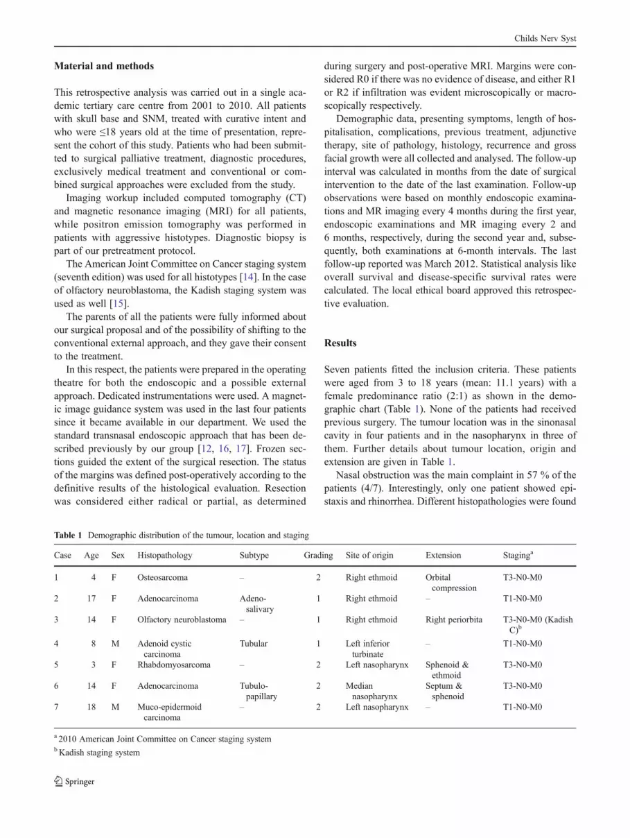
Seven patients fitted the inclusion criteria. These patients
were aged from 3 to 18 years (mean: 11.1 years) with a
female predominance ratio (2:1) as shown in the demo-
graphic chart (Table 1). None of the patients had received
previous surgery. The tumour location was in the sinonasal
cavity in four patients and in the nasopharynx in three of
them. Further details about tumour location, origin and
extension are given in Table 1.
Nasal obstruction was the main complaint in 57 % of the
patients (4/7). Interestingly, only one patient showed epi-
staxis and rhinorrhea. Different histopathologies were found
Table 1 Demographic distribution of the tumour, location and staging
Case Age Sex Histopathology Subtype Grading Site of origin Extension Staginga
1 4 F Osteosarcoma – 2 Right ethmoid Orbital
compression
T3-N0-M0
2 17 F Adenocarcinoma Adeno-
salivary
1 Right ethmoid – T1-N0-M0
3 14 F Olfactory neuroblastoma – 1 Right ethmoid Right periorbita T3-N0-M0 (Kadish
C)b
4 8 M Adenoid cystic
carcinoma
Tubular 1 Left inferior
turbinate
– T1-N0-M0
5 3 F Rhabdomyosarcoma – 2 Left nasopharynx Sphenoid &
ethmoid
T3-N0-M0
6 14 F Adenocarcinoma Tubulo-
papillary
2 Median
nasopharynx
Septum &
sphenoid
T3-N0-M0
7 18 M Muco-epidermoid
carcinoma
– 2 Left nasopharynx – T1-N0-M0
a 2010 American Joint Committee on Cancer staging systembKadish staging system
Childs Nerv Syst

among the patients. All cases had localised pathology with
no evidence of loco-regional or distant metastases at the
time of presentation (Table 1).
All patients affected by sinonasal malignancies under-
went exclusively EER (Figs. 1 and 2). In this series, there
was no bony invasion of the ethmoidal roof in any of the
cases, so a craniectomy was never required. In patients with
nasopharyngeal lesions, a nasopharyngeal endoscopic
resection, tailored to the extension of disease, was per-
formed (type 2 resection in two cases; type 3 resection in
one case) [17].
All surgeries were considered radical. No intraoperative
complication occurred. In one case, the results of the histo-
logical evaluation showed presence of microscopic disease
in the margin (R1). In this case, adjuvant chemotherapy was
administered. Detailed data are given in Table 2.
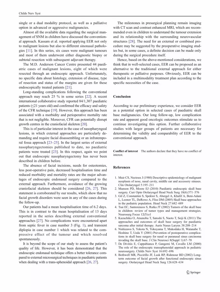
Fig. 1 Pre-operative MR scan, in coronal (a), axial (b), and sagittal (c)
view, of a right ethmoidal neuroblastoma with periorbital extension,
staged T3N0M0. Postoperative MR scan, in coronal (d), axial (e), and
sagittal (f) view, performed 7 years after the endoscopic endonasal
resection (EER) of the lesion. (Case number 3) 191×106 mm (300×
300 DPI)
Fig. 2 Coronal (a) and sagittal
(b) MR images showing a
nasopharyngeal
adenocarcinoma extended to
the sphenoid and nasal septum
staged T3N0M0. Post-operative
MR scan, in coronal (c) and
sagittal (d) view, performed
3 years after the endoscopic
endonasal resection (EER) of
the lesion. (Case number 6)
176×128 mm (300×300 DPI)
Childs Nerv Syst

Mean hospitalisation time was 6.2 days (2–17 days).
Regarding the complications, one patient developed a septic
fever 2 days after surgery that required an intravenous
antibiotic. Another patient with osteosarcoma of the ethmoi-
dal sinus compressing the orbit had a late complication
(after 1 month) of transient diplopia that was treated con-
servatively. No significant deformity in facial appearance
was observed in any of the cases during follow-up.
None of the patients received neoadjuvant therapy. The
patient with recurrent nasopharyngeal rhabdomyosarcoma
received primary chemo-radiation as first line treatment.
Four patients received adjuvant therapy: chemotherapy
alone (two patients), radiotherapy alone (one patient) and
both chemo-radiotherapy (one patient), as summarised in
Table 3.
According to the last follow-up, performed in March
2012, 6/7 (85.7 %) patients were alive with no evidence of
disease; only one patient died of causes other than SNM.
Notably, oncologic control of the sinonasal disease was
obtained in all the cases, without evidence of loco-regional
or distant recurrences. The mean follow-up was 67.7 months
(ranging from 24 to 109 months).
In detail, the patient who died during the follow-up was a
4-year-old girl who had previously received radiotherapy for
acute T-cell lymphoblastic leukaemia. She then developed a
radio-induced sinonasal osteosarcoma that was resected
endoscopically. Surgery along with adjuvant chemotherapy
achieved good oncologic control of the sinonasal osteosar-
coma but the patient died after 97 months due to a radio-
induced meningosarcoma of the spinal cord.
Discussion
Sinonasal and skull base malignancies are rare diseases in
the general population, characterised by histological hetero-
geneity, late diagnosis and poor prognosis [18]. The inci-
dence of the disease in children is unknown, but it is
probably less frequent in this population [19, 20].
The choice of treatment depends upon many aspects like
tumour biology, behaviour, histotype, and the consequences
of the treatment modality on the patient. It is commonly
accepted that aggressive tumours are better treated primarily
with radiotherapy, chemotherapy or both. Surgical treatment
is unfavourable when it ends up with poor outcome or
devastating complications, especially in children. On the
other hand, the best surgical outcome is obtained when the
tumour is resected radically, the margins maintain negative
results and when there are no serious complications.
These considerations are extremely important in the pae-
diatric population, in which physicians must be concerned
about future skeletal development and the incidence of
radiation-induced secondary lesions [21].
In our series, we were able to achieve radical tumour
resection despite narrow spaces. EER gives us the opportu-
nity to view the origin of the tumour and then resect it
radically while preserving the adjacent vital structures.
According to our long follow-up (mean: 67.7 months), there
is no clinical or radiological evidence of residual disease or
recurrence. Statistically, the survival rate and oncological
control rate were 85.7 and 100 %, respectively. Due to the
rarity of these tumours and our small number of cases, the
impact of these statistical outcomes is difficult to ascertain.
EER of SNM and skull base tumours without tragic com-
plications can be considered a therapeutic option, either as a
Table 2 Intraoperative measures with post-operative complications
Case Location Type of resection Frozen
sections
Margins Complications Treatment of
complication
Hospitalisation
(days)
1 Sinonasal EER Negative R0 Diplopia Conservative 8
2 Sinonasal EER Negative R0 – – 3
3 Sinonasal EER Negative R0 Septic fever Antibiotics 17
4 Sinonasal EER Negative R0 – – 2
5 Nasopharynx NER type 3 Negative R1 – – 7
6 Nasopharynx NER type 2 Negative R0 – – 3
7 Nasopharynx NER type 2 Negative R0 – – 4
EER endoscopic endonasal resection, NER nasopharyngeal endoscopic resection, R0 no infiltration, R1 micro-infiltration, R2 macro-infiltration
Table 3 Adjuvant therapy and follow-up
Number Adjuvant therapy F/U (months) Status
1 CHT 97 DOC
2 – 109 NED
3 RT + CHT 91 NED
4 – 74 NED
5 CHT 40 NED
6 – 36 NED
7 RT 27 NED
DOC died of other causes, NED no evidence of the disease
Childs Nerv Syst
single or a dual modality protocol, as well as a palliative
option in advanced or aggressive malignancies.
Almost all the available data regarding the surgical man-
agement of SNM in children have discussed the convention-
al approach. Kassam et al. reported applying EER not only
to malignant lesions but also to different sinonasal patholo-
gies [11]. In this series, six cases were malignant tumours
and most of them underwent either diagnostic biopsy or
subtotal resection with subsequent adjuvant therapy.
The M.D. Anderson Cancer Centre presented 44 paedi-
atric cases of malignant lesions, eight of which were
resected through an endoscopic approach. Unfortunately,
no specific data about histology, extension of disease, type
of resection and status of the margins are given for these
endoscopically treated patients [21].
Long-standing complications following the conventional
approach may reach 25 % in some series [22]. A recent
international collaborative study reported 84/1,307 paediatric
patients (≤21 years old) and confirmed the efficacy and safety
of the CFR technique [19]. However, this approach has been
associated with a morbidity and perioperative mortality rate
that is not negligible. Moreover, CFR can potentially disrupt
growth centres in the craniofacial skeleton [20].
This is of particular interest in the case of nasopharyngeal
lesions, in which external approaches are particularly de-
manding and require facial disassembling or an infratempo-
ral fossa approach [23–25]. In the largest series of external
nasopharyngectomies published to date, no paediatric
patients were treated [23]. In this respect, again we point
out that endoscopic nasopharyngectomy has never been
described in children before.
The absence of facial incisions, needs for osteotomies,
less post-operative pain, decreased hospitalisation time and
reduced morbidity and mortality rates are the major advan-
tages of endoscopic endonasal surgery compared to the
external approach. Furthermore, avoidance of the growing
craniofacial skeleton should be considered [26, 27]. This
statement is corroborated by our results, which show that no
facial growth disorders were seen in any of the cases during
the follow-up.
Our patients had a mean hospitalisation time of 6.2 days.
This is in contrast to the mean hospitalisation of 13 days
reported in the series describing external conventional
approaches [27]. No complications were encountered apart
from septic fever in case number 3 (Fig. 1), and transient
diplopia in case number 1 which was related to the com-
pressive effect of the tumour and which resolved
spontaneously.
It is beyond the scope of our study to assess the patient’s
quality of life. However, it has been demonstrated that the
endoscopic endonasal technique offers a better tolerance com-
pared to external microsurgical techniques in paediatric patients
when dealing with a trans-sphenoidal approach [26, 27].
The milestones in presurgical planning remain imaging
with CT scan and contrast enhanced MRI, which are recom-
mended even in children to understand the tumour extension
and its relationship with the surrounding neurovascular
structures [28]. The need for an external or combined pro-
cedure may be suggested by the preoperative imaging stud-
ies but, in some cases, a definite decision can be made only
during the surgical procedure itself.
Hence, based on the above-mentioned considerations, we
think that in well-selected cases, EER can be proposed as an
alternative to the traditional external approach for either
therapeutic or palliative purposes. Obviously, EER can be
included in a multimodality treatment plan according to the
specific necessities of the case.
Conclusion
According to our preliminary experience, we consider EER
as a potential option in selected cases of paediatric skull
base malignancies. Our long follow-up, low complication
rate and apparent good oncologic outcomes stimulate us to
continue investigating this option. Further collaborative
studies with larger groups of patients are necessary for
determining the validity and comparability of EER to the
conventional approach.
Conflict of interest The authors declare that they have no conflict of
interest.
References
1. Muir CS, Nectoux J (1980) Descriptive epidemiology of malignant
neoplasm of nose, nasal cavity, middle ear and accessory sinuses.
Clin Otolaryngol 5:195–211
2. Munson PD, Moore EJ (2010) Paediatric endoscopic skull base
surgery. Curr Opin Otolaryngol Head Neck Surg 18(6):571–576
3. Gil Z, Constantini S, Spektor S, Abergel A, Khafif A, Beni-Adani
L, Leonor TL, DeRowe A, Fliss DM (2005) Skull base approaches
in the pediatric population. Head Neck 27:682–689
4. Tsai EC, Santoreneos S, Rutka JT (2002) Tumors of the skull base
in children: review of tumor types and management strategies.
Neurosurg Focus 12(5):e1
5. Kazuchika O, Atsunobu T, Satoshi S, Naoto T, Seiji K (2011) The
approaches and outcomes of skull base surgery for paediatric
sarcoma after initial therapy. Auris Nasus Larynx 38:208–214
6. Nishizawa S, Yokota N, Yokoyama T, Mukodaka H, Watanabe T,
Hoshino T, Ueda Y (2001) Prevention of postoperative complica-
tions in skull base surgery for nasal or paranasal sinus carcinoma
invading the skull base. J Clin Neurosci 8(Suppl 1):67–70
7. De Divitiis E, Cappabianca P, Gangemi M, Cavallo LM (2000)
The role of the endoscopic transsphenoidal approach in pediatric
neurosurgery. Childs Nerv Syst 16:692–696
8. Bothwell MR, Piccirillo JF, Lusk RP, Ridenour BD (2002) Long-
term outcome of facial growth after functional endoscopic sinus
surgery. Otolaryngol Head Neck Surg 126:628–634
Childs Nerv Syst

9. Lusk RP, Muntz HR (1990) Endoscopic sinus surgery in children
with chronic sinusitis: a pilot study. Laryngoscope 100:654–658
10. Gross CW, Gurucharri MJ, Lazar RH, Thomas E (1989) Function-
al endoscopic sinus surgery (FESS) in the paediatric age group.
Laryngoscope 99:272–275
11. Kassam A, Thomas AJ, Snyderman C, Carrau R, Gardner P, Mintz
A, Kanaan H, Horowitz M, Pollack IF (2007) Fully endoscopic
expanded endonasal approach treating skull base lesions in pedi-
atric patients. J Neurosurg 106(2 Suppl):75–86
12. Nicolai P, Battaglia P, Bignami M, Bolzoni Villaret A, Delù G,
Khrais T, Lombardi D, Castelnuovo P (2008) Endoscopic surgery
for malignant tumors of the sinonasal tract and adjacent skull base:
a 10-year experience. Am J Rhinol 22(3):308–316, May-Jun
13. Hanna E, DeMonte F, Ibrahim S, Roberts D, Levine N, Kupferman
M (2009) Endoscopic resection of sinonasal cancers with and
without craniotomy: oncologic results. Arch Otolaryngol Head
Neck Surg Dec 135(12):1219–1224
14. Edge SB, Byrd DR, Compton CC, Fritz AG, Greene FL, Trotti A
(2010) Nasal cavity and paranasal sinuses. American Joint Com-
mittee on Cancer AJCC Cancer Staging Manual, 7th edn.
Springer-Verlag, New York
15. Kadish S, Goodman M, Wang CC (1976) Olfactory neuroblasto-
ma: a clinical analysis of 17 cases. Cancer 37:1571–1576
16. Castelnuovo P, Battaglia P, Locatelli D, Delù G, Sberze F, Bignami
M (2006) Endonasal micro-endoscopic treatment of the malignant
tumors of paranasal sinuses and anterior skull base. Oper Tech
Otolaryngol 17:152–167
17. Castelnuovo P, Dallan I, Bignami M, Battaglia P, Mauri S, Bolzoni
Villaret A, Bizzoni A, Tomenzoli D, Nicolai P (2010) Nasopha-
ryngeal endoscopic resection in the management of selected ma-
lignancies: ten-year experience. Rhinology 48:84–89
18. Lund VJ, Stammberger H, Nicolai P, Castelnuovo P (2010) Euro-
pean position paper on endoscopic management of tumours of the
nose, paranasal sinuses and skull base. Rhinol Suppl 22:1–143
19. Gil Z, Patel SG, Cantu G, Fliss DM, Kowalsky LP, Singh B,
Snyderman C, Kraus DH, Shah JP, International Collaborative Study
Group, Bridger PG, Cheesman AD, Donald P, Gullane P, Janecka I,
Kamata SE, Levine PA,Medina LR, Pradhan S, SchrammV,WeiWI
(2009) Outcome of craniofacial surgery in children and adolescents
with malignant tumors involving the skull base: an international
collaborative study. Head Neck 31(3):308–317
20. Hanbali F, Tabrizi P, Lang FF, DeMonte F (2004) Tumors of the
skull base in children and adolescents. J Neurosurg 100(2 Suppl
Pediatrics):169–178
21. Zevallos JP, Jain K, Roberts D, El-Naggar A, Hanna E, Kupferman
M (2011) Sinonasal malignancies in children: a 10-year, single-
institutional review. Laryngoscope 121(9):2001–2003
22. Ketcham AS, Wilkins RH, Van Buren JM, Smith RR (1963) A
combined intracranial facial approach to the paranasal sinuses. Am
J Surg 166:698–703
23. Wei WI, Chan JYW, Man RW, Ho WK (2011) Surgical salvage of
persistent or recurrent nasopharyngeal carcinoma with maxillary
swing approach—critical appraisal after 2 decades. Head Neck
33:969–975
24. Fisch U (1983) The infratemporal fossa approach for nasopharyn-
geal tumors. Laryngoscope 93:36–44
25. Fee WE Jr, Robertson JB Jr, Goffinet DR (1991) Long term
survival after surgical resection for recurrent nasopharyngeal can-
cer after radiotherapy failure. Arch Otolaryngol Head Neck Surg
117:1233–1236
26. Locatelli D, Massimi L, Rigante M, Custodi V, Paludetti G,
Castelnuovo P, Di Rocco C (2010) Endoscopic endonasal
transsphenoidal surgery for sellar tumors in children. Int J
Pediatr Otorhinolaryngol 74(11):1298–1302
27. Massimi L, Rigante M, D’Angelo L, Paternoster G, Leonardi P,
Paludetti G, Di Rocco C (2011) Quality of postoperative course in
children: endoscopic endonasal surgery versus sublabial microsur-
gery. Acta Neurochir (Wien) 153(4):843–849
28. Benoit MM, Silvera VM, Nichollas R, Jones D, McGill T, Rahbar
R (2009) Image guidance systems for minimally invasive sinus
and skull base surgery in children. Int J Pediatr Otorhinolaryngol
73(10):1452–1457
Childs Nerv Syst
Related Documents