World J Gastrointest Endosc 2011 August 16; 3(8): 157-161 ISSN 1948-5190 (online) © 2011 Baishideng. All rights reserved. Online Submissions: http://www.wjgnet.com/1948-5190office [email protected] doi:10.4253/wjge.v3.i8.157 Endoscopic and histopathological features of gastrointestinal amyloidosis Akira Hokama, Kazuto Kishimoto, Manabu Nakamoto, Chiharu Kobashigawa, Tetsuo Hirata, Nagisa Kinjo, Fukunori Kinjo, Seiya Kato, Jiro Fujita Akira Hokama, Kazuto Kishimoto, Tetsuo Hirata, Jiro Fujita, Department of Infectious, Respiratory, and Digestive Medi- cine, Faculty of Medicine, University of the Ryukyus, Okinawa 903-0125, Japan Chiharu Kobashigawa, Manabu Nakamoto, Nagisa Kinjo, Fukunori Kinjo, Department of Endoscopy, University Hospital of the Ryukyus, Okinawa 903-0125, Japan Seiya Kato, Department of Pathology and Cell Biology, Faculty of Medicine, University of the Ryukyus, Okinawa 903-0125, Japan Author contributions: Hokama A wrote the manuscript; Ho- kama A, Kishimoto K, Nakamoto M, Kobashigawa C, Hirata T and Kinjo N performed endoscopic examinations and treated the patients; Hokama A and Kato S performed the pathological examinations; Kinjo F and Fujita J supervised treatment of the patients and preparation of the manuscript. Correspondence to: Akira Hokama, MD, PhD, Assistant Pro- fessor, Department of Infectious, Respiratory, and Digestive Medi- cine, Faculty of Medicine, University of the Ryukyus, Okinawa 903-0125, Japan. [email protected] Telephone: +81-98-8951144 Fax: +81-98-8951414 Received: March 3, 2011 Revised: July 18, 2011 Accepted: August 6, 2011 Published online: August 16, 2011 Abstract Amyloidosis is a rare disorder, characterized by the ex- tracellular deposition of an abnormal fibrillar protein, which disrupts tissue structure and function. Amyloi- dosis can be acquired or hereditary, and systemic or localized to a single organ, such as the gastrointestinal (GI) tract. Clinical manifestations may vary from as- ymptomatic to fatal forms. Primary amyloidosis (mono- clonal immunoglobulin light chains, AL) is the most common form of amyloidosis. AL amyloidosis has been associated with plasma cell dyscrasias, such as, mul- tiple myeloma. Secondary amyloidosis is caused by the deposition of fragments of the circulating acute-phase reactant, serum amyloid A protein (SAA). Common causes of AA amyloidosis are chronic inflammatory dis- orders. Although GI symptoms are usually nonspecific, histopathological patterns of amyloid deposition are as- sociated with clinical and endoscopic features. Amyloid deposition in the muscularis mucosae, submucosa, and muscularis propria has been dominant in AL amyloi- dosis, leading to polypoid protrusions and thickening of the valvulae conniventes, whereas granular amyloid deposition mainly in the propria mucosae has been re- lated to AA amyloidosis, resulting in the fine granular appearance, mucosal friability, and erosions. As a re- sult, AL amyloidosis usually presents with constipation, mechanical obstruction, or chronic intestinal pseudo- obstruction while AA amyloidosis presents with diar- rhea and malabsorption Amyloidotic GI symptoms are mostly refractory and have a negative impact on quality of life and survival. Diagnosing GI amyloidosis requires high suspicion of evaluating endoscopists. Because of the absence of specific treatments for reducing the abundance of the amyloidogenic precursor protein, we should be aware of certain associations between pat- terns of amyloid deposition and clinical and endoscopic features. © 2011 Baishideng. All rights reserved. Key words: Amyloidosis; Amyloid; Congo red; Endos- copy; Gastrointestinal tract; Histopathology Peer reviewers: Shinji Tanaka, MD, PhD, Professor, Department of Endoscopy, Hiroshima University Hospital, 1-2-3 Kasumi, Minami-ku, Hiroshima 734-8551, Japan; Omar Javed Shah, Professor, Head, Department of Surgical Gastroenterology, Sher- i-Kashmir Institute of Medical Sciences, Srinagar, Kashmir 900100, India Hokama A, Kishimoto K, Nakamoto M, Kobashigawa C, Hirata T, Kinjo N, Kinjo F, Kato S, Fujita J. Endoscopic and histo- pathological features of gastrointestinal amyloidosis. World J Gastrointest Endosc 2011; 3(8): 157-161 Available from: URL: http://www.wjgnet.com/1948-5190/full/v3/i8/157.htm DOI: http://dx.doi.org/10.4253/wjge.v3.i8.157 EDITORIAL 157 August 16, 2011|Volume 3|Issue 8| WJGE|www.wjgnet.com

Welcome message from author
This document is posted to help you gain knowledge. Please leave a comment to let me know what you think about it! Share it to your friends and learn new things together.
Transcript

World J Gastrointest Endosc 2011 August 16; 3(8): 157-161ISSN 1948-5190 (online)
© 2011 Baishideng. All rights reserved.
Online Submissions: http://www.wjgnet.com/[email protected]:10.4253/wjge.v3.i8.157
Endoscopic and histopathological features of gastrointestinal amyloidosis
Akira Hokama, Kazuto Kishimoto, Manabu Nakamoto, Chiharu Kobashigawa, Tetsuo Hirata, Nagisa Kinjo, Fukunori Kinjo, Seiya Kato, Jiro Fujita
Akira Hokama, Kazuto Kishimoto, Tetsuo Hirata, Jiro Fujita, Department of Infectious, Respiratory, and Digestive Medi-cine, Faculty of Medicine, University of the Ryukyus, Okinawa 903-0125, JapanChiharu Kobashigawa, Manabu Nakamoto, Nagisa Kinjo, Fukunori Kinjo, Department of Endoscopy, University Hospital of the Ryukyus, Okinawa 903-0125, JapanSeiya Kato, Department of Pathology and Cell Biology, Faculty of Medicine, University of the Ryukyus, Okinawa 903-0125, JapanAuthor contributions: Hokama A wrote the manuscript; Ho-kama A, Kishimoto K, Nakamoto M, Kobashigawa C, Hirata T and Kinjo N performed endoscopic examinations and treated the patients; Hokama A and Kato S performed the pathological examinations; Kinjo F and Fujita J supervised treatment of the patients and preparation of the manuscript. Correspondence to: Akira Hokama, MD, PhD, Assistant Pro-fessor, Department of Infectious, Respiratory, and Digestive Medi-cine, Faculty of Medicine, University of the Ryukyus, Okinawa 903-0125, Japan. [email protected]: +81-98-8951144 Fax: +81-98-8951414Received: March 3, 2011 Revised: July 18, 2011Accepted: August 6, 2011Published online: August 16, 2011
AbstractAmyloidosis is a rare disorder, characterized by the ex-tracellular deposition of an abnormal fibrillar protein, which disrupts tissue structure and function. Amyloi-dosis can be acquired or hereditary, and systemic or localized to a single organ, such as the gastrointestinal (GI) tract. Clinical manifestations may vary from as-ymptomatic to fatal forms. Primary amyloidosis (mono-clonal immunoglobulin light chains, AL) is the most common form of amyloidosis. AL amyloidosis has been associated with plasma cell dyscrasias, such as, mul-tiple myeloma. Secondary amyloidosis is caused by the deposition of fragments of the circulating acute-phase reactant, serum amyloid A protein (SAA). Common causes of AA amyloidosis are chronic inflammatory dis-
orders. Although GI symptoms are usually nonspecific, histopathological patterns of amyloid deposition are as-sociated with clinical and endoscopic features. Amyloid deposition in the muscularis mucosae, submucosa, and muscularis propria has been dominant in AL amyloi-dosis, leading to polypoid protrusions and thickening of the valvulae conniventes, whereas granular amyloid deposition mainly in the propria mucosae has been re-lated to AA amyloidosis, resulting in the fine granular appearance, mucosal friability, and erosions. As a re-sult, AL amyloidosis usually presents with constipation, mechanical obstruction, or chronic intestinal pseudo-obstruction while AA amyloidosis presents with diar-rhea and malabsorption Amyloidotic GI symptoms are mostly refractory and have a negative impact on quality of life and survival. Diagnosing GI amyloidosis requires high suspicion of evaluating endoscopists. Because of the absence of specific treatments for reducing the abundance of the amyloidogenic precursor protein, we should be aware of certain associations between pat-terns of amyloid deposition and clinical and endoscopic features.
© 2011 Baishideng. All rights reserved.
Key words: Amyloidosis; Amyloid; Congo red; Endos-copy; Gastrointestinal tract; Histopathology
Peer reviewers: Shinji Tanaka, MD, PhD, Professor, Department of Endoscopy, Hiroshima University Hospital, 1-2-3 Kasumi, Minami-ku, Hiroshima 734-8551, Japan; Omar Javed Shah, Professor, Head, Department of Surgical Gastroenterology, Sher-i-Kashmir Institute of Medical Sciences, Srinagar, Kashmir 900100, India
Hokama A, Kishimoto K, Nakamoto M, Kobashigawa C, Hirata T, Kinjo N, Kinjo F, Kato S, Fujita J. Endoscopic and histo-pathological features of gastrointestinal amyloidosis. World J Gastrointest Endosc 2011; 3(8): 157-161 Available from: URL: http://www.wjgnet.com/1948-5190/full/v3/i8/157.htm DOI: http://dx.doi.org/10.4253/wjge.v3.i8.157
EDITORIAL
157 August 16, 2011|Volume 3|Issue 8|WJGE|www.wjgnet.com

Hokama A et al . Gastrointestinal amyloidosis
INTRODUCTIONAmyloidosis is a rare disorder, characterized by the extra-cellular deposition of an abnormal fibrillar protein, which disrupts tissue structure and function. Types of amyloi-dosis are classified based on the identity of the respective precursor protein[1]. Amyloidosis can be acquired or he-reditary, and systemic or localized to a single organ, such as the gastrointestinal (GI) tract. Clinical manifestations may vary from asymptomatic to fatal forms. We review the endoscopic and histopathological characteristics of GI amyloidosis with the presentation of our experiences.
TYPES OF AMYLOIDOSISPrimary amyloidosis (monoclonal immunoglobulin light chains, AL) is the most common form of amyloidosis. AL amyloidosis has been associated with plasma cell dyscra-sias, such as multiple myeloma. Secondary amyloidosis is caused by the deposition of fragments of the circulating acute-phase reactant, serum amyloid A protein (SAA). Common causes of AA amyloidosis are chronic inflam-matory disorders and infections, including rheumatoid arthritis, Crohn’s disease, familial Mediterranean fever, lep-rosy and tuberculosis[1,2]. Due to a predominance of infec-tions before 1990, the AA/AL ratio was 1:3; however, the ratio has been 1:17 to 1:38 due to fewer chronic infections and an increasing recognition of AL amyloidosis[3]. Other types of amyloidosis are dialysis-related amyloidosis with the deposition of β2-microglobulins, and autosomal domi-nant systemic amyloidosis, such as familial amyloidotic polyneuropathy (FAP) with the deposition of genetically variant transthyretin[1,2]. The incidence of the former has declined with the use of high flux hemodialysis.
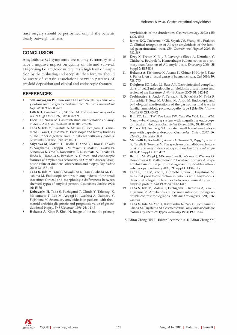
THE ASSOCIATION OF CLINICAL FEATURES AND ENDOSCOPIC FINDINGSPresentations of systemic amyloidosis include weakness, weight loss, neuropathy, cardiopathy, nephropathy and arthropathy, all of which can be refractory[1,2]. Among patients with systemic amyloidosis, the involvement in the GI tract is very common. The small intestine is most commonly affected in the GI tract[4,5]. Diagnosis requires confirmation of the presence of amyloid by histopathol-ogy using Congo red staining (Figure 1). Although GI symptoms are usually nonspecific and include macroglos-sia, dysphagia, abdominal pain, hemorrhage, constipation, diarrhea and malabsorption, patterns of amyloid depo-sition are associated with clinical and endoscopic fea-tures[6,7]. Amyloid deposition in the muscularis mucosae, submucosa and muscularis propria has been dominant in AL amyloidosis, leading to polypoid protrusions and thickening of the valvulae conniventes, whereas granu-lar amyloid deposition mainly in the propria mucosae has been related to AA amyloidosis, resulting in the fine granular appearance, mucosal friability and erosions[6]. As a result, AL amyloidosis usually presents with constipa-tion, mechanical obstruction or chronic intestinal pseudo-obstruction, while AA amyloidosis presents with diar-rhea and malabsorption[6]. Typical endoscopic images of duodenal lesions in AL amyloidosis at our institute[8] are shown in Figure 1. Characteristic polypoid protrusions and thickening of the folds are presented. In Figure 2, gastroduodenal lesions in AA amyloidosis caused by rheumatoid arthritis are depicted. More friable duodenal mucosa and reddish colonic mucosa of AA amyloidosis caused by familial Mediterranean fever are disclosed in Figures 3 and 4. Table 1 shows a brief comparison of characteristics of AL and AA amyloidosis. In addition,
158 August 16, 2011|Volume 3|Issue 8|WJGE|www.wjgnet.com
C
A
B
Figure 1 Endoscopic view of amyloid light chains amyloidosis in a 64-year-old man without multiple myeloma presenting abdominal fullness. A: Characteristic multiple yellowish-white polypoid protrusions and thickening of the folds in the descending duodenum are presented; B: Biopsy specimens showed marked homogenous eosinophilic deposition in the mucosae and submucosa (HE, × 40); C: Congo red stain confirmed a unique “apple-green” birefringence of the amyloid deposition under polarized light (× 40). All figures and legends are reproduced from[8] with permission from Elsevier.

submucosal hematoma, ulcers and hemorrhagic bullous colitis, which may be caused by amyloid infiltration, are other features in the setting of GI bleeding in AL amy-loidosis[9,10]. Our experience with hemorrhagic colonic lesions in AL amyloidosis[11] is shown in Figure 5. Charac-teristic yellowish plaque-like infiltrative lesions, submuco-sal hematoma and ulceration are presented.
As for other types of amyloidosis, dialysis-related β2-microglobulin amyloidosis has a similar present ation to AL amyloidosis[12]. In FAP, endoscopic findings of GI tract are mostly a mild, fine, granular appearance and the
159 August 16, 2011|Volume 3|Issue 8|WJGE|www.wjgnet.com
B
A
Figure 3 Endoscopic views of amyloid A amyloidosis in a 45-year-old man with familial Mediterranean fever. A: Friable granular mucosa with in the descending duodenum; B: Closer observation revealing whitish dilatated villi with multiple reddish erosions.
Figure 4 Endoscopic views of amyloid A amyloidosis in a 55-year-old man with familial Mediterranean fever. Patchy reddish mucosa was presented along with submucosal veins. Histopathological examination confirmed amyloid deposition.
D
C
B
A
Figure 2 Endoscopic views of amyloid A amyloidosis in a 45-year-old woman with rheumatoid arthritis. A: A round ulcer surrounded by longitudinal reddish mucosa is presented in the gastric antrum. Histopathological examination confirmed amyloid deposition; B: Fine granular mucosa in the descending duodenum; C: Biopsy of the duodenal lesion showing marked amorphous eosinophilic deposition in the lamina propria mucosae (HE, × 100); D: Congo red staining showing amy-loid deposition (× 100).
Hokama A et al . Gastrointestinal amyloidosis

amount of amyloid deposition in the mucosa is small compared with that in AL and AA amyloidosis. However, a significant amount of deposition is evident in the nerves of the GI tract, which may be the cause of severe diar-rhea and malabsorption occasionally observed in FAP pa-tients despite the mild macroscopic findings[13]. Although recent advances in endoscopy, including narrow-band imaging[14], capsule endoscopy[15,16] and double-balloon enteroscopy[17], have been widely applied to diagnose GI amyloidosis, plain radiographs and radiological barium examination, basic techniques, are still useful in evaluating GI amyloidosis, especially in the small intestine[18,19]. These methods can clearly reveal fold thickening of AL amyloi-dosis or fine granular mucosa of AA amyloidosis, which corroborate well with the histopathological findings[19,20].
TREATMENT OF AMYLOIDOSISBecause of the absence of specific treatments for GI amyloidosis, therapy is aimed at reducing the abundance of the amyloidogenic precursor protein, leading to the improvement of amyloidotic organ dysfunction[1]. Treat-ment of AL amyloidosis includes myeloma-type chemo-therapy with melphalan and prednisone and high-dose chemotherapy with hematopoietic stem cell transplanta-tion. Prokinetic agents may benefit dysmotility-related symptoms. Treatment of AA amyloidosis is control of the underlying inflammatory disorders, leading to the reduction of SAA. Diarrhea and malabsorption are often refractory. Supportive measures such as total parenteral nutrition and antidiarrheal agents can be beneficial[1]. GI
160 August 16, 2011|Volume 3|Issue 8|WJGE|www.wjgnet.com
Table 1 Comparison of characteristics of amyloid light chains and amyloid A amyloidosis[1,2,6,7]
amyloid light chains amyloidosis amyloid A amyloidosis
Causes Idiopathy and plasma cell dyscrasias Chronic inflammatory disorders and infectionsDeposition Monoclonal immunoglobulin light chains Serum amyloid A proteinGastrointestinal site of amyloid deposition The muscularis mucosae, submucosa and
muscularis propriaThe propria mucosae
Gastrointestinal symptoms Constipation, mechanical obstruction and chronic intestinal pseudo-obstruction
Diarrhea, malabsorption and weight loss
Endoscopic and radiological features Polypoid protrusions and thickening of the folds Fine granular appearance and mucosal friabilityTreatments Prokinetic agents and myeloma-type chemotherapy Control of the underlying inflammatory disorders
Figure 5 Endoscopic views of amyloid light chains amyloidosis in an 80-year-old woman without multiple myeloma presenting hematochezia. A: Colo-noscopy showing a round ulcer in the cecum; B: A patchy yellowish plaque-like infiltrative lesion (arrowheads) relatively normal-appearing intervening mucosae in the transverse colon; C: A tiny submucosal hematoma within pinky plaque-like erythema in the sigmoid colon; D: Biopsy of the colonic lesion showing marked amor-phous eosinophilic deposition in the mucosa and submucosa (HE, × 100); E: Congo red staining showing amyloid deposition (× 100). All figures and legends are reproduced from[11] with permission from BMJ Publishing Group Ltd.
D
CBA
E
Hokama A et al . Gastrointestinal amyloidosis
tract surgery should be performed only if the benefits clearly outweigh the risks.
CONCLUSIONAmyloidotic GI symptoms are mostly refractory and have a negative impact on quality of life and survival. Diagnosing GI amyloidosis requires a high level of suspi-cion by the evaluating endoscopists; therefore, we should be aware of certain associations between patterns of amyloid deposition and clinical and endoscopic features.
REFERENCES1 Sattianayagam PT, Hawkins PN, Gillmore JD. Systemic am-
yloidosis and the gastrointestinal tract. Nat Rev Gastroenterol Hepatol 2009; 6: 608-617
2 Falk RH, Comenzo RL, Skinner M. The systemic amyloido-ses. N Engl J Med 1997; 337: 898-909
3 Ebert EC, Nagar M. Gastrointestinal manifestations of amy-loidosis. Am J Gastroenterol 2008; 103: 776-787
4 Tada S, Iida M, Iwashita A, Matsui T, Fuchigami T, Yama-moto T, Yao T, Fujishima M. Endoscopic and biopsy findings of the upper digestive tract in patients with amyloidosis. Gastrointest Endosc 1990; 36: 10-14
5 Miyaoka M, Matsui T, Hisabe T, Yano Y, Hirai F, Takaki Y, Nagahama T, Beppu T, Murakami Y, Maki S, Takatsu N, Ninomiya K, Ono Y, Kanemitsu T, Nishimata N, Tanabe H, Ikeda K, Haraoka S, Iwashita A. Clinical and endoscopic features of amyloidosis secondary to Crohn’s disease: diag-nostic value of duodenal observation and biopsy. Dig Endosc 2011; 23: 157-165
6 Tada S, Iida M, Yao T, Kawakubo K, Yao T, Okada M, Fu-jishima M. Endoscopic features in amyloidosis of the small intestine: clinical and morphologic differences between chemical types of amyloid protein. Gastrointest Endosc 1994; 40: 45-50
7 Kobayashi H, Tada S, Fuchigami T, Okuda Y, Takasugi K, Matsumoto T, Iida M, Aoyagi K, Iwashita A, Daimaru Y, Fujishima M. Secondary amyloidosis in patients with rheu-matoid arthritis: diagnostic and prognostic value of gastro-duodenal biopsy. Br J Rheumatol 1996; 35: 44-49
8 Hokama A, Kinjo F, Kinjo N. Image of the month: primary
amyloidosis of the duodenum. Gastroenterology 2003; 125: 1302, 1565
9 James DG, Zuckerman GR, Sayuk GS, Wang HL, Prakash C. Clinical recognition of Al type amyloidosis of the lumi-nal gastrointestinal tract. Clin Gastroenterol Hepatol 2007; 5: 582-588
10 Dray X, Treton X, Joly F, Lavergne-Slove A, Uzunhan Y, Chiche A, Bouhnik Y. Hemorrhagic bullous colitis as a pri-mary manifestation of AL amyloidosis. Endoscopy 2006; 38 Suppl 2: E15-E16
11 Hokama A, Kishimoto K, Azama K, Chinen H, Kinjo F, Kato S, Fujita J. An unusual cause of haematochezia. Gut 2010; 59: 728, 793
12 Dulgheru EC, Balos LL, Baer AN. Gastrointestinal complica-tions of beta2-microglobulin amyloidosis: a case report and review of the literature. Arthritis Rheum 2005; 53: 142-145
13 Yoshimatsu S, Ando Y, Terazaki H, Sakashita N, Tada S, Yamashita T, Suga M, Uchino M, Ando M. Endoscopic and pathological manifestations of the gastrointestinal tract in familial amyloidotic polyneuropathy type I (Met30). J Intern Med 1998; 243: 65-72
14 Hui YT, Lam TW, Yee Lam PW, Yan Wu WH, Lam WM. Narrow-band imaging system with magnifying endoscopy for rectal amyloidosis. Gastrointest Endosc 2008; 68: 400-401
15 Pollack MJ, Isenberg GA. Isolated small bowel amyloidosis seen with capsule endoscopy. Gastrointest Endosc 2007; 66: 829-830; discussion 830
16 Mandelli G, Radaelli F, Amato A, Terreni N, Paggi S, Spinzi G, Ceretti E, Terruzzi V. The spectrum of small-bowel lesions of AL-type amyloidosis at capsule endoscopy. Endoscopy 2009; 41 Suppl 2: E51-E52
17 Bellutti M, Weigt J, Mönkemüller K, Röcken C, Wieners G, Dombrowski F, Malfertheiner P. Localized primary AL-type amyloidosis of the jejunum diagnosed by double-balloon enteroscopy. Endoscopy 2007; 39 Suppl 1: E134-E135
18 Tada S, Iida M, Yao T, Kitamoto T, Yao T, Fujishima M. Intestinal pseudo-obstruction in patients with amyloidosis: clinicopathologic differences between chemical types of amyloid protein. Gut 1993; 34: 1412-1417
19 Tada S, Iida M, Matsui T, Fuchigami T, Iwashita A, Yao T, Fujishima M. Amyloidosis of the small intestine: findings on double-contrast radiographs. AJR Am J Roentgenol 1991; 156: 741-744
20 Tada S, Iida M, Yao T, Kawakubo K, Yao T, Fuchigami T, Okada M, Fujishima M. Gastrointestinal amyloidosiadiologic features by chemical types. Radiology 1994; 190: 37-42
S- Editor Zhang HN L- Editor Roemmele A E- Editor Zheng XM
161 August 16, 2011|Volume 3|Issue 8|WJGE|www.wjgnet.com
Hokama A et al . Gastrointestinal amyloidosis
Related Documents