Endocellular polyamine availability modulates epithelial-to-mesenchymal transition and unfolded protein response in MDCK cells Marco Prunotto 1,2,8 , Alessandra Compagnone 3,8 , Maurizio Bruschi 1,2 , Giovanni Candiano 1 , Sebastiano Colombatto 3 , Andrea Bandino 3 , Andrea Petretto 4 , Solange Moll 5 , Marie Luce Bochaton-Piallat 6 , Giulio Gabbiani 6 , Veronica Dimuccio 1 , Maurizio Parola 3 , Lorenzo Citti 7 and GianMarco Ghiggeri 1 Epithelial-to-mesenchymal transition (EMT) is involved in embryonic development as well as in several pathological conditions. Literature indicates that polyamine availability may affect transcription of c-myc, matrix metalloproteinase (MMP)1, MMP2, TGFb 1 , and collagen type I mRNA. The aim of this study was to elucidate polyamines role in EMT in vitro. Madin-Darby canine kidney (MDCK) cells were subjected to experimental manipulation of intracellular levels of poly- amines. Acquisition of mesenchymal phenotype was evaluated by means of immunofluorescence, western blots, and zymograms. MDCK cells were then subjected to 2D gel proteomic study and incorporation of a biotinilated polyamine (BPA). Polyamine endocellular availability modulated EMT process. Polyamine-depleted cells treated with TGFb 1 showed enhanced EMT with a marked decrease of E-cadherin expression at plasma membrane level and an increased expression of mesenchymal markers such as fibronectin and a-smooth muscle actin. Polyamine-depleted cells showed a twofold increased expression of the rough endoplasmic reticulum (ER)-stress proteins GRP78, GRP94, and HSP90 a/b in 2D gels. The latter data were confirmed by western blot analysis. Administration of BPA showed that polyamines are covalently linked, within the cell, to ER-stress proteins. Intracellular polyamine availability affects EMT in MDCK cells possibly through the modulation of ER-stress protein homeostasis. Laboratory Investigation (2010) 90, 929–939; doi:10.1038/labinvest.2010.65; published online 8 March 2010 KEYWORDS: MDCK; polyamines; EMT; UPR; DFMO; ER-stress Epithelial–mesenchymal transition (EMT), paradigmatic of the cell plasticity concept, is involved in embryonic devel- opment as well as in several pathological conditions, including organ fibrosis and cancer progression. 1 During EMT, epithelial cells loose polarization, disassemble cell–cell junctions, and switch to a mesenchymal-type gene expression program. Cells undergoing EMT start to synthesize fibronectin, collagen type I, and vimentin acquiring a fibro- blast-like phenotype. In some cases, cells undergoing EMT express a-smooth muscle actin (a-SMA) assuming the myofibroblastic phenotype. Several signaling factors, including TGFb 1 , trigger this process. 1–3 It has been shown that TGFb 1 downregulates the sper- midine synthase gene 4 and that fibroblasts treated with difluoromethylornithine (DFMO), a specific inhibitor of the enzyme ornithine decarboxylase (ODC), responsible in mammalian cells of ornithine decarboxylation to putrescine, exhibit a significant increase in c-myc, matrix metallo- proteinase (MMP)1, MMP2, TGFb 1 , and collagen type I mRNAs, 5 suggesting that intracellular polyamine levels affects transcription of EMT-related genes. Polyamines are organic polycations present in all living organisms. Higher polyamines are synthesized by the sequen- tial addition of aminopropyl groups to putrescine by a reaction involving S-adenosyl methionine decarboxylase (SAMDC) as well as spermidine and spermine synthases. ODC and SAMDC are considered rate-limiting enzymes in polyamine biosynthesis. In addition to these multi-level Received 10 September 2009; revised 11 January 2010; accepted 27 January 2010 1 Nephrology Laboratory, Giannina Gaslini Children’s Hospital, Genoa, Italy; 2 RenalChild Foundation, c/o Giannina Gaslini Children’s Hospital, Genoa, Italy; 3 Department of Experimental Medicine and Oncology, University of Torino, Torino, Italy; 4 Mass Spectrometry Core Facility, Giannina Gaslini Children Hospital, Genoa, Italy; 5 Service de Pathologie Clinique, Ho ˆ pitaux Universitaires de Gene ` ve, Geneva, Switzerland; 6 Division of Pathology, Geneva University Hospital, Geneva, Switzerland and 7 Department of Clinical Physiology—CNR, Pisa, Italy Correspondence: Dr M Prunotto, PhD, Nephrology Laboratory, Giannina Gaslini Children’s Hospital, Largo G. Gaslini, 5-16147 Genoa, Italy. E-mail: [email protected] 8 These authors contributed equally to this work Laboratory Investigation (2010) 90, 929–939 & 2010 USCAP, Inc All rights reserved 0023-6837/10 $32.00 www.laboratoryinvestigation.org | Laboratory Investigation | Volume 90 June 2010 929

Welcome message from author
This document is posted to help you gain knowledge. Please leave a comment to let me know what you think about it! Share it to your friends and learn new things together.
Transcript
Endocellular polyamine availability modulatesepithelial-to-mesenchymal transition and unfoldedprotein response in MDCK cellsMarco Prunotto1,2,8, Alessandra Compagnone3,8, Maurizio Bruschi1,2, Giovanni Candiano1,Sebastiano Colombatto3, Andrea Bandino3, Andrea Petretto4, Solange Moll5, Marie Luce Bochaton-Piallat6,Giulio Gabbiani6, Veronica Dimuccio1, Maurizio Parola3, Lorenzo Citti7 and GianMarco Ghiggeri1
Epithelial-to-mesenchymal transition (EMT) is involved in embryonic development as well as in several pathologicalconditions. Literature indicates that polyamine availability may affect transcription of c-myc, matrix metalloproteinase(MMP)1, MMP2, TGFb1, and collagen type I mRNA. The aim of this study was to elucidate polyamines role in EMT in vitro.Madin-Darby canine kidney (MDCK) cells were subjected to experimental manipulation of intracellular levels of poly-amines. Acquisition of mesenchymal phenotype was evaluated by means of immunofluorescence, western blots, andzymograms. MDCK cells were then subjected to 2D gel proteomic study and incorporation of a biotinilated polyamine(BPA). Polyamine endocellular availability modulated EMT process. Polyamine-depleted cells treated with TGFb1 showedenhanced EMT with a marked decrease of E-cadherin expression at plasma membrane level and an increased expressionof mesenchymal markers such as fibronectin and a-smooth muscle actin. Polyamine-depleted cells showed a twofoldincreased expression of the rough endoplasmic reticulum (ER)-stress proteins GRP78, GRP94, and HSP90 a/b in 2D gels.The latter data were confirmed by western blot analysis. Administration of BPA showed that polyamines are covalentlylinked, within the cell, to ER-stress proteins. Intracellular polyamine availability affects EMT in MDCK cells possibly throughthe modulation of ER-stress protein homeostasis.Laboratory Investigation (2010) 90, 929–939; doi:10.1038/labinvest.2010.65; published online 8 March 2010
KEYWORDS: MDCK; polyamines; EMT; UPR; DFMO; ER-stress
Epithelial–mesenchymal transition (EMT), paradigmatic ofthe cell plasticity concept, is involved in embryonic devel-opment as well as in several pathological conditions,including organ fibrosis and cancer progression.1 DuringEMT, epithelial cells loose polarization, disassemble cell–celljunctions, and switch to a mesenchymal-type gene expressionprogram. Cells undergoing EMT start to synthesizefibronectin, collagen type I, and vimentin acquiring a fibro-blast-like phenotype. In some cases, cells undergoing EMTexpress a-smooth muscle actin (a-SMA) assuming themyofibroblastic phenotype. Several signaling factors,including TGFb1, trigger this process.1–3
It has been shown that TGFb1 downregulates the sper-midine synthase gene4 and that fibroblasts treated with
difluoromethylornithine (DFMO), a specific inhibitor of theenzyme ornithine decarboxylase (ODC), responsible inmammalian cells of ornithine decarboxylation to putrescine,exhibit a significant increase in c-myc, matrix metallo-proteinase (MMP)1, MMP2, TGFb1, and collagen type ImRNAs,5 suggesting that intracellular polyamine levels affectstranscription of EMT-related genes.
Polyamines are organic polycations present in all livingorganisms. Higher polyamines are synthesized by the sequen-tial addition of aminopropyl groups to putrescine by areaction involving S-adenosyl methionine decarboxylase(SAMDC) as well as spermidine and spermine synthases.ODC and SAMDC are considered rate-limiting enzymes inpolyamine biosynthesis. In addition to these multi-level
Received 10 September 2009; revised 11 January 2010; accepted 27 January 2010
1Nephrology Laboratory, Giannina Gaslini Children’s Hospital, Genoa, Italy; 2RenalChild Foundation, c/o Giannina Gaslini Children’s Hospital, Genoa, Italy; 3Departmentof Experimental Medicine and Oncology, University of Torino, Torino, Italy; 4Mass Spectrometry Core Facility, Giannina Gaslini Children Hospital, Genoa, Italy; 5Servicede Pathologie Clinique, Hopitaux Universitaires de Geneve, Geneva, Switzerland; 6Division of Pathology, Geneva University Hospital, Geneva, Switzerland and7Department of Clinical Physiology—CNR, Pisa, ItalyCorrespondence: Dr M Prunotto, PhD, Nephrology Laboratory, Giannina Gaslini Children’s Hospital, Largo G. Gaslini, 5-16147 Genoa, Italy.E-mail: [email protected]
8These authors contributed equally to this work
Laboratory Investigation (2010) 90, 929–939
& 2010 USCAP, Inc All rights reserved 0023-6837/10 $32.00
www.laboratoryinvestigation.org | Laboratory Investigation | Volume 90 June 2010 929
controls of synthesis, uptake and efflux into extracellularenvironment co-operate in the fine-tuning of intracellularlevels of polyamines. The availability of specific inhibitors ofODC and SAMDC has allowed to establish the essential roleof polyamines in many processes, including cell growth,6–9
differentiation,10,11 cell adhesion,12 induction of apoptosis,13
and signal transduction.14–16 On this basis, DFMO admin-istration has been suggested for the treatment of infectiousdiseases,17 protozoal parasitism18 or, with modest results,cancer19 and hyper proliferative diseases.20
Polyamines may act as ligands of DNA, RNA, phospho-lipids, nucleotide triphosphates, or proteins and act aseffectors of gene expression and signal transduction. Poly-amines are also substrates for transglutaminase-2 (TG-2),which catalyses the covalent incorporation of severallow molecular weight amines into protein in the form ofg-carboxyl group of a peptide-bound glutamic acid. Keycellular processes are modulated by the amount of both freeand protein-conjugated polyamines formed by TG activity.21
The aim of this study was to elucidate whether polyamineshave a role in EMT modulation of Madin-Darbycanine kidney (MDCK) cells derived from distal tubularepithelial cells.
MATERIALS AND METHODSCell CultureMDCK cells (ATCC, Middlesex, UK) were cultured in Dul-becco’s modified Eagle medium (DMEM; Sigma, Saint-Louis,USA) and supplemented with 10% fetal bovine serum (FBS).Polyamine-related experiments were instead performed inRPMI 1640 (Gibco BRL, Paisley, UK) supplemented with10% horse serum (HS) with cells cultured in 150 cm2 flasks.Addition of HS to all experimental conditions instead of FBSis an established procedure aimed at minimizing cytotoxiceffects due to high levels of amine-oxidases in FBS, which, onthe contrary, is very low in HS. Addition of HS instead of FBSis necessary because of the experimental protocol design that,in some conditions, requires addition of polyamines (i.e. tocounteract effect of DFMO). FBS high-level content ofamino-oxidases can in fact generate significant amounts ofhydrogen peroxide, ammonia, and aldehydes through poly-amine degradation, leading to cell injury and death.22 Cellswere plated and pre-treated, when scheduled, alternativelywith 3 mM DFMO for 24 h, leading to the depletion ofendocellular polyamines, or DFMOþ a mix of polyamines(P, containing 10 mM putrescine and 10 mM spermidine) tovalidate the specific effect of DFMO. Medium was changedafter 24 h, and cells were exposed to the following experi-mental conditions: control (RPMIþ 10% HS), DFMO(RPMIþ 10% HSþ 3 mM DFMO), TGFb1 (RPMIþ 10%HSþ 10 ng/ml TGFb1), DFMOþTGFb1 (3 mM DMFO and10 ng/ml TGFb1, respectively, in RPMIþ 10% HS) orDFMOþTGFb1þ P (3 mM DMFO and 10 ng/ml TGFb1,respectively, plus polyamine mix, in RPMIþ 10% HS). Cellswere allowed to grow up to 96 h and processed for western
blot or immunofluorescence. Cells pre-treated with 3 mMDMFO for 24 h were used to evaluate proteins linked topolyamines that were assessed through the administration of100 mM EZ-link penthylamine-biotin (BPA, Pierce, Rockford,USA) for further 6 h. Cells were then finally processed fortwo-dimensional (2D) electrophoresis or immunofluore-scence, in this case, BPA administration was performed in theabsence or presence of a specific siRNA against TG-2.
Assessment of TGFb1 mRNA and Protein inPolyamine-Depleted CellsTGFb1 mRNA presence was routinely assessed throughRT-PCR in control and DFMO-treated cells. TGFb1 proteinwas evaluated with two different methods: secreted proteinwas evaluated on 5-day treated cells pre-treated on day 4 withBrefeldin A (Sigma-Aldrich), this to prevent cytokine secre-tion. Cells were then fixed, stained with an anti-TGFb1
antibody (Clone TB21, Serotec GmbH, Dusseldorf, Germany)and detected with corresponding FITC-conjugated secondaryantibody. Cells were then observed through a FACS Excalibur(Becton & Dickinson, Mountain View, USA). TGFb1 in thesupernatant was dosed at day 5 of treatment through appli-cation of a specific ELISA test (Human TGFb1 Instant ELISA,Bender MedSystems, Wien, Austria). In both cases, positivecontrol was obtained by treatment of MDCK cells with4.2 mM Cyclosporin as described earlier by Feldman et al.23
TG-2 KnockdownThe TG-2 knockdown was performed by the administrationof a specific siRNA addressed to the known incompletecanine transcript sequence (accession N1 DR103953) targetinga 23 nucleotide long stretch starting at 573 nucleotide. Thesequences of siRNA were: CAAGCAGGCTTTAGAGCCACAAA (sense strand) and UGUGGCUCUAAAGCCUGCUUGAA (antisense strand). The two oligonucleotides were dis-solved in 20 mM Tris-HCl buffer, 100 mM NaCl, pH 7.5 andthen annealed by slow cooling after 1 min denaturation inboiling water bath. The siRNA was administered to cultures ina 12-well plate at 100 nM final concentration using alipoplexed vehicle GeneSilencer (GenLantis, San Diego CA,USA) according to the manufacturer’s instructions in serum-free medium 8 h later, cells were then washed and usedfor BPA administration experiments. As control, the siRNAaddressed to fluorescent protein GFP was administered toparallel cultures. Sense strand: CAAGCUGACCCUGAAGUUCUAA; antisense strand: AGAACUUCAGGGUCAGCUUGAA.
Matrix Metalloproteinase AssayMMP evaluation was performed at 120 h on extracellularmedium of cells submitted exactly to the above-describedexperimental protocol. Medium was changed 24 h beforemedium sampling. Briefly, samples were loaded onto astandard 7.5% acrylamide gel containing 0.1% gelatin andrun at 120 V for 2 h. Gelatin digestion was counterstainedwith Coomassie Blue R-250 (Sigma-Aldrich). Zymograms
Polyamines modulate EMT and UPR
M Prunotto et al
930 Laboratory Investigation | Volume 90 June 2010 | www.laboratoryinvestigation.org

were loaded based on cellular protein concentration asevaluated by BCA assay.
Polyamine Content EvaluationCells were scraped in 0.2 M HClO4 and subsequentlycentrifuged at 15 000 g. Supernatant was dansylated andanalyzed by HPLC as described earlier.24 Detectionwas performed with a fluorimeter (Waters 2475, WatersCorporation, Watford, UK).
Apoptosis Assessment in Polyamine-Depleted CellsApoptosis was assessed in control and polyamine-depletedcells through Annexin V/propidium iodide labeling andsubsequent FACS analysis (FACS Excalibur).
2D Electrophoresis and Western BlotCells collected for western blot and 2D were trypsinized,counted, and washed in TBS. For western blot, 4� 104 cells/mlwere resuspended in Laemmli sample buffer-containing phos-phatase and protease inhibitor cocktail. Samples were thenboiled for 100 and proteins concentration was determined usingBCA assay.
Western blot samples (10–50 mg) were load onto a SDS-PAGE (T% 8–16) in a Protean II XI system (Bio-Rad,Hercules, CA, USA), transferred onto nitrocellulose mem-brane (Protean BA, Schleicher & Schuell, Dassel, Germany)with a Novablot semidry system, 3% w/v BSA saturated inTBS and incubated separately with e-cadherin (Santa CruzBiotechnologies, CA, USA, 1:1000), fibronectin (Santa Cruz,1:1000), a-SMA (kindly provided by Professor G Gabbiani,1:750), b-tubulin (AbCam, 1:1000), GRP78 (Santa CruzBiotechnology, 1:1000), GRP94 (Sigma, 1:1000), HSP90 a/b(Santa Cruz Biotechnology, 1:1000) a-enolase (AbD Serotec,1:1000) or vimentin (Novocastra, 1:1000) in 3% w/v BSA inTBS-Tween 0.15% v/v (TBS-T). Membranes were then rinsedin TBS-T and incubated with corresponding secondaryantibodies. Chemioluminescence was used for immuno-detection. Images were digitalized by mean of VersaDoc 4000(Bio-Rad) and analyzed with QuantityOne software (Bio-Rad).
For analytical (100 mg) and preparative (400 mg) 2D elec-trophoresis, samples were solubilized in the reduction/alkylation solution, ie 8 M urea, 4% CHAPS, 5 mM tributyl-phosphine, 20 mM iodoacetamide (IAA), 40 mM Tris, and0.1 mM EDTA pH 8.5. Before isoelectric focusing (IEF),samples were incubated in this solution for 3 h, to allowproper reduction and alkylation. To prevent over alkylationduring the IEF step, excess of IAA was neutralized by addingan equimolar amount of DTT. Samples were then cleanedusing an ice-cool solution consisting of tri-n-butyl-phos-phate: acetone: methanol (1:12:1). Fourteen milliliters of thismixture were added to each sample to reach a final acetoneconcentration of 80% (v/v) and incubated at 41C for 900. Theprecipitate was pelleted by centrifugation at 2800 g for 200 at41C. After washing with the same de-lipidizing solution,it was centrifuged again and then air- dried. Finally, samples
were dissolved in the focusing solution, ie 7 M urea, 2 Mthiourea, 4% CHAPS, and 50 mM dithioerythritol (DTE) andload onto home-made non-linear pH 3–10 strips (18 cmlong, 3 mm wide, 0.5 mm thick).25 Strip re-swelling wascarried out overnight at room temperature in the focusingsolution, ie 7 M urea, 2 M thiourea, 2% (w/v) CHAPS,15 mM DTE, and a 0.6% (v/v) carrier ampholyte cocktail,containing 40% of the pH 3.5–10 and 60% of the pH 4–8intervals.
The proteins were focused at r50 mA per strip at 201C,using progressively increasing voltage for a total of 80 000 Vh.Equilibration step was carried out for 300 in the equilibrationbuffer, ie 6 M Urea, 50 mM Tris-HCl pH 8.8, 2% w/v SDS,30% v/v glycerol, and 0.01% w/v bromophenol blue. Insecond dimension was used a SDS-PAGE (T% 8–16) in aProtean II XI system (Bio-Rad).
Blue silver colloidal staining26 or silver staining were usedfor preparative or analytical proteins detection. Images weredigitalized by mean of GS-800 (Bio-Rad) and analyzed withPDQuest software (Bio-Rad).
Procedure described above was also adopted for 2Dwestern blot of BPA-labeled proteins (BPAþ ). Briefly, pro-teins were transferred to nitrocellulose membrane, saturatedin 3% w/v BSA in TBS, and incubated with neutravidinHRP-conjugated (Pierce Biotech., Rockford, USA) in 1% w/vBSA in TBS-T. Chemioluminescence was used for immuno-detection. Images were analyzed with PDQuest software(Bio-Rad); 2D experiments were performed in triplicate.
Statistical AnalysisDensitometric values of 2D gel and western blot wereanalyzed using non-parametric Mann–Whitney U test. In thecase of 2D gels, spots with at least twofold increase relative tocontrol with corresponding two-tailed P-value o0.05 wereaccepted as statistically significant. Mass spectrometryidentification was limited to BPAþ spots retaining thesecharacteristics. Densitometric western blot differences wereconsidered statistically significant at two-tailed P-valueo0.05.
Tryptic Digestion and Protein Identification by LC-ESIMS-MS/MSProtein spots were excised from gel, rinse in 50% v/v aceto-nitrile (ACN) in 5 mM ammonium bicarbonate pH 8.9 untilfull destaining, rinse twice in 100% v/v ACN, and brieflyrinse in 1 mM CaCl2 and 100 mM ammonium bicarbonatepH 8.9. Enzymatic digestions were performed using trypsinin 100 mM ammonium bicarbonate buffer pH 7.8 overnightat 371C. After incubation, the reaction was quenched by theaddition of formic acid to pH 2.
All mass spectrometric measurements were performedusing a LTQ linear ion trap mass spectrometer (ThermoElectron, San Jose, USA) coupled to a HPLC Surveyor(Thermo Electron) and equipped with a Jupiter C18 column250� 1 mm (Phenomenex). Peptides were eluted from the
Polyamines modulate EMT and UPR
M Prunotto et al
www.laboratoryinvestigation.org | Laboratory Investigation | Volume 90 June 2010 931

column using an acetonitrile gradient, 5% B for 60 followedby 5 to 90% B within 1090 (eluent A: 0.1% formic acid inwater; eluent B: 0.1% formic acid in acetonitrile) at flow rateof 50 ml/min. The column effluent was directed into theelectro spray source. The spray voltage was 5.0 kV. The iontrap capillary was kept at 2001C and the voltage at 2.85 V.Spectra were obtained in automated MS/MS mode: each fullMS scan (m/z 400–1800) was followed by five MS/MS ofthe most abundant ions. The ions analyzed this way wereautomatically excluded for 3000.
The raw data acquired by the mass spectrometer wereconverted in a peak list file for database search usingExtract_msn in Bioworks 3.3.1 Sp1 with default parametersfor LTQ spectra.
Protein identification was performed using SEQUESTsoftware 3.3.1 from Thermo Electron, operating on a10-processor computer cluster (AETHIA, Torino, Italia) andsearched against a Canis familiaris protein database (no. ofprotein entries 41427) downloaded on 11/2008 by NCBIusing the taxonomy browser tool. Peptide MS/MS assign-ments were filtered following very high stringent criteria:Xcorr Z1.9 for the singly charged ions, XcorrZ2.2 fordoubly charged ions, and XcorrZ3.7 for triply charged ions,peptide probability r0.01, Delta CnZ0.1, and Rsp r4according to the HUPO criteria.27 The mass tolerance forprecursor ions was set to 2 amu and the mass tolerance forfragment ions was 1 amu. To identify the largest panel ofpeptides, the option no enzyme was used for the in silicadigestion of human databases, so every combination ofhuman peptides was evaluated. Moreover, the searchparameters no included variable or fix modifications.
Immunofluorescence AnalysisIndirect immunofluorescence analysis was performed asdescribed earlier.28 Briefly, cells were gently rinsed in warmRPMI/2% HEPES buffer and fixed for 200 at 371C in 1%paraformaldehyde dissolved in RPMI/2% HEPES buffer.Cells were then rinsed in PBS and stained for e-cadherin(1:100), fibronectin (1:100), zonula occludens 1 (ZO-1,1:100), TG-2 (clone CUB 7402, Thermo Fisher Scientific,Fremont, USA), GRP78 (ABcam, Cambridge, USA) or a-SMA(1:50). Detection was performed with corresponding Alexa 488or Alexa 568 conjugated secondary antibody. Nuclei werecounterstained with DAPI. Images were acquired on a confocalApotome Axiovert microscope (Carl Zeiss AG, Jena, Germany).
RESULTSDFMO Administration Reduces Intracellular PolyamineContent in MDCK CellsDFMO administration reduces MDCK intracellular levels ofpolyamines (Figure 1).
DFMO administration reduced intracellular polyaminecontent to B20% of controls values (ie 5.922±0.296nmol/mg of total protein extract in DFMO-treated cells vs29.215±1.416 nmol/mg in control cells, Po0.001). TGFb1
alone did not statistically affect polyamine content, whereascombined administration of DFMO and TGFb1 reducedintracellular polyamines to 9.635±0.482 nmol/mg (Po0.001vs control value). Exogenous polyamine addition to theculture medium restored values comparable to those detectedin control cells (31.179±1.559 nmol/mg).
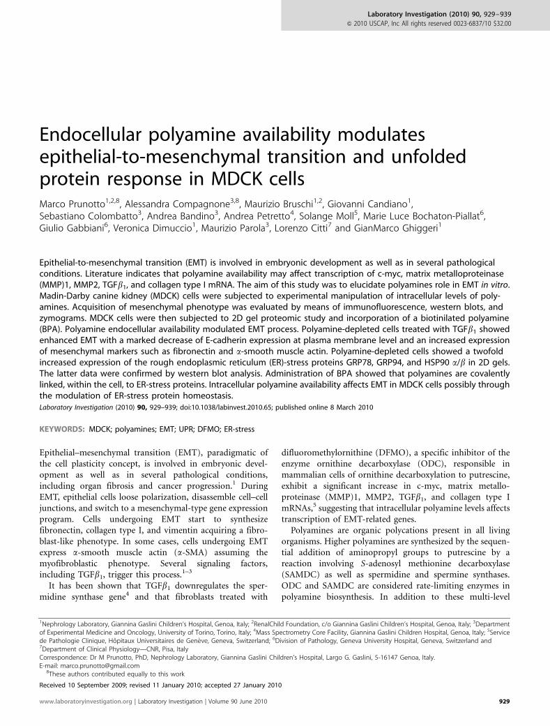
Polyamine Depletion Enhances EMT in MDCK CellsPolyamine-depleted MDCK cells showed a significantmorphological rearrangement into large polygonal cells(Figure 2b, phase contrast panel), associated with a markedredistribution of E-cadherin staining (decreased at plasmamembrane levels and increased in the cytoplasm) (Figure 2g).Immunofluorescence also showed some scattered cells exhi-biting positivity for fibronectin and an increase in a-SMApositive staining that were limited to sparse cytoplasmic rod-like structures (Figure 2n and s). Combined administrationof DFMO and TGFb1 resulted in a morphological re-ar-rangement at confluence (Figure 2c); very similar to the oneinduced by TGFb1 alone. Treatment up to 96 h resulted in areduced expression of the epithelial markers E-cadherin, asshown by western blot (Figure 3, panel A; SupplementaryFigure 1), and ZO-1 (data not shown). Immunofluorescenceanalysis revealed that combined treatment resulted in thedownregulation of plasma membrane E-cadherin (Figure 2i)and acquisition of a dense network of fibronectin (Figure 2p)associated to an increased expression of a-SMA in stressfibers in each cell (Figure 2u), whereas the treatment withTGFb1 alone resulted in an increased positive staining fora-SMA detectable in only in 20 to 30% of cells, this feature
Figure 1 Bar graph showing the effect of difluoromethylornithine (DFMO),
TGFb1, DFMOþ TGFb1, or DFMOþ TGFb1þ administration of exogenous
polyamines on total (ie including putrescine, spermidine, and spermine)
intracellular polyamine levels. DFMO decreased polyamines to B20%
compared with controls; TGFb1 alone did not statistically influence
polyamine content, whereas coupled administration of DFMO and TGFb1
decreased polyamines. Exogenous polyamine addition to the culture
medium restored values comparable to control. ***Po0.001 values are
referred compared with control conditions.
Polyamines modulate EMT and UPR
M Prunotto et al
932 Laboratory Investigation | Volume 90 June 2010 | www.laboratoryinvestigation.org

being mainly limited to the external edge of MDCK cell islets.Exogenous polyamine addition to the medium preventedall these phenomena: E-cadherin expression on plasmamembrane was maintained (Figure 2l), whereas fibronectinand a-SMA expression was negligible (Figure 2q and v,respectively). Addition of polyamines alone showed noinfluence on expression of all considered markers, whereasaddition of polyamines to TGFb1 showed a moderateinhibition of TGFb1-stimulated phenotypical modulation(Supplementary Figure 1).
Zymograms (Figure 3, panel B) showed that polyaminedepletion resulted in an increased activity of MMP2,pro-MMP9, and MMP9; moreover, polyamine depletiononce again synergized with TGFb1 and was partially pre-vented by polyamine addition to the medium (ie by restoringlevels similar to those observed after TGFb1 alone).
Polyamine Depletion Induces Limited TGFb1 Secretionbut not ApoptosisRT-PCR showed the presence of mRNA for TGFb1 in controland polyamine-depleted cells (Supplementary Figure 2aA).FACS analysis of labeled cells (Supplementary Figure 2B)showed expression of TGFb1 in polyamine-depleted cellsequal to 5.6% of cell population whereas percentage elevated
to 29.7% compared with control in the case of MDCK treatedwith 4.2 mM Cyclosporin. TGFb1 dosed in the supernatantcollected at day 5 (Supplementary Figure 2C) displayed aconcentration of TGFb1 equal to 450 pg/ml in polyamine-depleted cells, 900 pg/ml of TGFb1 in Cyclosporin-stimulatedcells, whereas unstimulated control cells showed an en-dogenous TGFb1 secretion of 90 pg/ml of TGFb1. Apoptosis,assessed through Annexin V/propidium iodide labeling andsubsequent FACS analysis showed no apoptosis induction inpolyamine-depleted cells (Supplementary Figure 2, panel D).
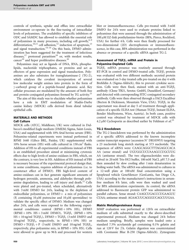
Proteomics2D proteomics showed, as expected, a great difference in theproteomic profile of TGFb1 treated cells compared withcontrols (Figure 4, panels A and B). The proteomic profile ofcontrol, TGFb1 treated, or polyamine-depleted TGFb1 treatedcells showed, respectively, 1584±6, 1848±6, or 1857±4spots. 1506±7 spots were matched across the three groups,whereas 1847±4 spots were matched when TGFb1
and polyamine-depleted TGFb1 treated were compared.Difference between polyamine-depleted TGFb1 and TGFb1
alone was limited to 30 differentially expressed spots (Figure 4,panels B and C), among them, 27 showed a statistically signi-ficant twofold upregulation compared with TGFb1 alone.
Figure 2 Phase contrast (a–e) or immunofluorescence (f–v) of MDCK cells stained for e-cadherin (f–l), fibronectin (m–q) or SMA (r–v) treated with DFMO,
TGFb1 (TGFb), DFMOþ TGFb1 (Dþ T), or DFMOþ TGFb1þ exogenous polyamines (Dþ Tþ P). MDCKs treated with Dþ T showed decreased expression of
e-cadherins and increased expression of fibronectin and a-SMA compared with TGFb1 alone. Polyamine addition to the medium inhibited cell phenotypical
modulation induced by Dþ T. Original magnification 200� (a–e) and 630x (f–v, bar¼ 20 mm).
Polyamines modulate EMT and UPR
M Prunotto et al
www.laboratoryinvestigation.org | Laboratory Investigation | Volume 90 June 2010 933
Spots were subsequently identified with LC-Mass spectro-metry (see Table 1), a-enolase (spots 4, 5, 6), vimentin (spots7, 8, 9), glucose-regulated protein 78 kDa (GRP78, spots 10,
11, 12), glucose-regulated protein 94 kDa (GRP94, spot 13), orHSP90 a/b (spots 16, 17, 18).
Each gel was performed in triplicate and analyzed usingPDQuest Advanced software (Bio-Rad). Average gels werecreated from the three gels for each type of condition.Normalization and statistical analysis of the results were thenperformed to compare each condition.
Functional Assessment Using a Biotinilated PolyamineBPA was efficiently incorporated in MDCK cells as revealedby immunofluorescence (Figure 5a). Competition withexogenous polyamines reduced BPA incorporation withincells, whereas administration of a TG-2 siRNA (Figure 5c)notably decreased TG-2-dependent covalent bond formationof polyamines on cellular target proteins. BPA is therebycovalently linked only in the presence of active TG-2.Moreover, 2D blots of BPA-labeled proteins showed that BPAis covalently linked on a limited subset of cellular proteins(Figure 5b). Statistically significant twofold upregulatedproteins were all BPAþ . A complete list of BPAþ proteins isdisplayed in Table 1.
Silencing of TG-2 Induces upregulation of EndoplasmicReticulum-Stress Proteins and Enhanced EMTSilencing of TG-2 in the presence of polyamines (Figure 6),added 4 h later, showed a response of cell to TGFb1 quitesimilar to administration of TGFb1 in combination withDFMO. In both conditions, DþT or TG-2 silenced cells,both GRP78 and a-SMA were upregulated compared withTGFb1 alone. These evidences suggest that TG-2-dependentpolyamine covalent bond formation on endoplasmic reti-culum (ER)-stress proteins is a relevant post-translationalmodification for homeostasis of ER-stress proteins. Whenpolyamines are depleted from cells or their covalent bondformation on ER-stress proteins is inhibited throughsilencing of TG-2, cells increase expression of GRP78 andGRP94 (data not shown) thereby increasing cells suscepti-bility to TGFb1.
Figure 3 Representative western blot (a) or for E-cadherin, fibronectin or
a-SMA of MDCK cells treated with DFMO, TGFb1 (TGFb), DFMOþ TGFb1
(Dþ T), or DFMOþ TGFb1þ exogenous polyamines (Dþ Tþ P).
(b) Zymogram for the same conditions. Polyamine depletion decreased
epithelial marker e-cadherins and increased expression of mesenchymal
markers fibronectin and SMA while at the same time increasing activity of
MMP2, pro-MMP9, and MMP9. b-Tubulin was charged as loading control.
Figure 4 Silver staining of representative 2D electrophoresis showing the effect of TGFb1 (TGFb) or DFMOþ TGFb1 (Dþ T) on proteomic profile of MDCK
cells. (a) Untreated cells, (b) TGFb1, or (c) DFMOþ TGFb1. Statistically differentially expressed spots between condition TGFb and Dþ T were labeled with
numbers. Correspondence with mass spectrometry identified proteins is reported in Table 1.
Polyamines modulate EMT and UPR
M Prunotto et al
934 Laboratory Investigation | Volume 90 June 2010 | www.laboratoryinvestigation.org
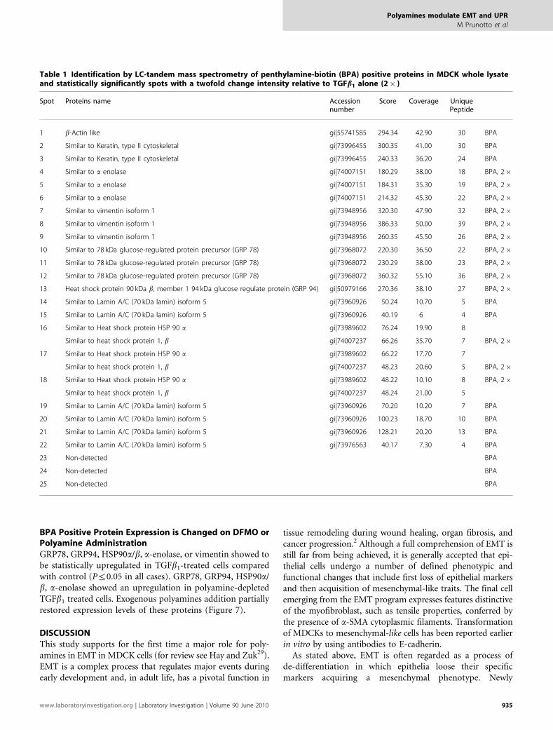
BPA Positive Protein Expression is Changed on DFMO orPolyamine AdministrationGRP78, GRP94, HSP90a/b, a-enolase, or vimentin showed tobe statistically upregulated in TGFb1-treated cells comparedwith control (Pr0.05 in all cases). GRP78, GRP94, HSP90a/b, a-enolase showed an upregulation in polyamine-depletedTGFb1 treated cells. Exogenous polyamines addition partiallyrestored expression levels of these proteins (Figure 7).
DISCUSSIONThis study supports for the first time a major role for poly-amines in EMT in MDCK cells (for review see Hay and Zuk29).EMT is a complex process that regulates major events duringearly development and, in adult life, has a pivotal function in
tissue remodeling during wound healing, organ fibrosis, andcancer progression.2 Although a full comprehension of EMT isstill far from being achieved, it is generally accepted that epi-thelial cells undergo a number of defined phenotypic andfunctional changes that include first loss of epithelial markersand then acquisition of mesenchymal-like traits. The final cellemerging from the EMT program expresses features distinctiveof the myofibroblast, such as tensile properties, conferred bythe presence of a-SMA cytoplasmic filaments. Transformationof MDCKs to mesenchymal-like cells has been reported earlierin vitro by using antibodies to E-cadherin.
As stated above, EMT is often regarded as a process ofde-differentiation in which epithelia loose their specificmarkers acquiring a mesenchymal phenotype. Newly
Table 1 Identification by LC-tandem mass spectrometry of penthylamine-biotin (BPA) positive proteins in MDCK whole lysateand statistically significantly spots with a twofold change intensity relative to TGFb1 alone (2� )
Spot Proteins name Accessionnumber
Score Coverage UniquePeptide
1 b-Actin like gi|55741585 294.34 42.90 30 BPA
2 Similar to Keratin, type II cytoskeletal gi|73996455 300.35 41.00 30 BPA
3 Similar to Keratin, type II cytoskeletal gi|73996455 240.33 36.20 24 BPA
4 Similar to a enolase gi|74007151 180.29 38.00 18 BPA, 2�5 Similar to a enolase gi|74007151 184.31 35.30 19 BPA, 2�6 Similar to a enolase gi|74007151 214.32 45.30 22 BPA, 2�7 Similar to vimentin isoform 1 gi|73948956 320.30 47.90 32 BPA, 2�8 Similar to vimentin isoform 1 gi|73948956 386.33 50.00 39 BPA, 2�9 Similar to vimentin isoform 1 gi|73948956 260.35 45.50 26 BPA, 2�10 Similar to 78 kDa glucose-regulated protein precursor (GRP 78) gi|73968072 220.30 36.50 22 BPA, 2�11 Similar to 78 kDa glucose-regulated protein precursor (GRP 78) gi|73968072 230.29 38.00 23 BPA, 2�12 Similar to 78 kDa glucose-regulated protein precursor (GRP 78) gi|73968072 360.32 55.10 36 BPA, 2�13 Heat shock protein 90 kDa b, member 1 94 kDa glucose regulate protein (GRP 94) gi|50979166 270.36 38.10 27 BPA, 2�14 Similar to Lamin A/C (70 kDa lamin) isoform 5 gi|73960926 50.24 10.70 5 BPA
15 Similar to Lamin A/C (70 kDa lamin) isoform 5 gi|73960926 40.19 6 4 BPA
16 Similar to Heat shock protein HSP 90 a gi|73989602 76.24 19.90 8
Similar to heat shock protein 1, b gi|74007237 66.26 35.70 7 BPA, 2�17 Similar to Heat shock protein HSP 90 a gi|73989602 66.22 17.70 7
Similar to heat shock protein 1, b gi|74007237 48.23 20.60 5 BPA, 2�18 Similar to Heat shock protein HSP 90 a gi|73989602 48.22 10.10 8 BPA, 2�
Similar to heat shock protein 1, b gi|74007237 48.24 21.00 5
19 Similar to Lamin A/C (70 kDa lamin) isoform 5 gi|73960926 70.20 10.20 7 BPA
20 Similar to Lamin A/C (70 kDa lamin) isoform 5 gi|73960926 100.23 18.70 10 BPA
21 Similar to Lamin A/C (70 kDa lamin) isoform 5 gi|73960926 128.21 20.20 13 BPA
22 Similar to Lamin A/C (70 kDa lamin) isoform 5 gi|73976563 40.17 7.30 4 BPA
23 Non-detected BPA
24 Non-detected BPA
25 Non-detected BPA
Polyamines modulate EMT and UPR
M Prunotto et al
www.laboratoryinvestigation.org | Laboratory Investigation | Volume 90 June 2010 935

differentiated cells exhibit activation of MMPs, enzymes thatcleave almost all extracellular matrix components, andactivate growth factors and growth factor receptors.30
The results of this study indicate that polyamine depletionenhances TGFb1-induced EMT in MDCKs by down-regulating epithelial markers ZO-1 and E-cadherin as well as
Figure 5 (a) Penthylamine-biotin (BPA) incorporation in polyamine-depleted MDCK cells or in presence of exogenous polyamines (poly) (b). BPA is actively
incorporated in MDCK cells. Competition with exogenous polyamines drastically reduced BPA staining. (b) 2D blots of BPA-labeled proteins revealed with
neutravidin-HRP showed incorporation of BPA on several cellular proteins, labels are reported in Table 1. (c) Transfection of siRNA designed against
transglutaminase-2 (TG-2) drastically reduced TG-2 expression hence reducing BPA incorporation in MDCK cells, siRNA for GFP was administered as control.
Figure 6 Silencing of TG-2 or polyamine depletion enhance EMT in MDCK cells. Cells silenced for TG-2 (g) and subsequent treated with TGFb1 in the
presence of polyamines showed an increased expression of GRP78 (h) and a-SMA (i) similar to polyamine-depleted cells (in e, f, respectively) treated
with control GFP siRNA (d) compared with cells treated with TGFb1 alone (b and c, respectively) and silenced with control GFP siRNA (a). GRP78
polyamination is TG-2 dependent and modulate EMT in MDCK cells. Original magnification 630� , bar¼ 20 mm.
Polyamines modulate EMT and UPR
M Prunotto et al
936 Laboratory Investigation | Volume 90 June 2010 | www.laboratoryinvestigation.org

by increasing expression of mesenchymal markers fibronectinand a-SMA. At the same time, polyamine depletion increasesactivity of MMP2, pro-MMP9, and MMP9, all enzymesinvolved in the break down of cell–cell and cell–ECM inter-actions, thus triggering EMT process.30–32 Polyamine deple-tion and TGFb1 stimulation show a synergic effect onEMT-related markers, as also confirmed by the fact thatexogenous polyamine addition, which restores intracellularpolyamine content, prevents these changes. Although alimited TGFb1 secretion is induced by the sole administrationof DFMO, this is in any case insufficient to induce theobserved enhanced EMT. Levels of TGFb1 induced by DFMOare in fact the half of those induced by 4.2 mM Cyclosporinadministration that proved earlier23 to be capable to alterparacellular permeability but not EMT. The irrelevance ofDFMO-induced TGFb1 secretion allowed us to look for apossible alternative explanation for the observed enhancedEMT in polyamine-depleted cells. For this reason, a pro-teomic approach was applied. 2D proteomic analysis ofTGFb1-treated and polyamine-depleted MDCKs vs TGFb1-treated MDCKs showed a statistically significant variationof 30 spots among the two conditions: we identifiedenhanced expression of a-enolase, vimentin, HSP90a/b,GRP78, and GRP94. A growing body of literature indicatesan essential function for GRP78 and GRP94 proteins inpreserving ER function. In particular, the 78 kDa molecularchaperone GRP78 is ubiquitously expressed in mammaliancells, and it has been reported to bind to hydrophobicpatches on nascent polypeptides in the ER. This is to preventaggregation of misfolded proteins and to contribute tothe development of proper secondary structure of matureproteins. In addition to preventing the physical aggregationof misfolded proteins, GRP78 is one of the initial compo-nents in the signaling cascade of the so-called unfoldedprotein response (UPR). Similarly to GRP78, GRP94,
identified as a 94-kDa protein also referred to as ERp9933
or endoplasmin,34 is an abundant ER glycoprotein. It shares50% amino acid identity to HSP90a/�, a well-characterized90-kDa member of the heat-shock protein family.Recent evidence suggests that GRP94 may also possessprotein chaperoning activity. Its synthesis is induced bythe accumulation of misfolded proteins in the ER.35,36 In theER, GRP78 and GRP94 act in tandem on folding inter-mediates of the newly synthesized immunoglobulin chains,with GRP94 being involved at a more advanced step in theirprocessing.37
Upregulation of both GRP78 and GRP94, together witha-enolase and vimentin, has been validated in our modelby western blot analysis performed at day 3 after TGFb1
administration. We substantially confirmed earlierimmunohistochemical and electron microscopy findingsby Parkkinen et al.38 These authors showed in fact redis-tribution, whorl formation, and autophagy of the ER inpolyamine-deprived BHK cells. This is of particular interestin view of recent in vitro studies39,40 demonstratingthat disturbances of the ER homeostasis results in a EMT-likeprocess characterized by cell morphology changes andextensive reorganization of the actin cytoskeleton.2,39,40
In our model, endocellular polyamine depletion dramati-cally enhances TGFb1-induced EMT, by increasing at thesame time the expression of typical ER-stress proteins likeGRP78 and GRP94, thus suggesting a direct involvement ofER-stress in the overall and polyamine depletion-dependentscenario.
Preliminary experiments, performed with a rat hepatomacell line (HTC) clonally selected to be defective for polyaminetransport across cell membrane (data not shown, manuscriptin preparation), show that endocellular polyamine avail-ability is crucial. In these mutant cells, exogenous polyaminesare ineffective in counteracting induction of fibronectin by
Figure 7 Representative western blots and corresponding bar graphs showing effect of TGFb1, DFMOþ TGFb1 (Dþ T), or DFMOþ TGFb1þ exogenous
polyamines (Dþ Tþ P) on cellular expression of GRP78, GRP94, HSP90a/b, a-enolase, or vimentin expression.
Polyamines modulate EMT and UPR
M Prunotto et al
www.laboratoryinvestigation.org | Laboratory Investigation | Volume 90 June 2010 937
combined DFMO and TGFb1 treatment, thus excluding adirect effect of polyamines on TGFb1 binding on its receptor.
Results of the experiments performed by loading MDCKswith a biotin-linked cadaverine (BPA), in the presence or inthe absence of DFMO, suggest that polyamines might bedirectly involved in the regulation of both GRP78 andGRP94. BPA, that is a mono-functional polyamine carryingonly one free -NH2 function, is efficiently up taken by MDCKcells, as demonstrated by immunofluorescence. Within thecell, BPA is than covalently linked to a specific and limitednumber of cellular proteins. These experiments revealedGRP78 and GRP94 as putative protein targets for polyaminecovalent bond formation.
Availability of endocellular polyamines is thereby criticalfor the regulation of GRP78 and GRP94 and, possibly, for theregulation of ER-stress and subsequent induction of an EMT-like process. Results showed that cell response to TGFb1
demonstrated to be very similar in polyamine-depleted cellsas well as in cells where TG-2 was previously silenced in thepresence of polyamines. In all cells, both ER-stress proteinGRP78 and a-SMA were upregulated compared with TGFb1
alone, suggesting that polyamination, through covalent bondformation induced by TG-2, may be a relevant post-trans-lational modification for ER-stress proteins homeostasis.In our experimental settings, TG-2-dependent polyaminecovalent bond formation onto ER-stress proteins mayrepresent an intracellular regulatory mechanism able tocounteract TGFb1 induced EMT. In this respect, it is note-worthy that a recent in vivo study has reported that TG-2inhibition ameliorates experimental fibrosis.41 Moreover,further studies are needed to fully comprehend relevance ofpolyamine biology in the cell, in particular related to TGFb1
pathway. Moderate inhibition of TGFb1-stimulated pheno-typical modulation by the sole addition of polyamines, sug-gests in fact involvement of polyamines also in other cellularpathways.
In conclusion, our work provides the first evidence for akey role of polyamines in EMT. 2D proteomic approachallowed suggesting a direct link between polyamine deple-tion, activation of ER-stress, and enhancement of EMT, thusreinforcing the concept that ER-stress may be crucial for theepithelial cell fate and the triggering of an EMT-like response.
Supplementary Information accompanies the paper on the Laboratory
Investigation website (http://www.laboratoryinvestigation.org)
ACKNOWLEDGEMENT
We acknowledge the RenalChild Foundation for the financial support and
Professor Rosanna Gusmano for critical discussion of results.
1. Radisky DC, Kenny PA, Bissell MJ. Fibrosis and cancer: domyofibroblasts come also from epithelial cells via EMT? J CellBiochem 2007;101:830–839.
2. Thiery JP, Sleeman JP. Complex networks orchestrate epithelial-mesenchymal transitions. Nat Rev Mol Cell Biol 2006;7:131–142.
3. McAnulty RJ. Fibroblasts and myofibroblasts: their source, functionand role in disease. Int J Biochem Cell Biol 2007;39:666–671.
4. Nishikawa Y, Kar S, Wiest L, et al. Inhibition of spermidine synthasegene expression by transforming growth factor-beta 1 in hepatomacells. Biochem J 1997;321(Pt 2):537–543.
5. Stabellini G, Moscheni C, Gagliano N, et al. Depletion of polyaminesand increase of transforming growth factor-beta1, c-myc, collagen-type I, matrix metalloproteinase-1, and metalloproteinase-2 mRNA inprimary human gingival fibroblasts. J Periodontol 2005;76:443–449.
6. Barnes RN, Bungay PJ, Elliott BM, et al. Alterations in the distributionand activity of transglutaminase during tumour growth andmetastasis. Carcinogenesis 1985;6:459–463.
7. Birckbichler PJ, Orr GR, Patterson Jr MK. Differential transglutaminasedistribution in normal rat liver and rat hepatoma. Cancer Res1976;36:2911–2914.
8. Birckbichler PJ, Patterson Jr MK. Cellular transglutaminase, growth, andtransformation. Ann NY Acad Sci 1978;312:354–365.
9. Pegg AE. Recent advances in the biochemistry of polyamines ineukaryotes. Biochem J 1986;234:249–262.
10. Kannagi R, Teshigawara K, Noro N, et al. Transglutaminase activityduring the differentiation of macrophages. Biochem Biophys ResCommun 1982;105:164–171.
11. Tabor CW, Tabor H. Polyamines. Annu Rev Biochem 1984;53:749–790.12. Gentile V, Thomazy V, Piacentini M, et al. Expression of tissue
transglutaminase in Balb-C 3T3 fibroblasts: effects on cellularmorphology and adhesion. J Cell Biol 1992;119:463–474.
13. Oliverio S, Amendola A, Di Sano F, et al. Tissue transglutaminase-dependent posttranslational modification of the retinoblastoma geneproduct in promonocytic cells undergoing apoptosis. Mol Cell Biol1997;17:6040–6048.
14. Koenig H, Goldstone AD, Lu CY. Beta-adrenergic stimulation of Ca2+fluxes, endocytosis, hexose transport, and amino acid transport inmouse kidney cortex is mediated by polyamine synthesis. Proc NatlAcad Sci USA 1983;80:7210–7214.
15. Koenig H, Goldstone AD, Lu CY. Polyamines are intracellularmessengers in the beta-adrenergic regulation of Ca2+ fluxes, [Ca2+]iand membrane transport in rat heart myocytes. Biochem Biophys ResCommun 1988;153:1179–1185.
16. Kohno H, Sasaki K, Yamaguchi M, et al. Spermine modulates calciumflux through the rat erythrocyte membrane. Biol Pharm Bull1997;20:153–157.
17. Kameji T, Pegg AE. Inhibition of translation of mRNAs for ornithinedecarboxylase and S-adenosylmethionine decarboxylase bypolyamines. J Biol Chem 1987;262:2427–2430.
18. Heby O, Persson L, Rentala M. Targeting the polyamine biosyntheticenzymes: a promising approach to therapy of African sleeping sickness,Chagas0 disease, and leishmaniasis. Amino Acids 2007;33:359–366.
19. Gerner EW, Meyskens Jr FL, Goldschmid S, et al. Rationale for, anddesign of, a clinical trial targeting polyamine metabolism for coloncancer chemoprevention. Amino Acids 2007;33:189–195.
20. Meyskens Jr FL, Gerner EW. Development of difluoromethylornithine(DFMO) as a chemoprevention agent. Clin Cancer Res 1999;5:945–951.
21. Lentini A, Abbruzzese A, Caraglia M, et al. Protein-polyamineconjugation by transglutaminase in cancer cell differentiation: reviewarticle. Amino Acids 2004;26:331–337.
22. Baydoun AR, Morgan DM. Inhibition of ornithine decarboxylasepotentiates nitric oxide production in LPS-activated J774 cells.Br J Pharmacol 1998;125:1511–1516.
23. Feldman G, Kiely B, Martin N, et al. Role for TGF-beta in cyclosporine-induced modulation of renal epithelial barrier function. J Am SocNephrol 2007;18:1662–1671.
24. Colombatto S, Fasulo L, Fulgosi B, et al. Transport and metabolism ofpolyamines in human lymphocytes. Int J Biochem 1990;22:489–492.
25. Bruschi M, Musante L, Candiano G, et al. Soft immobilized pH gradientgels in proteome analysis: a follow-up. Proteomics 2003;3:821–825.
26. Candiano G, Bruschi M, Musante L, et al. Blue silver: a very sensitivecolloidal Coomassie G-250 staining for proteome analysis.Electrophoresis 2004;25:1327–1333.
27. Adamski M, Blackwell T, Menon R, et al. Data management andpreliminary data analysis in the pilot phase of the HUPO PlasmaProteome Project. Proteomics 2005;5:3246–3261.
28. Brisset AC, Hao H, Camenzind E, et al. Intimal smooth muscle cells ofporcine and human coronary artery express S100A4, a marker of therhomboid phenotype in vitro. Circ Res 2007;100:1055–1062.
Polyamines modulate EMT and UPR
M Prunotto et al
938 Laboratory Investigation | Volume 90 June 2010 | www.laboratoryinvestigation.org

29. Hay ED, Zuk A. Transformations between epithelium andmesenchyme: normal, pathological, and experimentally induced.Am J Kidney Dis 1995;26:678–690.
30. Sternlicht MD, Werb Z. How matrix metalloproteinases regulate cellbehavior. Annu Rev Cell Dev Biol 2001;17:463–516.
31. Cicchini C, Laudadio I, Citarella F, et al. TGFbeta-induced EMT requiresfocal adhesion kinase (FAK) signaling. Exp Cell Res 2008;314:143–152.
32. Hojilla CV, Mohammed FF, Khokha R. Matrix metalloproteinases andtheir tissue inhibitors direct cell fate during cancer development.Br J Cancer 2003;89:1817–1821.
33. Mazzarella RA, Green M. ERp99, an abundant, conserved glycoproteinof the endoplasmic reticulum, is homologous to the 90-kDa heat shockprotein (hsp90) and the 94-kDa glucose regulated protein (GRP94).J Biol Chem 1987;262:8875–8883.
34. Koch G, Smith M, Macer D, et al. Endoplasmic reticulum containsa common, abundant calcium-binding glycoprotein, endoplasmin.J Cell Sci 1986;86:217–232.
35. Kim YK, Kim KS, Lee AS. Regulation of the glucose-regulated proteingenes by beta-mercaptoethanol requires de novo protein synthesisand correlates with inhibition of protein glycosylation. J Cell Physiol1987;133:553–559.
36. Kozutsumi Y, Segal M, Normington K, et al. The presence ofmalfolded proteins in the endoplasmic reticulum signals theinduction of glucose-regulated proteins. Nature 1988;332:462–464.
37. Melnick J, Aviel S, Argon Y. The endoplasmic reticulum stressprotein GRP94, in addition to BiP, associates with unassembledimmunoglobulin chains. J Biol Chem 1992;267:21303–21306.
38. Parkkinen JJ, Lammi MJ, Agren U, et al. Polyamine-dependentalterations in the structure of microfilaments, Golgi apparatus,endoplasmic reticulum, and proteoglycan synthesis in BHK cells.J Cell Biochem 1997;66:165–174.
39. Ulianich L, Garbi C, Treglia AS, et al. ER stress is associated withdedifferentiation and an epithelial-to-mesenchymal transition-likephenotype in PC Cl3 thyroid cells. J Cell Sci 2008;121(Pt 4):477–486.
40. Seki K, Fujimori T, Savagner P, et al. Mouse Snail family transcriptionrepressors regulate chondrocyte, extracellular matrix, type II collagen,and aggrecan. J Biol Chem 2003;278:41862–41870.
41. Ishii Y, Ichikawa M, Yamaguchi K, et al. Localization ofcephalosporinase in Enterobacter cloacae by immunocytochemicalexamination. J Antibiot (Tokyo) 1991;44:1088–1095.
Polyamines modulate EMT and UPR
M Prunotto et al
www.laboratoryinvestigation.org | Laboratory Investigation | Volume 90 June 2010 939
Related Documents