Encystation of Giardia lamblia: A model for other parasites Tineke Lauwaet, Barbara J. Davids, David S. Reiner, and Frances D. Gillin Department of Pathology, Division of Infectious Disease, UCSD, 214 Dickinson Street, San Diego, CA 92103-8416, USA Summary To colonize the human small intestine, Giardia lamblia monitors a dynamic environment. Trophozoites attach to enterocytes that mature and die. The parasites must “decide” whether to re- attach or differentiate into cysts that survive in the environment and re-activate when ingested. Other intestinal parasites face similar challenges. Study of these parasites is limited because they do not encyst in vitro. Giardia trophozoites were persuaded to encyst in vitro by mimicking physiologic stimuli. Cysts are dormant, yet “spring-loaded for action” to excyst upon ingestion. Giardial encystation has been studied from morphological, cell-biological, biochemical and molecular viewpoints. Yet important gaps remain and the mechanisms that co-ordinate responses to external signals remain enigmatic. Introduction As a major cause of waterborne diarrheal disease, Giardia contributes to the burden of malnutrition worldwide [1••]. Giardia’s simple, two-stage life cycle is central to its success as a parasite. G. lamblia cysts can survive in cold fresh water for months and fewer than 10 cysts are needed for human infection. Exposure of ingested cysts to gastric acid triggers excystation, a rapid and dramatic differentiation. After entry into the small intestine, the cyst wall opens and the parasite emerges. Trophozoites colonize below the entry of the common bile duct and can cause disease, although they do not invade. If they are carried downstream, trophozoites must encyst to survive outside the host. In vitro, Giardia encysts in response to the physiologic stimuli of increased bile and slightly alkaline pH [2]. The “gold standard” for successful encystation is the ability of cysts to excyst. Other important intestinal parasites, including Entamoeba, Toxoplasma, Cryptosporidium, several tapeworms and nematodes, are transmitted as cysts or oocysts. However, study of these organisms is limited by the inability to generate mature cysts in vitro. The giardial encystation pathway is a key virulence mechanism whose “biological goal” is differentiation into a form that can survive in the environment and infect a new host. Encystation also promotes immune evasion and is a target for vaccine and drug development [3-5]. The construction of the extracellular cyst wall (CW) is of primary importance as it allows the parasite to persist in fresh water, resist disinfectants, pass through the new host’s stomach and infect in the small intestine. This 300 nm thick fibrous structure excludes small molecules Correspondence to: Frances D. Gillin, [email protected]. Publisher's Disclaimer: This is a PDF file of an unedited manuscript that has been accepted for publication. As a service to our customers we are providing this early version of the manuscript. The manuscript will undergo copyediting, typesetting, and review of the resulting proof before it is published in its final citable form. Please note that during the production process errors may be discovered which could affect the content, and all legal disclaimers that apply to the journal pertain. NIH Public Access Author Manuscript Curr Opin Microbiol. Author manuscript; available in PMC 2009 July 13. Published in final edited form as: Curr Opin Microbiol. 2007 December ; 10(6): 554–559. doi:10.1016/j.mib.2007.09.011. NIH-PA Author Manuscript NIH-PA Author Manuscript NIH-PA Author Manuscript

Welcome message from author
This document is posted to help you gain knowledge. Please leave a comment to let me know what you think about it! Share it to your friends and learn new things together.
Transcript
Encystation of Giardia lamblia: A model for other parasites
Tineke Lauwaet, Barbara J. Davids, David S. Reiner, and Frances D. GillinDepartment of Pathology, Division of Infectious Disease, UCSD, 214 Dickinson Street, San Diego,CA 92103-8416, USA
SummaryTo colonize the human small intestine, Giardia lamblia monitors a dynamic environment.Trophozoites attach to enterocytes that mature and die. The parasites must “decide” whether to re-attach or differentiate into cysts that survive in the environment and re-activate when ingested. Otherintestinal parasites face similar challenges. Study of these parasites is limited because they do notencyst in vitro. Giardia trophozoites were persuaded to encyst in vitro by mimicking physiologicstimuli.
Cysts are dormant, yet “spring-loaded for action” to excyst upon ingestion. Giardial encystation hasbeen studied from morphological, cell-biological, biochemical and molecular viewpoints. Yetimportant gaps remain and the mechanisms that co-ordinate responses to external signals remainenigmatic.
IntroductionAs a major cause of waterborne diarrheal disease, Giardia contributes to the burden ofmalnutrition worldwide [1••]. Giardia’s simple, two-stage life cycle is central to its success asa parasite. G. lamblia cysts can survive in cold fresh water for months and fewer than 10 cystsare needed for human infection. Exposure of ingested cysts to gastric acid triggers excystation,a rapid and dramatic differentiation. After entry into the small intestine, the cyst wall opensand the parasite emerges. Trophozoites colonize below the entry of the common bile duct andcan cause disease, although they do not invade. If they are carried downstream, trophozoitesmust encyst to survive outside the host. In vitro, Giardia encysts in response to the physiologicstimuli of increased bile and slightly alkaline pH [2]. The “gold standard” for successfulencystation is the ability of cysts to excyst.
Other important intestinal parasites, including Entamoeba, Toxoplasma, Cryptosporidium,several tapeworms and nematodes, are transmitted as cysts or oocysts. However, study of theseorganisms is limited by the inability to generate mature cysts in vitro.
The giardial encystation pathway is a key virulence mechanism whose “biological goal” isdifferentiation into a form that can survive in the environment and infect a new host.Encystation also promotes immune evasion and is a target for vaccine and drug development[3-5]. The construction of the extracellular cyst wall (CW) is of primary importance as it allowsthe parasite to persist in fresh water, resist disinfectants, pass through the new host’s stomachand infect in the small intestine. This 300 nm thick fibrous structure excludes small molecules
Correspondence to: Frances D. Gillin, [email protected]'s Disclaimer: This is a PDF file of an unedited manuscript that has been accepted for publication. As a service to our customerswe are providing this early version of the manuscript. The manuscript will undergo copyediting, typesetting, and review of the resultingproof before it is published in its final citable form. Please note that during the production process errors may be discovered which couldaffect the content, and all legal disclaimers that apply to the journal pertain.
NIH Public AccessAuthor ManuscriptCurr Opin Microbiol. Author manuscript; available in PMC 2009 July 13.
Published in final edited form as:Curr Opin Microbiol. 2007 December ; 10(6): 554–559. doi:10.1016/j.mib.2007.09.011.
NIH
-PA Author Manuscript
NIH
-PA Author Manuscript
NIH
-PA Author Manuscript

such as water, but transmits the physiological stimuli that regulate excystation. It is a modelextracellular matrix with both protective and signaling functions.
Encystation is a gradual transformation of the motile, flagellated binucleate (4N), half-pear-shaped trophozoite (Figure 1). Trophozoites lose the ability to attach; the attachment diskfragments [6••] and the flagella are internalized. Metabolism also decreases as cells round upand enter dormancy. The oval, immotile, quadrinucleate (16N) cyst is encased in the refractileCW that contains protein (CWP) and glycopolymer (CWG) in insoluble fibrils [7]. Synthesisof CWP begins early in encystation, and leads to formation of novel large encystation secretoryvesicles (ESV), which export CWP. Several excellent reviews have focused on the ESVpathway [8-11•].
This review presents a global overview of major events in giardial encystation, emphasizingrecent progress and important areas where further research is needed.
Biogenesis of the cyst wallThe CW composition, formation, and supramolecular architecture are incompletelyunderstood. Currently, only four structural CWP are known. Three CWP are related leucine-rich repeat-containing proteins, while the fourth resembles trophozoite variant surface proteins(VSP) [12-15••]. All are sorted, concentrated within and exported to the nascent CW by ESV,the earliest cellular manifestation of encystation [16]. Recent studies focus on complementaryaspects of ESV biogenesis. The Lujan lab [17] proposed that CWP aggregate and interact withspecific membranes and drive ESV formation. Maturation requires complex interactionsbetween ESV contents and membrane receptors. Using CWP chimeras, they reported thatCWP2 is a key regulator of ESV formation and acts as an aggregation factor for CWP1 andCWP3, and as a ligand for sorting via its C-terminal basic extension. They postulate that theCWP2 extension must be removed for transport to the CW. However, we found CWP2 withits tagged C-terminus in the mature CW [14]. They propose that the necessary sorting receptorsare lipid molecules [18], which bud off the ESV in a specialized ER or Golgi-like compartment.The CWP have 14 positionally conserved cysteine residues [14] and form extensive disulfidebonded complexes [14]. The importance of the cargo is supported by our finding that reducingthese complexes in situ with DTT reversibly disrupted the ESV [19], transforming them toflattened ER-like cisternae [20••].
The Hehl laboratory emphasizes peripheral secretory system proteins (Table 1) and Golgi-likeproperties of the ESV [9]. Their limited proteomic analysis implicated several cytoplasmic andluminal ER quality control factors [20••], including the ER chaperone HSP70-BiP that cyclesbetween the ESV and ER. Several proteasome subunits relocalize near ESV, suggestingpossible cytoplasmic quality control.
In contrast to CWP1-3, whose exclusive destination is the CW, the high cysteine non-variantcyst protein (HCNCp) differs [15••]. HCNCp is detected in trophozoites and it co-localizeswith CWP to the ESV during encystation. Although HCNCp is in the wall of mature cysts,much of it remains in the cell body. HCNCp is much larger than CWP and resembles VSP.HCNCp lacks LRR and has ~14% cysteines with many “CxxC” or “CxC” motifs and adivergent, VSP-like C-terminal transmembrane domain. The roles of HCNCp and the 60 othernon-VSP high cysteine proteins [15••] in the genome remain enigmatic.
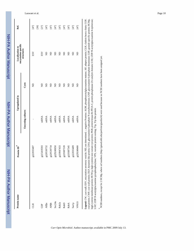
The ESV contents must attain their insoluble architecture after secretion [10]. Severalenzymatic activities have been implicated in post-translational processing in the ESV pathway(Table 1):
Lauwaet et al. Page 2
Curr Opin Microbiol. Author manuscript; available in PMC 2009 July 13.
NIH
-PA Author Manuscript
NIH
-PA Author Manuscript
NIH
-PA Author Manuscript
a. The major known post-translational modification of the three CWP is formation ofextensive intermolecular disulfide bonds by protein disulfide isomerases (PDI)[12-14,19]. Giardia has five protein disulfide isomerases [21] and the three that arecharacterized localize to ESV matrix but not CW [22].
b. PDI 1-3 also have transglutaminase activity which forms isopeptide protein crosslinksthat are resistant to degradation [23,24]. Isopeptide bonds increase during encystationand transglutaminase inhibition decreases cyst formation. However, the cross-linkedproteins remain to be identified.
c. A lysosomal cysteine proteinase was implicated in cleavage of the C-terminalextension of CWP2, suggesting cross-talk between the lysosomal compartment andESV [25]. HCNCp is cleaved [15••] by a yet unknown protease.
d. CWP 1 and 2 are phosphorylated [26], but no kinase has been implicated.
e. The Giardia granule-specific protein (gGSP) has a calsequentrin domain, bindscalcium, is upregulated in encystation, and localizes to the ESV [27]. Knockdown ofgGSP inhibits ESV release, suggesting a calcium-dependent process [27].
Thus, a number of independent studies show that the ESV are central to CW biogenesis as anygenetic or chemical manipulation that interferes with the ESV pathway blocks all downstreamevents [19,23,27,28••].
Many cells and organisms have extracellular walls that permit them to survive in theenvironment. Sugar polymers are key components of these walls and are often composed ofrepeating hexose units. Although the monomers are closely related, the polymers have distinctphysical properties. Beta 1-3 polyhexoses do not associate as strongly as beta (1-4)-linkedpolysaccharides. Chitin, (beta 1-4 linked N-acetyl glucosamine) of arthropod and insectexoskeleton and fungal cell walls, is widespread in evolution [29,30]. Pioneering studies fromthe Jarroll group showed beta 1-3 polymer of galNAc as the major CWG [31]. Their insolublematerial was purified by extensive enzymatic and chemical extractions that might haveremoved other important CW components. They defined an enzymatic pathway for synthesisof UDP-galNAc from glucose by an encystation-specific cytosolic pathway (Table 1)[12-15••]. An activity in crude cyst wall particles, termed “cyst wall synthase” (CWS),specifically incorporates galNAc from UDP-galNAc into insoluble material. However, noCWS gene has been identified. Based on the complexity of chitin synthase systems [32],“CWS” activity may require more than one protein.
Despite its central importance and the accessibility of the giardial life cycle, many gaps remainin our knowledge of the CW composition, formation, and architecture.
Transcriptional regulation of encystationThe molecular control of encystation is not well understood. RNA expression of the three CWPand the CWG biosynthetic enzymes, is largely upregulated transcriptionally (Table 1). Inaddition, several other proteins, whose roles in encystation are yet to be discovered, areupregulated at the transcriptional level (Table 1).
To date, three giardial DNA-binding transcription factors have been described. Only GARPglp1 and Myb2 are upregulated in encystation [33-35]. Myb2 binds to target sequences in theproximal upstream regions of the CWP genes and of g6PI-B, the first enzyme in the galNAcbiosynthetic pathway, and of Myb2 itself [33]. Transcripts of most giardial genes initiate in A/T-rich initiator-like sequences near the start of translation [2]. This and several upstreamsequences have been implicated in transcription of encystation genes [33,36-39]. Adownstream region was reported to affect transcript stability [38,39].
Lauwaet et al. Page 3
Curr Opin Microbiol. Author manuscript; available in PMC 2009 July 13.
NIH
-PA Author Manuscript
NIH
-PA Author Manuscript
NIH
-PA Author Manuscript
Signal transduction in encystationTrophozoites in the small intestine constantly monitor and respond to their environment. Thelumenal composition varies with location and host nutrition. Trophozoites that are attached toenterocytes are beneath a mucus blanket and bathed in a serum-like microfiltrate, near neutralpH and at low bile concentration. As enterocytes mature, they are sloughed off and trophozoitesmust swim upstream to re-attach. If they remain in the lumen, trophozoites are exposed to theslightly alkaline pH and increased bile that lead to encystation.
During encystation, morphological modifications are coordinated with cell cycle exit anddecreased metabolism. However, the proteins and pathways involved in transducing thephysiological signals into effective responses are only beginning to be understood. Certainintracellular signaling proteins have been implicated in encystation based on their increasedmRNA or protein expression and/or their localization to ESV and CW (Table 1). ERK1/2,PKAc, PKAr, PP2A-C and a PKCβ were reported to play a role in Giardia encystation [28••,40••-43]. PKA and ERK1/2 activities and ERK1/2 phosphorylation increase during encystation[28••,40••-43]. Importantly, inhibition of PP2A-C and of PKCβ decreases encystation [28••,42••].
These signaling proteins are all universal regulators of growth and differentiation in otherorganisms. Their specific functions in Giardia, however, are dependent on their cellulargeography. All of these signaling proteins (except ERK2 and PKCβ) localize constitutively tothe Giardia basal bodies/centrosomes. They also localize to cytoskeletal structures unique toGiardia, such as characteristic paraflagellar rods and the attachment disk. Their diversetargeting suggests that each signaling protein has a distinct role in encystation. The localizationof PKAc/r, PP2A-C, PKCβ and ERK1/2 changes in response to the physiologic stimuli thatinduce encystation [28••,40••-42••,44]. Much additional research is needed to elucidate thecomplex cell signaling pathways that regulate encystation. Individual signaling proteins areregulated, often in cascades, by addition and removal of phosphates. Giardia has fewtransmembrane kinases (H.G. Morrison et al., in press) and the surface receptors for detectingand transmitting the extracellular encystation signals have not been defined.
Conclusions and perspectivesWe have summarized progress in understanding giardial encystation from molecular and cellbiological points of view. What emerges is the need for additional research to unmask thecomplexities of this important differentiation. In addition to being a model for other parasites,Giardia may provide useful hypotheses and paradigms for the entry into and exit fromdormancy of a wide variety of cell types.
AcknowledgmentsResearch in our laboratory is supported by grants RO1 AI 42488, RO1 AI 51687, and RO1 GM 61896 from theNational Institutes of Health awarded to Frances Gillin.
References1••. Huang DB, White AC. An updated review on Cryptosporidium and Giardia. Gastroenterol Clin North
Am 2006;35:291–314.314 viii. [PubMed: 16880067]. This recent review summarizes thepathogenesis, clinical symptoms and epidemiology of giardiasis compared with cryptosporidiosis.
2. Adam RD. Biology of Giardia lamblia. Clin Microbiol Rev 2001;14:447–475. [PubMed: 11432808]3. Lee P, Faubert GM. Oral immunization of BALB/c mice by intragastric delivery of Streptococcus
gordonii-expressing Giardia cyst wall protein 2 decreases cyst shedding in challenged mice. FEMSMicrobiol Lett 2006;265:225–236. [PubMed: 17081198]
Lauwaet et al. Page 4
Curr Opin Microbiol. Author manuscript; available in PMC 2009 July 13.
NIH
-PA Author Manuscript
NIH
-PA Author Manuscript
NIH
-PA Author Manuscript
4. Lee P, Faubert GM. Expression of the Giardia lamblia cyst wall protein 2 in Lactococcus lactis.Microbiology 2006;152:1981–1990. [PubMed: 16804173]
5. Larocque R, Nakagaki K, Lee P, Abdul-Wahid A, Faubert GM. Oral immunization of BALB/c micewith Giardia duodenalis recombinant cyst wall protein inhibits shedding of cysts. Infect Immun2003;71:5662–5669. [PubMed: 14500486]
6••. Palm D, Weiland M, McArthur AG, Winiecka-Krusnell J, Cipriano MJ, Birkeland SR, Pacocha SE,Davids B, Gillin F, Linder E, et al. Developmental changes in the adhesive disk during Giardiadifferentiation. Mol Biochem Parasitol 2005;141:199–207.207 [PubMed: 15850703]. In this paper,the authors describe the structure and protein composition of the attachment disk in the Giardia lifecycle. In addition, they characterize the new disk protein SALP-1. This is one of the few papersshowing immunofluorescence images of excysting cells.
7. Svard SG, Hagblom P, Palm JE. Giardia lamblia—a model organism for eukaryotic cell differentiation.FEMS Microbiol Lett 2003;218:3–7. [PubMed: 12583890]
8. Marti M, Hehl AB. Encystation-specific vesicles in Giardia: a primordial Golgi or just another secretorycompartment? Trends Parasitol 2003;19:440–446. [PubMed: 14519581]
9. Hehl AB, Marti M. Secretory protein trafficking in Giardia intestinalis. Mol Microbiol 2004;53:19–28. [PubMed: 15225300]
10. Lujan HD, Touz MC. Protein trafficking in Giardia lamblia. Cell Microbiol 2003;5:427–434.[PubMed: 12814433]
11•. Chavez-Munguia B, Omana-Molina M, Gonzalez-Lazaro M, Gonzalez-Robles A, Cedillo-RiveraR, Bonilla P, Martinez-Palomo A. Ultrastructure of cyst differentiation in parasitic protozoa.Parasitol Res 2007;100:1169–1175.1175 [PubMed: 17252271]. This review illustrates andcompares the ultrastructure of E. invadens, A. castellanii and G. lamblia ESV and cyst walls.
12. Mowatt MR, Lujan HD, Cotten DB, Bowers B, Yee J, Nash TE, Stibbs HH. Developmentallyregulated expression of a Giardia lamblia cyst wall protein gene. Mol Microbiol 1995;15:955–963.[PubMed: 7596296]
13. Lujan HD, Mowatt MR, Conrad JT, Bowers B, Nash TE. Identification of a novel Giardia lambliacyst wall protein with leucine-rich repeats. Implications for secretory granule formation and proteinassembly into the cyst wall. J Biol Chem 1995;270:29307–29313. [PubMed: 7493963]
14. Sun CH, McCaffery JM, Reiner DS, Gillin FD. Mining the Giardia lamblia genome for new cyst wallproteins. J Biol Chem 2003;278:21701–21708. [PubMed: 12686559]
15••. Davids BJ, Reiner DS, Birkeland SR, Preheim SP, Cipriano MJ, McArthur AG, Gillin FD. A NewFamily of Giardial Cysteine-Rich Non-VSP Protein Genes and a Novel Cyst Protein. PLoS ONE2006;1:e44. [PubMed: 17183673]. This paper describes a protein, HCNCp, with sequence similarityto Giardia variant surface proteins, which surprisingly does not localize to the Giardia trophozoitesurface membrane, but traffics via the ESV to the cyst wall It belongs to a group of 61 unusualcysteine-rich proteins.
16. Reiner DS, McCaffery M, Gillin FD. Sorting of cyst wall proteins to a regulated secretory pathwayduring differentiation of the primitive eukaryote, Giardia lamblia. Eur J Cell Biol 1990;53:142–153.[PubMed: 2076701]
17. Gottig N, Elias EV, Quiroga R, Nores MJ, Solari AJ, Touz MC, Lujan HD. Active and passivemechanisms drive secretory granule biogenesis during differentiation of the intestinal parasiteGiardia lamblia. J Biol Chem 2006;281:18156–18166. [PubMed: 16611634]
18. Thiele C, Gerdes HH, Huttner WB. Protein secretion: puzzling receptors. Curr Biol 1997;7:R496–500. [PubMed: 9259537]
19. Reiner DS, McCaffery JM, Gillin FD. Reversible interruption of Giardia lamblia cyst wall proteintransport in a novel regulated secretory pathway. Cell Microbiol 2001;3:459–472. [PubMed:11437832]
20••. Stefanic S, Palm D, Svard SG, Hehl AB. Organelle proteomics reveals cargo maturation mechanismsassociated with Golgi-like encystation vesicles in the early-diverged protozoan Giardia lamblia. JBiol Chem 2006;281:7595–7604.7604 [PubMed: 16407213]. In this paper, the authors report thefirst limited proteomic analysis of isolated ESV The ESV were isolated from encysting trophozoitesby sucrose density fractionation and proteins in the ESV containing fraction were resolved by 2D-PAGE and identified by mass spectrometry.
Lauwaet et al. Page 5
Curr Opin Microbiol. Author manuscript; available in PMC 2009 July 13.
NIH
-PA Author Manuscript
NIH
-PA Author Manuscript
NIH
-PA Author Manuscript
21. McArthur AG, Knodler LA, Silberman JD, Davids BJ, Gillin FD, Sogin ML. The evolutionary originsof eukaryotic protein disulfide isomerase domains: new evidence from the Amitochondriate protistGiardia lamblia. Mol Biol Evol 2001;18:1455–1463. [PubMed: 11470836]
22. Knodler LA, Noiva R, Mehta K, McCaffery JM, Aley SB, Svard SG, Nystul TG, Reiner DS, SilbermanJD, Gillin FD. Novel protein-disulfide isomerases from the early-diverging protist Giardia lamblia.J Biol Chem 1999;274:29805–29811. [PubMed: 10514458]
23. Davids BJ, Mehta K, Fesus L, McCaffery JM, Gillin FD. Dependence of Giardia lamblia encystationon novel transglutaminase activity. Mol Biochem Parasitol 2004;136:173–180. [PubMed: 15478797]
24. Knodler LA, Noiva R, Mehta K, McCaffery JM, Aley SB, Svard SG, Nystul TG, Reiner DS, SilbermanJD, Gillin FD. Novel protein-disulfide isomerases from the early-diverging protist giardia lamblia.J Biol Chem 2000;275:28339. [PubMed: 10970904]
25. Touz MC, Nores MJ, Slavin I, Carmona C, Conrad JT, Mowatt MR, Nash TE, Coronel CE, LujanHD. The activity of a developmentally regulated cysteine proteinase is required for cyst wallformation in the primitive eukaryote Giardia lamblia. J Biol Chem 2002;277:8474–8481. [PubMed:11773053]
26. Slavin I, Saura A, Carranza PG, Touz MC, Nores MJ, Lujan HD. Dephosphorylation of cyst wallproteins by a secreted lysosomal acid phosphatase is essential for excystation of Giardia lamblia.Mol Biochem Parasitol 2002;122:95–98. [PubMed: 12076774]
27. Touz MC, Gottig N, Nash TE, Lujan HD. Identification and characterization of a novel secretorygranule calcium-binding protein from the early branching eukaryote Giardia lamblia. J Biol Chem2002;277:50557–50563. [PubMed: 12354777]
28••. Lauwaet T, Davids BJ, Torres-Escobar A, Birkeland SR, Cipriano MJ, Preheim SP, Palm D, SvardSG, McArthur AG, Gillin FD. Protein phosphatase 2A plays a crucial role in Giardia lambliadifferentiation. Mol Biochem Parasitol 2007;152:80–89.89 [PubMed: 17204341]. Using antisensemRNA and immunofluorescence analysis, authors demonstrate a role for PP2A-C in Giardiaencystation and excystation and show its localization throughout the life cycle.
29. Merzendorfer H. Insect chitin synthases: a review. J Comp Physiol [B] 2006;176:1–15.30. Lesage G, Bussey H. Cell wall assembly in Saccharomyces cerevisiae. Microbiol Mol Biol Rev
2006;70:317–343. [PubMed: 16760306]31. Gerwig GJ, van Kuik JA, Leeflang BR, Kamerling JP, Vliegenthart JF, Karr CD, Jarroll EL. The
Giardia intestinalis filamentous cyst wall contains a novel beta(1-3)-N-acetyl-D-galactosaminepolymer: a structural and conformational study. Glycobiology 2002;12:499–505. [PubMed:12145190]
32. Van Dellen KL, Bulik DA, Specht CA, Robbins PW, Samuelson JC. Heterologous expression of anEntamoeba histolytica chitin synthase in Saccharomyces cerevisiae. Eukaryot Cell 2006;5:203–206.[PubMed: 16400183]
33. Sun CH, Palm D, McArthur AG, Svard SG, Gillin FD. A novel Myb-related protein involved intranscriptional activation of encystation genes in Giardia lamblia. Mol Microbiol 2002;46:971–984.[PubMed: 12421304]
34. Yang H, Chung HJ, Yong T, Lee BH, Park S. Identification of an encystation-specific transcriptionfactor, Myb protein in Giardia lamblia. Mol Biochem Parasitol 2003;128:167–174. [PubMed:12742583]
35. Sun CH, Su LH, Gillin FD. Novel plant-GARP-like transcription factors in Giardia lamblia. MolBiochem Parasitol 2006;146:45–57. [PubMed: 16310259]
36. Davis-Hayman SR, Hayman JR, Nash TE. Encystation-specific regulation of the cyst wall protein 2gene in Giardia lamblia by multiple cis-acting elements. Int J Parasitol 2003;33:1005–1012.[PubMed: 13129521]
37. Knodler LA, Svard SG, Silberman JD, Davids BJ, Gillin FD. Developmental gene regulation inGiardia lamblia: first evidence for an encystation-specific promoter and differential 5’ mRNAprocessing. Mol Microbiol 1999;34:327–340. [PubMed: 10564476]
38. Hehl AB, Marti M, Kohler P. Stage-specific expression and targeting of cyst wall protein-greenfluorescent protein chimeras in Giardia. Mol Biol Cell 2000;11:1789–1800. [PubMed: 10793152]
Lauwaet et al. Page 6
Curr Opin Microbiol. Author manuscript; available in PMC 2009 July 13.
NIH
-PA Author Manuscript
NIH
-PA Author Manuscript
NIH
-PA Author Manuscript
39. Marti M, Li Y, Schraner EM, Wild P, Kohler P, Hehl AB. The secretory apparatus of an ancienteukaryote: protein sorting to separate export pathways occurs before formation of transient Golgi-like compartments. Mol Biol Cell 2003;14:1433–1447. [PubMed: 12686599]
40••. Gibson C, Schanen B, Chakrabarti D, Chakrabarti R. Functional characterisation of the regulatorysubunit of cyclic AMP-dependent protein kinase A homologue of Giardia lamblia: Differentialexpression of the regulatory and catalytic subunits during encystation. Int J Parasitol. 2006. Theauthors report an increased activity of PKAc and describe the expression and localization patternsof PKAc and PKAr in encystation.
41. Ellis, JGt; Davila, M.; Chakrabarti, R. Potential involvement of extracellular signal-regulated kinase1 and 2 in encystation of a primitive eukaryote, Giardia lamblia. Stage-specific activation andintracellular localization. J Biol Chem 2003;278:1936–1945. [PubMed: 12397063]
42••. Bazan-Tejeda ML, Arguello-Garcia R, Bermudez-Cruz RM, Robles-Flores M, Ortega-Pierres G.Protein kinase C isoforms from Giardia duodenalis: identification and functional characterizationof a beta-like molecule during encystment. Arch Microbiol 2007;187:55–66.66 [PubMed:17028847]. In this paper, the presence of PKC-like activity in Giardia is reported for the first time.Inhibition of this activity blocks encystation.
43. Kim KT, Mok MT, Edwards MR. Protein kinase B from Giardia intestinalis. Biochem Biophys ResCommun 2005;334:333–341. [PubMed: 16018966]
44. Abel ES, Davids BJ, Robles LD, Loflin CE, Gillin FD, Chakrabarti R. Possible roles of protein kinaseA in cell motility and excystation of the early diverging eukaryote Giardia lamblia. J Biol Chem2001;276:10320–10329. [PubMed: 11104758]
45. Lopez AB, Sener K, Jarroll EL, van Keulen H. Transcription regulation is demonstrated for five keyenzymes in Giardia intestinalis cyst wall polysaccharide biosynthesis. Mol Biochem Parasitol2003;128:51–57. [PubMed: 12706796]
46. Gallego E, Alvarado M, Wasserman M. Identification and expression of the protein ubiquitinationsystem in Giardia intestinalis. Parasitol Res 2007;101:1–7. [PubMed: 17252268]
47. Marti M, Regos A, Li Y, Schraner EM, Wild P, Muller N, Knopf LG, Hehl AB. An ancestral secretoryapparatus in the protozoan parasite Giardia intestinalis. J Biol Chem 2003;278:24837–24848.[PubMed: 12711599]
Lauwaet et al. Page 7
Curr Opin Microbiol. Author manuscript; available in PMC 2009 July 13.
NIH
-PA Author Manuscript
NIH
-PA Author Manuscript
NIH
-PA Author Manuscript
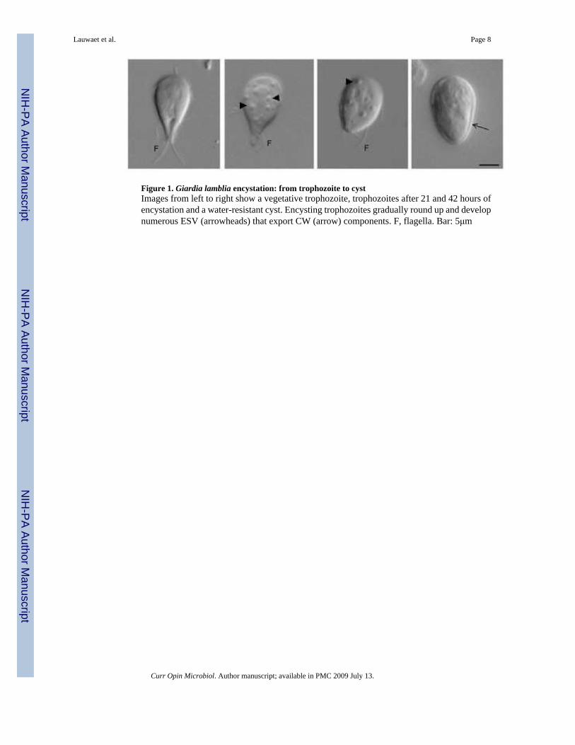
Figure 1. Giardia lamblia encystation: from trophozoite to cystImages from left to right show a vegetative trophozoite, trophozoites after 21 and 42 hours ofencystation and a water-resistant cyst. Encysting trophozoites gradually round up and developnumerous ESV (arrowheads) that export CW (arrow) components. F, flagella. Bar: 5μm
Lauwaet et al. Page 8
Curr Opin Microbiol. Author manuscript; available in PMC 2009 July 13.
NIH
-PA Author Manuscript
NIH
-PA Author Manuscript
NIH
-PA Author Manuscript

NIH
-PA Author Manuscript
NIH
-PA Author Manuscript
NIH
-PA Author Manuscript
Lauwaet et al. Page 9Ta
ble
1Pr
otei
ns a
nd/o
r mR
NA
upr
egul
ated
in e
ncys
tatio
n, a
nd/o
r pre
sent
in th
e ES
V o
r CW
Prot
ein
nam
ePr
otei
n ID
*U
preg
ulat
ed in
Loc
aliz
atio
n to
ency
stat
ion
spec
ific
stru
ctur
es
Ref
.
Enc
ystin
g cu
lture
sC
ysts
Sign
alin
g pr
otei
ns
PP2A
-Cgi
|292
4613
9-
mR
NA
/ pr
otei
nC
W[2
8••]
PKB
gi|6
2744
95m
RN
AN
DN
D[4
3]
Enz
ymes
GN
P (U
DP-
galN
AC
synt
hesi
s)gi
|609
0573
mR
NA
/ pr
otei
nN
DN
D[3
7]
GN
A
“gi
|282
6121
5m
RN
A /
prot
ein
ND
ND
[45]
AG
M
“gi
|282
6121
7m
RN
A /
prot
ein
ND
ND
[45]
UA
P
“gi
|283
9613
7m
RN
A /
prot
ein
ND
ND
[45]
UA
E
“gi
|283
9614
0m
RN
A /
prot
ein
ND
ND
[45]
Ubi
quiti
n ac
tivat
ing
enzy
me
E1gi
|292
5114
5m
RN
AN
DN
D[4
6]
“
gi|2
9246
853
mR
NA
ND
ND
[46]
Tra
nscr
iptio
n fa
ctor
s
Myb
2gi
|279
7955
8m
RN
AN
D-
[34]
GA
RP
glp1
gi|5
6407
639
mR
NA
ND
-[3
5]
Hig
h cy
stei
ne m
embr
ane
prot
eins
HC
Mp
Gro
up 1
orf:2
5816
mR
NA
ND
ND
[15•
•]
HC
Mp
EGF-
like
orf:1
1321
3m
RN
AN
DN
D[1
5••]
HC
NC
pgi
|756
7809
5m
RN
A /
prot
ein
prot
ein
ESV
/ C
W[1
5••]
Cys
t wal
l pro
tein
s
CW
P1gi
|606
009
mR
NA
/ pr
otei
nm
RN
A /
prot
ein
ESV
/ C
W[1
2]
CW
P2gi
|903
940
mR
NA
/ pr
otei
nm
RN
A /
prot
ein
ESV
/ C
W[1
3]
CW
P3gi
|190
6814
7m
RN
A /
prot
ein
mR
NA
/ pr
otei
nES
V /
CW
[14]
Secr
etor
y pr
otei
ns
Rab
11gi
|286
2853
9m
RN
AN
DES
V[4
7]
b’ C
OP
gi|1
9401
686
mR
NA
ND
ESV
[47]
Yip
gi|2
8974
725
ND
ND
ESV
[47]
DLP
gi|1
9401
683
ND
ND
ESV
[47]
Curr Opin Microbiol. Author manuscript; available in PMC 2009 July 13.
NIH
-PA Author Manuscript
NIH
-PA Author Manuscript
NIH
-PA Author Manuscript
Lauwaet et al. Page 10
Prot
ein
nam
ePr
otei
n ID
*U
preg
ulat
ed in
Loc
aliz
atio
n to
ency
stat
ion
spec
ific
stru
ctur
es
Ref
.
Enc
ystin
g cu
lture
sC
ysts
CLH
gi|2
2035
407
-N
DES
V[4
7]
[39]
GSP
gi|1
5419
593
mR
NA
ND
ESV
[27]
APβ
agi
|205
3073
2m
RN
AN
DN
D[4
7]
APβ
bgi
|205
3073
4m
RN
AN
DN
D[4
7]
Sec2
4agi
|292
4905
0m
RN
AN
DN
D[4
7]
Rab
2agi
|100
4743
3m
RN
AN
DN
D[4
7]
Rab
Dgi
|193
8753
9m
RN
AN
DN
D[4
7]
Rab
Agi
|193
8754
5m
RN
AN
DN
D[4
7]
Sar1
pgi
|220
3540
9m
RN
AN
DN
D[4
7]
VPS
33gi
|292
4840
0m
RN
AN
DN
D[4
7]
Leg
end:
CW
, cys
t wal
l; ES
V, e
ncys
tatio
n se
cret
ory
vesi
cle;
ND
, not
det
erm
ined
; -, n
egat
ive
Prot
eins
: AG
M, p
hosp
hoac
etyl
gluc
osam
ine
mut
ase;
AP,
ada
ptor
pro
tein
; CLH
, cla
thrin
hea
vy c
hain
; CO
P,co
at p
rote
in; C
WP,
cyst
wal
l pro
tein
; DLP
, dyn
amin
like
pro
tein
; GN
A, g
luco
sam
ine 6
-pho
spha
te N
-ace
tyltr
ansf
eras
e; G
NP,
glu
cosa
min
e 6-p
hosp
hate
dea
min
ase;
GSP
, gra
nule
spec
ific p
rote
in; H
CM
p,hi
gh cy
stei
ne m
embr
ane p
rote
in; H
CN
Cp,
hig
h cy
stei
ne n
on-v
aria
nt cy
st p
rote
in; P
KB
, pro
tein
kin
ase B
; PP2
A-C
, pro
tein
pho
spha
tase
2A
cata
lytic
dom
ain;
UA
E, U
DP-
N-a
cety
lglu
cosa
min
e 4-e
pim
eras
e;U
AP,
UD
P-N
-ace
tylg
luco
sam
ine
pyro
phos
phor
ylas
e; V
PS, v
acuo
lar p
rote
in so
rting
; Yip
, Yip
like
pro
tein
* NC
BI n
umbe
rs, e
xcep
t for
2 H
CM
p, w
here
orf
num
bers
(http
://gm
od.m
bl.e
du/p
erl/s
ite/g
iard
ia14
) wer
e us
ed b
ecau
se n
o N
CB
I num
bers
hav
e be
en a
ssig
ned
yet.
Curr Opin Microbiol. Author manuscript; available in PMC 2009 July 13.
Related Documents


![Giardia Lamblia [Modo de Compatibilidad]](https://static.cupdf.com/doc/110x72/577cc3cd1a28aba7119738fc/giardia-lamblia-modo-de-compatibilidad.jpg)