ENAMEL Dr. Muznah Sultan Lecturer DIDC- DUHS Dental Anatomy

Welcome message from author
This document is posted to help you gain knowledge. Please leave a comment to let me know what you think about it! Share it to your friends and learn new things together.
Transcript
ENAMEL
Dr. Muznah Sultan
Lecturer DIDC-DUHS
Dental Anatomy
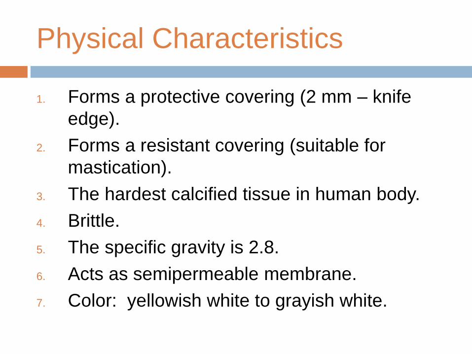
Physical Characteristics
1. Forms a protective covering (2 mm – knife
edge).
2. Forms a resistant covering (suitable for
mastication).
3. The hardest calcified tissue in human body.
4. Brittle.
5. The specific gravity is 2.8.
6. Acts as semipermeable membrane.
7. Color: yellowish white to grayish white.
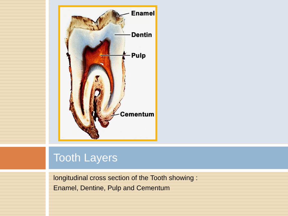
Tooth Layers
longitudinal cross section of the Tooth showing :
Enamel, Dentine, Pulp and Cementum
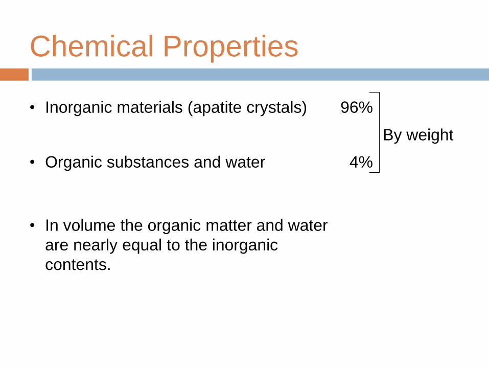
Chemical Properties
• Inorganic materials (apatite crystals) 96%
By weight
• Organic substances and water 4%
• In volume the organic matter and water
are nearly equal to the inorganic
contents.
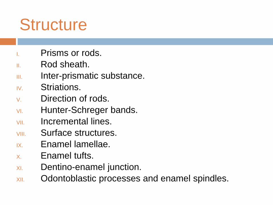
Structure
I. Prisms or rods.
II. Rod sheath.
III. Inter-prismatic substance.
IV. Striations.
V. Direction of rods.
VI. Hunter-Schreger bands.
VII. Incremental lines.
VIII. Surface structures.
IX. Enamel lamellae.
X. Enamel tufts.
XI. Dentino-enamel junction.
XII. Odontoblastic processes and enamel spindles.
Enamel Rods or Prisms
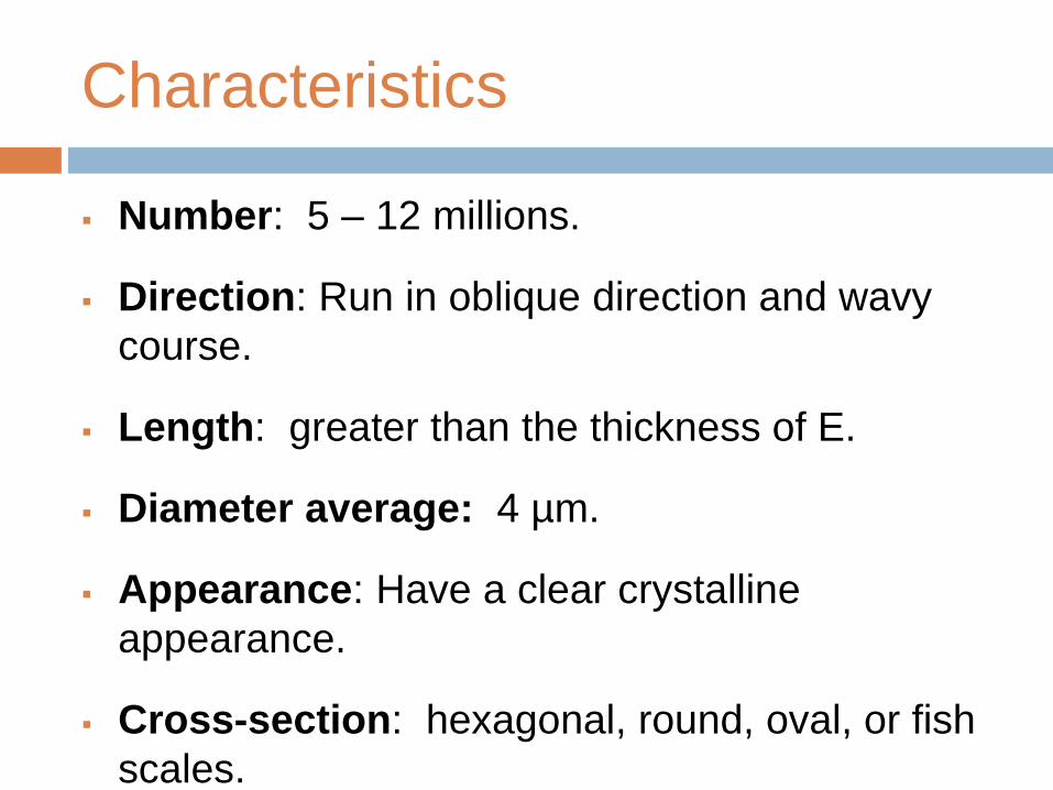
Characteristics
Number: 5 – 12 millions.
Direction: Run in oblique direction and wavy
course.
Length: greater than the thickness of E.
Diameter average: 4 µm.
Appearance: Have a clear crystalline
appearance.
Cross-section: hexagonal, round, oval, or fish
scales.
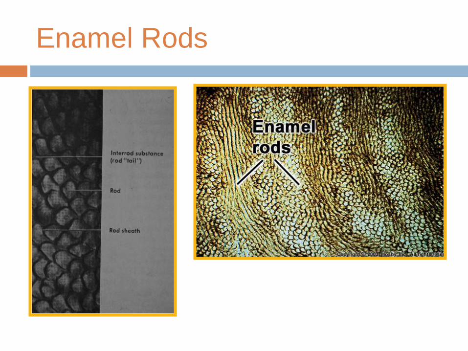
Enamel Rods
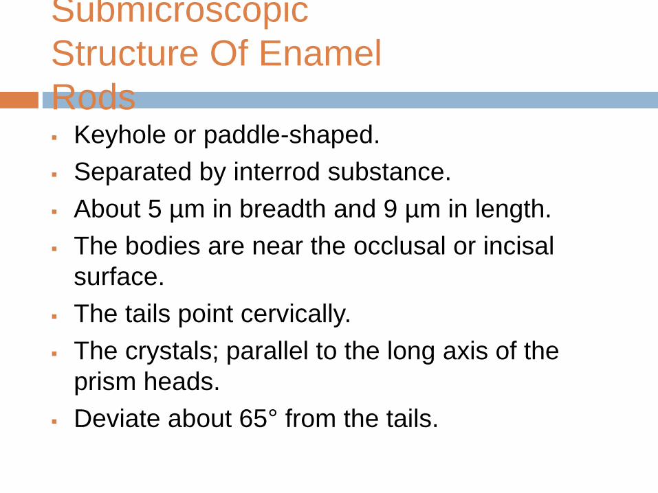
Submicroscopic
Structure Of Enamel
Rods Keyhole or paddle-shaped.
Separated by interrod substance.
About 5 µm in breadth and 9 µm in length.
The bodies are near the occlusal or incisal
surface.
The tails point cervically.
The crystals; parallel to the long axis of the
prism heads.
Deviate about 65° from the tails.
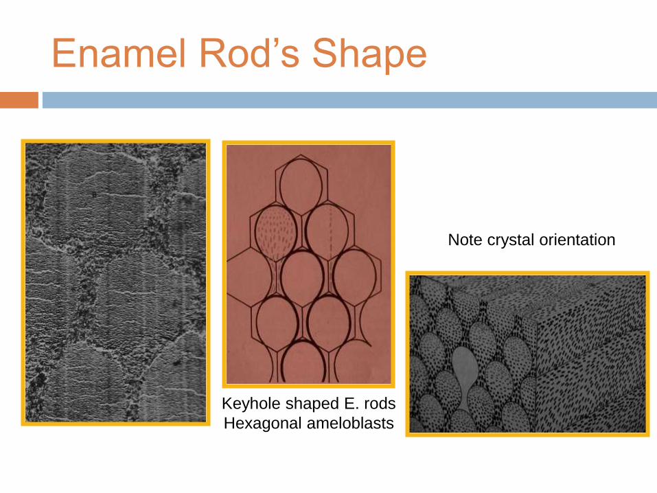
Keyhole shaped E. rods
Hexagonal ameloblasts
Note crystal orientation
Enamel Rod’s Shape
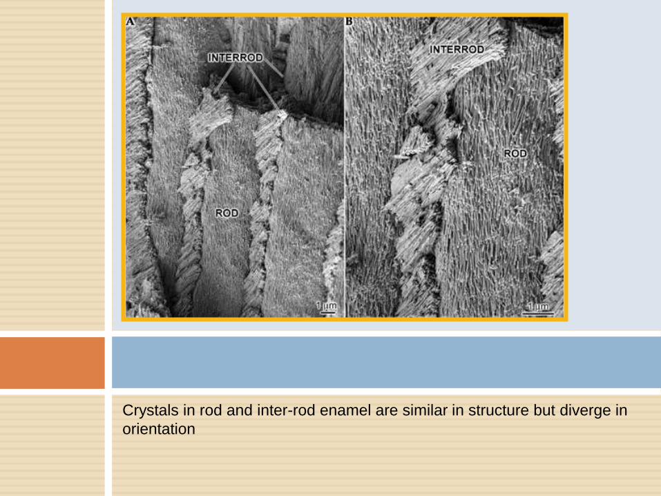
Crystals in rod and inter-rod enamel are similar in structure but diverge in
orientation
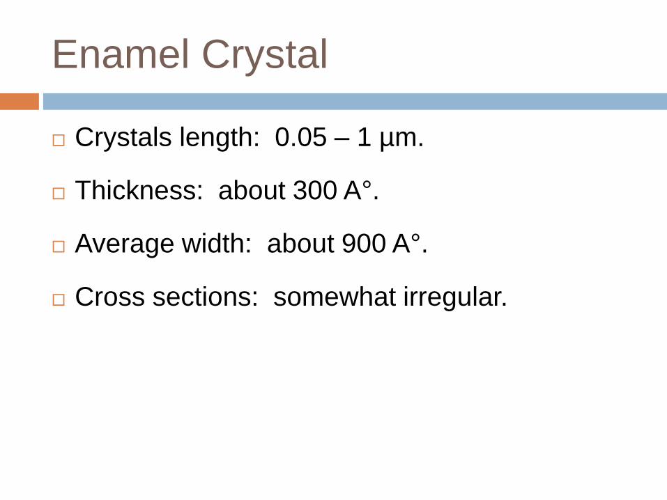
Enamel Crystal
Crystals length: 0.05 – 1 µm.
Thickness: about 300 A°.
Average width: about 900 A°.
Cross sections: somewhat irregular.
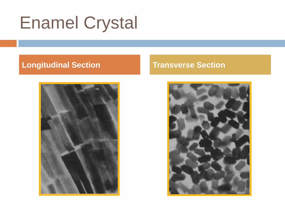
Enamel Crystal
Longitudinal Section Transverse Section
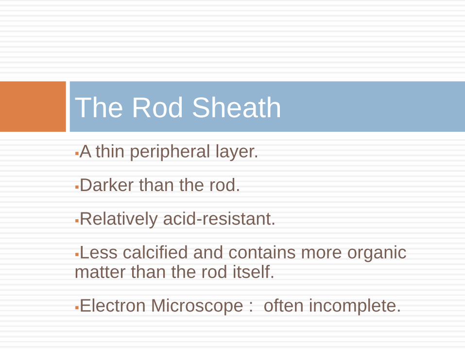
A thin peripheral layer.
Darker than the rod.
Relatively acid-resistant.
Less calcified and contains more organic matter than the rod itself.
Electron Microscope : often incomplete.
The Rod Sheath
•Cementing E. rods together.
•More calcified than the rod sheath.
•Less calcified than the rod itself.
•Appears to be minimum in human teeth.
Inter-prismatic Substance
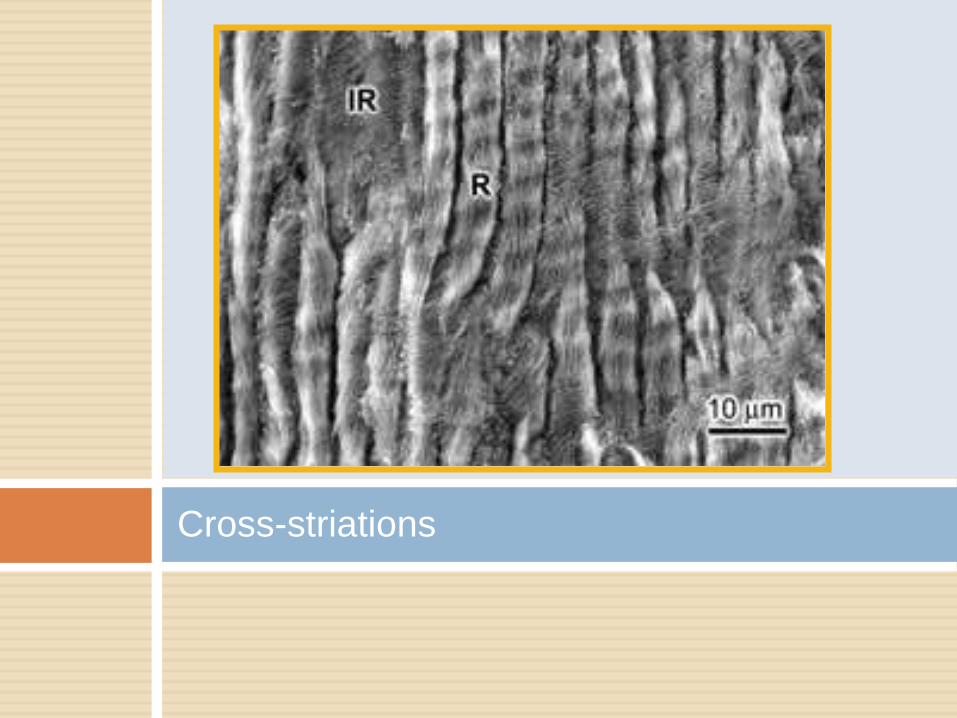
Cross-striations
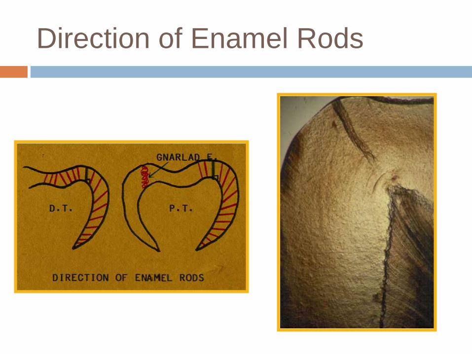
•Usually at right angles to the D. surface.
•Follow a wavy course in clockwise and
anticlockwise deviation.
•At the cusps or incisal edges: gnarled
enamel.
•At pits and fissures: rods converge in their
outward course.
Direction of Rods
Direction of Enamel Rods
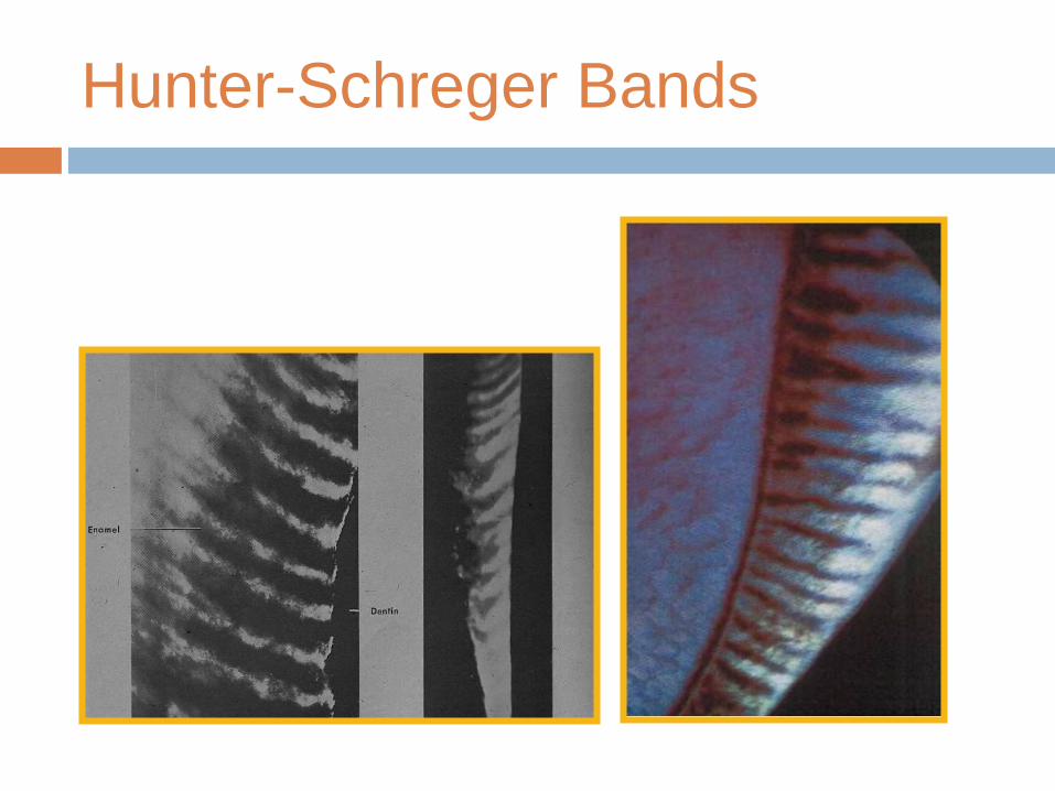
•Alternating dark and light strips.
•Have varying width.
•Seen in large ground section (oblique
reflected light).
•Originate from the DEJ.
Hunter-Schreger Bands
Hunter-Schreger Bands
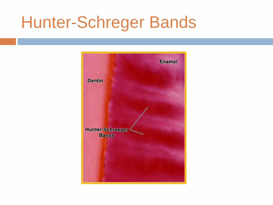
Hunter-Schreger Bands
Hunter-Schreger Bands
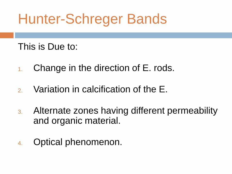
This is Due to:
1. Change in the direction of E. rods.
2. Variation in calcification of the E.
3. Alternate zones having different permeability and organic material.
4. Optical phenomenon.
A. Incremental Lines of Retzius
B. Neonatal Line
Incremental Lines
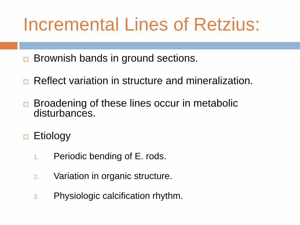
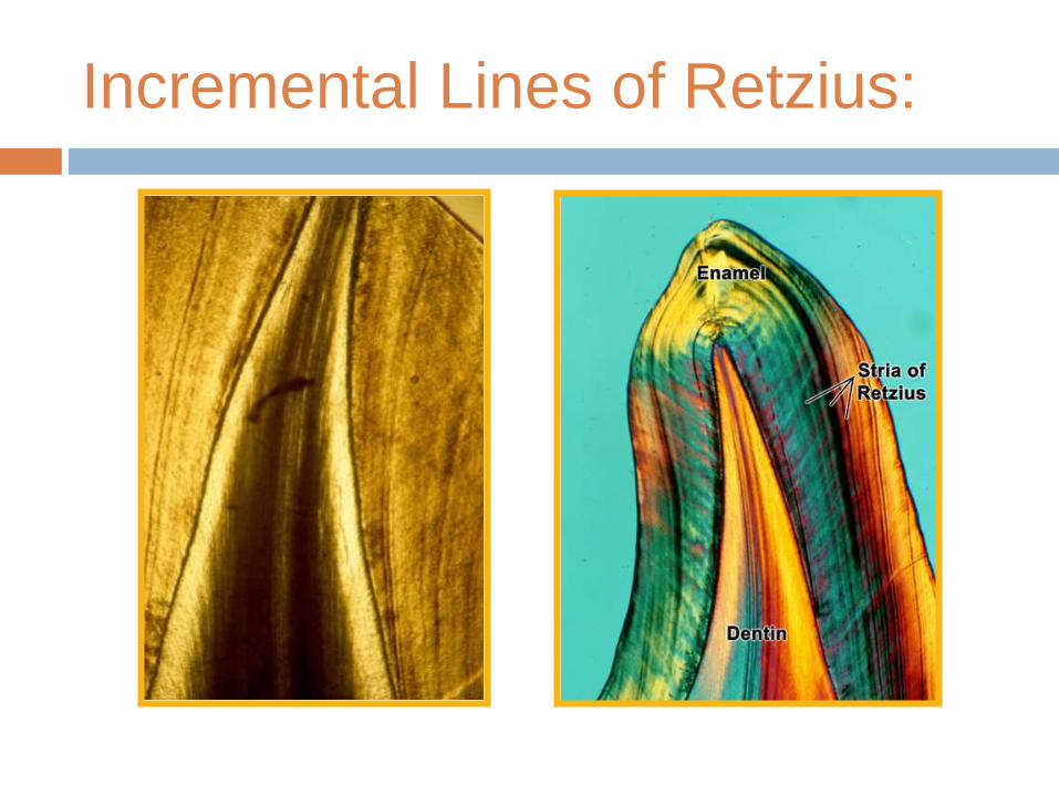
Incremental Lines of Retzius:
Brownish bands in ground sections.
Reflect variation in structure and mineralization.
Broadening of these lines occur in metabolic disturbances.
Etiology
1. Periodic bending of E. rods.
2. Variation in organic structure.
3. Physiologic calcification rhythm.
Incremental Lines of Retzius:
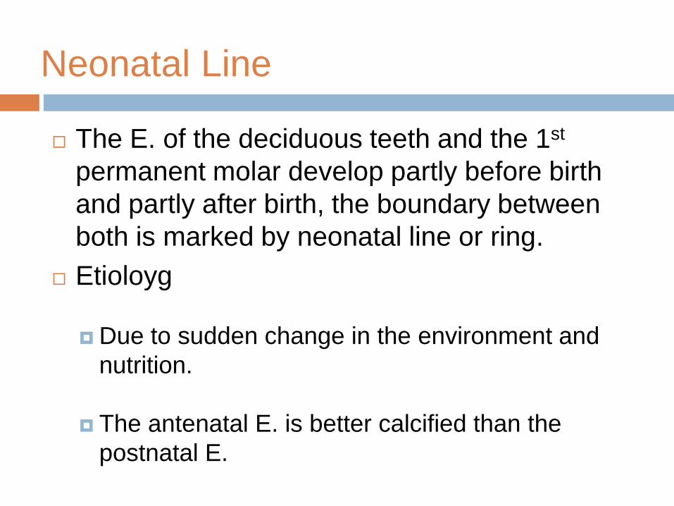
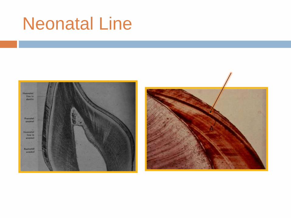
Neonatal Line
The E. of the deciduous teeth and the 1st
permanent molar develop partly before birth
and partly after birth, the boundary between
both is marked by neonatal line or ring.
Etioloyg
Due to sudden change in the environment and
nutrition.
The antenatal E. is better calcified than the
postnatal E.
Neonatal Line
SURFACE
STRUCTURES
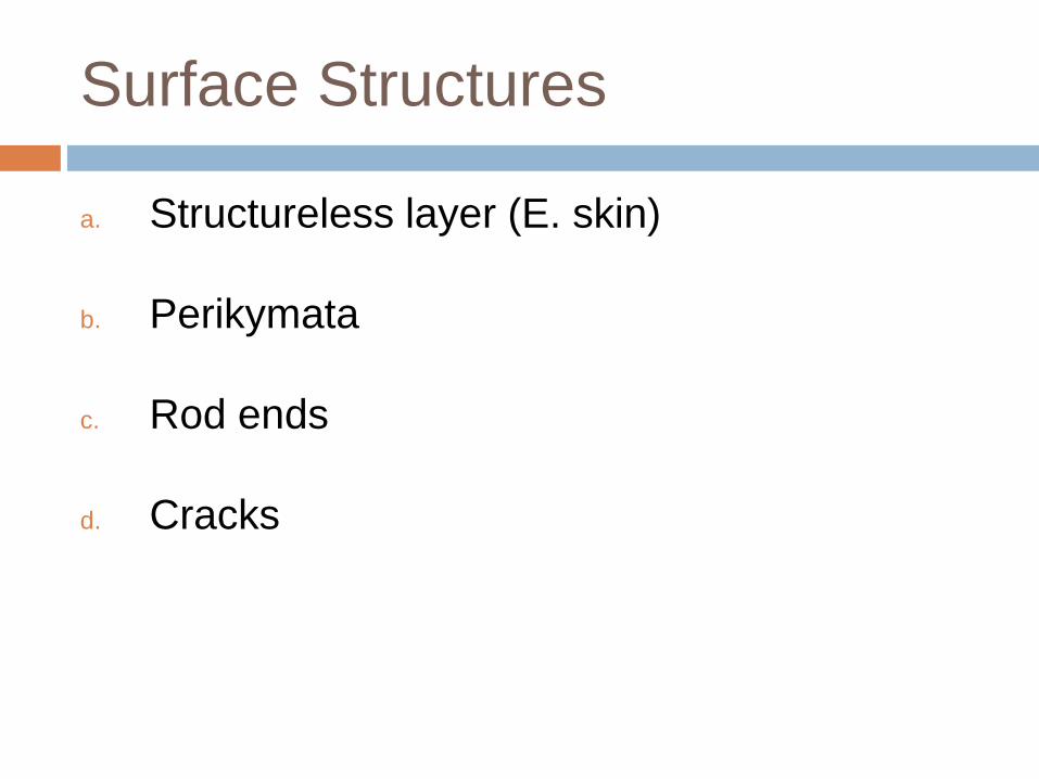
Surface Structures
a. Structureless layer (E. skin)
b. Perikymata
c. Rod ends
d. Cracks
a. Structureless layer
About 30 µm thick.
In 70% permanent teeth and all deciduous teeth.
Found least often over the cusp tips.
Found commonly in the cervical areas.
No E. prisms.
All the apatite crystals area parallel to one another and
perpendicular to the striae of Retzius.
More mineralized than the bulk of E. beneath it.
b. Perikymata
Transverse wave like grooves.
Thought to be the external manifestation of the striae of Retzius.
Lie parallel to each other and to CEJ.
Number:
About 30 perik./mm at the CEJ.
About 10 perik./mm near the incisal edge.
Their course is regular, but in the cervical region, it may be quite irregular.
Powdered graphite demonstrates them.
It is absent in the occlusal part of deciduous teeth but present in postnatal cervical part (due to undisturbed and even development of E. before birth)
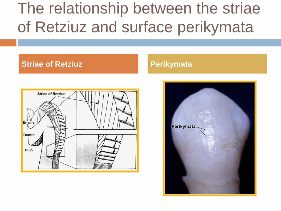
The relationship between the striae
of Retziuz and surface perikymata
Striae of Retziuz Perikymata
c. Rod ends
Are concave and vary in depth and shape.
Are shallow in the cervical regions.
Deep near the incisal or occlusal edges.
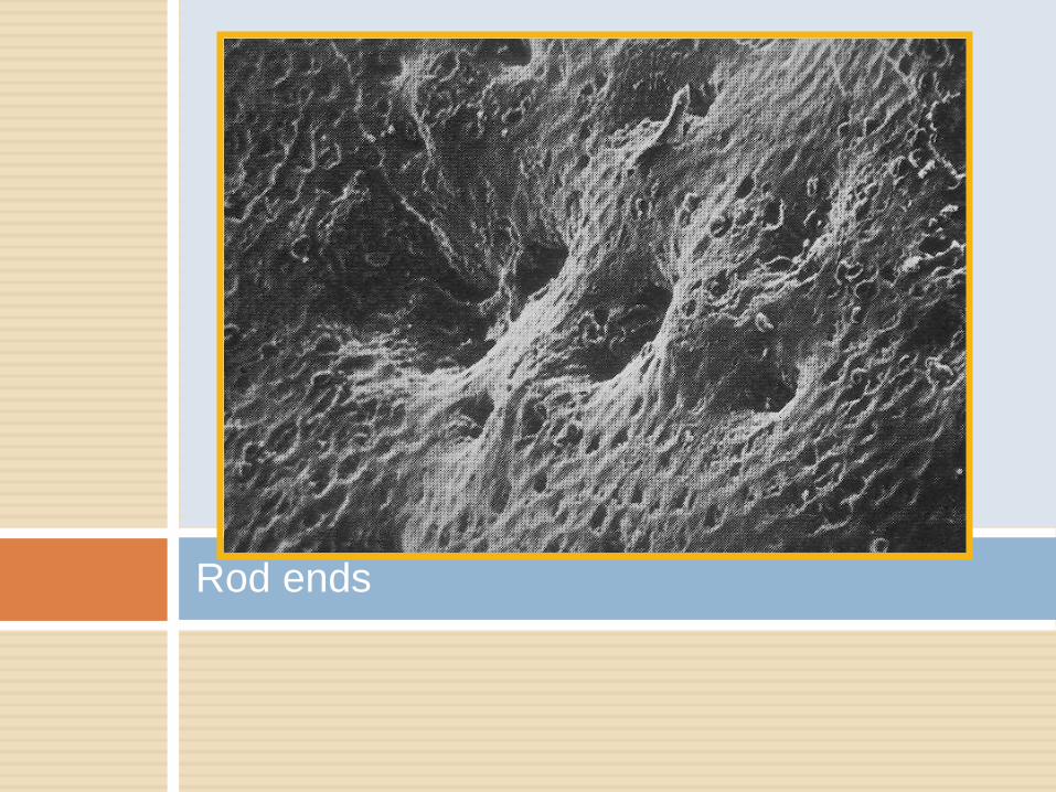
Rod ends
d. Cracks
Narrow fissure like structure.
Seen on almost all surfaces.
They are the outer edges of lamellae.
Extend for varying distance along the surface.
At right angles to CEJ.
Long cracks are thicker than the short one.
May reach the occlusal or incisal edge.

Cracks
ENAMEL LAMELLAE
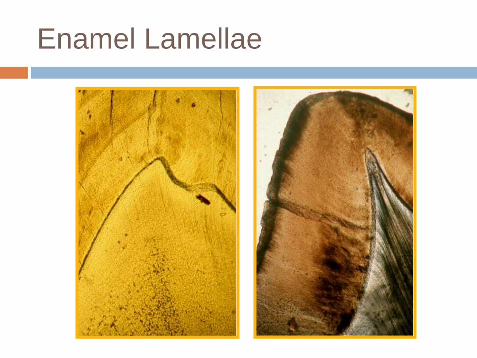
Enamel Lamellae
Are thin, leaf like structures,
Develop in planes of tension.
Extends from E. surface towards the DEJ.
Confused with cracks caused by grinding (decalcification).
Extend in longitudinal and radial direction.
Represent site of weakness in the tooth and three types; A, B, and C.

Enamel Lamellae
Enamel Lamellae
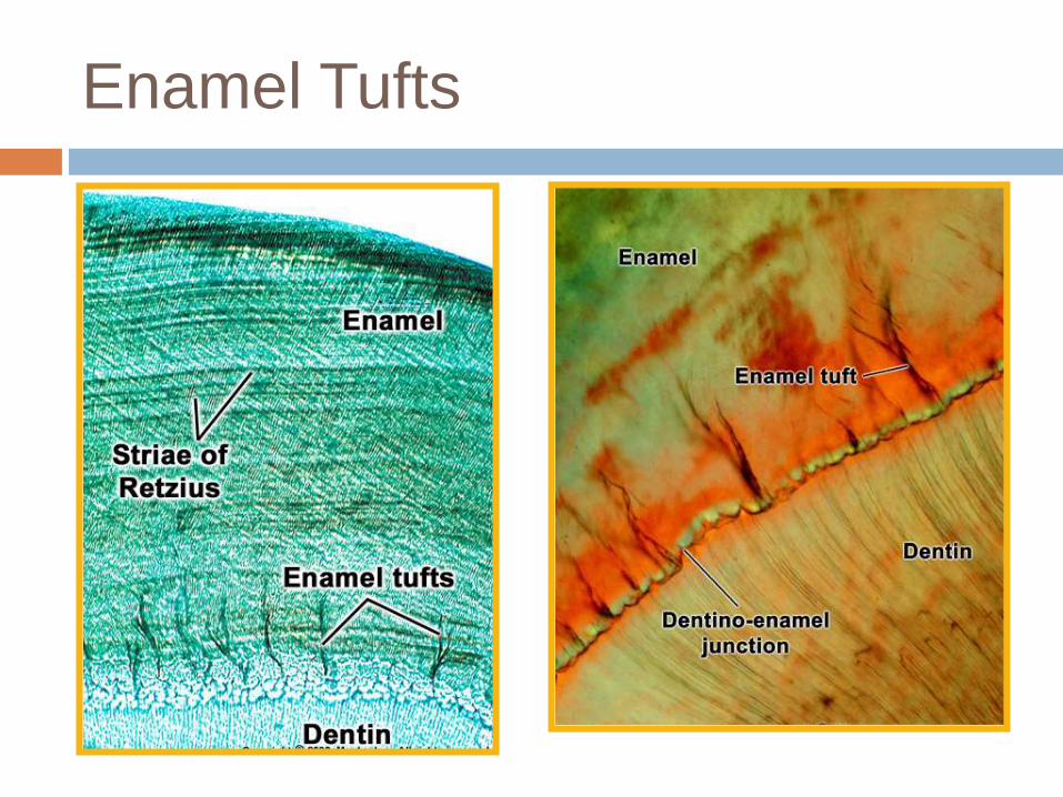
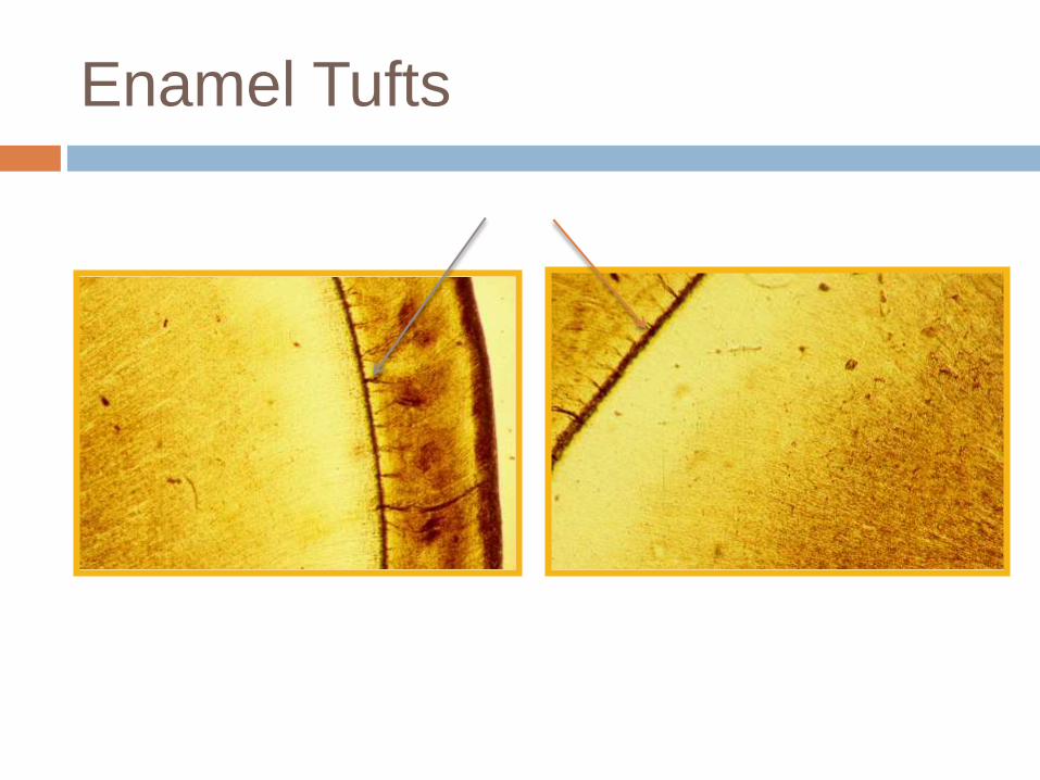
ENAMEL TUFTS
Enamel Tufts
Arise from DEJ.
Reach to 1/5 – 1/3 the thickness of E.
In ground section: resemble tufts of grass.
Do not spring from a single small area.
The inner end arises at the dentin.
Consist of hypocalcified E. rods and interprismatic substance.
The extend in the direction of the long axis of the crown (best seen in horizontal sections).
Enamel Tufts
Enamel Tufts
DENTINO-ENAMEL
JUNCTION
Dentino-Enamel Junction
Scalloped junction – the convexities towards
D.
At this junction, the pitted D. surface fit
rounded projections of the enamel.
The outline of the junction is performed by the
arrangement of the ameloblasts and the B. M.

Dentino-Enamel Junction
ODONTOBLASTIC
PROCESSES AND
ENAMEL SPINDLES
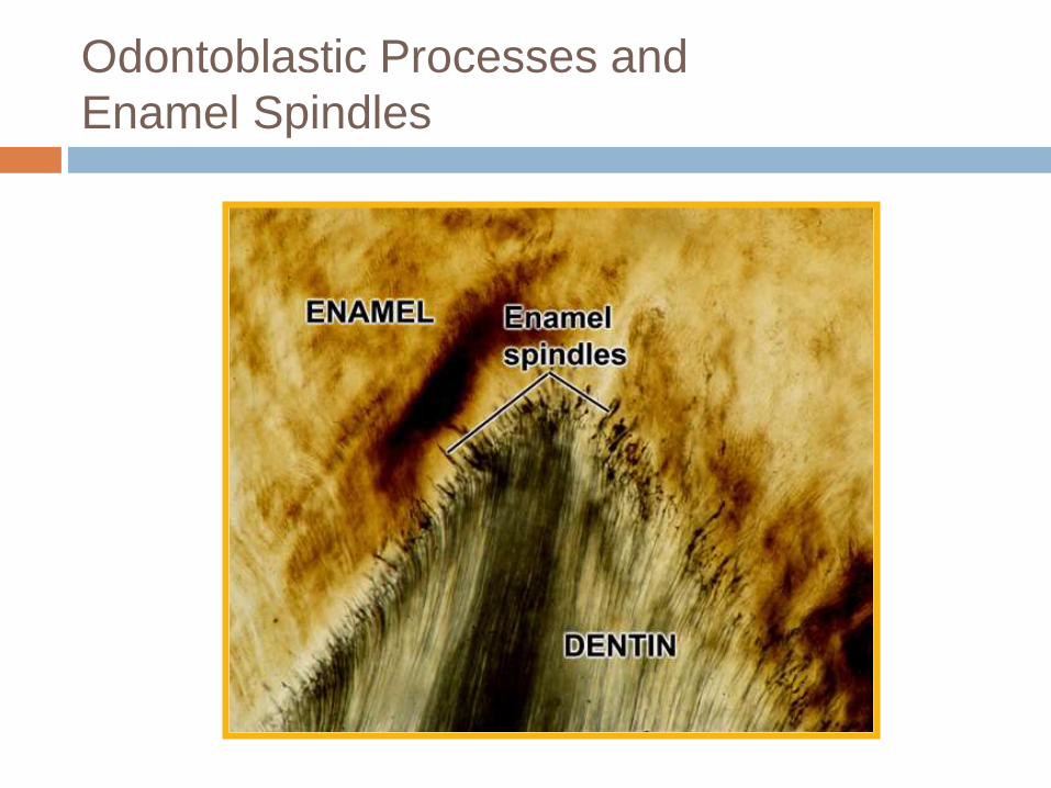
Odontoblastic Processes and
Enamel Spindles
The odontoblasts processes may cross DEJ (before the hard substance is formed) to the E. and ends as E. spindles.
They are filled with organic matter.
The processes and spindles are at right angle to the surface of the dentin.
The direction of spindles and rods is divergent.
Spindles appear dark in ground sections under transmitted light.
Odontoblastic Processes and
Enamel Spindles
Abnormalities
Interference during E. matrix formation may
cause Enamel hypoplasia.
Interference during Enamel maturation may
cause Enamel hypocalcification.
Each condition may be caused by systemic,
local, or hereditary factors.
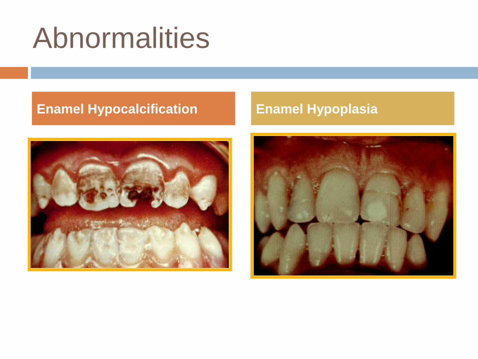
Abnormalities
Enamel Hypocalcification Enamel Hypoplasia

Thank you!!
Related Documents