Vedic Research International Biolological Medicinal Chemistry Journal home page at www.VedicJournals.com ISSN Review Nagiat T Hwisa 1 , Prakash Katakam 1* , Babu Rao Chandu 1 , Shanta Kumari Adiki 2 *Corresponding Author Prakash Katakam, PhD Faculty of Pharmacy, University of Zawia, Al-Zawia, Libya. Email: [email protected] Article Info 1 Faculty of Pharmacy, University of Zawia, Al-Zawia, Libya, 2 Nirmala College of Pharmacy, Mangalagiri, Guntur, AP, India. Received: June 13th, 2013 Accepted: June 20th, 2013 Keywords © 2013 by the Vedic Research International Journals, Vedic Research Inc. All rights reserved. Solvent evaporation, Microencapsulation, Microcapsules, Review, Technique. Abstract In recent times solvent evaporation techniques have gained prominence in microencapsulation process. Solvent evaporation techniques are broadly classified into emulsification solvent- evaporation and extraction methods. Several variations have been developed recently based on this technology. Using solvent evaporation methods we can regulate microsphere morphology and other characteristics to the desired level for the targeted delivery of bioactives like peptides and vaccines using various biomaterials as carriers. Several methods of solvent evaporation, core and coat materials used, emulsion stabilizers, and process variables were discussed in detail with due interest of recent advancements in this area of research. This technology is showing a promising future for drug targeting and throwing challenges to pharmaceutical scientist such as: scale-up problems, use of non-organic solvents, use of alternative biodegradable polymers, and the application of a viable hybrid technology by amalgamating various techniques of microencapsulation to overcome the problems of peptide degradation during the process and stability of microspheres after the process. Introduction Solvent Evaporation Techniques as Promising Advancement in Microencapsulation Proteins and peptides delivery in the form of controlled release creates new challenges to pharmaceutical scientist. Investigators and pharmacologists have been trying to develop delivery systems that allow the fate of a drug to be controlled and the optimal drug dosage to arrive at the site of action in the body by means of novel microparticulate dosage forms. During the past two decades, researchers have succeeded in part in controlling the drug-absorption process to sustain adequate and effective plasma drug levels over a prolonged period of time by designing controlled release microspheres intended for either oral or parenteral administration. Targeted or site-specific microparticulate delivery systems were also developed to alter the pharmacokinetic profiles of various therapeutic classes of drugs resulting in maintaining effective drug concentration for a prolonged period and result in decreased side effects associated with lower plasma concentrations in the peripheral blood without attempting to modify the normal buffet of the active molecules in the body after administration and absorption. Microspheres can be defined as solid, approximately spherical particles ranging in size from 1 to 1000 μm. They are made of polymeric, waxy, or other protective materials, that is, biodegradable synthetic polymers and modified natural products such as starches, gums, proteins, fats and waxes. The natural polymers include gelatin and albumin whereas the synthetic polymers include polylactic acid and polyglycolic acid [1-4]. The influence of hydrophilic protective colloids was studied by Lin et al. [5]. Microencapsulation is a process by which a drug substance is entrapped within discrete free-flowing polymeric particle microcapsule products [6-16]. Different types of coated particles can be obtained depending on coating process used. The particles can be embedded within a polymeric or proteinic matrix network in either a solid aggregated state or a molecular dispersion, resulting in the formulation of micromatrices. Alternatively, the particles can be coated by a solidified VRI Biol Med Chem, Volume 1, Issue 1, July 2013 Page No 8

Emulsion Solvent Evaporation Microencapsulation Review
Dec 06, 2015
k;n;pzNC
Welcome message from author
This document is posted to help you gain knowledge. Please leave a comment to let me know what you think about it! Share it to your friends and learn new things together.
Transcript
Vedic Research International
Biolological Medicinal ChemistryJournal home page at www.VedicJournals.com
ISSN
Review
Nagiat T Hwisa1, Prakash Katakam1*, Babu Rao Chandu1, Shanta Kumari Adiki2
*Corresponding Author Prakash Katakam, PhDFaculty of Pharmacy, University of Zawia, Al-Zawia, Libya.Email: [email protected]
Article Info
1Faculty of Pharmacy, University of Zawia, Al-Zawia, Libya, 2Nirmala College of Pharmacy, Mangalagiri, Guntur, AP, India.
Received: June 13th, 2013Accepted: June 20th, 2013
Keywords
© 2013 by the Vedic Research International Journals, Vedic Research Inc. All rights reserved.
Solvent evaporation, Microencapsulation, Microcapsules, Review, Technique.
AbstractIn recent times solvent evaporation techniques have gained prominence in microencapsulation process. Solvent evaporation techniques are broadly classified into emulsification solvent- evaporation and extraction methods. Several variations have been developed recently based on this technology. Using solvent evaporation methods we can regulate microsphere morphology and other characteristics to the desired level for the targeted delivery of bioactives like peptides and vaccines using various biomaterials as carriers. Several methods of solvent evaporation, core and coat materials used, emulsion stabilizers, and process variables were discussed in detail with due interest of recent advancements in this area of research. This technology is showing a promising future for drug targeting and throwing challenges to pharmaceutical scientist such as: scale-up problems, use of non-organic solvents, use of alternative biodegradable polymers, and the application of a viable hybrid technology by amalgamating various techniques of microencapsulation to overcome the problems of peptide degradation during the process and stability of microspheres after the process.
Introduction
Solvent Evaporation Techniques as Promising Advancement in Microencapsulation
Proteins and peptides delivery in the form of controlled release creates new challenges to pharmaceutical scientist. Investigators and pharmacologists have been trying to develop delivery systems that allow the fate of a drug to be controlled and the optimal drug dosage to arrive at the site of action in the body by means of novel microparticulate dosage forms. During the past two decades, researchers have succeeded in part in controlling the drug-absorption process to sustain adequate and effective plasma drug levels over a prolonged period of time by designing controlled release microspheres intended for either oral or parenteral administration. Targeted or site -specif ic microparticulate delivery systems were also developed to alter the pharmacokinetic profiles of various therapeutic classes of drugs resulting in maintaining effective drug concentration for a prolonged period and result in decreased side effects associated
with lower plasma concentrations in the peripheral blood without attempting to modify the normal buffet of the active molecules in the body after administration and absorption.
Microspheres can be defined as solid, approximately spherical particles ranging in size from 1 to 1000 μm. They are made of polymeric, waxy, or other protective materials, that is, biodegradable synthetic polymers and modified natural products such as starches, gums, proteins, fats and waxes. The natural polymers include gelatin and albumin whereas the synthetic polymers include polylactic acid and polyglycolic acid [1-4]. The influence of hydrophilic protective colloids was studied by Lin et al. [5].
Microencapsulation is a process by which a drug substance is entrapped within discrete free-flowing polymeric particle microcapsule products [6-16]. Different types of coated particles can be obtained depending on coating process used. The particles can be embedded within a polymeric or proteinic matrix network in either a solid aggregated state or a molecular dispersion, resulting in the formulation of micromatrices. Alternatively, the particles can be coated by a solidified
VRI Biol Med Chem, Volume 1, Issue 1, July 2013
Page No 8
Page No 9© 2013 by the Vedic Research International Journals, Vedic Research Inc. All rights reserved.
Hwisa et al, 2013
polymeric or proteinic envelope, leading to the formation of microcapsules.A large number of microencapsulation processes and modifications of processes have been reported in the literature and in various patents, offering a variety of opportunities for the pharmaceutical technologist to choose from [17]. Crainich Jr has stressed upon the need to develop better scale-up technologies for microencapsulation processes [18]. Solvent evaporation technique has been successful in recent years for the preparation of microspheres in various applications and showing a promising future for its advantages over other techniques. Using this technique, controlled delivery of peptide drugs and vaccines has become realistic today in the form of micro- and nanospheres [19-22]. Thus solvent-evaporation technique does pave a way for identifying a better share in microencapsulation technology. The solvent evaporation technique and characteristics of biodegradable polylactic acid and poly(lactic-co-glycolic) acid (poly-DL-lactic acid-co-glycolic acid) microspheres produced by this method is presented by O’Donnell and McGinity [23, 24]. In this article we review the method of solvent evaporation covering the techniques and variations involved, core and coat materials used, process variables, problems involved and prospective scope of the method extending the coverage to all polymers along with biodegradable polymers.
Solvent evaporation technique Way back in 1933 Hickey et al. prepared microcapsules using this technique. But the solvent evaporation technique was fully
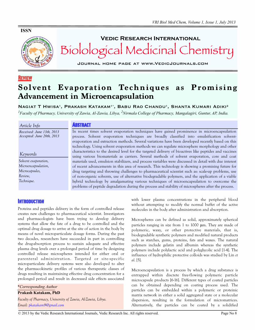
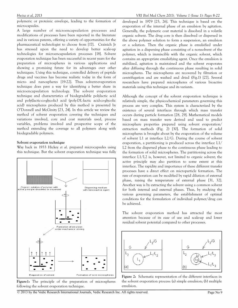
developed in 1979 [25, 26]. This technique is based on the evaporation of the internal phase of an emulsion by agitation. Generally, the polymeric coat material is dissolved in a volatile organic solvent. The drug core is then dissolved or dispersed in the above polymer solution to form a suspension, an emulsion or a solution. Then the organic phase is emulsified under agitation in a dispersing phase consisting of a nonsolvent of the polymer, which is immiscible with the organic solvent, which contains an appropriate emulsifying agent. Once the emulsion is stabilized, agitation is maintained and the solvent evaporates after diffusing through the continuous phase resulting in solid microspheres. The microspheres are recovered by filtration or centrifugation and are washed and dried (Fig.1) [27]. Several researchers have prepared microspheres of various bioactive materials using this technique and its variants.
Although the concept of the solvent evaporation technique is relatively simple, the physicochemical parameters governing this process are very complex. This system is characterized by the existence of several interfaces through which mass transfer occurs during particle formation [28, 29]. Mathematical models based on mass transfer were derived and used to predict microsphere properties prepared using solvent evaporation/extraction methods (Fig. 2) [30]. The formation of solid microspheres is brought about by the evaporation of the volume of solvent L1 at interface L2/G. During the course of solvent evaporation, a partitioning is produced across the interface L1/L2 from the dispersed phase to the continuous phase leading to the formation of solid microspheres. The partitioning across the interface L1/L2 is, however, not limited to organic solvent; the active principle may also partition to some extent at this interface. The rapidity and importance of these different transfer processes have a direct effect on microparticle formation. The rate of evaporation can be modified by rapid dilution of external phase, raising the temperature of external phase [31, 32]. Another way is by extracting the solvent using a common solvent for both internal and external phases. Thus, by studying the various governing parameters, the establishment of optimal conditions for the formulation of individual polymer/drug can be achieved.
The solvent evaporation method has attracted the most attention because of its ease of use and scale-up and lower residual solvent potential compared to other processes.
VRI Biol Med Chem 2013; Volume 1 (Issue 1): Pages 8--22
Figure 2: Schematic representation of the different interfaces in the solvent evaporation process: (a) simple emulsion; (b) multiple emulsion.
Figure1: The principle of the preparation of microspheres following the solvent evaporation technique.

Page No 10© 2013 by the Vedic Research International Journals, Vedic Research Inc. All rights reserved.
The systems of solvent evaporation method can be based on;(a) the nature of the external phase either aqueous or non
aqueous; (b) the incorporated mode of the core material in the organic
solution of the polymer either dissolved, dispersed or emulsified; and
(c) the elimination procedure of the organic solvent by either evaporation or extraction.
Classification of solvent evaporation techniqueThe classification proposed by Aftabrouchad and Doelker is applicable to microspheres prepared using solvent evaporation technique [33].
A. Solvent Evaporation (Emulsification-Evaporation):1. Oil-in-Water Emulsion (o/w)2. Multiple Emulsions: Water-in-Oil-in-Water (w/o/w)3. Nonaqueous Emulsions
B. Solvent Extraction (Emulsification-Extraction)
A: Solvent evaporation (emulsification-evaporation)A1: Oil-in-water emulsion (o/w): In this technique water acts as a non-solvent to the polymer. This method is also known as “in-water drying” developed for encapsulation of water-insoluble drugs and its use with polylactide polymers was reviewed in detail [34-37]. This process is very economical and eliminates recycling of an external phase [38-40]. In this system, the polymer is dissolved in an organic solvent such as methylene chloride or chloroform. The drug is dissolved or dispersed in the same medium and then entire mixture is emulsified in an aqueous solution containing an appropriate surfactant. Beck et al. were the first to propose this procedure for the encapsulation of progesterone in poly (D,L-lactic acid) (PLA) microparticles [26]. Nanospheres were also prepared using same technique [41]. The residual organic solvent is usually removed under reduced pressure. The emulsifier can be removed by dialysis of the suspension or by washings following the separation of free particles by filtration or ultra centrifugation. This technique is mostly useful for several lipophilic drugs. This procedure was applied to the encapsulation of a few water-insoluble peptides such as salmon calcitonin and cyclosporine [30, 42-44]. A rise in temperature for solvent evaporation causes formation of large core and thin polymer coat and the dilution technique produced porous microcapsules.
The physicochemical characteristics of the core materials like partition coefficient, degree of ionization will decide the entrapment efficiency of the microspheres formed. Water-soluble drugs cannot be encapsulated efficiently in the o/w system because they partition out into the external phase resulting in negligible entrapment in the microspheres. The drug should be made soluble in the polymer solution (oil phase). The solvation of core substance in organic solutions of polymers can be enhanced by the addition of hydrophilic cosolvents [45-47]. Conversion of hydrophilic drugs such as floxuridine to lipophilic drugs is another way to increase microencapsulation yields [48]. Loss of active ingredient from oily dispersed phase to aqueous continuous phase can also be reduced by saturating the continuous phase with the drug
substance which is less water soluble, adjusting the pH of same phase provided there is no degradation of polymers or even adding electrolytes [34, 49-52]. The pH sensitive polymers such as eudragits and cellulose acetate butyrate (CAB) were employed for preparation of microspheres for oral delivery [53-55]. Recently the mechanisms of burst release from pH-responsive polymeric microparticles using this technique was studied [56]. Mucoadhesive microspheres can be formulated using CAB and dextran derivatives [57]. Floating microspheres were also prepared using this technique [58, 59].
A2: Multiple emulsions (Water-in-oil-in-water or w/o/w): This procedure was first patented by Vrancken and Claeys in 1970 and further by DeJaeger and Tavermier in 1971 [60, 61]. Typically, an aqueous solution of the active principle was poured into an organic solution of the polymer to form an emulsion of the type water-in-oil (w/o). This primary emulsion is itself emulsified in an external aqueous phase leading to a multiple emulsion of the type water-in-oil-in-water (w/o/w). This procedure was further modified to enhance encapsulation of highly water-soluble peptides and anaesthetics [8, 62, 63]. Several authors have reviewed about biodegradable microspheres for peptide delivery using this technique [29, 64-66]. This process is very useful for the encapsulation of drugs in weak doses, which are strongly water soluble like hormones and trophic factors and antigens [13, 14, 67-75].
Various modes of mixing have been used for primary emulsification including high-speed homogenization, microfluidization, probe sonication, vortexing and static mixers. High-speed homogenization is by far the most popular method because of the different configurations of rotors, stators and their combination available, the ease of scale-up when compared to other procedures. As the homogenization speed increases in rotor-stator homogenization the droplet size decreases until an equilibrium droplet size is achieved whereas the homogenization time does not affect much on the equilibrium droplet size [76]. Drug loading and encapsulation efficiency decrease and drug release rate increases with high-pressure homogenization during the emulsification process [77]. The high-pressure emulsifier is
produced by Microfluidics [18]. This equipment can emulsify oil droplets down to particle size of 400 nm. This process can achieve the microsphere size suitable for injection through a normal 22-23 gauge needle. Biodegradable polymeric drug carriers to pass across blood-brain barrier have been prepared using high-pressure emulsification process [78]. The organic phase acts as a barrier between the two aqueous compartments preventing the diffusion of the drug substance toward the external phase [40].
The viscosity of the primary emulsion influences the entrapment of the drug, the particle size and morphology [79]. The role of viscosity of the primary emulsion (w/o) in the prevention of the diffusion of the active principle toward the external aqueous phase was studied by Ogawa et al. [8]. The viscosity can be increased by increasing the polymer concentration in the organic solvent, addition of drug retaining
Hwisa et al, 2013 VRI Biol Med Chem 2013; Volume 1 (Issue 1): Pages 8--22

Page No 11© 2013 by the Vedic Research International Journals, Vedic Research Inc. All rights reserved.
substance, lowering of temperature and adjusting the ratio of internal aqueous phase to the polymer-organic solution phase. Viscosity increasing agents like gelatin, pectin and agarose could prevent the diffusion of active principle but at the same time they were found to be incompatible with active principles.
The United States Food and Drug Administration (USFDA) has drawn certain standards about the following parameters of microspheres for parenteral depot administration [80]. These are: polymer/copolymer, organic solvents, copolymer-peptide complexes, sterilization, in vitro in vivo correlations, particle size and diluent-suspending vehicle. The first FDA approved system for controlled delivery of a peptide was an injectable poly(lactide-coglycolide) microsphere formulation of leuprolide acetate. Commercially it is available with brand name Lupron Depot. In France, it was marketed under the name of Enantone LP and is prescribed in the treatment of prostate cancer along with Décapeptyl L.P. Another drug, goserelin acetate is being sold as Zoldex by I.C.I.
A3: Influence of process variablesThe sphericity, size and yields of the microspheres are influenced by several process variables like nature of solvent, polymer concentration, emulsifier type and concentration, phase volume ratio, temperature and drug to polymer ratio [81, 82]. Influence of process parameters was also investigated and optimized [83-85].
The selection of solvent and external continuous phase determines the microsphere formation and entrapment efficiencies. The solvent properties should include:
• good solvency for the polymer for better drug entrapment,
• poor solvency for the drug to minimize partitioning of the drug into external phase,
• low boiling point for easy evaporation, • immiscibility with continuous phase yet should have a
finite solubility with it for solvent evaporation to occur,• does not cause the degradation of the dug substance, and• acceptable for human use.
Methylene chloride is most widely used solvent in microencapsulation technique. It suffers from the drawback of being carcinogenic and has a low solubility in water [65]. Other solvents with a lower toxicity than methylene chloride, such as ethyl acetate have also been used in microencapsulation. Ethyl acetate suffers from the disadvantage of being a poor solubilizer for higher molecular weight polymers and those with a monomer ratio of 50:50 mole % (lactide:glycolide) [86].
The external phase should be high boiling, non-toxic, immiscible with the organic solvent used and less expensive. Water is the only medium that fulfills all these requirements. Other vehicles like vegetable oils, mineral oils, organic solvents, etc., have also been used to enhance entrapment efficiencies but they suffer from the disadvantage of residual levels in the finished product [87]. Ethylcellulose microspheres of aspirin were prepared avoiding surfactant and using non-toxic solvents
[88].
A good emulsifier is required for the stabilization of an external phase to prevent the formation of agglomerates of microspheres during an early evaporation stage. As the evaporation proceeds, the emulsifier film helps to maintain the spherical shape of droplets till such time as the droplets are hardened enough to be harvested. The surfactant such as albumin or polyoxyether-polyoxypropylene copolymers is employed for the stabilization of primary emulsion. But one problem persists as the primary surfactant could leach out into the external aqueous phase during secondary emulsification along with the diffusion of the internal water and organic solvent during solvent evaporation. This would probably generate a microsphere matrix that is more porous and would result in a large burst effect and a drug release via a diffusion process rather than through the process of degradation of the polymer. Lipophilic surfactants do not much affect on the physical characteristics of microcapsules but particle charge depends on the concentration and type of the surfactant used [89]. By selecting the appropriate HLB value of emulsifier can control the encapsulation efficiency, size and morphology of microspheres [90]. The emulsifiers that have been used include polyvinyl alcohol (PVA), poly (vinylpyrrolidone) (PVP), polysorbate 80, gelatin, alginate, methylcellulose, polysorbates, hydroxypropyl methylcellulose, sodium lauryl sulfate, etc. [87, 91]. Poloxamer 407 and poloxamine 908 were also used as surfactants to prepare poly(DL-lactide-coglycolide (polyglactin 370) nanospheres [92]. Carbopol and poloxamer were employed and found to be alternatives to polyvinyl alcohol as emulsion stabilizers [77, 93].
Polyethylene glycol (PEG)-dextran conjugates were used as combined stabilizer and surface modifier to produce PLG microparticles that have a mean particle size of 480 nm [94]. The major problem associated with the use of polymeric microspheres, their natural surface characteristics favor protein binding and thereby phagocytosed due to opsonization. PEG conjugates of hydrophobic drugs allow surface modification of biomaterials that does not favor microspheres for protein binding and hence they are not phagocytosed. This technology has been commercialized by Nottingham Technology Ventures. The effect of different surfactants on entrapment and drug release was studied using a peptide pBC-264 from polyglactin 370 microspheres resulting in reduced drug loading into microspheres and increased burst release [95]. Smoothing agents like aluminium tristearate and polyisobutylene can be employed to yield very smooth microspheres [96, 97]. Mechanism of elimination of solvent influences the microparticle morphology [98].
The use of drug retaining substances such as gelatin, albumin, pectin, etc., in the inner aqueous phase aids in achieving higher drug loading through an increased viscosity of inner aqueous phase. A reduction in the temperature of the primary emulsion aids in enhancing the entrapment ratio of the drug. But, as the overall viscosity of the primary emulsion increases the particle size also increases where above a certain viscosity the particles are actually deformed upon secondary emulsification [62].
Hwisa et al, 2013 VRI Biol Med Chem 2013; Volume 1 (Issue 1): Pages 8--22
Page No 12© 2013 by the Vedic Research International Journals, Vedic Research Inc. All rights reserved.
Temperature also influences emulsion-stabilizer incorporation [99]. Solidification time influences the final particle size of microspheres [100].
Several biomaterials were tried to prepare microspheres using this technique several of them are biodegradable. Low molecular weight polyesters; PLA, PLGA and PV (poly delta-valerolactone) are more effective for parenteral drug delivery [101]. PLGA microcapsules sterilized by gamma irradiation are stable [102]. Vandervoort and Ludwig studied the effect of drug-polymer ratio on drug entrapment using w/o/w technique [103]. An increase in the concentration of the polymer in the organic solvent causes an increase in the viscosity of the polymer solution, the viscosity of the primary emulsion, the stability of the primary emulsion and also the rate at which the polymer precipitation occurs upon secondary emulsification. This results in higher entrapment efficiency upon secondary emulsification [65]. Effect of primary emulsion stability was also studied by Nihant et al. Microsphere morphology and porosity are influenced by emulsion stability [104, 105]. An attempt was made to reduce the toxicity of cyanoacrylate polymer by PEGylation and has shown reduced toxicity of the polymer towards mouse peritoneal macrophages. This also increased biodegradability of the polymer in the calf serum, which is essential for parenteral controlled delivery of drugs [106]. The nature of polymer also plays an important role in particle characteristics. The effect of the molecular structure of hydrophobic polymers on their interfacial activity at the methylene chloride-water interface, as well as on the emulsifying ability and the size of nanoparticles obtained by emulsification-solvent evaporation, has been studied [107]. A blend of polymers such as PLA and PLGA, can be used to obtain microspheres having desired release rate profiles [108]. In another study poly (D, L-lactide) was employed for vaccine delivery [71]. When polymer combinations were used other factors such as structural effects must be considered [109]. Polymer blends may alter the net glass transition temperature and thus influence the drug release from the microspheres [110]. The molecular weight of the polymer also influences the microsphere characteristics and drug release properties from microspheres [111, 112]. Microspheres for lysozyme delivery were prepared from hydrophilic poly(ethylene glycol) (PEG) blocks and hydrophobic poly(butylenes terephthalate) (PBT) resulting in perfectly spherical microspheres [113]. The stability of lysozyme was found to be good in poly(ethylene glycol terephthalate)-poly(butylenes terephthalate) (PEGT/PBT) [114]. In a recent study a novel emulsification technique assisted with amphiphilic block copolymers was developed for PEG-PLA/PLGA microparticles for pulmonary drug delivery [115].
The increase in the phase volume ratio between internal aqueous phase to the oil phase results in insufficient availability of polymer for entrapment of internal aqueous droplets. This results in the loss of the drug as well as internal water from the microsphere matrix into external aqueous phase during secondary emulsification stage and creates a microsphere system with a high porosity through which the drug substance is released at a faster rate [15].
The pH of internal aqueous phase determined the state of ionization and thereby interaction of the polymer and the drug substance. Such an interaction is beneficial to enhance entrapment, to reduce the burst effect and to control the release rate of the drugs. This phenomenon was demonstrated for the encapsulation of somatostatin acetate within PLA microspheres and for leuprolide acetate and thyrotropin within PLGA microspheres [15, 62]. The drug-polymer interaction may also result in degradation of drug or poor release profiles [86]. Use of additives such as NaHCO3 and sucrose can enhance the entrapment efficiency and stability of microcapsules [116].
Recovery of the microspheres is done by filtration or centrifugation. Drying of the wet microspheres is usually done by lyophilization process for peptide drugs. Other techniques like rising of the temperature of the product above the glass transition temperature of the polymer results in the agglomeration of the microspheres. So a careful control on the drying conditions is essential to achieve a product of reproducible quality. Freeze drying can be employed on embryonic microspheres to achieve the increased extent of burst release [117].
A3: Nonaqueous Emulsions: In this technique both the continuous and dispersed phases are oily in nature but are immiscible with each other. The continuous phase is usually a mineral or vegetable oil or a non-volatile organic solvent [34-37]. This technique has only been used for the encapsulation of a very limited number of drugs including cytostatics, antiinflammatories, antimalarials, anxiolytics and serum albumin [45, 54, 81, 118-127]. This process can be utilized to provide a protective barrier between the drug and the polymer-solvent phase and to prevent the highly water soluble drugs from partitioning out of the microspheres. Lamivudine and stavudine microcapsules were prepared and characterized us by using acetone as solvent and liquid paraffin as dispersion medium [128, 129]. A variation of this technique consists of replacing the solvent evaporation by sublimation process through lyophilization after emulsification step resulting in porous microspheres [130]. A multiunit floating drug delivery system of rosiglitazone maleate has been developed by encapsulating into Eudragit RS100 through this method [131]. This process gives increased entrapment of water-soluble drugs; help prevent the eventual hydrolysis of drug or polymer. But, compared with aqueous emulsions, this technique exhibits a number of disadvantages related to the use of non-aqueous solvents that may be costly and the residual solvents are difficult to eliminate from microspheres [132].
B: Solvent extraction (emulsification-extraction)The evaporation stage in the solvent evaporation process can be avoided by using large volumes of dispersing phase with respect to dispersed phase or by choosing a dispersed phase consisting of cosolvents, of which at least one has great affinity for dispersing phase, which acts as solvent extractor [71, 72, 133]. This technique was employed to prepare microspheres of albumin and naproxen [134-136]. Solvent extraction process could be
Hwisa et al, 2013 VRI Biol Med Chem 2013; Volume 1 (Issue 1): Pages 8--22
Page No 13© 2013 by the Vedic Research International Journals, Vedic Research Inc. All rights reserved.
advantageous compared to solvent evaporation and spray drying processes resulting in particles that are more regular in shape, smaller with narrow size distribution and high porosity [137].
New modifications of solvent evaporation techniquesSeveral innovative modifications of emulsification solvent evaporation/extraction techniques have been developed, including water-in-oil-in-water-in-oil (w/o/w/o), water-in-oil-in-oil (w/o/o), water-in-oil-in-oil-in-oil (w/o/o/o) and solid-in-oil-in-water (s/o/w), water-in-oil (w/o), thus widening the scope of these techniques [23, 88, 114, 135, 138-143].
A hybrid technology may be employed where emulsification-evaporation is initialized then interrupted before the volatile solvent is totally eliminated. This is done by transferring the microspheres into a large volume of continuous phase where the remaining solvent is eliminated by extraction [144]. This technique was used for the prevention of crystal formation of the drug on the microspheres during solvent evaporation stage [145, 146]. A very interesting alteration of this technology involves injection of a polymeric support solution containing the drug in a solvent-extracting medium. The entire mixture is placed in a tubular system with reservoirs designed to permit the total extraction of the solvent from the polymer. This system replaces the mechanical stirring process and can be used for the production of microspheres on a continual basis [147]. Another variation in solvent evaporation/extraction techniques is the use of membrane emulsification technique in which microporous glass membranes are used for preparing monodisperse microspheres [83, 148]. Buoyant hollow microspheres can be prepared by dissolving carbon dioxide gas under pressure into the drug-polymer dispersions. Upon the release of the pressure, the gas bubbles are formed and entrapped in the dispersed drug-polymer droplets and eventually formed internal cavities in the microspheres [59]. Supercritical (SC) fluid technology can be employed to achieve desired porous properties to microspheres. In another method SC CO2 pressure-quench treatment was applied to prepare large porous deslorelin-PLGA particles with reduced residual solvent content, which retained deslorelin integrity, sustained deslorelin release, and reduced cellular uptake [149]. An interesting novel concept was developed for the continuous, contact- and contamination-free treatment of fluid mixtures with ultrasound. It is based on exciting a steel jacket with an ultrasonic transducer, which transmitted the sound waves via pressurised water to a glass tube installed inside the jacket. This avoids the release of metallic particles and contamination of fluid from environment making the method highly suitable for aseptic production of microspheres [150]. The summary of research works involving solvent evaporation techniques using several core and coat materials is shown in the Table 1.
characteristics in recent times especially for peptide and vaccine delivery. It can be observed from the above discussion that a variety of factors are responsible for achieving the correct microsphere characteristics of particle size, entrapment efficiency and drug release. Certain regulatory concerns like residual solvent and emulsifiers, sterility and syringeability have become prime importance today. The major challenges facing the pharmaceutical scientists include, scale-up problems, use of alternative solvents to replace methylene chloride, use of alternative biodegradable polymers to the polylactides/glycosides, application of hybrid technology for encapsulation of high molecular weight proteins, to overcome the problems of peptide degradation during the process and stability of microspheres after the process. The focus of research should be positioned on the above aspects resulting in a new variety of microspheres in the market with improved stability and therapeutic efficacy in the coming years.
ConclusionMicroencapsulation of various bioactive materials is a challenging field for research especially for peptide or protein drugs. Solvent evaporation technique has been a better choice for the microencapsulation process to get desired product
References1. Yapel AP: US Patent, 4,147,767. 1979, Apr. 3.2. Burgess DJ, David SS: Potential use of albumin microspheres as
a drug delivery system. Part 2. In vivo deposition and release of steroids. Int J Pharm 1988, 46:69-76.
3. Redmon MP, Hickey AJ, DeLuca PP: Prednisolone-21-acetate poly(glycolic acid) microspheres : influence of matrix characteristic on release. J Control Rel 1989, 32:99-109.
4. Lzumikawa S, Yoshioka S, Aso Y Takeda Y: Preparation of poly(l-lactide) microspheres of different crystalline morphology and effect of crystalline morphology on drug release rate. J Control Rel 1991, 15:133-140.
5. Lin SY, Chen KS, Teng HH: Functionality of protective colloids affecting the formation, size uniformity and morphology of drug-free polylactic acid microspheres. J Microencapsul 1998, 15:383-390.
6. Sanders LM, Kell BA, McRae GI, Whitehead GW: Prolonged controlled-release of nafarelin a leutenizing hormone-releasing hormone analogue, from biodegradable polymeric implants: influence of composition and molecular weight of polymer. J Pharm Sci 1986, 75:356-360.
7. Mason-Garcia M, Vaccarella M, Horvath J Redding TW, Groot K, Orsolini P, Schally AV: Radioimmunoassay for octapeptide analogs of somatostatin: measurement of serum levels after administration of long-acting microcapsule formulations. Proc. Natl. Acad. Sci., USA 1988, 85:5688-5692.
8. Ogawa Y, Yamamoto M, Okada H, Yashiki T, Shimamoto T: A new technique to efficiently entrap leuprolide acetate into microcapsules of polylactic acid or copoly(lactic/glycolic) acids. Chem Pharm Bull 1988a, 36:1095-1102.
9. Ogawa Y, Yamamoto M, Okada H, Yashiki T, Shimamoto T: Controlled release of leuprolide acetate from polylactic acid or copoly(lactic/glycolic) acid microcapsules: Influence of molecular weight and copolymer ratio of polymer. Chem Pharm Bull 1988b, 36:1502-1507.
10. Ogawa Y, Okada H, Yamamoto M, Shimamoto T: In-vivo release profiles of leuprolide acetate from microcapsules prepared with polylactic acids or copoly (lactic/
Hwisa et al, 2013 VRI Biol Med Chem 2013; Volume 1 (Issue 1): Pages 8--22
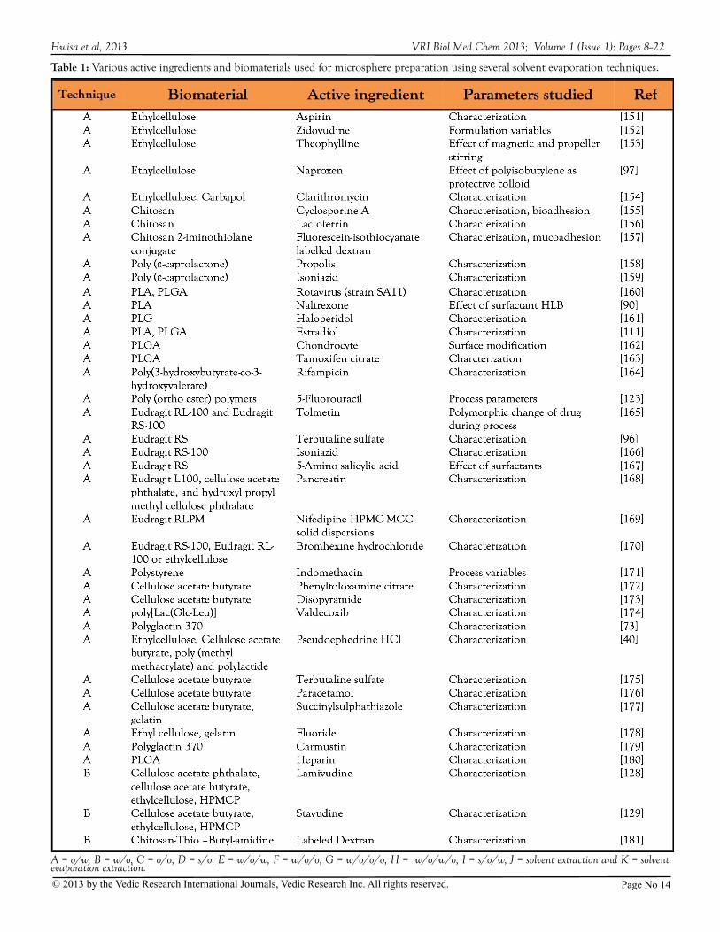
Table 1: Various active ingredients and biomaterials used for microsphere preparation using several solvent evaporation techniques.
A = o/w, B = w/o, C = o/o, D = s/o, E = w/o/w, F = w/o/o, G = w/o/o/o, H = w/o/w/o, I = s/o/w, J = solvent extraction and K = solvent evaporation extraction.
Hwisa et al, 2013 VRI Biol Med Chem 2013; Volume 1 (Issue 1): Pages 8--22
Page No 14© 2013 by the Vedic Research International Journals, Vedic Research Inc. All rights reserved.
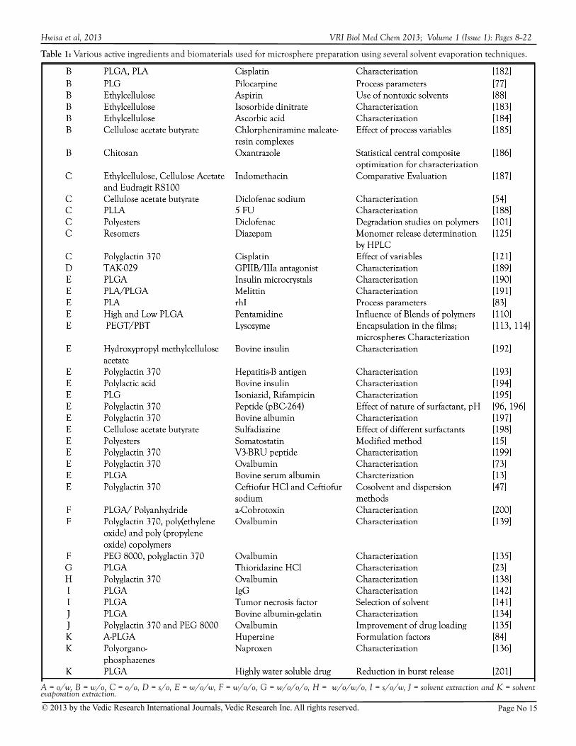
Table 1: Various active ingredients and biomaterials used for microsphere preparation using several solvent evaporation techniques.
A = o/w, B = w/o, C = o/o, D = s/o, E = w/o/w, F = w/o/o, G = w/o/o/o, H = w/o/w/o, I = s/o/w, J = solvent extraction and K = solvent evaporation extraction.
Hwisa et al, 2013 VRI Biol Med Chem 2013; Volume 1 (Issue 1): Pages 8--22
Page No 15© 2013 by the Vedic Research International Journals, Vedic Research Inc. All rights reserved.
glycolic acids) and in-vivo degradation of the polymers. Chem Pharm Bull 1988c, 36:2576-2581.
11. Ruiz JM, Tissier B, Benoit JP: Microencapsulation of peptide: a study of the phase separation of poly(DL-lactic acid-co-glycolic acid) copolymers by silicone oil. Int J Pharm 1989, 49:69-77.
12. Csernus VJ, Szende B, Schally AV: Release of peptides from sustained delivery systems (microcapsules and microparticles) in vivo. Int J Peptide Protein Res 1990, 35:557-565.
13. Cohen S, Yoshioka T, Lucarelli M, Hwang LH, Langer R: Controlled delivery systems for proteins based on poly(lactid-glycolic acid) microspheres. Pharm Res 1991, 8:713-720.
14. Heya T, Okada H, Tanigawara Y, Ogawa Y, Toguchi H: Effect of counteranion of TRH and loading amount on control of TRH release from copoly (dl-lactic-glycolic acid) microspheres prepared by an in-water drying method. Int J Pharm 1991a, 69:69-75.
15. Hermann J, Bodmeier R: Somatostatin containing biodegradable microspheres prepared by a modified solvent evaporation method based on w/o/w-multiple emulsions. Int J Pharm 1995, 126:129-138.
16. Bittner B, Morlock M, Koll H, Winter G, Kissel T: Recombinant human erythropoietin (rhEPO loaded poly(lactide-co-g lycol ide) microspheres : inf luence of the encapsulation technique and polymer purity on microsphere characteristics. Eur J Pharm Biopharm 1998, 45:295-305.
17. Deasy PB: Microencapsulation and related drug processes, Marcel Dekker, New York, 1984, p. 34.
18. Crainich Jr VA: Microencapsulation: Scale-up considerations and production technology. In: Tyle P (ed.) Specialized drug delivery systems: manufacturing and production technology, Marcel Dekker Inc., New York, 1990, p. 221-255.
19. O’Hogan DT, McGee JP, Holmgren J, Mowat A, Donachie AM, Mills W. Gaisford KHG, Rahman D, Challacombe SJ: Biodegradable microparticles for oral immunization. Vaccine 1993, 11:149-154.
20. Schwendeman SP, Cardamone M, Klibanov A, Langer R, Brandon MR: Stability of proteins and their delivery from biodegradable polymer microspheres. In: Cohen S (ed.) Microparticulate systems for the delivery of proteins and vaccines, Marcel Dekker Inc., New York, 1996, p.10.
21. Alonso JM: Nanoparticulate drug carrier technology. In: Cohen S (ed.) Microparticulate systems for the delivery of proteins and vaccines, Marcel Dekker Inc., New York, 1996, p.203-242.
22. Brunner A, Göpferich : The characterization of polyanhydride microspheres. In: Cohen S (ed.) Microparticulate systems for the delivery of proteins and vaccines, Marcel Dekker Inc., New York, 1996, p.169-202.
23. O’Donell PB, McGinity JW: Influence of processing on the stability and release properties of biodegradable microspheres containing thioridazine hydrochloride. Eur J Pharm Biopharm 1998, 45:83-94.
24. O’Donnell PB, McGinity JW: Preparation of microspheres by the solvent evaporation technique. Adv Drug Deliv Rev 1997, 28:85-96.
25. Hickey AJ, Tian Y, Parasrampuria D, Kanke M: Biopharmaceut. Drug Dispos 1933, 14:181-186.
26. Beck LR, Cowsar DR, Lewis DH, Cosgrove RJ, Riddle CT,
Lowry SL Epperly T: A new long-acting injectable microcapsule system for the administration of progesterone. Fertil Steril 1979, 31:545-551.
27. Watts PJ, Davies MC, Melia CD: Microencapsulation using emulsification/solvent evaporation : an overview of techniques and applications. Crit Rev Ther Drug Carrier Syst 1990, 7:235-259.
28. Thies C: Formation of degradable drug-loaded microparticles by in-liquid drying processes In: Donbrow M (ed.) Microcapsules and Nanoparticles in Medicine and Pharmacy, CRC Press, Boca Raton, FL, 1992, p.47-71.
29. Benoit JP, Herve M, Rolland H, Velde VV: Biodegradable microspheres: Advances in production technology In: Simon Benita (ed.) Microencapsulation: Methods and industrial applications, Marcel Dekker, New York, 1996, p.35-72.
30. Li W-I, Anderson KW, DeLuca PP: Kinetic and thermodynamic modeling of the formation of polymeric microspheres using solvent extraction/evaporation method. J Control Rel 1995a, 37:187-198.
31. Li W-I, Anderson KW, Mehta RC, DeLuca PP: Prediction of solvent removal profile and effect on properties for peptide-loaded PLGA microspheres prepared by solvent extraction/evaporation method. J Control Rel 1995b, 37:199-214.
32. Jeyanthi R, Thanoo BC, Mehta RC, DeLuca PP: Effect of solvent removal technique on the matrix characteristics of polylactide/glycolide microspheres for peptide delivery. J Control Rel 31996, 8:235-244.
33. Aftabrouchad C, Doelker E: Méthods de préparation des microparticules biodégradables chargées en principes actifs hydrosolubles. STP Pharma Sci.1992, 5:365-380.
34. Jalil R, Nixon JR: Microencapsulation using poly(L-lactic acid). I. Microcapsule properties affected by the preparative technique. J Microencapsul 1989, 6:473-484.
35. Jalil R, Nixon JR: Microencapsulation using poly(L-lactic acid). II. Preparative variables affecting microcapsules properties. J Microencapsul 1990a, 7:25-39.
36. Jalil R, Nixon JR: Microencapsulation using poly(L-lactic acid). III. Effect on polymer molecular weight on microcapsule properties. J Microencapsul 1990ab, 7:41-52.
37. Jalil R, Nixon JR: Microencapsulation using poly(L-lactic acid). IV. Release properties of microcapsules containing phenobarbitone. J Microencapsul 1990c, 7:53-66.
38. Fong JW: Microencapsulation by solvent evaporation and organic phase separation process In: Hsieh DST (ed.) Controlled Release Systems, CRC Press, Boca Raton, FL, 1988, p.81-108.
39. Alex R, Bodmeier R: Encapsulation of water-soluble drugs by a modified solvent evaporation method. I. Effect of process and formulation variables on drug entrapment. J Microencaps 1990, 7:347-355.
40. Bodmeier R, Chen H, Tyle P, Jarosz P: Pseudoephedrine HCl microspheres formulated into an oral suspension dosage form. J Control Rel 1991, 15:65-77.
41. Gurny R, Peppas NA, Harrington DD, Banker GS: Development of biodegradable and injectable lattices for controlled release of potent drugs. Drug Dev Ind Pharm 1981, 7:1-25.
42. Mehta RC Jeyanthi R, Calis S, Thanoo BC, Burton KW and
Hwisa et al, 2013 VRI Biol Med Chem 2013; Volume 1 (Issue 1): Pages 8--22
Page No 16© 2013 by the Vedic Research International Journals, Vedic Research Inc. All rights reserved.
DeLuca PP: Biodegradable microspheres as depot system for parenteral delivery peptide drugs. J Control Rel 1994, 29:375-384.
43. Jeyanthi R, Mehta RC, Thanoo BC and DeLuca PP: Effect of processing parameters on the properties of peptide-containing PLGA microspheres. J Microencapsul 1997, 14:163-174.
44. Chacon M, Molpeceres J, Berges L, Guzman M, Aberturas MR:, Stability and freeze-drying of cyclosporine loaded poly (D,L lectide-glycolide) carriers. Eur J Pharm Sci 1999, 8:99-107.
45. Wada R, Hyon SH, Ikada Y: Lactic acid oligomer microspheres containing hydrophilic drugs. J Pharm Sci 1990, 79:919-924.
46. Hardee GE, Davidson GWR, Chen H, Bodmeier R: Microencapsulation of antimicrobial agents for extended
release after injection, Proceedings of 18th Int. Symp. Control. Rel. Bioact. Mater., Amsterdam, 1991, p.203-204.
47. Bodmeier R, Chen H, Davidson GWR, Hardee GE: Microencapsulation of antimicrobial ceftiofur drugs. Pharm Dev Tech 1997, 2:323-334.
48. Seki Y, Kawaguchi T, Endoh H, Ishikawa K, Juni K, Nakano M: Controlled release of 3,5-diester products of 5-floro-2-deoxyuridine from poly-L-lactic acid microspheres. J Pharm Sci 1990, 79:985-987.
49. Spenlehauer G, Vert M, Benoit JP, Chabot F, Veillard M: Biodegradable cisplatin microspheres prepared by the solvent evaporation method: morphology and release characteristics. J Control Rel 1988, 7:217-229.
50. Bodmeier R, McGinity JW: Polylactic acid microspheres containing quinidine base and quinidine sulfate prepared by the solvent evaporation technique. I. Methods and morphology. J Microencapsul 1987a , 4:279-288.
51. Bodmeier R, McGinity JW: Polylactic acid microspheres containing quinidine base and quinidine sulfate prepared by the solvent evaporation technique. II. Some process parameters inf luencing the preparation and properties of microspheres. J Microencapsul 1987b, 4:289-297.
52. Mestiri M, Puisieux F, Benoit JP: Preparation and characterization of cisplatin-loaded polymethyl methacrylate microspheres. Int J Pharm 1993, 89: 229-234.
53. Rodriguez M, Vila-Jato JL, Torres D: Design of a new multiparticulate system for potential site-specific and controlled drug delivery to the colonic region. J Control Rel 1998, 55:67-77.
54. Prakash K, Suresh S, Kumari KS, Rao MEB, Patro SS: Design of cellulose acetate butyrate microspheres of diclofenac for colon
targeting. Poster presentations, 55th Indian Pharmaceutical Congress, AP38, 1998, p.159.
55. Obeidat WM, Price JC: Evaluation of enteric matrix microspheres prepared by emulsion-solvent evaporation using scanning electron microscopy. J Microencapsul 2004, 21:47-57.
56. Rizi K, Green RJ, Khutoryanskaya O, Donaldson M, Williams AC: Mechanisms of burst release from pH-responsive polymeric microparticles. J Pharm Pharmacol. 2011 63:1141-55.
57. Miyazaki Y, Ogihara K, Yakou S, Nagai T, Takayama K:
Bioavailability of theophylline and thiamine disulfide incorporated into mucoadhesive microspheres consisting of dextran derivatives and cellulose acetate butyrate. Biol Pharm Bull 2003, 26:1744-1747.
58. Umamaheshwari RB, Jain S, Bhadra D, Jain NK: Floating microspheres bearing acetohydraxamic acid for the treatment of Helicobacter pylori. J Pharm Pharmcol 2003, 55:1607-1613.
59. Stithit S, Chen W, Price JC: Development and characterization of buoyant theophylline microspheres with near zero order release kinetics. J Microencapsul 1998, 15:725-737.
60. Vrancken MN, Claeys DA: US Patent 3,526,906: 1970.61. Dejaeger NC, Tavernier BH: GB Patent 1,405,108: 1971.62. Okada H, Toguchi H: Biodegradable microspheres in drug
delivery. Critical reviews in Therapeutic Drug Carrier Systems 1995, 12:1-99.
63. Uchida T, Yoshida K, Nakada Y, Nagareya N, konishi Y, Nakai A, Nihikata M, Matsuyama K: Preparation and characterization of polylactic acid microspheres containing water-soluble anesthetics with small molecular weight. Chem Pharm Bull 1997b, 45:513-517.
64. Kissel T, Koneberg R: Injectable biodegradable microspheres for vaccine delivery. In: Cohen S (ed.) Microparticulate systems for the delivery of proteins and vaccines, Marcel Dekker Inc., New York, 1996, p.51-87.
65. Bhagwatwar HP: Biodegradable microparticles of peptide drugs using polylactide polymers. In: Jain NK (ed.) Advances in Controlled and Novel Drug Delivery, CBS Publishers and Distributors, New Delhi, 2001, p.1-17.
66. Vyas SP, Khar RK: Microspheres. In: Targeted and controlled Drug Delivery: Novel Carrier Systems, CBS Publishers & Distributors, New Delhi, 2002, p.417-457.
67. Heya T, Okada H, Ogawa Y, Toguchi H: Factors influencing the profiles of TRH release from copoly (D,L-lactic/glycolic acid) microspheres. Int J Pharm 1991b, 72:199-205.
68. Bodmer D and Kissel T: Sustained release of the somatostatin
analogue octeotide from microspheres, Proceedings of 18th Int Symp Control Rel Bioact Mater, Amsterdam, 1991, p.597-598.
69. Camarata PJ, Suryanarayanan R, Turner DA, Parker RG, Ebner TJ: Sustained release of nerve growth factor from biodegradable polymer microspheres. Neurosurgery 1992, 30:313-319.
70. Eldridge JH, Hammond CJ, Meulbroek JA, Staas JK, Gilley RM, Tice Tr: Controlled vaccine release in the gut-associated lymphoid tissues. I. Orally administered biodegradable microspheres target the Peyer’s patches. J Control Rel 1990, 11:205-214.
71. Singh M, Singh A and Talwar GP: Controlled delivery of diphtheria toxoid using biodegradable poly (D,L-lactide) microcapsules. Pharm Res 1991, 8:958-961.
72. Wang HT, Schmitt E, Flanagan DR, Linhardt RJ: Influence of formulation methods on the in vitro controlled release of protein from poly (ester) microspheres. J Control Rel 1991, 17:23-32.
73. Jeffery H, Davis SS, O’Hagan DT: The preparation and characterization of poly )lactide-co-glycolide) microparticles. II. The entrapment of a model protein using a (water-in-oil)-in-water emulsion solvent evaporation technique. Pharm Res 1993,
Hwisa et al, 2013 VRI Biol Med Chem 2013; Volume 1 (Issue 1): Pages 8--22
Page No 17© 2013 by the Vedic Research International Journals, Vedic Research Inc. All rights reserved.
Page No 18© 2013 by the Vedic Research International Journals, Vedic Research Inc. All rights reserved.
10:362-368.74. Alonso MJ, Cohen S, Park TG, Gupta RK, Siber GR, Langer R:
Determinants of release rate of tetanus vaccine from polyester microspheres. Pharm Res 1993, 10:945-953.
75. Sah HK, Chien YW: Evaluation of a microreservoir-type biodegradable microcapsule for controlled release of proteins. Drug Dev Ind Pharm 1993, 19:1243-1263.
76. Maa Y-F, Hsu C: Liquid-liquid emulsification by rotor/stator homogenization. J Control Rel 1996, 38:219-228.
77. Yoncheva K, Vandervoort J, Ludwig A: Influence of process parameters of high-pressure emulsification method on the proper t i e s o f p i l o ca r p ine - l oaded nanoparticles. J Microencapsul 2003, 20:449-458.
78. Ueda M, Kreuter J: Optimization of the preparation of loperamide-loaded poly (L-lactide) nanoparticles by high-pressure emulsification-solvent evaporation. J Microencapsul 1997, 14:593-605.
79. Maa Y-F, Hsu C: Effect of primary emulsions on microsphere size and protein-loading in the double emulsion process. J Microencapsul 1997, 14: 225-241.
80. Niu CH, Chiu YY: FDA perspective on peptide formulation and stability issues. J Pharm Sci 1998, 87:1331-1334.
81. Sato T, Kanke M, Schroeder HG, DeLuca PP: Porous biodegradable microspheres for controlled drug delivery. I. Assessment of processing conditions and solvent removal techniques. Pharm Res 1988, 5:21-30.
82. Zambaux MF, Bonneaux F, Gref R, Maincent P, E Dellacherie, MJ Alonso, P Labrude, Vigneron C: Influence of experimental parameters on the characteristics of poly(lactic acid nanoparticles prepared by a double emulsion method. J Control Rel 1998, 50:31-40.
83. Liu R, Ma GH, Wan YH, Su ZG: Influence of process parameters on the size distribution of PLA microcapsules prepared by combining membrane emulsification technique and double emulsion-solvent evaporation method. Colloids Surf B Biointerfaces 2005, 45:144-153.
84. Fu X, Ping Q, Gao Y: Effects of formulation factors on encapsulation efficiency and release behaviour in vitro of huperzine A-PLGA microspheres. J Microencapsul 2005, 22:57-66.
85. Parikh RH, Parikh JR, Dubey RR, Soni HN, Kapadia KN: Poly(D,L -lactide-co- glycolide) microspheres containing 5-fluorouracil: optimization of process parameters. AAPS PharmSciTech 2003, 4(2):E13.
86. Cleland JL, Lim A, Barron L, Duenas ET, Powell MF: Development of a single-shot subunit vaccine for HIV-1: Part 4. Optimizing microencapsulation and pulsatile release of MN rgp120 from biodegradable microspheres. J Control Release 1997, 47:135-150.
87. Jain R, Shah NH, Waseem Malick A, Rhodes CT: Controlled drug delivery by biodegradable (poly(ester) devices: Different preparative approaches. Drug Dev Ind Pharm 1998, 24:703-727.
88. Yang CY, Tsay SY, Tsiang RC: Encapsulating aspirin into a surfactant-free ethyl cellulose microsphere using non-toxic solvents by emulsion-solvent evaporation technique. J Microencapsul 2001, 18:223-236.
89. Graves RA, Moiseyev R, Freeman T, Mandal TK: Effect of surfactant on the characteristics of biodegradable microcapsules. J Biomater Sci Polym Ed 2005, 16:585-596.
90. Dinarvand R, Moghadam SH, Sheikhi A, Atyabi F. Effect of surfactant HLB and different formulation variables on the properties of poly-D,L-lactide microspheres of naltrexone prepared by double emulsion technique. J Microencapsul. 2005, 22:139-51.
91. Kristmundsdottir T, Ingvarsdottir K: Influence of emulsifying agents on the properties of cellulose acetate butyrate and ethylcellulose microcapsules. J Microencapsul. 1994, 11:633-639.
92. Scholes PD, Coombes AG, Illum L, Davis SS, JF Watts, C Ustariz, M Vert, Davies MC: Detection and detemination of surface levels of poloxamer and PVA surfactant on biodegradable nanospheres using SSIMS and XPS. J Control Rel 1999, 59:261-278.
93. Vandervoort J, Ludwig A: Biocompatible stabilizers in the preparation of PLGA nanoparticles: a factorial design study. Int J Pharm 2002, 238:77-92.
94. Coombes AG, Tasker S, Lindblad M, Holmgren J, Hoste K, Toncheva V, Schacht E, Davies MC, Illum L, Davis SS: Biodegradable polymeric microparticles for drug delivery and vaccine formulation: the surface attachment of hydrophilic species using the concept of poly(ethylene glycol) anchoring segments. Biomaterials 1997, 18:1153-1161.
95. Blanco-Prieto MJ, Leo E, Delie F, Gulik A, Couvreur P, Fattal E: Study of influence of several stabilizing agents on the entrapment and in vitro release of pBC 264 from poly(lactide-co-glycolide)microspheres prepared by a w/o/w solvent evaporation method. Pharm Res 1996, 13:1127-1129.
96. Kim CK, Kim MJ, Oh KH: Preparation and evaluation of sustained release microspheres of terbutaline sulfate. Int J Pharm 1994, 106:213-219.
97. Sveinsson SJ, Kristmundsdottir T: Naproxen microcapsules: preparation and in vitro characterization. Int J Pharm 1992, 82:129-133.
98. Rosca ID, Watari F, Uo M: Microparticle formation and its mechanism in single and double emulsion solvent evaporation. J Control Rel 2004, 99:271-280.
99. Mateovik-Rojnik T, Frlan R, Bogataj M, Bukovec P, Mrhar A: Effect of preparation temperature in solvent evaporation process on Eudragit RS microsphere properties. Chem Pharm Bull 2005, 53:143-146.
100. Mateovik T, Ratnik M, Bogataj M, Mrhar A: Determination of microsphere solidification time in the solvent evaporation process. J Microencapsul 2005, 22:81-90.
101. Lin SY, Chen KS,Teng HH, Li MJ: In vitro degradation and dissolution behaviors of microspheres prepared by three low molecular weight polyesters. J Microencapsul 2000, 17:577-586.
102. Fernandez-Carballido A, Herrero-Vanrell R, Molina-Martinez IT, Pastoriza P: Sterilized ibuprofen-loaded poly (D,L-lactide-co-glycolide) microspheres for intra-articular administration: effect of gamma-irradiation and storage. J Microencapsul 2004, 21:653-665.
103. Vandervoort J, Ludwig A: Preparation factors affecting the properties of polylactide nanoparticles : a factorial design study. Pharmazie 2001, 56:484-488.
104. Nihant N, Shugens Ch, Grandfils Ch, Jerome R, Teyssie Ph: Polylactide microparticles prepared by a double emulsion/evaporation technique. I. Effect of primary emulsion stability. Pharm Res 1994, 11:1479-1484.
Hwisa et al, 2013 VRI Biol Med Chem 2013; Volume 1 (Issue 1): Pages 8--22
Page No 19© 2013 by the Vedic Research International Journals, Vedic Research Inc. All rights reserved.
105. Shugens Ch, Laruelle N, Nihant N, Grandfils Ch, Jerome R, Teyssie PH: Effect of emulsion stability on the morphology and porosity of semicrystalline poly l-lactide microparticles prepared by w/o/w double emulsion-evaporation. J Control Rel 1994, 32:161-176.
106. Peracchia MT, Vauthier C, Desmaele D, Gulik A, Dedieu J, Demoy M, Angelo J, Couvreur P: Pegylated nanoparticles from a novel methoxypolyethylene glycol cyanoacrylate-hexadecyl cyanoacrylate amphiphilic copolymer. Pharm Res 1998, 15:550-556.
107. Yulia VC, Valery GB, Natalia RK, Franck B, Jean PB, Nathalie U, Philippe M: Effect of the type of hydrophilic polymers on the size of nanoparticles obtained by emulsification-solvent evaporation. Mendeleev Commun 2003, 13: 65-68.
108. Dinarvand R, Moghadam SH, Boorboor M: Effect of PLA and PLGA blending on drug release from microspheres. Poster Session 2–Drug Delivery, BPC Science Proceedings, 104, 2003, p.S-48.
109. Obeidat WM, Price JC: Preparation and in vitro evaluation of propylthiouracil microspheres made of Eduragit RL 100 and cellulose acetate butyrate polymers using the emulsion-solvent evaporation method. J Microencapsul 2005, 22:281-289.
110. Graves RA, Pamujula S, Moiseyev R, Freeman T, Bostanina LA, Mandal TK: Effect of different ratios of high and low molecular weight PLGA blend on the characteristics of pentamidine microcapsules. Int J Pharm 2004, 270: 251-262.
111. Xinteng Z, Weisan P, Ruhua Z, Feng Z: Preparation and evaluation of poly (D,L-lactic acid) (PLA) or D,L-lactide/glycolide copolymer (PLGA) microspheres with estradiol. Pharmazie 2002, 57:695-697.
112. Obeidat WM, Price JC: Viscosity of polymer solution phase and other factors controlling the dissolution of theophylline microspheres prepared by the emulsion solvent evaporation method. J Microencapsul 2003, 20:57-65.
113. Bezemer JM, Radersma R, Grijpma DW, Dijkstra PJ, van Blitterswijk CA, Feijen J: Microspheres for protein delivery prepared from amphiphilic multiblock copolymers. 1. Influence of preparation techniques on particle. J Control Rel 2000, 67:233-248.
114. van de Weert M, van Dijkhuizen-Radersma R, Benzemer JM, Hennink WE, Crommelin DJ: Reversible aggregation of lysozyme in a biodegradable amphiphilic multiblock copolymer. Eur J Pharm Biopharm 2002, 54:89-93.
115. Takami T, Murakami Y: Development of PEG-PLA/PLGA microparticles for pulmonary drug delivery prepared by a novel emulsification technique assisted with amphiphilic block copolymers. Colloids Surf B Biointerfaces 2011, 87:433-8.
116. Srinivasan C, Katare YK, Muthukumaran T, Panda AK: Effect of additives on encapsulation efficiency, stability and bioactivity of entrapped lysozyme from biodegradable polymer particles. J Microencapsul 2005, 22:127-138.
117. Kim TH, Park TG: Critical effect of freezing/freeze-drying on sustained release of FITC-dextran encapsulated within PLGA microspheres. Int J Pharm 2004, 271:207-214.
118. Tsai DC, Howard SA, Hogan TF, Malanga CJ, Kandzari SJ, Ma JKH: Preparation and in vitro evaluation of polylactic acid mitomycin C microcapsules. J Microencapsul 1986, 3:181-193.
119. Hazrati AM, DeLuca PP: 5-Fluorouracil in biodegradable polymeric
microspheres, Proceedings of 16th Int. Symp. Control. Rel. Bioact. Mater., Chicago, 1989, 1989, p.79-80.
120. Ike O, Shimizu Y, Wada R, Hyon SH, Ikada Y: Controlled cisplatin delivery system using poly (D,L-lactic acid). Biomaterials 1992, 13:230-234.
121. Kyo M, Hyon SH, Ikada Y: Effects of preparation conditions of cisplatin loaded microspheres on the in vitro release. J. Control. Rel. 1995, 35:73-82.
122. Goto S, Kawata M, Nakamura M, Maekawa, Aoyama T: Eudragit RS and RL (acrylic resins) microcapsules as pH insensitive and sustained release preparations of ketoprofen. J Microencapsul 1986, 3:293-304.
123. Lin YH, Vasavada RC: Studies on microencapsulation of 5-fluorouracil with poly(ortho ester) polymers. J Microencapsul 2000, 17:1-11.
124. Bontemps J, Pirson P, Falmagne JB, Jérome, Teysssié P, Delattre L, Evrard B: Microparticules comportant un polymère biodegradable contrôlant la liberation d’un actif antimalarique, compositions pharmaceutiques en comprenant et procédé de preparation. EP Patent 0301969, 1989.
125. Giunchedi P, Conti B, Scalia S, Conte U: In vitro degradation study of polyester microspheres by a new HPLC method for monomer release determination. J Control Rel 1998, 56:53-62.
126. Gardner DL: Process of preparing microcapsules of lactides or lactide copolymers with glycolides and/or -caprolactone. US Patent 4,637,905, 1987.
127. Spenlehauer G, Spenlehauer-Bonthonneau F, Thies C: Biodegradable microparticles for delivery of polypeptides and proteins. In: Butterworth DA (ed.) Biological and Synthetic Membranes, Alan R. Liss, Inc., New York, 1989, p.283-291.
128. Prakash K, Narayana PR, Shanta KK, Lakshmi: Preparation and characterization of lamivudine microcapsules using various cellulose polymers. Trop. J. Pharm. Res., 2007, 6 (4):841-847.
129. Narayana RP, Prakash K, Reddy CSB, Krishnaveni B, Shantakumari K and Lakshmi NM. Stavudine Loaded Microcapsules using various Cellulose Polymers: Preparation and In-Vitro Evaluation. Int. J. Pharm. Sci. Nanotech. 2009, 2(2):551-556.
130. DeLuca PP, Kanke M, Sato T, Scroeder HG: Porous microspheres for drug delivery and methods for making the same. US Patent 4,818,542, 1989.
131. Mohan Kamila M, Mondal N, Kanta Ghosh L, Kumar Gupta B: Multiunit f loating drug delivery system of rosiglitazone maleate: development, characterization, statistical optimization of drug release and in vivo evaluation. AAPS PharmSciTech. 2009, 10:887-99.
132. Bitz C, Doelker E: Influence of the preparation method on residual solvents in biodegradable microspheres. Int J Pharm 1996, 131:171-181.
133. Boisdron-Celle M, Menei P, Benoit JP: Preparation and ch a r a c t e r i z a t i o n o f 5 - f l u o ro ur a c i l - l o a d e d microparticles as biodegradable anticancer drug carriers. J Pharm Pharmacol 1995, 47:108-114.
134. Li JK, Wang N, Wu XS: Novel biodegradable system based on
Hwisa et al, 2013 VRI Biol Med Chem 2013; Volume 1 (Issue 1): Pages 8--22
gelating nanoparticles and poly(lactic-co-glycolic acid) microspheres for protein and peptide drug delivery. J Pharm Sci 1997, 86:891-895.
135. Yeh MK, Jenkins PG, Davis SS, Coombes AG: Improving the delivery capacity of microparticle systems using blends of poy(DL-lactide co-glycolide) and poly (ethylene glycol). J Control Rel 1995b, 37:1-9.
136. Veronese FM, Marsilio F, Caliceti P, De Filippis P, Giunchedi P, Lora S: Polyorganophosphazene microspheres for drug release: polymer synthesis, microsphere preparation, in vitro and in vivo naproxen release. J Control Rel 1998, 52:227-237.
137. Pavenetto F, Conti B, Genta I, Giunchedi P: Solvent evaporation, solvent extraction and spray drying for polylactide microsphere preparation. Int J Pharm 1992, 84:151-159.
138. Yeh MK, Coombes AG, Jenkins PG, Davis SS: Novel emulsification-solvent extraction technique for production of protein loaded biodegradable microparticles for vaccine and drug delivery. J Control Rel 1995a, 33:437-445.
139. Yeh MK, Davis SS, Coombes A: Improving protein delivery from microparticles using blends of poly (DL-lactide-co-glycolide)and poly (ethylene oxide)-poly(propylene oxide) copolymers. Pharm Res 1996, 13:1693-1698.
140. Iwata M, McGinity JW: Preparation of multi-phase microspheres of poly (D,L-lactic acid) and poly (D,L-lactic-co-glycolic acid)containing a w/o emulsion by a multiple emulsion solvent evaporation technique. J Microencaps 1992, 9:201-214.
141. Iwata M, Tanaka T, Nakamura Y, McGinity JW: Selection of the solvent system for the preparation of poly (D,L-lactic-co-glycolic acid) microspheres containing tumor necrosis factor-alpha (TNF-alpha). Int J Pharm 1998, 160: 145-156.
142. Wang J, Chua KM, Wang CH: Stabilization and encapsulation of human immunoglobulin G into biodegradable microspheres. J Colloid Interface Sci. 2004, 271:92-101.
143. Kockisch S, Rees GD, Young SA, Tribouklis J, Smart JD: Polymeric microspheres for drug delivery to the oral cavity: an in vitro evaluation of mucoadhesive potential. J Pharm Sci 2003, 92:1614-1623.
144. Benita S, Benoit JP, Puisieux F, Thies C: Characterization of drug loaded poly (D,L-lactide) microspheres. J Pharm Sci 1984, 73:1721-1724.
145. Cowsar DR, Tice TR, Gilley RM, English JP: Poly(lactide-co-glycolide) microcapsules for controlled release of steroids. In: Widder KH, Green R (eds.) Methods in Enzymology: Drug and Enzyme Targeting, Academic Press, Orlando, FL, 1985, p.101-116.
146. Kwong AK, Chou S, Sun AM, Sefton MV, Goosen MFA: In vitro and in vivo release of insulin from poly(lactic acid) microbeads and pellets. J Control Rel 1986, 4:47-62.
147. Leelarasamee N, Howard SA, Malanga CJ, Ma JKH: A method for preparation of polylactic and microcapsules of controlled particle size and drug loading. J Microencapsul 1988, 5:147-157.
148. Shiga K, Muramatsu N, Kondo T: Preparation of poly (D,L-lactide) and copoly (lactide-glycolide) microspheres of uniform size. J Pharm Pharmacol 1996, 48:891-895.
149. Koushik K, Kompella UB: Preparation of large porous
deslorelin-PLGA microparticles with reduced residual solvent and cellular uptake using a supercritical carbon dioxide process. Pharm Res 2004, 21:524-535.
150. Freitas S, Hielscher G, Merkle HP, Gander B: Continuous contact - and contamination-free ultrasonic emulsification-a useful tool for pharmaceutical development and production. Ultrason Sonochem. 2006, 13:76-85.
151. Yang CY, Tsay SY, Tsiang RC: An enhanced process for encapsulating aspirin in ethyl cellulose microcapsules by solvent evaporation in an o/w emulsion. J Microencapsul 2000, 17:269-277.
152. Abu-Izza K, Garcia-Contreras L, Lu DR: Preparation and evaluation of sustained release AZT-loaded microspheres : optimizat ion of the release characteristics using response surface methodology. J Pharm Sci 1996, 85:144-149.
153. Amperiadou A , Georgarakis M: Preparat ion and characterization of ethylcellulose-walled theophylline microcapsules using the emulsion-solvent evaporation technique. Drug Dev Ind Pharm 1995, 21:1339-1346.
154. Rajinikanth PS, Karunagaran LN, Balasubramaniam J, Mishra B: Formulation and evaluation of clarithromycin microspheres for eradication of Helicobacter pylori. Chem Pharm Bull 2008, 56:1658-64.
155. Malaekeh-Nikouei B, Sajadi Tabassi SA, Jaafari MR: Preparation, characterization, and mucoadhesive properties of chitosan-coated microspheres encapsulated with cyclosporine A. Drug Dev Ind Pharm. 2008, 34:492-8.
156. Onishi H, Machida Y, Koyama K: Preparation and in vitro characteristics of lactoferrin-loaded chitosan microparticles. Drug Dev Ind Pharm. 2007, 33:641-7.
157. Maculotti K, Genta I, Perugini P, Imam M, Bernkop-Schnürch A, Pavanetto F: Preparation and in vitro evaluation of thiolated chitosan microparticles. J Microencapsul. 2005, 22:459-70.
158. Durán N, Marcato PD, Buffo CM, De Azevedo MM, Esposito E: Po l y ( - c a p r o l a c t o n e ) / p r o p o l i s e x t r a c t : microencapsulation and antibacterial activity evaluation. Pharmazie. 2007, 62:287-90.
159. Durán N, De Oliveira AF, De Azevedo MM: In vitro studies on the release of isoniazid incorporated in poly(-caprolactone). J Chemother. 2006, 18:473-9.
160. Nayak B, Panda AK, Ray P, Ray AR: Formulation, characterization and evaluation of rotavirus encapsulated PLA and PLGA particles for oral vaccination. J Microencapsul. 2009, 26:154-65.
161. Cheng YH, Illum L, Davis SS: A poly(D,L-lactide-co-glycolide) microsphere depot system for delivery of haloperidol. J Control Rel 1998, 55:203-212.
162. Chun KW, Yoo HS, Yoon JJ, Park TG: Biodegradable PLGA microcarriers for injectable delivery of chondrocytes: effect of surface modification on cell attachment and function. Biotechnol Prog 2004, 20:1797-1801.
163. Sehra S, Dhake AS: Formulation and evaluation of sustained release microspheres of poly-lactide-co-glycolide containing tamoxifen citrate. J Microencapsul. 2005, 22:521-8.
164. Durán N, Alvarenga MA, Da Silva EC, Melo PS, Marcato PD: Microencapsulation of antibiotic rifampicin in poly(3-hydroxybutyrate-co-3-hydroxyvalerate). Arch Pharm Res. 2008, 31:1509-16.
Hwisa et al, 2013 VRI Biol Med Chem 2013; Volume 1 (Issue 1): Pages 8--22
Page No 20© 2013 by the Vedic Research International Journals, Vedic Research Inc. All rights reserved.
165. Pignatello R Vandelli MA, Giunchedi P, Puglisi G: Properties of Tolmetin-loaded Eudragit RL-100 and Eudragit Rs-100 microparticles prepared by different techniques. STP Pharm Sci 1997, 7:148-157.
166. Muhuri G, Pal TK: Computation of release kinetics of isoniazid microcapsules. Bullettino Chimico Farmaceutico 1991, 130:169-171.
167. Watts PJ, Davies MC, Melia CD: Encapsulation of 5-aminosalicylic acid into Eduragit RS microspheres and modulation of their release characteristics by use of surfactants. J Control Rel 1991, 16:311-318.
168. Naikwade SR, Meshram RN, Bajaj AN: Preparation and in vivo efficacy study of pancreatin microparticles as an enzyme replacement therapy for pancreatitis. Drug Dev Ind Pharm. 2009, 35:417-32.
169. Chowdary KP, Sankar GG: Eudragit microcapsules of nifedipine and its dispersions in HPMC-MCC: physicochemical characterization and drug release studies. Drug Dev Ind Pharm 1997, 23:325-330.
170. Bhattacharyya M, Mandal SC, Gupta BK: Controlled delivery of bromhexine hydrochloride using rate-controlling polymers. STP Pharma Sci 1992, 2:481-487.
171. Tamilvanan S, Sa B: Effect of production variables on the physical characteristics of indomethacin-loaded polystyrene microparticles. Pharmacy and Pharmacology Communications 1998, 4:427-432.
172. Samy EM, Mohamed FA, Ali AS, Anwar MM, Aboutaleb AE: Preparation and evaluation of phenyltoloxamine citrate microspheres. Bull Pharm Sci Assiut Univeristy 1995, 18:125-134.
173. Radwan MA, Price JC, Tackett RL: In vitro release of disopyramide from cellulose acetate butyrate microspheres. Drug Dev Ind Pharm 1995, 21:1453-1462.
174. Agnihotri SM, Vavia PR: Drug loaded poly[Lac(Glc-Leu)] m i c ro pa r t i c l e s : fo r m ul a t i o n a n d re l e a s e characteristics. Colloids Surf B Biointerfaces. 2009, 74:336-9.
175. Ruiz R, Sakr A, Sprockel OL: A Study on the Manufacture and in Vitro Dissolution of Terbutaline Sulfate Microcapsules and their Tablets. Drug Dev Ind Pharm 1990, 11:1829-1842.
176 . S p ro cke l O L , P r a p a i t r a k u l W: C o mp a r i s o n o f microencapsulation by various emulsion techniques. Int J Pharm 1990, 58:123-127.
177. Chiao CS, Price JC: Modification of gelatin beadlets for zero order sustained release. Pharm Res 1989, 6:517-520.
178. de Francisco LMB, Cerquetani JA, Bruschi ML: Development and characterization of gelatin and ethylcellulose microparticles designed as platforms to delivery fluoride. Drug Dev Ind Pharm. 2012, 1-7.
179. Painbeni T, Venier-Julienne MC,Benoit JP: Internal morphology of poly(D,L -lactide-co-glycolide) BCNU-loaded microspheres: Influence of drug stability. Eur J Pharm Biopharm 1998, 45:31-39.
180. Oliveira SS, Oliveira FS, Gaitani CM, Marchetti JM: Microparticles as a strategy for low-molecular-weight heparin delivery. J Pharm Sci. 2011, 100:1783-92.
181. Elhassan IM, Berhnkop-Schnurch A: Controlled drug delivery systems based on thiolated chitosan microspheres. Drug Dev Ind Pharm 2005, 31:557-65.
182. Matsumoto A, Matsukawa Y, Suzuki T, Yoshino H: Drug release characteristics of multi-reservoir type microspheres
with poly (dl-lactide-co-glycolide) and poly (dl-lactide). J Control Rel 2005, 106:172-180.
183. Dinarvand R, Mirfattahi S, Atyabi F: Preparation, characterization and in vitro drug release of isosorbide dinitrate microspheres. J Microencapsul. 2002, 19(1):73-81.
184. Vanichtanunkul D, Vayumhasuwan P, Nimmannit U: The effect of core-to-wall ratio and Span 80 concentration on the properties of ascorbic acid microcapsules. J Microencapsul 1998, 15:753-759.
185. Sayed HA: Effect of process variables on the properties of coated resonates. Part 1. Formulation of liquid dosage forms. Egyptian J Pharm Sci 1991, 32: 937-949.
186. Hassan EE, Parish RC, Gallo JM: Optimized formulation of magnetic chitosan microspheres containing the anticancer agent, oxantrazole. Pharm Res 1992, 9:390-397.
187. Chowdary KPR, Bhanoji Rao ME, Prakash K: A Comparative Evaluation of Indomethacin Release and Permeability of Ethylcellulose, Cellulose Acetate and Eudragit RS 100
Microspheres. Poster presentations, 56th Indian Pharmaceutical Congress, AP14, 2004, p.128.
188. Zhu KJ, Zhang JX, Wang C,Yasuda H, Ichimaru A, Yamamoto K: Preparation and in vitro release behaviour of 5-fluorouracil-loaded microsphres based on poly (L-lactide) and its carbonate copolymers. J Microencapsul 2003, 20:731-743.
189. Takada S, Kurokawa T, Miyazaki K, Iwasa S, Ogawa Y: Utilization of an amorphous form of a water-soluble GPIIb/IIIa antagonist for controlled release from biodegradable microspheres. Pharm Res 1997, 14:1146-1150.
190. Choi SH, Kwon JH, Kim CW: Microencapsulation of insulin microcrystals. Biosci Biotechnol Biochem. 2004, 68:749-52.
191. Cui F, Cun D, Tao A, Yang M, Shi K, Zhao M, Guan Y: Preparation and characterization of melittin-loaded poly (DL-lactic acid) or poly (DL-lactic-co-glycolic acid) microspheres made by the double emulsion method. J Control Rel 2005, 107:310-319.
192. Nagareya N, Uchida T, Matsuyama K: Preparation and characterization of enteric microspheres containing bovine insulin by a w/o/w emulsion solvent evaporation method. Chem Pharm Bull. 1998, 46:1613-1617.
193. Uchida T, Shiosaki K, Nakada Y, Fukada K, Eda Y, Tokiyoshi S, Nagarey N, Matsuyam K: Microencapsulation of hepatitis B core antigen for vaccine preparation. Pharm Res 1998, 15:1708-1713.
194. Uchida T, Nagareya N, Sakakibara S, Konishi Y, Nakai A, Nishikata M, Matsuyama K, Yoshida K: Preparation and characterization of polylactic acid microspheres containing bovine insulin by a w/o/w emulsion solvent evaporation method. Chem Pharm Bull. 1997a, 45:1539-1543.
195. Dutt M, Khuller GK: Therapeutic efficacy of poly(DL-lactide-co-glycolide)-encapsulated antitubercular drugs against Mycobacterium tuberculosis Infection Induced in Mice. Antimicrob Agents Chemother 2001, 45:363-366.
196. Blanco-Prieto MJ, Fattal E, Gulik A, Dedieu JC, Roques BP, Couvreur P: Characterization and morphological analysis of a cholecystokinin derivative in peptide-
Hwisa et al, 2013 VRI Biol Med Chem 2013; Volume 1 (Issue 1): Pages 8--22
Page No 21© 2013 by the Vedic Research International Journals, Vedic Research Inc. All rights reserved.
loaded poly(lactide-co-glycolide) microspheres prepared by a water-in-oil-in-water emulsion solvent evaporation method. J Control Rel 1997, 43:81-87.
197. Rafati H, Coombes AG, Adler J, Holland J, Davis SS. Protein-loaded poly(DL-latide-co-glycolide) microparticles for oral administration: formulation, structural and release characteristics. J Control Rel 1997, 43:89-102.
198. Kondo T, Hafez E, Abdel-Monem H, Muramatsu N, El-Harras S, El-Gibaly I: Microencapsulation of sulfadiazine by water/oil/water complex emulsion technique. Pharmazeutische Industrie 1996, 58:552-556.
199. Blanco-Prieto MJ, Delie F, Fattal E, Tartar A, Puisieux F, Gulik
A, Couvreur P: Characterization of V3-BRU peptide-loaded small PLGA microspheres prepared by a (w1/o)w2 emulsion solvent evaporation method. Int J Pharm 1994, 111:137-145.
200. Li Y, Jiang HL, Zhu KJ, Liu JH, Hao YL: Preparation, characterization and nasal delivery of α-cobrotoxin - loaded poly (lactide-co-glycolide) / polyanhydride microspheres. J Control Rel 2005, 108:10-20.
201. Ahmed AR, Elkharraz K, Irfan M, Bodmeier R: Reduction in burst release after coating poly(D,L-lactide-co-glycolide) (PLGA) microparticles with a drug-free PLGA layer. Pharm Dev Technol. 2012, 17:66-72.
Hwisa et al, 2013 VRI Biol Med Chem 2013; Volume 1 (Issue 1): Pages 8--22
Page No 22© 2013 by the Vedic Research International Journals, Vedic Research Inc. All rights reserved.
Babu Rao Chandu, M.Pharm., Ph.D., has done his Ph.D. (Pharmaceutical Sciences) from Andhra University, India. He has completed his post-graduation in Pharmaceutical Chemistry specialization from Dr.MGR Medical University, Chennai, India. During his highly noticeable research experience over 20 years he has published over 100 papers in various international impact journals and one Indian patent. His research areas of interest include, Phytochemistry, Natural medicine, Drug design and synthesis, Bioanalytical method development and Formulation of dosage forms. He is a member of various professional bodies and editorial member of several international journals. He has guided five Ph.D. students and several M.Pharm students. Presently he is working on bioanalytical method development and validation and synthesis of biomolecules.
Note: Vedic Research International, Vedic Research Inc is not responsible for any data in the present article including, but not limited to, writeup, figures, tables. If you have any questions, directly contact authors.
Nagiat T Hwisa, M.Pharm., has completed her Master Degree in Pharmacy from Tripoli University, Libya. She has over 25 years rich experience in research and published over 20 papers in reputed and impact journals. Her areas of research include, Pharmacology of natural biomolecules, Bioanalytical method development and formulation development of biomolecules.
Prakash Katakam, M.Pharm., Ph.D., has done his Ph.D. (Pharmaceutical Sciences) from Berhampur University, Orissa, India. He has completed his under- and post-graduation in Pharmaceutical Technology specialization from Andhra University, Visakhapatnam, India. Research contribution includes design and biomedical evaluation in vitro and in vivo, of various controlled release drug delivery systems like subgingival delivery films, microcapsules and matrix tablets and conventional dosage forms. Other areas of research interest are biomaterials, bioanalytical method development and natural medicine. He has guided two Ph.D. students and several M.Pharm students. He has 61 peer reviewed research articles and four Indian patents to his credit. He is a member of various professional bodies and editorial board member of several international journals. Presently he is working on biomaterials and their novel modifications for pharmaceutical application.
Shanta Kumari Adiki, M.Tech., Ph.D. (Pharmaceutical Sciences), has completed her Ph.D. from Berhampur University, India. She has over 12 years of research experience. She has rich research experience in developing all kinds of analytical method development including Bioanalytical methods for drug molecules. She has published over 25 research articles in reputed journals and has two Indian patents. Her areas of research include, bioanalytical method development and validation, instrumental analysis using HPLC, UV-visible spectroscopy, novel formulation development of drugs.
Related Documents