CASE REPORT PEER REVIEWED | OPEN ACCESS www.edoriumjournals.com International Journal of Case Reports and Images (IJCRI) International Journal of Case Reports and Images (IJCRI) is an international, peer reviewed, monthly, open access, online journal, publishing high-quality, articles in all areas of basic medical sciences and clinical specialties. Aim of IJCRI is to encourage the publication of new information by providing a platform for reporting of unique, unusual and rare cases which enhance understanding of disease process, its diagnosis, management and clinico-pathologic correlations. IJCRI publishes Review Articles, Case Series, Case Reports, Case in Images, Clinical Images and Letters to Editor. Website: www.ijcasereportsandimages.com Emphysematous pyelonephritis in a non-diabetic patient associated with nephrolithiasis: A rare case report Sônia M. H. A. Araújo, Mateus P. M. Feitosa, Priscila D. Evangelista, Sarah F. Sá, Jarinne C. L. Nasserala, Yuri Nóbrega, Pedro Miguel A. Pinheiro, Nicole Almeida de Alencar Araújo, Elizabeth F. Daher, Geraldo B. Silva Junior ABSTRACT Introduction: Emphysematous pyelonephritis (EP) is an acute renal infection, potentially fatal, with high mortality, which is more frequent in women at fifth decade of life. Herein, we describe a case of EP successfully treated with surgery. Case Report: A 50-year-old female was admitted with complaints of lumbar pain, irradiating to ipsilateral lower limb, associated with nausea and fever (38.9ºC), with chills. Abdominal computed tomography (CT) scan showed amorphous echogenic image, with gaseous component, inside the right kidney, and pyelogram showing lithiasis in the right renal pelvis and filling defect. Urine culture was positive for Escherichia coli. Antibiotic therapy was initiated with piperacillin- tazobactam and opioids. On 15th day of hospital stay, she underwent a right nephrectomy. The histopathological analysis evidenced numerous sclerosed glomeruli, tubules with atrophy, inflammatory infiltrate in the interstitium. She was discharged with complete resolution of the infection, asymptomatic and with normal renal function. Conclusion: Emphysematous pyelonephritis is a rare, atypical, and severe form of renal parenchyma infection. Early nephrectomy (<1 week) is associated with increased mortality in comparison to conservative treatment. In this case, the patient underwent an elective nephrectomy due to the chronicity of the disease, with successful recovery. (This page in not part of the published article.)

Welcome message from author
This document is posted to help you gain knowledge. Please leave a comment to let me know what you think about it! Share it to your friends and learn new things together.
Transcript

case RePORT PeeR ReVIeWeD | OPeN access
www.edoriumjournals.com
International Journal of Case Reports and Images (IJCRI)International Journal of Case Reports and Images (IJCRI) is an international, peer reviewed, monthly, open access, online journal, publishing high-quality, articles in all areas of basic medical sciences and clinical specialties.
Aim of IJCRI is to encourage the publication of new information by providing a platform for reporting of unique, unusual and rare cases which enhance understanding of disease process, its diagnosis, management and clinico-pathologic correlations.
IJCRI publishes Review Articles, Case Series, Case Reports, Case in Images, Clinical Images and Letters to Editor.
Website: www.ijcasereportsandimages.com
Emphysematous pyelonephritis in a non-diabetic patient associated with nephrolithiasis: A rare case report
Sônia M. H. A. Araújo, Mateus P. M. Feitosa, Priscila D. Evangelista, Sarah F. Sá, Jarinne C. L. Nasserala, Yuri Nóbrega,
Pedro Miguel A. Pinheiro, Nicole Almeida de Alencar Araújo, Elizabeth F. Daher, Geraldo B. Silva Junior
ABSTRACT
Introduction: Emphysematous pyelonephritis (EP) is an acute renal infection, potentially fatal, with high mortality, which is more frequent in women at fifth decade of life. Herein, we describe a case of EP successfully treated with surgery. Case Report: A 50-year-old female was admitted with complaints of lumbar pain, irradiating to ipsilateral lower limb, associated with nausea and fever (38.9ºC), with chills. Abdominal computed tomography (CT) scan showed amorphous echogenic image, with gaseous component, inside the right kidney, and pyelogram showing lithiasis in the right renal pelvis and filling defect. Urine culture was positive for Escherichia coli. Antibiotic therapy was initiated with piperacillin-tazobactam and opioids. On 15th day of hospital stay, she underwent a right nephrectomy. The histopathological analysis evidenced numerous sclerosed glomeruli, tubules with atrophy, inflammatory infiltrate in the interstitium. She was discharged with complete resolution of the infection, asymptomatic and with normal renal function. Conclusion: Emphysematous pyelonephritis is a rare, atypical, and severe form of renal parenchyma infection. Early nephrectomy (<1 week) is associated with increased mortality in comparison to conservative treatment. In this case, the patient underwent an elective nephrectomy due to the chronicity of the disease, with successful recovery.
(This page in not part of the published article.)
International Journal of Case Reports and Images, Vol. 6 No. 3, March 2015. ISSN – [0976-3198]
Int J Case Rep Images 2015;6(3):138–141. www.ijcasereportsandimages.com
Araújo et al. 138
CASE REPORT OPEN ACCESS
Emphysematous pyelonephritis in a non-diabetic patient associated with nephrolithiasis: A rare case report
Sônia M. H. A. Araújo, Mateus P. M. Feitosa, Priscila D. Evangelista, Sarah F. Sá, Jarinne C. L. Nasserala, Yuri Nóbrega,
Pedro Miguel A. Pinheiro, Nicole Almeida de Alencar Araújo, Elizabeth F. Daher, Geraldo B. Silva Junior
AbstrAct
Introduction: Emphysematous pyelonephritis (EP) is an acute renal infection, potentially fatal, with high mortality, which is more frequent in women at fifth decade of life. Herein, we describe a case of EP successfully treated with surgery. case report: A 50-year-old female was admitted with complaints of lumbar pain, irradiating to ipsilateral lower limb, associated with nausea and fever (38.9ºc), with chills. Abdominal computed tomography (ct) scan showed amorphous echogenic image, with gaseous component, inside the right kidney, and pyelogram showing lithiasis in the right renal pelvis and filling defect. Urine culture was positive for Escherichia coli. Antibiotic therapy was initiated with piperacillin-tazobactam and opioids. On 15th day of hospital stay, she underwent a right nephrectomy. the histopathological analysis evidenced numerous sclerosed glomeruli, tubules with atrophy, inflammatory infiltrate in the interstitium. she was discharged with complete resolution of the infection, asymptomatic and with normal renal function. conclusion: Emphysematous
Sônia M. H. A. Araújo, Mateus P. M. Feitosa, Priscila D. Evangelista, Sarah F. Sá, Jarinne C. L. Nasserala, Yuri Nóbrega, Pedro Miguel A. Pinheiro, Nicole Almeida de Alencar Araújo, Elizabeth F. Daher, Geraldo B. Silva JuniorAffiliations: School of Medicine, University of Fortaleza. Fortaleza, Ceará, Brazil; School of Medicine, Federal University of Ceará. Fortaleza, Ceará, Brazil.Corresponding Author: Sônia M.H.A. Araújo, Curso de Medicina. Universidade de Fortaleza. Av. Washington Soares, 1321. CEP: 60811-905. Fortaleza, Ceará, Brazil; Email: [email protected], [email protected]
Received: 09 October 2014Accepted: 19 November 2014Published: 01 March 2015
pyelonephritis is a rare, atypical, and severe form of renal parenchyma infection. Early nephrectomy (<1 week) is associated with increased mortality in comparison to conservative treatment. In this case, the patient underwent an elective nephrectomy due to the chronicity of the disease, with successful recovery.
Keywords: Emphysematous pyelonephritis, In-fection, Nephrolithiasis, Non-diabetic patient, renal failure, Urinary infection
How to cite this article
Araújo SMHA, Feitosa MPM, Evangelista PD, Sá SF, Nasserala JCL, Nóbrega Y, Pinheiro PMA, Araújo ADA, Daher EF, Silva Junior GB. Emphysematous pyelonephritis in a non-diabetic patient associated with nephrolithiasis: A rare case report. Int J Case Rep Images 2015;6(3):138–141.
doi:10.5348/ijcri-201525-CR-10486
INtrODUctION
The first case of emphysematous pyelonephritis (EP) was described in 1898 by Kelly and MacCallum, and it was called ‘pneumaturia’. This term was then replaced by EP in 1962 by Schultz and Klorfein [1].
Emphysematous pyelonephritis is an acute renal infection, potentially fatal, with mortality around 70–80% [2, 3], and there are approximately 200 cases reported in literature. The disease is more frequent in women at fifth decade of life, in the proportion 5.9:1 [1]. The main risk factors are diabetes mellitus (80–90%) and urinary tract obstruction (40%).
Its pathophysiology is not completely understood, but it is related to bacterial infection by gram negative and anaerobes, such as Escherichia coli, Klebsiella
CASE REPORT PEER REviEwEd | OPEN ACCESS
International Journal of Case Reports and Images, Vol. 6 No. 3, March 2015. ISSN – [0976-3198]
Int J Case Rep Images 2015;6(3):138–141. www.ijcasereportsandimages.com
Araújo et al. 139
pneumoniae, Proteus mirabilis and Pseudomonas aeruginosa. The production of carbon dioxide and hydrogen from the glucose fermentation by these bacteria causes inflammation and necrosis of renal parenchyma with progressive renal function loss.
The clinical manifestations are similar to an acute pyelonephritis, characterized by the triad: lumbar pain, fever and vomiting. However, a more severe evolution can be seen, mainly associated with thrombocytopenia, renal failure, sepsis and shock.
The gold-standard complimentary test for the diagnosis of EP is abdominal computed tomography (CT) scan without contrast with the finding of gas in the genitourinary tract. It is unilateral in 90% of cases. Approximately, half of the cases present is extra-renal involvement.
There is still no consensus about the best treatment for this infection, since the prevalence of this disorder is low and there are a few data in literature. Treatment can be conservative, with endovenous antibiotics and percutaneous drainage, or surgical, with nephrectomy [4].
cAsE rEPOrt
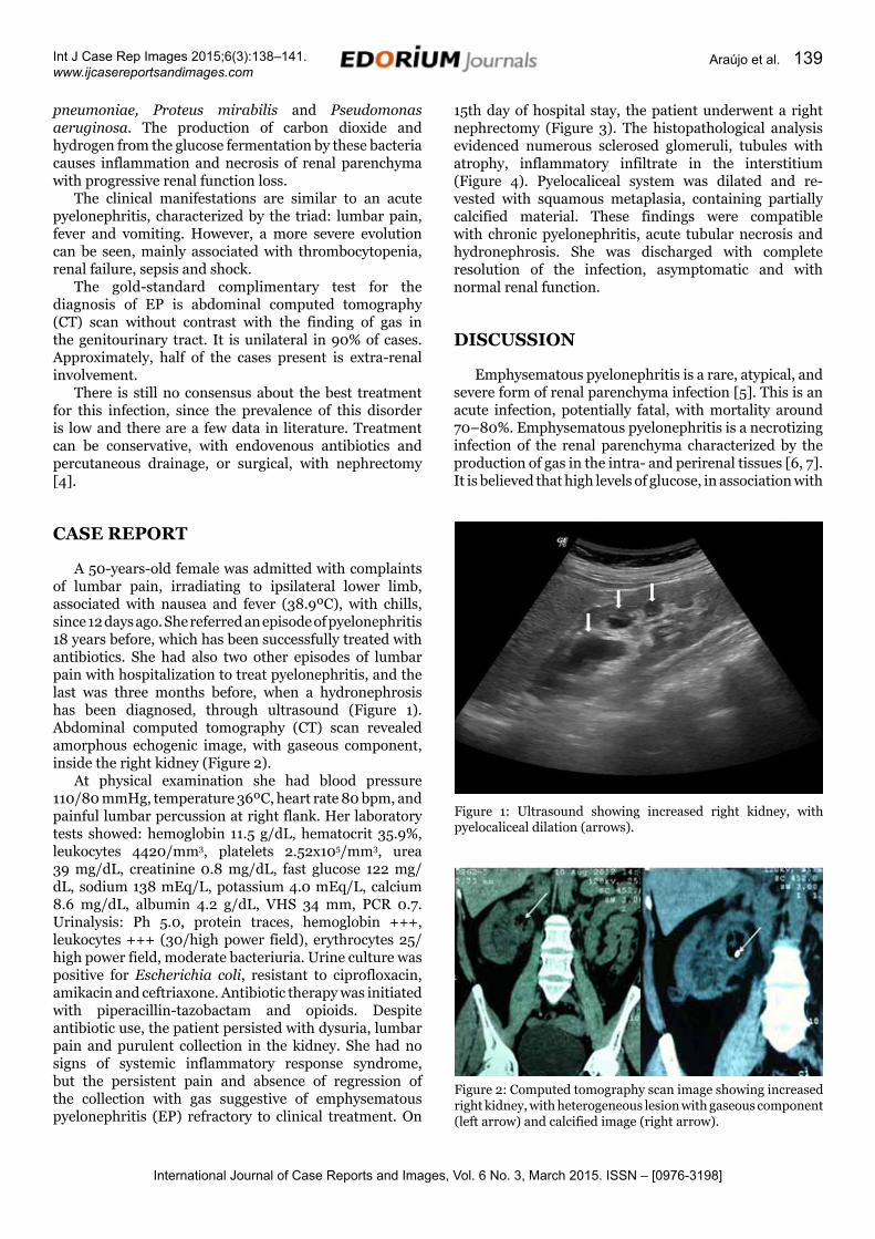
A 50-years-old female was admitted with complaints of lumbar pain, irradiating to ipsilateral lower limb, associated with nausea and fever (38.9ºC), with chills, since 12 days ago. She referred an episode of pyelonephritis 18 years before, which has been successfully treated with antibiotics. She had also two other episodes of lumbar pain with hospitalization to treat pyelonephritis, and the last was three months before, when a hydronephrosis has been diagnosed, through ultrasound (Figure 1). Abdominal computed tomography (CT) scan revealed amorphous echogenic image, with gaseous component, inside the right kidney (Figure 2).
At physical examination she had blood pressure 110/80 mmHg, temperature 36ºC, heart rate 80 bpm, and painful lumbar percussion at right flank. Her laboratory tests showed: hemoglobin 11.5 g/dL, hematocrit 35.9%, leukocytes 4420/mm3, platelets 2.52x10⁵/mm3, urea 39 mg/dL, creatinine 0.8 mg/dL, fast glucose 122 mg/dL, sodium 138 mEq/L, potassium 4.0 mEq/L, calcium 8.6 mg/dL, albumin 4.2 g/dL, VHS 34 mm, PCR 0.7. Urinalysis: Ph 5.0, protein traces, hemoglobin +++, leukocytes +++ (30/high power field), erythrocytes 25/high power field, moderate bacteriuria. Urine culture was positive for Escherichia coli, resistant to ciprofloxacin, amikacin and ceftriaxone. Antibiotic therapy was initiated with piperacillin-tazobactam and opioids. Despite antibiotic use, the patient persisted with dysuria, lumbar pain and purulent collection in the kidney. She had no signs of systemic inflammatory response syndrome, but the persistent pain and absence of regression of the collection with gas suggestive of emphysematous pyelonephritis (EP) refractory to clinical treatment. On
15th day of hospital stay, the patient underwent a right nephrectomy (Figure 3). The histopathological analysis evidenced numerous sclerosed glomeruli, tubules with atrophy, inflammatory infiltrate in the interstitium (Figure 4). Pyelocaliceal system was dilated and re-vested with squamous metaplasia, containing partially calcified material. These findings were compatible with chronic pyelonephritis, acute tubular necrosis and hydronephrosis. She was discharged with complete resolution of the infection, asymptomatic and with normal renal function.
DIscUssION
Emphysematous pyelonephritis is a rare, atypical, and severe form of renal parenchyma infection [5]. This is an acute infection, potentially fatal, with mortality around 70–80%. Emphysematous pyelonephritis is a necrotizing infection of the renal parenchyma characterized by the production of gas in the intra- and perirenal tissues [6, 7]. It is believed that high levels of glucose, in association with
Figure 1: Ultrasound showing increased right kidney, with pyelocaliceal dilation (arrows).
Figure 2: Computed tomography scan image showing increased right kidney, with heterogeneous lesion with gaseous component (left arrow) and calcified image (right arrow).

International Journal of Case Reports and Images, Vol. 6 No. 3, March 2015. ISSN – [0976-3198]
Int J Case Rep Images 2015;6(3):138–141. www.ijcasereportsandimages.com
Araújo et al. 140
inadequate perfusion, lead to a favorable environment for the growth of anaerobic organisms. This disease affects individuals of all ages, but women are six times more likely to be affected [6, 7].
Emphysematous pyelonephritis is more common in patients with diabetes [1]. Our patient did not have diagnosis of diabetes, but had altered fast glucose. An obstructive factor was found in the present case, nephrolithiasis, which is a known risk factor for chronic
kidney disease. She had history of recurrent urinary tract infections, with increased frequency in the last years. Emphysematous pyelonephritis was suspected after doing a CT scan, which showed a gaseous component in the right kidney.
Huang and Tseng [8] proposed a radiological classification for emphysematous pyelonephritis in four classes: (1) gas in the collecting system only, (2) gas in the renal parenchyma without extension to extrarenal space, (3A) extension of gas or abscess to perinephric space, (3B) extension of gas or abscess to pararenal space, and (4) bilateral EPN or solitary kidney with emphysematous pyelonephritis. According to this classification, the case presented here falls in class II, which is associated with a better prognosis. Some factors are associated with poor prognosis such as thrombocytopenia, renal failure, hyponatremia and sepsis. No one of these factors was present in our patient.
Based on the cases published by now, there is no consensus regarding the best treatment conservative management, with endovenous antibiotics and percutaneous drainage, or surgery, with nephrectomy of the involved kidney should be chosen according to each case. Early nephrectomy (<1 week) is associated with increased mortality in comparison to conservative treatment. In the present case, the patient underwent an elective nephrectomy due to the chronicity of the disease. Patients with chronic pyelonephritis and renal function <10% evidenced by scintigraphy benefits with nephrectomy.
cONcLUsION
In summary, we reported a rare case of emphysematous pyelonephritis in a non-diabetic patient. Renal lithiasis was evidenced in this case, and it represents a possible risk factor for pyelonephritis due to chronic urinary tract obstruction. Repeated history of pyelonephritis is another important factor for emphysematous pyelonephritis.
*********
Author contributionsSônia M. H. A. Araújo – Substantial contributions to conception and design, Drafting the article, Revising it critically for important intellectual content, Final approval of the version to be publishedMateus P. M. Feitosa – Substantial contributions to conception and design, Drafting the article, Revising it critically for important intellectual content, Final approval of the version to be publishedPriscila D. Evangelista – Substantial contributions to conception and design, Drafting the article, Revising it critically for important intellectual content, Final approval of the version to be publishedSarah F. Sá – Substantial contributions to conception and design, Drafting the article, Revising it critically
Figure 3: Macroscopic image of right kidney after nephrectomy showing purulent secretion and thickening of the renal parenchyma.
Figure 4: Histopathological analysis evidencing sclerosed glomerulus, tubules with atrophy and inflammatory infiltrate in the interstitium.

International Journal of Case Reports and Images, Vol. 6 No. 3, March 2015. ISSN – [0976-3198]
Int J Case Rep Images 2015;6(3):138–141. www.ijcasereportsandimages.com
Araújo et al. 141
for important intellectual content, Final approval of the version to be publishedJarinne C. L. Nasserala – Substantial contributions to conception and design, Drafting the article, Revising it critically for important intellectual content, Final approval of the version to be publishedYuri Nóbrega – Substantial contributions to conception and design, Drafting the article, Revising it critically for important intellectual content, Final approval of the version to be publishedPedro Miguel A. Pinheiro – Substantial contributions to conception and design, Drafting the article, Revising it critically for important intellectual content, Final approval of the version to be publishedNicole Almeida de Alencar Araújo – Substantial contributions to conception and design, Drafting the article, Revising it critically for important intellectual content, Final approval of the version to be publishedElizabeth F. Daher – Substantial contributions to conception and design, Drafting the article, Revising it critically for important intellectual content, Final approval of the version to be publishedGeraldo B. Silva Junior – Substantial contributions to conception and design, Drafting the article, Revising it critically for important intellectual content, Final approval of the version to be published
GuarantorThe corresponding author is the guarantor of submission.
conflict of InterestAuthors declare no conflict of interest.
copyright© 2015 Sônia M. H. A. Araújo et al. This article is distributed under the terms of Creative Commons Attribution License which permits unrestricted use,
distribution and reproduction in any medium provided the original author(s) and original publisher are properly credited. Please see the copyright policy on the journal website for more information.
rEFErENcEs
1. Carvalho M, Goulão J, Monteiro C, et al. Pielonefrite enfisematosa: revisão da literatura a propósito de um caso clínico. Acta Urol 2006;23:75–80.
2. Junior M, Ferreira G, Lopes H, Amaral K. Pielonefrite enfisematosa: revisão e atualização da abordagem terapêutica. HU Revista (Juiz de Fora) 2010;36:161–5.
3. Cavalli AC, Tambara Filho R, Cavalli RC, Bressan F. Emphysematous pyelonephritis: importance of early diagnosis. Rev Méd Paraná (Curitiba) 2006;64:13–15.
4. Eloubeidi MA, Fowler VG Jr. Images in clinical medicine. Emphysematous pyelonephritis. N Engl J Med 1999 Sep 2;341(10):737.
5. Lim CS, Kim WB, Kim YS, et al. Bilateral emphysematous pyelonephritis with perirenal abscess cured by conservative therapy. J Nephrol 2000 Mar-Apr;13(2):155–8.
6. McGorry DM, Kroser J, Taylor LT, Howard Lewis H, Gabale D. Emphysematous Pyelonephritis Presenting as an Acute Abdomen. Infect Urol 1999;12:162–5.
7. Jain SK, Agarwal N, Chaturvedi SK. Emphysematous pyelonephritis: A rare presentation. J Postgrad Med 2000 Jan-Mar;46(1):31–2.
8. Huang JJ, Tseng CC. Emphysematous pyelonephritis: clinicoradiological classification, management, prognosis, and pathogenesis. Arch Intern Med 2000 Mar 27;160(6):797–805.
Access full text article onother devices
Access PDF of article onother devices

EDORIUM JOURNALS AN INTRODUCTION
Edorium Journals: On Web
About Edorium JournalsEdorium Journals is a publisher of high-quality, open ac-cess, international scholarly journals covering subjects in basic sciences and clinical specialties and subspecialties.
Edorium Journals www.edoriumjournals.com
Edorium Journals et al.
Edorium Journals: An introduction
Edorium Journals Team
But why should you publish with Edorium Journals?In less than 10 words - we give you what no one does.
Vision of being the bestWe have the vision of making our journals the best and the most authoritative journals in their respective special-ties. We are working towards this goal every day of every week of every month of every year.
Exceptional servicesWe care for you, your work and your time. Our efficient, personalized and courteous services are a testimony to this.
Editorial ReviewAll manuscripts submitted to Edorium Journals undergo pre-processing review, first editorial review, peer review, second editorial review and finally third editorial review.
Peer ReviewAll manuscripts submitted to Edorium Journals undergo anonymous, double-blind, external peer review.
Early View versionEarly View version of your manuscript will be published in the journal within 72 hours of final acceptance.
Manuscript statusFrom submission to publication of your article you will get regular updates (minimum six times) about status of your manuscripts directly in your email.
Our Commitment
Mentored Review Articles (MRA)Our academic program “Mentored Review Article” (MRA) gives you a unique opportunity to publish papers under mentorship of international faculty. These articles are published free of charges.
Favored Author programOne email is all it takes to become our favored author. You will not only get fee waivers but also get information and insights about scholarly publishing.
Institutional Membership programJoin our Institutional Memberships program and help scholars from your institute make their research accessi-ble to all and save thousands of dollars in fees make their research accessible to all.
Our presenceWe have some of the best designed publication formats. Our websites are very user friendly and enable you to do your work very easily with no hassle.
Something more...We request you to have a look at our website to know more about us and our services.
We welcome you to interact with us, share with us, join us and of course publish with us.
Browse Journals
CONNECT WITH US
Invitation for article submissionWe sincerely invite you to submit your valuable research for publication to Edorium Journals.
Six weeksYou will get first decision on your manuscript within six weeks (42 days) of submission. If we fail to honor this by even one day, we will publish your manuscript free of charge.
Four weeksAfter we receive page proofs, your manuscript will be published in the journal within four weeks (31 days). If we fail to honor this by even one day, we will pub-lish your manuscript free of charge and refund you the full article publication charges you paid for your manuscript.
This page is not a part of the published article. This page is an introduction to Edorium Journals and the publication services.
Related Documents