Embryology of the dioecious Australian endemic Lomandra longifolia (Lomandraceae) Nabil M. Ahmad A , Peter M. Martin A,B and John M. Vella A A Amenity Horticulture Unit, University of Sydney Plant Breeding Institute, PMB 11, Camden, NSW 2570, Australia. B Corresponding author. Email: [email protected] Abstract. Microsporogenesis, embryogeny and endosperm development of Lomandra longifolia Labill. are described in detail. The formation of the anther wall is the basic type composed of four cell layers, namely an epidermis, an endothecium, one middle layer and a tapetum. The tapetum layer has glandular, uninucleate cells. Successive cytokinesis follows meiosis, subsequently forming a tetrahedral tetrad of microspores. The ovule in each carpel is hemitropous, crassinucellate and bitegmic, with the micropyle formed by the inner integument. The archesporial cell divides periclinally to form the primary parietal and primary sporogenous cells. The sporogenous cell functions as the megaspore mother cell, whereas the parietal cell divides to give rise to two parietal layers. The mature megagametophyte, which has enlarged synergids and antipodals, is of the Polygonum type, with the normal complement of seven cells and eight nuclei. Nucellar tissue in the mature ovule consists of enlarged dermal cells and irregular subdermal cells surrounding a central strand of markedly smaller cells. Endosperm development is of the nuclear type. Embryo development is of the Graminad type, characterised by oblique zygotic and early pro-embryonic divisions. Introduction Lomandra is a genus of 50 species native to Australia, of which two species extend to New Guinea and one of these to New Caledonia (Lee and Macfarlane 1986). L. longifolia, the subject of this paper, is one of the most widely distributed Lomandra species, with a range along the coast and tablelands from North Queensland to south-eastern South Australia and eastern and northern Tasmania (Lee and Macfarlane 1986). The systematic position of this genus has long been problematic; it was placed in the family Juncaceae by Bentham (1878) and by Bentham and Hooker (1880), then in Liliaceae (Krause 1930), Xanthorrhoeaceae (Cronquist 1981), Dasypogonaceae (Dahlgren et al. 1985), Xanthorrhoeaceae (Bedford et al. 1986) and more recently Lomandraceae (Brummitt 1992; Rudall and Chase 1996) and Laxmanniaceae (APG 1998; APGII 2003). The Australian Plant Name Index (2008) currently places Lomandra in the family Lomandraceae. There is considerable activity concerning relationships within the Asparagales (Rudall 1994; Rudall and Chase 1996; APGII 2003), suggesting that there will be further changes in relation to the preferred family placement of the genus Lomandra. Rudall (1994) examined the ovule, the mature embryo sac and some aspects of megagametophyte development in several genera grouped by Bedford et al. (1986) under Xanthorrhoeaceae. She noted in Lomandra markedly enlarged cells in the chalazal dermal layer of the nucellus, and very large (‘giant’) synergids and antipodals. On the basis of this work and other anatomical and molecular studies, Rudall and Chase (1996) concluded that there are several generic groupings in Xanthorrhoeaceae sensu lato deserving recognition at family level and proposed a recircumscription of the family Lomandraceae (as originally defined by Lotsy in 1911) to include Lomandra, Acanthocarpus, Chamaexeros, Romnalda, Xerolirion and certain Arthropodioid genera such as Thysanotus and Cordyline. Davis (1966), summarising the account of Schnarf and Wunderlich (1939), described the embryo-sac development of Lomandra rigida as follows: ‘the archesporial cell cuts off a primary parietal cell which gives rise to three parietal layers, and the apical cells of the nucellar epidermis divide periclinally to form a nucellar cap three cells in thickness. Cytokinesis in the megaspore mother cell accompanies meiosis and the chalazal megaspore of a linear tetrad develops into a Polygonum type embryo sac. The synergids are pyriform and exhibit the filiform apparatus, the polar nuclei fuse before fertilisation, and three large antipodal cells are formed’. The paper of Schnarf and Wunderlich (1939) contains what seems to be the only published account of the sequence of embryo-sac development in any species of Lomandra; no information has been found in the literature on embryo development, endosperm development or microsporogenesis in Lomandra. In the present paper, we describe in detail the embryological characters and endosperm development of L. longifolia as well as the process of microsporogenesis. It is hoped that this data, together with similar information on other Lomandra species and related genera, will contribute to furthering understanding of relationships and phylogeny within the family Lomandraceae and to evaluating relationships with allied families. Materials and methods Source of material Plants of L. longifolia grown by Leppington Speedy Ò Seedlings Pty Ltd, Leppington, New South Wales, from seed collected in the Southern Highlands region of New South Wales, were supplied in nursery trays when ~5 cm high. These were grown to maturity and CSIRO PUBLISHING www.publish.csiro.au/journals/ajb Australian Journal of Botany, 2008, 56, 651–665 Ó CSIRO 2008 10.1071/BT07222 0067-1924/08/080651

Welcome message from author
This document is posted to help you gain knowledge. Please leave a comment to let me know what you think about it! Share it to your friends and learn new things together.
Transcript
Embryology of the dioecious Australian endemic Lomandra longifolia(Lomandraceae)
Nabil M. AhmadA, Peter M. MartinA,B and John M. VellaA
AAmenityHorticultureUnit, University of Sydney Plant Breeding Institute, PMB11,Camden,NSW2570, Australia.BCorresponding author. Email: [email protected]
Abstract. Microsporogenesis, embryogeny and endosperm development of Lomandra longifolia Labill. are described indetail. The formation of the anther wall is the basic type composed of four cell layers, namely an epidermis, an endothecium,one middle layer and a tapetum. The tapetum layer has glandular, uninucleate cells. Successive cytokinesis followsmeiosis,subsequently forming a tetrahedral tetrad of microspores. The ovule in each carpel is hemitropous, crassinucellate andbitegmic, with the micropyle formed by the inner integument. The archesporial cell divides periclinally to form the primaryparietal and primary sporogenous cells. The sporogenous cell functions as the megaspore mother cell, whereas the parietalcell divides to give rise to two parietal layers. Thematuremegagametophyte, which has enlarged synergids and antipodals, isof the Polygonum type, with the normal complement of seven cells and eight nuclei. Nucellar tissue in the mature ovuleconsists of enlarged dermal cells and irregular subdermal cells surrounding a central strand of markedly smaller cells.Endosperm development is of the nuclear type. Embryo development is of the Graminad type, characterised by obliquezygotic and early pro-embryonic divisions.
Introduction
Lomandra is a genus of 50 species native to Australia, of whichtwo species extend to New Guinea and one of these to NewCaledonia (Lee andMacfarlane1986).L. longifolia, the subject ofthis paper, is one of the most widely distributed Lomandraspecies, with a range along the coast and tablelands fromNorth Queensland to south-eastern South Australia and easternand northern Tasmania (Lee and Macfarlane 1986).
The systematic position of this genus has long beenproblematic; it was placed in the family Juncaceae byBentham (1878) and by Bentham and Hooker (1880), then inLiliaceae (Krause 1930), Xanthorrhoeaceae (Cronquist 1981),Dasypogonaceae (Dahlgren et al. 1985), Xanthorrhoeaceae(Bedford et al. 1986) and more recently Lomandraceae(Brummitt 1992; Rudall and Chase 1996) and Laxmanniaceae(APG 1998; APGII 2003). The Australian Plant Name Index(2008) currently places Lomandra in the family Lomandraceae.There is considerable activity concerning relationships within theAsparagales (Rudall 1994;Rudall andChase 1996;APGII 2003),suggesting that there will be further changes in relation to thepreferred family placement of the genus Lomandra.
Rudall (1994) examined the ovule, themature embryo sac andsomeaspects ofmegagametophytedevelopment in several generagrouped by Bedford et al. (1986) under Xanthorrhoeaceae. Shenoted inLomandramarkedly enlarged cells in the chalazal dermallayer of the nucellus, and very large (‘giant’) synergids andantipodals. On the basis of this work and other anatomical andmolecular studies, Rudall and Chase (1996) concluded that thereare several generic groupings in Xanthorrhoeaceae sensu latodeserving recognition at family level and proposed arecircumscription of the family Lomandraceae (as originallydefined by Lotsy in 1911) to include Lomandra,
Acanthocarpus, Chamaexeros, Romnalda, Xerolirion andcertain Arthropodioid genera such as Thysanotus and Cordyline.
Davis (1966), summarising the account of Schnarf andWunderlich (1939), described the embryo-sac development ofLomandra rigida as follows: ‘the archesporial cell cuts off aprimary parietal cell which gives rise to three parietal layers, andthe apical cells of the nucellar epidermis divide periclinally toform a nucellar cap three cells in thickness. Cytokinesis in themegaspore mother cell accompanies meiosis and the chalazalmegaspore of a linear tetrad develops into a Polygonum typeembryo sac. The synergids are pyriform and exhibit the filiformapparatus, thepolar nuclei fuse before fertilisation, and three largeantipodal cells are formed’. The paper of Schnarf andWunderlich(1939) contains what seems to be the only published account ofthe sequence of embryo-sac development in any species ofLomandra; no information has been found in the literatureon embryo development, endosperm development ormicrosporogenesis in Lomandra.
In the present paper, we describe in detail the embryologicalcharacters and endosperm development of L. longifolia as well asthe process of microsporogenesis. It is hoped that this data,together with similar information on other Lomandra speciesand related genera, will contribute to furthering understanding ofrelationships andphylogenywithin the familyLomandraceae andto evaluating relationships with allied families.
Materials and methodsSource of materialPlants of L. longifolia grown by Leppington Speedy� SeedlingsPtyLtd,Leppington,NewSouthWales, fromseedcollected in theSouthernHighlands regionofNewSouthWales,were supplied innursery trayswhen~5 cmhigh.Thesewere grown tomaturity and
CSIRO PUBLISHING
www.publish.csiro.au/journals/ajb Australian Journal of Botany, 2008, 56, 651–665
� CSIRO 2008 10.1071/BT07222 0067-1924/08/080651
maintained in the field at the Plant Breeding Institute at Cobbitty,New South Wales. Floral and fruit materials (buds, flowers, andfruits) of various developmental stages of L. longifolia werecollected periodically from this plantation (every 2 days, frominflorescence emergence to seed ripening). Individual plantsoften had floral and fruit material at various stages of devel-opment, ranging from unopened buds through to immature fruitson different inflorescences. Vouchers of the male and femaleplants examined have been deposited in the Australian NationalHerbarium CANB (CANB 763117–CANB 763124).
For embryological studies, individual buds, flowers andseeds at different developmental stages were excised, dissectedunder a stereomicroscope and fixed in formalin acetic alcohol(FAA; 5 parts formalin : 5 parts glacial acetic acid : 90 parts50% ethanol (v/v/v)) and stored in 70% ethanol. They were
dehydrated through an ethanol series and then embedded inparaffin with melting point 58–60�C for microtoming. Serialsections (longitudinal and transverse) cut with a rotarymicrotome (Spencer 820: American Optical Co, Buffalo, NY,USA) at 6–8mm in thickness were stained with safranin-O andfast green FCF (Sass 1958), dehydrated through an alcohol seriesto 100% ethanol and mounted with DPX (BDH, Poole,UK). The samples were observed with normal brightfieldoptics on a Nikon Eclipse E800 light microscope (NikonOptical Co, Tokyo, Japan) and photographed with a mountedNikon Photo Head V-TP Sensicam camera (PCO CCD imaging:PCO Imaging, Kelheim, Germany).
For observations on food reserves in mature seeds, they werefirst softened by soaking in water and then sectioned by hand.Sections were tested immediately for the existence of oil droplets
t
en
ep en
t vn
g
p
pore
porespc
ep
enml
tst
ep
enml
t
tmmc
cw
epen
ml
t
cw
1 2 3
6 5 4
8 9 10 7 vn
ms t
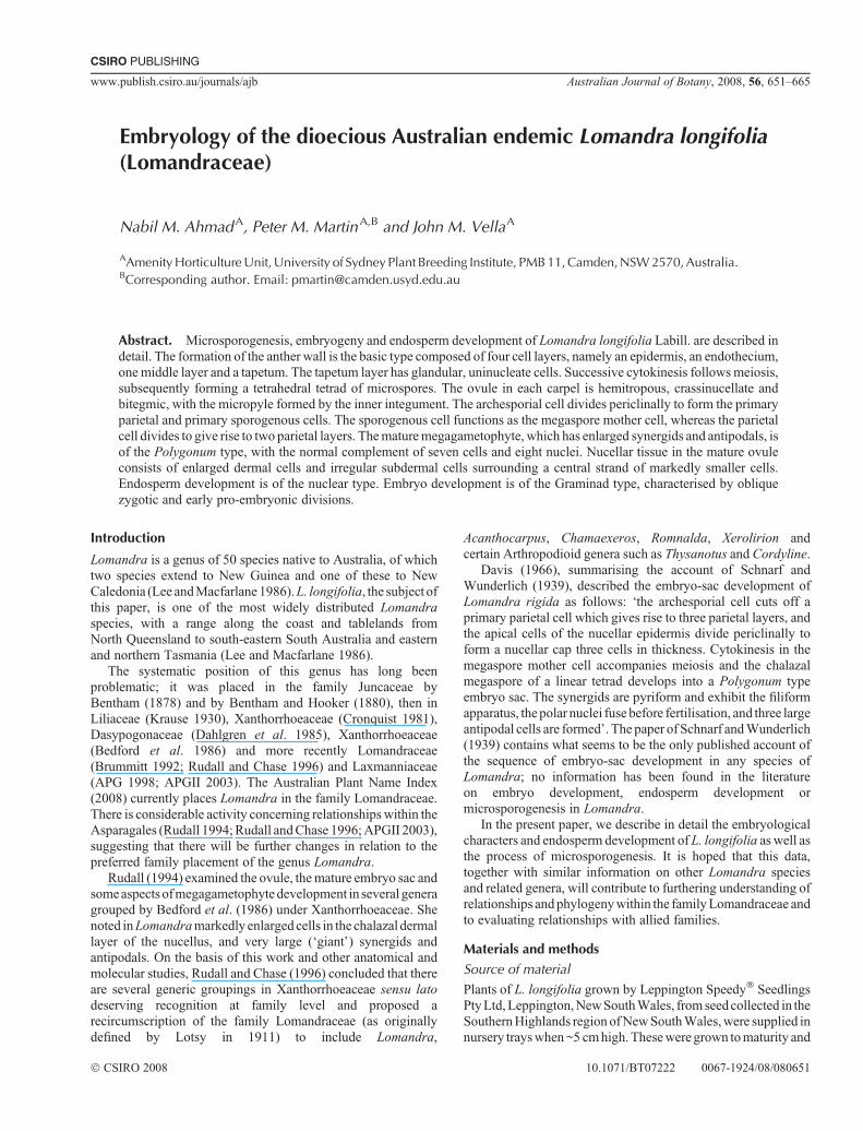
Figs 1–10. Development of an anther and microspores in Lomandra longifolia. Fig. 1. Transverse section of a young anther showing two lobes and fourmicrosporangia. Fig. 2. Four-cell-layered wall structure. Fig. 3. Isolation of individual microspore mother cells by callose wall. Fig. 4. Dyadformation. Fig. 5. Microsporangium showing the glandular tapetum and the tetrads in the callose wall. Fig. 6. Multilayered endothecium-like tissue.Fig. 7.Releaseofmicrospores bydissolutionof callosewall. Fig. 8.Maturemicrosporangiumwith themiddle layer degeneratedand the cells of the endotheciumenlarged and developing thickenings. Fig. 9. Scanning electronmicrograph showingmature pollen grains in face and rear view. Fig. 10.Mature pollen grainwith two sperm cells embedded in vegetative cell (nucleus on the right side). cw, callosewall; en, endothecium; ep, epidermis; g, generative cell; ml,middle layer;mmc, microspore mother cell; ms, microspore; p, pollen; spc, sperm cell; st, sporogenous tissue; t, tapetum; vn, vegetative nucleus. Scale bar = 33mm (Figs 1–8),10mm (Figs 9, 10).
652 Australian Journal of Botany N. M. Ahmad et al.
Epidermis
Endothecium
Middle layer
TapetumPollen mother cell
Connective tissue
Petal
Filament
Anther
Pollen grain
Microspores
Microspore mother cells
Microspore mother cellat metaphase I
Callose wall
Dyads of microsporesafter telophase I
Callose wall
Tetrad of microsporesafter telophase II
Callose wall
Meiosis I
Meiosis II
Vacuole
Mitosis
Mitosis
Vegetative Nucleus
Sperm Cells
Vegetative Nucleus
Generative Cell
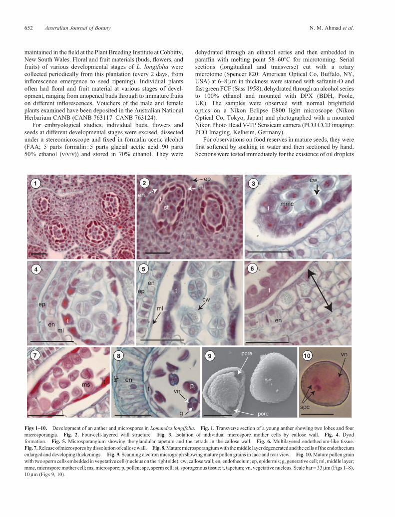
Fig. 11. Diagrammatic representation of pollen development in Lomandra longifolia. Scale bars = 33mm.
Embryology of Lomandra longifolia Australian Journal of Botany 653
with Sudan IV, for starchwith iodine in potassium iodide solutionand for protein with Millon’s reagent.
To observe and analyse the mature ovules without sectioning,flowerswerefixed in FAA (5 parts formalin : 5 parts glacial aceticacid : 90 parts 50% ethanol (v/v/v)) and stored in 70% ethanol.The ovules were dissected out with fine forceps and soaked inHoyer’s fluid (3 g of gum arabic, 20 g of chloral hydrate, 2mL ofglycerol and 5mLof distilledwater as described inRuzin (1999))for 24 h at room temperature. The cleared ovulesweremounted inHoyer’s fluid and examined by differential interference contrast(DIC) optics on a Nikon Eclipse E800 light microscope andphotographedwith amountedNikonPhotoHeadV-TPSensicamcamera (PCO CCD imaging).
For scanning electron microscopy (SEM), dehiscing antherswerefixed in 2.5%glutaraldehyde (in 0.1Mpotassiumphosphatebuffer, pH 7.1) at 20�C for 2 h. After thrice rinsing in the samebuffer (5min each), theyweredehydrated sequentially in agradedethanol series (50, 70, 95 and 100%). Samples were critical pointdried in CO2 (BAL-TEC 030 critical point dryer: Bal-Tec,Balzers, Leichtenstein). Pollen grains were dusted on stickytape affixed to aluminium stubs. Mounted pollens were coatedwith 20 nm gold palladium in a sputter coater (Edwards E306 A:Edwards Vacuum Systems, Crawley, UK), examined with aPhilips 505 SEM (Philips, Eindhoven, Netherlands) operatingat an accelerating voltage of 15KV and photographed with amounted digital camera.
ii
ii
ii
oi
oi
ac
pa
loc
nu
fg
ovi
mi
mmc
(a)
(b)(c)
(d )
(e)
(f )
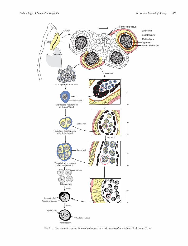
Fig. 12. Ovule and integument initiation inLomandra longifolia. (a) Early erect stage. (b, c)Developingovule undergoingcurvature. (d)Recurvingyoungovulewith enlarged megaspore mother cell in nucellus; inner integument starting to extend over nucellus. (e) Recurving ovule with inner integument just overtoppingnucellus andouter integumentpartially formed. ( f )Completely invertedovulewithnucellusovertoppedbyboth integuments; themicropyle is formedby the innerintegument which protrudes through the outer integument. The axis of themicropyle forms an angle of ~90�with the plane of attachment of the ovule to the ovary(hemitropous type). ac, archesporial cell; fg, female gametophyte; ii, inner integument; loc, loculus; mi, micropyle; mmc, megaspore mother cell; nu, nucellus;oi, outer integument; ovi, ovule initial; pa, place of attachment. Scale bar = 33mm.
654 Australian Journal of Botany N. M. Ahmad et al.
mmc
oi
ii
sc
pc
ac
oi
ii
fc
dc
nu
n
n
nn
v
nu
n
n
n
n
vv
nu
n n
v
nu
nn
n
n
nu
n
n
nn
nu
dc
13 14 15
16 17 18
19 20 21
22 23 24
dc
dc
ii
nu
nue
pl
ii
ac
oi
pl
nc
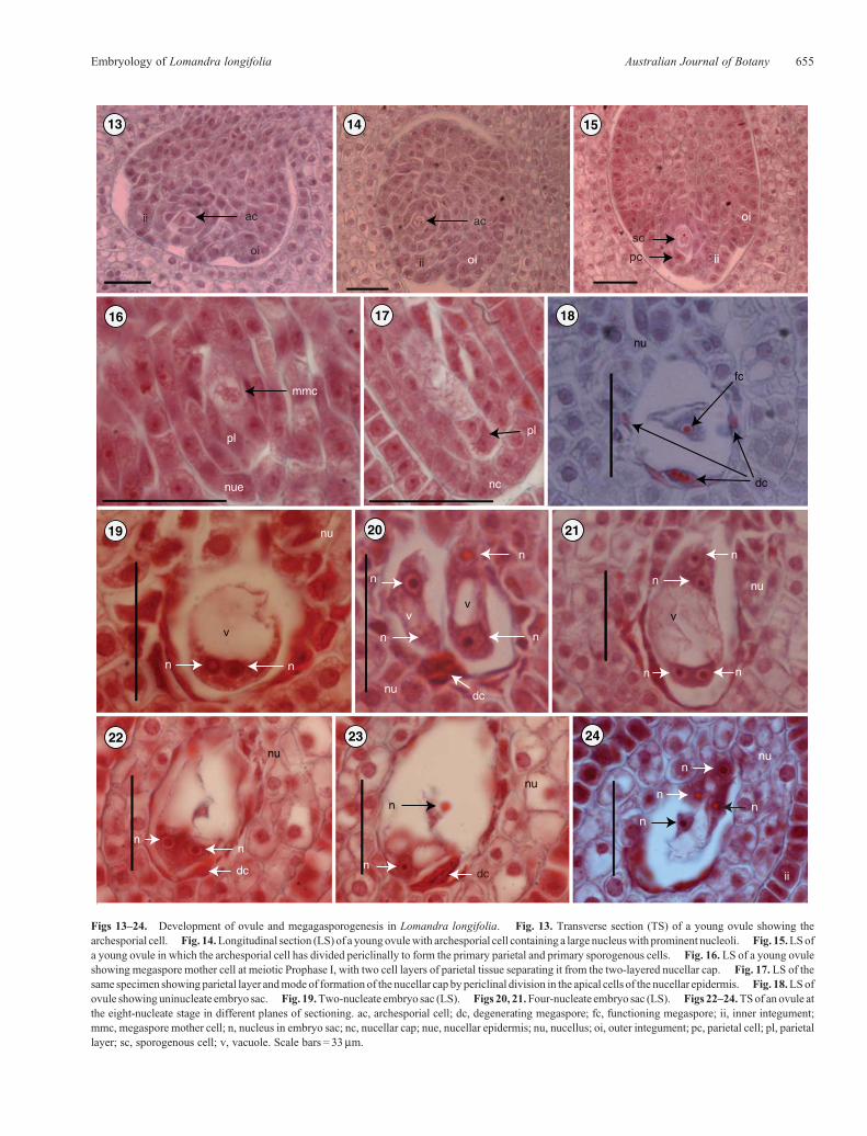
Figs 13–24. Development of ovule and megagasporogenesis in Lomandra longifolia. Fig. 13. Transverse section (TS) of a young ovule showing thearchesporial cell. Fig. 14.Longitudinal section (LS) of a youngovulewith archesporial cell containing a large nucleuswith prominent nucleoli. Fig. 15.LSofa young ovule in which the archesporial cell has divided periclinally to form the primary parietal and primary sporogenous cells. Fig. 16. LS of a young ovuleshowing megaspore mother cell at meiotic Prophase I, with two cell layers of parietal tissue separating it from the two-layered nucellar cap. Fig. 17. LS of thesame specimen showing parietal layer andmode of formation of the nucellar cap by periclinal division in the apical cells of the nucellar epidermis. Fig. 18.LSofovule showinguninucleate embryo sac. Fig. 19.Two-nucleate embryo sac (LS). Figs 20, 21.Four-nucleate embryo sac (LS). Figs 22–24.TSof anovule atthe eight-nucleate stage in different planes of sectioning. ac, archesporial cell; dc, degenerating megaspore; fc, functioning megaspore; ii, inner integument;mmc, megaspore mother cell; n, nucleus in embryo sac; nc, nucellar cap; nue, nucellar epidermis; nu, nucellus; oi, outer integument; pc, parietal cell; pl, parietallayer; sc, sporogenous cell; v, vacuole. Scale bars = 33mm.
Embryology of Lomandra longifolia Australian Journal of Botany 655
29 31 32
sysy
aa sn
30
sy
sy
sysy
a
a
sysy
mi
iioi
sn
34
cns
cns
3533
nu
nu
nu
cns
a
ii
oi
aa
cns
nc
nu
36 37
25 26 27 28pnaa
asysy
fanc
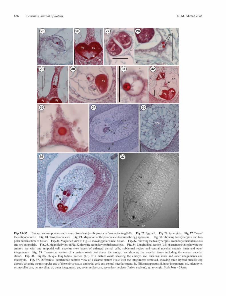
Figs 25–37. Embryo sac components andmature (8-nucleate) embryo sacs inLomandra longifolia. Fig. 25.Egg cell. Fig. 26.Synergids. Fig. 27.Two ofthe antipodal cells. Fig. 28. Two polar nuclei. Fig. 29.Migration of the polar nuclei towards the egg apparatus. Fig. 30. Showing two synergids, and twopolar nuclei at time of fusion. Fig. 31.Magnified viewof Fig. 30 showing polar nuclei fusion. Fig. 32.Showing the two synergids, secondary (fusion) nucleusand two antipodals. Fig. 33.Magnified view in Fig. 32 showing secondary or fusion nucleus. Fig. 34.Longitudinal section (LS) of amature ovule showing theembryo sac with one antipodal cell, nucellus (two layers of enlarged dermal cells, subdermal region and central nucellar strand), inner and outerinteguments. Fig. 35. Transverse section of a mature ovule just above the embryo sac showing the nucellar tissue including the central nucellarstrand. Fig. 36. Slightly oblique longitudinal section (LS) of a mature ovule showing the embryo sac, nucellus, inner and outer integuments andmicropyle. Fig. 37. Differential interference contrast view of a cleared mature ovule with the integuments removed, showing three layered nucellar capdirectly covering the micropylar end of the embryo sac. a, antipodal cell; cns, central nucellar strand; fa, filiform apparatus; ii, inner integument; mi, micropyle;nc, nucellar cap; nu, nucellus; oi, outer integument; pn, polar nucleus; sn, secondary nucleus (fusion nucleus); sy, synergid. Scale bars = 33mm.
656 Australian Journal of Botany N. M. Ahmad et al.
Results and discussion
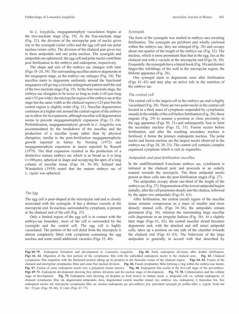
Microsporangium
Anthers of L. longifolia are tetrasporangiate, with twomicrosporangia in each lobe (Figs 1, 2). The anther wallbefore maturity comprises the following four cell layers: anepidermis, an endothecium, one middle layer and a tapetum(Figs 2–5).
The endothecium is a single layer of cells that lies justunder the epidermis. During the maturation of the anther,the endothecial cells enlarge and acquire fibrous thickenings.
In cross-section, these cells form a complete ring around eachlocule. The endothecium becomes irregularly multilayeredtowards the inner side. This endothecium-like tissue may becontributed by the subepidermal layer of the neighbouringconnective tissues which develop fibrous thickening towardsthe inner side of the anther (Fig. 6). The middle layeris ephemeral and becomes obliterated before themicrosporangium is fully mature and ready to dehisce, leavingonly a remnant layer against the endothecial wall (Fig. 5).
The anther of L. longifolia has one tapetum layer, which is theinnermost layer of the anther wall surrounding the sporogenoustissue (Fig. 2). The tapetal cells are uninucleate and remain intact,being of the glandular type (Figs 3, 4).
Microspore mother cells lose contact with the tapetal cells asthey enter the phases of meiosis and become enclosed in thickcallose walls (Fig. 3).
Meiosis in the microspore mother cell is accompanied bysuccessive cytokinesis. The first meiotic division separates twodyads (Fig. 4); each dyad undergoes Division II, resulting in atetrad. The cell-plate formation takes place after Meiosis I. Theend product of the meiotic divisions is the formation ofmicrospore tetrads with a tetrahedral shape (Fig. 5). Eachtetrad is initially enclosed within the mother cell wall whichhas a gelatinous appearance owing to deposition of callose. Thebreakdown of this wall liberates individual microspores into thesporangium (Figs 7, 8).
Atmaturity, the anther epidermis is persistent, themiddle layercompletely degenerates and the endothecium develops fibrousthickenings in the formof bars that assist the anther to dehisce andrelease pollen. Thus, the mature anther wall is composed of apersistent epidermis and a fibrous endothecium (Fig. 8). Thepollen is shed as single grains (Fig. 9). Developing pollen grainsstained deeply with safranin, preventing observation of pollenmitosis. However, staining with acetocarmine allowedidentification of a 3-celled pollen (Fig. 10). A diagrammaticrepresentation of all stages of pollen development inL. longifolia is presented in Fig. 11.
Ovule morphology
Lomandra longifolia fruit has three loculi and only one ovuleper loculus, whereas several ovules per loculus is the most
fasy
sy
cns
nua
essn
ea
oi ii
mi
eg
cen
fa
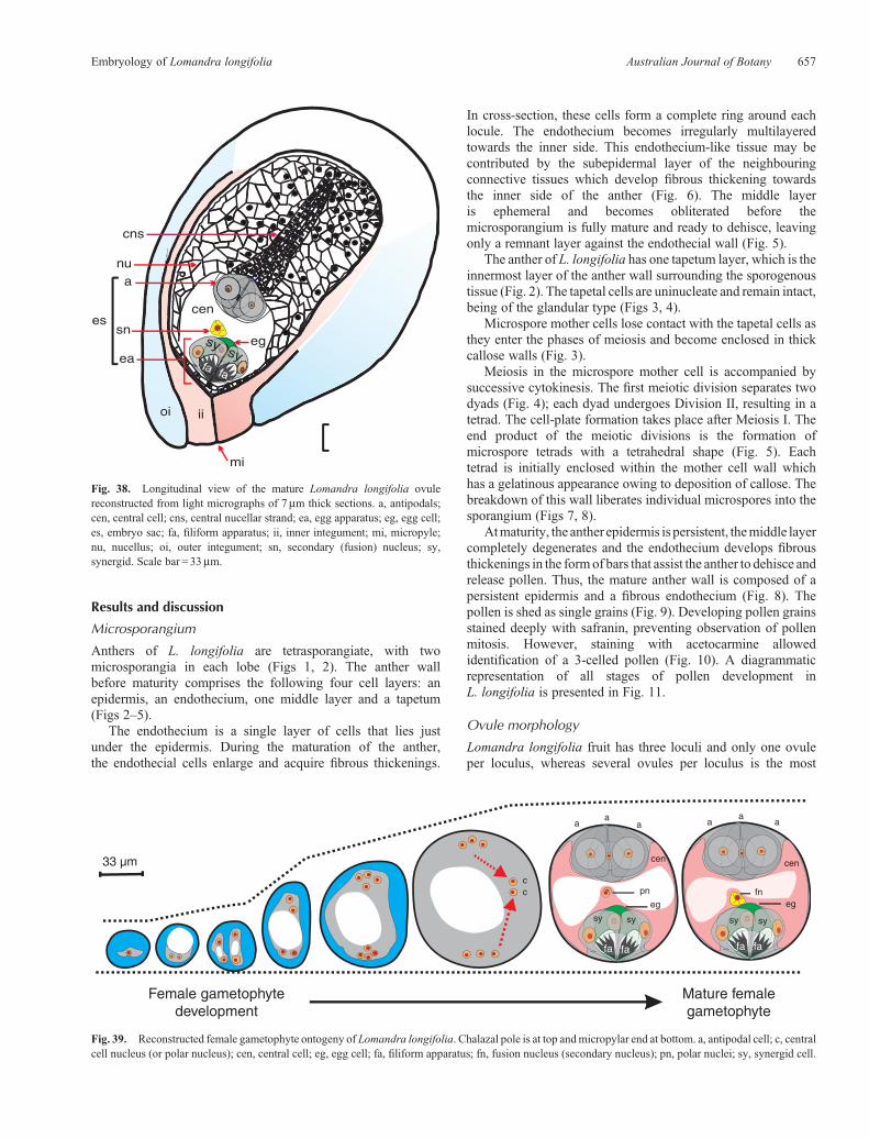
Fig. 38. Longitudinal view of the mature Lomandra longifolia ovulereconstructed from light micrographs of 7mm thick sections. a, antipodals;cen, central cell; cns, central nucellar strand; ea, egg apparatus; eg, egg cell;es, embryo sac; fa, filiform apparatus; ii, inner integument; mi, micropyle;nu, nucellus; oi, outer integument; sn, secondary (fusion) nucleus; sy,synergid. Scale bar = 33mm.
cen
pn
eg eg
cen
fn
sy sy sysy
cc
aaa a
aa
Female gametophytedevelopment
Mature femalegametophyte
Fig. 39. Reconstructed female gametophyte ontogeny of Lomandra longifolia. Chalazal pole is at top andmicropylar end at bottom. a, antipodal cell; c, centralcell nucleus (or polar nucleus); cen, central cell; eg, egg cell; fa, filiform apparatus; fn, fusion nucleus (secondary nucleus); pn, polar nuclei; sy, synergid cell.
Embryology of Lomandra longifolia Australian Journal of Botany 657
eg
sy
sy
m
ci
ca
cc
I
I′
cb
ca
cbpt
z
ps
enspen
z psy
z
psy
pe
s
pe
s
pe
em
s
pc
pc
se
mc
pb
lp
lp
lp
sa
ra cr
40 41 42 43
44
48
45 47
49 50 51
52 53 54 55
56 57 58 59
46
em
s
pt
ensensens
658 Australian Journal of Botany N. M. Ahmad et al.
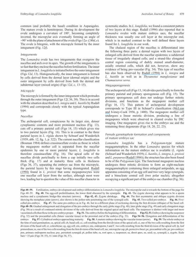
common (and probably the basal) condition in Asparagales.The mature ovule is hemitropous. During its development theovule undergoes a curvature of 180�, becoming completelyinverted, the micropylar axis eventually forming an angle of~90�with theplaneof attachment of theovule (Figs12a–f, 80d,e).The ovule is bitegmic, with the micropyle formed by the innerintegument (Fig. 12f).
Integuments
The Lomandra ovule has two integuments that overgrow thenucellus and arch over its apex. The growth of the integuments isso fast that they enclose the nucellus before fertilisation. The innerintegument inL. longifolia is initiated before the outer integument(Figs 12d, 13). Histogenetically, the inner integument is formedby cells derived from the dermal layer (dermal origin) and theouter integument by cells derived from both the dermal andsubdermal layer (mixed origin) (Figs 12d, e, 13–15).
Micropyle
Themicropyle is formedby the inner integumentwhich protrudesthrough the outer integument (Figs 12f, 36, 38). This is in accordwith the situation described in L. integra and L. hastilis byRudall(1994) and corresponds closely with the typical Asparagaleantype.
Nucellus
The archesporial cell, conspicuous by its larger size, densercytoplasmic contents and more prominent nucleus (Fig. 13),cuts off a primary parietal cell (Figs 14, 15) which gives riseto two parietal layers (Fig. 16). This is in contrast to the threeparietal layers in L. rigida shown by Schnarf and Wunderlich(1939; abb. 12, fig. 1, p. 322). The current nucellar classification(Bouman 1984) defines crassinucellate ovules as those in whichthe megaspore mother cell is separated from the nucellarepidermis by one or more parietal layers; L. longifolia istherefore crassinucellate (Fig. 16). The apical cells of thenucellus divide periclinally to form a cap initially two cellsthick (Fig. 17) and at maturity three cells in thickness(Figs 36, 37), separating the embryo sac from the micropyle,the parietal layers by this stage having disintegrated. Rudall(1994) found in L. preissii that some megasporocytes wereone nucellar cell layer from the surface, although most weretwo, leading her to question the value of this nucellar character in
systematic studies. In L. longifolia, we found a consistent patternof two layers at this stage. Rudall (1994) also reported that inLomandra ovules with mature embryo sacs, the nucellarthickness was usually one cell layer at the micropylar end,which is in marked contrast to the well defined three layersfound for L. longifolia in our work.
The chalazal region of the nucellus is differentiated intothe following three parts: a dermal region with two layers ofenlarged cells derived from the nucellar epidermis; a subdermaltissue of irregularly shaped cells; and a strand-like elongatedcentral region consisting of darkly stained small-diameter,axially oriented cells, which did not, however, show anyevidence of being pro-vascular (Fig. 34). This type of nucellushas also been observed by Rudall (1994) in L. integra andL. hastilis as well as in Thysanotus manglesianus andChamaexeros serra.
Megasporogenesis
The archesporial cell (Figs 13, 14) divides periclinally to form theprimary parietal and primary sporogenous cell (Fig. 15). Theprimary sporogenous cell does not undergo further mitoticdivisions, and functions as the megaspore mother cell(Figs 16, 17). This pattern of archesporial developmentcorresponds to Type III in Schnarf’s classification (McLeanand Ivimey-Cook 1956, p. 1400). The megaspore mother cellundergoes a linear meiotic division, producing a line ofmegaspores which were observed in cleared ovules by DICimaging. One megaspore gives rise to the embryo sac and theremaining three degenerate (Figs 18, 20, 22, 23).
Female gametophyte formation and componentsof mature embryo sac
Lomandra longifolia has a Polygonum-type maturemegagametophyte. In the other Lomandra species for whichinformation on the mature embryo sac is available (L. rigida(Schnarf andWunderlich 1939); L. hastilis, L. integra, L. preissiiand L. purpurea (Rudall 1994)), the structure has also been foundto be of the Polygonum type. The functional megaspore nucleusundergoes three mitotic divisions to form an eight-nucleatemegagametophyte containing three enlarged antipodals, an eggapparatus consisting of an egg cell and two very large synergids,and a binucleate central cell (two polar nuclei), altogetheroccupying a significant part of the embryo sac chamber (Fig. 36).
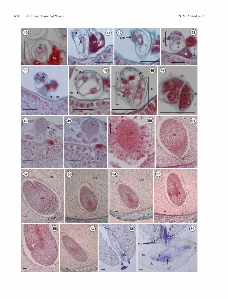
Figs 40–59. Fertilisation, embryo development and embryo differentiation in Lomandra longifolia. The micropylar end is towards the bottom of the page forFigs 40–59. Fig. 40. The egg-cell prefertilisation, the lower third obscured by the synergids. Fig. 41. The zygote showing what appears to be a spermnucleus and a cytoplasmic bridge-like structure (arrows). Figs 42, 43. Zygote shrinkage. Fig. 44. The first asymmetric and oblique zygotic divisionshowing the metaphase plate (arrow), also shown is the pollen tube penetrating one of the synergid cells. Fig. 45. Two-celled pro-embryo. Fig. 46. Five-celled pro-embryo. Fig. 47. The same pro-embryo as in Fig. 46, but in a different plane of sectioning showing the first division (oblique) of the basal cell.Figs 48–50.Gradual enlargement and elongation of the pro-embryo through the early globe stage (Fig. 48), later globe stage (Fig. 49) and early elongation stage(Fig. 50) as a result of continued mitotic divisions of the cells derived from the original apical and basal cells. The suspensor is distinct with larger and morevacuolatedcells than those in thepro-embryoproper. Fig. 51.Theembryobefore thebeginningof differentiation. Figs52, 53.Embryo showing the suspensor(Fig. 52) and the procambial cells (future vascular tissue) at the proximal end of the embryo (Fig. 53). Figs 54–56. Elongation and differentiation of theembryo. Fig. 57.Cylindrical embryo approachingmaturity. Fig. 58.Amature embryo showing the vascular tissue (arrow). Fig. 59.Thewhole embryo atthe time of seed germination. ca, apical cell; cb, basal cell; cc, cell resulting from the first division of the apical cell; ci, one of the two cells resulting from the firstdivision of the basal cell; cr, coleorhizae; eg, egg cell; em, embryo; ens, endosperm; I and I0, two cells resulting from the third division of the apical cell; lp, leafprimordium;m, oneof the twocells resulting from thefirst divisionof the basal cell;mc,micropylar cap; pb, protective bract; pc, procambial cells; pe, pro-embryo;pen, primary endosperm nucleus; psy, persistent synergid; pt, pollen tube; ra, root apex; s, suspensor; sa, shoot apex; se, seed; sy, synergid; z, zygote. Scalebars = 33mm (Figs 38–55), 0.1mm (Figs 56, 57).
Embryology of Lomandra longifolia Australian Journal of Botany 659
ce
f
ce
77 78
cf
mf
z
a
acf
ens
pt
aa
fen
ne
f
em
fne
ce
ne
psy
pe
cf
da
mf
peds
cf
mf
pe
da
ce
60 61 62 63
64 65 66 67
68 69 70 71
72 73 74
76 79
da
75
ne
dcns
ce
z
cecene
em
em
660 Australian Journal of Botany N. M. Ahmad et al.
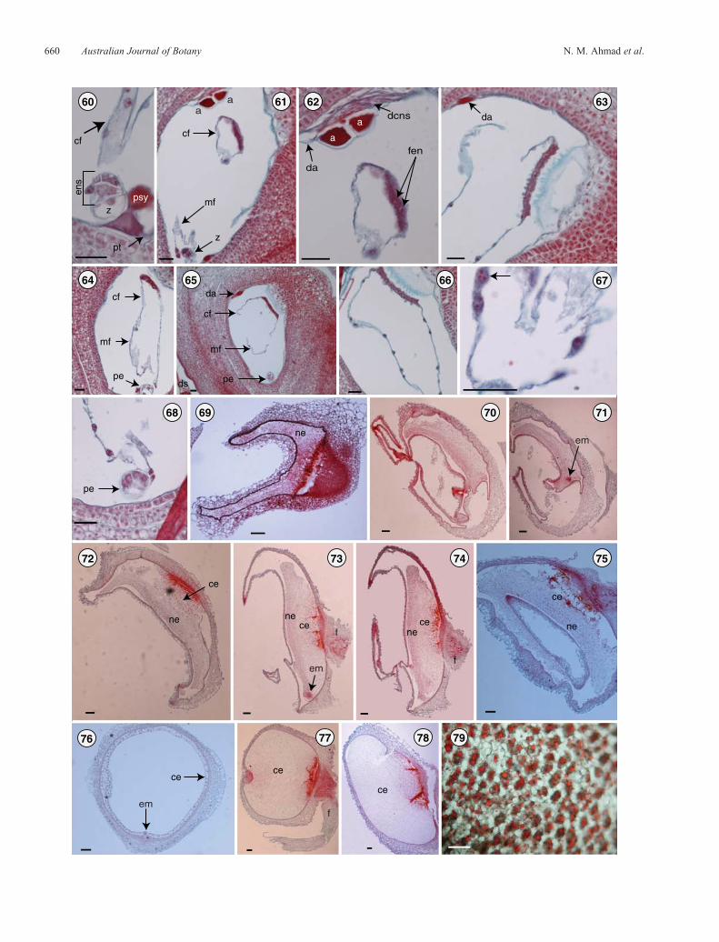
In L. longifolia, megagametophyte vacuolation begins atthe two-nucleate stage (Fig. 19). At the four-nucleate stage(Fig. 21), the division of the micropylar pair of nuclei givesrise to the synergids (sister cells) and the egg cell and one polarnucleus (sister cells). The division of the chalazal pair gives riseto three antipodals and one polar nucleus. The synergids andantipodals are ephemeral; the egg cell and polar nuclei contributepost fertilisation to the embryo and endosperm, respectively.
The shape and size of the embryo sac changes with time(Figs 18–24, 39). The surrounding nucellus starts to degenerate atthe megaspore stage, as the embryo sac enlarges (Fig. 18). Thenucellus starts to degenerate uniformly around the functionalmegaspore cell giving a circular enlargement pattern until the endof the two-nucleate stage (Fig. 19). At the four-nucleate stage, theembryo sac elongates to be twice as long as wide (�65mm longand�35mmwide); themicropylar regionof the embryo sac at thisstage has the same width as the chalazal region (�25mm) but thecentral region is slightly wider (Fig. 21). Nucellar degenerationcontinues at a higher rate around the central region of the embryosac to allow for its expansion, althoughnucellar-cell degenerationseems to precede megagametophyte expansion (Figs 21–24).Prefertilisation, megagametophytic growth in L. longifolia wasaccommodated by the breakdown of the nucellus and theproduction of a nucellar lysate rather than by physicaldisruption, similar to the post-fertilisation megagametophyticgrowth reported in barley by Norstog (1972) andmegagametophyte expansion in maize reported by Russell(1979). The final expansion resulted in the production of adistinctive mature embryo sac which is as broad as it is long(�100mm), spherical in shape and occupying the apex of a longcolumn of nucellar tissue (Figs 34, 36–38). Schnarf andWunderlich (1939) noted that the mature embryo sac ofL. rigida was spherical.
The Egg
The egg cell is pear-shaped at the micropylar end and is closelyassociated with the synergids. It has a distinct vacuole at themicropylar end. Its nucleus, surrounded by cytoplasm, is presentat the chalazal end of the cell (Fig. 25).
Only a limited region of the egg cell is in contact with theembryo-sac boundary, most of the cell is surrounded by thesynergids and the central cell. The egg cell is highlyvacuolated. The portion of the cell distal from the micropyle isalmost completely filled with cytoplasm containing the eggnucleus and some small additional vacuoles (Figs 25, 40).
Synergids
The form of the synergids was studied in embryo sacs awaitingfertilisation. The synergids are pyriform and wholly enclosedwithin the embryo sac; they are enlarged (Fig. 26) and occupyabout one-quarter of the length of the embryo sac (Fig. 32). Thenucleus, which is more prominent than that in the egg, lies at thechalazal end with a vacuole at the micropylar end (Figs 26, 30).Frequently, the synergids have a lateral hook (Fig. 36) anddistinctfinger-like infoldings of the wall in the micropylar region, thefiliform apparatus (Fig. 26).
One synergid starts to degenerate soon after fertilisation(Figs 41–43) and may play an active role in the nutrition ofthe embryo sac.
The central cell
The central cell is the largest cell in the embryo sac and is highlyvacuolated (Fig. 38). There are two polar nuclei in the central celllocated in a thick mass of cytoplasm suspended by cytoplasmicstrands in themiddle of the cell before fertilisation (Fig. 28); thesemigrate (Fig. 29) to assume a position in close proximity tothe egg apparatus (Figs 30, 31) and subsequently fuse to formthe secondary nucleus (Figs 32, 33). Fusion occurs beforefertilisation, and after the resulting secondary nucleus isfertilised, it forms the primary endosperm nucleus. The polarnuclei and fusion nucleus are the largest nuclei observed in theembryo sac (Figs 28, 30–33). The central cell contains complexorganised cytoplasm which is rich in organelles.
Antipodals and post-fertilisation nucellus
In the undifferentiated 8-nucleate embryo sac, cytokinesis isinitiated at the chalazal pole and proceeds in an orderlymanner towards the micropyle. The three antipodal nucleipersist as three cells into the post-fertilisation stages (Fig. 27).
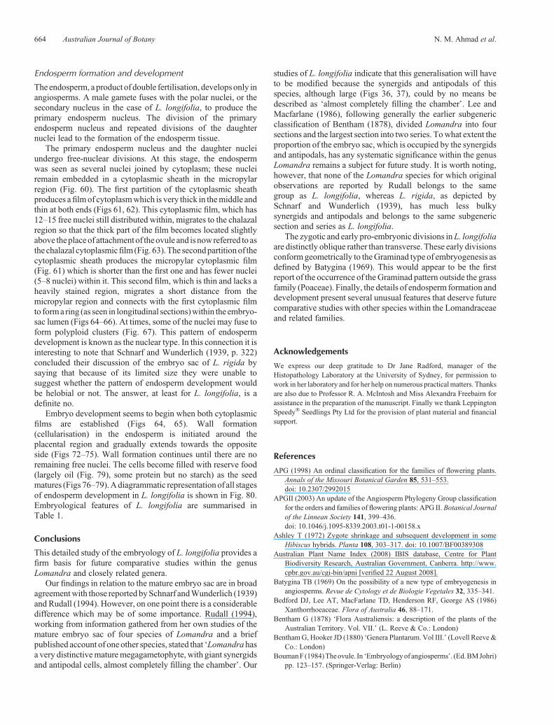
The antipodals occupy about one-third of the length of theembryo sac (Fig. 37).Degeneration of the lowest antipodal beginsinitially, after the cell penetrates deeply into the chalaza, followedby the upper two antipodals (Figs 61–63).
After fertilisation, the central (axial) region of the nucellartissue remains conspicuous as a mass of smaller and moredensely stained cells (Figs 34–36), the antipodals remainprominent (Fig. 36), whereas the surrounding large nucellarcells degenerate in an irregular fashion (Fig. 36). At a slightlylater stage (Figs 61, 62), the central nucellar strand becomesdegenerate and, with the attached and still intact antipodalcells, takes up a position on one side of the chamber towardsthe chalazal end (Figs 61–63). The behaviour of the largeantipodals is generally in accord with that described by
Figs 60–79. Endosperm formation and development in Lomandra longifolia. Fig. 60. Early endosperm divisions after double fertilisation.Figs 61, 62. Migration of the first portion of the cytoplasmic film with the embedded endosperm nuclei to the chalazal area. Fig. 63. Chalazalcytoplasmic film migration with the thickened portion taking up its position in the funicular corner of the chalazal region. Figs 64, 65. Fusion of thechalazal and micropylar cytoplasmic films and some free nuclear division. Fig. 66. Fused cytoplasmic films forming a ring within the embryo-sac lumen.Fig. 67. Fusion of some endosperm nuclei into a polyploid cluster (arrow). Fig. 68. Endosperm free nuclei at the five-cell stage of the pro-embryo.Figs 69–71. Endosperm development showing free mitotic divisions and the nuclear stage of development. Fig. 72–78. Cellularisation and the cellularstage of development. Fig. 79. Endosperm cells showing oil droplets as food reserve in mature seeds. a, antipodal cell; ce, cellular endosperm; cf,chalazal cytoplasmic film; da, degenerated antipodals; dcns, degenerated central nucellar strand; em, embryo; ens, endosperm; f, funiculus; fen, freeendosperm nuclei; mf, micropylar cytoplasmic film; ne, nuclear endosperm; pe, pro-embryo; psy, persistent synergid; pt, pollen tube; z, zygote. Scale barfor = 33mm (Figs 58–66), 0.1mm (Figs 67–77).
Embryology of Lomandra longifolia Australian Journal of Botany 661
Rudall (1994) for the mature megagametophyte of L. integraand L. purpurea.
Fertilisation and embryogeny
In L. longifolia, the pollen tube first comes into contact with theovule near the funicular end where it begins to grow along the
outer surface of the outer integument towards the micropyle.When the pollen tube reaches the micropylar end of the ovule,it grows through the micropyle (porogamous entry), through thethree layers of nucellar cap tissue and into the embryo sac(Fig. 44). The pollen tube then enters into one of the synergids(Figs 43, 44) through the filiform apparatus to release the malegametes, the vegetative nucleus and presumably a limited amount
(b)
(c)
(a)(d)
(e)
(f )
pe
mf
cf
da
pene
ne
em
em
f
em
cece
ce
testa
egsy sy
aa
afnv
cen
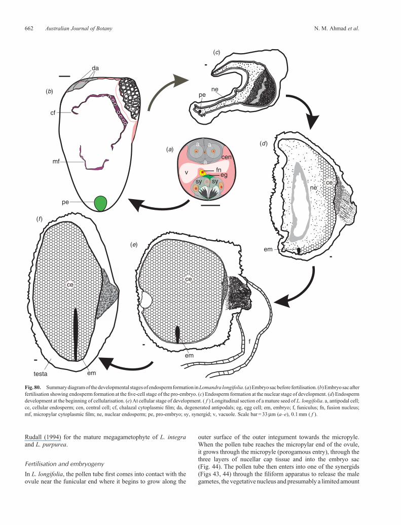
Fig. 80. Summarydiagramof thedevelopmental stagesof endospermformation inLomandra longifolia. (a)Embryosacbefore fertilisation. (b)Embryosacafterfertilisation showing endosperm formation at the five-cell stage of the pro-embryo. (c) Endosperm formation at the nuclear stage of development. (d) Endospermdevelopment at the beginning of cellularisation. (e) At cellular stage of development. ( f ) Longitudinal section of a mature seed of L. longifolia. a, antipodal cell;ce, cellular endosperm; cen, central cell; cf, chalazal cytoplasmic film; da, degenerated antipodals; eg, egg cell; em, embryo; f, funiculus; fn, fusion nucleus;mf, micropylar cytoplasmic film; ne, nuclear endosperm; pe, pro-embryo; sy, synergid; v, vacuole. Scale bar = 33mm (a–e), 0.1mm ( f ).
662 Australian Journal of Botany N. M. Ahmad et al.
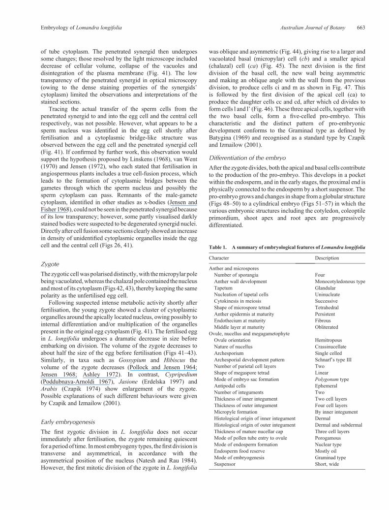
of tube cytoplasm. The penetrated synergid then undergoessome changes; those resolved by the light microscope includeddecrease of cellular volume, collapse of the vacuoles anddisintegration of the plasma membrane (Fig. 41). The lowtransparency of the penetrated synergid in optical microscopy(owing to the dense staining properties of the synergids’cytoplasm) limited the observations and interpretations of thestained sections.
Tracing the actual transfer of the sperm cells from thepenetrated synergid to and into the egg cell and the central cellrespectively, was not possible. However, what appears to be asperm nucleus was identified in the egg cell shortly afterfertilisation and a cytoplasmic bridge-like structure wasobserved between the egg cell and the penetrated synergid cell(Fig. 41). If confirmed by further work, this observation wouldsupport the hypothesis proposed by Linskens (1968), van Went(1970) and Jensen (1972), who each stated that fertilisation inangiospermous plants includes a true cell-fusion process, whichleads to the formation of cytoplasmic bridges between thegametes through which the sperm nucleus and possibly thesperm cytoplasm can pass. Remnants of the male-gametecytoplasm, identified in other studies as x-bodies (Jensen andFisher 1968), could not be seen in the penetrated synergidbecauseof its low transparency; however, some partly visualised darklystained bodies were suspected to be degenerated synergid nuclei.Directly after cell fusion somesections clearly showedan increasein density of unidentified cytoplasmic organelles inside the eggcell and the central cell (Figs 26, 41).
Zygote
The zygotic cellwas polarised distinctly,with themicropylar polebeingvacuolated,whereas the chalazal pole contained thenucleusandmost of its cytoplasm (Figs 42, 43), thereby keeping the samepolarity as the unfertilised egg cell.
Following suspected intense metabolic activity shortly afterfertilisation, the young zygote showed a cluster of cytoplasmicorganelles around the apically located nucleus, owing possibly tointernal differentiation and/or multiplication of the organellespresent in the original egg cytoplasm (Fig. 41). The fertilised eggin L. longifolia undergoes a dramatic decrease in size beforeembarking on division. The volume of the zygote decreases toabout half the size of the egg before fertilisation (Figs 41–43).Similarly, in taxa such as Gossypium and Hibiscus thevolume of the zygote decreases (Pollock and Jensen 1964;Jensen 1968; Ashley 1972). In contrast, Cypripedium(Poddubnaya-Arnoldi 1967), Jasione (Erdelska 1997) andArabis (Czapik 1974) show enlargement of the zygote.Possible explanations of such different behaviours were givenby Czapik and Izmailow (2001).
Early embryogenesis
The first zygotic division in L. longifolia does not occurimmediately after fertilisation, the zygote remaining quiescentfor a periodof time. Inmost embryogeny types, thefirst division istransverse and asymmetrical, in accordance with theasymmetrical position of the nucleus (Natesh and Rau 1984).However, the first mitotic division of the zygote in L. longifolia
was oblique and asymmetric (Fig. 44), giving rise to a larger andvacuolated basal (micropylar) cell (cb) and a smaller apical(chalazal) cell (ca) (Fig. 45). The next division is the firstdivision of the basal cell, the new wall being asymmetricand making an oblique angle with the wall from the previousdivision, to produce cells ci and m as shown in Fig. 47. Thisis followed by the first division of the apical cell (ca) toproduce the daughter cells cc and cd, after which cd divides toform cells l and l’ (Fig. 46). These three apical cells, together withthe two basal cells, form a five-celled pro-embryo. Thischaracteristic and the distinct pattern of pro-embryonicdevelopment conforms to the Graminad type as defined byBatygina (1969) and recognised as a standard type by Czapikand Izmailow (2001).
Differentiation of the embryo
After the zygote divides, both the apical and basal cells contributeto the production of the pro-embryo. This develops in a pocketwithin the endosperm, and in the early stages, the proximal end isphysically connected to the endosperm by a short suspensor. Thepro-embryo grows and changes in shape from a globular structure(Figs 48–50) to a cylindrical embryo (Figs 51–57) in which thevarious embryonic structures including the cotyledon, coleoptileprimordium, shoot apex and root apex are progressivelydifferentiated.
Table 1. A summary of embryological features of Lomandra longifolia
Character Description
Anther and microsporesNumber of sporangia FourAnther wall development Monocotyledonous typeTapetum GlandularNucleation of tapetal cells UninucleateCytokinesis in meiosis SuccessiveShape of microspore tetrad TetrahedralAnther epidermis at maturity PersistentEndothecium at maturity FibrousMiddle layer at maturity Obliterated
Ovule, nucellus and megagametophyteOvule orientation HemitropousNature of nucellus CrassinucellateArchesporium Single celledArchesporial development pattern Schnarf’s type IIINumber of parietal cell layers TwoShape of megaspore tetrad LinearMode of embryo sac formation Polygonum typeAntipodal cells EphemeralNumber of integuments TwoThickness of inner integument Two cell layersThickness of outer integument Four cell layersMicropyle formation By inner integumentHistological origin of inner integument DermalHistological origin of outer integument Dermal and subdermalThickness of mature nucellar cap Three cell layersMode of pollen tube entry to ovule PorogamousMode of endosperm formation Nuclear typeEndosperm food reserve Mostly oilMode of embryogenesis Graminad typeSuspensor Short, wide
Embryology of Lomandra longifolia Australian Journal of Botany 663
Endosperm formation and development
The endosperm, a product of double fertilisation, develops only inangiosperms. A male gamete fuses with the polar nuclei, or thesecondary nucleus in the case of L. longifolia, to produce theprimary endosperm nucleus. The division of the primaryendosperm nucleus and repeated divisions of the daughternuclei lead to the formation of the endosperm tissue.
The primary endosperm nucleus and the daughter nucleiundergo free-nuclear divisions. At this stage, the endospermwas seen as several nuclei joined by cytoplasm; these nucleiremain embedded in a cytoplasmic sheath in the micropylarregion (Fig. 60). The first partition of the cytoplasmic sheathproduces afilmof cytoplasmwhich is very thick in themiddle andthin at both ends (Figs 61, 62). This cytoplasmic film, which has12–15 free nuclei still distributed within, migrates to the chalazalregion so that the thick part of the film becomes located slightlyabove theplaceof attachment of theovule and is nowreferred to asthe chalazal cytoplasmicfilm (Fig. 63).The secondpartitionof thecytoplasmic sheath produces the micropylar cytoplasmic film(Fig. 61) which is shorter than the first one and has fewer nuclei(5–8 nuclei) within it. This second film, which is thin and lacks aheavily stained region, migrates a short distance from themicropylar region and connects with the first cytoplasmic filmto forma ring (as seen in longitudinal sections)within the embryo-sac lumen (Figs 64–66). At times, some of the nuclei may fuse toform polyploid clusters (Fig. 67). This pattern of endospermdevelopment is known as the nuclear type. In this connection it isinteresting to note that Schnarf and Wunderlich (1939, p. 322)concluded their discussion of the embryo sac of L. rigida bysaying that because of its limited size they were unable tosuggest whether the pattern of endosperm development wouldbe helobial or not. The answer, at least for L. longifolia, is adefinite no.
Embryo development seems to begin when both cytoplasmicfilms are established (Figs 64, 65). Wall formation(cellularisation) in the endosperm is initiated around theplacental region and gradually extends towards the oppositeside (Figs 72–75). Wall formation continues until there are noremaining free nuclei. The cells become filled with reserve food(largely oil (Fig. 79), some protein but no starch) as the seedmatures (Figs 76–79).Adiagrammatic representationof all stagesof endosperm development in L. longifolia is shown in Fig. 80.Embryological features of L. longifolia are summarised inTable 1.
Conclusions
This detailed study of the embryology of L. longifolia provides afirm basis for future comparative studies within the genusLomandra and closely related genera.
Our findings in relation to the mature embryo sac are in broadagreementwith those reported bySchnarf andWunderlich (1939)and Rudall (1994). However, on one point there is a considerabledifference which may be of some importance. Rudall (1994),working from information gathered from her own studies of themature embryo sac of four species of Lomandra and a briefpublished account of one other species, stated that ‘Lomandrahasa very distinctivematuremegagametophyte, with giant synergidsand antipodal cells, almost completely filling the chamber’. Our
studies of L. longifolia indicate that this generalisation will haveto be modified because the synergids and antipodals of thisspecies, although large (Figs 36, 37), could by no means bedescribed as ‘almost completely filling the chamber’. Lee andMacfarlane (1986), following generally the earlier subgenericclassification of Bentham (1878), divided Lomandra into foursections and the largest section into two series. Towhat extent theproportion of the embryo sac, which is occupied by the synergidsand antipodals, has any systematic significance within the genusLomandra remains a subject for future study. It is worth noting,however, that none of the Lomandra species for which originalobservations are reported by Rudall belongs to the samegroup as L. longifolia, whereas L. rigida, as depicted bySchnarf and Wunderlich (1939), has much less bulkysynergids and antipodals and belongs to the same subgenericsection and series as L. longifolia.
The zygotic and early pro-embryonic divisions inL. longifoliaare distinctly oblique rather than transverse. These early divisionsconformgeometrically to theGraminad type of embryogenesis asdefined by Batygina (1969). This would appear to be the firstreport of the occurrence of the Graminad pattern outside the grassfamily (Poaceae). Finally, the details of endosperm formation anddevelopment present several unusual features that deserve futurecomparative studies with other species within the Lomandraceaeand related families.
Acknowledgements
We express our deep gratitude to Dr Jane Radford, manager of theHistopathology Laboratory at the University of Sydney, for permission towork in her laboratory and for her help on numerous practicalmatters. Thanksare also due to Professor R. A. McIntosh and Miss Alexandra Freebairn forassistance in the preparation of the manuscript. Finally we thank LeppingtonSpeedy� Seedlings Pty Ltd for the provision of plant material and financialsupport.
References
APG (1998) An ordinal classification for the families of flowering plants.Annals of the Missouri Botanical Garden 85, 531–553.doi: 10.2307/2992015
APGII (2003) An update of the Angiosperm Phylogeny Group classificationfor the orders and families of flowering plants: APG II. Botanical Journalof the Linnean Society 141, 399–436.doi: 10.1046/j.1095-8339.2003.t01-1-00158.x
Ashley T (1972) Zygote shrinkage and subsequent development in someHibiscus hybrids. Planta 108, 303–317. doi: 10.1007/BF00389308
Australian Plant Name Index (2008) IBIS database, Centre for PlantBiodiversity Research, Australian Government, Canberra. http://www.cpbr.gov.au/cgi-bin/apni [verified 22 August 2008].
Batygina TB (1969) On the possibility of a new type of embryogenesis inangiosperms. Revue de Cytology et de Biologie Vegetales 32, 335–341.
Bedford DJ, Lee AT, MacFarlane TD, Henderson RF, George AS (1986)Xanthorrhoeaceae. Flora of Australia 46, 88–171.
Bentham G (1878) ‘Flora Australiensis: a description of the plants of theAustralian Territory. Vol. VII.’ (L. Reeve & Co.: London)
BenthamG, Hooker JD (1880) ‘Genera Plantarum. Vol III.’ (Lovell Reeve &Co.: London)
BoumanF(1984)Theovule. In ‘Embryologyofangiosperms’. (Ed.BMJohri)pp. 123–157. (Springer-Verlag: Berlin)
664 Australian Journal of Botany N. M. Ahmad et al.
Brummitt RK (1992) ‘Vascular plant families and genera.’ (Royal BotanicGardens, Kew: London)
Cronquist A (1981) ‘An integrated system of classification of floweringplants.’ (Columbia University Press: New York)
Czapik R (1974) Embryology of five species of the Arabis hirsuta complex.Acta Biologica Cracoviensia. Series; Botanica 27, 13–25.
Czapik R, Izmailow R (2001) Zygotic embryogenesis, structural aspects. In‘Current trends in the embryology of angiosperms’. (Eds SS Bhojwani,WY Soh) pp. 197–222. (Kluwer Academic Publishers: Dordrecht,The Netherlands)
Dahlgren RM, Clifford HT, Yeo PF (1985) ‘The families of themonocotyledons.’ (Springer-Verlag: Berlin)
DavisGL(1966) ‘Systematic embryologyof the angiosperms.’ (JohnWiley&Sons: New York)
Erdelska O (1997) Structural aspects of fertilization and embryogenesis inangiosperms. Acta Universitatis Carolinae Biologica 41, 49–66.
Jensen WA (1968) Cotton embryogenesis. The zygote. Planta 79, 346–366.doi: 10.1007/BF00386917
JensenWA (1972) ‘The embryo sac and fertilization in angiosperms. HaroldL Lyon Arboretum Lecture Number Three.’ (University of Hawaii:Honolulu)
Jensen WA, Fisher DB (1968) Cotton embryogenesis: the entrance anddischarge of the pollen tube in the embryo sac. Planta 78, 158–193.doi: 10.1007/BF00406648
Krause K (1930) Liliaceae. In ‘Die Natürlichen Pflanzenfamilien’. 2nd edn.15a. (Eds A Engler, K Prantl) pp. 227–386 (Engelmann: Leipzig,Germany)
Lee AL, Macfarlane TD (1986) Lomandra. Flora of Australia 46, 100–141.Linskens HF (1968) Egg-sperm interaction in higher plants. Accademia
Nazionale dei Lincei 104, 47–60.
McLean RC, Ivimey-Cook RW (1956) ‘Textbook of theoretical botany.Vol. II.’ (Longmans, Green & Co: London)
Natesh S, Rau MA (1984) The embryo. In ‘Embryology of angiosperms’.(Ed. BM Johri) (Springer-Verlag: Berlin)
Norstog K (1972) Early development of the barley embryo: fine structure.American Journal of Botany 59, 123–132. doi: 10.2307/2441390
Poddubnaya-Arnoldi VA (1967) Comparative embryology of theOrchidaceae. Phytomorphology 17, 312–320.
PollockEG, JensenWA(1964)Cell development duringearly embryogenesisin Capsella and Gossypium. American Journal of Botany 51, 915–921.doi: 10.2307/2440240
Rudall P (1994) The ovule and embryo sac in Xanthorrhoeaceae sensu lato.Flora 189, 335–351.
Rudall P, Chase MW (1996) Systematics of Xanthorrhoeaceae sensu lato:evidence for polyphyly. Telopea 6, 629–647.
Russell SD (1979) Fine structure of megagametophyte development in Zeamays.Canadian Journal of Botany 57, 1093–1110. doi: 10.1139/b79-134
Ruzin SE (1999) ‘Plantmicrotechnique andmicroscopy.’ (OxfordUniversityPress: New York)
Sass JE (1958) ‘Botanical microtechnique.’ 3rd edn. (The Iowa StateUniversity Press: Ames, IO)
Schnarf K, Wunderlich R (1939) Zur vergleichenden Embryologie derLiliaceae–Asphodeloideae. Flora 133, 297–327.
vanWent JL (1970) The ultrastructure of the fertilized embryo sac ofPetunia.Acta Botanica Neerlandica 19, 468–480.
Manuscript received 17 December 2007, accepted 30 October 2008
Embryology of Lomandra longifolia Australian Journal of Botany 665
http://www.publish.csiro.au/journals/ajb
Related Documents