RESEARCH ARTICLE Open Access Elucidating how the saprophytic fungus Aspergillus nidulans uses the plant polyester suberin as carbon source Isabel Martins 1 , Diego O Hartmann 1 , Paula C Alves 1 , Celso Martins 1,2 , Helga Garcia 1 , Céline C Leclercq 3 , Rui Ferreira 1 , Ji He 4 , Jenny Renaut 3 , Jörg D Becker 5 and Cristina Silva Pereira 1,2* Abstract Background: Lipid polymers in plant cell walls, such as cutin and suberin, build recalcitrant hydrophobic protective barriers. Their degradation is of foremost importance for both plant pathogenic and saprophytic fungi. Regardless of numerous reports on fungal degradation of emulsified fatty acids or cutin, and on fungi–plant interactions, the pathways involved in the degradation and utilisation of suberin remain largely overlooked. As a structural component of the plant cell wall, suberin isolation, in general, uses harsh depolymerisation methods that destroy its macromolecular structure. We recently overcame this limitation isolating suberin macromolecules in a near-native state. Results: Suberin macromolecules were used here to analyse the pathways involved in suberin degradation and utilisation by Aspergillus nidulans. Whole-genome profiling data revealed the complex degrading enzymatic machinery used by this saprophytic fungus. Initial suberin modification involved ester hydrolysis and ω-hydroxy fatty acid oxidation that released long chain fatty acids. These fatty acids were processed through peroxisomal β-oxidation, leading to up-regulation of genes encoding the major enzymes of these pathways (e.g. faaB and aoxA). The obtained transcriptome data was further complemented by secretome, microscopic and spectroscopic analyses. Conclusions: Data support that during fungal growth on suberin, cutinase 1 and some lipases (e.g. AN8046) acted as the major suberin degrading enzymes (regulated by FarA and possibly by some unknown regulatory elements). Suberin also induced the onset of sexual development and the boost of secondary metabolism. Keywords: Aspergillus nidulans, β-oxidation, Cutinase, Long chain fatty acids, Suberin, Whole-genome profiling Background Plant lipid polymers, particularly cutin and suberin, are the third most abundant of the plant polymers [1], yet the least understood since the underlying polyester structure remains partially unresolved. High recalcitrance is an in- herent property of their molecular structure and hallmark monomers are often identified in soils [2,3]. Filamentous fungi are key recyclers and compose nearly 75% of the soil microbial biomass [4] but their role in the turnover (biodegradation) of plant polyesters remains largely overlooked [5]. Suberin is a structural component of the secondary cell walls in specialised tissues, namely in the phellem of tree barks and subterranean organs [5,6] and in the endodermis of roots [7]. Cutin, together with waxes (viz. cuticle), covers the cell walls in the epidermis of aerial tissues [8,9]. Biosynthesis of either suberin or cutin is developmentally regulated and triggered as response to infection or wounding, among other challenges [8-13]. These lipid hydrophobic barriers constrain apoplastic water and solute translocation, physically strengthen the cell wall and, might also play roles in plant–pathogen interactions [14,15]. In general, both suberin and cutin contain fatty acids (FAs), ω-hydroxy FAs and glycerol but suberin also con- tains high levels of α,ω-dicarboxylic acids, ferulic acid and fatty alcohols and its saturated aliphatics have longer chain lengths than in cutin (>C20 and C16-18, respectively) * Correspondence: [email protected] 1 Instituto de Tecnologia Química e Biológica, Universidade Nova de Lisboa, Av. da República, 2780-157 Oeiras, Portugal 2 Instituto de Biologia Experimental e Tecnológica (iBET), Av. da República, 2781-901 Oeiras, Portugal Full list of author information is available at the end of the article © 2014 Martins et al.; licensee BioMed Central Ltd. This is an Open Access article distributed under the terms of the Creative Commons Attribution License (http://creativecommons.org/licenses/by/4.0), which permits unrestricted use, distribution, and reproduction in any medium, provided the original work is properly credited. The Creative Commons Public Domain Dedication waiver (http://creativecommons.org/publicdomain/zero/1.0/) applies to the data made available in this article, unless otherwise stated. Martins et al. BMC Genomics 2014, 15:613 http://www.biomedcentral.com/1471-2164/15/613

Welcome message from author
This document is posted to help you gain knowledge. Please leave a comment to let me know what you think about it! Share it to your friends and learn new things together.
Transcript
Martins et al. BMC Genomics 2014, 15:613http://www.biomedcentral.com/1471-2164/15/613
RESEARCH ARTICLE Open Access
Elucidating how the saprophytic fungusAspergillus nidulans uses the plant polyestersuberin as carbon sourceIsabel Martins1, Diego O Hartmann1, Paula C Alves1, Celso Martins1,2, Helga Garcia1, Céline C Leclercq3,Rui Ferreira1, Ji He4, Jenny Renaut3, Jörg D Becker5 and Cristina Silva Pereira1,2*
Abstract
Background: Lipid polymers in plant cell walls, such as cutin and suberin, build recalcitrant hydrophobic protectivebarriers. Their degradation is of foremost importance for both plant pathogenic and saprophytic fungi. Regardless ofnumerous reports on fungal degradation of emulsified fatty acids or cutin, and on fungi–plant interactions, thepathways involved in the degradation and utilisation of suberin remain largely overlooked. As a structural componentof the plant cell wall, suberin isolation, in general, uses harsh depolymerisation methods that destroy itsmacromolecular structure. We recently overcame this limitation isolating suberin macromolecules in a near-native state.
Results: Suberin macromolecules were used here to analyse the pathways involved in suberin degradation andutilisation by Aspergillus nidulans. Whole-genome profiling data revealed the complex degrading enzymatic machineryused by this saprophytic fungus. Initial suberin modification involved ester hydrolysis and ω-hydroxy fatty acid oxidationthat released long chain fatty acids. These fatty acids were processed through peroxisomal β-oxidation, leading toup-regulation of genes encoding the major enzymes of these pathways (e.g. faaB and aoxA). The obtainedtranscriptome data was further complemented by secretome, microscopic and spectroscopic analyses.
Conclusions: Data support that during fungal growth on suberin, cutinase 1 and some lipases (e.g. AN8046) acted asthe major suberin degrading enzymes (regulated by FarA and possibly by some unknown regulatory elements).Suberin also induced the onset of sexual development and the boost of secondary metabolism.
Keywords: Aspergillus nidulans, β-oxidation, Cutinase, Long chain fatty acids, Suberin, Whole-genome profiling
BackgroundPlant lipid polymers, particularly cutin and suberin, arethe third most abundant of the plant polymers [1], yet theleast understood since the underlying polyester structureremains partially unresolved. High recalcitrance is an in-herent property of their molecular structure and hallmarkmonomers are often identified in soils [2,3]. Filamentousfungi are key recyclers and compose nearly 75% of thesoil microbial biomass [4] but their role in the turnover(biodegradation) of plant polyesters remains largelyoverlooked [5]. Suberin is a structural component of the
* Correspondence: [email protected] de Tecnologia Química e Biológica, Universidade Nova de Lisboa,Av. da República, 2780-157 Oeiras, Portugal2Instituto de Biologia Experimental e Tecnológica (iBET), Av. da República,2781-901 Oeiras, PortugalFull list of author information is available at the end of the article
© 2014 Martins et al.; licensee BioMed CentralCommons Attribution License (http://creativecreproduction in any medium, provided the orDedication waiver (http://creativecommons.orunless otherwise stated.
secondary cell walls in specialised tissues, namely in thephellem of tree barks and subterranean organs [5,6] and inthe endodermis of roots [7]. Cutin, together with waxes(viz. cuticle), covers the cell walls in the epidermis of aerialtissues [8,9]. Biosynthesis of either suberin or cutin isdevelopmentally regulated and triggered as response toinfection or wounding, among other challenges [8-13].These lipid hydrophobic barriers constrain apoplasticwater and solute translocation, physically strengthen thecell wall and, might also play roles in plant–pathogeninteractions [14,15].In general, both suberin and cutin contain fatty acids
(FAs), ω-hydroxy FAs and glycerol but suberin also con-tains high levels of α,ω-dicarboxylic acids, ferulic acid andfatty alcohols and its saturated aliphatics have longer chainlengths than in cutin (>C20 and C16-18, respectively)
Ltd. This is an Open Access article distributed under the terms of the Creativeommons.org/licenses/by/4.0), which permits unrestricted use, distribution, andiginal work is properly credited. The Creative Commons Public Domaing/publicdomain/zero/1.0/) applies to the data made available in this article,
Martins et al. BMC Genomics 2014, 15:613 Page 2 of 19http://www.biomedcentral.com/1471-2164/15/613
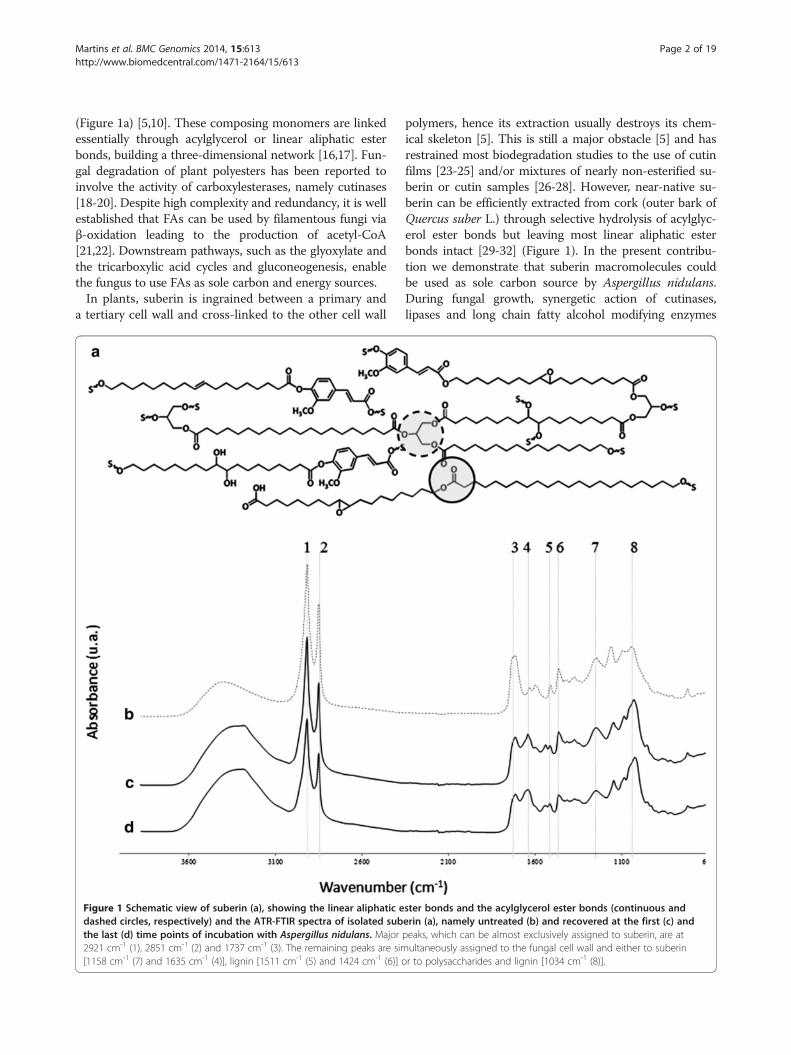
(Figure 1a) [5,10]. These composing monomers are linkedessentially through acylglycerol or linear aliphatic esterbonds, building a three-dimensional network [16,17]. Fun-gal degradation of plant polyesters has been reported toinvolve the activity of carboxylesterases, namely cutinases[18-20]. Despite high complexity and redundancy, it is wellestablished that FAs can be used by filamentous fungi viaβ-oxidation leading to the production of acetyl-CoA[21,22]. Downstream pathways, such as the glyoxylate andthe tricarboxylic acid cycles and gluconeogenesis, enablethe fungus to use FAs as sole carbon and energy sources.In plants, suberin is ingrained between a primary and
a tertiary cell wall and cross-linked to the other cell wall
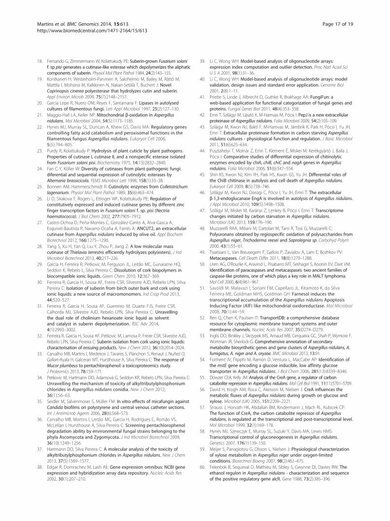
Figure 1 Schematic view of suberin (a), showing the linear aliphatic edashed circles, respectively) and the ATR-FTIR spectra of isolated subthe last (d) time points of incubation with Aspergillus nidulans. Major2921 cm-1 (1), 2851 cm-1 (2) and 1737 cm-1 (3). The remaining peaks are sim[1158 cm-1 (7) and 1635 cm-1 (4)], lignin [1511 cm-1 (5) and 1424 cm-1 (6)] o
polymers, hence its extraction usually destroys its chem-ical skeleton [5]. This is still a major obstacle [5] and hasrestrained most biodegradation studies to the use of cutinfilms [23-25] and/or mixtures of nearly non-esterified su-berin or cutin samples [26-28]. However, near-native su-berin can be efficiently extracted from cork (outer bark ofQuercus suber L.) through selective hydrolysis of acylglyc-erol ester bonds but leaving most linear aliphatic esterbonds intact [29-32] (Figure 1). In the present contribu-tion we demonstrate that suberin macromolecules couldbe used as sole carbon source by Aspergillus nidulans.During fungal growth, synergetic action of cutinases,lipases and long chain fatty alcohol modifying enzymes
ster bonds and the acylglycerol ester bonds (continuous anderin (a), namely untreated (b) and recovered at the first (c) andpeaks, which can be almost exclusively assigned to suberin, are atultaneously assigned to the fungal cell wall and either to suberinr to polysaccharides and lignin [1034 cm-1 (8)].
Martins et al. BMC Genomics 2014, 15:613 Page 3 of 19http://www.biomedcentral.com/1471-2164/15/613
released suberin long chain fatty acids (LCFAs) that weremetabolised via peroxisomal β-oxidation. Other major al-terations included the onset of sexual development andthe boost of secondary metabolism.
MethodsChemicalsIf not otherwise stated chemicals were of the highest analyt-ical grade and purchased from Sigma Aldrich (USA). Su-berin was extracted from cork using cholinium hexanoateas previous described [30], afterwards freeze dried and keptat -20°C until used. The same process was used to recoverthe residual suberin upon fungal incubation. All suberinsamples were analysed using attenuated total reflectanceFourier transform infrared spectroscopy (ATR-FTIR) aspreviously described [30,32]. The selected ATR-FTIR spec-tra should be regarded as representative (ten replicates).
Culture conditionsFungal cultures with a density of A. nidulans (FGSC A4)conidia of 105 per mL (5 mL six-well plates, minimalmedia [33] containing 1% w/v glucose) were grown fortwo days (27°C, dark, 90 rpm) for the establishment of amycelia mat against the bottom of the wells (control).Afterwards, old media were replaced by minimal mediacontaining 0.1% w/v of suberin as sole carbon source, andcultures were grown, in the same conditions, for add-itional two, four, six or fifteen days. At each time pointmycelia and the corresponding media filtrate were recov-ered and preserved at -80°C [33]. Additional control cul-tures were prepared containing 0.1% w/v of C16 glyceryltripalmitate or C8 octyl octanoate (contain acylglycerolester and linear ester bonds, respectively) instead of su-berin. Controls were processed as described above.
Microscopic analysesScanning Electron Microscopy (SEM) analysis of lyophi-lised fungal mycelia were performed using an analyticalfield emission gun scanning electron microscope (FEG-SEM: JEOL 7001 F with Oxford light elements EDS de-tector) operated at 5-10 kV. After the LIVE/DEAD assay[34] (evaluates culture viability) and the safranin staining(detects the extracellular matrix typical of fungal biofilm[35]), mycelia were evaluated using a DM5500 B micro-scope (Leica) under fluorescent (49 DAPI and N21 filtersets) or white light, respectively. 10x and 63x magnificationobjectives were used, respectively and images were cap-tured with a DFC420 C camera (Leica). In all tests the se-lected micrographs should be regarded as representative.
Secretome analysesExtracellular protein was recovered from the culture fil-trates using denaturing precipitation conditions [33].25 μg of protein (bovine serum albumin equivalents
accordingly to the Bradford protocol) were loaded in aprecast gel (Criterion™ XT precast 1D gel 4-12% Bis-Tris) and allow to migrate for 1 cm. The gel was stainedwith Instant Blue (Gentaur BVBA), sliced into 5 bands;(each cut into 1-2 mm cubes), first reduced, alkylatedand de-stained, then digested by trypsin (Promega). Thepeptides were first desalted and finally fractionated byreverse phase separation in an Ultimate 3000 NanoLCSystem coupled to a LTQ-OrbiTrap Elite MS that was op-erated in data-dependent mode, automatically switchingbetween MS and MS2, using XCalibur software. Data wasprocessed in Mascot using Proteome Discoverer bysearching against the SwissProt Fungi (released on January21st, 2013) and NCBI databases. Protein identification wasdone using a set of pre-defined filters and a minimumconfidence of 95%. Full details in Additional file 1. Glycerolin the culture media filtrates (40 times concentrated, dupli-cates) was analysed by chromatography as reported before[36]. Quantification detection limits were 0.01 g · L−1. Theadditional control cultures (see above) were also analysed.
RNA isolation and cRNA preparationTotal RNA was isolated from fungal mycelia (groundedto powder using mortar and pestle in liquid nitrogen)using the RNeasy Plant Mini Kit (QIAGEN) and furtherpurified by ethanol precipitation [37]. Quantification andpurity of RNA were determined on a NanoDrop 1000Spectrophotometer (Thermo Scientific) and RNA integrityanalysed using an Agilent 2100 Bioanalyser with a RNA6000 Nano Assay (Agilent Technologies). Fragmented andbiotinylated cRNA was obtained by following GeneChip 3’IVT Express Kit protocols. Briefly, 100 ng of total RNAwere used for the synthesis of cDNA, which was furtherin vitro transcribed into labelled cRNA. After purificationand fragmentation, the size distribution of the cRNA andfragmented cRNA were assessed in an Agilent 2100 Bioa-nalyzer with a RNA 6000 Nano Assay.
Microarray processingThe custom array FungiANC (Affymetrix) contains a totalof 20,012 transcripts from the genetic information ofA. nidulans and Neurospora crassa available at the BroadInstitute database (http://www.broadinstitute.org) and isbased on a Perfect Match-only (PM) design with 11 mi-cron feature size. Each transcript is represented by 11probes (25-mer each). See full details in Additional file 2.The array processing was performed accordingly to Affy-metrix GeneChip protocols (biological triplicates). A totalof 200 μl of the hybridisation mixture containing 10 μg offragmented cRNA was hybridised on arrays for 16 hoursat 45°C. Standard post hybridisation washes and double-stain protocols (FS450_0001) were used on an AffymetrixGeneChip Fluidics Station 450, in conjunction with theGeneChip Hybridisation Wash and Stain Kit (Affymetrix).
Martins et al. BMC Genomics 2014, 15:613 Page 4 of 19http://www.biomedcentral.com/1471-2164/15/613
Arrays were scanned on an Affymetrix GeneChip Scanner3000 7G. Array quality parameters were analysed by Ex-pression Console Software (Affymetrix) for Robust Multi-array Averaging summarised data and confirmed to be inthe recommended range. The data discussed in this publi-cation have been deposited in NCBI’s Gene ExpressionOmnibus [38] and are accessible through GEO Series ac-cession number GSE54427 (http://www.ncbi.nlm.nih.gov/geo/query/acc.cgi?acc=GSE54427).
Microarray data analysisDNA-Chip Analyzer (dChip) 2010 (http://www.dchip.org)was used applying a probeset mask file considering onlythe A. nidulans probes (9675 transcripts). The normalisedCEL intensities of the 12 arrays (Invariant Set Normalisa-tion Method [39,40]), were used to obtain model-basedgene expression indices based on a Perfect Match-onlymodel [39,40]. dChip Log2 expression data were importedinto R v2.13.0. Differentially expressed genes (analysis wascarried out with Bioconductor LIMMA package, www.bio-conductor.org), included only the probe sets with adjustedp-value ≤0.01 and fold-change (FC) ≥2. See full details inAdditional file 1.
Quantitative real-time PCR analysis (qRT-PCR)Based on the sequences of A. nidulans genes (AspergillusGenome Database, http://www.aspergillusgenome.org/),all oligonucleotide pairs were designed using the Gene-Fisher2 web tool (http://bibiserv.techfak.uni-bielefeld.de/genefisher2) and produced by Thermo Fisher Scientific(see Additional file 1 for the list of the oligonucleotidesused in this study). The qRT-PCR analysis was per-formed in a CFX96 Thermal Cycler (Bio-Rad), using theSsoFast EvaGreen Supermix (Bio-Rad), 250 nM of eacholigonucleotide and the cDNA template equivalent to1 ng of total RNA, at a final volume of 10 μl per well, inthree technical and three biological replicates. The PCRconditions were: enzyme activation at 95°C for 30 s;40 cycles of denaturation at 95°C for 10 s and annealing/extension at 59°C for 15 s; and melting curve obtainedfrom 65°C to 95°C, consisting of 0.5°C increments for5 s. Data analyses were performed using the CFX Man-ager software (Bio-Rad). The expression of each gene wastaken as the relative expression in pair-wise comparisonsof consecutive time points. The expression of all targetgenes was normalised by the expression of the 60S riboso-mal protein L33-A gene, AN2980, selected as internalcontrol due to its constant levels in all time points.
Functional annotationAnnotations for all the genes represented on the FungiANCgenome array were obtained from the Broad Institute data-base (www.broadinstitute.org) and the Aspergillus GenomeDatabase (AspGD, www.aspgd.org). See full details in
Additional file 2. The differentially expressed genes for eachbiological condition were classified using the FungiFunweb annotation tool [41] and the Functional Catalogue(FunCat). The significant hits (p-value ≤0.05) were definedusing the identities present on the chip as the background.
Results & discussionAspergillus nidulans transcriptome on suberin - enrich-ment analysisPair-wise comparisons were used to identify differentiallyexpressed genes (adjusted p-value < 0.01 and |FC| > 2) be-tween the control (grown on glucose) and during growth onsuberin for two, four or six days (hereafter defined as first,mid and last time points, respectively) Additional file 2.Within the differentially expressed genes (4198 constitutingnearly half of the transcripts in the microarray), 32% (1357genes) can be specifically associated with the switch of thesubstrate (control vs first time point) and were enriched infunctional categories associated with the metabolism of al-kanes, alkenes, alkanals and alkanols (MIPS 01.20.05.03)and the oxidation of fatty acids (MIPS 2.25), as well as cel-lular sensing and response to external stimulus (MIPS33.11) and cell type differentiation (MIPS 43), amongothers (Figure 2b, Additional file 3). Pair-wise comparisonof consecutive time points, henceforward systematicallyused, showed that among the enriched functional categor-ies at the mid time point some were associated with in-creased nutrient starvation response (MIPS 32.01.11) andalterations in fatty acid metabolism (MIPS 01.06.05), alongwith major alterations in cell cycle (MIPS 10) and cell fate(MIPS 40) Additional file 3. In addition, those enriched atthe last time point revealed e.g. an increased stress re-sponse (MIPS 32.01) and development of ascospores(MIPS 43.01.03.09). In particular, the degradation/modifi-cation of exogenous ester compounds (MIPS 32.10.09)can be associated with cleavage of ester bonds in suberin.The intensity of the major peak assigned to ester bonds(1737 cm-1 which can be exclusively assigned to the C =Ostretch of ester groups) in the ATR-FTIR spectra ofsuberin decreased significantly after fungal incubation(Figure 1b).
Autolysis and primary metabolismAutolysis occurred after switching from glucose to su-berin media. Major up-regulation of pepJ (AN7962,Table 1) and up-regulation of prtA (AN5558) resulted inaccumulation of the encoded proteins in the secretome(Table 2). Both proteases play a role in the degradation ofempty hyphae during starvation [42,43]. Out of the 25protein species identified in the secretome, ten are in-volved in cell wall remodelling events typical of autolysisand show, in general, high consistency with the transcrip-tome data (Table 2). Several other cell wall remodellinggenes related with autolysis were up-regulated along
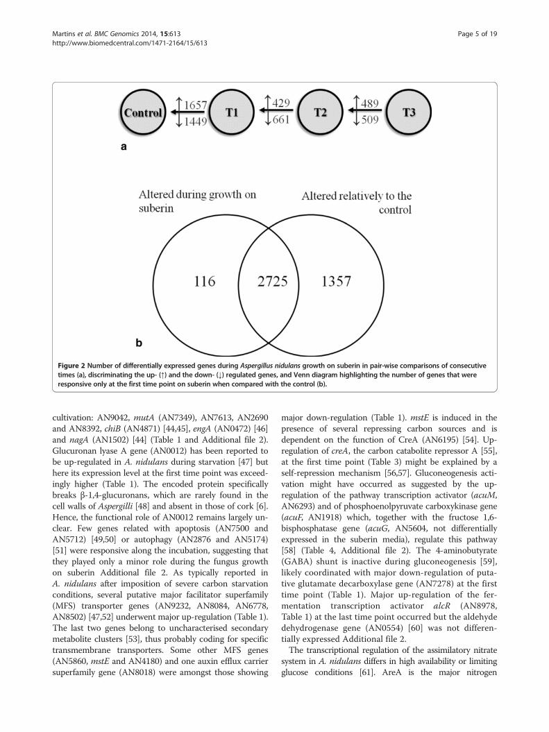
Figure 2 Number of differentially expressed genes during Aspergillus nidulans growth on suberin in pair-wise comparisons of consecutivetimes (a), discriminating the up- (↑) and the down- (↓) regulated genes, and Venn diagram highlighting the number of genes that wereresponsive only at the first time point on suberin when compared with the control (b).
Martins et al. BMC Genomics 2014, 15:613 Page 5 of 19http://www.biomedcentral.com/1471-2164/15/613
cultivation: AN9042, mutA (AN7349), AN7613, AN2690and AN8392, chiB (AN4871) [44,45], engA (AN0472) [46]and nagA (AN1502) [44] (Table 1 and Additional file 2).Glucuronan lyase A gene (AN0012) has been reported tobe up-regulated in A. nidulans during starvation [47] buthere its expression level at the first time point was exceed-ingly higher (Table 1). The encoded protein specificallybreaks β-1,4-glucuronans, which are rarely found in thecell walls of Aspergilli [48] and absent in those of cork [6].Hence, the functional role of AN0012 remains largely un-clear. Few genes related with apoptosis (AN7500 andAN5712) [49,50] or autophagy (AN2876 and AN5174)[51] were responsive along the incubation, suggesting thatthey played only a minor role during the fungus growthon suberin Additional file 2. As typically reported inA. nidulans after imposition of severe carbon starvationconditions, several putative major facilitator superfamily(MFS) transporter genes (AN9232, AN8084, AN6778,AN8502) [47,52] underwent major up-regulation (Table 1).The last two genes belong to uncharacterised secondarymetabolite clusters [53], thus probably coding for specifictransmembrane transporters. Some other MFS genes(AN5860, mstE and AN4180) and one auxin efflux carriersuperfamily gene (AN8018) were amongst those showing
major down-regulation (Table 1). mstE is induced in thepresence of several repressing carbon sources and isdependent on the function of CreA (AN6195) [54]. Up-regulation of creA, the carbon catabolite repressor A [55],at the first time point (Table 3) might be explained by aself-repression mechanism [56,57]. Gluconeogenesis acti-vation might have occurred as suggested by the up-regulation of the pathway transcription activator (acuM,AN6293) and of phosphoenolpyruvate carboxykinase gene(acuF, AN1918) which, together with the fructose 1,6-bisphosphatase gene (acuG, AN5604, not differentiallyexpressed in the suberin media), regulate this pathway[58] (Table 4, Additional file 2). The 4-aminobutyrate(GABA) shunt is inactive during gluconeogenesis [59],likely coordinated with major down-regulation of puta-tive glutamate decarboxylase gene (AN7278) at the firsttime point (Table 1). Major up-regulation of the fer-mentation transcription activator alcR (AN8978,Table 1) at the last time point occurred but the aldehydedehydrogenase gene (AN0554) [60] was not differen-tially expressed Additional file 2.The transcriptional regulation of the assimilatory nitrate
system in A. nidulans differs in high availability or limitingglucose conditions [61]. AreA is the major nitrogen
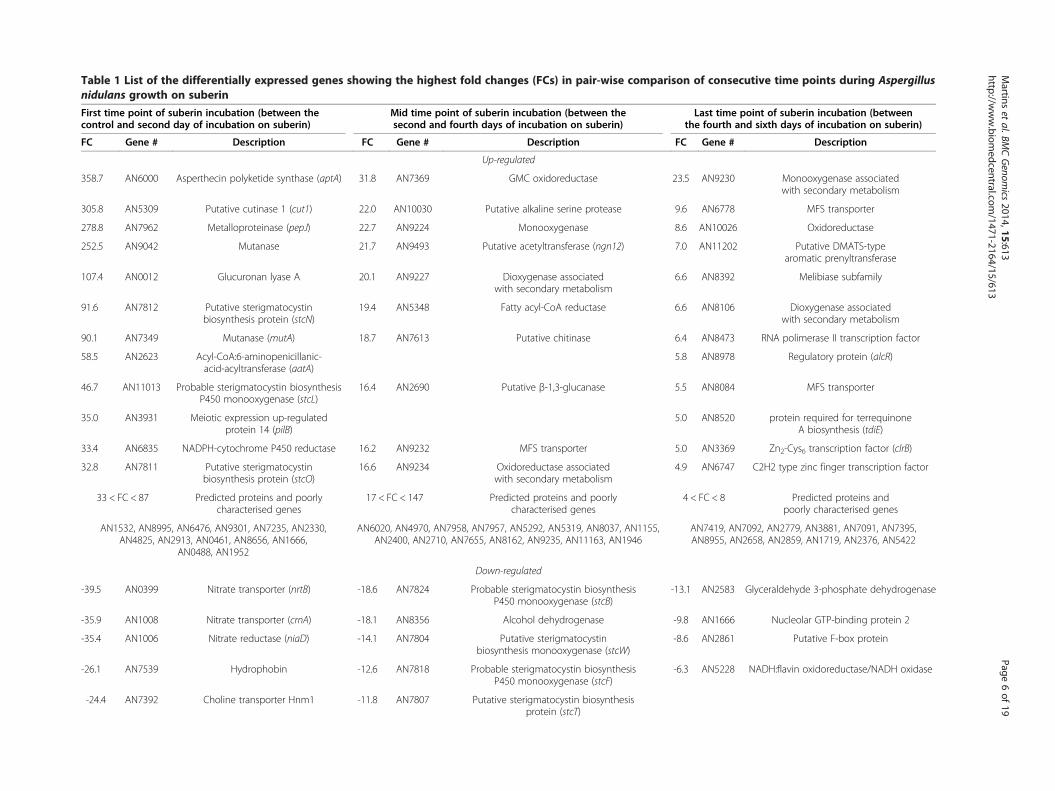
Table 1 List of the differentially expressed genes showing the highest fold changes (FCs) in pair-wise comparison of consecutive time points during Aspergillusnidulans growth on suberin
First time point of suberin incubation (between thecontrol and second day of incubation on suberin)
Mid time point of suberin incubation (between thesecond and fourth days of incubation on suberin)
Last time point of suberin incubation (betweenthe fourth and sixth days of incubation on suberin)
FC Gene # Description FC Gene # Description FC Gene # Description
Up-regulated
358.7 AN6000 Asperthecin polyketide synthase (aptA) 31.8 AN7369 GMC oxidoreductase 23.5 AN9230 Monooxygenase associatedwith secondary metabolism
305.8 AN5309 Putative cutinase 1 (cut1) 22.0 AN10030 Putative alkaline serine protease 9.6 AN6778 MFS transporter
278.8 AN7962 Metalloproteinase (pepJ) 22.7 AN9224 Monooxygenase 8.6 AN10026 Oxidoreductase
252.5 AN9042 Mutanase 21.7 AN9493 Putative acetyltransferase (ngn12) 7.0 AN11202 Putative DMATS-typearomatic prenyltransferase
107.4 AN0012 Glucuronan lyase A 20.1 AN9227 Dioxygenase associatedwith secondary metabolism
6.6 AN8392 Melibiase subfamily
91.6 AN7812 Putative sterigmatocystinbiosynthesis protein (stcN)
19.4 AN5348 Fatty acyl-CoA reductase 6.6 AN8106 Dioxygenase associatedwith secondary metabolism
90.1 AN7349 Mutanase (mutA) 18.7 AN7613 Putative chitinase 6.4 AN8473 RNA polimerase II transcription factor
58.5 AN2623 Acyl-CoA:6-aminopenicillanic-acid-acyltransferase (aatA)
5.8 AN8978 Regulatory protein (alcR)
46.7 AN11013 Probable sterigmatocystin biosynthesisP450 monooxygenase (stcL)
16.4 AN2690 Putative β-1,3-glucanase 5.5 AN8084 MFS transporter
35.0 AN3931 Meiotic expression up-regulatedprotein 14 (pilB)
5.0 AN8520 protein required for terrequinoneA biosynthesis (tdiE)
33.4 AN6835 NADPH-cytochrome P450 reductase 16.2 AN9232 MFS transporter 5.0 AN3369 Zn2-Cys6 transcription factor (clrB)
32.8 AN7811 Putative sterigmatocystinbiosynthesis protein (stcO)
16.6 AN9234 Oxidoreductase associatedwith secondary metabolism
4.9 AN6747 C2H2 type zinc finger transcription factor
33 < FC < 87 Predicted proteins and poorlycharacterised genes
17 < FC < 147 Predicted proteins and poorlycharacterised genes
4 < FC < 8 Predicted proteins andpoorly characterised genes
AN1532, AN8995, AN6476, AN9301, AN7235, AN2330,AN4825, AN2913, AN0461, AN8656, AN1666,
AN0488, AN1952
AN6020, AN4970, AN7958, AN7957, AN5292, AN5319, AN8037, AN1155,AN2400, AN2710, AN7655, AN8162, AN9235, AN11163, AN1946
AN7419, AN7092, AN2779, AN3881, AN7091, AN7395,AN8955, AN2658, AN2859, AN1719, AN2376, AN5422
Down-regulated
-39.5 AN0399 Nitrate transporter (nrtB) -18.6 AN7824 Probable sterigmatocystin biosynthesisP450 monooxygenase (stcB)
-13.1 AN2583 Glyceraldehyde 3-phosphate dehydrogenase
-35.9 AN1008 Nitrate transporter (crnA) -18.1 AN8356 Alcohol dehydrogenase -9.8 AN1666 Nucleolar GTP-binding protein 2
-35.4 AN1006 Nitrate reductase (niaD) -14.1 AN7804 Putative sterigmatocystinbiosynthesis monooxygenase (stcW)
-8.6 AN2861 Putative F-box protein
-26.1 AN7539 Hydrophobin -12.6 AN7818 Probable sterigmatocystin biosynthesisP450 monooxygenase (stcF)
-6.3 AN5228 NADH:flavin oxidoreductase/NADH oxidase
-24.4 AN7392 Choline transporter Hnm1 -11.8 AN7807 Putative sterigmatocystin biosynthesisprotein (stcT)
Martins
etal.BM
CGenom
ics2014,15:613
Page6of
19http://w
ww.biom
edcentral.com/1471-2164/15/613
Table 1 List of the differentially expressed genes showing the highest fold changes (FCs) in pair-wise comparison of consecutive time points during Aspergillusnidulans growth on suberin (Continued)
-11.5 AN7815 Fatty acid synthase subunit α -5.6 AN4180 MFS transporter
-11.4 AN5860 MFS monosaccharide transporter (mstE)
-24.1 AN2859 Dihydrodipicolinate synthetase -11.2 AN7825 Putative sterigmatocystin biosynthesispolyketide synthase (stcA)
-5.6 AN10619 Glutamate decarboxylase
-11.2 AN4135 Stearoyl-CoA desaturase (sdeB)
-23.9 AN4131 Na+/H+ antiporter -11.1 AN7814 Fatty acid synthase subunit β -5.5 AN7169 Flavohemoprotein (fhbA)
-23.8 AN7278 Glutamate decarboxylase -10.8 AN7806 Versicolorin reductase
-21.7 AN4119 MFS multidrug transporter -10.6 AN3763 Siderochrome-iron transporter -5.5 AN3264 MFS multidrug transporter
-19.0 AN3776 MFS transporter -10.0 AN7811 Putative sterigmatocystin biosynthesisprotein (stcO)
-61 < FC < -18 Predicted proteins and poorlycharacterised genes
-28 < FC < -10 Predicted protein and poorly characterised genes -16 < FC < -5 Predicted protein and poorlycharacterised genes
AN2595, AN7214, AN5305, AN8081, AN6946, AN4108, AN7378,AN9220, AN6128, AN8981, AN3333, AN6930, AN4128, AN6932,
AN8670
AN7397, AN3251, AN8544, AN6661, AN7817, AN8314,AN2722, AN0169, AN7812, AN7809, AN7915, AN11013
AN2571, AN9310, AN7949, AN5489, AN0421, AN0461,AN9102, AN9378, AN7960, AN3305, AN0728, AN0247,
AN0288, AN8974, AN0838, AN8544, AN3314
Martins
etal.BM
CGenom
ics2014,15:613
Page7of
19http://w
ww.biom
edcentral.com/1471-2164/15/613
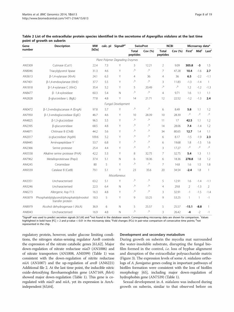
Table 2 List of the extracellular protein species identified in the secretome of Aspergillus nidulans at the last timepoint of growth on suberin
Genenumber
Description MW[kDa]
calc. pI SignalPa SwissProt NCBI Microarray data*
Totalpeptides
Cov (%) Totalpeptides
Cov (%) Firstc Midc Lastc
Plant Polymer Degrading Enzymes
AN5309 Cutinase (Cut1) 22.4 7.3 Y 3 12.21 2 9.09 305.8 -3 1.5
AN8046 Triacylglycerol lipase 31.3 4.6 Y -b -b 7 47.28 10.4 -1.6 2.7
AN3613 β-1,4-xylanase (XlnA) 24.1 6.3 Y 4 36 4 36 6.5 -2.2 -1.1
AN7401 β-1,4-endoxylanase (XlnE) 37.7 5.5 Y -b -b 3 11.83 -1.3 -1.4 1
AN1818 β-1,4-xylanase C (XlnC) 35.4 5.2 Y 5 20.49 -b -b 1.2 -1.2 -1.3
AN8477 β -1,4-xylosidase 60.3 5.4 N -b -b 4 9.71 1.6 1.1 1.1
AN2828 β-glucosidase L (BglL) 77.8 4.8 Y 14 21.71 12 22.52 -1.2 -1.3 2.4
Fungal Development
AN0472 β-1,3-endoglucanase A (EngA) 97.8 5.7 Y -b -b 6 8.49 5.8 1.1 1.2
AN7950 β-1,3-endoglucosidase (EglC) 46.7 4.6 Y 10 28.39 10 28.39 -d -d -d
AN4825 β-1,3-glucosidase 96.5 5.5 Y -b -b 11 17 42.5 1.1 1.2
AN2395 β-glucuronidase 68.5 4.8 Y -b -b 14 28.06 7.4 -1.4 1.2
AN4871 Chitinase B (ChiB) 44.2 5.6 Y -b -b 34 80.65 12.7 1.4 1.1
AN2017 α-glucosidase (AgdA) 109.6 5.2 Y -b -b 6 8.17 -1.5 -1.9 2.3
AN8445 Aminopeptidase Y 53.7 6.8 Y -b -b 6 19.68 1.8 -1.5 1.6
AN2366 Serine protease 25.4 4.4 Y -b -b 3 17.27 -d -d -d
AN5558 Alkaline serine protease (PrtA) 42.2 5.3 Y 8 32.75 7 32.75 5.4 1.3 1.1
AN7962 Metalloproteinase (PepJ) 37.4 5.1 N 6 18.36 6 18.36 278.8 1.8 1.2
AN4245 Ceramidase 80 5 Y -b -b 7 14.8 1.6 1.5 1.8
AN9339 Catalase B (CatB) 79.1 5.1 Y 23 35.6 20 34.54 -2.4 1.8 1
Miscellaneous
AN3351 Uncharacterised 63.2 5.1 Y -b -b 5 12.91 1.6 -1.4 -1.1
AN3246 Uncharacterised 22.3 6.4 N -b -b 4 29.8 2 -1.3 2
AN6273 Allergenic Asp F13 16.3 4.8 Y -b -b 3 32.91 -1 -1.5 -1.4
AN5879 Phosphatidylglycerol/phosphatidylinositoltransfer protein
18.3 5 Y 9 53.25 9 53.25 1 1 -1
AN8979 Alcohol dehydrogenase I (AlcA) 36.9 6 N 5 25.57 5 25.57 -15.1 -8.8 1
AN8043 Uncharacterised 16.9 4.8 N -b -b 3 26.42 -4 -2 -1aSignalP was used to predict secretion signals [67,68] and bnot found in the database search. Corresponding microarray data are shown for comparison. *Valueshighlighted in bold have |FC| > 2 and p-value < 0.01 in the microarray data; cFold changes (FCs) in pair-wise comparison of consecutive time points; dnotrepresented in the chip.
Martins et al. BMC Genomics 2014, 15:613 Page 8 of 19http://www.biomedcentral.com/1471-2164/15/613
regulatory protein, however, under glucose limiting condi-tions, the nitrogen status-sensing regulator AreB controlsthe expression of the nitrate catabolic genes [61,62]. Majordown-regulation of nitrate reductase niaD (AN1006) andof nitrate transporters (AN1008, AN0399) (Table 1) wasconsistent with the down-regulation of nitrite reductaseniiA (AN1007) and the up-regulation of areB (AN6221)Additional file 2. At the last time point, the inducible nitricoxide-detoxifying flavohaemoglobin gene (AN7169, fhbA)showed major down-regulation (Table 1). This gene is co-regulated with niaD and niiA, yet its expression is AreA-independent [63,64].
Development and secondary metabolismDuring growth on suberin the mycelia mat surroundedthe water-insoluble substrate, disrupting the fungal bio-film formed in the control, i.e. loss of hyphae alignmentand disruption of the extracellular polysaccharide matrix(Figure 3). The expression levels of some A. nidulans ortho-logs of A. fumigatus genes coding in important pathways ofbiofilm formation were consistent with the loss of biofilmmorphology [65], including major down-regulation ofhydrophobin gene (AN7539) (Table 1).Sexual development in A. nidulans was induced during
growth on suberin, similar to that observed before on
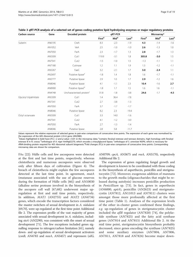
Table 3 qRT-PCR analysis of a selected set of genes coding putative lipid hydrolysing enzymes or major regulatory proteins
Carbon source Gene Encoded protein qRT-PCR Microarray*
Firstd Midd Lastd Firstd Midd Lastd
Suberin AN6195 CreA 8.3 -2.3 -1.9 4.3 -1.4 -1.6
AN1052 VeA 2.5 -1.0 -1.0 2.6 -1.3 1.0
AN7050 FarA 2.3 -1.7 1.3 2.8 -1.7 1.3
AN5309 Cut1 119.9 -3.1 1.8 305.8 -3.0 1.5
AN7541 Cut2 -1.5 -1.0 1.5 -1.3 -1.1 1.1
AN7180 Cutinase 1.2 1.1 1.9 1.3 -1.2 -1.1
AN5267 FaeC 3.2 -2.1 1.7 3.0 -2.4 1.1
AN2697 Putative lipasea -1.8 1.4 1.8 1.6 -1.7 -1.1
AN5777 Putative lipase 2.9 1.0 1.7 2.9 -1.2 1.6
AN8046 Putative lipase 4.1 -1.1 2.3 10.4 -1.6 2.7
AN8900 Putative lipaseb -1.8 1.7 1.5 1.6 1.6 -1.1
AN4748 Uncharacterised proteinc 31.8 -1.8 -3.8 24.6 -1.7 -4.5
Glyceryl tripalmitate AN5309 Cut1 1.4 36.4 -7.8
AN7541 Cut2 2.7 -3.8 -1.3
AN7050 FarA 3.7 -1.7 -1.7
AN8046 Putative lipase 8.2 2.3 -19.4
Octyl octanoate AN5309 Cut1 3.3 14.0 -1.6
AN7541 Cut2 4.1 -1.2 -3.0
AN7050 FarA 2.7 -1.3 -1.4
AN8046 Putative lipase 2.8 5.8 -11.7
Values represent the relative expression of selected genes in pair-wise comparisons of consecutive time points. The expression of each gene was normalised bythe expression of the 60S ribosomal protein L33-A gene (AN2980).*Values highlighted in bold have |FC| > 2 and p-value < 0.01 in the microarray data; acontains feruloyl esterase and tannase domains, high homology with feruloylesterase B in N. crassa; borthologue of A. niger An09g02270, which encodes a triacylglycerol lipase; corthologue of S. cerevisiae NOP6, which is necessary forrRNA-binding protein required for 40S ribosomal subunit biogenesis.dFold changes (FCs) in pair-wise comparison of consecutive time points. Correspondingmicroarray data are shown for comparison.
Martins et al. BMC Genomics 2014, 15:613 Page 9 of 19http://www.biomedcentral.com/1471-2164/15/613
FAs [22]: Hülle cells and few ascospores were detectedat the first and last time points, respectively; whereascleistothecia and numerous ascospores were observedonly after fifteen days of cultivation (Figure 4). Thebreach of cleistothecia might explain the few ascosporesdetected at the last time point. In agreement, mutA(mutanase associated with the use of glucose reservesduring the formation of Hülle cells [66]) and AN10030(alkaline serine protease involved in the biosynthesis ofthe ascopore cell wall [67,68]) underwent major up-regulation at first and mid time points, respectively.In addition, MAT1 (AN2755) and MAT2 (AN4734)genes, which encode the transcription factors consideredthe master switchers of sexual development in A. nidulans[69,70], were up-regulated at the first time point Additionalfile 2. The expression profile of the vast majority of genesassociated with sexual development in A. nidulans, includ-ing steA (AN2290), was consistent with the onset of sexualdevelopment [71]. The few exceptions included genes sig-nalling response to nitrogen/carbon limitation [61], namelydown- and up-regulation of sexual development activators(csnB, AN4783 and noxA, AN5457) and repressors (silG,
AN0709; cpcA, AN3675 and rosA, AN5170), respectivelyAdditional file 2.The expression of genes regulating fungal growth and
development is known to be coordinated with those codingin the biosynthesis of asperthecin, penicillin and sterigma-tocystin [72]. Moreover, exogenous addition of mannansto the growth media (oligosaccharides that might be re-leased during autolysis) increases penicillin productionin Penicillium sp. [73]. In fact, genes in asperthecin(AN6000, aptA), penicillin (AN2623) and sterigmato-cystin (AN7812, AN11013 and AN7811) clusters wereamongst those more profoundly affected at the firsttime point (Table 1). Analyses of the expression levelsof the other in-cluster genes confirmed these findings,e.g. up-regulation of genes in sterigmatocystin clusterincluded the aflR regulator (AN7820) [74], the polyke-tide synthase (AN7825) and the fatty acid synthasegenes (AN7814 and AN7815) Additional file 2. At themid time point, sterigmatocystin biosynthesis probablydecreased, since genes encoding the synthase (AN7825)and some auxiliary enzymes (AN7804, AN7806,AN7811, AN7818 and AN7824) become major down-
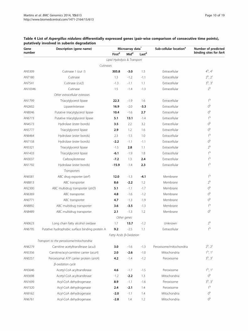
Table 4 List of Aspergillus nidulans differentially expressed genes (pair-wise comparison of consecutive time points),putatively involved in suberin degradation
Genenumber
Description (gene name) Microarray data* Sub-cellular locationa Number of predictedbinding sites for farAFirstd Midd Lastd
Lipid Hydrolysis & Transport
Cutinases
AN5309 Cutinase 1 (cut 1) 305.8 -3.0 1.5 Extracellular 4b, 4c
AN7180 Cutinase 1.3 -1.2 -1.1 Extracellular 2b, 2c
AN7541 Cutinase (cut2) -1.3 -1.1 1.1 Extracellular 3b, 3c
AN10346 Cutinase 1.5 -1.4 -1.3 Extracellular 2b
Other extracellular esterases
AN1799 Triacylglycerol lipase 22.3 -1.9 1.6 Extracellular 1b
AN2602 Lipase/esterase 16.9 -2.0 -3.3 Extracellular 0b
AN8046 Putative triacylglycerol lipase 10.4 -1.6 2.7 Extracellular 0b
AN6773 Putative triacylglycerol lipase 5.1 13.1 -1.4 Extracellular 1b
AN4573 Hydrolase (ester bonds) 3.5 2.2 3.2 Extracellular 0b
AN5777 Triacylglycerol lipase 2.9 1.2 1.6 Extracellular 0b
AN6464 Hydrolase (ester bonds) 2.3 -1.5 1.0 Extracellular 1b
AN7158 Hydrolase (ester bonds) -2.2 -1.1 -1.1 Extracellular 0b
AN5321 Triacylglycerol lipase -1.5 2.8 1.1 Extracellular 2b
AN1433 Triacylglycerol lipase -6.1 -1.9 1.8 Extracellular 1b
AN3037 Carboxylesterase -7.2 1.3 2.4 Extracellular 1b
AN1792 Hydrolase (ester bonds) -15.9 -1.4 2.3 Extracellular 1b
Transporters
AN6581 ABC drug exporter (atrF) 12.0 -1.3 -4.1 Membrane 1b
AN8813 ABC transporter 8.6 -2.2 1.2 Membrane 2b
AN2300 ABC multidrug transporter (atrD) 5.1 -1.1 -1.7 Membrane 0b
AN6369 ABC transporter 4.8 -1.6 -1.2 Membrane 0b
AN0771 ABC transporter 4.7 -1.3 -1.9 Membrane 0b
AN8892 ABC multidrug transporter 3.6 -3.5 -1.3 Membrane 1b
AN8489 ABC multidrug transporter 2.1 -1.3 1.2 Membrane 0b
Other genes
AN0623 Long chain fatty alcohol oxidase 1.7 13.7 -1.2 Unknown 2b
AN6795 Putative hydrophobic surface binding protein A 9.2 -2.5 1.1 Extracellular 1b
Fatty Acids β-Oxidation
Transport to the peroxisome/mitochondria
AN6279 Carnitine acetyltransferase (acuJ) 3.0 -1.6 -1.3 Peroxisome/mitochondria 2b, 2c
AN5356 Carnitine/acyl-carnitine carrier (acuH) 2.0 -2.6 -1.0 Mitochondria 1b, 1c
AN0257 Peroxisomal ATP carrier protein (antA) 4.2 -1.4 -1.2 Peroxisome 3b, 3c
β-oxidation cycle
AN5646 Acetyl-CoA acyltransferase 4.6 -1.7 -1.5 Peroxisome 1b, 1c
AN5698 Acetyl-CoA acyltransferase -1.2 -2.2 1.3 Mitochondria 0b
AN1699 Acyl-CoA dehydrogenase 8.9 -1.1 -1.6 Peroxisome 3b, 3c
AN7320 Acyl-CoA dehydrogenase 2.4 -2.1 1.4 Peroxisome 1b
AN9162 Acyl-CoA dehydrogenase -2.0 -1.1 1.4 Mitochondria 0b
AN6761 Acyl-CoA dehydrogenase -2.8 1.4 1.2 Mitochondria 0b
Martins et al. BMC Genomics 2014, 15:613 Page 10 of 19http://www.biomedcentral.com/1471-2164/15/613
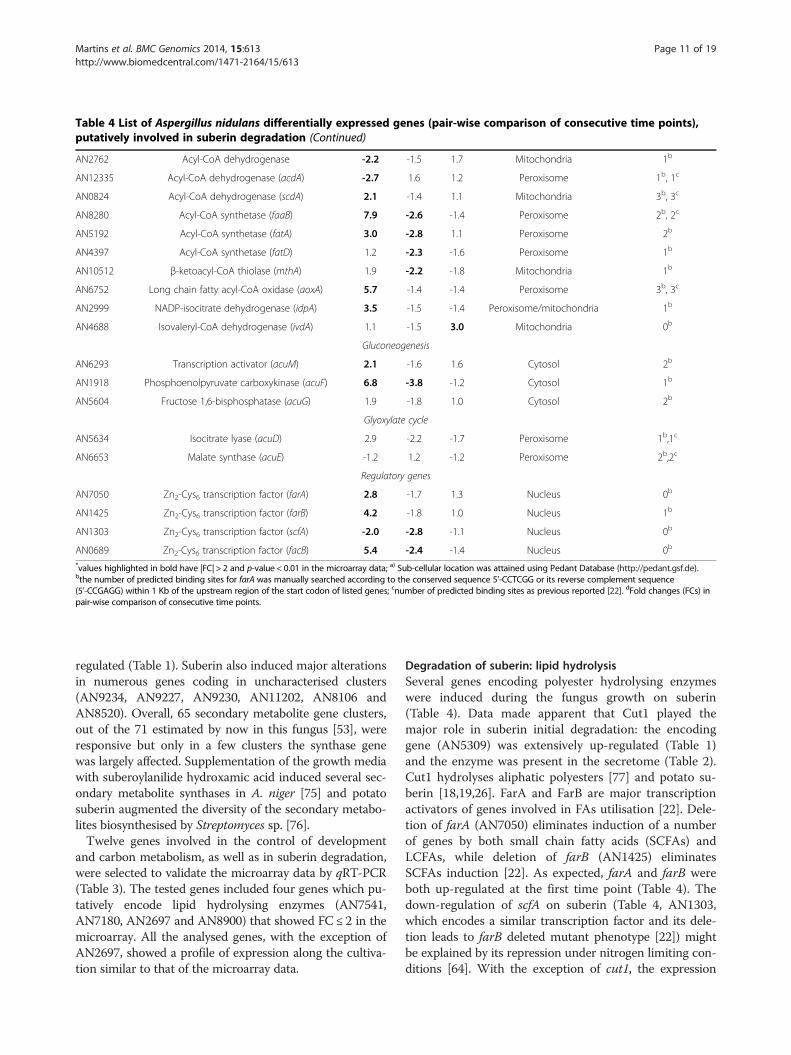
Table 4 List of Aspergillus nidulans differentially expressed genes (pair-wise comparison of consecutive time points),putatively involved in suberin degradation (Continued)
AN2762 Acyl-CoA dehydrogenase -2.2 -1.5 1.7 Mitochondria 1b
AN12335 Acyl-CoA dehydrogenase (acdA) -2.7 1.6 1.2 Peroxisome 1b, 1c
AN0824 Acyl-CoA dehydrogenase (scdA) 2.1 -1.4 1.1 Mitochondria 3b, 3c
AN8280 Acyl-CoA synthetase (faaB) 7.9 -2.6 -1.4 Peroxisome 2b, 2c
AN5192 Acyl-CoA synthetase (fatA) 3.0 -2.8 1.1 Peroxisome 2b
AN4397 Acyl-CoA synthetase (fatD) 1.2 -2.3 -1.6 Peroxisome 1b
AN10512 β-ketoacyl-CoA thiolase (mthA) 1.9 -2.2 -1.8 Mitochondria 1b
AN6752 Long chain fatty acyl-CoA oxidase (aoxA) 5.7 -1.4 -1.4 Peroxisome 3b, 3c
AN2999 NADP-isocitrate dehydrogenase (idpA) 3.5 -1.5 -1.4 Peroxisome/mitochondria 1b
AN4688 Isovaleryl-CoA dehydrogenase (ivdA) 1.1 -1.5 3.0 Mitochondria 0b
Gluconeogenesis
AN6293 Transcription activator (acuM) 2.1 -1.6 1.6 Cytosol 2b
AN1918 Phosphoenolpyruvate carboxykinase (acuF) 6.8 -3.8 -1.2 Cytosol 1b
AN5604 Fructose 1,6-bisphosphatase (acuG) 1.9 -1.8 1.0 Cytosol 2b
Glyoxylate cycle
AN5634 Isocitrate lyase (acuD) 2.9 -2.2 -1.7 Peroxisome 1b,1c
AN6653 Malate synthase (acuE) -1.2 1.2 -1.2 Peroxisome 2b,2c
Regulatory genes
AN7050 Zn2-Cys6 transcription factor (farA) 2.8 -1.7 1.3 Nucleus 0b
AN1425 Zn2-Cys6 transcription factor (farB) 4.2 -1.8 1.0 Nucleus 1b
AN1303 Zn2-Cys6 transcription factor (scfA) -2.0 -2.8 -1.1 Nucleus 0b
AN0689 Zn2-Cys6 transcription factor (facB) 5.4 -2.4 -1.4 Nucleus 0b
*values highlighted in bold have |FC| > 2 and p-value < 0.01 in the microarray data; a) Sub-cellular location was attained using Pedant Database (http://pedant.gsf.de).bthe number of predicted binding sites for farA was manually searched according to the conserved sequence 5’-CCTCGG or its reverse complement sequence(5’-CCGAGG) within 1 Kb of the upstream region of the start codon of listed genes; cnumber of predicted binding sites as previous reported [22]. dFold changes (FCs) inpair-wise comparison of consecutive time points.
Martins et al. BMC Genomics 2014, 15:613 Page 11 of 19http://www.biomedcentral.com/1471-2164/15/613
regulated (Table 1). Suberin also induced major alterationsin numerous genes coding in uncharacterised clusters(AN9234, AN9227, AN9230, AN11202, AN8106 andAN8520). Overall, 65 secondary metabolite gene clusters,out of the 71 estimated by now in this fungus [53], wereresponsive but only in a few clusters the synthase genewas largely affected. Supplementation of the growth mediawith suberoylanilide hydroxamic acid induced several sec-ondary metabolite synthases in A. niger [75] and potatosuberin augmented the diversity of the secondary metabo-lites biosynthesised by Streptomyces sp. [76].Twelve genes involved in the control of development
and carbon metabolism, as well as in suberin degradation,were selected to validate the microarray data by qRT-PCR(Table 3). The tested genes included four genes which pu-tatively encode lipid hydrolysing enzymes (AN7541,AN7180, AN2697 and AN8900) that showed FC ≤ 2 in themicroarray. All the analysed genes, with the exception ofAN2697, showed a profile of expression along the cultiva-tion similar to that of the microarray data.
Degradation of suberin: lipid hydrolysisSeveral genes encoding polyester hydrolysing enzymeswere induced during the fungus growth on suberin(Table 4). Data made apparent that Cut1 played themajor role in suberin initial degradation: the encodinggene (AN5309) was extensively up-regulated (Table 1)and the enzyme was present in the secretome (Table 2).Cut1 hydrolyses aliphatic polyesters [77] and potato su-berin [18,19,26]. FarA and FarB are major transcriptionactivators of genes involved in FAs utilisation [22]. Dele-tion of farA (AN7050) eliminates induction of a numberof genes by both small chain fatty acids (SCFAs) andLCFAs, while deletion of farB (AN1425) eliminatesSCFAs induction [22]. As expected, farA and farB wereboth up-regulated at the first time point (Table 4). Thedown-regulation of scfA on suberin (Table 4, AN1303,which encodes a similar transcription factor and its dele-tion leads to farB deleted mutant phenotype [22]) mightbe explained by its repression under nitrogen limiting con-ditions [64]. With the exception of cut1, the expression
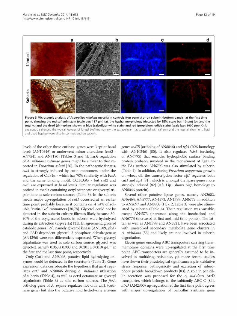
Figure 3 Microscopic analysis of Aspergillus nidulans mycelia in controls (top panels) or on suberin (bottom panels) at the first timepoint, showing the red safranin stain (scale bar: 137 μm) (a), the hyphal morphology (detected by SEM, scale bar: 10 μm) (b), and thetotal (c) and the dead (d) hyphae, shown in blue (calcofluor white stain) and red (propidium iodide stain) (scale bar: 1000 μm). Onlythe controls showed the typical features of fungal biofilms, namely the extracellular matrix stained with safranin and the hyphal alignment. Totaland dead hyphae were alike in controls and on suberin.
Martins et al. BMC Genomics 2014, 15:613 Page 12 of 19http://www.biomedcentral.com/1471-2164/15/613
levels of the other three cutinase genes were kept at basallevels (AN10346) or underwent minor alterations (cut2 -AN7541 and AN7180) (Tables 3 and 4). FarA regulationof A. nidulans cutinase genes might be similar to that re-ported in Fusarium solani [26]. In the pathogenic fungus,cut1 is strongly induced by cutin monomers under theregulation of CTF1α - which has 70% similarity with FarAand the same binding motif, CCTCGG - but cut2 andcut3 are expressed at basal levels. Similar regulation wasnoticed in media containing octyl octanoate or glyceryl tri-palmitate as sole carbon sources (Table 3). In the suberinmedia major up-regulation of cut1 occurred at an earliertime point probably because it contains ca. 4 wt% of sol-uble “cutin-like” monomers [30,78]. Glycerol could not bedetected in the suberin culture filtrates likely because 80-90% of the acylglycerol bonds in suberin were hydrolysedduring its extraction (Figure 1a) [31]. In agreement, glycerolcatabolic genes [79], namely glycerol kinase (AN5589, glcA)and FAD-dependent glycerol 3-phosphate dehydrogenase(AN1396) were not differentially expressed. When glyceryltripalmitate was used as sole carbon source, glycerol wasdetected, namely 0.063 ± 0.003 and 0.0201 ± 0.0018 g⋅L-1 atthe first and the last time point, respectively.Only Cut1 and AN8046, putative lipid hydrolysing en-
zymes, could be detected in the secretome (Table 2). Geneexpression data corroborate the hypothesis that farA regu-lates cut1 and AN8046 during A. nidulans utilisationof suberin (Table 4), as well as octyl octanoate or glyceryltripalmitate (Table 3) as sole carbon sources. The farAortholog gene of A. oryzae regulates not only cutL (cuti-nase gene) but also the putative lipid hydrolysing enzyme
genes mdlB (ortholog of AN8046) and tglA (70% homologywith AN10346) [80]. It also regulates hsbA (orthologof AN6795) that encodes hydrophobic surface bindingprotein probably involved in the recruitment of CutL tothe FAs surface. AN6795 was also stimulated by suberin(Table 4). In addition, during Fusarium oxysporum growthon wheat oil, the transcription factor ctf1 regulates bothcut1 and lip1 [81], which is amongst the lipase genes morestrongly induced [82] (n.b. Lip1 shows high homology toAN8046 protein).Several other putative lipase genes, namely AN2602,
AN6464, AN5777, AN4573, AN1799, AN6773, in additionto AN2697 and AN8900 (FC < 2, Table 3) were also stimu-lated by suberin (Table 4). Their regulation was variable,except AN4573 (increased along the incubation) andAN6773 (increased at first and mid time points). The lat-ter, as well as AN1799 and AN5321, have been associatedwith unresolved secondary metabolite gene clusters inA. nidulans [53] and likely are not involved in suberindegradation.Eleven genes encoding ABC transporters carrying trans-
membrane domains were up-regulated at the first timepoint. ABC transporters are generally assumed to be in-volved in multidrug resistance, yet more recent studieshave shown their physiological significance e.g. in oxidativestress response, pathogenicity and excretion of sidero-phore peptide breakdown products [83]. A role in penicil-lin secretion was proposed for the A. nidulans AtrDtransporter, which belongs to the subfamily ABC-C [84].atrD (AN2300) up-regulation at the first time point agreeswith major up-regulation of penicillin synthase gene
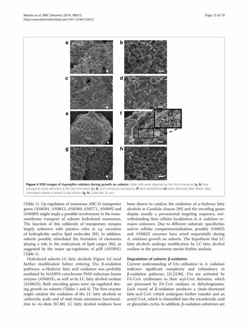
Figure 4 SEM images of Aspergillus nidulans during growth on suberin. Hülle cells were detected at the first time point (a, b), fewascospores were detected at the last time point (c, d), and numerous ascospores (f) and cleistothecia (e) were detected after fifteen days.Untreated suberin (control) is also shown (g, h). Scale bar: 10 μm.
Martins et al. BMC Genomics 2014, 15:613 Page 13 of 19http://www.biomedcentral.com/1471-2164/15/613
(Table 1). Up-regulation of numerous ABC-G transportergenes (AN6581, AN8813, AN6369, AN0771, AN8892 andAN8489) might imply a possible involvement in the trans-membrane transport of suberin hydrolysed monomers.The function of this subfamily of transporters remainslargely unknown with putative roles in e.g. excretionof hydrophobic and/or lipid molecules [85]. In addition,suberin possibly stimulated the formation of eisosomesplaying a role in the endocytosis of lipid cargos [86], assuggested by the major up-regulation of pilB (AN3931)(Table 1).Hydrolysed suberin LC fatty alcohols (Figure 1a) need
further modification before entering FAs β-oxidationpathways. ω-Hydroxy fatty acid oxidation was probablymediated by NADPH-cytochrome P450 reductase fusionenzyme (AN6835), as well as by LC fatty alcohol oxidase(AN0623). Both encoding genes were up-regulated dur-ing growth on suberin (Tables 1 and 4). The first enzymemight catalyse the oxidation of the LC fatty alcohols tocarboxylic acids and of mid-chain saturation functional-ities to vic-diols [87,88]. LC fatty alcohol oxidases have
been shown to catalyse the oxidation of ω-hydroxy fattyalcohols in Candida cloacae [89] and the encoding genesdisplay usually a peroxisomal targeting sequence, not-withstanding their cellular localisation in A. nidulans re-mains unknown. Due to different substrate specificitiesand/or cellular compartmentalisation, possibly AN6835and AN0623 enzymes have acted sequentially duringA. nidulans growth on suberin. The hypothesis that LCfatty alcohols undergo modification by LC fatty alcoholoxidase in the peroxisome merits further analysis.
Degradation of suberin: β-oxidationCurrent understanding of FAs utilisation in A. nidulansindicates significant complexity and redundancy inβ-oxidation pathways [21,22,90]. FAs are activated byFA-CoA synthetases to their acyl-CoA derivates, whichare processed by FA-CoA oxidases or dehydrogenases.Each round of β-oxidation produces a chain-shortenedfatty-acyl-CoA (which undergoes further rounds) and anacetyl-CoA, which is channelled into the tricarboxylic acidor glyoxylate cycles. In addition, β-oxidation substrates are
Martins et al. BMC Genomics 2014, 15:613 Page 14 of 19http://www.biomedcentral.com/1471-2164/15/613
actively transported across the mitochondria and/or theperoxisome membrane by carnitine shuttles, ADP/ATPcarriers or ABC transporter proteins. Suberin stimulatedtwo peroxisomal FA-CoA synthetase genes, namely faaB(AN8280) and fatA (AN5192) (Table 4) but none of thewell characterised mitochondrial ones (e.g. facA (AN5626)and pcsA (AN5833), Additional file 2). It seems thathydrolysed suberin monomers were essentially processedvia peroxisomal β-oxidation pathways (Table 4) and that,as previously suggested, FaaB is the major peroxisomalFA-CoA synthetase, while the remaining ones (FatA-D(AN5192, AN5877, AN6649, AN4397), FaaA (AN6114)and AN4659) display high functional redundancy [91]. Ac-tivation of peroxisomal β-oxidation agrees with the up-regulation of antA, a peroxisomal ATP carrier (AN0257)and of acuJ, a mitochondrial/peroxisomal carnitine acetyl-transferase (AN6279) (Table 4). Uncharacterised trans-porters for the peroxisomal translocation of activatedLCFAs have been suggested [22]; justifying the basal ex-pression levels of pxa1 and pxa2 (AN10078 and AN1014,ABC transporter proteins).Suberin stimulated the expression of aoxA (AN6752) but
not aoxB (AN6765), both encoding peroxisomal fatty-acyl-CoA oxidases (Table 4) but AoxA plays the major roleduring growth on LCFAs [92]. Deletion of aoxA leads togrowth impairment on LCFAs but not on SCFAs or verylong chain fatty acids (VLCFAs) [21]. aoxA stimulation isconsistent with the observation that suberin hydrolysis re-leases three times more LCFAs than VLCFAs [30,78]. Onlythree peroxisomal fatty-acyl-CoA dehydrogenase genes(out of seven) were responsive at the first time point,namely acdA (AN12335), AN1699 and AN7320 genes(Table 4). Deletion of either acdA or AN7320 has not led toany growth impairment on FAs, suggesting that theencoded enzymes display high redundancy [92]. Botrytiscinerea BC1G_13535 gene, which displays 78% of homologywith AN1699, was amongst the highest up-regulated genescoding in FAs β-oxidation during Lactuca sativa infection[93]. The hypothesis that this fatty-acyl-CoA dehydrogenaseplays a major role in A. nidulans degradation of plant FAscalls for its functional characterisation.LCFAs β-oxidation is shuttled between the peroxisomes
and the mitochondria, typically when the produced chain-shortened fatty-acyl-CoA is a butyryl-CoA (C4) [92]. Atthe first time point, most mitochondrial fatty-acyl-CoAdehydrogenase genes were down-regulated (AN2762,AN6761 and AN9162), except scdA (AN0824) [22]that was up-regulated. The remaining mitochondrialβ-oxidation genes, in general, decreased at the mid and/orlast time points, including the well characterised mthA, aβ-ketoacyl-CoA thiolase gene (AN10512), as well as acuH,a mitochondrial carnitine/acyl-carnitine carrier (AN5356)(Table 4). The only exception was ivdA, an isovaleryl-CoAdehydrogenase gene (AN4688) that was up-regulated,
together with mccB (AN4687), at the last time point(Table 4). This might imply that the fungus started usingleucine as a catabolic source; both genes are clustered withmccA (AN4690) in the leucine catabolic pathway [94].The glyoxylate bypass is absolutely required for growth
on carbon sources that produce acetyl-CoA and isdependent on isocitrate lyase (AcuD) and malate synthase(AcuE) activities. Transcription of acuD (AN5634) andacuE (AN6653) is regulated by FA and acetate inductionvia the FacB activator, but facB (AN0689) mutations donot prevent growth on FAs [22]. Up-regulation of farAand farB (FA-induced) and of facB (acetate-induced) ledto up-regulation of acuD but not of acuE (Table 4). Previ-ous studies have demonstrated that the imposition of sev-eral stresses might lead to both fluctuating mRNA andirregular protein expression levels in A. nidulans [95].
Degradation of phenolic suberinRelease of ferulic acid during fungal growth on potatosuberin has been suggested to involve the activity of fer-uloyl esterases (Fae) [5], notwithstanding a direct proofis still lacking. Ferulic acid release probably justifies theup-regulation of faeC (AN5267) (Table 3). Ferulic aciddegradation might involve the activity of 2,3-dihydroxy-benzoate carboxylyase [96], of which the encoding genedhbD (AN6723) was up-regulated at the first time point(Additional file 2). Several downstream products havebeen reported in different fungal strains, however the as-sociated enzymes remain largely unknown [96]. Neverthe-less, final degradation of the formed aromatics mightinvolve the homogentisate, benzoate or the salicylate path-ways [97], and several genes coding in these pathwayswere found to be up-regulated (e.g. AN10950, hmgA(AN1897), hpdA (AN1899), maiA (AN1895), Additionalfile 2) in the suberin media.
Degradation of other cell wall constituentsSeveral polysaccharides degrading enzymes were identi-fied in the A. nidulans secretome on suberin at the lasttime point (Table 2, Additional file 4), in general, agree-ing with the transcriptome data (Additional file 2). Theyincluded the β-glucosidase BglL (AN2828) and the xyla-nase XlnA (AN3613), of which the encoding genes werefound up-regulated at the last and the first time points,respectively. The other β-glucosidase genes were up-regulated at the first (bglM, AN7396) and mid timepoints (eglB (AN5214) and AN3046) and could not bedetected in the secretome. xlnR (AN7610), which en-codes the xylanolytic/cellulolytic transcriptional activa-tor, was up-regulated at the last time point (consistentwith creA decrease [98]), notwithstanding the up-regulation of the xlnA and xlnB (AN9365) at the earliertime points. The additional polysaccharide degrading en-zymes detected in the secretome were not consistent
Martins et al. BMC Genomics 2014, 15:613 Page 15 of 19http://www.biomedcentral.com/1471-2164/15/613
with the transcriptome data (Table 2), including XlnC(AN1818) and XlnE (AN7401). Probably the up-regulation of their corresponding genes occurred at anintermediate time point. The poor correlation betweentranscriptomic and proteomic data has been previouslyreported [95,99], often related to mechanisms associatedwith mRNA turnover and/or efficiency of translation[100] or to transcription on demand of certain mRNApools [101], among other reasons, including experimen-tal noise [102]. Protein species grouped in the miscellan-eous category (six, of which half have a predictedintracellular localisation) might be associated with celllysis, as highlighted by the presence of alcohol dehydro-genase I (alcA, AN8979) (Table 2).
ConclusionsPrevious studies on A. nidulans colonisation of cork re-vealed that suberin remained unaltered [103] probablybecause the outermost lignin-enriched cell wall layershampered its degradation. Here we have shown that
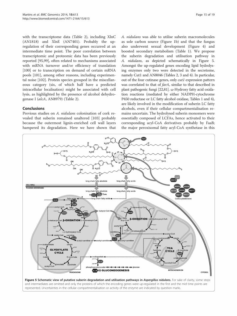
Figure 5 Schematic view of putative suberin degradation and utilisatand intermediates are omitted and only the proteins of which the encodinrepresented. Uncertainties in the cellular compartmentalisation or activity o
A. nidulans was able to utilise suberin macromoleculesas sole carbon source (Figure 1b) and that the fungusalso underwent sexual development (Figure 4) andboosted secondary metabolism (Table 1). We proposethe suberin degradation and utilisation pathway inA. nidulans, as depicted schematically in Figure 5.Amongst the up-regulated genes encoding lipid hydrolys-ing enzymes only two were detected in the secretome,namely Cut1 and AN8046 (Tables 2, 3 and 4). In particular,out of the four cutinase genes, only cut1 expression patternwas correlated to that of farA, similar to that described inplant pathogenic fungi [22,81]. ω-Hydroxy fatty acid oxida-tion reactions (mediated by either NADPH-cytochromeP450 reductase or LC fatty alcohol oxidase, Tables 1 and 4),are likely involved in the modification of suberin LC fattyalcohols, even if their cellular compartmentalisation re-mains uncertain. The hydrolysed suberin monomers wereessentially composed of LCFAs, hence activated to theircorresponding acyl-CoA derivatives probably by FaaB;the major peroxisomal fatty acyl-CoA synthetase in this
ion pathways in Aspergillus nidulans. For sake of clarity, some stepsg genes were up-regulated in the first and the mid time points aref the enzyme are indicated by question marks.
Martins et al. BMC Genomics 2014, 15:613 Page 16 of 19http://www.biomedcentral.com/1471-2164/15/613
fungus [92]. Despite high functional redundancy of add-itional peroxisomal FA-CoA synthetase genes, fatAshowed the highest up-regulation on suberin. Up-regulation of aoxA also occurred, agreeing with the ideathat the encoded fatty-acyl-CoA oxidase plays a majorrole during growth on LCFAs [21]. In addition, amongthe peroxisomal fatty-acyl-CoA dehydrogenase genes up-regulated here, AN1699 underwent the highest up-regulation (Table 4), similar to that reported for its B.cinerea ortholog during plant infection [93]. The corebinding sequence for FarA, typically overrepresented inthe promoter region of genes required for growth on FAs,is not present in all related genes up-regulated during A.nidulans growth on suberin (Table 3). Some unknownregulatory elements are certainly yet to be discovered, fur-ther emphasised by the down-regulation of some lipidhydrolysing genes carrying the FarA recognition site.
Availability of supporting dataThe data sets supporting the results of this article are in-cluded within the article (and its additional files).
Additional files
Additional file 1: Materials and Methods details.
Additional file 2: List of the differentially expressed genes.
Additional file 3: Functional categorisation of the genessignificantly altered during suberin incubation.
Additional file 4: Protein identification data.
AbbreviationsFC: Fold change; FAs: Fatty acids; SCFAs: Small chain fatty acids; LCFAs: Longchain fatty acids; VLCFAs: Very long chain fatty acids; ATR-FTIR: Attenuatedtotal reflectance Fourier transform infrared spectroscopy; SEM: Scanningelectron microscopy; qRT-PCR: Quantitative real-time PCR analysis;MIPS: Munich Information Center for Protein Sequences.
Competing interestsThe authors declare that they have no competing interests.
Authors’ contributionsCSP supervised the project and the interpretation of data and prepared thefinal version of the manuscript. All authors have done substantialcontributions to the acquisition, analysis and interpretation of data, andcontributed to the drafting of the manuscript: IM (experimental set up,transcriptomics and secretome analyses and preparation of the initial draft ofthe manuscript), DOH (PCR analyses), CM (microscopy studies), PCA, JH andJDB (set up of the custom microarray), HG and RF (suberin isolation andspectroscopy), HG and JDB (statistics), JR and CL (secretome analyses). Allauthors read and approved the final version of the manuscript.
Authors’ informationThis research constitutes an important milestone of C. Silva Pereira’s team(established in 2008) that is addressing a major scientific question: How fungiperceive and interact with the environment? What makes our researchapproach distinctive is the merging of fundamental biology research withchemical expertise. Such multidisciplinary environment has inspired thedevelopment of a new extraction method for suberin - a structural componentof the plant cell wall. Elucidating how fungi utilise and degrade plant polyestersstrongly impacts on our understanding of plant-fungi interactions and nutrientscycling. Complementary to this research, we have reported that the plantpolyester can be reconstituted as a material that combines bactericidal and
antibiofouling properties. We are particularly interested in further elucidatingplant polyesters physiological roles and to promote the clinical use of plantpolyester based materials.
AcknowledgementsThe work was partially supported by a grant from Iceland, Liechtenstein andNorway through the EEA financial mechanism (Project PT015), and FCTthrough the grants PEst-OE/EQB/LA0004/2013, PTDC/QUI-QUI/120982/2010and PTDC/AAC-CLI/119100/2010.IM, DOH, PCA and RF are grateful to Fundação para a Ciência e a Tecnologia(FCT), Portugal, for the fellowships SFRH/BD/38378/2007, SFRH/BD/66396/2009, SFRH/BD/66030/2009, SFRH/BD/48286/2008, respectively and HG toFundação Calouste Gulbenkian, Portugal for the fellowship 21-95587-B. Weare thankful to Dr. Tiago Martins (ITQB), who read and critically commentedthe final manuscript.
Author details1Instituto de Tecnologia Química e Biológica, Universidade Nova de Lisboa,Av. da República, 2780-157 Oeiras, Portugal. 2Instituto de BiologiaExperimental e Tecnológica (iBET), Av. da República, 2781-901 Oeiras,Portugal. 3Proteomics Platform, Centre de Recherche Public - Gabriel Lipp-mann, Belvaux, Luxembourg. 4Cancer Genomics Research Laboratory,Division of Cancer Epidemiology and Genetics, National Cancer Institute, NIH,DHHS, 8717 Grovemont Circle, 20877 Gaithersburg, MD, USA (previously, theScientific Computing department, Samuel Roberts Noble Foundation, USA).5Instituto Gulbenkian de Ciência, Rua da Quinta Grande 6, 2780-156 Oeiras,Portugal.
Received: 18 March 2014 Accepted: 16 July 2014Published: 21 July 2014
References1. Heredia A, Matas A, Dominguez E: Investigating plant lipid biopolymers.
Biochem Educ 2000, 28(1):50–51.2. Winkler A, Haumaier L, Zech W: Insoluble alkyl carbon components in soils
derive mainly from cutin and suberin. Org Geochem 2005, 36(4):519–529.3. Hamer U, Rumpel C, Dignac MF: Cutin and suberin biomarkers as tracers for
the turnover of shoot and root derived organic matter along achronosequence of Ecuadorian pasture soils. Eur J Soil Sci 2012, 63(6):808–819.
4. Harms H, Schlosser D, Wick LY: Untapped potential: exploiting fungi inbioremediation of hazardous chemicals. Nat Rev Microbiol 2011, 9(3):177–192.
5. Kolattukudy PE, Scheper T, Babel W, Steinbüchel A: Polyesters in higherplants. In Biopolyesters, Volume 71; 2001:1–49.
6. Pereira H: Cork: Biology, Production and Uses. Amsterdam: Elsevier; 2007.7. van Fleet DS: Histochemistry and function of the endodermis. Bot Rev
1961, 27(2):165–220.8. Yeats TH, Rose JKC: The formation and function of plant cuticles.
Plant Physiol 2013, 163(1):5–20.9. Schreiber L: Transport barriers made of cutin, suberin and associated
waxes. Trends Plant Sci 2010, 15(10):546–553.10. Pollard M, Beisson F, Li YH, Ohlrogge JB: Building lipid barriers:
biosynthesis of cutin and suberin. Trends Plant Sci 2008, 13(5):236–246.11. Javelle M, Vernoud V, Rogowsky PM, Ingram GC: Epidermis: the formation
and functions of a fundamental plant tissue. New Phytol 2011, 189(1):17–39.12. Kolattukudy PE: Biopolyester membranes of plants: cutin and suberin.
Science 1980, 208(4447):990–1000.13. Lulai EC, Corsini DL: Differential deposition of suberin phenolic and
aliphatic domains and their roles in resistance to infection during potatotuber (Solanum tuberosum L.) wound-healing. Physiol Mol Plant Pathol1998, 53(4):209–222.
14. Ranathunge K, Schreiber L, Franke R: Suberin research in the genomicsera: new interest for an old polymer. Plant Sci 2011, 180(3):399–413.
15. Garcia H, Ferreira R, Martins C, Sousa AF, Freire CSR, Silvestre AJD, Kunz W,Rebelo LPN, Silva Pereira C: Ex situ reconstitution of the plantbiopolyester suberin as a film. Biomacromolecules 2014, 15(5):1806–1813.
16. Graça J, Schreiber L, Rodrigues J, Pereira H: Glycerol and glyceryl esters ofω-hydroxyacids in cutins. Phytochemistry 2002, 61(2):205–215.
17. Graça J, Lamosa P: Linear and branched poly(ω-hydroxyacid) esters inplant cutins. J Agr Food Chem 2010, 58(17):9666–9674.
Martins et al. BMC Genomics 2014, 15:613 Page 17 of 19http://www.biomedcentral.com/1471-2164/15/613
18. Fernando G, Zimmermann W, Kolattukudy PE: Suberin-grown Fusarium solanif. sp pisi generates a cutinase-like esterase which depolymerizes the aliphaticcomponents of suberin. Physiol Mol Plant Pathol 1984, 24(2):143–155.
19. Kontkanen H, Westerholm-Parvinen A, Saloheimo M, Bailey M, Rättö M,Mattila I, Mohsina M, Kalkkinen N, Nakari-Setälä T, Buchert J: NovelCoprinopsis cinerea polyesterase that hydrolyzes cutin and suberin.Appl Environ Microb 2009, 75(7):2148–2157.
20. Garcia Lepe R, Nuero OM, Reyes F, Santamaria F: Lipases in autolysedcultures of filamentous fungi. Lett Appl Microbiol 1997, 25(2):127–130.
21. Maggio-Hall LA, Keller NP: Mitochondrial β-oxidation in Aspergillusnidulans. Mol Microbiol 2004, 54(5):1173–1185.
22. Hynes MJ, Murray SL, Duncan A, Khew GS, Davis MA: Regulatory genescontrolling fatty acid catabolism and peroxisomal functions in thefilamentous fungus Aspergillus nidulans. Eukaryot Cell 2006,5(5):794–805.
23. Purdy R, Kolattukudy P: Hydrolysis of plant cuticle by plant pathogens.Properties of cutinase I, cutinase II, and a nonspecific esterase isolatedfrom Fusarium solani pisi. Biochemistry 1975, 14(13):2832–2840.
24. Fan C-Y, Köller W: Diversity of cutinases from plant pathogenic fungi:diferential and sequential expression of cutinolytic esterases byAlternaria brassicicola. FEMS Microbiol Lett 1998, 158(1):33–38.
25. Bonnen AM, Hammerschmidt R: Cutinolytic enzymes from Colletotrichumlagenarium. Physiol Mol Plant Pathol 1989, 35(6):463–474.
26. Li D, Sirakova T, Rogers L, Ettinger WF, Kolattukudy PE: Regulation ofconstitutively expressed and induced cutinase genes by different zincfinger transcription factors in Fusarium solani f. sp. pisi (Nectriahaematococca). J Biol Chem 2002, 277:7905–7912.
27. Castro-Ochoa D, Peña-Montes C, González-Canto A, Alva-Gasca A,Esquivel-Bautista R, Navarro-Ocaña A, Farrés A: ANCUT2, an extracellularcutinase from Aspergillus nidulans induced by olive oil. Appl BiochemBiotechnol 2012, 166:1275–1290.
28. Yang S, Xu H, Yan Q, Liu Y, Zhou P, Jiang Z: A low molecular masscutinase of Thielavia terrestris efficiently hydrolyzes poly(esters). J IndMicrobiol Biotechnol 2013, 40:217–226.
29. Garcia H, Ferreira R, Petkovic M, Ferguson JL, Leitão MC, Gunaratne HQ,Seddon K, Rebelo L, Silva Pereira C: Dissolution of cork biopolymers inbiocompatible ionic liquids. Green Chem 2010, 12:367–369.
30. Ferreira R, Garcia H, Sousa AF, Freire CSR, Silvestre AJD, Rebelo LPN, SilvaPereira C: Isolation of suberin from birch outer bark and cork usingionic liquids: a new source of macromonomers. Ind Crop Prod 2013,44:520–527.
31. Ferreira R, Garcia H, Sousa AF, Guerreiro M, Duarte FJS, Freire CSR,Calhorda MJ, Silvestre AJD, Rebelo LPN, Silva Pereira C: Unravellingthe dual role of cholinium hexanoate ionic liquid as solventand catalyst in suberin depolymerization. RSC Adv 2014,4(1):2993–3002.
32. Ferreira R, Garcia H, Sousa AF, Petkovic M, Lamosa P, Freire CSR, Silvestre AJD,Rebelo LPN, Silva Pereira C: Suberin isolation from cork using ionic liquids:characterisation of ensuing products. New J Chem 2012, 36(10):2014–2024.
33. Carvalho MB, Martins I, Medeiros J, Tavares S, Planchon S, Renaut J, Nuñez O,Gallart-Ayala H, Galceran MT, Hursthouse A, Silva Pereira C: The response ofMucor plumbeus to pentachlorophenol: a toxicoproteomics study.J Proteomics 2013, 78:159–171.
34. Petkovic M, Hartmann DO, Adamová G, Seddon KR, Rebelo LPN, Silva Pereira C:Unravelling the mechanism of toxicity of alkyltributylphosphoniumchlorides in Aspergillus nidulans conidia. New J Chem 2012,36(1):56–63.
35. Seidler M, Salvenmoser S, Müller FM: In vitro effects of micafungin againstCandida biofilms on polystyrene and central venous catheter sections.Int J Antimicrob Agents 2006, 28(6):568–573.
36. Carvalho MB, Martins I, Leitão MC, Garcia H, Rodrigues C, Romão VS,McLellan I, Hursthouse A, Silva Pereira C: Screening pentachlorophenoldegradation ability by environmental fungal strains belonging to thephyla Ascomycota and Zygomycota. J Ind Microbiol Biotechnol 2009,36(10):1249–1256.
37. Hartmann DO, Silva Pereira C: A molecular analysis of the toxicity ofalkyltributylphosphonium chlorides in Aspergillus nidulans. New J Chem2013, 37(5):1569–1577.
38. Edgar R, Domrachev M, Lash AE: Gene expression omnibus: NCBI geneexpression and hybridization array data repository. Nucleic Acids Res2002, 30(1):207–210.
39. Li C, Wong WH: Model-based analysis of oligonucleotide arrays:expression index computation and outlier detection. Proc Natl Acad SciU S A 2001, 98(1):31–36.
40. Li C, Wong WH: Model-based analysis of oligonucleotide arrays: modelvalidation, design issues and standard error application. Genome Biol2001, 2(8):1–11.
41. Priebe S, Linde J, Albrecht D, Guthke R, Brakhage AA: FungiFun: aweb-based application for functional categorization of fungal genes andproteins. Fungal Genet Biol 2011, 48(4):353–358.
42. Emri T, Szilágyi M, László K, M-Hamvas M, Pócsi I: PepJ is a new extracellularproteinase of Aspergillus nidulans. Folia Microbiol 2009, 54(2):105–109.
43. Szilágyi M, Kwon NJ, Bakti F, M-Hamvas M, Jámbrik K, Park H, Pócsi I, Yu JH,Emri T: Extracellular proteinase formation in carbon starving Aspergillusnidulans cultures - physiological function and regulation. J Basic Microbiol2011, 51(6):625–634.
44. Pusztahelyi T, Molnár Z, Emri T, Klement É, Miskei M, Kerékgyártó J, Balla J,Pócsi I: Comparative studies of differential expression of chitinolyticenzymes encoded by chiA, chiB, chiC and nagA genes in Aspergillusnidulans. Folia Microbiol 2006, 51(6):547–554.
45. Shin KS, Kwon NJ, Kim YH, Park HS, Kwon GS, Yu JH: Differential roles ofthe ChiB chitinase in autolysis and cell death of Aspergillus nidulans.Eukaryot Cell 2009, 8(5):738–746.
46. Szilágyi M, Kwon NJ, Dorogi C, Pócsi I, Yu JH, Emri T: The extracellularβ-1,3-endoglucanase EngA is involved in autolysis of Aspergillus nidulans.J Appl Microbiol 2010, 109(5):1498–1508.
47. Szilágyi M, Miskei M, Karányi Z, Lenkey B, Pócsi I, Emri T: Transcriptomechanges initiated by carbon starvation in Aspergillus nidulans.Microbiol (UK) 2013, 159:176–190.
48. Muzzarelli RAA, Miliani M, Cartolari M, Tarsi R, Tosi G, Muzzarelli C:Polyuronans obtained by regiospecific oxidation of polysaccharides fromAspergillus niger, Trichoderma reesei and Saprolegnia sp. Carbohyd Polym2000, 43(1):55–61.
49. Tsiatsiani L, Van Breusegem F, Gallois P, Zavialov A, Lam E, Bozhkov PV:Metacaspases. Cell Death Differ 2011, 18(8):1279–1288.
50. Uren AG, O’Rourke K, Aravind L, Pisabarro MT, Seshagiri S, Koonin EV, Dixit VM:Identification of paracaspases and metacaspases: two ancient families ofcaspase-like proteins, one of which plays a key role in MALT lymphoma.Mol Cell 2000, 6(4):961–967.
51. Savoldi M, Malavazi I, Soriani FM, Capellaro JL, Kitamoto K, da SilvaFerreira ME, Goldman MHS, Goldman GH: Farnesol induces thetranscriptional accumulation of the Aspergillus nidulans ApoptosisInducing Factor (AIF) like mitochondrial oxidoreductase. Mol Microbiol2008, 70(1):44–59.
52. Ren Q, Chen K, Paulsen IT: TransportDB: a comprehensive databaseresource for cytoplasmic membrane transport systems and outermembrane channels. Nucleic Acids Res 2007, 35:D274–D279.
53. Inglis DO, Binkley J, Skrzypek MS, Arnaud MB, Cerqueira GC, Shah P, Wymore F,Wortman JR, Sherlock G: Comprehensive annotation of secondarymetabolite biosynthetic genes and gene clusters of Aspergillus nidulans, A.fumigatus, A. niger and A. oryzae. BMC Microbiol 2013, 13:91.
54. Forment JV, Flipphi M, Ramón D, Ventura L, MacCabe AP: Identification ofthe mstE gene encoding a glucose inducible, low affinity glucosetransporter in Aspergillus nidulans. J Biol Chem 2006, 281(13):8339–8346.
55. Dowzer CEA, Kelly JM: Analysis of the CreA gene, a regulator of carboncatabolite repression in Aspergillus nidulans. Mol Cell Biol 1991, 11(11):5701–5709.
56. David H, Krogh AM, Roca C, Akesson M, Nielsen J: CreA influences themetabolic fluxes of Aspergillus nidulans during growth on glucose andxylose. Microbiol (UK) 2005, 151:2209–2221.
57. Strauss J, Horvath HK, Abdallah BM, Kindermann J, Mach RL, Kubicek CP:The function of CreA, the carbon catabolite repressor of Aspergillusnidulans, is regulated at the transcriptional and post-transcriptional level.Mol Microbiol 1999, 32(1):169–178.
58. Hynes MJ, Szewczyk E, Murray SL, Suzuki Y, Davis MA, Lewis HMS:Transcriptional control of gluconeogenesis in Aspergillus nidulans.Genetics 2007, 176(1):139–150.
59. Meijer S, Panagiotou G, Olsson L, Nielsen J: Physiological characterizationof xylose metabolism in Aspergillus niger under oxygen-limitedconditions. Biotechnol Bioeng 2007, 98(2):462–475.
60. Felenbok B, Sequeval D, Mathieu M, Sibley S, Gwynne DI, Davies RW: Theethanol regulon in Aspergillus nidulans - characterization and sequenceof the positive regulatory gene alcR. Gene 1988, 73(2):385–396.
Martins et al. BMC Genomics 2014, 15:613 Page 18 of 19http://www.biomedcentral.com/1471-2164/15/613
61. Macios M, Caddick MX, Weglenski P, Scazzocchio C, Dzikowska A: The GATAfactors AREA and AREB together with the co-repressor NMRA, negativelyregulate arginine catabolism in Aspergillus nidulans in response tonitrogen and carbon source. Fungal Genet Biol 2012, 49(3):189–198.
62. Wong KH, Hynes MJ, Davis MA: Recent advances in nitrogen regulation: acomparison between Saccharomyces cerevisiae and filamentous fungi.Eukaryot Cell 2008, 7(6):917–925.
63. Berger H, Basheer A, Böck S, Reyes-Dominguez Y, Dalik T, Altmann F,Strauss J: Dissecting individual steps of nitrogen transcription factorcooperation in the Aspergillus nidulans nitrate cluster. Mol Microbiol2008, 69(6):1385–1398.
64. Schinko T, Berger H, Lee W, Gallmetzer A, Pirker K, Pachlinger R, Buchner I,Reichenauer T, Güldener U, Strauss J: Transcriptome analysis of nitrateassimilation in Aspergillus nidulans reveals connections to nitric oxidemetabolism. Mol Microbiol 2010, 78(3):720–738.
65. Muszkieta L, Beauvais A, Pähtz V, Gibbons JG, Anton Leberre V, Beau R,Shibuya K, Rokas A, Francois JM, Kniemeyer O, Brakhage AA, Latge JP:Investigation of Aspergillus fumigatus biofilm formation by various“omics” approaches. Front Microbiol 2013, 4:13.
66. Wei HJ, Scherer M, Singh A, Liese R, Fischer R: Aspergillus nidulans α-1,3glucanase (mutanase), mutA, is expressed during sexual developmentand mobilizes mutan. Fungal Genet Biol 2001, 34(3):217–227.
67. Arnaud M, Chibucos M, Costanzo M, Crabtree J, Inglis D, Lotia A, Orvis J,Shah P, Skrzypek M, Binkley G, Miyasato SR, Wortman JR, Sherlock G: TheAspergillus Genome Database, a curated comparative genomicsresource for gene, protein and sequence information for theAspergillus research community. Nucleic Acids Res 2010,38(Database issue):D420–427.
68. Apweiler R, Martin MJ, O’Donovan C, Magrane M, Alam-Faruque Y, Alpi E,Antunes R, Arganiska J, Casanova EB, Bely B, Apweiler R, Martin MJ,O’Donovan C, Magrane M, Alam-Faruque Y, Alpi E, Antunes R, Arganiska J,Casanova EB, Bely B, Bingley M, Bonilla C, Britto R, Bursteinas B, Chan WM,Chavali G, Cibrian-Uhalte E, Da Silva A, De Giorgi M, Dimmer E, et al: Updateon activities at the Universal Protein Resource (UniProt) in 2013.Nucleic Acids Res 2013, 41(D1):D43–D47.
69. Dyer PS, O’Gorman CM: Sexual development and cryptic sexuality infungi: insights from Aspergillus species. FEMS Microbiol Rev 2012,36(1):165–192.
70. Krijgsheld P, Bleichrodt R, van Veluw GJ, Wang F, Müller WH, Dijksterhuis J,Wosten HAB: Development in Aspergillus. Stud Mycol 2013, 74(1):1–29.
71. Paoletti M, Seymour FA, Alcocer MJC, Kaur N, Caivo AM, Archer DB, Dyer PS:Mating type and the genetic basis of self-fertility in the model fungusAspergillus nidulans. Curr Biol 2007, 17(16):1384–1389.
72. Bayram O, Braus GH: Coordination of secondary metabolism anddevelopment in fungi: the velvet family of regulatory proteins.FEMS Microbiol Rev 2012, 36(1):1–24.
73. Ariyo B, Tamerler C, Bucke C, Keshavarz T: Enhanced penicillinproduction by oligosaccharides from batch cultures of Penicilliumchrysogenum in stirred-tank reactors. FEMS Microbiol Lett 1998,166(1):165–170.
74. Yin W-B, Amaike S, Wohlbach DJ, Gasch AP, Chiang Y-M, Wang CCC, BokJW, Rohlfs M, Keller NP: An Aspergillus nidulans bZIP response pathwayhardwired for defensive secondary metabolism operates through aflR.Mol Microbiol 2012, 83(5):1024–1034.
75. Fisch KM, Gillaspy AF, Gipson M, Henrikson JC, Hoover AR, Jackson L,Najar FZ, Waegele H, Cichewicz RH: Chemical induction of silentbiosynthetic pathway transcription in Aspergillus niger. J Ind MicrobiolBiotechnol 2009, 36(9):1199–1213.
76. Lerat S, Forest M, Lauzier A, Grondin G, Lacelle S, Beaulieu C: Potato suberininduces differentiation and secondary metabolism in the genusStreptomyces. Microbes Environ 2012, 27(1):36–42.
77. Baker PJ, Poultney C, Liu Z, Gross R, Montclare JK: Identification andcomparison of cutinases for synthetic polyester degradation.Appl Microbiol Biotechnol 2012, 93(1):229–240.
78. Gandini A, Pascoal Neto C, Silvestre AJD: Suberin: a promisingrenewable resource for novel macromolecular materials. Prog PolymSci 2006, 31(10):878–892.
79. Salazar M, Vongsangnak W, Panagiotou G, Andersen MR, Nielsen J:Uncovering transcriptional regulation of glycerol metabolism inAspergilli through genome-wide gene expression data analysis.Mol Genet Genomics 2009, 282(6):571–586.
80. Garrido SM, Kitamoto N, Watanabe A, Shintani T, Gomi K: Functionalanalysis of FarA transcription factor in the regulation of the genesencoding lipolytic enzymes and hydrophobic surface binding protein forthe degradation of biodegradable plastics in Aspergillus oryzae. J BiosciBioeng 2012, 113(5):549–555.
81. Rocha ALM, Di Pietro A, Ruiz-Roldan C, Roncero MIG: Ctf1, a transcriptionalactivator of cutinase and lipase genes in Fusarium oxysporum is dispensablefor virulence. Mol Plant Pathol 2008, 9(3):293–304.
82. Bravo-Ruiz G, Ruiz-Roldan C, Roncero MIG: Lipolytic System of the TomatoPathogen Fusarium oxysporum f. sp. lycopersici. Mol Plant Microbe In 2013,26(9):1054–1067.
83. Kovalchuk A, Driessen AJM: Phylogenetic analysis of fungal ABCtransporters. BMC Genomics 2010, 11(1):177.
84. Andrade AC, Van Nistelrooy JGM, Peery RB, Skatrud PL, De Waard MA: Therole of ABC transporters from Aspergillus nidulans in protection againstcytotoxic agents and in antibiotic production. Mol Gen Genet 2000,263(6):966–977.
85. Holyoak CD, Bracey D, Piper PW, Kuchler K, Coote PJ: TheSaccharomyces cerevisiae weak acid inducible ABC transporterPdr12 transports fluorescein and preservative anions from thecytosol by an energy dependent mechanism. J Bacteriol 1999,181(15):4644–4652.
86. Vangelatos I, Roumelioti K, Gournas C, Suarez T, Scazzocchio C,Sophianopoulou V: Eisosome organization in the filamentous ascomyceteAspergillus nidulans. Eukaryot Cell 2010, 9(10):1441–1454.
87. Kelly DE, Kraševec N, Mullins J, Nelson DR: The CYPome (cytochromeP450 complement) of Aspergillus nidulans. Fungal Genet Biol 2009,46:S53–S61.
88. Kitazume T, Takaya N, Nakayama N, Shoun H: Fusarium oxysporumfatty-acid subterminal hydroxylase (CYP505) is a membrane-boundeukaryotic counterpart of Bacillus megaterium cytochrome P450BM3.J Biol Chem 2000, 275(50):39734–39740.
89. Vanhanen S, West M, Kroon JTM, Lindner N, Casey J, Cheng Q, ElboroughKM, Slabas AR: A consensus sequence for long-chain fatty-acid alcoholoxidases from Candida identifies a family of genes involved in lipidomega-oxidation in yeast with homologues in plants and bacteria.J Biol Chem 2000, 275(6):4445–4452.
90. Hynes MJ, Murray SL, Khew GS, Davis MA: Genetic analysis of the role ofperoxisomes in the utilization of acetate and fatty acids in Aspergillusnidulans. Genetics 2008, 178(3):1355–1369.
91. Reiser K, Davis MA, Hynes MJ: Aspergillus nidulans contains six possiblefatty acyl-CoA synthetases with FaaB being the major synthetase forfatty acid degradation. Arch Microbiol 2010, 192(5):373–382.
92. Reiser K, Davis MA, Hynes MJ: AoxA is a major peroxisomal long chainfatty acyl-CoA oxidase required for beta-oxidation in A. nidulans.Curr Genet 2010, 56(2):139–150.
93. De Cremer K, Mathys J, Vos C, Froenicke L, Michelmore RW, Cammue BPA,De Coninck B: RNAseq-based transcriptome analysis of Lactuca sativainfected by the fungal necrotroph Botrytis cinerea. Plant Cell Environ 2013,36(11):1992–2007.
94. Maggio-Hall LA, Lyne P, Wolff JA, Keller NP: A single acyl-CoAdehydrogenase is required for catabolism of isoleucine, valine andshort-chain fatty acids in Aspergillus nidulans. Fungal Genet Biol 2008,45(3):180–189.
95. Pusztahelyi T, Klement É, Szajli E, Klem J, Miskei M, Karányi Z, Emri T,Kóvacs S, Orosz G, Kóvacs KL, Medzihradszky KF, Prade RA, Pócsi I:Comparison of transcriptional and translational changes caused bylong-term menadione exposure in Aspergillus nidulans. Fungal Genet Biol2011, 48(2):92–103.
96. Mathew S, Abraham TE: Bioconversions of ferulic acid, anhydroxycinnamic acid. Crit Rev Microbiol 2006, 32(3):115–125.
97. Caspi R, Altman T, Dreher K, Fulcher CA, Subhraveti P, Keseler IM, Kothari A,Krummenacker M, Latendresse M, Mueller LA, Ong Q, Paley S, Pujar A,Shearer AG, Travers M, Weerasinghe D, Zhang PF, Karp PD: The MetaCycdatabase of metabolic pathways and enzymes and the BioCyccollection of pathway/genome databases. Nucleic Acids Res 2012,40(D1):D742–D753.
98. Tamayo EN, Villanueva A, Hasper AA, de Graaff LH, Ramón D, Orejas M:CreA mediates repression of the regulatory gene xlnR which controls theproduction of xylanolytic enzymes in Aspergillus nidulans. Fungal GenetBiol 2008, 45(6):984–993.
Martins et al. BMC Genomics 2014, 15:613 Page 19 of 19http://www.biomedcentral.com/1471-2164/15/613
99. Gygi SP, Rist B, Gerber SA, Turecek F, Gelb MH, Aebersold R: Quantitativeanalysis of complex protein mixtures using isotope-coded affinity tags.Nat Biotechnol 1999, 17(10):994–999.
100. Hinnebusch AG: Translational regulation of GCN4 and the general aminoacid control of yeast. Annu Rev Microbiol 2005, 59:407–450.
101. Beyer A, Hollunder J, Nasheuer HP, Wilhelm T: Post-transcriptionalexpression regulation in the yeast Saccharomyces cerevisiae on agenomic scale. Mol Cell Proteomics 2004, 3(11):1083–1092.
102. Laloo B, Simon D, Veilla V, Lauzel D, Guyonnet-Duperat V, Moreau-Gaudry F,Sagliocco F, Grosset C: Analysis of post-transcriptional regulations by afunctional, integrated, and quantitative method. Mol Cell Proteomics 2009,8(8):1777–1788.
103. Martins I, Garcia H, Varela A, Núñez O, Planchon S, Galceran MT, Renaut J,Rebelo LPN, Silva Pereira C: Investigating Aspergillus nidulans secretomeduring colonisation of cork cell walls. J Proteomics 2014, 26(98):175–188.
doi:10.1186/1471-2164-15-613Cite this article as: Martins et al.: Elucidating how the saprophyticfungus Aspergillus nidulans uses the plant polyester suberin as carbonsource. BMC Genomics 2014 15:613.
Submit your next manuscript to BioMed Centraland take full advantage of:
• Convenient online submission
• Thorough peer review
• No space constraints or color figure charges
• Immediate publication on acceptance
• Inclusion in PubMed, CAS, Scopus and Google Scholar
• Research which is freely available for redistribution
Submit your manuscript at www.biomedcentral.com/submit
Related Documents