Elsevier Editorial System(tm) for Gait and Posture Manuscript Draft Manuscript Number: GAIPOS-D-10-00494R1 Title: Postural control after traumatic brain injury in patients with neuro-ophthalmic deficits Article Type: Original Paper Keywords: Traumatic Brain Injury; Balance; Neuro-ophthalmic deficits; Static posturography; Quiet standing Corresponding Author: Dr. Valentina Agostini, Ph.D. Corresponding Author's Institution: Politecnico di Torino First Author: Valentina Agostini, PhD Order of Authors: Valentina Agostini, PhD; Emma Chiaramello, M.Sc.; Carla Bredariol, M.D.; Chanda Cavallini, M.D.; Marco Knaflitz, PhD Abstract: Postural instability is a common and devastating consequence of Traumatic Brain Injury (TBI). The majority of TBI patients also suffer from neuro-ophthalmic deficits that may be an important contributing element to their sensation of vertigo and dizziness. Static posturography aims at the objective evaluation of patient balance impairment, but it is usually affected by large inter- and intra- subject variability. Here we propose a protocol based on ten randomized trials stimulating in different ways the visual and vestibular systems. Due to its completeness, our protocol highlights the specific residual difficulties of each patient in the various conditions. In this way, it was possible to evidence significant balance abnormalities in TBI patients with respect to controls. Moreover, by means of a multivariate analysis we were able to discriminate different levels of residual neuro- ophthalmic impairment. Authors' version. To be published in: Gait &Posture, ISSN 0966-6362, Editore Elsevier Science B.V., Amsterdam. Link to publisher version: http://dx.doi.org/10.1016/j.gaitpost.2011.05.008

Welcome message from author
This document is posted to help you gain knowledge. Please leave a comment to let me know what you think about it! Share it to your friends and learn new things together.
Transcript

Elsevier Editorial System(tm) for Gait and Posture Manuscript Draft Manuscript Number: GAIPOS-D-10-00494R1 Title: Postural control after traumatic brain injury in patients with neuro-ophthalmic deficits Article Type: Original Paper Keywords: Traumatic Brain Injury; Balance; Neuro-ophthalmic deficits; Static posturography; Quiet standing Corresponding Author: Dr. Valentina Agostini, Ph.D. Corresponding Author's Institution: Politecnico di Torino First Author: Valentina Agostini, PhD Order of Authors: Valentina Agostini, PhD; Emma Chiaramello, M.Sc.; Carla Bredariol, M.D.; Chanda Cavallini, M.D.; Marco Knaflitz, PhD Abstract: Postural instability is a common and devastating consequence of Traumatic Brain Injury (TBI). The majority of TBI patients also suffer from neuro-ophthalmic deficits that may be an important contributing element to their sensation of vertigo and dizziness. Static posturography aims at the objective evaluation of patient balance impairment, but it is usually affected by large inter- and intra- subject variability. Here we propose a protocol based on ten randomized trials stimulating in different ways the visual and vestibular systems. Due to its completeness, our protocol highlights the specific residual difficulties of each patient in the various conditions. In this way, it was possible to evidence significant balance abnormalities in TBI patients with respect to controls. Moreover, by means of a multivariate analysis we were able to discriminate different levels of residual neuro-ophthalmic impairment.
Authors' version. To be published in: Gait &Posture, ISSN 0966-6362, Editore Elsevier Science B.V., Amsterdam.
Link to publisher version: http://dx.doi.org/10.1016/j.gaitpost.2011.05.008

1
Title of the manuscript:
Postural control after traumatic brain injury in patients with neuro-ophthalmic deficits
Authors:
Valentina Agostini, Dipartimento di Elettronica, Politecnico di Torino, Torino, Italy,
e-mail: [email protected]
Emma Chiaramello, Dipartimento di Elettronica, Politecnico di Torino, Torino, Italy,
e-mail: [email protected]
Carla Bredariol, Clinica C. Sperino, Ospedale Oftalmico di Torino, Torino, Italy,
e-mail: [email protected]
Chanda Cavallini, Clinica C. Sperino, Ospedale Oftalmico di Torino, Torino, Italy,
e-mail: [email protected]
Marco Knaflitz, Dipartimento di Elettronica, Politecnico di Torino, Torino, Italy,
e-mail: [email protected]
Corresponding Author:
Valentina Agostini, Dipartimento di Elettronica, Politecnico di Torino,
Corso Duca degli Abruzzi 24, 10129 Torino, Italy
Tel. +39 011 5644136
Fax. +39 011 5644217
E-mail: [email protected]
*4. Title Page (with authors and addresses)

1
Abstract 1
Postural instability is a common and devastating consequence of Traumatic Brain Injury (TBI). The 2
majority of TBI patients also suffer from neuro-ophthalmic deficits that may be an important 3
contributing element to their sensation of vertigo and dizziness. Static posturography aims at the 4
objective evaluation of patient balance impairment, but it is usually affected by large inter- and 5
intra- subject variability. Here we propose a protocol based on ten randomized trials stimulating in 6
different ways the visual and vestibular systems. Due to its completeness, our protocol highlights 7
the specific residual difficulties of each patient in the various conditions. In this way, it was possible 8
to evidence significant balance abnormalities in TBI patients with respect to controls. Moreover, by 9
means of a multivariate analysis we were able to discriminate different levels of residual neuro-10
ophthalmic impairment. 11
12
1. Introduction 13
Traumatic Brain Injury (TBI) is an important cause of disability at all ages [1]. In the USA the 14
annual incidence of emergency department visits and hospital admission are respectively 403 per 15
100,000 and 85 per 100,000 [2]. The mean annual incidence rate of hospitalized and fatal TBI for 16
Europe is 235 per 100,000 [3]. Approximately 80% of injuries are classified as mild, 10% as 17
moderate, and 10% as severe [3]. Severity is usually described by the Glasgow Coma Scale (GCS) 18
[4], evaluated when the patient enters the emergency department. However, GCS may change 19
during hospitalization and it does not describe the nature and the entity of the residual impairments. 20
One of the most common complaints among TBI patients is postural instability and balance 21
impairment [5-6]. 22
Neuro-ophthalmic deficits commonly follow TBI, since the afferent and efferent pathways are 23
vulnerable to traumatic injury. Commonly described categories of oculomotor dysfunctions are 24
anomalies of accommodation, version, vergence (nonstrabismic, as well as strabismic), 25
*5. Manuscript

2
photosensitivity, visual field integrity, and ocular health [7]. Authors indicate different percentages 1
of neuro-ophthalmic impairments following TBI, ranging from 39% to 90%, as described in [8-11]. 2
Neuro-ophthalmic deficits may have important consequences on balance, since postural control 3
integrates information from the visual, vestibular, and somatosensory systems. 4
Subjective complaints of dizziness that occur in the absence of objective clinical signs are difficult 5
to assess [12-13]. Static stabilometry may provide an objective evaluation of postural instability 6
[14-18] by characterizing the performance of the postural control system during quiet standing. 7
This technique is based on the study of the trajectories of the Center of Pressure (CoP) on the 8
support surface. CoP trajectories are recorded by a force platform and analyzed using different 9
techniques and extracting different kinds of parameters [16,18]. A possible limit of static 10
stabilometry was highlighted by [15,19] due to the high inter-subject and intra-subject variability 11
that many studies report. 12
Previous studies [12-13, 20-25] addressed the problem of quantifying the consequences of TBI on 13
balance assessment using static stabilometry. None of the studies published in the past specifically 14
considered a group of TBI patients with a significant residual visual impairment. 15
Studies on static posturography are usually based on an acquisition protocol consisting of two trials, 16
with eyes open and closed respectively, to take into account the role of the visual system. 17
Our study differs from the previous ones for two aspects. First, we consider a group of TBI patients 18
with residual neuro-ophthalmic deficits. Secondly, this study is based on a more complete 19
acquisition protocol that adds to frontal open- and closed-eye trials, trials in which quiet standing of 20
the subject is evaluated after a fast or a slow head rotation, both with eyes open and closed. In this 21
way, it is possible to highlight the specific difficulties of each patient in various conditions that 22
stimulate the visual and vestibular systems. 23
The aim of this study is to present a more complete acquisition protocol that allows to evaluate 24
balance impairments in TBI patients and to demonstrate that such a protocol can discriminate 25

3
between controls and patients. Furthermore, we demonstrate that the presented protocol can also 1
distinguish patients with different levels of visual impairment. 2
2. Materials and Methods 3
2.1 Subjects 4
TBI patients were recruited from the outpatients of the Clinica Oculistica “C. Sperino”, Ospedale 5
Oftalmico (Torino), Italy, where they were referred to for a neuro-ophthalmologic examination. On 6
the average, 73% of approximately 70 TBI patients that refer to Clinica Sperino in a year have 7
neuro-ophthalmic impairments. The assessment of the severity of trauma was based on patient’s 8
history and medical records obtained from the Post-traumatic Rehabilitation Centre of Caraglio 9
(Cuneo, Italy) where they were treated after the injury. Our greater sample was formed by 50 10
subjects. The inclusion criteria were the typology of brain injury, its localization, and the presence 11
of visual impairment only at the time of the test. We considered patients whose injuries were 12
localized in the frontal, fronto-temporal, and fronto-temporo-parietal lobe, to select subjects with a 13
high probability of suffering from neuro-ophthalmic deficits caused by the trauma. We excluded 14
patients who showed residual sensorimotor or vestibular impairments. Thus, 13 TBI patients out of 15
50 were included in this study. These were 4 females (age 28 - 41 years, mean 34.5±6.0 years; 16
height 160 - 170 cm, mean 163.0±4.8 cm; weight 53 - 85 kg, mean 62.5±15.1 kg) and 9 males (age 17
22 - 63 years, mean 33.7±13.9 years; height 170 - 186 cm, mean 181.0±3.4 cm; weight 70 - 90 kg, 18
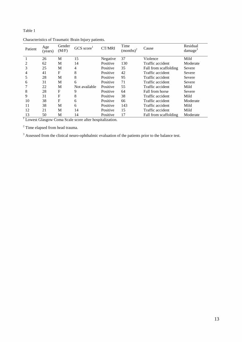
mean 79.0±6.4 kg). Table 1 shows patient characteristics. 19
The control group consisted of 43 healthy subjects, 26 females and 17 males, matched for age, 20
height and body mass index, with no orthopedic, neurological or visual problems. 21
Both TBI patients and controls underwent a neuro-ophthalmologic examination prior to the test to 22
evaluate the visual system. They were examined for pupillary reflex, smooth pursuit, saccades and 23
optokinetic nystagmus. The last column of Table 1 reports the clinical evaluation of the residual 24
visual impairment at the time of the balance test. In all patients abnormal saccades were observed. 25
In five patients global deficits of the eyes version were found. These patients were classified as 26

4
“severe” in the last column of Table1. Three patients showed both saccades and smooth pursuit 1
anomalies and were classified as “moderate”. Patients in which only abnormal saccades were 2
observed were classified as “mild”. All the subjects belonging to the control group did not show any 3
neuro-ophthalmologic abnormality. 4
The experimental protocol was approved by the local ethical committee and all participants gave 5
their written informed consent to the study. 6
2.2 Acquisition protocol 7
Subjects were asked to stand quietly, in upright position, over a Kistler 9286A force platform. The 8
inter-malleolar distance was fixed at 4 cm and the feet opening angle was 30°. The acquisition 9
protocol consisted of 10 different trial conditions, five with eyes open (looking at a visual target) 10
and five with eyes closed. The head positions were: 1) frontal: Open Eyes Frontal (OEF), Closed 11
Eyes Frontal (CEF), 2) head rotated after a slow left rotation: Open Eyes Left slow (OELs), Closed 12
Eyes Left slow (CELs) 3) head rotated after a slow right rotation: Open Eyes Right slow (OERs), 13
Closed Eyes Right slow (CERs) 4) head rotated after a fast left rotation: Open Eyes Left fast 14
(OELf), Closed Eyes Left fast (CELf), 5) head rotated after a fast right rotation: Open Eyes Right 15
fast (OERf), Closed Eyes Right fast (CERf). At the operator order, the subject reached the requested 16
head position and then the signal acquisition started. A biaxial accelerometer fixed on the forehead 17
of the subject was employed for monitoring the head rotation. Each recording started at the end of 18
the head rotation and lasted 60 s. 19
The sequence of trials was randomized to avoid learning and/or fatigue effects [26]. Every two trials 20
the subject rested for one minute moving away from the platform. 21
The platform signal was recorded with a sampling frequency of 2 kHz and then down-sampled to 50 22
Hz. The acquisition system was Step32 (DemItalia, Italy). 23
2.3 Data analysis 24
We calculated the major geometrical and time-domain parameters based on the CoP trajectory [16-25
17]. Table 2 describes the set of parameters we considered. 26
5
First, we compared TBI and controls - for each trial condition and CoP parameter - by means of a 1
two-sample t-test, after having verified the gaussianity of the distributions. 2
Moreover, we were interested in taking into account the inter-relations among CoP parameters in 3
the different trials, using the global information arising from the complete protocol: for each subject 4
we have a total of 70 dependent variables (10 trials × 7 parameter values). To this purpose, we 5
applied a multivariate analysis of variance (MANOVA) approach [27-29]. We reduced the number 6
of CoP parameters considered, preserving those containing non-redundant information and 7
discarding parameters highly correlated among them or with high within-group variability. To 8
select the reduced set of parameters we used Wilks’ Lambda statistic (Λ) [27]. Λ is an index of the 9
parameters’ discrimination capability. It is defined as the ratio between the within-groups 10
generalized variability and the total generalized variability, the latter being the sum of the within-11
groups and between-groups generalized variability. This index takes values between zero and one, 12
lower Λ-values indicating a better discrimination among groups. 13
The procedure we adopted is the following. As a first step, we calculated Λ for each parameter 14
separately and sorted the parameters in Λ ascending order. We kept the parameter with lower Λ-15
value. Then we considered all the possible combinations of two parameters, recalculated the 16
corresponding Λ-values and sorted them in ascending order, keeping the combination with lower Λ-17
value. The process was carried out iteratively adding one parameter at a time, each time 18
recalculating the Λ-value and choosing the combination of parameters showing the lowest Λ-value. 19
The parameter selection stopped when, adding more parameters, Λ did not significantly decrease 20
[27]. 21
After the selection of the reduced set of CoP parameters we summarized the information arising 22
from the ten-trial protocol applying a canonical variate analysis (CVA) [27]. The canonical 23
variables C are linear combinations of the original variables, chosen to maximize the separation 24
among groups. Specifically, the first canonical variable C1 is the linear combination of the original 25
variables that has the maximum separation among groups. This means that among all possible linear 26
6
combinations, it is the one with the most significant F statistic in a one-way analysis of variance. 1
The second canonical variable C2 has the maximum separation while being orthogonal to C1, and 2
so on. We represented the two populations of TBI and controls in the plane of the first two 3
canonical variables. 4
3. Results 5
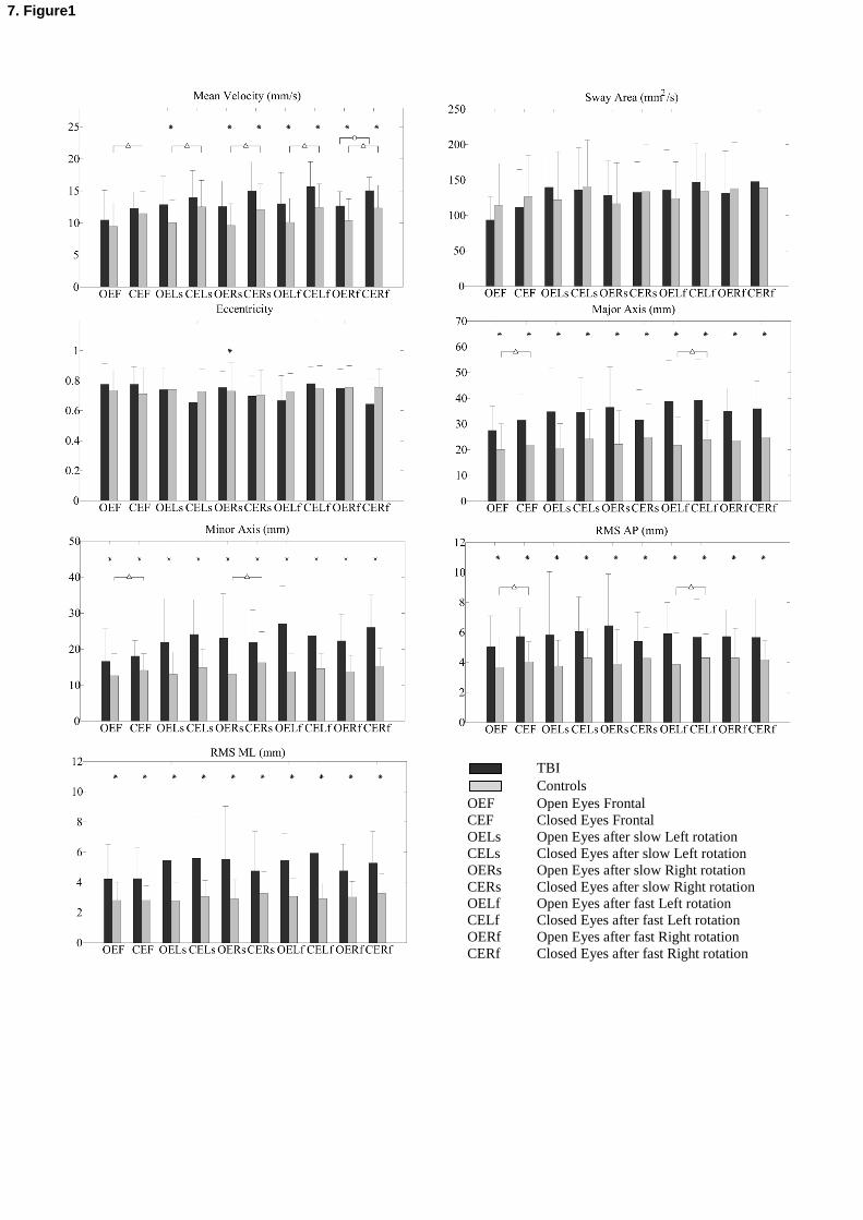
Fig. 1 shows, for each parameter, mean and standard deviation of TBI patients and controls in the 6
ten typologies of acquisition. Differences between groups which are statistically significant (two-7
sample t-test, p 0.05) are indicated with an asterisk. Major and Minor Axis and the RMS values 8
show significant differences in all of the trials. Mean Velocity highlights significant differences 9
between TBI and controls mainly in trials after head rotation (slow or fast). On the contrary, Mean 10
Velocity is not significantly different in trials with a frontal head position, both with eyes open and 11
closed. Sway Area and Eccentricity do not differentiate the two groups. 12
We tested also open eyes vs. closed eyes performances: significant differences are indicated with 13
triangles in controls and with circles in TBI patients. In controls, differences were observed in all 14
the test conditions for the Mean Velocity. For the other parameters, statistically significant 15
differences were observed only in a few test conditions. In TBI patients there were significant 16
differences between open eyes and closed eyes trials only in a single test condition (Mean Velocity, 17
OERf vs. CERf). 18
Fig. 2 shows the values of Λ on which we based the parameter selection. The single parameter that 19
better differentiates the two populations is Minor Axis (Λ = 0.42), the best combination of two 20
parameters is Minor and Major Axes (Λ = 0.31), that of three parameter is Minor Axis, Major Axis, 21
and RMS AP (Λ = 0.15), that of four parameters is Minor Axis, Major Axis, RMS AP, and 22
Eccentricity (Λ = 0.076), and, finally, that of five parameters is Minor Axis, Major Axis, RMS AP, 23
Eccentricity, and Sway Area (Λ = 0.0035). Therefore, the Λ-value decreases remarkably each time 24
a parameter is added to the set of the best CoP parameters and it falls below 0.05 when considering 25
the best combination of five parameters. Hence, in the rest of the analysis, we consider only these 26

7
five parameters. Note that Eccentricity and Sway Area do not play a role in differentiating the two 1
populations if they are considered as standalone parameters, but they become useful if they are 2
considered in combination with the other parameters. 3
The parameter selection procedure was performed considering all the 10 trials. The effect of 4
considering a smaller number of trials is evidenced by Fig. 3, which shows multivariate data from 5
TBI patients and controls plotted against the first two canonical variables C1 and C2. Fig. 3a) and 6
3b) show the results of multivariate analysis to compare controls and TBI patients, while fig. 3c) 7
and 3d) show the differences among the three sub-groups of TBI patients and controls. The 8
procedure of parameter selection was not redone, while we recomputed the canonical variables for 9
this specific case. Fig. 3a shows the results on two acquisition trials only (Open Eyes Frontal and 10
Closed Eyes Frontal), while Fig. 3b refers to the complete set of ten trials. In Fig. 3a TBI patients 11
and controls are partially overlapped, even if some of the TBI patients fall outside the control group 12
cloud (Λ = 0.63, p = 0.014). In Fig. 3b the two populations are completely separated (Λ = 0.0035, p 13
= 3.3×10⁻¹³
). Therefore, considering all the ten trials, TBI patients are completely differentiated 14
from controls. 15
Fig. 3c and Fig. 3d show controls and patients suffering from mild, moderate, and severe residual 16
visual impairment, as reported by Table1. When only two trials are considered, the various groups 17
are scarcely separated (Fig. 3c). On the contrary, when all the ten trials are taken into account, not 18
only the patients are well differentiated from controls, but also the three groups are completely 19
separated among them (Fig. 3d). Moreover, the distance between controls and the three TBI groups 20
increases with increasing level of visual impairment. 21
22
4. Discussion 23
The most widely used parameters in posturography are the total length of the CoP path (Sway Path 24
Length) and the Mean Velocity. They are essentially the same parameter, except that Mean Velocity 25
is normalized with respect to the test duration and hence does not depend on it. They are usually 26
8
evaluated with the subject in quiet stance on the platform with the head in frontal position, both 1
with eyes open and closed. It is important to notice that velocity integrates both amplitude and 2
frequency changes, thus a concomitant reduction in sway frequency can reduce the discriminant 3
power of velocity. Dehail et al. [20] studied a group of sixty-eight TBI patients (60 of which with a 4
GCS score < 8, and 33 with a residual neurological impairment) and found that Sway Path Length 5
was significantly increased, compared to controls, both with eyes open and closed. In our sample 6
population, we found that Mean Velocity did not separate TBI patients from controls in the trials 7
with the head in frontal position (both with eyes open and closed), while it separated the two 8
populations in the newly proposed test conditions (after slow or fast head rotation). The difference 9
between our results and those reported in [20] may be explained by the fact that in our study 10
patients reported, in general, less severe TBI and suffered from no vestibular or sensorimotor 11
impairment. 12
We hypothesized that subjects with visual impairment rely less than controls on the information 13
arising from the visual system. Geurts et al. [13], working with a group of TBI patients who 14
complained of reduced gross motor skills without sensory-motor impairments, report that visual 15
deprivation was most detrimental for TBI patients, particularly for the Medio-Lateral control. In our 16
patients, differences between the open- and closed-eyes balance performances are almost never 17
statistically significant, while they are significant in controls for the parameter Mean Velocity. 18
These results are coherent with our hypothesis, since we found that visual deprivation is less 19
detrimental for patients. 20
Among others, Visser et al. [12] pointed out that the poor discriminative ability (between health and 21
disease) of posturography may relate to the substantial inter-subject and intra-subject variability. 22
Given these uncertainties, many researchers record a broad range of different parameters and/or 23
perform repeated tests in the same or in different test conditions. As a consequence, for each 24
subject, many parameters and many test conditions are considered which are partially correlated 25
among them. 26
9
To take into account all the information arising from the complete protocol, we used a multivariate 1
approach. Multivariate analysis requires a prior variable selection, as described in [27]. The results 2
of variables selection are often counterintuitive. In our study, we excluded from the 'best 3
combination of five parameters' parameters that were discriminative in univariate analysis. This is 4
not surprising, since univariate analysis does not take into consideration the correlation among 5
parameters. 6
Thanks to the representation of subjects in the plane of the first two canonical variables, we 7
demonstrated (see Fig. 3) that it is possible to obtain a complete separation of patients from controls 8
when all the 10 test conditions are considered and that it is also possible to discriminate among 9
groups of patients with different residual visual impairment. Specifically, C1 discriminates patients 10
from controls, while C2 summarizes the information needed to separate patients according to the 11
degree of their residual visual impairment. 12
5. Conclusions 13
Using the proposed 10-trial protocol it was possible to clearly distinguish balance abnormalities of 14
TBI patients with respect to controls. Moreover, we found that the severity of the residual neuro-15
ophthalmic deficit is correlated to the severity of the balance impairment. This is of paramount 16
importance from a clinical perspective since it demonstrates that static posturography, associated to 17
the presented protocol, can be applied to objectively evaluate the balance performances of a patient 18
enrolled in a rehabilitation program and assess his/her progresses. 19
20
21
Conflict of interest statement 22
None of the Authors on this manuscript had or has any financial and personal relationships with 23
other people or organizations that could inappropriately influence (bias) this work. 24

10
References
[1] Langlois JA, Rutland-Brown JA and Thomas KE, Traumatic brain injury in the United States:
emergency department visits, hospitalizations, and deaths, Centers for Disease Control and
Prevention, National Center for Injury Prevention and Control, Atlanta (GA), 2006.
[2] Maas AIR, Stocchetti N, Bullock R. Moderate and severe traumatic brain injury in adults.
Lancet Neurol 2008; 7:728–741.
[3] Tagliaferri F, Compagnone C, Korsic M, Servadei F, Kraus J. A systematic review of brain
injury epidemiology in Europe. Acta Neurochirurgica 2006; 148:255–68.
[4] Teasdale G, Jennett B. Assessment of coma and impaired consciousness. A practical scale.
Lancet 1974; 2(7872):81–84.
[5] Chamelian L, Feistein A. Outcome after mild to moderate traumatic brain injury: the role of
dizziness. Arch Phys Med Rehabil 2004; 85:1662–6.
[6] Thornhill S, Teasdale GM, Murray GD, McEwen J, Roy CW, KI Penny. Disability in young
people and adults one year after head injury: prospective cohort study. British Medical Journal
2000; 320:1631–5.
[7] Kapoor N, Ciuffreda KJ. Vision Disturbances Following Traumatic Brain Injury. Current
Treatment Options in Neurology 2002; 4:271–280.
[8] Suchoff IB, Kapoor N, Ciuffreda KJ, Rutner D, Han E, Craig S. The frequency of occurrence,
types, and characteristics of visual field defects in acquired brain injury: A retrospective analysis.
Optometry 2008; 79:259–265.
[9] Stavern GP et al. Neuro-Ophthalmic Manifestations of Head Trauma. Journal of Neuro-
Ophthalmology 2001; 21(2):112–117.
[10] Kulkarni AR, Aggarwal1 SP, Kulkarni RR, Deshpande MD, Walimbe PB, Labhsetwar AS.
Ocular manifestations of head injury: a clinical study. Eye 2005; 19:1257–1263.

11
[11] Ciuffreda KJ, Kapoor N, Rutner D, Suchoff IB, Han E, Craig S. Occurrence of oculomotor
dysfunctions in acquired brain injury: A retrospective analysis. Optometry 2007; 78:155-161.
[12] Basford JR et al. An assessment of gait and balance deficits after traumatic brain injury. Arch
Phys Med Rehabil 2003; 84:343–349.
[13] Geurts ACH, Ribbers GM, Knoop JA, van Limbeek J. Identification of static and dynamic
postural instability following traumatic brain injury. Arch Phys Med Rehabil 1996; 77:639–644.
[14] Winter DA. Human balance and posture control during standing and walking. Gait and posture
1995; 3:193-214.
[15] Visser JE, Carpenter MG, van der Kooij H, Bloem BR. The clinical utility of posturography.
Clinical Neurophysiology 2008; 119:2424–2436.
[16] Prieto T et al. Measures of postural steadiness: differences between healthy young and elderly
adults. IEEE Transactions on biomedical engineering 1996; 43(9):956-966.
[17] L. Rocchi, L. Chiari and A. Cappello, Feature selection of stabilometric parameters based on
principal component analysis, Med. Biol. Eng. Comput. 42 (2004), pp. 71–79.
[18] Raymakers JA, Samson MM, Verhaar HJJ. The assessment of body sway and the choice of the
stability parameter(s). Gait and posture 2005; 21:48–58.
[19] Samson M, Crowe A. Intra-subject inconsistencies in quantitative assessments of body sway.
Gait and posture 1996; 4:252–257.
[20] Dehail P et al. An assessment of postural instability in patients with traumatic brain injury
upon enrolment in a vocational adjustment programme. J Rehabil Med 2007; 39:531–536.
[21] Geurts ACH, Knoop JA, van Limbeek J, Is Postural Control Associated With Mental
Functioning in the Persistent Postconcussion Syndrome? Arch Phys Med Rehabil; 80:144–149,
1999.
[22] Kaufman KR et al. Comparison of subjective and objective measurements of balance disorders
following traumatic brain injury. Medical Engineering & Physics 2006; 28:234–239.

12
[23] Lahat E, Barr J, Klin B, Dvir Z, Bistrizer T, Eshel G. Postural stability by computerized
posturography in minor head trauma. Pediatr Neurol 1996; 15:299-301.
[24] Slobounov S, Cao C, Sebastianelli W, Slobounov E, Newell K. Residual deficits from
concussion as revealed by virtual time-to-contact measures of postural stability. Clinical
Neurophysiology 2008; 119:281–289.
[25] Wade LD et al. Changes in postural sway and performance of functional tasks during
rehabilitation after traumatic brain injury. Arch Phys Med Rehabil 1997; 78:1107-1111.
[26] Tarantola J, Nardone A, Tacchini E, Schieppati M. Human stance stability improves with the
repetition of the task: effect of foot position and visual condition. Neuroscience Letters 1997, 228:
75-78.
[27] Krzanowski WJ, Principles of Multivariate Analysis: A User’s Perspective. Oxford: Clarendon
Press; 1988.
[28] Johnson R, Wichern D, Applied multivariate statistical analysis. Fifth ed. Prentice Hall,
International; 2002.
[29] Mardia KV, Kent JT, Bibby JM, Multivariate Analysis. London: Academic Press; 1979.
13
Table 1
Characteristics of Traumatic Brain Injury patients.
Patient Age (years)
Gender
(M/F) GCS score
1 CT/MRI
Time
(months)2
Cause Residual
damage3
1 26 M 15 Negative 37 Violence Mild
2 62 M 14 Positive 130 Traffic accident Moderate
3 25 M 4 Positive 35 Fall from scaffolding Severe
4 41 F 8 Positive 42 Traffic accident Severe
5 28 M 8 Positive 95 Traffic accident Severe
6 31 M 6 Positive 71 Traffic accident Severe
7 22 M Not available Positive 55 Traffic accident Mild
8 28 F 9 Positive 64 Fall from horse Severe
9 31 F 8 Positive 38 Traffic accident Mild
10 38 F 6 Positive 66 Traffic accident Moderate
11 38 M 6 Positive 143 Traffic accident Mild
12 21 M 14 Positive 15 Traffic accident Mild
13 50 M 14 Positive 17 Fall from scaffolding Moderate 1 Lowest Glasgow Coma Scale score after hospitalization.
2 Time elapsed from head trauma.
3 Assessed from the clinical neuro-ophthalmic evaluation of the patients prior to the balance test.

14
Table 2
Posturographic parameters.
1AP and ML are respectively the antero-posterior and the medio-lateral coordinates of the displacement of the CoP on
the platform surface.
Parameter Dimension Description Definition1
Mean velocity
mm/s
Length of CoP trajectory on the base of support in the unit of time
Sway area mm2/s
Area of the surface enclosed by the CoP path per unit of time
RMS AP mm
Root mean square of the antero-posterior time series
RMS ML mm Root mean square of the medio-lateral time series
Major Axis mm
Length of the major axis of the smallest ellipse containing the CoP trajectory on the base of support
2a
2b
Minor Axis mm
Length of the minor axis of the smallest ellipse containing the CoP trajectory on the base of support
Eccentricity adimention
al
Eccentricity of the smallest ellipse containing the CoP trajectory on the base of support
15
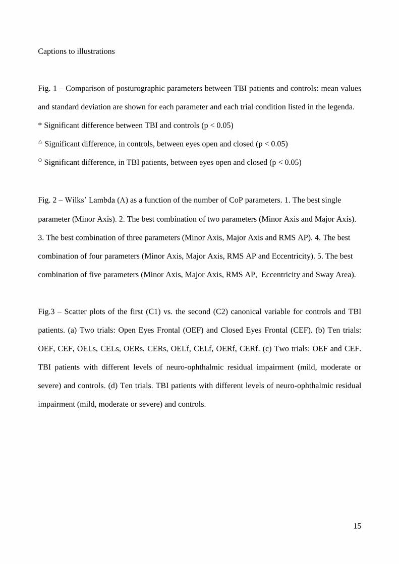
Captions to illustrations
Fig. 1 – Comparison of posturographic parameters between TBI patients and controls: mean values
and standard deviation are shown for each parameter and each trial condition listed in the legenda.
* Significant difference between TBI and controls (p < 0.05)
Significant difference, in controls, between eyes open and closed (p < 0.05)
Significant difference, in TBI patients, between eyes open and closed (p < 0.05)
Fig. 2 – Wilks’ Lambda () as a function of the number of CoP parameters. 1. The best single
parameter (Minor Axis). 2. The best combination of two parameters (Minor Axis and Major Axis).
3. The best combination of three parameters (Minor Axis, Major Axis and RMS AP). 4. The best
combination of four parameters (Minor Axis, Major Axis, RMS AP and Eccentricity). 5. The best
combination of five parameters (Minor Axis, Major Axis, RMS AP, Eccentricity and Sway Area).
Fig.3 – Scatter plots of the first (C1) vs. the second (C2) canonical variable for controls and TBI
patients. (a) Two trials: Open Eyes Frontal (OEF) and Closed Eyes Frontal (CEF). (b) Ten trials:
OEF, CEF, OELs, CELs, OERs, CERs, OELf, CELf, OERf, CERf. (c) Two trials: OEF and CEF.
TBI patients with different levels of neuro-ophthalmic residual impairment (mild, moderate or
severe) and controls. (d) Ten trials. TBI patients with different levels of neuro-ophthalmic residual
impairment (mild, moderate or severe) and controls.

16
Figure 1
TBI
Controls
OEF Open Eyes Frontal
CEF Closed Eyes Frontal
OELs Open Eyes after slow Left rotation
CELs Closed Eyes after slow Left rotation
OERs Open Eyes after slow Right rotation
CERs Closed Eyes after slow Right rotation
OELf Open Eyes after fast Left rotation
CELf Closed Eyes after fast Left rotation
OERf Open Eyes after fast Right rotation
CERf Closed Eyes after fast Right rotation

17
Figure 2

18
Figure 3
(a) (b)
(d) (c)

Table 1
Characteristics of Traumatic Brain Injury patients.
Patient Age (years)
Gender
(M/F) GCS score
1 CT/MRI
Time
(months)2
Cause Residual
damage3
1 26 M 15 Negative 37 Violence Mild
2 62 M 14 Positive 130 Traffic accident Moderate
3 25 M 4 Positive 35 Fall from scaffolding Severe
4 41 F 8 Positive 42 Traffic accident Severe
5 28 M 8 Positive 95 Traffic accident Severe
6 31 M 6 Positive 71 Traffic accident Severe
7 22 M Not available Positive 55 Traffic accident Mild
8 28 F 9 Positive 64 Fall from horse Severe
9 31 F 8 Positive 38 Traffic accident Mild
10 38 F 6 Positive 66 Traffic accident Moderate
11 38 M 6 Positive 143 Traffic accident Mild
12 21 M 14 Positive 15 Traffic accident Mild
13 50 M 14 Positive 17 Fall from scaffolding Moderate 1 Lowest Glasgow Coma Scale score after hospitalization.
2 Time elapsed from head trauma.
3 Assessed from the clinical neuro-ophthalmic evaluation of the patients prior to the balance test.
6. Table1

Table 2
Posturographic parameters.
1AP and ML are respectively the antero-posterior and the medio-lateral coordinates of the displacement of the CoP
on the platform surface.
Parameter Dimension Description Definition1
Mean velocity
mm/s
Length of CoP trajectory on the base of support in the unit of time
Sway area mm2/s
Area of the surface enclosed by the CoP path per unit of time
RMS AP mm
Root mean square of the antero-posterior time series
RMS ML mm Root mean square of the medio-lateral time series
Major Axis mm
Length of the major axis of the smallest ellipse containing the CoP trajectory on the base of support
2a
2b
Minor Axis mm
Length of the minor axis of the smallest ellipse containing the CoP trajectory on the base of support
Eccentricity adimention
al
Eccentricity of the smallest ellipse containing the CoP trajectory on the base of support
6. Table2
TBI
Controls
OEF Open Eyes Frontal
CEF Closed Eyes Frontal
OELs Open Eyes after slow Left rotation
CELs Closed Eyes after slow Left rotation
OERs Open Eyes after slow Right rotation
CERs Closed Eyes after slow Right rotation
OELf Open Eyes after fast Left rotation
CELf Closed Eyes after fast Left rotation
OERf Open Eyes after fast Right rotation
CERf Closed Eyes after fast Right rotation
7. Figure1

7. Figure2

1
(a) (b)
(d) (c)
7. Figure3
Related Documents


![FunctionalMedicineAOMA2018 (003).pptx [Read-Only]...Musculoskeletal: Rolled shoulders and poor posture noted, normal gait, no joint effusions/swelling Skin: Excoriations over bilateral](https://static.cupdf.com/doc/110x72/5f106f3c7e708231d449178d/functionalmedicineaoma2018-003pptx-read-only-musculoskeletal-rolled-shoulders.jpg)


