LINKÖPING MEDICAL DISSERTATIONS No. 960 CHROMATIN, HISTONES, AND EPIGENETIC TAGS Elisavet Koutzamani Division of Cell Biology Department of Biomedicine and Surgery Faculty of Health Sciences Linköping University SE-581 85 Linköping, Sweden Linköping 2006

Welcome message from author
This document is posted to help you gain knowledge. Please leave a comment to let me know what you think about it! Share it to your friends and learn new things together.
Transcript

LINKÖPING MEDICAL DISSERTATIONS No. 960
CHROMATIN, HISTONES, AND EPIGENETIC TAGS
Elisavet Koutzamani
Division of Cell Biology
Department of Biomedicine and Surgery Faculty of Health Sciences
Linköping University SE-581 85 Linköping, Sweden
Linköping 2006

Cover picture: unfolding of a chromosome (courtesy of Darryl Leja, National Human Genome Research Institute). ©Elisavet Koutzamani All rights reserved. ISBN 91-85523-10-0 ISSN 0345-0082 Published articles have been reprinted with permission from the publishers. Papers I–III © American Society for Biochemistry and Molecular Biology Printed in Sweden by LTAB Linköpings Tryckeri AB, 2006-875.

This thesis is dedicated to
my brother and my mother and father for believing in me and for making me a better person

Είπα στη μυγδαλιά: “Αδελφή, μίλησέ μου για το Θεό”. Κι η μυγδαλιά άνθισε.
Νίκος Καζαντζάκης (από το βιβλίο του ”Αναφορά στον Γκρέκο”)
TABLE OF CONTENTS ABSTRACT POPULÄRVETENSKAPLIG SAMMANFATTNING LIST OF PUBLICATIONS LIST OF ABBREVIATIONS INTRODUCTION 15 Chromatin History and Research 16 Nobel Prizes Related to Chromatin 19 AIMS OF THE STUDY 21 CHROMATIN STRUCTURE 23 DNA 23 Composition of DNA 23 The Nucleosome 26 Location of Linker Histones 27 Higher-Order Chromatin Structure 28 Euchromatin and Heterochromatin 30 THE HISTONES 31 Histone Variants 32 Core Histones 33 H2A 33 H2B 33 H3 33 H4 34 Linker Histones 34 H1.1 35 H1.2 36 H1.3 36 H1.4 37 H1.5 37 H1°/H5 38 H1.t 38 H1.oo 39 H1x 39 HISTONE EPIGENETICS 41 Post-Translational Modifications 41 Acetylation 41 Methylation 42 Phosphorylation 43

ADP-Ribosylation 45 Ubiquitination 46 Sumoylation 46 Glycosylation 47 The Histone Code 49 LINKER HISTONE FUNCTION 51 CONCLUSIONS 59 ACKNOWLEDGEMENTS 61 REFERENCES 65 PAPER I-V
ABSTRACT
The fundamental building blocks of chromatin are the nucleosomes. Each such unit is composed of about 200 bp of DNA, the well-conserved core histones (H2A, H2B, H3, and H4) and a linker histone (H1). The DNA is wound around two dimers of H2A–H2B and a tetramer comprising two molecules each of H3 and H4, and there is approximately one linker histone molecule positioned on the exterior of the DNA–protein octamer complex. The nucleosome directs the various structural transitions in chromatin that are needed for proper transcriptional regulation during differentiation and development of the organism in question. The gene activity can be regulated by different histone variants, DNA–protein interactions, and protein–protein interactions, all of which are influenced by the enormous amounts of post-translational modifications that occur in the histone tails. The research underlying this thesis focused on different aspects of post-translational modifications during aging, differentiation, and progression of the cell cycle, and also on expression of linker histone variants and linker histone-chromatin interactions in a variety of cells and tissues.
The present results are the first to show that H4 can be trimethylated at lysine 20 in mammalian cells. The trimethylated H4K20 was found in rat kidney and liver at levels that rose with increasing age of the animals, and it was also detected in trace amounts in human cell lines. Furthermore, in differentiating MEL cells, trimethylated H4K20 was localized to heterochromatin, and levels of trimethylated H4K20 increased during the course of cell differentiation and were correlated with the increasing compaction of the chromatin.
The chromatin of terminally differentiated chicken and frog erythrocytes is highly condensed, and the linker histone variants it contains vary between the two species. Cytofluorometric analyses revealed that the linker histones in the chicken erythrocytes exhibited higher affinity for chromatin than did those in the frog erythrocytes. Characterization of the H1° in frog erythrocytes proved it to be the H1°-2 subvariant. Other experiments demonstrated that normal human B lymphocytes expressed the linker histone variants H1.2, H1.3, H1.4, and H1.5, and that B cells from patients with B-CLL expressed the same variants although in different amounts. The most striking dissimilarity was that amounts of H1.3 in the cells were decreased or undetectable in some samples. Sequencing did not discern any defects in the H1.3 gene, and thus the absence of H1.3 is probably regulated at the post-translational level. It was also observed that the levels of linker histone phosphorylation in EBV-transformed B lymphocytes were already increased in the G1 phase of the cell cycle, which is earlier than previously thought. This increase in phosphorylation is probably responsible for the lower affinity of linker histones for chromatin in EBV-transformed cells in the G1 phase of the cell cycle.


POPULÄRVETENSKAPLIG SAMMANFATTNING
Vår arvsmassa utgörs av en 2 meter lång DNA helix, uppdelad på 23 par kromosomer, som bland annat med hjälp av en grupp basiska proteiner, histonerna (H1, H2A, H2B, H3 och H4), lindas, veckas och kompakteras för att få plats i en cellkärna av storleksordningen 5–10 µm. I våra celler finns inte DNA i dess nakna form utan det är associerat till proteiner, histoner och andra proteiner, som binder till DNA. Strukturen med DNA och dess proteiner kallas för kromatin och är grundstrukturen för vår arvsmassa.
Avhandlingsarbetet är fokuserat på histonerna. Alla gener har en gemensam grundstruktur i form av en kromatinfiber som är uppbyggd av repetitiva element, nukleosomer. Nukleosomerna i sin tur är uppbyggda av en proteinkärna som består av corehistonerna H2A, H2B, H3 och H4, och runt proteinerna lindas DNA i två fulla varv. På utsidan av DNA-proteinkomplexet sitter en molekyl av ett linkerhiston (kallas även för H1). Hos mammalier finns linkerhistonerna i 9 olika varianter.
Det står nu klart att histonerna inte bara har en strukturell roll i arvsmassan i samband med dess kompaktering, utan att de också medverkar i regleringen av olika geners uttryck. De olika histonerna kan dessutom modifieras posttranslatoriskt, framför allt genom acetylering, metylering eller fosforylering. Varianterna och deras modifieringar har tillskrivits regulatoriska egenskaper i samband med cellers tillväxt, utveckling och mognad. För linkerhistonvarianterna har det föreslagits att deras regulatoriska funktioner kan bero på olikheter i deras inbindning till kromatin. Vi har studerat post-translatoriska modifieringar av H4 och H1, linkerhistonernas affinitet till kromatin och mönstret av H1 varianter under cellers mognad, åldrande och transformation för att få klarhet i en del epigenetiska fenomen som kan ligga bakom de förändringar som vår arvsmassa genomgår i samband med olika cellulära processer.
H4 kan metyleras och kan innehålla upp till 3 metylgrupper. Den trimetylerade formen av H4 ökar i samband med åldrandet och dess förekomst är kopplat till delar av kromatin som innehåller avstängda gener. Olika kombinationer av metylerat och acetylerat H4 kan fungera som signaler för att antingen slå på eller stänga av gener.
Linkerhistonernas bindning till kromatin, verkar inte vara den faktor som kompakterar kromatinet, vilket man tidigare ansett. Normala B-lymfocyter och leukemiska B-lymfocyter har olikheter i sin H1-sammansättning, som kan ha betydelse for kromatinets kompaktering. Fosforylering av H1 minskar dess binding till kromatin och öppnar samtidigt upp kromatinet och gör det tillgängligt för andra faktorer. Transformerade celler i snabb celldelning innehåller högre mängder

fosforylerat H1 jämfört med normala celler. Linkerhistonernas fosforyleringsgrad kan vara en faktor som reglerar progressionen genom cellcykeln.
LIST OF PUBLICATIONS This thesis is based on the following papers, which will be referred to in the text by the Roman numerals: I Bettina Sarg, Elisavet Koutzamani, Wilfried Helliger, Ingemar
Rundquist and Herbert Lindner. Postsynthetic trimethylation of histone H4 at lysine 20 in mammalian tissues is associated with aging. (2002) Journal of Biological Chemistry, 277:39195–39201
II Bettina Sarg1, Wilfried Helliger1, Heribert Talasz, Elisavet
Koutzamani and Herbert Lindner. Histone H4 hyperacetylation precludes histone H4 lysine 20 trimethylation. (2004) Journal of Biological Chemistry, 279: 53458–53464
1These authors contributed equally to this work. III Elisavet Koutzamani1, Helena Loborg1, Bettina Sarg, Herbert
Lindner and Ingemar Rundquist. Linker histone subtype composition and affinity for chromatin in situ in nucleated mature erythrocytes. (2002) Journal of Biological Chemistry, 277: 44688–44694
1These authors contributed equally to this work. IV Elisavet Koutzamani, Bettina Sarg, Anna Gréen, Ola Söderberg,
Gunnar Juliusson, Herbert Lindner and Ingemar Rundquist. Differences in H1 subtype composition between normal human B cells and B-cell chronic lymphocytic leukemia cells. Manuscript.
V Elisavet Koutzamani, Bettina Sarg, Herbert Lindner and Ingemar
Rundquist. Cell cycle dependent phosphorylation of histone H1 in Epstein-Barr virus-transformed B cells. Manuscript.


LIST OF ABBREVIATIONS B-CLL B-cell chronic lymphocytic leukaemia bp base pairs CDK1 cyclin-dependent kinase 1 CDK2 cyclin-dependent kinase 2 ChIP chromatin immunoprecipitation CS cleavage stage DAPI 4’,6-diamidino-2-phenylindole DMSO dimethyl sulfoxide DNA deoxyribonucleic acid EBV Epstein-Barr virus EM electron microscopy Ezh2 enhancer of Zeste homologue FRAP fluorescence recovery after photobleaching GFP green fluorescent protein HAT histone acetyl transferase HDAC histone deacetylase HILIC hydrophilic interaction liquid chromatography HILS1 histone H1-like protein in spermatides 1 HMBA hexamethylenebisacetamide HMG high mobility group HMT histone methyl transferase HP1 heterochromatin protein 1 HPCE high performance capillary electrophoresis kDa kilodalton LCL lymphoblastoid cell line LSD1 lysine-specific demethylase 1 MAP mitogen-activated protein MEL murine erythroleukaemia MENT myeloid and erythroid nuclear termination stage-specific protein MMTV mouse mammary tumour virus mRNA messenger RNA NCP nucleosome core particle NRL nucleosome repeat length PAD4 peptidylarginine deiminase 4 ParaT parathymosin PCR polymerase chain reaction PMRT protein arginine methyltransferase

PP1 protein phosphatase 1 ProTα prothymosin α RNA ribonucleic acid RP-HPLC reversed phase high performance liquid chromatography SNP single nucleotide polymorphism SUMO small ubiquitin-related modifier TNP transition nuclear protein TSA trichostatin A u ubiquitinated wt wild-type
Chromatin, histones, and epigenetic tags
15
'Eureka—I have found it!' Archimedes (280–211 BC), Greek mathematician
INTRODUCTION
he human genome is composed of deoxyribonucleic acid (DNA) that is maintained and sheltered within the boundaries of the cell nucleus and it is
made up of 23 pairs of chromosomes that consist of approximately 6 x 10⁹ base pairs (bp). The DNA in a single cell has a total length of approximately 2 meters, and it must be assembled within a nucleus that is only 5-10 µm in diameter. Therefore, in all eukaryotic cells, the linear DNA molecule is found in the form of a DNA–protein complex called chromatin. The numerous DNA-binding proteins (histones and non-histone proteins) compact DNA into different ordered levels, from the interphase chromatin to the mitotic chromosomes in which the DNA shows the highest degree of compaction. The association between DNA-binding proteins and DNA prevents regulatory proteins from anchoring to their targets on the underlying DNA template. The chromatin has a highly intricate structure that enables it to open up and allow access to the DNA, and also to close up and prevent access, as required by the cell. The positioning of the proteins also protects the genome from radiation-induced damage and genotoxic stress. DNA is a rather static molecule, whereas the chromatin fibre is highly dynamic. Modulation of the structure of chromatin and the sequential unravelling of this fibre are essential for regulation of gene expression. These events determine the accessibility and recruitment of regulatory factors, and they are necessary for many cellular processes, such as transcription, replication, repair, and recombination. Most somatic cells in an organism contain identical genetic material, and differentiation into the many cell identities that are possible is determined by which sets of genes are expressed. The term epigenetics was first defined by Conrad Waddington in 1942 (Waddington, 1942), at which time it had already been postulated that genomic function is regulated by additional information that exists
T

Elisavet Koutzamani
16
beyond the level of the genetic code. Epigenetics is thus defined as the overlaying molecular factors that control under which circumstances the nucleotide sequence become transcribed. At present, epigenetics includes DNA methylation, histone variants and their post-translational modifications, and the interaction of microRNAs with the genome. Over the past few years, it has been proposed that malfunctions in chromatin, histone variants, and epigenetic tags in the form of modifications in the histone tails can lead to cancer and autoimmune disorders. More work is needed to provide further knowledge in this field that can be used for diagnostic and therapeutic purposes. Chromatin History and Research No individual has influenced the systematic study of life as much as the Greek philosopher, universal scientist, and impressive thinker Aristotle (384–322 BC). His contributions to the field of biology include ideas about the inheritance of human traits. Aristotle’s work was both theoretical and practical, and of course it was burdened with major limitations due to the lack of appropriate tools to conduct analyses and evaluate hypotheses. Even so, when his theories are viewed in light of the knowledge that is available today, they are fully in line with modern genetics (Muller, 1996; Vinci and Robert, 2005). The scientific community had to wait until 1871 before Friedrich Miescher (Miescher, 1871) discovered the molecular substrate of the genetic code—the nucleic acids. Miescher focused his work on developing methods to isolate nuclei from human leukocytes, and he reported the finding of an acidic substance which he named nuclein. He subsequently performed additional experiments on salmon spermatozoa in which he carried out stepwise extraction with hydrochlorid acid to remove a basic component that he called protamine. Next, Miescher subjected the remaining material to alkali treatment, which yielded an acidic component that was highly similar to the nuclein he had previously obtained from human leukocytes. This was a breakthrough because it meant that nuclear material was composed of an acidic component and a basic component. The next major advance was made in 1884 by Albrecht Kossel (Kossel, 1911), who studied nucleated geese erythrocytes and succeeded in isolating basic proteins that he named histones, which differed from the protamines present in salmon sperms. Around the time of Miescher’s discovery, the Austrian monk Gregor Mendel and the British scientist Charles Darwin were both publishing papers on the theories of genetics and evolution.

Chromatin, histones, and epigenetic tags
17
However, no one suspected that Miescher’s new compound was the key to all the queries in this area. DNA was in the right place to control heredity, in the chromosomes inside the cell nucleus, but it was such a simple molecule that some doubted it had any function at all. The proteins in the chromosomes of cell nuclei were considered much better candidates for carrying the information necessary to build a living organism. Walter Flemming is believed to be the first scientist to study cell division. The development of dyes useful for microscopy of nuclear structures made it possible to visualize a fibrous scaffold in the nucleus that Flemming designated chromatin, which comes from the Greek word χρωμα (colour) and refers to the stainable material. Flemming published his results in 1879 (Paweletz, 2001). At the beginning of the 20th century, chromatin research was facing an uneventful period, and progress was instead being made in the field of genetics. In 1928, Franklin Griffith, discovered that genetic information can be transferred from heat-killed to live bacteria, a phenomenon called transformation (Olins and Olins, 2003). In 1944, Oswald Avery and his colleagues Maclyn McCarty and Colin MacLeod identified Griffith’s transforming agent as DNA (Avery et al., 1944). This breakthrough was greeted with much scepticism, and scientists could/would not accept the notion that it might be the DNA molecule, not the proteins, that represented the genetic tool. Luckily for us, this uncertainty opened a window of opportunity for the scientists who were less biased in this context.
Around this time, advances in X-ray diffraction techniques enabled Maurice Wilkins and Rosalind Franklin to look directly at DNA, and their results showed that DNA probably had the corkscrew structure of a helix. The April issue of Nature published in 1953 included three articles dealing with the structure of DNA. One of those was the classical paper by Watson and Crick which described DNA as being composed “of two helical chains each coiled round the same axis” (Watson and Crick, 1953). Watson and Crick also stated that the purine and pyrimidine bases in the two opposing helices interact with each other via hydrogen bonds and occur in pairs. Each pair must comprise a purine and pyrimidine base, thus adenine interacts with thymine and guanine with cytosine. The second paper was written by Wilkins, Stokes, and Wilson (1953), who showed that the DNA structure revealed by X-ray diffraction studies was the same in several biological systems. The third paper by Franklin and Gosling (1953) presented data about the existence of an A and a B form of the DNA helix, and concluded that the phosphate backbone lies on the outside of the molecule.

Elisavet Koutzamani
18
In the 1970s, the focus turned back to chromatin structure. Previously, the development of preparative methods had fractionated the histones into five groups (Johns and Butler, 1962; Johns, 1964; Phillips and Johns, 1965; Johns, 1967), which, when translated into present-day nomenclature, correspond to linker histones (H1) and the core histones H2A, H2B, H3, and H4. Chromatin isolated from eukaryotic nuclei was found to harbour fibres that contain spherical particles termed ν bodies which were assumed to be a nucleohistone complex composed of double-stranded DNA and two molecules of each type of histone (Olins and Olins, 1974). It was later shown that the histones in the ν bodies are core histones and occur pairwise in chromatin: H3 and H4 form pairs that are further assembled into a tetramer, whereas pairs containing H2A and H2B are found as two dimers (Kornberg and Thomas, 1974). Kornberg (1974) further postulated that the chromatin fibre is composed of units of 200 bp stretches of DNA coiled around the octamer of histones to give the appearance “beads on a string”. The findings published by Olins (Olins and Olins, 1974) and Kornberg (Kornberg and Thomas, 1974; Kornberg, 1974) were confirmed by additional experimental work conducted by Woodcock and colleagues (1976a, 1976b). In 1975, the spherical ν bodies were named nucleosomes, and that designation still stands today (Oudet et al., 1975). The term nucleosome refers to the nu (ν) bodies described by Olins and Olins (1974) and also to the nuclear origin of such units. The core histones were found to be an integral part of the nucleosome, whereas the linker histones were demonstrated to interact with the linker DNA between nucleosomes (Kornberg and Thomas, 1974; Noll and Kornberg, 1977). Much effort has been made to elucidate the structure of the nucleosome and assembly of the histones (Luger et al., 1997), and additional information has been reported regarding the positioning of linker histones on the nucleosome (Thomas, 1999; Brown et al., 2006). In the last decade, attention was turned towards the functional role of the histones, which will be dealt with in the chapter headed “Histones” in this thesis. For in-depth reviews, see Olins and Olins (2003) and van Holde (1988).

Chromatin, histones, and epigenetic tags
19
Nobel Prizes Related to Chromatin Albrech Kossel (1853–1927) was awarded the Nobel Prize in Physiology or Medicine in 1910 “in recognition of the contributions to our knowledge of cell chemistry made through his work on proteins, including nucleic substances” (http://nobelprize.org/nobel_prizes/medicine/laureates/1910/index.html). Thomas Morgan (1866–1945) received the Nobel Prize in Physiology or Medicine in 1933 “for his discoveries concerning the role played by the chromosome heredity (http://nobelprize.org/nobel_prizes/medicine/laureates/1933/index.html). Francis Crick (1916–2004), James Watson (b. 1928) and Maurice Wilkins (1916-2004), were given the Nobel Prize in Physiology or Medicine in 1962 “for their discoveries concerning the molecular structure of nucleic acids and its significance for information transfer in living material” (http://nobelprize.org/nobel_prizes/medicine/laureates/1962/index.html). Aaron Klug (b. 1926) was awarded the Nobel Prize in Chemistry in 1982 “for his development of crystallographic electron microscopy and his structural elucidation of biologically important nucleic acid-protein complexes” (http://nobelprize.org/nobel_prizes/chemistry/laureates/1982/index.html).

Elisavet Koutzamani
20

Chromatin, histones, and epigenetic tags
21
AIMS OF THE STUDY The general aim of this thesis was to study the expression pattern and post-translational modifications of histones in correlation to chromatin structure. Specific aims: 1. Upon the discovery of the trimethylated form of H4, to study its distribution
and biological function. 2. To study the subtype composition and the affinity of linker histones for
chromatin in the highly condensed chromatin of chicken and frog erythrocytes.
3. To study the expression of linker histone variants in normal and leukaemic
human cells. 4. To study the phosphorylation pattern and the affinity of linker histones for
chromatin in normal resting cells and in transformed proliferating cells.

Elisavet Koutzamani
22

Chromatin, histones, and epigenetic tags
23
Any living cell carries with it the experiences of a billion years of experimentation by its ancestors. Max Delbruck–Nobel Prize winner in Physiology or Medicine 1969
CHROMATIN STRUCTURE Chromatin is the complex composed of DNA, histones, and non-histone proteins that arrange the DNA molecule into several hierarchical levels of organization. DNA The central dogma in the field of cell biology is as follows: DNA → RNA → protein. Every somatic cell in humans (with the exception of red blood cells) contains 46 chromosomes, 23 inherited from the mother and 23 from the father, which make up the genome. Additional DNA that comes solely from the mother is found in the mitochondria. The DNA possesses all the information that is needed to determine the amino acid sequence of each protein, and thus it is responsible for production of all the proteins required by each cell and thereby also the formation and maintenance of the individual. The DNA is copied into ribonucleic acid (RNA), which specifies the precise order of the building blocks—the amino acids—in the protein (Lodish, 2003). Composition of DNA DNA is composed of monomers referred to as nucleotides that occur in four different forms named adenine, guanine, cytosine, and thymine (designated A, G, C, and T) (Figure 1b). The nucleotides have a common structure: a phosphate group coupled via a phosphoester bond to a pentose (a five carbon sugar molecule), which is in turn linked to a nitrogen- and carbon-containing ring structure known as a base. The bases are divided into two groups, purines and pyrimidines. Adenine and

Elisavet Koutzamani
24
guanine are purines and contain a pair of fused rings; thymine and cytosine are pyrimidines and contain a single ring (Figure 1b). The nucleotides are linked together by phosphodiester bonds to generate a linear sequence of polynucleotides (Lodish, 2003). DNA consists of two linear strands of polynucleotides that are intertwined like a staircase in a double helix (Figure 1a). The sugar-phosphate parts of the nucleotides are positioned on the outside of the double helix, and the bases are directed towards the inside. The nucleotides are stacked on top of each other, so the plane of the base pair is perpendicular to the axis of the helix. The bases from the two opposing strands interact with each other via hydrogen bonds, and they occur in pairs that always consist of a purine and a pyrimidine. A connects with T via two hydrogen bonds, and C pairs with G via three hydrogen bonds, which stabilizes the double helix (Figure 1a). Stabilization is promoted by additional interactions involving hydrophobic and van der Waals bonds between the stacked bases. The twining of the helices creates two grooves on the outside of the DNA, one larger and one smaller, which are termed the major and the minor groove (Lodish, 2003). The double helix is most often in B conformation, which is predominant in living cells, and it can be described as being a right-handed spiral that makes a complete turn every 3.6 nm and contains 10.5 nucleotide pairs per turn. Two other well-known DNA conformations are designated A and Z. The A conformation is seen during dehydration, and it is also a right-handed helix, albeit more compact than the B form, and it contains 11 base pairs per turn, and the base pairs are more tilted. The Z conformation of DNA is unusual, because it consists of left-handeded helices. Z-DNA was detected by use of high salt concentrations, and it can exist in vivo within stretches of B-DNA and often at transcription sites (Rich and Zhang, 2003).

Chromatin, histones, and epigenetic tags
25
a) b) Figure 1. a) Nucleotide base pairing in DNA (left) and the DNA double helix (right). b) Chemical structure of the four bases found in DNA. (Figures courtesy of Darryl Leja, National Human Genome Research Institute).
Base pair
Guanine Cytosine
Adenine Thymine
Elisavet Koutzamani
26
The Nucleosome The lack of hydrogen bonds between the stacks of nucleotides in the DNA helix enables the bending of the DNA that is induced by protein complexes such as the histones. Upon bending, the major and minor grooves of the DNA are affected as follows: they are compressed and become narrower on the inside of the curve, whereas they expand on the outside of the bend (Travers, 1990). The connections between DNA and histones are mainly non-specific and include non-polar interactions with the pentose groups in the DNA, hydrogen bonds to the phosphate groups of DNA, and electrostatic interactions between the positively charged amino groups of the histones and the negatively charged DNA phosphate backbone (Widom, 1997). The nucleosome constitutes the primary level of chromatin organization. The structure of this unit has been elucidated through pioneering work conducted using the enzyme micrococcal nuclease to digest chromatin, which resulted in the detection of several minor components that differ with regard to length of the DNA they include and their histone composition. The smallest nucleoprotein complex is the nucleosome core particle (NCP), which contains 146 bp of DNA and an octamer of the core histones. The association of one molecule of H1 with the NCP yields 167 bp of DNA and thus protects approximately 20 bp of DNA from digestion. This structure was originally termed the chromatosome (Simpson, 1978). An entire nucleosome is associated with about 200 bp of DNA (Figure 2). Nucleosomes are connected with one another to form what are called nucleosomal arrays.
There has been some discrepancy in the literature as to whether linker histones are present or absent in the nucleosome unit. Kornberg and Lorch (1999) proposed that it is composed of an octamer of the histones H2A, H2B, H3, and H4 in association with about 200 base bp of DNA and has one molecule of linker histone on the outside. According to the definition used in this thesis, the nucleosome includes the NCP, the linker DNA, and the linker histone (Luger et al., 1997). In the NCP, the protein octamer consists of a tetramer of two molecules each of H3 and H4, and two histone H2A–H2B dimers. The DNA is wrapped around the NCP in 1.65 turns of a left-handed superhelix. The histones interact with DNA at fourteen sites, resulting in the immense distortion of the DNA helix. The DNA lies on the outside of the core particle and makes two superhelical turns, and the structure is symmetrical with the mid point of the DNA representing the dyad axis.

Chromatin, histones, and epigenetic tags
27
Location of Linker Histones Over the years, several models have been proposed for the positioning of the globular part of the linker histone in the nucleosome (for reviews, see Belikov and Karpov, 1998; Thomas, 1999). The original view placed the globular portion near the dyad axis in contact with the DNA where it enters and exits the nucleosome, thus sealing the two turns of DNA (Allan et al., 1980); this position was believed to be symmetrical and to protect 10 bp of DNA at both ends. Another model proposed asymmetrical binding of linker histones, protecting the 20 bp of DNA on either side of the core particle (An et al., 1998). In studies using 5S RNA from Xenopus borealis, it was found that addition of linker histones to reconstituted NCP protected the additional 20 bp asymmetrically (Hayes and Wolffe, 1993), with 5 bp and 15 bp on either side of the NCP (Hayes et al., 1994). Hayes and co-workers (1994) placed the globular part of linker histone H5 65 bp away from the dyad axis and asymmetrically located inside the DNA gyres (Pruss et al., 1996). However, that model was not supported by the findings of Zhou et al. (1998), who assumed that the globular portion of H5 associates with two strands of the chromatosomal DNA, with one contact point at the terminus and the other at a DNA site near the dyad axis. Recently, Brown and colleagues (2006) were able to map the interaction of a linker histone (H1º) to the chromatosome in vivo by performing fluorescence recovery after photobleaching (FRAP) and molecular modelling. The globular part of the linker histone was found to display two binding sites, one within the major groove close to the dyad axis, and the other within the minor groove on the linker DNA in the proximity of the core particle. Amino acid replacement experiments revealed that the basic amino acids, arginine and lysine, were in contact with the two binding sites on the chromatosome through electrostatic interactions. In support of that model, atomic force microscopy of chromatosomes containing the linker histone H5 has shown that linker histones bind on the outside of the core particle near the dyad axis, although that method did not provide information about whether binding resulted in asymmetry (Sheng et al., 2006). In another study published this year (Fan and Roberts, 2006), computational analysis indicated that there are three, not two, H5-nucleosome contact sites.
Elisavet Koutzamani
28
Higher-Order Chromatin Structure Initial electron micrographs of chromatin showed a fibre with a diameter of 10 nm and the previously mentioned “beads on a string” appearance. This fibre consisted of regularly spaced nucleosomes and was believed to represent the lowest level of chromatin organization. However, it was later demonstrated that this structure did not represent the actual organization in vivo, but was instead an artefact resulting from isolation under non-physiological, low-salt conditions and preparation for electron microscopy (EM) (Widom, 1989). Folding around the NCP leads to at most fivefold compaction of the DNA (van Holde, 1988), but the chromatin must be further condensed to be able to package itself into the nucleus. When chromatin is isolated under physiological salt conditions, a structure with a diameter of 30 nm appears that is assumed to be the chromatin conformation normally found in nuclei. Scientists have favoured two models of the 30-nm fibre that are based on internucleosomal interactions: the solenoid model and the zigzag model. In the solenoid model, the nucleosomes are structured in a helical array containing 6–8 nucleosomes per turn; the linker histones are positioned on the inside of the fibre, and the linker DNA is curved and connects adjacent nucleosomes. In the zigzag model (also known as the two-start model), the nucleosomes form a zigzag pattern and the linker is straight, and it is presumed that the linker histones are necessary for compaction of chromatin into the 30-nm fibre. The 30-nm fibre compacts the DNA 50-fold (van Holde, 1988), which is still not sufficient condensation to result in the interphase and metaphase chromatin structures. Very little is known about the conformations that lead to these dense structures, and several models have been proposed over the years. According to the radial loop model, the 30-nm fibre can be further organized in loops that are attached to a scaffold composed of non-histone proteins, positioned along the chromosome axis (van Holde, 1988). In the chromonema model, fibres with diameters of 60–80 nm are coiled into 100–130-nm fibres, which are in their turn coiled into the 200–300 nm fibres that constitute the most condensed chromatin structure in metaphase (Belmont and Bruce, 1994). In the G1 phase of the cell cycle, the fibre has a diameter of 100-130 nm. There is also another model that suggests the involvement of both loops and helical coils in metaphase chromosomes (Rattner and Lin, 1985).

Chromatin, histones, and epigenetic tags
29
Figure 2. The higher-order structure of chromatin. (Figure courtesy of Darryl Leja, National Human Genome Research Institute).

Elisavet Koutzamani
30
Euchromatin and Heterochromatin The two terms euchromatin and heterochromatin refer to the two different configurations in which chromatin can exist in the nuclei of eukaryotic cells. Euchromatin unfolds during interphase and is actively transcribed, whereas it is usually said that heterochromatin does not unfold during the cell cycle, and is condensed and thus contains transcriptionally inactive genes. Heterochromatin is further divided into facultative heterochromatin and constitutive heterochromatin. Facultative heterochromatin is formed when silencing of genes is required by the cell; it appears during the development of an organism and exhibits varying degrees of condensation that correspond to the pattern of maturation that is specific for the species in question. In contrast, constitutive heterochromatin is a permanent structural entity that occurs in certain chromosomal structures, such as at the telomeres at the end of chromosomes; the function of telomeric heterochromatin is to protect the free ends of chromosomes. Centromeric heterochromatin is found in the chromosomal substructures called centromeres, and pericentric heterochromatin appears in large clusters adjacent to centromeres; these two heterochromatic regions are necessary for proper segregation of chromosomes (Babu and Verma, 1987; Craig, 2005). DNA and histone methylation, as well as heterochromatin protein 1 (HP1) and RNA, are factors involved in the assembly of highly packed heterochromatin and they have been reviewed by Craig (2005).

Chromatin, histones, and epigenetic tags
31
There is nothing impossible to him who will try. Alexander the Great (356–323 BC)
THE HISTONES The family of histones is divided into five classes designated H1, H2A, H2B, H3, and H4. H2A, H2B, H3 and H4 show greater sequence and structural similarity with each other than with H1, and they are grouped together under the generic term of core histones. H1 is known as the linker histone. The histones are small basic proteins, and core histones have molecular weights ranging from 11 to 16 kilodalton (kDa), whereas the linker histones are slightly larger at 20–30 kDa (van Holde, 1988; Wolffe, 1998). Characteristically, all histones contain large but varying numbers of the basic residues arginine and lysine. The linker histones, which were originally called lysine rich histones, include mainly lysine residues with a small contribution of arginine. Arginine dominates over lysine in H3 and H4, while the number of lysines is slightly higher than the number of arginines in H2A and H2B (van Holde, 1988). The structure of the core histones consists of a histone fold in the C-terminal part of the protein and an unstructured N-terminal tail. The histone fold contains 3 α-helices separated by two regions of shorter loop segments and β-sheets, and it is involved in forming dimers with its partner (Arents et al., 1991; Arents and Moudrianakis, 1995). In the nucleosome, the unstructured tails extend outside of the octamer and are targets of numerous regulatory post-translational modifications. The core histones are highly conserved, and the histone fold is the most conserved region in each histone. The most extensive conservation is seen in H3 and H4. Comparison of the amino acid sequence of H4 between calf and pea has shown a difference of only two residues (DeLange et al., 1969).

Elisavet Koutzamani
32
The linker histones share an asymmetrical tripartite structure, and they do not contain the histone fold. The structured central globular domain is flanked by a highly charged short N-terminal tail and a longer C-terminal tail. The central part contains a winged-helix domain consisting of three α-helices attached to three β-sheets (Ramakrishnan et al., 1993). Under native conditions the tails lack a well-defined conformation, but, upon interaction with DNA, they acquire α-helical structures (Vila et al., 2001a, 2001b; Roque et al., 2005). The linker histones are less conserved than the core histones, and their tails exhibit the greatest amino acid sequence variation. The variation in the primary sequence is more prominent within than between species. Histone Variants About 16 core histone variants and nine linker histone variants have been identified in humans, all of which are non-allelic and are found in clusters in the genome. In humans, the major cluster containing genes encoding core histones and six of the linker histone variants (H1.1–H1.5 and H1.t) is located on chromosome 6. An additional smaller cluster consisting solely of genes for core histones is located on chromosome 1, and the gene for another linker histone variant, H1º, is present on chromosome 22. The genes lack introns, and, with the exception of H2A.X, H2A.Z, H3.3, and H1º, their messenger RNAs (mRNAs) are not polyadenylated. In general, histone synthesis is coupled to the S-phase of the cell cycle to allow the subsequent incorporation into newly synthesized DNA, and hence the proteins are called replication-dependent variants. A few variants can be expressed independent of the cell cycle stage, and thus they are referred to as replication-independent or replacement histones (for review, see Marzluff et al., 2002). The number and distribution of the linker histone variants differ with progressing development, differentiation, and age and also with respect to turnover rates. Although the replication-dependent variants are expressed exclusively in the S-phase of the cell cycle, reports have suggested that basal synthesis can occur in the G1 phase as well. The G1 pool of newly synthesized linker histones comprises about 15% of these proteins synthesized during mid-S phase (Tarnowka et al., 1978). The same phenomenon has been described for some of the core histone variants, for which the basal synthesis constitutes 10% of the total production during the S-phase (Wu and Bonner, 1981).

Chromatin, histones, and epigenetic tags
33
Core Histones H2A Histone H2A differs in structure from the other core histones in that it contains a flexible C-terminal region that protrudes out of the nucleosome. The main variants H2A.1 and H2A.2 are most common in chromatin, and they show high sequence similarity and no functional differences have been found between them. The variant H2A.X represents about 2–25% of the H2A pool in the cell, and phosphorylation of H2A.X has been detected in experiments using ionizing radiation to induce double-strand breaks in DNA (Rogakou et al., 1998). Occurrence of such breaks, resulted in co-localization of H2A.X with foci of DNA repair factors, which suggests that H2A.X plays a role as both a marker of DNA damage and a recruiter of repair factors (Paull et al., 2000). The variant H2A.Z represents about 10% of the H2A pool, and it influences chromatin on a large scale as well as at the nucleosome level. H2A.Z is enriched in pericentric heterochromatin in postmeiotic X and Y chromosomes (Greaves et al., 2006) and also in pericentric heterochromatin (Rangasamy et al., 2003). It is present in nucleosomes of promoters during inactivity and may act as a turn off switch until appropriate activation signals are received (Li et al., 2005). MacroH2A has three subvariants: mH2A1.1; mH2A1.2; and mH2A2, and mH2As appear to be associated with inactive X chromosomes and have similar although not identical distribution (Costanzi and Pehrson, 1998, 2001). The variant H2A.Bbd (Barr body deficient) is absent from inactive X chromosomes and has been suggested to be present in active chromatin (Chadwick and Willard, 2001). H2B Very little is known of the two H2B variants H2B.1 and H2B.2. Expression of these proteins has been observed to change during neuron development (Pina and Suau, 1985; Bosch and Suau, 1995). The testis-specific variant TH2B is expressed at low levels in spermatogonia cells and increases thereafter (Meistrich et al., 1985). H3 H3 has three somatic variants (H3.1, H3.2, and H3.3) and a testis-specific variant (H31.t). H3.1 and H3.2 are replication-dependent proteins that are associated with heterochromatic domains of the chromosomes and are involved in gene silencing. H3.3 is a replication-independent variant that is often located in euchromatic domains of the chromosomes and takes part in transcriptional activation. CENP-A is an extreme variant of H3 that is associated with centromeres (for review, see Hake and Allis, 2006).

Elisavet Koutzamani
34
H4 H4 is the only core histone protein that does not have any variants (Pusarla and Bhargava, 2005). Linker Histones There are several linker histone nomenclatures in the literature, and Parseghian et al. (1994) found at least 12 different systems for naming the variants. The nomenclatures have been adapted based on the different species and tissues in which they were discovered and the separation methods employed. Table 1 summarizes the most commonly used nomenclatures with the aid of information presented in the original papers described below and reported by Parseghian et al. (1994) and in the excellent review published by that research group (Parseghian and Hamkalo, 2001). The naming of human variants is based on gene sequencing (Albig et al., 1993). Ohe’s nomenclature derives from the amino acid sequencing of variants obtained from human spleen (Ohe et al., 1986, 1989), and designation of mouse linker histones is related to separation of the proteins by two dimensional gel electrophoresis (Lennox et al., 1982). The nomenclature reported by Parseghian (1994) is the result of efforts to establish a coherent classification of somatic mammalian linker histone variants by comparing amino acid sequences. Seyedin and Kistler based naming on the order in which rat linker histones were eluted with ion-exchange chromatography on Bio-Rex 70 (Seyedin and Kistler, 1979). Methods based on gel electrophoresis have major limitations in identifying the individual linker histones and their modified forms. Thus, the development of highly specific and sensitive analytical methods, using reversed phase high performance liquid chromatography (RP-HPLC) (Lindner et al., 1986a, 1986b, 1988, 1990), high performance capillary electrophoresis (HPCE) (Lindner et al., 1992, 1993, 2003), and hydrophilic interaction liquid chromatography (HILIC) (Lindner and Helliger, 2004) has proven to be invaluable tools for the separation of the linker histone variants and their post-translationally modified forms.

Chromatin, histones, and epigenetic tags
35
Human Human
spleen Mouse
cell line
Coherent nomenclature
for somatic variants
Rat testis
Doenecke Ohe Lennox Parseghian Seyedin and Kistler H1.5 H1a H1b H1s-3 H1b H1.4 H1b H1d H1s-4 H1e H1.3 H1c H1e H1s-2 H1d H1.2 H1d H1c H1s-1 H1c H1.1 H1a H1a H1a H1° H1° H1.t H1.t
Table 1. The most common linker histone nomenclatures and cross-references Doenecke’s nomenclature for human linker histone variants is used in this thesis, and, for the sake of simplicity, the other nomenclatures are cross-referenced to that system.
H1.1 H1.1 has a narrow pattern of expression that is restricted to thymus, testis, and spleen. This variant is present in several tissues at birth but the levels decreases as the tissues differentiate or become quiescent. Pina and colleagues (1987) studied post-natal development of cortical neurons in rat brain and found that H1.1 was present on day 0, but early during development it decreased from a level of 5% to 0.5% and was replaced by the H1.4 variant. H1.1 has the highest turnover rate, with a half-life of five days (Pina et al., 1987). H1.1 has been reported to occur in lymphocytes (Lennox and Cohen, 1983), but that observation is not supported by our findings, which show that HPCE did not detect any H1.1 in purified B cells from peripheral blood of healthy human blood donors (paper IV). Furthermore, we found no H1.1 in human peripheral blood mononuclear cells (data not shown). The discrepancy between our studies and those of Lennox and Cohen (1983) could be due to the use of methods and varying reliability of identification. Another plausible explanation is that they used lymphocytes from mouse, whereas our experimental cells came from human subjects. It might seem that the patterns of variants in humans and mice should be similar due to similarities in the development of

Elisavet Koutzamani
36
lymphocytes in the two species. Nonetheless, the mentioned disparities between our studies might be explained by interspecies differences and thus support the notion of species-specific expression of H1.1. H1.2 H1.2 is expressed in all types of tissue that have been analysed, with the highest levels of mRNA of all the somatic variants (Meergans et al., 1997). Compared with H1.1, H1.2 has a slow turnover rate, and it is substituted more slowly (Pina et al., 1987). A function outside of the cell nucleus has been attributed to H1.2, because it has been shown that, in cells undergoing radiation-induced apoptosis, this protein is released from the nucleus to the cytoplasm, where it liberates cytochrome C from mitochondria (Konishi et al., 2003). In other words, H1.2 seems to function as an apoptotic messenger between cellular compartments. In a recent investigation (Sarg et al., 2005), HILIC detected a microsequence variation in H1.2 from the human erythroleukaemia cell line K562. Sequencing of the corresponding gene confirmed a non-synonymous, single-nucleotide polymorphism (SNP) that resulted in the substitution of alanine (present in the wild-type (wt) protein), to a valine (Sarg et al., 2005). The consequence of this alteration in the primary sequence might be a change in the binding behaviour of chromatin, which in turn could affect a variety of nuclear processes. H1.3 The H1.3 subtype is generally present in both quiescent and non-dividing cells in all tissues that have been examined, and it has a low turnover rate (Pina et al., 1987). In the study reported in paper IV, we noted that B-cell chronic lymphocytic leukaemia (B-CLL) cells had lower amounts of H1.3, compared with normal B lymphocytes, and in some samples H1.3 was absent. Olins et al. (2001) have also observed the absence of H1.3 in the human cell line HL-60, but Eggena et al. (2000) reported the presence of H1.3 in HL-60 cells, although the authors used the same method as Olins et al. (2001). The lack of H1.3 in some of our B-CLL samples might have been due to deregulation at the gene or the mRNA level. To address the possibility of a defect in the H1.3 gene, we performed multiplex polymerase chain reaction (PCR) to analyse the presence of N- and C-terminal sequences. All samples contained PCR products of the correct length, as deduced from the gene sequence, which implied that no parts of the N- and C-terminal regions were missing (paper IV). Since this method does not give information about smaller substitutions and deletions, we sequenced the entire H1.3 gene, which proved to be feasible in only 15 out of 25 samples. In the 15 samples that we were able to sequence, we detected only two known synonymous polymorphisms, which are known not to cause changes in the amino acid sequence of the H1.3 protein (paper IV). These results

Chromatin, histones, and epigenetic tags
37
indicate that no genetic deviation in the coding region is responsible for the absence of H1.3 in some of our samples. The next facing us was whether the deregulation of H1.3 might occur at the mRNA level. The synthesis and stability of H1 mRNA are regulated by several processes, and degradation occurs very soon after synthesis. Since the B-CLL cells we used were in the G0 phase of the cell cycle, we expected that H1 mRNA would be absent or else present at nearly undetectable levels in our samples. Due to that assumption, we decided not to pursue assessment of H1.3 mRNA. Alterations in the processes that regulate H1 mRNA can result in the expression at the mRNA level without concomitant translation of the protein (Tonjes et al., 1997). It also occurred to us that the absence or low amounts of H1.3 in B-CLL samples might be explained by degradation of the protein during storage in liquid nitrogen, although the linker histones in a portion of our samples were isolated from fresh blood. Accordingly, we designed an experiment to compare the profiles of linker histones in fresh blood from a B-CLL patient with the profiles of frozen counterparts. We also wanted to evaluate the effects of different anti-coagulants on the stability of H1.3, so we obtained fresh blood in tubes containing EDTA or heparin. The leukaemic B cells from the B-CLL patient who donated blood lacked H1.3, as shown by HPCE analysis, and we found no difference between the EDTA- and heparin-treated blood (data not shown). Linker histones were isolated from samples of cells frozen in liquid nitrogen for 55 days, and no changes in the subtype pattern were detected in either EDTA- or heparin treated blood (data not shown). The results of this experiment exclude the possibility that the absence of H1.3 in our B-CLL samples was due to breakdown of the protein. H1.4 H1.4 protein is expressed at high levels in all tissues studied so far, and during development it replaces a portion of the other H1 variants (Pina et al., 1987). A synonymous SNP with a shift from the wt lysine to arginine has also been described for H1.4. The protein with this microsequence variation was detected in the human lymphoblastic cell line Raji (Sarg et al., 2005). H1.5 Like H1.2 and H1.3, levels of H1.5 decrease after completion of development and differentiation (Pina et al., 1987). Moreover, H1.5 is present in reduced amounts in quiescent cells (Lennox and Cohen 1983; paper IV).
Elisavet Koutzamani
38
H1º/H5
H1º is 190 amino acids long and thereby the smallest H1 variant. By comparison, all the other variants are at least 210 amino acids long. H1º was originally detected in non-dividing cells (Panyim and Chalkley, 1969), and it is known to be a replication-independent variant. This protein accumulates during differentiation in neuroblastoma cells (Hall and Cole, 1986) and in the murine erythroleukaemia cell line (MEL) (Helliger et al., 1992). Expression of H1º has been linked to cessation of DNA synthesis (Gabrielli et al., 1984), although it has also been observed in the dividing cells of the hepatoma cell line HepG2 (Hochhuth and Doenecke, 1990). D’Incalci et al. (1986) did not detect H1º in quiescent lymphocytes, and that finding was confirmed by our results (paper IV). Two subvariants of H1° (H1ºa and H1ºb) were initially described in mammals, (Smith and Johns, 1980). H1ºa and H1ºb were present in most tissues studied, although in different amounts (Harris and Smith, 1983; Pina and Suau, 1987), but only one gene has been found (Doenecke et al., 1994). Lindner et al. (1998) determined the nature of the two subvariants. The authors were able to further resolve the two subvariants into totally four subfractions and found that H1ºa actually consists of a mixture of two subfractions, the acetylated H1º and the acetylated form of the deamidated H1º. H1ºb consists of a mixture of the two subfractions of the unacetylated forms of H1º and deamidated H1º (Lindner et al., 1998). H1º-1 and H1º-2 are the amphibian homologues of the mammalian H1º, and the genes encoding the two amphibian proteins have been described (Rutledge et al., 1984, 1988; Brocard et al., 1997). Both subvariants are present in frog liver, whereas only one is known to occur in mature nucleated frog erythrocytes (Risley and Eckhardt, 1981). In the experiments reported in paper III, we discovered that erythrocytes of the African claw frog Xenopus laevis contain only the H1°-2 subvariant. H5 is the replication-independent variant found in mature avian erythrocytes. Levels of this protein increase during the differentiation of these blood cells, and, in the chicken, H5 constitutes at least 60% of the total amount of linker histones in terminally differentiated erythrocytes (Bates and Thomas, 1981; paper III). H1.t H1.t is a testis-specific variant that is expressed during male germ cell development, from the pachytene spermatocytes to the elongating spermatids stage (Seyedin and Kistler, 1979, 1980; Meistrich et al., 1985). H1.t replaces almost all other somatic variants in pachytene spermatocytes, with the exception of H1.1 (Bucci et al., 1982). In the spermatid stage, the chromatin undergoes major transitions, and H1.t

Chromatin, histones, and epigenetic tags
39
is replaced by other specialized proteins, first by the transition nuclear proteins (TNP) and thereafter by the basic protamines. A spermatid-specific linker histone-like protein, histone H1-like protein in spermatides 1 (HILS1) has been described in certain stages of spermatid development (Yan et al., 2003). H1.oo H1.oo is an oocyte-specifik variant that was first described in mammals in 2001 (Tanaka et al., 2001). H1.oo shows homology to the specialized linker histone variants called cleavage stage linker histones (CS-H1) and B4/H1M, which are found in sea urchin and frog unfertilized oocytes, and are present until early stages of embryogenesis. H1oo is expressed from the secondary follicle to the two-cell stage embryo (Tanaka et al., 2001). In general the maternally expressed oocyte-specific variants may have special functions in chromatin that are required for regulation of specific genes during development. H1x The most deviant linker histone variant is H1x, which shows 20-30% similarity with the other linker histone variants. The H1x gene is not located together with the main histone gene cluster but is instead found on chromosome 3. H1.x mRNA is polyadenylated like H1º mRNA, which suggests replication-independent expression, and it is expressed in a variety of tissues (Yamamoto and Horikoshi, 1996). Comparison of the expression patterns of H1º and H1x has revealed disparities indicating that H1x is differentially regulated. In chromatin, H1x is non-randomly distributed and is found in less accessible regions (Happel et al., 2005a).

Elisavet Koutzamani
40

Chromatin, histones, and epigenetic tags
41
Look and you will find it - what is unsought will go undetected. Sophocles (496–406 BC), Greek playwrighter
HISTONE EPIGENETICS During differentiation and development, the phenotype of a cell is changed without any alteration of the genotype. This is the essence of epigenetics, and the underlying mechanisms largely involve DNA methylation and post-translational modifications of the core histone tails. The modifications that occur in both the core and linker histone tails are summarized below. Post-Translational Modifications Acetylation Acetylation of specific lysine (K) residues in the N-terminal of the core histones is a reversible process. All core histone variants are known to be acetylated, although at different positions. The dynamic addition and removal of acetyl groups is regulated by two groups of enzymes: histone acetyl transferases (HATs) and histone deacetylases (HDACs). Depending on the variant, four to five lysine residues can be maximally acetylated (and are then referred to as being hyperacetylated), with the exception of just only one acetylation site in H2A (van Holde, 1988; Davie and Chadee, 1998). H4 is acetylated at K5, K8, K12 and K16, and H3 is acetylated at K9, K14, K18 and K23 (Figure 3). The acetylation of histones was discovered about 40 years ago, (Phillips, 1963), and it was proposed that this event influences transcriptional activation (Allfrey et al., 1964). In general terms, acetylated tails are associated with active chromatin, and hypoacetylated tails are found in silent parts of the genome. However the picture is actually more complex (O'Neill and Turner, 1995; Kurdistani et al., 2004). There are numerous transcriptional co-regulators that

Elisavet Koutzamani
42
posses HAT or HDAC activity. Core histone acetylation is now accepted as a central component of epigenetic transcription control. Research has demonstrated that linker histones are acetylated at the N terminus (Phillips, 1963; Rall and Cole, 1971). Non- acetylated and acetylated forms of H1º have been found to co-exist (Lindner et al., 1998), and the amount of the acetylated form increases with age (Sarg et al., 1999). H5 has also been found to be acetylated, and the forms that are and are not acetylated occur simultaneously at a ratio of 70:30, which is constant (i.e., does not change with age) (Sarg et al., 1999). Amino acid sequencing of the N-terminal peptide of Xenopus laevis H1º-2 indicated that that part of the molecule was blocked, probably due to acetylation (paper III). Other investigators have reported that the human histone deacetylase SirT1 can deacetylate linker histones at lysine 25 (Vaquero et al., 2004). Methylation H3 and H4 are the major recipients of methyl groups on specific lysine and arginine residues in their N termini. H3 is mehylated at lysines K4, K9, K27, K36, and K79, and H4 at lysine K20 (Figure 3). The positions of methylated arginines (R) are R2, R17 and R26 in H3, and H4 contains a single site at R3 (Biel et al., 2005). The lysine residues can be mono-, di-, or trimethylated, whereas the arginine residues can harbour a maximum of two methyl groups and are thus mono-, or dimethylated. The dimethylated arginine can be symmetrical or assymetrical (Shilatifard, 2006). The methyl groups present on K or R vary in number, and they have different functions related to transcription and chromatin structure. Development has been explosive in the field of work focused on characterizing the enzymes responsible for methylation. It is now recognized that lysines are modified by histone methyl transferases (HMTs), which contain a conserved SET domain. Protein arginine methyltransferases (PMRTs) methylate the arginines. Several of the enzymes show specificity towards the different positions of the residues. Inasmuch as candidate enzymes for the removal of methyl groups were long lacking, scientists considered methylation to be a stable epigenetic mark, although it is now known that methylation is reversible. Lysine-specific demethylase 1 (LSD1) demethylates mono- and dimethylated H3K4, but not the trimethylated form (Shi et al., 2004). LSD1 has also been observed to remove one or two methyl groups from H3K9 (Metzger et al., 2005). Furthermore, histone demethylase 1 (JHDM1) containing JmjC domain was recently identified that catalyses the removal of mono- and dimethylated H3K36 (Tsukada et al., 2006). Monomethyl groups of arginine
Chromatin, histones, and epigenetic tags
43
are removed by way of the peptidylargenine deiminase 4 (PAD4), in which methylarginine is converted to citrulline (Wang et al., 2004). The phenomenon of methylation has been accepted for about 40 years (Murray, 1964), but it was not until 2002 that we presented data confirming the existence of the trimethylated form of H4 at lysine 20 in mammals (paper I). The development of a HILIC method in combination with mass spectrometry and amino acid sequence analysis enabled us to characterize trimethylated H4K20 (paper I). We found that the trimethylation increased with age in animals and in cell lines in the stationary phase. Trimethylated H4K20 has been localized to heterochromatin and implicated in gene silencing (Schotta et al., 2004). In addition, it has been noted that amounts of the H4K20 trimethyl mark are reduced in rat liver tumours (Pogribny et al., 2006). It is plausible that the levels of trimethylated H4K20 are correlated with the turning off of genes and also with the formation of heterochromatic regions such as those found in the chromatin of aging cells, which might explain the elevated levels we have observed (paper I). In agreement with that assumption, Pogribny et al (2006) have proposed that amounts of trimethylated H4K20 might be reduced in tumour cells that are transcriptionally active and exhibit an open chromatin conformation. Loss of the trimethylated form of H4 has been reported in a variety of cancer cells accompanied by hyperacetylation of H4 although the monoacetylated from of H4K16 is being lost in cancer cells (Fraga et al., 2005; Tryndyak et al., 2006). Methylation of linker histones can occur at K25 (Garcia et al., 2004). This event is induced by the HMT human enhancer of Zeste homolog (Ezh2), and it is linked to transcriptional repression (Kuzmichev et al., 2004). Phosphorylation Although all histones can be phosphorylated, attention has focused on the phosphorylation of H3 and H1 because of the immense impact these two proteins have on the modulation of chromatin structure (Figure 3). The N-terminal tail of H3 contains two phosphorylation sites, serines (S) 10 and 28, and characterization of H3S10 (Taylor, 1982) led to extensive research. Phosphorylation of H3 occurs non-randomly within pericentromeric heterochromatin of G2 cells, and this tag is spread to other parts of the chromatin as the cell cycle progresses (Hendzel et al., 1997). All H3 molecules have been found to be phosphorylated during prophase, metaphase and anaphase. They undergo dephosphorylation when cells exit anaphase, and this process is completed in telophase (Gurley et al., 1978). H3S10

Elisavet Koutzamani
44
phosphorylation is correlated with mitotic cells (Ajiro et al., 1983; Ajiro and Nishimoto, 1985), and treatment with the kinase inhibitor staurosporine leads to dephosphorylation of H3 and linker histones and decondensation of chromatin (Th'ng et al., 1994). H3S10 has also been detected in the ciliate Tetrahymena, where the phosphorylation is coupled with chromosome condensation and is necessary to initiate chromosome condensation and the proper segregation of chromosomes, but is not required for the maintenance of the compacted chromatin in mitosis (Van Hooser et al., 1998; Wei et al., 1998, 1999). Little is known about H3S28 phosphorylation, although it has been observed that this site is associated with early mitosis and chromosome condensation (Goto et al., 1999). Mitogen-activated protein (MAP) kinases are responsible for phosphorylating H3S28 upon exposure to UV-B irradiation (Zhong et al., 2001a, 2001b). Moreover, Aurora-B has been shown to be responsible for mitotic phosphorylation of H3S10 and H3S28, and protein phosphatase 1 (PP1) is the enzyme involved in removing the phosphate groups (Murnion et al., 2001; Goto et al., 2002). Phosphorylation of linker histones is also regulated in relation to stages of the cell cycle, and it is generally assumed that it is during the G1 phase that these histones contain the lowest number of phosphorylated sites. The phosphorylation increases as the cell cycle progresses, reaching a maximum during mitosis when the sites are considered to be hyperphosphorylated. The individual linker histone variants differ regarding their capacity to harbour phosphate groups, and they are phosphorylated on serine and threonine (T) residues located in both the N- and C-terminal tails of the molecule (Figure 3). During interphase, the phosphorylated residues are found within the consensus sequence designated (S/T)PXZ, (X indicating any amino acid, and Z lysine or arginine), while one mitotically phosphorylated threonine residue occurs in the non-consensus sequence TPAP (Sarg et al., 2006). It has been suggested that the phosphorylation pattern in human cell lines is non-random and follows a precise order of events that ends with the addition of one phosphate group to the newly characterized serine 10 located in the non-consensus sequence at the N-terminal part of H1.5 (Sarg et al., 2006). Cyclin-dependent kinase 2 (CDK2) phosphorylates linker histones in the G1 and S phases of the cell cycle (Contreras et al., 2003), whereas cyclin-dependent kinase 1 (CDK1) exerts its effects during mitosis (Langan et al., 1989). PP1 dephosphorylates linker histones at the end of mitosis (Paulson et al., 1996). The cell cycle phosphorylation pattern is the consequence of appropriate balance between kinases and phosphatases. The hyperphosphorylation seen during mitosis has given rise to the concept that this process is involved in chromatin condensation. However, there are also several

Chromatin, histones, and epigenetic tags
45
other models indicating that chromatin is condensed in the absence of phosphorylation. For example, the linker histones in the highly compacted chromatin of mature chicken erythrocytes are dephosphorylated (Wagner et al., 1977). We performed immunofluorescence analysis using antibodies recognizing phosphorylated histones, but the approach did not detect any linker histone phosphorylation in the dense chromatin of terminally differentiated frog erythrocytes (paper III). Consequently, it seems that there is no clear causal relationship between linker histone phosphorylation and chromatin condensation (Roth and Allis, 1992). Phosphorylation of linker histones may act to destabilize chromatin structure in order to allow other regulatory proteins with condensing properties to interact with chromatin. Another possibility is that phosphorylation is required during the S phase of the cell cycle to ensure proper replication (Halmer and Gruss, 1995; Alexandrow and Hamlin, 2005). One conceivable mechanism for the opening of chromatin in interphase could involve disruption of the proposed interaction of linker histones with HP1. Unphosphorylated linker histones interact with HP1 and may cause it to translocate to heterochromatic regions; upon phosphorylation, this interaction is disrupted, which opens the chromatin conformation (Hale et al., 2006). Phosphorylation has also been implicated in regulation of gene expression. For instance, in Tetrahymena, dephosphorylation of linker histones is correlated with specific gene activation (Dou et al., 1999). In another system using the mouse mammary tumour virus (MMTV) promoter, it has been observed that dephosphorylation of linker histones leads to transcriptional activation (Bhattacharjee and Archer, 2006). Linker histones have been found to be dephosphorylated in the course of apoptosis (Talasz et al., 2002a). Later, it was found that dephosphorylation may be a result of the cell cycle arrest caused by the apoptotic inducers (Happel et al., 2005b). ADP-Ribosylation Homopolymers of ADP-ribose are added to all histones, but little is known about the impact of this histone modification, which occurs in the C-terminal tail of linker histones and in the N-terminal tail of the core histones (Panzeter et al., 1993). After fertilization in sea urchins, poly-ADP ribosylation protects maternal CS linker histones from being degraded, whereas the non poly-ADP ribosylated sperm specific variants are broken down and removed (Morin et al., 1999). During DNA excision repair, this modification displaces histones, to open the nucleosome and allow the repair enzymes to access the naked DNA (Realini and Althaus, 1992;

Elisavet Koutzamani
46
Althaus et al., 1994). Compared with non-proliferating cells, dividing cells contain higher levels of poly-ADP ribosylated core histones, which suggests a role in DNA replication (Boulikas, 1990). Of all histones, the linker histones have the greatest affinity for ADP-ribosylation (Panzeter et al., 1992). Indeed, poly-ADP ribosylation of linker histones caused opening of chromatin conformation without causing displacement of the linker histones themselves (Poirier et al., 1982). Ubiquitination Ubiquitin is a conserved 8.6-kDa protein, the main task of which is to mark and direct other proteins within a cell so that they can undergo ATP-dependent degradation. In the core histones H2A and H2B, ubiquitin is covalently attached to lysines in the C terminus (Biel et al., 2005). In general H2A and H2B have been described as being monoubiquitinated, and there is also evidence that they can exist in polyubiquitinated forms (Nickel et al., 1989; Nickel and Davie, 1989). Chen and colleagues (1998) found H3 to be ubiquitinated (u) in elongating spermatides, and those investigators noted that the protein lacks sites of ubiquitin modification in the C-terminal tail, whereas such sites are available in the other regions of the protein. Ubiquitination of histones is not associated with protein degradation, but rather it affects chromatin structure and constitutes another building block in the histone code. It is possible that uH3 in spermatides helps to open up chromatin structure so that histones can be replaced with other proteins (Chen et al., 1998). In Tetrahymena, uH2B is present in transcriptionally active regions of chromatin, whereas uH2A occurs in both active and repressed parts of chromatin, albeit predominantly in the former (Nickel et al., 1989). In yeast, it has been suggested that ubiquitination of H2B by Rad6 is an initial step in a cascade of events that leads to methylation of either H3K4 or H3K79 and ultimately results in gene silencing (Dover et al., 2002; Ng et al., 2002; Briggs et al., 2002). Also in yeast, ubiquitination of H2B by RAD6 is requiered for optimal cell growth (Robzyk et al., 2000). Sumoylation The protein small ubiquitin-related modifier (SUMO) resembles ubiquitin but has its own distinct features. SUMO modifies lysines, and, unlike ubiquitin, it does not promote protein degradation (for review, see Gill, 2004). Sumoylation of core histones has been described, which targets H4 more efficiently than the other core
Chromatin, histones, and epigenetic tags
47
histone variants. H4 sumolylation occurs in vivo, which causes transcriptional repression by attracting HDAC and HP1 (Shiio and Eisenman, 2003). Glycosylation Glycosylation is a non-enzymatic process that adds sugar groups to the lysine residues of histones. In vitro, glycosylation favours the core histones over the linker histones. However, Talasz et al. (2002b) incubated cell nuclei with glucose, fructose, or ribose and observed that glycosylation favoured linker histones over the core histones, although the level of glycosylation was reduced. This preference was explained by the orientation of linker histones on the outside of the nucleosome, which implies that they are more accessible for the reaction than the sheltered core histones Furthermore, there is evidence that glycosylation of histones occurs in vitro and in vivo in liver cells of diabetic rats (Gugliucci, 1994; Gugliucci and Bendayan, 1995).
Elisavet Koutzamani
48
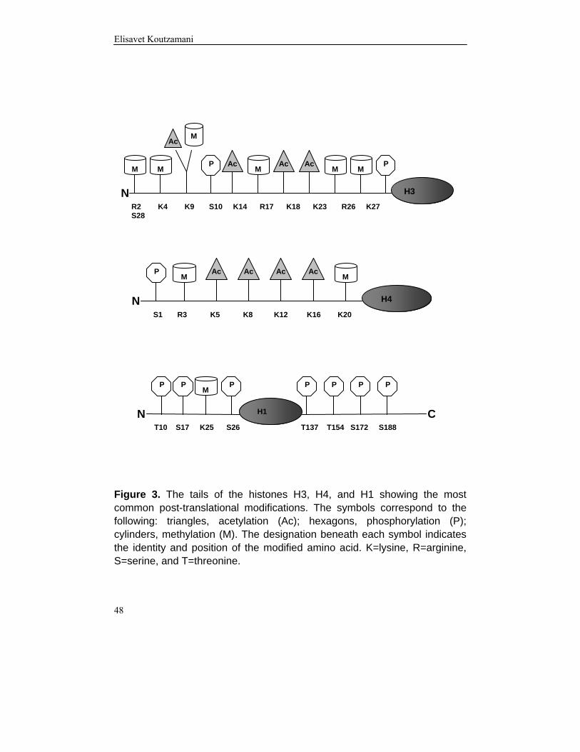
Figure 3. The tails of the histones H3, H4, and H1 showing the most common post-translational modifications. The symbols correspond to the following: triangles, acetylation (Ac); hexagons, phosphorylation (P); cylinders, methylation (M). The designation beneath each symbol indicates the identity and position of the modified amino acid. K=lysine, R=arginine, S=serine, and T=threonine.
H3
M M P Ac
M Ac Ac
M M P
Ac M
R2 K4 K9 S10 K14 R17 K18 K23 R26 K27 S28
N
H4
P M M
Ac Ac Ac Ac
S1 R3 K5 K8 K12 K16 K20 N
P P P P P P P
T10 S17 K25 S26 T137 T154 S172 S188
M
N C H1

Chromatin, histones, and epigenetic tags
49
The Histone Code An exciting and never-ending era of histone research began with the proposal of the histone code by Strahl and Allis (2000) and Turner (2000). According to this “multiple histone modifications, acting in combinatorial or sequential fashion on one or multiple histone tails, specify unique functions along the eukaryotic genome” (Strahl and Allis, 2000). The rapid advances in this field have provided a vast amount of information about the sites of modification that are involved, the types of modifications on the affected residues, and the enzymes that participate in these processes. The research findings relevant to formulation of the histone code are summarized below. The post-translational modifications seen in the tails of different histones can separately or in combination, function as epigenetic tags for either the activity or inactivity of genes. In a global perspective, these tags can direct the formation of euchromatic or heterochromatic chromatin. The chromodomain of HP1 specifically recognizes and binds to methylated H3K9, and that interaction leads to gene repression (Bannister et al., 2001; Lachner et al., 2001). Thus, modification tags are recognized by effector proteins such as HP1, which have distinct structural functions in chromatin. The presence of one, two, or three methyl groups on lysines adds complexity to the histone code and dictates different outcomes. Trimethylation of H3K4 occurs on active genes, and dimethylation of the same site is found in both active and inactive genes (Santos-Rosa et al., 2002). Trimethylated H3K9 and monomethylated H3K27 are repressive marks localized to pericentric heterochromatin (Rice et al., 2003; Peters et al., 2003). The mono- and dimethylated forms of H3K9 are localized to silent euchromatic regions (Rice et al., 2003). Trimethylated H3K9 is also seen in methylated DNA, and it has been implicated in initiating DNA methylation of chromatin regions (Tamaru et al., 2003). Furthermore, it has been reported that monomethylated H4K20 and H3K9 occur within the same heterochromatic regions, and this co-existence was interpreted as being a mark for distinct heterochromatic regions (Sims et al., 2006). The inactive female X chromosome has been found to contain an abundance of monomethylated H4K20 (Sims et al., 2006) and methylated H3K9 in the facultative heterochromatin, whereas methylated H3K4 has been detected in a few regions that are still active in the essentially inactive X chromosome (Boggs et al., 2002). The different methylated forms of H4K20 can also reside within distinct compartments in the nucleus. Research has shown monomethylated H4K20 at the
Elisavet Koutzamani
50
nuclear periphery, dimethylated H4K20 evenly dispersed throughout the nucleus and also in nucleoli, and trimethylated H4K20 in pericentric heterochromatin (Sims et al., 2006). In mouse mammary tumour cells, trimethylated H4K20 is localized to constitutive heterochromatin during interphase and metaphase (Kourmouli et al., 2004). In contrast, it appears that monomethylated H4K20 associates with transcriptionally active chromatin, as indicated by the observation that H4K20 monomethylation correlated with H4 hyperacetylation on the induced promoter of the β-globin gene in MEL cells differentiating in vitro (Talasz et al., 2005). It is also known that promoters of active genes are associated with methylated H3R17 (Chen et al., 1999), and methylation of H4R3 participates in transcriptional activation by recruiting p300, which results in acetylation of H4K8 and H4K12 (Wang et al., 2001). Several other studies have revealed interplay between different modifications in the establishment of chromatin conformations. In yeast, ubiquitination of H2B at lysine 123 by Rad6 is required for methylation of H3K4 and leads to transcriptional silencing (Sun and Allis, 2002). Phosphorylation of H3K10 inhibits methylation of H3K9 (Rea et al., 2000), and, in EGF-stimulated cells, phosphorylation of H3K10 precedes and induces H3K14 acetylation (Cheung et al., 2000). Recently, the histone code was extended to also include the linker histones. HP1 binds to methylated K25 in H1.4, and this binding can be dissolved by phosphorylation of the neighbouring S26 (Daujat et al., 2005). The experiments reported in paper II provided an additional piece of the histone code. We exposed MEL cells in vitro to dimethyl sulfoxide (DMSO) and hexamethylenebisacetamide (HMBA), as well as butyrate and trichostatin A (TSA) (both HDAC inhibitors), to induce erythroid differentiation and the concomitant increase in chromatin condensation. Regardless of the type of inducing agent, hypoacetylated (non- and monoacetylated H4) trimethylated H4K20 was increased with differentiation. In contrast, diacetylated H4 at K16 and K12 contained mainly mono- or dimethylated H4K20 with small amounts of trimethylated H4K20 in samples induced with HDAC inhibitors. Hyperacetylated H4 was not found to be trimethylated but instead contained one or two methyl groups. In the light of these results, it is plausible that these epigenetic tags are involved in the transitions between euchromatin and heterochromatin. Further information on the histone code is available in a number of interesting articles (Biel et al., 2005; Fuchs et al., 2006; Jenuwein and Allis, 2001; Lachner et al., 2003).

Chromatin, histones, and epigenetic tags
51
True knowledge exists in knowing that you know nothing.
Socrates (470–399 BC), Greek philosopher
LINKER HISTONE FUNCTION
The precise function of the linker histones in the nucleus of the cell has not yet been fully elucidated, and many fundamental questions remain to be answered. It was initially believed that the linker histone positioned on the nucleosome serves as a lid to close up the nucleosome and render the enclosed DNA inaccessible, thereby suppressing gene activity. Accordingly, it was long assumed that linker histones act as general repressors of gene activity (Weintraub, 1984), but that view has changed radically over the past few years. Linker histones are non-randomly distributed on chromatin, and the active parts of the genome are relatively depleted of H1.3 and H1.4 (Parseghian et al., 2000), two variants that are connected with inactive genes (Alami et al., 2003). In addition, H1.4 may be more abundant than H1.3 in inactive genes, and such relative enrichment might be correlated with increased condensation of the affected chromatin regions (Parseghian and Hamkalo, 2001). H1.5 has been found to be more extensively associated with active parts of the genome (Parseghian et al., 2000), but it has also been localized to the nuclear periphery, which contains heterochromatin (Parseghian et al., 1993). Partial depletion of linker histones over the transcriptionally competent β-globin gene has been described, whereas the inactive ovalbumin gene contains the full complement of linker histones (Kamakaka and Thomas, 1990). In the present research (paper IV), the depletion of H1.3 in some B-CLL samples was compensated by an increase in H1.4. According to the studies cited above, the rise in H1.4 suggests that the level of chromatin condensation is higher in H1.3-depleted B-CLL cells than in normal B cells. That finding agrees with an ultrastructural investigation showing that CLL lymphocytes contain increased amounts of highly condensed heterochromatin compared with normal B lymphocytes (Manzoli et al., 1975). However, analyses using green

Elisavet Koutzamani
52
fluorescent protein (GFP)-tagged linker histone variants have detected the variants H1.1, H1.2, and H1.3 in euchromatic regions of chromatin, whereas H1.4 and H1.5 were found primarily in heterochromatic regions (Th'ng et al., 2005). The existence of multiple, evolutionarily conserved linker histone variants suggests individual functions for each of these proteins. However, knockout experiments have shown a substantial redundancy in this system, where the loss of one or more variants can be counterbalanced by increased levels of other variants. (Sirotkin et al., 1995) noted that knockout of the gene for the differentiation-specific H1° in mice did not alter the phenotype of the animals, nor did it result in any visible pathological condition. The absence of H1° was compensated by elevated levels of all other variants present, and the stoichiometry of 0.7 linker histones per nucleosome was preserved. Thus the amount of linker histones needed per nucleosome was somehow maintained to ensure proper functioning and regulation of chromatin. On the other hand, in experiments on H1° transgenic mice (Tonjes et al., 1997), overexpression of H1° was detected only at the mRNA level, not at the protein level. Presumably, this stability is post-translationally regulated to create a balance and in that way prevent overexpression of the protein. Results using H1.1-knockout mice (Rabini et al., 2000) have led to the same conclusion. The cited investigators found that absence of H1.1 was offset by increased levels of H1.2, H1.3, and H1.4, and the animals developed normally in that they were fertile and showed no signs of defective spermatogenesis or other abnormalities. Knockout of the testis-specific variant H1.t in mice also resulted in viable fertile animals, and the loss of H1.t was fully counterbalanced by the other variants, with maintained linker histone nucleosome stoichiometry (Drabent et al., 2000; Lin et al., 2000). In another H1.t-knockout study, H1.t deficiency resulted in spermatids that contained only 75% of the normal total amount of linker histones (Fantz et al., 2001). In experiments on the chicken DT40 B cell line (Seguchi et al., 1995), it was found that disruption of one allele of one of the six linker histone variants was compensated by the other variants. This disruption affected a cluster of 21 histone genes, and it had an impact on the expression of several cellular proteins, although the DT40 B cells were still viable and proliferating. These findings suggest that both linker and core histones have compensatory abilities, and only half a set of histone genes is needed for proper cell growth (Takami et al., 1997). In another study on the same cell line (Takami and Nakayama, 1997), it was noted that DT40 mutant cells devoid of five of the six linker histone genes showed no defects in cell growth, even though they contained only half the amount of linker histones compared with the wt cells. Furthermore, there were no changes in the chromatin structure. It is plausible that an increase in levels of the chromatin-associated high

Chromatin, histones, and epigenetic tags
53
mobility group (HMG) proteins that was observed in the mutant cells compensated for the absence of linker histones and thereby maintained a similar chromatin structure. In mice, separate disruption of any of the variants H1.2, H1.3, and H1.4 has shown that these variants are dispensable (Fan et al., 2001). In that study, double knockout mice of any one of those three variants together with H1° also maintained a normal stoichiometry, and the compensation was achieved through increasing levels of two of the three variants H1.2, H1.3, and H1.4. Fan et al. (2003) have observed that mice can tolerate double knock-outs of linker histones, whereas triple-homozygous knockouts of H1.2H1.3H1.4- - - / - - - have been found to result in embryonic lethality. Those investigators were able to produce two viable triple knock-outs, H1°H1.2H1.3- - - / - - - and H1.2H1.3H1.4- - + / - - -, which were smaller than the wt animals and showed a 20%–50% reduction in the linker histone content. The latter change altered the linker-histone-nucleosome stoichiometry and also resulted in a shorter nucleosome repeat length (NRL) compared with the wt. Fan et al. (2005) extended this research by establishing triple-null homozygous knockouts of mouse embryonic stem cells with regard to H1.2, H1.3, and H1.4. These mutant cells also exhibited a 50% decrease in the linker histone content, which was accompanied by an approximately 15-bp reduction in the NRL. Indeed there is a correlation between the presence of linker histones and NRL (Woodcock et al., 2006). In the study by Fan et al. (2003), the investigators observed more limited modifications in H4K12 acetylation and H3K27 trimethylation, and the chromatin of the mutants was more extensively decondensed. Microarray analyses of the mutants showed that a fraction of the genes were affected (i.e., up- or downregulated). Most notably, the expression of a group of imprinted genes was found to be elevated, and analyses of DNA methylation of the H19 gene showed reduced levels of CpG methylation compared with wt embryonic stem cells. Chromatin immunoprecipitation (ChIP) assay revealed an interaction between linker histones and the H19 gene, which suggests that linker histones are involved in regulation of that gene and in establishment of the DNA methylation pattern. In a study of H1°-deficient mice, Gabrilovitch et al. (2002) noted that proliferation of dendritic cells was impaired, while the growth of other leukocytes was normal, and the animals also responded poorly to vaccination with antigens. These findings imply that the absence of linker histone variants has an impact on the biological functions of an organism at the macrolevel, and it would be extremely interesting to ascertain how the double- and triple-knockout mice described above would respond to different exogenous stimuli.

Elisavet Koutzamani
54
Microarray gene expression analyses of germ cells from mice double null for H1t and H1.1 have also linked the reduced amount of linker histones to alterations in the levels of expression of specific genes, rather than to a global effect (Lin et al., 2004). Several studies have confirmed the view of a gene-specific regulatory function of linker histones. During frog development, linker histones selectively repress the 5S rRNA genes (Bouvet et al., 1994) as well as the genes needed for muscle development (Steinbach et al., 1997). In a study of Tetrahymena devoid of linker histones (Shen and Gorovsky, 1996), it was demonstrated that two genes were affected: the CyP gene was activated, and the ngoA gene was repressed. In mice, H1.4 has been shown to interact with the transcription factor Msx1, resulting in repression of the MyoD gene, which is responsible for myogenic differentiation (Lee et al., 2004). In the the chicken DT40 B cell line, homozygous knockouts of each of the individual variants changed the profile of cellular proteins (Takami et al., 2000). Consequently, it seems that the linker histone variants play individual roles in regulating gene expression. Within a living organism, the linker histones are highly mobile molecules that are translocated between different chromatin regions. The mobility of GFP-labelled linker histone molecules within the nuclei of cells has been analysed by FRAP, which has indicated a residence time of about 220 seconds (Lever et al., 2000; Misteli et al., 2000). The linker histones that are exchanged are found in a large mobile pool, and also in a less mobile pool that is larger in heterochromatin than in euchromatin (Misteli et al., 2000). The C-terminal domain of linker histones is responsible for the high affinity binding of these proteins to chromatin, which stabilizes the folding of the chromatin fibre (Allan et al., 1986; Hendzel et al., 2004). The N-terminal part of linker histones is assumed to contribute to positioning of the globular part of the protein on DNA (Allan et al., 1986). The individual linker histone variants differ with respect to their affinity for chromatin, which is lowest for H1.1 and H1.2, and increasingly higher for H1.3, H1.4, and H1.5, in correlation with increasing length of the C-terminal domain (Th'ng et al., 2005). The ability of that domain to bind to nucleosomal DNA is not achieved by the entire region per se, but is instead due to the effects of subdomains within the C-terminal domain (Lu and Hansen, 2004). A novel cytofluorometric method has been developed to analyse the affinity of linker histones for chromatin in situ (Loborg and Rundquist, 1997, 2000). This technique is based on using sodium chloride to extract linker histones and the fluorochrome 4’,6-diamidino-2-phenylindole (DAPI) as a probe to detect the dissociation of linker histones from chromatin. The linker histones attach to

Chromatin, histones, and epigenetic tags
55
chromatin mainly via electrostatic interactions, and it is known that they can be selectively extracted using salt concentrations in the range 0.3–0.7 M; by comparison, higher salt concentrations are needed to dissociate core histones from chromatin (van Holde, 1988). We applied the cytofluorometric method in two studies (papers III and V) to determine the affinity of linker histones for chromatin in chicken and frog erythrocytes, normal B cells, and Epstein-Barr virus (EBV)-transformed cells. The nuclei of terminally differentiated chicken and frog erythrocytes consist of highly condensed and transcriptionally inert chromatin. Despite these similarities, we observed that the chicken linker histones showed substantially higher affinity for chromatin than the frog linker histones did. This disparity might be explained by the presence of the differentiation-specific variant H5, which represents 60–70% of the total amount of linker histones (Bates and Thomas, 1981; paper III). H5 has been reported to bind tighter to chromatin compared with the other variants that are present (Kumar and Walker, 1980). Frog erythrocytes contain the differentiation-specific variant H1°, but the level of that protein is much lower than the amount of H5 seen in chicken erythrocytes. In a mouse cell line, H1° has been found to exhibit a slightly higher affinity for chromatin compared with the other somatic variants (Misteli et al., 2000). We also confirmed the differences in affinity by analysing the degree to which different concentrations of sodium chloride caused linker histones to dissociate from chromatin in chicken and frog erythrocytes. HPCE was applied to investigate the chicken linker histones, whereas RP-HPLC was used to separate the frog linker histone variants because they could not be successfully separated by HPCE. These analyses showed that dissociation of H5 was achieved primarily at a sodium chloride concentration above 0.5 M, and only very small amounts of this protein were detected at 0.5 M. A sodium chloride level of 0.5 M was sufficient to extract a substantial portion of the H1°, and small amounts were retrieved at 0.45 M. Our cytofluorometric and protein analysis data agree with the results of the above-mentioned studies, which confirms that, compared with H1° and all other somatic variants, H5 has a stronger binding affinity for chromatin. The difference in affinity between linker histone in chicken and frog erythrocytes is probably an effect of the dissimilar subtype compositions of these proteins in the respective cell types. Both chicken and frog erythrocytes contain highly condensed chromatin, but they differ with regard to linker histone affinities for chromatin, and hence our data suggest that there is no direct correlation between these two features. Interestingly, it has been reported that mature avian erythrocytes, as well as other cell types, contain the myeloid and erythroid nuclear termination stage-specific protein (MENT), which has been found to be associated with peripheral heterochromatic regions (Grigoryev and Woodcock, 1993). Furthermore, it has been observed that

Elisavet Koutzamani
56
expression of MENT is more strongly correlated with chromatin compaction than with binding of linker histones (Grigoryev et al., 1992). In the present research (paper V), cytofluorometric analyses of linker histone affinity for chromatin in situ in both normal and EBV-transformed B lymphocytes indicated that the affinity was higher in the normal cells. Normal B cells are non-proliferating and positioned in the G0 phase of the cell cycle, but they can be transformed in vitro by exposure to EBV to obtain the immortalized proliferating lymphoblastoid cell line (LCL), which is synonymous with EBV-transformed lymphocytes. The abbreviation LCL is used here. The underlying dissimilarities in affinity for chromatin exhibited by the normal B cell and LCL models might be due to differences in the linker histone composition. To address that possibility, we performed HPCE to analyse linker histone samples from the respective cell types. For the LCL, this meant that the protein samples were prepared from cells undergoing exponential growth (i.e., they were in all phases of the cell cycle). Both cell models expressed the variants H1.2–H1.5, and the LCLs also showed several HPCE peaks corresponding to the phosphorylated variants. This was not surprising, because it is known that phosphorylation of linker histones increases as the cell cycle progresses. However, our cytofluorometric affinity analyses were done only on cells in the G1 phase of the cell cycle, and, since the results of those assessments showed a significant reduction in the affinity of linker histones for chromatin, we were interested in also using HPCE to study the phosphorylation pattern in purified G1 cells. To achieve that goal, we sorted LCL cells in a flow cytometer, and we discriminated between the cell cycle phases by vital staining with the DNA-binding fluorochrome Hoechst 33343. The sorting gave two populations, G1 cells and S/G2/M cells, and linker histones from these cells were subsequently analysed by HPCE. G1 cells were found to contain all variants previously described for LCL, and, surprisingly, there were also multiple phosphorylation peaks in a pattern resembling the linker histone profile of exponentially growing LCL. Thus our results suggest that LCL cells in the G1 phase contain highly phosphorylated linker histone variants, whereas normal B cells contain only minor amounts of monophosphorylated H1.4. The affinity for chromatin exhibited by the C-terminal domain of linker histones is known to be affected by phosphorylation of these proteins (Dou et al., 2002; Contreras et al., 2003). Consequently, our cytofluorometric data agree with our HPCE data on purified G1 cells (paper V), and thus our results indicate that proliferating cells contain phosphorylated linker histones over a larger part of the cell cycle than previously assumed (Herrera et al., 1996). This might be a property of transformed cells, since increased levels of phosphorylation in combination with

Chromatin, histones, and epigenetic tags
57
a more open chromatin structure have been observed in oncogene-altered cells (Chadee et al., 1995; Taylor et al., 1995; Chadee et al., 2002), and pRB-deficient fibroblasts have also been reported to exhibit augmented linker histone phosphorylation together with elevated CDK2 activity and a relaxed chromatin structure (Herrera et al., 1996). Therefore, varying levels of phosphorylation in the different linker histone subtypes might be considered epigenetic tags involved in regulation of the cell cycle. In addition to phosphorylation, the interactions between linker histones and chromatin can be modulated by several other factors. For example, acetylation of the N termini of core histones induced by treatment with the histone deacetylase inhibitor TSA has been found to increase the mobility of linker histones (Misteli et al., 2000). Furthermore, the non-histone HMG proteins in chromatin, which are also nucleosome-binding proteins, may compete for the same binding sites as the linker histones. FRAP studies have indicated that HMG proteins increase the mobility of linker histones, and the elevated exchange rate implies that the affinity of linker histones for chromatin will be reduced (Catez et al., 2002, 2004). Prothymosin α (ProTα) is a nuclear protein involved in cell proliferation, and it interacts with linker histones to displace parts of those molecules from chromatin (Karetsou et al., 1998). Another protein of interest is parathymosin (ParaT), which is similar to ProTα with regard to primary structure; ParaT also interacts with linker histones, and that interplay might lead to chromatin decondensation (Martic et al., 2005). In addition, it has been proposed that linker histone binding to chromatin is affected by an as-of-yet unidentified ATP-dependent process (Dou et al., 2002). For reviews concerning modulation of interactions between linker histones and chromatin, see Zlatanova et al. (2000) and Catez et al. (2006). In conclusion, the linker histones are either directly or indirectly important components of the chromatin fibre. They are needed to stabilize and to some extent also to condense the chromatin, and they are involved in gene-specific regulation of several cellular processes. They are definitely necessary for the viability of higher organisms, although a certain reduction in the linker histone content and/or to some extent the absence of certain combinations of linker histone variants can be tolerated. The orientation of these proteins on the nucleosome stops regulatory factors from gaining access to the nucleosomal DNA, but that effect can be overcome by phosphorylation of the linker histones and by competition or displacement of a variety of factors, resulting in changes in chromatin conformation.

Elisavet Koutzamani
58

Conclusionss
59
CONCLUSIONS
The results and conclusions of the research reported in papers I–V are summarized as follows:
• Mammalian cells were found to contain trimethylated H4K20. Levels in rat kidney and liver increased with increasing age of the animals. Trimethylated H4K20 was also detected in small but increasing amounts in cell lines in the stationary phase, as compared with exponentially growing cells.
• Trimethylated H4K20 was localized to heterochromatin. Levels increased
in MEL cells undergoing differentiation, and this change correlated with H4 hypoacetylation.
• Linker histone affinity for chromatin, in the highly compacted chromatin of
chicken and frog erythrocytes, was very high in chicken erythrocytes compared with frog erytrhocytes. These two cell models differed with regard to the composition and levels of linker histone variants. These findings suggest that linker histone affinity is not the key factor in chromatin compaction. It was also observed that frog erythrocytes contain only one differentiation-specific H1° subvariant, which was identified as the subvariant H1°-2.
• Normal B lymphocytes were noted to contain the linker histone variants
H1.2, H1.3, H1.4, and H1.5. The same linker histone variants, although in different amounts, were detected in most samples of leukaemic cells from patients with B-CLL. H1.3 was generally expressed at lower levels or was indeed lacking, in the leukaemic cells. The low levels or absence of H1.3 was compensated by increased levels of H1.4. No genetic abnormality was found in the coding region of H1.3 that could explain its absence.
• Compared with normal resting B lymphocytes, proliferating LCL cells were
observed to have reduced affinity for chromatin when in the G1 phase of the cell cycle. The G1 cells contained multiple phosphorylated variants, whereas only low levels of monophosphorylated H1.4 were found in normal B cells. The increased phosphorylation might explain the reduced affinity for chromatin shown by linker histones in LCL cells.

Elisavet Koutzamani
60

Acknowledgements
61
No man is an island; entire of itself every man is a piece of the continent, a part of the main. John Donne (1572–1631), English poet and cleric
ACKNOWLEDGEMENTS I would like to express my deepest gratitude to the following people: My advisor, Ingemar Rundquist, for sharing with me your incredible energy, enthusiasm, knowledge and visions; for providing me with a good combination of advice and independence; and for caring. My co-advisor, Herbert Lindner, for opening your laboratory in Innsbruck to me and for making me feel at home. Your vast knowledge about histones has helped me immensely. Words like “why not” will always echo in my mind. Thanks for all the unforgivable evenings at Innrain! Bettina Sarg, a very dear friend and histone expert. Thank you for taking care of me and showing me everything there is to know about analysing linker histones, and also for all the unforgettable moments in Innsbruck. Miss you! Gunnar Juliusson, my second co-advisor, and also Ola Söderström and Anders Rosén. I appreciate your expertise in the field of B-CLL biology, and thanks for always having time for discussions and to help with sequencing of the Ig genes. Anita Lönn, the angel in the lab, for teaching me everything about cell culturing, for trying to bring order into my chaos, for great fun and chats, for sharing my enthusiasm for salty liquorice, and for putting up with listening to Radio Match all those year. Peter Söderkvist, Åsa Schippert and Annette Molbaek, for your expertise in molecular biology. Many thanks to Peter for always having time for discussions; to Åsa for introducing me to RT-PCR, for being so patient, and also for excellent collaboration on the laboratory course “celltillväxt och celldifferentiering”; to Annette for your warm smile whenever I came running to you with incessant simple questions.

Elisavet Koutzamani
62
Jan-Ingvar Jönsson and Uno Johansson, for your good spirits and for supplying me with cells and always taking the time to answer my questions about flow cytometry. Wilfried Helliger and Heribert Talasz, the other histone researchers in Innsbruck, for your invaluable help and advice. Astrid Devich, for your kindness, helpfulness, and nice chats in the lab in Innsbruck and for patiently, listening to me trying to speak German. Michael Rittinger, for fun in the cell culture lab in Innsbruck and for showing me everything about acetic acid-urea-Triton gel electrophoresis of histones. Karin Schütz, a very dear friend of mine who is in New Zeeland, physically far, far, far away from me yet only a phone call away. We had so many smashing and crazy times together. Many thanks for love and support! Come home soon! Miss you! Anna Green, my friend, my roommate and my partner in histone research, which would not have been the same without you. Late evenings by the flow cytometer filled with laughter and moral support and who can forget the awesome time at the Keystone Conference. We need to have those Margaritas soon! Looking forward to your thesis! Line Bjørge, for friendship, support, and wonderful hiking excursions. I wish you all the best! My “big sister” Annelie Lindström, my mentor in pedagogics and life, and my personal coach, for always being concerned about me and for all your kindness. You have increased my English vocabulary with self-composed sentences that have proven to be very useful in all kinds of situations. You have been so patient in trying to get me to jump and run on the floor at “Friskis och Svettis” sessions and then spoiling me afterwards with nice dinners and crazy discussions. My “little sister”Maria “Lillan” Dahlström, for slowing me down whenever I wanted things to happen too fast (det ska ju gå snabbt, hela tiden ☺), and for trying to keep me in a good mood with words of wisdom and funny anecdotes. Who can forget all those late night philosophical discussions over bottles of wine and our insane New York shopping? “Always and forever” lillan!

Acknowledgements
63
Cecilia, the calmest and coolest person I ever met. Thanks for dragging me from work to go shopping and have pleasant times at different coffee houses. I am still amazed at how you put up with my messy part of our office. Ian, my British roommate for a year, thanks for improving my English and for all the awesome and crazy moments in and outside the office. Anna I, Anna-Mia, Carina, Caroline, Christine, Karin E., Mai-Le, Paulina, and Åsa–my ”sötnosar” outside the laboratory, who remind me of how real life is supposed to be and how to enjoy it. Thanks for putting up with me for the last couple of months! Vi är bäst och det vet vi om! Lotta and Jesper, the sunshine at the division of Cell Biology, always there to give good advice and unforgivable laughter. Alexey, Anna L, Anna E., Björn, Eva, Emelie, Johan, Lorena, Maria H, Marcus and Rickard–the people next-door on floor 13–for bringing such good spirits to work every day and for all the crazy after-work events, like laserdome, bowling, golf, paintball and poker, just to mention a few of the innovative things you persuaded me to try and that sometimes hurt. To all the other fantastic present and past co-workers that I have had the privilege to meet during this journey and who made my working place so enjoyable: Alex, Anna-Lotta, Ana-Maria, Annelie K., Anette, Anita S., Anita L., Avni, Bita, Cornelia, Eva B., Eva D., Joackim, Helena, Henrik, Håkan, Ingrid, Karin F, Karin T., Kerstin, Kristina, Lena, Lennart, Lotta B., Lotta H., Margareta, Masha, Monica, Pia, Peter S., Ragnhild, Thomas, Viveka and Åke. To the Greek party of five: Argiris, Georgios, Giannis, Sakis, and Stergios. Thanks for endless conversations about nothing and everything, for the summer barbeques, and for the “Friday fika” at Santini’s with tea and blueberry muffins. Hope we can all get together again. Alberto, Andrea, Christa, Christian, Christina, Esther, Gerald, Jessica, Katharina, Mariana, and Paul for all the fun we had in Innsbruck: nice dinners, wonderful trips, evenings by the pool table or just with a beer and the unsolved questions of life and science.

Elisavet Koutzamani
64
Maria, Michael, Panagyota, and Sofia Tsahalis, my New York sweethearts for always taking such good care of me and spoiling me with Crispy Cream doughnuts and tremendous laughter whenever I am in New York. Miss you so much! My cousin–Georgios–the most misunderstood person on this planet. Your tremendous imagination and great thinking has had a great influence on me. Never give up trying to convince people of your ideas! Blodcentralen, for providing me with all those buffy coats! To everyone at the HUB library of the Faculty of Health Sciences, for help with obtaining articles I had given up hope on finding. DJ Stan, don’t you ever stop playing the music. Keep up the rhythm and shake it baby! Jonas, for being such an inspiration in the writing of this thesis and for all the pleasant late night company at the computer. My relatives in Greece, for all the nice summers spent together and for your familial love and encouragement. See you next summer! My brother Andreas, for being my best friend and giving me continuous support and wisdom. No one can make me laugh like you do, and you bring so much joy into my life. I admire you and wish I was more like you! My mother and father, Agathi and Dimitrios Koutzamanis, for being the steady anchor in my life. You have always been there for me, on happy and stormy days. Your love has made me feel safe and strong. Σας αγαπω παρα πολυ!

References
65
REFERENCES Ajiro, K., and Nishimoto, T. 1985. Specific site of histone H3 phosphorylation
related to the maintenance of premature chromosome condensation. Evidence for catalytically induced interchange of the subunits. J Biol Chem. 260: 15379-15381.
Ajiro, K., Nishimoto, T., and Takahashi, T. 1983. Histone H1 and H3
phosphorylation during premature chromosome condensation in a temperature-sensitive mutant (tsBN2) of baby hamster kidney cells. J Biol Chem. 258: 4534-4538.
Alami, R., Fan, Y., Pack, S., Sonbuchner, T.M., Besse, A., Lin, Q., et al. 2003.
Mammalian linker-histone subtypes differentially affect gene expression in vivo. Proc Natl Acad Sci U S A. 100: 5920-5925.
Albig, W., Drabent, B., Kunz, J., Kalff-Suske, M., Grzeschik, K.H., and Doenecke,
D. 1993. All known human H1 histone genes except the H1(0) gene are clustered on chromosome 6. Genomics. 16: 649-654.
Alexandrow, M.G., and Hamlin, J.L. 2005. Chromatin decondensation in S-phase
involves recruitment of Cdk2 by Cdc45 and histone H1 phosphorylation. J Cell Biol. 168: 875-886.
Allan, J., Hartman, P.G., Crane-Robinson, C., and Aviles, F.X. 1980. The structure
of histone H1 and its location in chromatin. Nature. 288: 675-679. Allan, J., Mitchell, T., Harborne, N., Bohm, L., and Crane-Robinson, C. 1986.
Roles of H1 domains in determining higher order chromatin structure and H1 location. J Mol Biol. 187: 591-601.
Allfrey, V.G., Faulkner, R., and Mirsky, A.E. 1964. Acetylation and Methylation of
Histones and Their Possible Role in the Regulation of Rna Synthesis. Proc Natl Acad Sci U S A. 51: 786-794.
Althaus, F.R., Hofferer, L., Kleczkowska, H.E., Malanga, M., Naegeli, H.,
Panzeter, P.L., and Realini, C.A. 1994. Histone shuttling by poly ADP-ribosylation. Mol Cell Biochem. 138: 53-59.
An, W., Leuba, S.H., van Holde, K., and Zlatanova, J. 1998. Linker histone protects
linker DNA on only one side of the core particle and in a sequence-dependent manner. Proc Natl Acad Sci U S A. 95: 3396-3401.

Elisavet Koutzamani
66
Arents, G., Burlingame, R.W., Wang, B.C., Love, W.E., and Moudrianakis, E.N. 1991. The nucleosomal core histone octamer at 3.1 A resolution: a tripartite protein assembly and a left-handed superhelix. Proc Natl Acad Sci U S A. 88: 10148-10152.
Arents, G., and Moudrianakis, E.N. 1995. The histone fold: a ubiquitous
architectural motif utilized in DNA compaction and protein dimerization. Proc Natl Acad Sci U S A. 92: 11170-11174.
Avery O.T., MacLeod C.M., and McCarthy M. 1944. Studies on the chemical
nature of the substance inducing trasnformation of pneumococcal types. Journal of Experimental Medicine. 79: 137-159
Babu, A., and Verma, R.S. 1987. Chromosome structure: euchromatin and
heterochromatin. Int Rev Cytol. 108: 1-60. Bannister, A.J., Zegerman, P., Partridge, J.F., Miska, E.A., Thomas, J.O., Allshire,
R.C., and Kouzarides, T. 2001. Selective recognition of methylated lysine 9 on histone H3 by the HP1 chromo domain. Nature. 410: 120-124.
Bates, D.L., and Thomas, J.O. 1981. Histones H1 and H5: one or two molecules per
nucleosome? Nucleic Acids Res. 9: 5883-5894. Belikov, S., and Karpov, V. 1998. Linker histones: paradigm lost but questions
remain. FEBS Lett. 441: 161-164. Belmont, A.S., and Bruce, K. 1994. Visualization of G1 chromosomes: a folded,
twisted, supercoiled chromonema model of interphase chromatid structure. J Cell Biol. 127: 287-302.
Bhattacharjee, R.N., and Archer, T.K. 2006. Transcriptional silencing of the mouse
mammary tumor virus promoter through chromatin remodeling is concomitant with histone H1 phosphorylation and histone H3 hyperphosphorylation at M phase. Virology. 346: 1-6.
Biel, M., Wascholowski, V., and Giannis, A. 2005. Epigenetics--an epicenter of
gene regulation: histones and histone-modifying enzymes. Angew Chem Int Ed Engl. 44: 3186-3216.
Boggs, B.A., Cheung, P., Heard, E., Spector, D.L., Chinault, A.C., and Allis, C.D.
2002. Differentially methylated forms of histone H3 show unique association patterns with inactive human X chromosomes. Nat Genet. 30: 73-76.

References
67
Bosch, A., and Suau, P. 1995. Changes in core histone variant composition in differentiating neurons: the roles of differential turnover and synthesis rates. Eur J Cell Biol. 68: 220-225.
Boulikas, T. 1990. Poly(ADP-ribosylated) histones in chromatin replication. J Biol
Chem. 265: 14638-14647. Bouvet, P., Dimitrov, S., and Wolffe, A.P. 1994. Specific regulation of Xenopus
chromosomal 5S rRNA gene transcription in vivo by histone H1. Genes Dev. 8: 1147-1159.
Briggs, S.D., Xiao, T., Sun, Z.W., Caldwell, J.A., Shabanowitz, J., Hunt, D.F., et al.
2002. Gene silencing: trans-histone regulatory pathway in chromatin. Nature. 418: 498.
Brocard, M.P., Triebe, S., Peretti, M., Doenecke, D., and Khochbin, S. 1997.
Characterization of the two H1(zero)-encoding genes from Xenopus laevis. Gene. 189: 127-134.
Brown, D.T., Izard, T., and Misteli, T. 2006. Mapping the interaction surface of
linker histone H1(0) with the nucleosome of native chromatin in vivo. Nat Struct Mol Biol. 13: 250-255.
Bucci, L.R., Brock, W.A., and Meistrich, M.L. 1982. Distribution and synthesis of
histone 1 subfractions during spermatogenesis in the rat. Exp Cell Res. 140: 111-118.
Catez, F., Brown, D.T., Misteli, T., and Bustin, M. 2002. Competition between
histone H1 and HMGN proteins for chromatin binding sites. EMBO Rep. 3: 760-766.
Catez, F., Ueda, T., and Bustin, M. 2006. Determinants of histone H1 mobility and
chromatin binding in living cells. Nat Struct Mol Biol. 13: 305-310. Catez, F., Yang, H., Tracey, K.J., Reeves, R., Misteli, T., and Bustin, M. 2004.
Network of dynamic interactions between histone H1 and high-mobility-group proteins in chromatin. Mol Cell Biol. 24: 4321-4328.
Chadee, D.N., Peltier, C.P., and Davie, J.R. 2002. Histone H1(S)-3 phosphorylation
in Ha-ras oncogene-transformed mouse fibroblasts. Oncogene. 21: 8397-8403.
Chadee, D.N., Taylor, W.R., Hurta, R.A., Allis, C.D., Wright, J.A., and Davie, J.R.
1995. Increased phosphorylation of histone H1 in mouse fibroblasts

Elisavet Koutzamani
68
transformed with oncogenes or constitutively active mitogen-activated protein kinase kinase. J Biol Chem. 270: 20098-20105.
Chadwick, B.P., and Willard, H.F. 2001. A novel chromatin protein, distantly
related to histone H2A, is largely excluded from the inactive X chromosome. J Cell Biol. 152: 375-384.
Chen, D., Ma, H., Hong, H., Koh, S.S., Huang, S.M., Schurter, B.T., et al. 1999.
Regulation of transcription by a protein methyltransferase. Science. 284: 2174-2177.
Chen, H.Y., Sun, J.M., Zhang, Y., Davie, J.R., and Meistrich, M.L. 1998.
Ubiquitination of histone H3 in elongating spermatids of rat testes. J Biol Chem. 273: 13165-13169.
Cheung, P., Tanner, K.G., Cheung, W.L., Sassone-Corsi, P., Denu, J.M., and Allis,
C.D. 2000. Synergistic coupling of histone H3 phosphorylation and acetylation in response to epidermal growth factor stimulation. Mol Cell. 5: 905-915.
Contreras, A., Hale, T.K., Stenoien, D.L., Rosen, J.M., Mancini, M.A., and Herrera,
R.E. 2003. The dynamic mobility of histone H1 is regulated by cyclin/CDK phosphorylation. Mol Cell Biol. 23: 8626-8636.
Costanzi, C., and Pehrson, J.R. 1998. Histone macroH2A1 is concentrated in the
inactive X chromosome of female mammals. Nature. 393: 599-601. Costanzi, C., and Pehrson, J.R. 2001. MACROH2A2, a new member of the
MARCOH2A core histone family. J Biol Chem. 276: 21776-21784. Craig, J.M. 2005. Heterochromatin--many flavours, common themes. Bioessays.
27: 17-28. Daujat, S., Zeissler, U., Waldmann, T., Happel, N., and Schneider, R. 2005. HP1
binds specifically to Lys26-methylated histone H1.4, whereas simultaneous Ser27 phosphorylation blocks HP1 binding. J Biol Chem. 280: 38090-38095.
Davie, J.R., and Chadee, D.N. 1998. Regulation and regulatory parameters of
histone modifications. J Cell Biochem Suppl. 30-31: 203-213. DeLange, R.J., Fambrough, D.M., Smith, E.L., and Bonner, J. 1969. Calf and pea
histone IV. 3. Complete amino acid sequence of pea seedling histone IV;

References
69
comparison with the homologous calf thymus histone. J Biol Chem. 244: 5669-5679.
D'Incalci, M., Allavena, P., Wu, R.S., and Bonner, W.M. 1986. H1 variant synthesis
in proliferating and quiescent human cells. Eur J Biochem. 154: 273-279. Doenecke, D., Albig, W., Bouterfa, H., and Drabent, B. 1994. Organization and
expression of H1 histone and H1 replacement histone genes. J Cell Biochem. 54: 423-431.
Dou, Y., Bowen, J., Liu, Y., and Gorovsky, M.A. 2002. Phosphorylation and an
ATP-dependent process increase the dynamic exchange of H1 in chromatin. J Cell Biol. 158: 1161-1170.
Dou, Y., Mizzen, C.A., Abrams, M., Allis, C.D., and Gorovsky, M.A. 1999.
Phosphorylation of linker histone H1 regulates gene expression in vivo by mimicking H1 removal. Mol Cell. 4: 641-647.
Dover, J., Schneider, J., Tawiah-Boateng, M.A., Wood, A., Dean, K., Johnston, M.,
and Shilatifard, A. 2002. Methylation of histone H3 by COMPASS requires ubiquitination of histone H2B by Rad6. J Biol Chem. 277: 28368-28371.
Drabent, B., Saftig, P., Bode, C., and Doenecke, D. 2000. Spermatogenesis
proceeds normally in mice without linker histone H1t. Histochem Cell Biol. 113: 433-442.
Eggena, M., Cohavy, O., Parseghian, M.H., Hamkalo, B.A., Clemens, D., Targan,
S.R., et al. 2000. Identification of histone H1 as a cognate antigen of the ulcerative colitis-associated marker antibody pANCA. J Autoimmun. 14: 83-97.
Fan, L., and Roberts, V.A. 2006. Complex of linker histone H5 with the
nucleosome and its implications for chromatin packing. Proc Natl Acad Sci U S A. 103: 8384-8389.
Fan, Y., Nikitina, T., Morin-Kensicki, E.M., Zhao, J., Magnuson, T.R., Woodcock,
C.L., and Skoultchi, A.I. 2003. H1 linker histones are essential for mouse development and affect nucleosome spacing in vivo. Mol Cell Biol. 23: 4559-4572.
Fan, Y., Nikitina, T., Zhao, J., Fleury, T.J., Bhattacharyya, R., Bouhassira, E.E., et
al. 2005. Histone H1 depletion in mammals alters global chromatin structure but causes specific changes in gene regulation. Cell. 123: 1199-1212.

Elisavet Koutzamani
70
Fan, Y., Sirotkin, A., Russell, R.G., Ayala, J., and Skoultchi, A.I. 2001. Individual somatic H1 subtypes are dispensable for mouse development even in mice lacking the H1(0) replacement subtype. Mol Cell Biol. 21: 7933-7943.
Fantz, D.A., Hatfield, W.R., Horvath, G., Kistler, M.K., and Kistler, W.S. 2001.
Mice with a targeted disruption of the H1t gene are fertile and undergo normal changes in structural chromosomal proteins during spermiogenesis. Biol Reprod. 64: 425-431.
Fraga, M.F., Ballestar, E., Villar-Garea, A., Boix-Chornet, M., Espada, J., Schotta,
G., et al. 2005. Loss of acetylation at Lys16 and trimethylation at Lys20 of histone H4 is a common hallmark of human cancer. Nat Genet. 37: 391-400.
Franklin, S.E., and Gosling, R.G. 1953. Molecular configuration in sodium
thymonucleate. Nature. 171: 740-741. Fuchs, J., Demidov, D., Houben, A., and Schubert, I. 2006. Chromosomal histone
modification patterns--from conservation to diversity. Trends Plant Sci. 11: 199-208.
Gabrielli, F., Aden, D.P., Carrel, S.C., von Bahr, C., Rane, A., Angeletti, C.A., and
Hancock, R. 1984. Histone complements of human tissues, carcinomas, and carcinoma-derived cell lines. Mol Cell Biochem. 65: 57-66.
Gabrilovich, D.I., Cheng, P., Fan, Y., Yu, B., Nikitina, E., Sirotkin, A., et al. 2002.
H1(0) histone and differentiation of dendritic cells. A molecular target for tumor-derived factors. J Leukoc Biol. 72: 285-296.
Garcia, B.A., Busby, S.A., Barber, C.M., Shabanowitz, J., Allis, C.D., and Hunt,
D.F. 2004. Characterization of phosphorylation sites on histone H1 isoforms by tandem mass spectrometry. J Proteome Res. 3: 1219-1227.
Gill, G. 2004. SUMO and ubiquitin in the nucleus: different functions, similar
mechanisms? Genes Dev. 18: 2046-2059. Goto, H., Tomono, Y., Ajiro, K., Kosako, H., Fujita, M., Sakurai, M., et al. 1999.
Identification of a novel phosphorylation site on histone H3 coupled with mitotic chromosome condensation. J Biol Chem. 274: 25543-25549.
Goto, H., Yasui, Y., Nigg, E.A., and Inagaki, M. 2002. Aurora-B phosphorylates
Histone H3 at serine28 with regard to the mitotic chromosome condensation. Genes Cells. 7: 11-17.

References
71
Greaves, I.K., Rangasamy, D., Devoy, M., Marshall Graves, J.A., and Tremethick, D.J. 2006. The X and Y chromosomes assemble into H2A.Z, containing facultative heterochromatin, following meiosis. Mol Cell Biol. 26: 5394-5405.
Grigoryev, S.A., Solovieva, V.O., Spirin, K.S., and Krasheninnikov, I.A. 1992. A
novel nonhistone protein (MENT) promotes nuclear collapse at the terminal stage of avian erythropoiesis. Exp Cell Res. 198: 268-275.
Grigoryev, S.A., and Woodcock, C.L. 1993. Stage-specific expression and
localization of MENT, a nuclear protein associated with chromatin condensation in terminally differentiating avian erythroid cells. Exp Cell Res. 206: 335-343.
Gugliucci, A. 1994. Advanced glycation of rat liver histone octamers: an in vitro
study. Biochem Biophys Res Commun. 203: 588-593. Gugliucci, A., and Bendayan, M. 1995. Histones from diabetic rats contain
increased levels of advanced glycation end products. Biochem Biophys Res Commun. 212: 56-62.
Gurley, L.R., D'Anna, J.A., Barham, S.S., Deaven, L.L., and Tobey, R.A. 1978.
Histone phosphorylation and chromatin structure during mitosis in Chinese hamster cells. Eur J Biochem. 84: 1-15.
Hake, S.B., and Allis, C.D. 2006. Histone H3 variants and their potential role in
indexing mammalian genomes: the "H3 barcode hypothesis". Proc Natl Acad Sci U S A. 103: 6428-6435.
Hale, T.K., Contreras, A., Morrison, A.J., and Herrera, R.E. 2006. Phosphorylation
of the linker histone H1 by CDK regulates its binding to HP1alpha. Mol Cell. 22: 693-699.
Hall, J.M., and Cole, R.D. 1986. Mechanisms of H1o accumulation in mouse
neuroblastoma cells differ with different treatments. J Biol Chem. 261: 5168-5174.
Halmer, L., and Gruss, C. 1995. Influence of histone H1 on the in vitro replication
of DNA and chromatin. Nucleic Acids Res. 23: 773-778. Happel, N., Schulze, E., and Doenecke, D. 2005a. Characterisation of human
histone H1x. Biol Chem. 386: 541-551.

Elisavet Koutzamani
72
Happel, N., Sommer, A., Hanecke, K., Albig, W., and Doenecke, D. 2005b. Topoisomerase inhibitor induced dephosphorylation of H1 and H3 histones as a consequence of cell cycle arrest. J Cell Biochem. 95: 1235-1247.
Harris, M.R., and Smith, B.J. 1983. A qualitative and quantitative study of
subfractions of the histone H10 in various mammalian tissues. Biochem J. 211: 763-766.
Hayes, J.J., Pruss, D., and Wolffe, A.P. 1994. Contacts of the globular domain of
histone H5 and core histones with DNA in a "chromatosome". Proc Natl Acad Sci U S A. 91: 7817-7821.
Hayes, J.J., and Wolffe, A.P. 1993. Preferential and asymmetric interaction of
linker histones with 5S DNA in the nucleosome. Proc Natl Acad Sci U S A. 90: 6415-6419.
Helliger, W., Lindner, H., Grubl-Knosp, O., and Puschendorf, B. 1992. Alteration
in proportions of histone H1 variants during the differentiation of murine erythroleukaemic cells. Biochem J. 288 ( Pt 3): 747-751.
Hendzel, M.J., Lever, M.A., Crawford, E., and Th'ng, J.P. 2004. The C-terminal
domain is the primary determinant of histone H1 binding to chromatin in vivo. J Biol Chem. 279: 20028-20034.
Hendzel, M.J., Wei, Y., Mancini, M.A., Van Hooser, A., Ranalli, T., Brinkley,
B.R., et al. 1997. Mitosis-specific phosphorylation of histone H3 initiates primarily within pericentromeric heterochromatin during G2 and spreads in an ordered fashion coincident with mitotic chromosome condensation. Chromosoma. 106: 348-360.
Herrera, R.E., Chen, F., and Weinberg, R.A. 1996. Increased histone H1
phosphorylation and relaxed chromatin structure in Rb-deficient fibroblasts. Proc Natl Acad Sci U S A. 93: 11510-11515.
Hochhuth, C., and Doenecke, D. 1990. The expression of the histone H1 (0) gene in
the human hepatoma cell line HepG2 is independent of the state of cell proliferation. Differentiation. 43: 212-219.
Jenuwein, T., and Allis, C.D. 2001. Translating the histone code. Science. 293:
1074-1080. Johns, E.W. 1964. Studies on histones. 7. Preparative methods for histone fractions
from calf thymus. Biochem J. 92: 55-59.

References
73
Johns, E.W. 1967. The electrophoresis of histones in polyacrylamide gel and their quantitative determination. Biochem J. 104: 78-82.
Johns, E.W., and Butler, J.A. 1962. Further fractionations of histones from calf
thymus. Biochem J. 82: 15-18. Kamakaka, R.T., and Thomas, J.O. 1990. Chromatin structure of transcriptionally
competent and repressed genes. Embo J. 9: 3997-4006. Karetsou, Z., Sandaltzopoulos, R., Frangou-Lazaridis, M., Lai, C.Y., Tsolas, O.,
Becker, P.B., and Papamarcaki, T. 1998. Prothymosin alpha modulates the interaction of histone H1 with chromatin. Nucleic Acids Res. 26: 3111-3118.
Konishi, A., Shimizu, S., Hirota, J., Takao, T., Fan, Y., Matsuoka, Y., et al. 2003.
Involvement of histone H1.2 in apoptosis induced by DNA double-strand breaks. Cell. 114: 673-688.
Kornberg, R.D. 1974. Chromatin structure: a repeating unit of histones and DNA.
Science. 184: 868-871. Kornberg, R.D., and Lorch, Y. 1999. Twenty-five years of the nucleosome,
fundamental particle of the eukaryote chromosome. Cell. 98: 285-294. Kornberg, R.D., and Thomas, J.O. 1974. Chromatin structure; oligomers of the
histones. Science. 184: 865-868. Kossel, A. 1911. Ueber die chemische Beschaffenheit des Zellkerns. Munchener
medizinische Wochenschrifte. 58: 65-69. Kourmouli, N., Jeppesen, P., Mahadevhaiah, S., Burgoyne, P., Wu, R., Gilbert,
D.M., et al. 2004. Heterochromatin and tri-methylated lysine 20 of histone H4 in animals. J Cell Sci. 117: 2491-2501.
Kumar, N.M., and Walker, I.O. 1980. The binding of histones H1 and H5 to
chromatin in chicken erythrocyte nuclei. Nucleic Acids Res. 8: 3535-3551. Kurdistani, S.K., Tavazoie, S., and Grunstein, M. 2004. Mapping global histone
acetylation patterns to gene expression. Cell. 117: 721-733. Kuzmichev, A., Jenuwein, T., Tempst, P., and Reinberg, D. 2004. Different EZH2-
containing complexes target methylation of histone H1 or nucleosomal histone H3. Mol Cell. 14: 183-193.

Elisavet Koutzamani
74
Lachner, M., O'Carroll, D., Rea, S., Mechtler, K., and Jenuwein, T. 2001. Methylation of histone H3 lysine 9 creates a binding site for HP1 proteins. Nature. 410: 116-120.
Lachner, M., O'Sullivan, R.J., and Jenuwein, T. 2003. An epigenetic road map for
histone lysine methylation. J Cell Sci. 116: 2117-2124. Langan, T.A., Gautier, J., Lohka, M., Hollingsworth, R., Moreno, S., Nurse, P., et
al. 1989. Mammalian growth-associated H1 histone kinase: a homolog of cdc2+/CDC28 protein kinases controlling mitotic entry in yeast and frog cells. Mol Cell Biol. 9: 3860-3868.
Lee, H., Habas, R., and Abate-Shen, C. 2004. MSX1 cooperates with histone H1b
for inhibition of transcription and myogenesis. Science. 304: 1675-1678. Lennox, R.W., and Cohen, L.H. 1983. The histone H1 complements of dividing and
nondividing cells of the mouse. J Biol Chem. 258: 262-268. Lennox, R.W., Oshima, R.G., and Cohen, L.H. 1982. The H1 histones and their
interphase phosphorylated states in differentiated and undifferentiated cell lines derived from murine teratocarcinomas. J Biol Chem. 257: 5183-5189.
Lever, M.A., Th'ng, J.P., Sun, X., and Hendzel, M.J. 2000. Rapid exchange of
histone H1.1 on chromatin in living human cells. Nature. 408: 873-876. Li, B., Pattenden, S.G., Lee, D., Gutierrez, J., Chen, J., Seidel, C., et al. 2005.
Preferential occupancy of histone variant H2AZ at inactive promoters influences local histone modifications and chromatin remodeling. Proc Natl Acad Sci U S A. 102: 18385-18390.
Lin, Q., Inselman, A., Han, X., Xu, H., Zhang, W., Handel, M.A., and Skoultchi,
A.I. 2004. Reductions in linker histone levels are tolerated in developing spermatocytes but cause changes in specific gene expression. J Biol Chem. 279: 23525-23535.
Lin, Q., Sirotkin, A., and Skoultchi, A.I. 2000. Normal spermatogenesis in mice
lacking the testis-specific linker histone H1t. Mol Cell Biol. 20: 2122-2128. Lindner, H., and Helliger, W. 2004. Hydrophilic interaction chromatography of
peptides and proteins. Methods Mol Biol. 251: 75-88. Lindner, H., Helliger, W., Dirschlmayer, A., Talasz, H., Wurm, M., Sarg, B., et al.
1992. Separation of phosphorylated histone H1 variants by high-performance capillary electrophoresis. J. Chromatogr. 608: 211-216.

References
75
Lindner, H., Helliger, W., and Puschendorf, B. 1986a. Histone separation by high-performance liquid chromatography on C4 reverse-phase columns. Anal Biochem. 158: 424-430.
Lindner, H., Helliger, W., and Puschendorf, B. 1986b. Rapid separation of histones
by high-performance liquid chromatography on C4 reversed-phase columns. J. Chromatogr. 357: 301-310.
Lindner, H., Helliger, W., and Puschendorf, B. 1988. Separation of Friend
erythroleukaemic cell histones and high-mobility-group proteins by reversed-phase high-performance liquid chromatography. J Chromatogr. 450: 309-316.
Lindner, H., Helliger, W., and Puschendorf, B. 1990. Separation of rat tissue
histone H1 subtypes by reverse-phase HPLC. Identification and assignment to a standard H1 nomenclature. Biochem. J. 269: 359-363.
Lindner, H., Sarg, B., and Helliger, W. 2003. Capillary electrophoresis analysis of
histones, histone variants, and their post-translationally modified forms: a review. J Capillary Electrophor. 8: 59-67.
Lindner, H., Sarg, B., Hoertnagl, B., and Helliger, W. 1998. The
microheterogeneity of the mammalian H1(0) histone. Evidence for an age-dependent deamidation. J Biol Chem. 273: 13324-13330.
Lindner, H., Wurm, M., Dirschlmayer, A., Sarg, B., and Helliger, W. 1993.
Application of high-performance capillary electrophoresis to the analysis of H1 histones. Electrophoresis. 14: 480-485.
Loborg, H., and Rundquist, I. 1997. DNA binding fluorochromes as probes for
histone H1-chromatin interactions in situ. Cytometry. 28: 212-219. Loborg, H., and Rundquist, I. 2000. Affinity of linker histones for chromatin in situ
analyzed using DAPI as a cytochemical probe. Cytometry. 40: 1-9. Lodish, B., Matsudaira, Kaiser, Kr 2003. Molecular Cell Biology. 5th ed. W.H.
Freeman and Company, New York. Lu, X., and Hansen, J.C. 2004. Identification of specific functional subdomains
within the linker histone H10 C-terminal domain. J Biol Chem. 279: 8701-8707.

Elisavet Koutzamani
76
Luger, K., Mader, A.W., Richmond, R.K., Sargent, D.F., and Richmond, T.J. 1997. Crystal structure of the nucleosome core particle at 2.8 A resolution. Nature. 389: 251-260.
Manzoli, F.A., Cocco, L., Maraldi, N.M., Facchini, A., Capitani, S., and
Torlontano, G. 1975. Nuclear proteins and chromatin ultrastructure in normal and CLL lymphocytes. Haematologica. 60: 400-408.
Martic, G., Karetsou, Z., Kefala, K., Politou, A.S., Clapier, C.R., Straub, T., and
Papamarcaki, T. 2005. Parathymosin affects the binding of linker histone H1 to nucleosomes and remodels chromatin structure. J Biol Chem. 280: 16143-16150.
Marzluff, W.F., Gongidi, P., Woods, K.R., Jin, J., and Maltais, L.J. 2002. The
human and mouse replication-dependent histone genes. Genomics. 80: 487-498.
Meergans, T., Albig, W., and Doenecke, D. 1997. Varied expression patterns of
human H1 histone genes in different cell lines. DNA Cell Biol. 16: 1041-1049.
Meistrich, M.L., Bucci, L.R., Trostle-Weige, P.K., and Brock, W.A. 1985. Histone
variants in rat spermatogonia and primary spermatocytes. Dev Biol. 112: 230-240.
Metzger, E., Wissmann, M., Yin, N., Muller, J.M., Schneider, R., Peters, A.H., et
al. 2005. LSD1 demethylates repressive histone marks to promote androgen-receptor-dependent transcription. Nature. 437: 436-439.
Miescher, F. 1871. Ueber die chemische Zusammensetzung der Eiterzellen. Hoppe-
Seyler. 4: 441-460. Misteli, T., Gunjan, A., Hock, R., Bustin, M., and Brown, D.T. 2000. Dynamic
binding of histone H1 to chromatin in living cells. Nature. 408: 877-881. Morin, V., Diaz, F., Montecino, M., Fothergill-Gilmore, L., Puchi, M., and
Imschenetzky, M. 1999. Poly(ADP-ribosylation) protects maternally derived histones from proteolysis after fertilization. Biochem J. 343 Pt 1: 95-98.
Muller, W.A. 1996. From the Aristotelian soul to genetic and epigenetic
information: the evolution of the modern concepts in developmental biology at the turn of the century. Int J Dev Biol. 40: 21-26.

References
77
Murnion, M.E., Adams, R.R., Callister, D.M., Allis, C.D., Earnshaw, W.C., and Swedlow, J.R. 2001. Chromatin-associated protein phosphatase 1 regulates aurora-B and histone H3 phosphorylation. J Biol Chem. 276: 26656-26665.
Murray, K. 1964. The Occurrence of Epsilon-N-Methyl Lysine in Histones.
Biochemistry. 3: 10-15. Ng, H.H., Xu, R.M., Zhang, Y., and Struhl, K. 2002. Ubiquitination of histone H2B
by Rad6 is required for efficient Dot1-mediated methylation of histone H3 lysine 79. J Biol Chem. 277: 34655-34657.
Nickel, B.E., Allis, C.D., and Davie, J.R. 1989. Ubiquitinated histone H2B is
preferentially located in transcriptionally active chromatin. Biochemistry. 28: 958-963.
Nickel, B.E., and Davie, J.R. 1989. Structure of polyubiquitinated histone H2A.
Biochemistry. 28: 964-968. Noll, M., and Kornberg, R.D. 1977. Action of micrococcal nuclease on chromatin
and the location of histone H1. J Mol Biol. 109: 393-404. Ohe, Y., Hayashi, H., and Iwai, K. 1986. Human spleen histone H1. Isolation and
amino acid sequence of a main variant, H1b. J Biochem (Tokyo). 100: 359-368.
Ohe, Y., Hayashi, H., and Iwai, K. 1989. Human spleen histone H1. Isolation and
amino acid sequences of three minor variants, H1a, H1c, and H1d. J Biochem (Tokyo). 106: 844-857.
Olins, A.L., Herrmann, H., Lichter, P., Kratzmeier, M., Doenecke, D., and Olins,
D.E. 2001. Nuclear envelope and chromatin compositional differences comparing undifferentiated and retinoic acid- and phorbol ester-treated HL-60 cells. Exp Cell Res. 268: 115-127.
Olins, A.L., and Olins, D.E. 1974. Spheroid chromatin units (v bodies). Science.
183: 330-332. Olins, D.E., and Olins, A.L. 2003. Chromatin history: our view from the bridge. Nat
Rev Mol Cell Biol. 4: 809-814. O'Neill, L.P., and Turner, B.M. 1995. Histone H4 acetylation distinguishes coding
regions of the human genome from heterochromatin in a differentiation-dependent but transcription-independent manner. Embo J. 14: 3946-3957.

Elisavet Koutzamani
78
Oudet, P., Gross-Bellard, M., and Chambon, P. 1975. Electron microscopic and biochemical evidence that chromatin structure is a repeating unit. Cell. 4: 281-300.
Panyim, S., and Chalkley, R. 1969. A new histone found only in mammalian tissues
with little cell division. Biochem Biophys Res Commun. 37: 1042-1049. Panzeter, P.L., Realini, C.A., and Althaus, F.R. 1992. Noncovalent interactions of
poly(adenosine diphosphate ribose) with histones. Biochemistry. 31: 1379-1385.
Panzeter, P.L., Zweifel, B., Malanga, M., Waser, S.H., Richard, M., and Althaus,
F.R. 1993. Targeting of histone tails by poly(ADP-ribose). J Biol Chem. 268: 17662-17664.
Parseghian, M.H., Clark, R.F., Hauser, L.J., Dvorkin, N., Harris, D.A., and
Hamkalo, B.A. 1993. Fractionation of human H1 subtypes and characterization of a subtype-specific antibody exhibiting non-uniform nuclear staining. Chromosome Res. 1: 127-139.
Parseghian, M.H., and Hamkalo, B.A. 2001. A compendium of the histone H1
family of somatic subtypes: an elusive cast of characters and their characteristics. Biochem Cell Biol. 79: 289-304.
Parseghian, M.H., Henschen, A.H., Krieglstein, K.G., and Hamkalo, B.A. 1994. A
proposal for a coherent mammalian histone H1 nomenclature correlated with amino acid sequences. Protein Sci. 3: 575-587.
Parseghian, M.H., Newcomb, R.L., Winokur, S.T., and Hamkalo, B.A. 2000. The
distribution of somatic H1 subtypes is non-random on active vs. inactive chromatin: distribution in human fetal fibroblasts. Chromosome Res. 8: 405-424.
Paull, T.T., Rogakou, E.P., Yamazaki, V., Kirchgessner, C.U., Gellert, M., and
Bonner, W.M. 2000. A critical role for histone H2AX in recruitment of repair factors to nuclear foci after DNA damage. Curr Biol. 10: 886-895.
Paulson, J.R., Patzlaff, J.S., and Vallis, A.J. 1996. Evidence that the endogenous
histone H1 phosphatase in HeLa mitotic chromosomes is protein phosphatase 1, not protein phosphatase 2A. J Cell Sci. 109 ( Pt 6): 1437-1447.
Paweletz, N. 2001. Walther Flemming: pioneer of mitosis research. Nat Rev Mol
Cell Biol. 2: 72-75.
References
79
Peters, A.H., Kubicek, S., Mechtler, K., O'Sullivan, R.J., Derijck, A.A., Perez-Burgos, L., et al. 2003. Partitioning and plasticity of repressive histone methylation states in mammalian chromatin. Mol Cell. 12: 1577-1589.
Phillips, D.M. 1963. The presence of acetyl groups of histones. Biochem J. 87: 258-
263. Phillips, D.M., and Johns, E.W. 1965. A Fractionation of the Histones of Group F2a
from Calf Thymus. Biochem J. 94: 127-130. Pina, B., Martinez, P., and Suau, P. 1987. Changes in H1 complement in
differentiating rat-brain cortical neurons. Eur J Biochem. 164: 71-76. Pina, B., and Suau, P. 1985. Core histone variants and ubiquitinated histones 2A
and 2B of rat cerebral cortex neurons. Biochem Biophys Res Commun. 133: 505-510.
Pina, B., and Suau, P. 1987. Changes in the proportions of histone H1 subtypes in
brain cortical neurons. FEBS Lett. 210: 161-164. Pogribny, I.P., Ross, S.A., Tryndyak, V.P., Pogribna, M., Poirier, L.A., and
Karpinets, T.V. 2006. Histone H3 lysine 9 and H4 lysine 20 trimethylation and the expression of Suv4-20h2 and Suv-39h1 histone methyltransferases in hepatocarcinogenesis induced by methyl deficiency in rats. Carcinogenesis. 27: 1180-1186.
Poirier, G.G., de Murcia, G., Jongstra-Bilen, J., Niedergang, C., and Mandel, P.
1982. Poly(ADP-ribosyl)ation of polynucleosomes causes relaxation of chromatin structure. Proc Natl Acad Sci U S A. 79: 3423-3427.
Pruss, D., Bartholomew, B., Persinger, J., Hayes, J., Arents, G., Moudrianakis,
E.N., and Wolffe, A.P. 1996. An asymmetric model for the nucleosome: a binding site for linker histones inside the DNA gyres. Science. 274: 614-617.
Pusarla, R.H., and Bhargava, P. 2005. Histones in functional diversification. Core
histone variants. Febs J. 272: 5149-5168. Rabini, S., Franke, K., Saftig, P., Bode, C., Doenecke, D., and Drabent, B. 2000.
Spermatogenesis in mice is not affected by histone H1.1 deficiency. Exp Cell Res. 255: 114-124.

Elisavet Koutzamani
80
Rall, S.C., and Cole, R.D. 1971. Amino acid sequence and sequence variability of the amino-terminal regions of lysine-rich histones. J Biol Chem. 246: 7175-7190.
Ramakrishnan, V., Finch, J.T., Graziano, V., Lee, P.L., and Sweet, R.M. 1993.
Crystal structure of globular domain of histone H5 and its implications for nucleosome binding. Nature. 362: 219-223.
Rangasamy, D., Berven, L., Ridgway, P., and Tremethick, D.J. 2003. Pericentric
heterochromatin becomes enriched with H2A.Z during early mammalian development. Embo J. 22: 1599-1607.
Rattner, J.B., and Lin, C.C. 1985. Radial loops and helical coils coexist in
metaphase chromosomes. Cell. 42: 291-296. Rea, S., Eisenhaber, F., O'Carroll, D., Strahl, B.D., Sun, Z.W., Schmid, M., et al.
2000. Regulation of chromatin structure by site-specific histone H3 methyltransferases. Nature. 406: 593-599.
Realini, C.A., and Althaus, F.R. 1992. Histone shuttling by poly(ADP-ribosylation).
J Biol Chem. 267: 18858-18865. Rice, J.C., Briggs, S.D., Ueberheide, B., Barber, C.M., Shabanowitz, J., Hunt, D.F.,
et al. 2003. Histone methyltransferases direct different degrees of methylation to define distinct chromatin domains. Mol Cell. 12: 1591-1598.
Rich, A., and Zhang, S. 2003. Timeline: Z-DNA: the long road to biological
function. Nat Rev Genet. 4: 566-572. Risley, M.S., and Eckhardt, R.A. 1981. H1 histone variants in Xenopus laevis. Dev
Biol. 84: 79-87. Robzyk, K., Recht, J., and Osley, M.A. 2000. Rad6-dependent ubiquitination of
histone H2B in yeast. Science. 287: 501-504. Rogakou, E.P., Pilch, D.R., Orr, A.H., Ivanova, V.S., and Bonner, W.M. 1998.
DNA double-stranded breaks induce histone H2AX phosphorylation on serine 139. J Biol Chem. 273: 5858-5868.
Roque, A., Iloro, I., Ponte, I., Arrondo, J.L., and Suau, P. 2005. DNA-induced
secondary structure of the carboxyl-terminal domain of histone H1. J Biol Chem. 280: 32141-32147.

References
81
Roth, S.Y., and Allis, C.D. 1992. Chromatin condensation: does histone H1 dephosphorylation play a role? Trends Biochem Sci. 17: 93-98.
Rutledge, R.G., Neelin, J.M., and Seligy, V.L. 1984. Uncoupled synthesis of H1o-
like histone H1s during late erythropoiesis in Xenopus laevis. Eur J Biochem. 144: 191-198.
Rutledge, R.G., Neelin, J.M., and Seligy, V.L. 1988. Isolation and expression of
cDNA clones coding for two sequence variants of Xenopus laevis histone H5. Gene. 70: 117-126.
Santos-Rosa, H., Schneider, R., Bannister, A.J., Sherriff, J., Bernstein, B.E., Emre,
N.C., et al. 2002. Active genes are tri-methylated at K4 of histone H3. Nature. 419: 407-411.
Sarg, B., Green, A., Soderkvist, P., Helliger, W., Rundquist, I., and Lindner, H.H.
2005. Characterization of sequence variations in human histone H1.2 and H1.4 subtypes. Febs J. 272: 3673-3683.
Sarg, B., Helliger, W., Hoertnagl, B., Puschendorf, B., and Lindner, H. 1999. The
N-terminally acetylated form of mammalian histone H1(o), but not that of avian histone H5, increases with age. Arch Biochem Biophys. 372: 333-339.
Sarg, B., Helliger, W., Talasz, H., Forg, B., and Lindner, H.H. 2006. Histone H1
phosphorylation occurs site-specifically during interphase and mitosis: identification of a novel phosphorylation site on histone H1. J Biol Chem. 281: 6573-6580.
Schotta, G., Lachner, M., Sarma, K., Ebert, A., Sengupta, R., Reuter, G., et al.
2004. A silencing pathway to induce H3-K9 and H4-K20 trimethylation at constitutive heterochromatin. Genes Dev. 18: 1251-1262.
Seguchi, K., Takami, Y., and Nakayama, T. 1995. Targeted disruption of 01H1
encoding a particular H1 histone variant causes changes in protein patterns in the DT40 chicken B cell line. J Mol Biol. 254: 869-880.
Seyedin, S.M., and Kistler, W.S. 1979. H1 histone subfractions of mammalian
testes. 1. Organ specificity in the rat. Biochemistry. 18: 1371-1375. Seyedin, S.M., and Kistler, W.S. 1980. Isolation and characterization of rat testis
H1t. An H1 histone variant associated with spermatogenesis. J Biol Chem. 255: 5949-5954.

Elisavet Koutzamani
82
Shen, X., and Gorovsky, M.A. 1996. Linker histone H1 regulates specific gene expression but not global transcription in vivo. Cell. 86: 475-483.
Sheng, S., Czajkowsky, D.M., and Shao, Z. 2006. Localization of linker histone in
chromatosomes by cryo-atomic force microscopy. Biophys J. 91: L35-37. Shi, Y., Lan, F., Matson, C., Mulligan, P., Whetstine, J.R., Cole, P.A., and Casero,
R.A. 2004. Histone demethylation mediated by the nuclear amine oxidase homolog LSD1. Cell. 119: 941-953.
Shiio, Y., and Eisenman, R.N. 2003. Histone sumoylation is associated with
transcriptional repression. Proc Natl Acad Sci U S A. 100: 13225-13230. Shilatifard, A. 2006. Chromatin Modifications by Methylation and Ubiquitination:
Implications in the Regulation of Gene Expression. Annu Rev Biochem. Simpson, R.T. 1978. Structure of the chromatosome, a chromatin particle
containing 160 base pairs of DNA and all the histones. Biochemistry. 17: 5524-5531.
Sims, J.K., Houston, S.I., Magazinnik, T., and Rice, J.C. 2006. A trans-tail histone
code defined by monomethylated H4 Lys-20 and H3 Lys-9 demarcates distinct regions of silent chromatin. J Biol Chem. 281: 12760-12766.
Sirotkin, A.M., Edelmann, W., Cheng, G., Klein-Szanto, A., Kucherlapati, R., and
Skoultchi, A.I. 1995. Mice develop normally without the H1(0) linker histone. Proc Natl Acad Sci U S A. 92: 6434-6438.
Smith, B.J., and Johns, E.W. 1980. Isolation and characterisation of subfractions of
nuclear protein H1 degree. FEBS Lett. 110: 25-29. Steinbach, O.C., Wolffe, A.P., and Rupp, R.A. 1997. Somatic linker histones cause
loss of mesodermal competence in Xenopus. Nature. 389: 395-399. Strahl, B.D., and Allis, C.D. 2000. The language of covalent histone modifications.
Nature. 403: 41-45. Sun, Z.W., and Allis, C.D. 2002. Ubiquitination of histone H2B regulates H3
methylation and gene silencing in yeast. Nature. 418: 104-108. Takami, Y., and Nakayama, T. 1997. One allele of the major histone gene cluster is
enough for cell proliferation of the DT40 chicken B cell line. Biochim Biophys Acta. 1354: 105-115.

References
83
Takami, Y., Nishi, R., and Nakayama, T. 2000. Histone H1 variants play individual roles in transcription regulation in the DT40 chicken B cell line. Biochem Biophys Res Commun. 268: 501-508.
Takami, Y., Takeda, S., and Nakayama, T. 1997. An approximately half set of
histone genes is enough for cell proliferation and a lack of several histone variants causes protein pattern changes in the DT40 chicken B cell line. J Mol Biol. 265: 394-408.
Talasz, H., Helliger, W., Sarg, B., Debbage, P.L., Puschendorf, B., and Lindner, H.
2002a. Hyperphosphorylation of histone H2A.X and dephosphorylation of histone H1 subtypes in the course of apoptosis. Cell Death Differ. 9: 27-39.
Talasz, H., Lindner, H.H., Sarg, B., and Helliger, W. 2005. Histone H4-lysine 20
monomethylation is increased in promoter and coding regions of active genes and correlates with hyperacetylation. J Biol Chem. 280: 38814-38822.
Talasz, H., Wasserer, S., and Puschendorf, B. 2002b. Nonenzymatic glycation of
histones in vitro and in vivo. J Cell Biochem. 85: 24-34. Tamaru, H., Zhang, X., McMillen, D., Singh, P.B., Nakayama, J., Grewal, S.I., et
al. 2003. Trimethylated lysine 9 of histone H3 is a mark for DNA methylation in Neurospora crassa. Nat Genet. 34: 75-79.
Tanaka, M., Hennebold, J.D., Macfarlane, J., and Adashi, E.Y. 2001. A mammalian
oocyte-specific linker histone gene H1oo: homology with the genes for the oocyte-specific cleavage stage histone (cs-H1) of sea urchin and the B4/H1M histone of the frog. Development. 128: 655-664.
Tarnowka, M.A., Baglioni, C., and Basilico, C. 1978. Synthesis of H1 histones by
BHK cells in G1. Cell. 15: 163-171. Taylor, S.S. 1982. The in vitro phosphorylation of chromatin by the catalytic
subunit of cAMP-dependent protein kinase. J Biol Chem. 257: 6056-6063. Taylor, W.R., Chadee, D.N., Allis, C.D., Wright, J.A., and Davie, J.R. 1995.
Fibroblasts transformed by combinations of ras, myc and mutant p53 exhibit increased phosphorylation of histone H1 that is independent of metastatic potential. FEBS Lett. 377: 51-53.
Th'ng, J.P., Guo, X.W., Swank, R.A., Crissman, H.A., and Bradbury, E.M. 1994.
Inhibition of histone phosphorylation by staurosporine leads to chromosome decondensation. J Biol Chem. 269: 9568-9573.

Elisavet Koutzamani
84
Th'ng, J.P., Sung, R., Ye, M., and Hendzel, M.J. 2005. H1 family histones in the nucleus. Control of binding and localization by the C-terminal domain. J Biol Chem. 280: 27809-27814.
Thomas, J.O. 1999. Histone H1: location and role. Curr Opin Cell Biol. 11: 312-
317. Tonjes, R.R., Paul, D., and Doenecke, D. 1997. Transgenic mice transcribing the
human H1 zero histone gene exhibit a normal phenotype. Eur J Biochem. 245: 97-102.
Travers, A.A. 1990. Why bend DNA? Cell. 60: 177-180. Tryndyak, V.P., Kovalchuk, O., and Pogribny, I.P. 2006. Loss of DNA methylation
and histone H4 lysine 20 trimethylation in human breast cancer cells is associated with aberrant expression of DNA methyltransferase 1, Suv4-20h2 histone methyltransferase and methyl-binding proteins. Cancer Biol Ther. 5: 65-70.
Tsukada, Y., Fang, J., Erdjument-Bromage, H., Warren, M.E., Borchers, C.H.,
Tempst, P., and Zhang, Y. 2006. Histone demethylation by a family of JmjC domain-containing proteins. Nature. 439: 811-816.
Turner, B.M. 2000. Histone acetylation and an epigenetic code. Bioessays. 22: 836-
845. Waddington, C. 1942. The Epigenotype. Endeavour: 18-20. Wagner, T.E., Hartford, J.B., Serra, M., Vandegrift, V., and Sung, M.T. 1977.
Phosphorylation and dephosphorylation of histone (V (H5): controlled condensation of avian erythrocyte chromatin. Appendix: Phosphorylation and dephosphorylation of histone H5. II. Circular dichroic studies. Biochemistry. 16: 286-290.
van Holde, K.E. 1988. Chromatin. Springer, New York. Van Hooser, A., Goodrich, D.W., Allis, C.D., Brinkley, B.R., and Mancini, M.A.
1998. Histone H3 phosphorylation is required for the initiation, but not maintenance, of mammalian chromosome condensation. J Cell Sci. 111 ( Pt 23): 3497-3506.
Wang, H., Huang, Z.Q., Xia, L., Feng, Q., Erdjument-Bromage, H., Strahl, B.D., et
al. 2001. Methylation of histone H4 at arginine 3 facilitating transcriptional activation by nuclear hormone receptor. Science. 293: 853-857.

References
85
Wang, Y., Wysocka, J., Sayegh, J., Lee, Y.H., Perlin, J.R., Leonelli, L., et al. 2004.
Human PAD4 regulates histone arginine methylation levels via demethylimination. Science. 306: 279-283.
Vaquero, A., Scher, M., Lee, D., Erdjument-Bromage, H., Tempst, P., and
Reinberg, D. 2004. Human SirT1 interacts with histone H1 and promotes formation of facultative heterochromatin. Mol Cell. 16: 93-105.
Watson, J.D., and Crick, F.H. 1953. Molecular structure of nucleic acids; a structure
for deoxyribose nucleic acid. Nature. 171: 737-738. Wei, Y., Mizzen, C.A., Cook, R.G., Gorovsky, M.A., and Allis, C.D. 1998.
Phosphorylation of histone H3 at serine 10 is correlated with chromosome condensation during mitosis and meiosis in Tetrahymena. Proc Natl Acad Sci U S A. 95: 7480-7484.
Wei, Y., Yu, L., Bowen, J., Gorovsky, M.A., and Allis, C.D. 1999. Phosphorylation
of histone H3 is required for proper chromosome condensation and segregation. Cell. 97: 99-109.
Weintraub, H. 1984. Histone-H1-dependent chromatin superstructures and the
suppression of gene activity. Cell. 38: 17-27. Widom, J. 1989. Toward a unified model of chromatin folding. Annu Rev Biophys
Biophys Chem. 18: 365-395. Widom, J. 1997. Chromatin: the nucleosome unwrapped. Curr Biol. 7: R653-655. Vila, R., Ponte, I., Collado, M., Arrondo, J.L., Jimenez, M.A., Rico, M., and Suau,
P. 2001a. DNA-induced alpha-helical structure in the NH2-terminal domain of histone H1. J Biol Chem. 276: 46429-46435.
Vila, R., Ponte, I., Collado, M., Arrondo, J.L., and Suau, P. 2001b. Induction of
secondary structure in a COOH-terminal peptide of histone H1 by interaction with the DNA: an infrared spectroscopy study. J Biol Chem. 276: 30898-30903.
Wilkins, M.H., Stokes, A.R., and Wilson, H.R. 1953. Molecular structure of
deoxypentose nucleic acids. Nature. 171: 738-740. Vinci, T., and Robert, J.S. 2005. Aristotle and modern genetics. J Hist Ideas. 66:
201-221.

Elisavet Koutzamani
86
Wolffe, A.P. 1998. Chromatin Structure and function. 3rd ed. Academic Press, San Diego, CA.
Woodcock, C.L., Safer, J.P., and Stanchfield, J.E. 1976a. Structural repeating units
in chromatin. I. Evidence for their general occurrence. Exp Cell Res. 97: 101-110.
Woodcock, C.L., Skoultchi, A.I., and Fan, Y. 2006. Role of linker histone in
chromatin structure and function: H1 stoichiometry and nucleosome repeat length. Chromosome Res. 14: 17-25.
Woodcock, C.L., Sweetman, H.E., and Frado, L.L. 1976b. Structural repeating units
in chromatin. II. Their isolation and partial characterization. Exp Cell Res. 97: 111-119.
Wu, R.S., and Bonner, W.M. 1981. Separation of basal histone synthesis from S-
phase histone synthesis in dividing cells. Cell. 27: 321-330. Yamamoto, T., and Horikoshi, M. 1996. Cloning of the cDNA encoding a novel
subtype of histone H1. Gene. 173: 281-285. Yan, W., Ma, L., Burns, K.H., and Matzuk, M.M. 2003. HILS1 is a spermatid-
specific linker histone H1-like protein implicated in chromatin remodeling during mammalian spermiogenesis. Proc Natl Acad Sci U S A. 100: 10546-10551.
Zhong, S., Jansen, C., She, Q.B., Goto, H., Inagaki, M., Bode, A.M., et al. 2001a.
Ultraviolet B-induced phosphorylation of histone H3 at serine 28 is mediated by MSK1. J Biol Chem. 276: 33213-33219.
Zhong, S., Zhang, Y., Jansen, C., Goto, H., Inagaki, M., and Dong, Z. 2001b. MAP
kinases mediate UVB-induced phosphorylation of histone H3 at serine 28. J Biol Chem. 276: 12932-12937.
Zhou, Y.B., Gerchman, S.E., Ramakrishnan, V., Travers, A., and Muyldermans, S.
1998. Position and orientation of the globular domain of linker histone H5 on the nucleosome. Nature. 395: 402-405.
Zlatanova, J., Caiafa, P., and Van Holde, K. 2000. Linker histone binding and
displacement: versatile mechanism for transcriptional regulation. Faseb J. 14: 1697-1704.
Related Documents