Elf5-centered transcription factor hub controls trophoblast stem cell self-renewal and differentiation through stoichiometry- sensitive shifts in target gene networks Paulina A. Latos, 1,2 Arnold R. Sienerth, 1,2 Alexander Murray, 1,2 Claire E. Senner, 1,2 Masanaga Muto, 3 Masahito Ikawa, 3 David Oxley, 4 Sarah Burge, 1 Brian J. Cox, 5,6 and Myriam Hemberger 1,2 1 Epigenetics Programme, The Babraham Institute, Cambridge CB22 3AT, United Kingdom; 2 Centre for Trophoblast Research, University of Cambridge, Cambridge CB2 3EG, United Kingdom; 3 Graduate School of Pharmaceutical Sciences, Animal Resource Center for Infectious Diseases, Research Institute for Microbial Diseases, Osaka University, Suita, Osaka 565-0871, Japan; 4 Proteomics Group, The Babraham Institute, Cambridge CB22 3AT, United Kingdom; 5 Department of Physiology, Faculty of Medicine, University of Toronto, Toronto, Ontario M5S 1A8, Canada; 6 Department of Obstetrics and Gynaecology, Faculty of Medicine, University of Toronto, Toronto, Ontario M5G 1E2, Canada Elf5 is a transcription factor with pivotal roles in the trophoblast compartment, where it reinforces a trophoblast stem cell (TSC)-specific transcriptional circuit. However, Elf5 is also present in differentiating trophoblast cells that have ceased to express other TSC genes such as Cdx2 and Eomes. In the present study, we aimed to elucidate the context-dependent role of Elf5 at the interface between TSC self-renewal and the onset of differentiation. We demonstrate that precise levels of Elf5 are critical for normal expansion of the TSC compartment and embryonic survival, as Elf5 overexpression triggers precocious trophoblast differentiation. Through integration of protein interactome, transcriptome, and genome-wide chromatin immunoprecipitation data, we reveal that this abundance- dependent function is mediated through a shift in preferred Elf5-binding partners; in TSCs, Elf5 interaction with Eomes recruits Tfap2c to triply occupied sites at TSC-specific genes, driving their expression. In contrast, the Elf5 and Tfap2c interaction becomes predominant as their protein levels increase. This triggers binding to double- and single-occupancy sites that harbor the cognate Tfap2c motif, causing activation of the associated differentiation- promoting genes. These data place Elf5 at the center of a stoichiometry-sensitive transcriptional network, where it acts as a molecular switch governing the balance between TSC proliferation and differentiation. [Keywords: trophoblast stem cells; transcriptional networks; transcription factor complexes; self-renewal; control of differentiation] Supplemental material is available for this article. Received July 15, 2015; revised version accepted October 30, 2015. The repertoire of transcription factors (TFs) expressed in any given cell is a critical determinant of cellular identity and developmental capacity. As such, TFs are pivotal in regulating the self-renewal and differentiation potential of stem cells. Much of our knowledge in this area has come from the analysis of embryonic stem cells (ESCs) as well as induced pluripotent stem cells (iPSCs). This work has elucidated a core set of TFs—namely, Oct4 and Sox2—that is essential for pluripotency and self-re- newal, in addition to numerous “accessory” factors that help promote naïve pluripotency (Nichols and Smith 2012; Martello and Smith 2014). Besides TF identity and combination, defined protein levels and the stoichiometry of TFs in relation to each oth- er influence the balance between stem cell self-renewal and differentiation. In ESCs, this concept is evidenced by Oct4, as either constitutively high expression levels or its down-regulation result in destabilization of the plu- ripotent state and differentiation (Niwa et al. 2000; Rad- zisheuskaya et al. 2013). Relative abundance of Oct4 in proportion to Cdx2 determines embryonic versus tropho- blast lineage fate in ESCs (Niwa et al. 2005). Similarly, the stoichiometry of reprogramming factors influences the bi- ological properties of iPSCs (Carey et al. 2011). Corresponding authors: [email protected], b.cox@ utorono.ca Article published online ahead of print. Article and publication date are on- line at http://www.genesdev.org/cgi/doi/10.1101/gad.268821.115. Freely available online through the Genes & Development Open Access option. © 2015 Latos et al. This article, published in Genes & Development, is available under a Creative Commons License (Attribution-NonCommer- cial 4.0 International), as described at http://creativecommons.org/ licenses/by-nc/4.0/. GENES & DEVELOPMENT 29:2435–2448 Published by Cold Spring Harbor Laboratory Press; ISSN 0890-9369/15; www.genesdev.org 2435 Cold Spring Harbor Laboratory Press on February 7, 2018 - Published by genesdev.cshlp.org Downloaded from

Welcome message from author
This document is posted to help you gain knowledge. Please leave a comment to let me know what you think about it! Share it to your friends and learn new things together.
Transcript

Elf5-centered transcription factor hubcontrols trophoblast stem cell self-renewaland differentiation through stoichiometry-sensitive shifts in target gene networksPaulina A. Latos,1,2 Arnold R. Sienerth,1,2 Alexander Murray,1,2 Claire E. Senner,1,2 Masanaga Muto,3
Masahito Ikawa,3 David Oxley,4 Sarah Burge,1 Brian J. Cox,5,6 and Myriam Hemberger1,2
1Epigenetics Programme, The Babraham Institute, Cambridge CB22 3AT, United Kingdom; 2Centre for Trophoblast Research,University of Cambridge, Cambridge CB2 3EG, United Kingdom; 3Graduate School of Pharmaceutical Sciences, Animal ResourceCenter for Infectious Diseases, Research Institute for Microbial Diseases, Osaka University, Suita, Osaka 565-0871, Japan;4Proteomics Group, The Babraham Institute, Cambridge CB22 3AT, United Kingdom; 5Department of Physiology, Faculty ofMedicine, University of Toronto, Toronto, Ontario M5S 1A8, Canada; 6Department of Obstetrics and Gynaecology, Faculty ofMedicine, University of Toronto, Toronto, Ontario M5G 1E2, Canada
Elf5 is a transcription factor with pivotal roles in the trophoblast compartment, where it reinforces a trophoblaststem cell (TSC)-specific transcriptional circuit. However, Elf5 is also present in differentiating trophoblast cells thathave ceased to express other TSC genes such as Cdx2 and Eomes. In the present study, we aimed to elucidate thecontext-dependent role of Elf5 at the interface between TSC self-renewal and the onset of differentiation. Wedemonstrate that precise levels of Elf5 are critical for normal expansion of the TSC compartment and embryonicsurvival, as Elf5 overexpression triggers precocious trophoblast differentiation. Through integration of proteininteractome, transcriptome, and genome-wide chromatin immunoprecipitation data, we reveal that this abundance-dependent function is mediated through a shift in preferred Elf5-binding partners; in TSCs, Elf5 interaction withEomes recruits Tfap2c to triply occupied sites at TSC-specific genes, driving their expression. In contrast, the Elf5and Tfap2c interaction becomes predominant as their protein levels increase. This triggers binding to double- andsingle-occupancy sites that harbor the cognate Tfap2c motif, causing activation of the associated differentiation-promoting genes. These data place Elf5 at the center of a stoichiometry-sensitive transcriptional network, where itacts as a molecular switch governing the balance between TSC proliferation and differentiation.
[Keywords: trophoblast stem cells; transcriptional networks; transcription factor complexes; self-renewal; control ofdifferentiation]
Supplemental material is available for this article.
Received July 15, 2015; revised version accepted October 30, 2015.
The repertoire of transcription factors (TFs) expressed inany given cell is a critical determinant of cellular identityand developmental capacity. As such, TFs are pivotal inregulating the self-renewal and differentiation potentialof stem cells. Much of our knowledge in this area hascome from the analysis of embryonic stem cells (ESCs)as well as induced pluripotent stem cells (iPSCs). Thiswork has elucidated a core set of TFs—namely, Oct4and Sox2—that is essential for pluripotency and self-re-newal, in addition to numerous “accessory” factors thathelp promote naïve pluripotency (Nichols and Smith2012; Martello and Smith 2014).
Besides TF identity and combination, defined proteinlevels and the stoichiometry of TFs in relation to each oth-er influence the balance between stem cell self-renewaland differentiation. In ESCs, this concept is evidencedby Oct4, as either constitutively high expression levelsor its down-regulation result in destabilization of the plu-ripotent state and differentiation (Niwa et al. 2000; Rad-zisheuskaya et al. 2013). Relative abundance of Oct4 inproportion to Cdx2 determines embryonic versus tropho-blast lineage fate in ESCs (Niwa et al. 2005). Similarly, thestoichiometry of reprogramming factors influences the bi-ological properties of iPSCs (Carey et al. 2011).
Corresponding authors: [email protected], [email protected] published online ahead of print. Article and publication date are on-line at http://www.genesdev.org/cgi/doi/10.1101/gad.268821.115. Freelyavailable online through theGenes &DevelopmentOpen Access option.
© 2015 Latos et al. This article, published in Genes & Development, isavailable under a Creative Commons License (Attribution-NonCommer-cial 4.0 International), as described at http://creativecommons.org/licenses/by-nc/4.0/.
GENES & DEVELOPMENT 29:2435–2448 Published by Cold Spring Harbor Laboratory Press; ISSN 0890-9369/15; www.genesdev.org 2435
Cold Spring Harbor Laboratory Press on February 7, 2018 - Published by genesdev.cshlp.orgDownloaded from

While the regulatory nature of TF stoichiometry hasbeen explored in ESCs and iPSCs, it is not knownwhethersimilar mechanisms of control over self-renewal and dif-ferentiation underpin the behavior of stem cells of the tro-phoblast lineage. Trophoblast stem cells (TSCs) are a stemcell type representative of the extraembryonic placentallineagemost akin to cells in the extraembryonic ectoderm(ExE), a structure that eventually gives rise to specializedplacental cell types (Tanaka et al. 1998; Adachi et al.2013). TSCs can self-renew and remain multipotent inthe presence of fibroblast growth factor (Fgf) and embryon-ic fibroblast-conditionedmedium (CM). Uponwithdrawalof Fgf andCM, TSCs differentiate into various trophoblastcell types of the chorioallantoic placenta (Tanaka et al.1998). A number of TFs are known to be involved in eitherthe determination of trophoblast cell fate and/or the abil-ity to derive or maintain TSCs. Among these are the TEAdomain family member Tead4 (Yagi et al. 2007; Nishiokaet al. 2008), the caudal-type homeobox factor Cdx2, the T-box gene Eomes, the SRY-box gene Sox2, and the estro-gen-related receptor Esrrb (Russ et al. 2000; Tremblayet al. 2001; Avilion et al. 2003; Strumpf et al. 2005; Ral-ston and Rossant 2008; Adachi et al. 2013; Latos et al.2015). Other TFs important for the establishment of aself-renewing TSC population include the AP-2 familymember Tfap2c (Auman et al. 2002; Werling and Schorle2002), the Ets family members Ets2 and Elf5 (Yamamotoet al. 1998; Donnison et al. 2005), and the Gata motif-con-taining factor Gata3 (Home et al. 2009; Ralston et al.2010). As all of these TFs are expressed in both ExE andTSCs, the latter provide an excellent in vitro model tostudy the trophoblast TF networks during self-renewaland the onset of differentiation.
Genome-wide occupancy analyses of Cdx2, Elf5,Eomes, Ets2, Tfap2c, Gata3, Sox2, and Esrrb in TSCs re-vealed that they cobind a significant number of targetloci, including themselves, to establish a self-reinforcingTSC-specific transcriptional network (Kidder and Palm-er 2010; Adachi et al. 2013; Chuong et al. 2013; Latoset al. 2015). However, despite coexpression in the TSCcompartment in vivo and in vitro, the precise temporaland spatial expression domains of these various TFsare not fully overlapping. For instance, Cdx2 and Elf5are coexpressed in TSCs, but, upon induction of differ-entiation, Cdx2 is down-regulated more rapidly thanElf5 (Ng et al. 2008). Similarly in vivo, Cdx2 and Eomesare restricted to a narrow region of the ExE immediatelyoverlying the epiblast, whereas Elf5 and Tfap2c havea much broader expression domain extending into theproximal ExE and ectoplacental cone (EPC) (Auman etal. 2002; Ng et al. 2008; Ralston and Rossant 2008;Kuckenberg et al. 2010; Adachi et al. 2013). These datapoint to a seeming discrepancy: Although core TFsare coexpressed and can mutually activate each otherin the TSC compartment, their expression domains be-come divergent with the onset of differentiation. Thisparadox prompted us to speculate whether the precisefunction of a TF in different trophoblast compartmentsdepends on its stoichiometric abundance in relation toother TFs.
In the present study, we hypothesized that Elf5 has acontext-dependent role at the interface between TSCself-renewal and differentiation. We addressed this ques-tion by manipulating Elf5 expression levels in vitro andin vivo and thereby demonstrate that precise amounts ofElf5 are critical for TSC maintenance, normal placenta-tion, and embryonic survival. By determining the Elf5 pro-tein interaction networks and integrating themwith geneexpression as well as TF-based chromatin immunoprecip-itation (ChIP) combined with deep sequencing (ChIP-seq)profiles, we reveal that Elf5 controls the balance betweenTSC self-renewal and differentiation through stoichiome-try-sensitive interactions with Eomes and Tfap2c. Thisstoichiometry-dependent composition of TF complexesdetermines their genomic distribution, activating eitherTSC or differentiation-associated genes, and provides amechanistic explanation of how self-renewal factors canalso drive the exit from the stem cell compartment.
Results
Elf5 levels are critical for the establishmentof a proliferative TSC compartment
Despite the mutual coactivation capacity of the three coreTSCTFsCdx2, Eomes, and Elf5, previous evidence showedthat their expression patterns do not fully overlap (Ng et al.2008). In outgrowths of trophoblast tissue, it was observedthat the central,most TSC-like cells are largely double pos-itive for Cdx2 and Elf5, while the surrounding, flatter cellsstill retain Elf5 but have largely lost Cdx2 (SupplementalFig.S1A).Quantificationof immunostainingsignals furtherindicated that theseCdx2-lowcells exhibitedoverall higherlevels of Elf5 protein than the central stem-like popula-tion (Supplemental Fig. S1B,C). These data prompted usto hypothesize that the precise quantities of Elf5 may beimportant for TSC maintenance and that increased Elf5amounts may trigger the exit from the proliferative TSCniche and prime trophoblast cells to differentiate.
To test this hypothesis in a physiologically meaningfulway, we generated Elf5 and vector (GFP) control transgen-ic conceptuses by lentiviral transduction of blastocysts,thereby driving trophoblast-specific Elf5 (or GFP) overex-pression (Fig. 1A; Supplemental Fig. S1D; Georgiadeset al. 2007; Okada et al. 2007). When dissected at embry-onic day 8.5 (E8.5) and E10.5, histological analysis re-vealed severe abnormalities in many Elf5 transgenicconceptuses (Fig. 1B). Already at E8.5, 50% of Elf5 trans-genic implantation sites were significantly smaller thanGFP-positive wild-type controls and exhibited a propor-tional overabundance of proliferin-positive (Prl2c2 = Plf)trophoblast giant cells at the expense of diploid Tpbpa-ex-pressing spongiotrophoblast precursors (Fig. 1C). In theremaining E8.5 conceptuses, milder defects were obviousin that the typically very columnar and regularly stackedE-Cadherin (Cdh1)-positive trophoblast cells at the base ofthe chorion appeared less well organized and had a morerounded morphology, indicative of epithelial integritydefects and precocious differentiation (Fig. 1D). To gainmore detailed insights into the precise effects of Elf5
Latos et al.
2436 GENES & DEVELOPMENT
Cold Spring Harbor Laboratory Press on February 7, 2018 - Published by genesdev.cshlp.orgDownloaded from
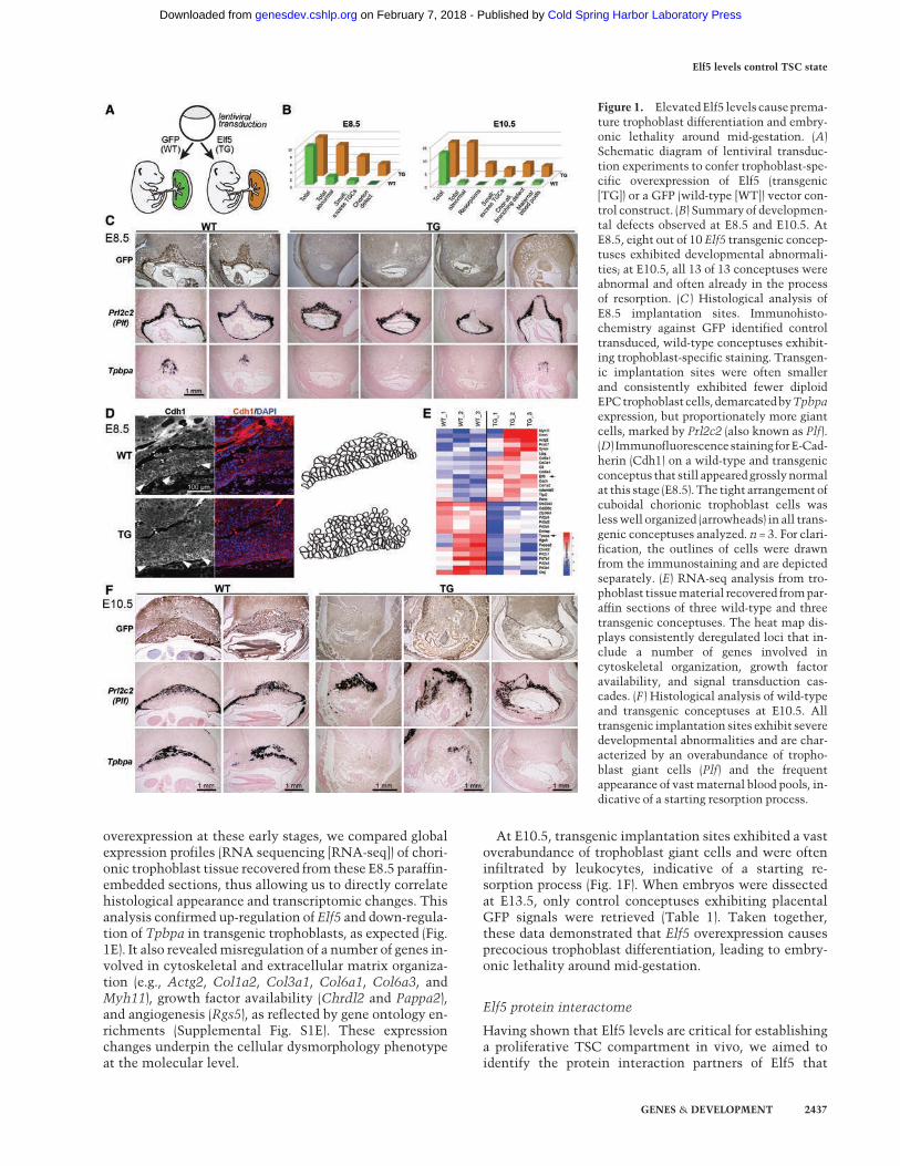
overexpression at these early stages, we compared globalexpression profiles (RNA sequencing [RNA-seq]) of chori-onic trophoblast tissue recovered from these E8.5 paraffin-embedded sections, thus allowing us to directly correlatehistological appearance and transcriptomic changes. Thisanalysis confirmed up-regulation of Elf5 and down-regula-tion of Tpbpa in transgenic trophoblasts, as expected (Fig.1E). It also revealedmisregulation of a number of genes in-volved in cytoskeletal and extracellular matrix organiza-tion (e.g., Actg2, Col1a2, Col3a1, Col6a1, Col6a3, andMyh11), growth factor availability (Chrdl2 and Pappa2),and angiogenesis (Rgs5), as reflected by gene ontology en-richments (Supplemental Fig. S1E). These expressionchanges underpin the cellular dysmorphology phenotypeat the molecular level.
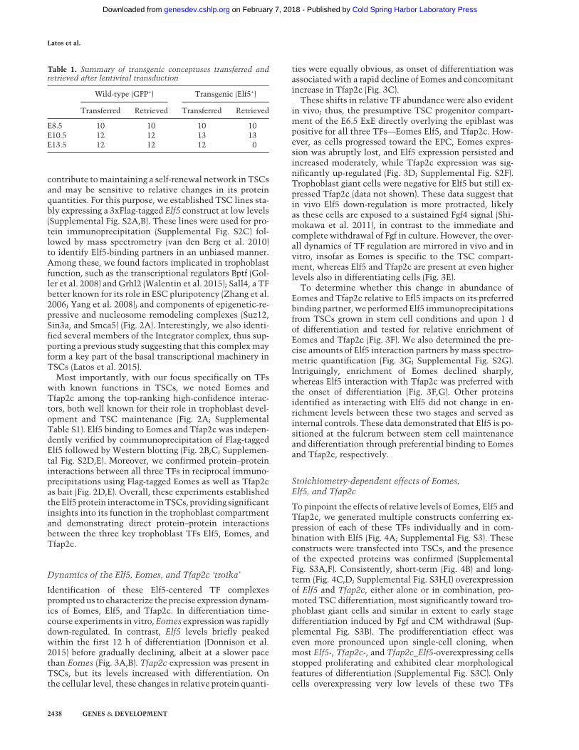
At E10.5, transgenic implantation sites exhibited a vastoverabundance of trophoblast giant cells and were ofteninfiltrated by leukocytes, indicative of a starting re-sorption process (Fig. 1F). When embryos were dissectedat E13.5, only control conceptuses exhibiting placentalGFP signals were retrieved (Table 1). Taken together,these data demonstrated that Elf5 overexpression causesprecocious trophoblast differentiation, leading to embry-onic lethality around mid-gestation.
Elf5 protein interactome
Having shown that Elf5 levels are critical for establishinga proliferative TSC compartment in vivo, we aimed toidentify the protein interaction partners of Elf5 that
Figure 1. ElevatedElf5 levels causeprema-ture trophoblast differentiation and embry-onic lethality around mid-gestation. (A)Schematic diagram of lentiviral transduc-tion experiments to confer trophoblast-spe-cific overexpression of Elf5 (transgenic[TG]) or a GFP (wild-type [WT]) vector con-trol construct. (B) Summary of developmen-tal defects observed at E8.5 and E10.5. AtE8.5, eight out of 10 Elf5 transgenic concep-tuses exhibited developmental abnormali-ties; at E10.5, all 13 of 13 conceptuses wereabnormal and often already in the processof resorption. (C ) Histological analysis ofE8.5 implantation sites. Immunohisto-chemistry against GFP identified controltransduced, wild-type conceptuses exhibit-ing trophoblast-specific staining. Transgen-ic implantation sites were often smallerand consistently exhibited fewer diploidEPCtrophoblastcells, demarcatedbyTpbpaexpression, but proportionately more giantcells, marked by Prl2c2 (also known as Plf).(D) Immunofluorescencestaining forE-Cad-herin (Cdh1) on a wild-type and transgenicconceptus that still appeared grosslynormalat this stage (E8.5). The tight arrangement ofcuboidal chorionic trophoblast cells waslesswell organized (arrowheads) in all trans-genic conceptuses analyzed. n = 3. For clari-fication, the outlines of cells were drawnfrom the immunostaining and are depictedseparately. (E) RNA-seq analysis from tro-phoblast tissuematerial recovered frompar-affin sections of three wild-type and threetransgenic conceptuses. The heat map dis-plays consistently deregulated loci that in-clude a number of genes involved incytoskeletal organization, growth factoravailability, and signal transduction cas-cades. (F ) Histological analysis of wild-typeand transgenic conceptuses at E10.5. Alltransgenic implantation sites exhibit severedevelopmental abnormalities and are char-acterized by an overabundance of tropho-blast giant cells (Plf) and the frequentappearance of vastmaternal blood pools, in-dicative of a starting resorption process.
Elf5 levels control TSC state
GENES & DEVELOPMENT 2437
Cold Spring Harbor Laboratory Press on February 7, 2018 - Published by genesdev.cshlp.orgDownloaded from
contribute tomaintaining a self-renewal network in TSCsand may be sensitive to relative changes in its proteinquantities. For this purpose, we established TSC lines sta-bly expressing a 3xFlag-tagged Elf5 construct at low levels(Supplemental Fig. S2A,B). These lines were used for pro-tein immunoprecipitation (Supplemental Fig. S2C) fol-lowed by mass spectrometry (van den Berg et al. 2010)to identify Elf5-binding partners in an unbiased manner.Among these, we found factors implicated in trophoblastfunction, such as the transcriptional regulators Bptf (Gol-ler et al. 2008) and Grhl2 (Walentin et al. 2015); Sall4, a TFbetter known for its role in ESC pluripotency (Zhang et al.2006; Yang et al. 2008); and components of epigenetic-re-pressive and nucleosome remodeling complexes (Suz12,Sin3a, and Smca5) (Fig. 2A). Interestingly, we also identi-fied several members of the Integrator complex, thus sup-porting a previous study suggesting that this complexmayform a key part of the basal transcriptional machinery inTSCs (Latos et al. 2015).
Most importantly, with our focus specifically on TFswith known functions in TSCs, we noted Eomes andTfap2c among the top-ranking high-confidence interac-tors, both well known for their role in trophoblast devel-opment and TSC maintenance (Fig. 2A; SupplementalTable S1). Elf5 binding to Eomes and Tfap2c was indepen-dently verified by coimmunoprecipitation of Flag-taggedElf5 followed by Western blotting (Fig. 2B,C; Supplemen-tal Fig. S2D,E). Moreover, we confirmed protein–proteininteractions between all three TFs in reciprocal immuno-precipitations using Flag-tagged Eomes as well as Tfap2cas bait (Fig. 2D,E). Overall, these experiments establishedtheElf5 protein interactome inTSCs, providing significantinsights into its function in the trophoblast compartmentand demonstrating direct protein–protein interactionsbetween the three key trophoblast TFs Elf5, Eomes, andTfap2c.
Dynamics of the Elf5, Eomes, and Tfap2c ‘troika’
Identification of these Elf5-centered TF complexespromptedus to characterize theprecise expressiondynam-ics of Eomes, Elf5, and Tfap2c. In differentiation time-course experiments in vitro,Eomes expressionwas rapidlydown-regulated. In contrast, Elf5 levels briefly peakedwithin the first 12 h of differentiation (Donnison et al.2015) before gradually declining, albeit at a slower pacethan Eomes (Fig. 3A,B). Tfap2c expression was present inTSCs, but its levels increased with differentiation. Onthe cellular level, these changes in relative protein quanti-
ties were equally obvious, as onset of differentiation wasassociated with a rapid decline of Eomes and concomitantincrease in Tfap2c (Fig. 3C).
These shifts in relative TF abundance were also evidentin vivo; thus, the presumptive TSC progenitor compart-ment of the E6.5 ExE directly overlying the epiblast waspositive for all three TFs—Eomes Elf5, and Tfap2c. How-ever, as cells progressed toward the EPC, Eomes expres-sion was abruptly lost, and Elf5 expression persisted andincreased moderately, while Tfap2c expression was sig-nificantly up-regulated (Fig. 3D; Supplemental Fig. S2F).Trophoblast giant cells were negative for Elf5 but still ex-pressed Tfap2c (data not shown). These data suggest thatin vivo Elf5 down-regulation is more protracted, likelyas these cells are exposed to a sustained Fgf4 signal (Shi-mokawa et al. 2011), in contrast to the immediate andcomplete withdrawal of Fgf in culture. However, the over-all dynamics of TF regulation are mirrored in vivo and invitro, insofar as Eomes is specific to the TSC compart-ment, whereas Elf5 and Tfap2c are present at even higherlevels also in differentiating cells (Fig. 3E).
To determine whether this change in abundance ofEomes and Tfap2c relative to Efl5 impacts on its preferredbinding partner, we performed Elf5 immunoprecipitationsfrom TSCs grown in stem cell conditions and upon 1 dof differentiation and tested for relative enrichment ofEomes and Tfap2c (Fig. 3F). We also determined the pre-cise amounts of Elf5 interaction partners bymass spectro-metric quantification (Fig. 3G; Supplemental Fig. S2G).Intriguingly, enrichment of Eomes declined sharply,whereas Elf5 interaction with Tfap2c was preferred withthe onset of differentiation (Fig. 3F,G). Other proteinsidentified as interacting with Elf5 did not change in en-richment levels between these two stages and served asinternal controls. These data demonstrated that Elf5 is po-sitioned at the fulcrum between stem cell maintenanceand differentiation through preferential binding to Eomesand Tfap2c, respectively.
Stoichiometry-dependent effects of Eomes,Elf5, and Tfap2c
To pinpoint the effects of relative levels of Eomes, Elf5 andTfap2c, we generated multiple constructs conferring ex-pression of each of these TFs individually and in com-bination with Elf5 (Fig. 4A; Supplemental Fig. S3). Theseconstructs were transfected into TSCs, and the presenceof the expected proteins was confirmed (SupplementalFig. S3A,F). Consistently, short-term (Fig. 4B) and long-term (Fig. 4C,D; Supplemental Fig. S3H,I) overexpressionof Elf5 and Tfap2c, either alone or in combination, pro-moted TSC differentiation, most significantly toward tro-phoblast giant cells and similar in extent to early stagedifferentiation induced by Fgf and CM withdrawal (Sup-plemental Fig. S3B). The prodifferentiation effect waseven more pronounced upon single-cell cloning, whenmost Elf5-, Tfap2c-, and Tfap2c_Elf5-overexpressing cellsstopped proliferating and exhibited clear morphologicalfeatures of differentiation (Supplemental Fig. S3C). Onlycells overexpressing very low levels of these two TFs
Table 1. Summary of transgenic conceptuses transferred andretrieved after lentiviral transduction
Wild-type (GFP+) Transgenic (Elf5+)
Transferred Retrieved Transferred Retrieved
E8.5 10 10 10 10E10.5 12 12 13 13E13.5 12 12 12 0
Latos et al.
2438 GENES & DEVELOPMENT
Cold Spring Harbor Laboratory Press on February 7, 2018 - Published by genesdev.cshlp.orgDownloaded from

were capable of forming colonies and could be propagatedlong-term (Fig. 4E; Supplemental Fig. S3D,G). These datawere entirely in line with the Elf5 overexpression pheno-type that we observed in vivo (Fig. 1).
In contrast, Eomes-overexpressing cells exhibited anundifferentiated, epithelial morphology and a TSC-like gene expression pattern (Fig. 4C,D; SupplementalFig. S3H,I). Significantly, the Elf5- and Tfap2c-induced
Figure 2. Identification of the Elf5 protein interactome inTSCs. (A) Summary of high-confidence Elf5 interactors as identified in three in-dependent immunoprecipitation (IP) andmass spectrometry experiments.Note the interaction of Elf5with the two other TSCTFs: EomesandTfap2c. The full list of interactors is provided in Supplemental Table S1. (B) Independent validation of the interaction between Elf5 andEomesbyanti-Flag immunoprecipitationof the3xFlag-tagged (3xF)Elf5protein followedbyWesternblotting. (C ) Independentvalidationofthe interactionbetweenElf5andTfap2cbyimmunoprecipitationfollowedbyWesternblotting.TheasteriskhighlightstheElf5-3xFlagbandin the input sample. Additional validation of the Elf5–Tfap2c interaction is provided in Figure 3F and Supplemental Figure S2, D and E. (D)Eomes interacts with Tfap2c and Elf5. Eomes-3xFlag immunoprecipitates analyzed by Western blot using anti-Elf5 and anti-Tfap2c anti-bodies.TheasteriskdemarcatesthepresenceofEomesinthe inputsamplesthatcanbeappreciateduponlongerexposure, showninthepanelbelow. (E) Tfap2c interacts with Eomes. Tfap2c-3xFlag immunoprecipitates analyzed byWestern blot using anti-Eomes antibody.
Elf5 levels control TSC state
GENES & DEVELOPMENT 2439
Cold Spring Harbor Laboratory Press on February 7, 2018 - Published by genesdev.cshlp.orgDownloaded from

Figure 3. Dynamics of Elf5, Eomes, and Tfap2c expression. (A) Expression dynamics of Eomes, Elf5, and Tfap2c by RT-qPCR in short-term (top row) and longer-term (bottom row) differentiation time-course experiments of TSCs. (B) Western blots confirming the dynamicregulation of Eomes, Elf5, andTfap2c upon trophoblast differentiation. (C ) Immunofluorescence stainings of TSCs grown in stem cell con-ditions (TSC) and after 1 d of differentiation (1 d diff.) for Eomes, Elf5, and Tfap2c. (D) Immunofluorescence stainings of E6.5 conceptusesfor Eomes, Elf5, and Tfap2c. All three TFs are expressed in the ExE region that harbors trophoblast cells with stem cell potential (bracket).Elf5 and Tfap2c expression is retained in trophoblast cells outside this compartment that start to differentiate. (E) Schematic representa-tion of the comparative expression patterns and relative protein levels (darker shading indicates higher expression levels) of Eomes, Elf5and Tfap2c in the trophoblast compartment of early post-implantation conceptuses. (F ) Immunoprecipitation (IP) of 3x-Flag-tagged (3xF)Elf5 and empty vector (vec) control from TSCs and 1-d (24-h)-differentiated TSCs followed by Western blotting for the factors indicated.Note the higher enrichment of Eomes in Elf5 immunoprecipitations from TSCs versus differentiated cells. Conversely, Tfap2c is moreabundant in Elf5 immunoprecipitations from differentiated trophoblasts. The Western blot for Elf5 confirms the presence of the taggedElf5-3xFlag protein in the corresponding cell lines that is of higher molecular weight than the endogenous Elf5 present in the input sam-ples. (G) Mass spectrometric quantification of Eomes and Tfap2c peptide enrichment in Elf5-3xFlag immunoprecipitations. Values werenormalized against Hspa8 that did not change enrichment with differentiation. Results show the significant decrease in Eomes and in-crease in Tfap2c binding to Elf5 upon TSC differentiation and are representative of two independent immunoprecipitation-tandemmass spectrometry (MS/MS) experiments.
Latos et al.
2440 GENES & DEVELOPMENT
Cold Spring Harbor Laboratory Press on February 7, 2018 - Published by genesdev.cshlp.orgDownloaded from
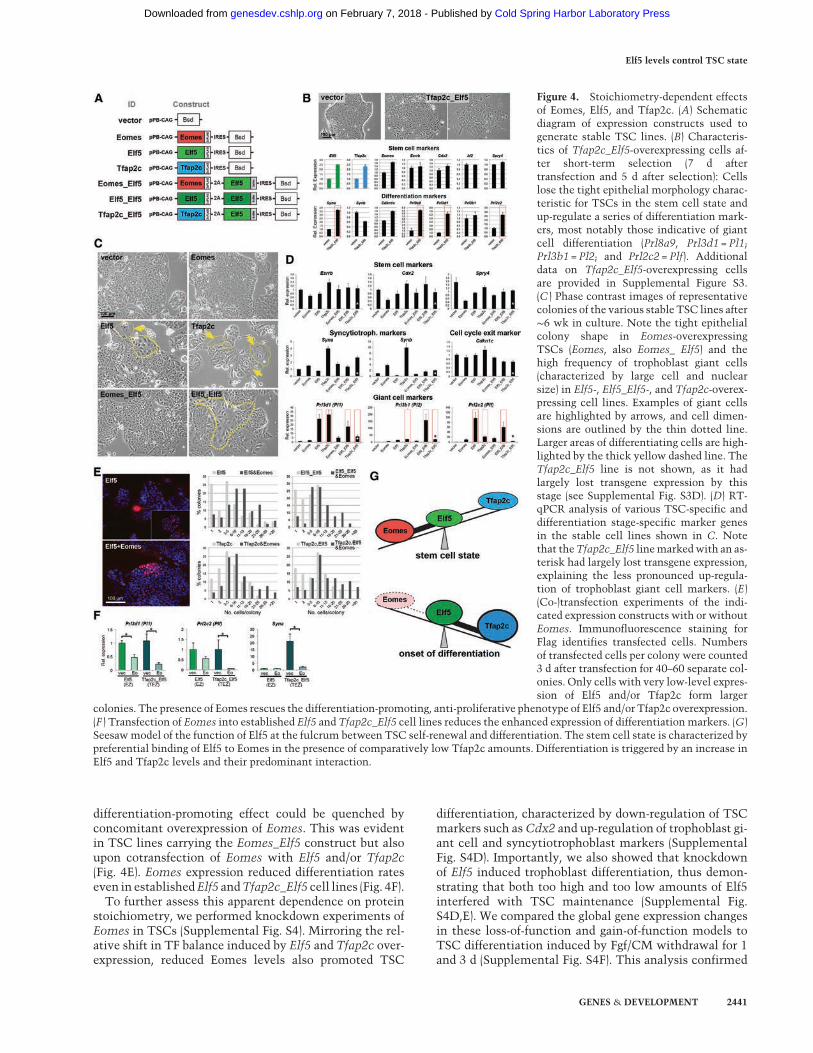
differentiation-promoting effect could be quenched byconcomitant overexpression of Eomes. This was evidentin TSC lines carrying the Eomes_Elf5 construct but alsoupon cotransfection of Eomes with Elf5 and/or Tfap2c(Fig. 4E). Eomes expression reduced differentiation rateseven in establishedElf5 andTfap2c_Elf5 cell lines (Fig. 4F).To further assess this apparent dependence on protein
stoichiometry, we performed knockdown experiments ofEomes in TSCs (Supplemental Fig. S4). Mirroring the rel-ative shift in TF balance induced by Elf5 and Tfap2c over-expression, reduced Eomes levels also promoted TSC
differentiation, characterized by down-regulation of TSCmarkers such asCdx2 and up-regulation of trophoblast gi-ant cell and syncytiotrophoblast markers (SupplementalFig. S4D). Importantly, we also showed that knockdownof Elf5 induced trophoblast differentiation, thus demon-strating that both too high and too low amounts of Elf5interfered with TSC maintenance (Supplemental Fig.S4D,E). We compared the global gene expression changesin these loss-of-function and gain-of-function models toTSC differentiation induced by Fgf/CM withdrawal for 1and 3 d (Supplemental Fig. S4F). This analysis confirmed
Figure 4. Stoichiometry-dependent effectsof Eomes, Elf5, and Tfap2c. (A) Schematicdiagram of expression constructs used togenerate stable TSC lines. (B) Characteris-tics of Tfap2c_Elf5-overexpressing cells af-ter short-term selection (7 d aftertransfection and 5 d after selection): Cellslose the tight epithelial morphology charac-teristic for TSCs in the stem cell state andup-regulate a series of differentiation mark-ers, most notably those indicative of giantcell differentiation (Prl8a9, Prl3d1 = Pl1;Prl3b1 = Pl2; and Prl2c2 = Plf). Additionaldata on Tfap2c_Elf5-overexpressing cellsare provided in Supplemental Figure S3.(C ) Phase contrast images of representativecolonies of the various stable TSC lines after∼6 wk in culture. Note the tight epithelialcolony shape in Eomes-overexpressingTSCs (Eomes, also Eomes_ Elf5) and thehigh frequency of trophoblast giant cells(characterized by large cell and nuclearsize) in Elf5-, Elf5_Elf5-, and Tfap2c-overex-pressing cell lines. Examples of giant cellsare highlighted by arrows, and cell dimen-sions are outlined by the thin dotted line.Larger areas of differentiating cells are high-lighted by the thick yellow dashed line. TheTfap2c_Elf5 line is not shown, as it hadlargely lost transgene expression by thisstage (see Supplemental Fig. S3D). (D) RT-qPCR analysis of various TSC-specific anddifferentiation stage-specific marker genesin the stable cell lines shown in C. Notethat theTfap2c_Elf5 linemarkedwith an as-terisk had largely lost transgene expression,explaining the less pronounced up-regula-tion of trophoblast giant cell markers. (E)(Co-)transfection experiments of the indi-cated expression constructs with or withoutEomes. Immunofluorescence staining forFlag identifies transfected cells. Numbersof transfected cells per colony were counted3 d after transfection for 40–60 separate col-onies. Only cells with very low-level expres-sion of Elf5 and/or Tfap2c form larger
colonies. The presence of Eomes rescues the differentiation-promoting, anti-proliferative phenotype of Elf5 and/or Tfap2c overexpression.(F ) Transfection of Eomes into established Elf5 and Tfap2c_Elf5 cell lines reduces the enhanced expression of differentiationmarkers. (G)Seesawmodel of the function of Elf5 at the fulcrum between TSC self-renewal and differentiation. The stem cell state is characterized bypreferential binding of Elf5 to Eomes in the presence of comparatively low Tfap2c amounts. Differentiation is triggered by an increase inElf5 and Tfap2c levels and their predominant interaction.
Elf5 levels control TSC state
GENES & DEVELOPMENT 2441
Cold Spring Harbor Laboratory Press on February 7, 2018 - Published by genesdev.cshlp.orgDownloaded from

that (1) Eomes-overexpressing cells are most similar toTSCs, (2) down-regulation of Eomes and Elf5 rapidly trig-gers significant differentiation, and (3) overexpression ofElf5 globally induces a state of early-onset differentiation,which corroborates the critical role of Elf5 levels preciselyat the tipping point between TSC self-renewal and exitfrom the stem cell state.
Collectively, this careful dissection of TF levels re-vealed that a balanced expression of Eomes and Elf5 pro-motes the stem cell state, while proportionally higherElf5 and Tfap2c levels trigger TSC differentiation (Fig.4G).
Dissecting the transcriptional networks establishedby Eomes, Elf5, and Tfap2c
To identify genomic loci occupied by the various bin-ary and ternary combinations between Eomes, Elf5, andTfap2c, we performed ChIP-seq for Tfap2c and integratedthese data with previously published Eomes and Elf5ChIP-seq profiles in TSCs (Chuong et al. 2013). In total,we identified 1254 loci cobound by all three TFs, and4254, 2035, and 766 sites cobound only by Elf5–Tfap2c,Elf5–Eomes, and Eomes–Tfap2c, respectively (Fig. 5A).Thesewere distributed across the genome in a largely sim-ilar pattern (Supplemental Fig. S5A). Importantly, thenumbers of observed combinatorial binding sites werehighly statistically significant over co-occupancy expect-ed at random, in particular for the triple, the Elf5–Tfap2c,and the Elf5–Eomes combinations (Fig. 5B). Interestingly,co-occupancy of Tfap2c with Sox2, another TF known tointeractwithTfap2c inTSCs (Adachi et al. 2013), was con-fined to a set of sites largely distinct from those bound byTfap2c and Elf5 (Fig. 5C). OverlapwithCdx2wasmarginal(Supplemental Fig. S5B). These chromatin-binding pat-terns strongly support our functional and protein–proteininteraction data of a concerted action of Eomes, Elf5, andTfap2c in TSCs.
Eomes–Elf5–Tfap2c triple occupancydemarcates active TSC genes
Analysis of genes associated with the various co-occupiedregions revealed that triply bound elements were highlyenriched for TSC genes, includingEomes,Elf5, andTfap2cthemselves as well as others such as Bmp4, Cdh1, Fgfr2,Gata3,Sox2,Spry4, andZic3 (Fig. 5D; SupplementalTableS2). This observation was corroborated by functional an-notation (Genomic Regions Enrichment of AnnotationsTool [GREAT]) analyses (McLean et al. 2010) in which tri-ply boundgeneswere tightly linkedwithblastocyst forma-tion and very early stages of trophoblast development (Fig.5E; Supplemental Fig. S5C). In contrast, genes associatedwith Elf5–Tfap2c doubly bound loci such as Ccne1,Fzd5,Gcm1, Peg10, Prl2c2 (Plf), and Tfebwere overall en-riched for processes related to trophoblast differentiationand placental development.
To further validate the correlation between triple-occu-pancy sites and expression of TSC genes, we performedtranscriptomics (RNA-seq) on TSCs in the stem cell state
and after 1 d of differentiation and integrated these datawith the ChIP-seq peakmaps. This analysis demonstratedthat triply bound loci are associatedwith genesmore high-ly expressed in stem cell conditions, underpinning thefunctional relevance of triple TF occupancy for transcrip-tional activation of stem cell genes (Fig. 5F).
Dynamic shift toward TF-binding enrichmentat Elf5–Tfap2c elements promotesdifferentiation
Our next aimwas to gain a refined view of the dynamics ofTF occupancy in correlationwith gene expression in TSCsand upon differentiation. Because Tfap2c levels are notlimiting in either condition (in contrast to Eomes andElf5), we used this TF as a readout of possible shifts in ge-nome-wide binding patterns. The ChIP-seq data revealedthat Tfap2c does not globally change genome occupancyafter 1 d of differentiation; however, its relative enrich-ment levels shift betweenbinding sites.Thus, triplyboundgenes (which are enriched for TSC expression) showed ahigher Tfap2c-binding intensity in stem cell conditions.In contrast, with onset of differentiation, Tfap2c enrich-ment was increased at a larger number of Elf5–Tfap2cdoubly bound and Tfap2c singly bound differentiation-associated genes (Fig. 6A,B).
The significance of this observation was further under-pinned on multiple levels: The peak intensity of Tfap2cat Elf5–Tfap2c-cobound elements strongly correlatedwith expression levels andhistoneH3Lys4 trimethylation(H3K4me3) (Rugg-Gunnet al. 2010) thatdemarcates activegenes (Supplemental Fig. S5D).Thus, peakswithhigher in-tensity in TSC conditions were associated with genesmore highly expressed in TSCs. Conversely, peaks thatbecame enriched upon differentiation correlated withgenes up-regulated in 1-d-differentiated TSCs (Fig. 6C);these peaks were also preferentially located closer to thetranscriptional start site (Supplemental Fig. S5E). We con-firmed up-regulation of a number of genes associatedwith differentiation-linked Elf5–Tfap2c peaks upon 1 d ofFgf/CM withdrawal (Supplemental Fig. S6A). During sub-sequent stages of differentiation, the correlation betweenElf5–Tfap2c sites and expression trailed off, as expectedfroman early function of Elf5 in the differentiation process(Supplemental Fig. S6B–D). This was corroborated by de-tailed cluster analysis of global gene expression changes,which revealed that genes whose expression changedonly in later differentiation (3 d vs. 1 d)were not associatedwith Elf5–Tfap2c-bound elements (Supplemental Fig.S6E). This finding strongly supported the notion that tip-ping the Elf5:Eomes ratio in favor of Elf5 (and Tfap2c) pro-vides the initial trigger of differentiation. Notably, genesassociated with Elf5–Tfap2c elements were also up-regu-lated in the Elf5- and Elf5_Elf5-overexpressing cells (Sup-plemental Fig. S6F).
Finally, to overcome the limitations of declining Elf5levels with TSC differentiation, we established TSCswith doxycycline (dox)-inducible Elf5 expression and con-firmed dose-dependent recruitment of Elf5 to Elf5–Tfap2celements (Fig. 6D). Together, these data unraveled in
Latos et al.
2442 GENES & DEVELOPMENT
Cold Spring Harbor Laboratory Press on February 7, 2018 - Published by genesdev.cshlp.orgDownloaded from

intricate detail the stoichiometry-driven changes of thetranscriptional networks regulating the onset of tropho-blast differentiation.
Differential TF-binding motif distribution underlies theshift toward a differentiation-promoting program
To reveal the mechanism underlying these shifts in rela-tive genome occupancy, we searched for TFmotifs withinthe triple, double, and single ChIP-seq peaks. Globally,Tfap2c ChIP-seq elements were very tightly correlatedwith the Tfap2c consensus binding sequence, with ∼76%of peaks overlapping the cognate Tfap2c motif. In starkcontrast, the Tfap2c motif was significantly underrepre-
sented at the 1254 triply bound sites, as only 405 (32%) ofthemcontainedone (Fig. 6E). In comparison, the frequencyofElf5motif recognitionwassimilarorevenenrichedattri-plyboundsitesoverallElf5peaks.TheEomesmotif didnotlend itself to this same type of analysis, as it is too poorlydefined. These findings implied that Tfap2c binding to tri-ple elements is mediated through its physical associationwith Elf5 and Eomes. In contrast, Tfap2c peaks that in-creased with differentiation harbored the cognate Tfap2cmotif.Overall, these findings provide a molecular mechanism
for how the same TFs can orchestrate both self-renewaland differentiation-promoting transcriptional programsthrough a stoichiometry-sensitive shift in function of a
Figure 5. Elf5, Eomes, and Tfap2c triply bound regions are associated with active TSC genes. (A) Venn diagram of the combinatorialChIP-seq binding peaks between Elf5, Eomes, and Tfap2c in TSCs. (B) Bioinformatic analysis assessing the statistical significance ofthe occurrence of cobound elements over randomly expected. (C ) Venn and chord diagrams depicting the genome-wide binding overlapof Tfap2c, Sox2, Eomes, and Elf5. Note that the Elf5–Tfap2c network is largely distinct from that established by the interaction betweenTfap2c and Sox2. (D) Genome browser views of the ChIP-seq peak distribution at the Eomes, Elf5, and Tfap2c loci. Triply bound elementsare highlighted in gray. (E) Functional annotation chart of the genes associated with single, double, and triple peaks. Only significant hitsare shown; the TF combinations not shown did not exhibit significantly enriched functional annotations. The thickness of the connectingbars is indicative of the proportion of genes near bound elements that are annotated to the enriched term. Genes associated with triplybound regions are most strongly associated with early trophoblast development at the blastocyst stage, whereas Elf5–Tfap2c-associatedgenes are more strongly associated with later stages of trophoblast differentiation. (F ) Integration of RNA-seq data from TSCs grown instem cell conditions and upon 1 d of differentiation with ChIP-seq peaks. The graph shows that genes more highly expressed in stemcell conditions are enriched for Elf5–Eomes–Tfap2c triply bound regions (red line) compared with all ChIP-seq loci.
Elf5 levels control TSC state
GENES & DEVELOPMENT 2443
Cold Spring Harbor Laboratory Press on February 7, 2018 - Published by genesdev.cshlp.orgDownloaded from

Figure 6. Elevated Elf5 andTfap2c levels trigger TSC differentiation. (A) Differential ChIP-seq binding enrichment of Tfap2c in TSCs andupon 1 d of differentiation. The 1254 triply bound elements predominantly have a higher Tfap2c abundance in TSCs, indicated by the blueshading of elements. By comparison, a significantly larger proportion of the 4254 Elf5–Tfap2c double and 28,473 (intergenic) Tfap2c singlepeaks exhibit a higher Tfap2c abundance upon differentiation, indicated by the red-shaded elements (highlighted by the dashed lines).Please note that, since this analysis is based on the differential enrichment of Tfap2c between TSCs and 1-d-differentiated cells, onlypeak combinations with Tfap2c can be analyzed. (B) Example browser views of the differential Tfap2c peak enrichment in TSCs grownin stem cell conditions and upon 1 d of differentiation. Tfap2c binding is higher in TSC conditions at triply bound sites at genes morehighly expressed in TSCs such as Spred1; conversely, Tfap2c binding is enriched at Tfap2c singly and Elf5–Tfap2c doubly occupied sitesin differentiation conditions surrounding genes up-regulated upon differentiation, such as Id3. (C ) Correlation of differential Tfap2c ChIP-seq peak binding intensity at Elf5–Tfap2c elements with expression levels of the associated genes. Higher enrichment of Tfap2c correlateswith elevated gene expression both for TSC genes in stem cell conditions (left of vertical dotted line) and at genes up-regulated with dif-ferentiation in differentiation-promoting conditions (right of vertical dotted line). (D) ChIP of Elf5 followed by qPCR for Elf5–Tfap2c targetloci in TSCs that harbor a doxycycline (dox)-inducible Elf5 expression construct. Elf5 recruitment to these sites is increased upon Elf5overexpression (Elf5 TSC + dox). (∗) P < 0.05. (E) Differential distribution of Tfap2c motifs: Globally, Tfap2c ChIP-seq peaks overlap ex-tremely closely with the Tfap2c consensus binding sequence, with 76% of peaks positioned over the cognate motif. In contrast, triplybound sites are significantly depleted for the cognate motif, with only 32% of peaks overlapping the consensus sequence. Significancewas calculatedwith a χ2 testwith Yates’ correction; the odds ratio is 6.7. The consensusmotif for Elf5 is less tightly defined, with, globally,41% of peaks overlapping the cognate sequence; this is increased to 52% at triply bound elements. (F ) Model of the stoichiometry-depen-dent functions of Elf5, Eomes, and Tfap2c in fine-tuning the balance between TSC self-renewal and commitment to differentiate.
Latos et al.
2444 GENES & DEVELOPMENT
Cold Spring Harbor Laboratory Press on February 7, 2018 - Published by genesdev.cshlp.orgDownloaded from

small group of interacting TFs centered on Elf5 acting as acell fate switch (Fig. 6F).
Discussion
Insights into the transcriptional networks required tomaintain the self-renewal state of TSCs have largely reliedon the analysis of mouse mutants and the inability toderive TSCs from them. These studies have revealed anumber of TFs that are essential to establish andmaintainTSCs, including Eomes, Elf5, and Tfap2c (Russ et al. 2000;Donnison et al. 2005; Kuckenberg et al. 2010). Here, weshow that these same three TFs physically bind to eachother to form multimeric TF complexes and co-occupytrophoblast genes, including themselves, driving their ex-pression. In this sense, the self-renewal network of TSCsshares important mechanistic features with pluripotencycircuits in ESCs not only in terms ofmutual self-reinforce-ment but also as far as protein–protein interactions andgenomic cobinding are concerned.In addition to the identification of these core tropho-
blast TF interactomes and the networks that they estab-lish, our findings add another important dimension tothe understanding of TSC regulation: Not only are certainTFs essential for TSC self-renewal, but their proportionalabundance is equally critical to maintain the stem cellstate. We show that precise levels of Elf5 determine thebalance between TSC self-renewal and onset of differenti-ation. Loss but also overexpression of Elf5 result in deple-tion of the proliferative stem cell compartment in vitroand in vivo and embryonic lethality aroundmid-gestation(Donnison et al. 2005). These data bear a strong resem-blance to Oct4 as a key node of the pluripotency networkin ESCs, whose fine-tuned expression levels control boththe entry into and the exit from naïve pluripotency (Rad-zisheuskaya et al. 2013). Complete depletion ofOct4 caus-es differentiation toward trophoblast-like cells, but,conversely, high Oct4 levels are also required for differen-tiation into embryonic lineages and the germline, albeitthe mechanism underlying this sensitivity to expressionlevels remains unknown (Niwa et al. 2000; Radzisheus-kaya et al. 2013). Our insights reveal an analogous finelytuned balance for Elf5 in TSCs and place Elf5 at the centerof the transcriptional networks governing self-renewal aswell as differentiation in TSCs.To gain a better molecular understanding of Elf5’s func-
tion in this circuit, we analyzed its interacting partners inTSCs using mass spectrometry. In addition to Eomes andTfap2c, we identified other Elf5 interactors with knownroles in trophoblast and placental development, includingGrhl2 (Walentin et al. 2015) and Bptf (Goller et al. 2008).Our data showed that Elf5 associates with componentsof both activating (e.g., Bptf and Chd7) and repressive(Sin3 and Polycomb) chromatin complexes, indicatingits role in both gene activation and repression. It is note-worthy that the Elf5 interactome did not include othercritical TSC TFs such as Cdx2 or Esrrb, and we did notidentify Elf5 associated with Cdx2 or Esrrb in the recipro-cal interactomes (Latos et al. 2015). Together with the
minimal genomic binding overlap between Cdx2 andElf5, it appears that these TFs function in mostly parallelcircuits to maintain self-renewal of TSCs, except at retro-viral elements where they have a joint function (Chuonget al. 2013).Our detailed dissection of the Eomes–Elf5–Tfap2c net-
work in TSCs shows that the relative levels of these threeTFs determine preferential interaction partners and, con-sequently, binding and activation of separate sets of geneswith opposing functions. The presence of Eomes in atleast equal stoichiometric quantities ensures occupancyat triply bound genomic sites and expression of the associ-ated TSC genes. This joint genome occupancy is facilitat-ed by formation of a ternary TF complex. However, whenElf5 and Tfap2c levels exceed those of Eomes, they be-come enriched at Elf5–Tfap2c double (and single) sites,triggering the exit from self-renewal through activationof a differentiation-promoting program. Mechanistically,this is enabled by the differential distribution of theTfap2c consensus motif; while triply occupied sites aredepleted for this sequence element, and Tfap2c bindingis indirect through association with Elf5 and Eomes, re-cruitment of Elf5:Tfap2c to sites containing the cognateTfap2c motif is favored as Elf5 and Tfap2c protein levelsincrease (Fig. 6F).Overall, our data reveal an intricate balance between
mutually interacting TFs that is critical to determinestem cell self-renewal versus differentiation in the tropho-blast compartment. This sensitivity of TSCs to precise TFstoichiometries is a likely reason for the significant spon-taneous differentiation rates commonly observed in TSCcultures. It may also explain why reprogramming strate-gies based on overexpression or activation of selectedTFs specifically between ESCs and TSCs have proved dif-ficult and tend to result in loss of self-renewal potentialand terminal differentiation (Niwa et al. 2000, 2005; Ral-ston et al. 2010; Cambuli et al. 2014). In the bigger picture,this precarious balance might explain why derivation andmaintenance of TSCs are difficult and have often provedinefficient or even unsuccessful from other species, in par-ticular humans (Kunath et al. 2014). Gaining detailed in-sights into the precise regulation of transcriptionalnetworks governing trophoblast development is thereforeessential to achieve these goals.
Materials and methods
Generation of trophoblast-specific Elf5-overexpressingconceptuses
Generation of lentivirally mediated trophoblast-specific overex-pression was performed largely as described previously (Okadaet al. 2007; Morioka et al. 2009). Briefly, B6D2F1 female micewere superovulated by intraperitoneal injection of 5 U of preg-nant mare’s serum gonadotropin followed by 5 U of human cho-rionic gonadotropin 48 h later and then mated with B6D2F1males. Two-cell stage embryos were collected from females 1.5d after copulation and then incubated for 2 d to obtain blastocysts.Zona-free blastocysts were prepared by treatment with acidicTyrode’s solution. Blastocysts were then incubated in groupsof four in 4 µL of medium containing lentiviral vector
Elf5 levels control TSC state
GENES & DEVELOPMENT 2445
Cold Spring Harbor Laboratory Press on February 7, 2018 - Published by genesdev.cshlp.orgDownloaded from

(pCAG-EGFP or p|CAG-Elf5; 1000 ng of p24 per mL) for5 h. Transduced blastocysts were implanted into the uteri ofE2.5 pseudopregnant females. Litters were dissected at the indi-cated days of development.
Stem cell culture
TSC lines EGFP, Rs26, and “CTRL WT” (a kind gift of the Ros-sant [Toronto, Canada] and Okano [Kobe, Japan] laboratories)were cultured as described previously (Tanaka et al. 1998). Fur-ther manipulations were performed with piggyBac vectors con-taining the complete ORFs of TFs as indicated. TFs were clonedand sequence-verified. All transformations to yield stable celllines were performed in the TS-EGFP cell line, which exhibitsproven placental contribution competence (Tanaka et al. 1998;Cambuli et al. 2014).
Histological analyses
Standard immunohistochemistry was performed on 7-µm sec-tions as detailed in the Supplemental Material. Images were tak-en on an Olympus BX41 or BX61 epifluorescencemicroscope or aZeiss LSM 780 confocal microscope.For RNA in situ hybridizations, linearized plasmids containing
cDNA inserts from Plf and Tpbpa cDNAs were used to generatedigoxigenin (DIG)-labeled riboprobes according to the manufac-turer’s instructions (Roche). Hybridizations were carried outovernight at 52°C using standard procedures. Signals were detect-ed with anti-DIG-alkaline phosphatase-conjugated antibody(Roche), and staining was performed overnight using NBT andBCIP reagents (Promega). Sections were counterstained with nu-clear Fast Red (Sigma).
Coimmunoprecipitation
TS-EGFP cells were transfected with a piggyBac-CAG-Avi-Elf5-3xFlag-Ires-Neo, piggyBac-CAG-Eomes-3xFlag-Ires-Bsd, or pig-gyBac-CAG-Tfap2c-3xFlag-Ires-Bsd construct containing theElf5, Eomes, Tfap2c coding sequences, respectively, or emptyvector alone using Lipofectamine 2000 (Invitrogen). Cells wereselected with G418 or Blasticidin S and expanded to 10 15-cmdishes. Cells were washed in PBS, harvested, and used for immu-noprecipitation exactly as described previously (van den Berget al. 2010). Precise details are given in the Supplemental Mate-rial. Eluates were pooled and analyzed by mass spectrometry orWestern blot.
Mass spectrometry
Immunoprecipitated proteins from three biological replicateseach of Elf5 and vector transfected TSCs were analyzed as before(Webster and Oxley 2009; Latos et al. 2015). Mass spectrometricdata were processed using Proteome Discoverer version 1.4(Thermo Scientific) and searched against the mouse entries inUniProt 2013.09 and a database of common contaminants usingMascot version 2.3 (Matrix Science), and the results were import-ed into Scaffold version 3.6 (Proteome Software, Inc.). With pro-tein/peptide thresholds of 50%/0% and a minimum of twopeptides, a total of 694 proteins was reported across the six sam-ples, with a calculated protein false discovery rate of 0.0%. Afterfurther filtering, (proteins identified in at least two of the threereplicates and having average spectral count ratios relative tothe controls >2), 109 proteins remained, which are shown in Sup-plemental Table S1.
RT-qPCRs
Total RNA was extracted using TRI reagent (Sigma, T9424) ac-cording to the manufacturer’s instructions, and any potentialDNA contamination was removed by treatment the TurboDNA-free kit (Life Technologies, AM1907) according to theman-ufacturer’s instructions. One micrgram to 2 µg of total RNAwasused for cDNA synthesis with RevertAid H-minus M-MuLV re-vert transcriptase (Fermentas, EP0451) and random hexamers(Promega, C118A).qPCR was performed using SYBR Green Jump Start Taq Ready
mix (Sigma, S4438) on a Bio-Rad CFX96 thermocycler.
Western blotting
Whole-cell extracts were prepared with transgenic buffer (20 mMTris-HCl at pH 7.5, 137 mM NaCl, 1mM EGTA, 1% TritonX-100, 10% glycerol, 1.5 mM MgCl2) supplemented with prote-ase inhibitor cocktail (Sigma, P2714). Nuclear extracts were pre-pared as described previously (van den Berg et al. 2010). Westernblotting was carried out according to a standard protocol (see theSupplemental Material). The antibodies used were anti-Flag(Sigma, F1804), anti-Elf5 (Santa Cruz Biotechnology, sc-9645),anti-Eomes (Abcam, ab23345; R&D Systems, MAB 6166), anti-Tfap2c (R&D System, AF5059), anti-tubulin (Abcam ab6160),and ImmunoCruz IP/WB Optima System C (sc-45040), E(sc-45042), or A (sc-45038).
ChIP-seq
Immunoprecipitations were carried out as described (Tavareset al. 2012; Latos et al. 2015) and are detailed in the SupplementalMaterial. DNA from four immunoprecipitations was pooled foreach library generated with the NEB Next DNA library prepara-tion master mix (New England Biolabs, E6040) according to themanufacturer’s instructions. Libraries were amplified using18 PCR cycles, purified using Agencourt AMPure XP SPRI beads(Beckman Coulter, A63881), and size-selected on an agarose gel.DNA was extracted using the QiaQuick gel extraction kit (Qia-gen), and its concentration was determined using the KAPA Illu-mina SYBR Universal Lid Q kit (KAPA Biosystems, KK4824) andBioanalyzer 2100 system (Agilent). Libraries were sequenced onan Illumina HiSeq 2500 sequencer.
Global expression profiling
RNA-seq was performed on chorion and trophoblast tissue mate-rial recovered from paraffin sections according to the FFPEmiRNeasy kit (Qiagen, 217504). rRNA depletion was performedusing the NEB Next kit (New England Biolabs, E6310S). ForRNA-seq from TSCs, total RNA was prepared using TRI reagent(Sigma, T9424) followed by DNase treatment using the TurboDNA-free kit (Life Technologies, AM1907). mRNA was isolatedfrom 150–240 ng of total DNA-free RNA using the DynabeadsmRNA purification kit (Life Technologies, 61006). Indexed,strand-specific librarieswere prepared using the ScriptSeq version2 RNA-seq library preparation kit (Epicentre, SSV21106). Librar-ies were quantified using both the KAPA library quantificationkit (KAPA Biosystems, KK4824) and Bioanalyzer 2100 system(Agilent). Indexed libraries were pooled and sequenced with a100-base-pair single-end protocol on an Illumina HiSeq 2500sequencer. Raw FastQ data were mapped to the Mus musculusNCBIM37 genome assembly using TopHat version 2.0.12 (Kimet al. 2013). Data were analyzed using DESeq and the RNA-seq
Latos et al.
2446 GENES & DEVELOPMENT
Cold Spring Harbor Laboratory Press on February 7, 2018 - Published by genesdev.cshlp.orgDownloaded from

quantitation pipeline in SeqMonk software (http://www.bioinformatics.babraham.ac.uk).
Bioinformatic analysis
Raw reads were aligned to mouse genome build mm9 using Bow-tie, and peak calling was performedwithMACS2with default pa-rameters (Zhang et al. 2008). To identify overlapping elementsfrom different ChIP experiments, BED files of ChIP elementswere compared using the BedTools (Quinlan and Hall 2010) func-tionmultiinter with the cluster option to reduce redundant inter-actions to the highest-order interactions. Overlapping regionsrequired a 50%overlap to be included. Tomodel the all by all pos-sible interactions, we used the shuffle function in the R libraryChIPseeker (Yu et al. 2015) to make randomized BED files ofeach ChIP experiment. We then used the multiinter function ofBedTools to generate a table of expected interactions of the ran-domized data. This was repeated 10,000 times to build averagemodels. We then tested our observed data against the randommodels using a Poisson exact test. P-valueswere adjusted formul-tiple testing (n = 175,123 to account the total number of all ChIPelements involved in the analysis).Annotation of binding sites according to genomic features was
performed by overlapping the sites with Ensembl version 77 an-notations. Motif analysis was performed with MEME-chip(Machanick and Bailey 2011) using the JASPAR_CORE_2014_vertebrates database, searching for zero or one occurrences of themotif per peaks and with a maximum number of motifs discov-ered by MEME of 12. Additional details are provided in the Sup-plemental Material; all high-throughput sequencing data weredeposited in publicly accessible repositories (accession no.PRJNA298763).
Acknowledgments
We are grateful to Kristina Tabbada for Illumina high-throughputsequencing, Judith Webster for assistance with mass spectrome-try experiments, Anne Segonds-Pichon for expert statisticalhelp, and Oliver Tam, Simon Andrews, and Felix Krueger forhelp with the bioinformatic analysis. This work was supportedby a Next-Generation Fellowship (awarded to P.A.L. by the Cen-tre for Trophoblast Research, University of Cambridge), the Bio-technology and Biological Sciences Research Council (BBSRC),and theWellcomeTrust. B.J.C. is supported by aCanadaResearchChair and an operating grant from the Canadian Institutes ofHealth Research.
References
Adachi K,Nikaido I, OhtaH, Ohtsuka S, UraH, KadotaM,Waka-yama T, Ueda HR, Niwa H. 2013. Context-dependent wiringof Sox2 regulatory networks for self-renewal of embryonicand trophoblast stem cells. Mol Cell 52: 380–392.
Auman HJ, Nottoli T, Lakiza O, Winger Q, Donaldson S, Wil-liams T. 2002. Transcription factor AP-2γ is essential in theextra-embryonic lineages for early postimplantation develop-ment. Development 129: 2733–2747.
Avilion AA, Nicolis SK, Pevny LH, Perez L, Vivian N, Lovell-Badge R. 2003. Multipotent cell lineages in early mousedevelopment depend on SOX2 function. Genes Dev 17:126–140.
Cambuli F,MurrayA,DeanW,DudzinskaD, Krueger F, AndrewsS, Senner CE, Cook SJ, Hemberger M. 2014. Epigenetic mem-
ory of the first cell fate decision prevents complete ES cell re-programming into trophoblast. Nat Commun 5: 5538.
Carey BW,Markoulaki S, Hanna JH, FaddahDA, BuganimY, KimJ, Ganz K, Steine EJ, Cassady JP, Creyghton MP, et al. 2011.Reprogramming factor stoichiometry influences the epigenet-ic state and biological properties of induced pluripotent stemcells. Cell Stem Cell 9: 588–598.
Chuong EB, Rumi MA, Soares MJ, Baker JC. 2013. Endogenousretroviruses function as species-specific enhancer elementsin the placenta. Nat Genet 45: 325–329.
Donnison M, Beaton A, Davey HW, Broadhurst R, L’Huillier P,Pfeffer PL. 2005. Loss of the extraembryonic ectoderm inElf5 mutants leads to defects in embryonic patterning.Devel-opment 132: 2299–2308.
Donnison M, Broadhurst R, Pfeffer PL. 2015. Elf5 and Ets2 main-tain the mouse extraembryonic ectoderm in a dosage depen-dent synergistic manner. Dev Biol 397: 77–88.
Georgiades P, CoxB,GertsensteinM,ChawengsaksophakK, Ros-sant J. 2007. Trophoblast-specific gene manipulation usinglentivirus-based vectors. Biotechniques 42: 317–318.
Goller T, Vauti F, Ramasamy S, Arnold HH. 2008. Transcription-al regulator BPTF/FAC1 is essential for trophoblast differenti-ation during early mouse development. Mol Cell Biol 28:6819–6827.
Home P, Ray S, Dutta D, Bronshteyn I, Larson M, Paul S. 2009.GATA3 is selectively expressed in the trophectoderm ofperi-implantation embryo and directly regulates Cdx2 geneexpression. J Biol Chem 284: 28729–28737.
Kidder BL, Palmer S. 2010. Examination of transcriptional net-works reveals an important role for TCFAP2C, SMARCA4,and EOMES in trophoblast stem cell maintenance. GenomeRes 20: 458–472.
Kim D, Pertea G, Trapnell C, Pimentel H, Kelley R, Salzberg SL.2013. TopHat2: accurate alignment of transcriptomes in thepresence of insertions, deletions and gene fusions. GenomeBiol 14: R36.
Kuckenberg P, Buhl S, Woynecki T, van Furden B, Tolkunova E,Seiffe F, Moser M, Tomilin A, Winterhager E, Schorle H.2010. The transcription factor TCFAP2C/AP-2γ cooperateswith CDX2 to maintain trophectoderm formation. Mol CellBiol 30: 3310–3320.
Kunath T, Yamanaka Y, Detmar J, MacPhee D, Caniggia I, Ros-sant J, Jurisicova A. 2014. Developmental differences in theexpression of FGF receptors between human and mouse em-bryos. Placenta 35: 1079–1088.
Latos PA, Goncalves A, Oxley D, Mohammed H, Turro E,Hemberger M. 2015. Fgf and Esrrb integrate epigenetic andtranscriptional networks that regulate self-renewal of tropho-blast stem cells. Nat Commun 6: 7776.
Machanick P, Bailey TL. 2011. MEME-ChIP: motif analysis oflarge DNA datasets. Bioinformatics 27: 1696–1697.
Martello G, Smith A. 2014. The nature of embryonic stem cells.Annu Rev Cell Dev Biol 30: 647–675.
McLean CY, Bristor D, Hiller M, Clarke SL, Schaar BT, Lowe CB,Wenger AM, Bejerano G. 2010. GREAT improves functionalinterpretation of cis-regulatory regions. Nat Biotechnol 28:495–501.
Morioka Y, Isotani A, Oshima RG, OkabeM, IkawaM. 2009. Pla-centa-specific gene activation and inactivation using inte-grase-defective lentiviral vectors with the Cre/LoxP system.Genesis 47: 793–798.
Ng RK, DeanW, Dawson C, Lucifero D,Madeja Z, ReikW, Hem-berger M. 2008. Epigenetic restriction of embryonic cell line-age fate by methylation of Elf5. Nat Cell Biol 10: 1280–1290.
Elf5 levels control TSC state
GENES & DEVELOPMENT 2447
Cold Spring Harbor Laboratory Press on February 7, 2018 - Published by genesdev.cshlp.orgDownloaded from

Nichols J, Smith A. 2012. Pluripotency in the embryo and in cul-ture. Cold Spring Harb Perspect Biol 4: a008128.
Nishioka N, Yamamoto S, Kiyonari H, Sato H, Sawada A, Ota M,NakaoK, Sasaki H. 2008. Tead4 is required for specification oftrophectoderm in pre-implantation mouse embryos. MechDev 125: 270–283.
Niwa H, Miyazaki J, Smith AG. 2000. Quantitative expression ofOct-3/4 defines differentiation, dedifferentiation or self-re-newal of ES cells. Nat Genet 24: 372–376.
Niwa H, Toyooka Y, Shimosato D, Strumpf D, Takahashi K, YagiR, Rossant J. 2005. Interaction between Oct3/4 and Cdx2 de-termines trophectoderm differentiation. Cell 123: 917–929.
Okada Y, Ueshin Y, Isotani A, Saito-Fujita T, Nakashima H,Kimura K, Mizoguchi A, Oh-Hora M, Mori Y, Ogata M,et al. 2007. Complementation of placental defects and embry-onic lethality by trophoblast-specific lentiviral gene transfer.Nat Biotechnol 25: 233–237.
Quinlan AR, Hall IM. 2010. BedTools: a flexible suite of utilitiesfor comparing genomic features. Bioinformatics 26: 841–842.
Radzisheuskaya A, Chia Gle B, dos Santos RL, Theunissen TW,Castro LF, Nichols J, Silva JC. 2013. A defined Oct4 level gov-erns cell state transitions of pluripotency entry and differenti-ation into all embryonic lineages. Nat Cell Biol 15: 579–590.
Ralston A, Rossant J. 2008. Cdx2 acts downstream of cell polari-zation to cell-autonomously promote trophectoderm fate inthe early mouse embryo. Dev Biol 313: 614–629.
Ralston A, Cox BJ, Nishioka N, Sasaki H, Chea E, Rugg-Gunn P,Guo G, Robson P, Draper JS, Rossant J. 2010. Gata3 regulatestrophoblast development downstream of Tead4 and in parallelto Cdx2. Development 137: 395–403.
Rugg-Gunn PJ, Cox BJ, Ralston A, Rossant J. 2010. Distinct his-tone modifications in stem cell lines and tissue lineagesfrom the early mouse embryo. Proc Natl Acad Sci 107:10783–10790.
Russ AP, Wattler S, Colledge WH, Aparicio SA, Carlton MB,Pearce JJ, Barton SC, Surani MA, Ryan K, Nehls MC, et al.2000. Eomesodermin is required for mouse trophoblast devel-opment and mesoderm formation. Nature 404: 95–99.
Shimokawa K, Kimura-Yoshida C, Nagai N,Mukai K, MatsubaraK, Watanabe H, Matsuda Y, Mochida K, Matsuo I. 2011. Cellsurface heparan sulfate chains regulate local reception ofFGF signaling in the mouse embryo. Dev Cell 21: 257–272.
Strumpf D, Mao CA, Yamanaka Y, Ralston A, Chawengsakso-phak K, Beck F, Rossant J. 2005. Cdx2 is required for correctcell fate specification and differentiation of trophectodermin the mouse blastocyst. Development 132: 2093–2102.
Tanaka S, Kunath T, Hadjantonakis AK, Nagy A, Rossant J. 1998.Promotion of trophoblast stem cell proliferation by FGF4. Sci-ence 282: 2072–2075.
Tavares L, Dimitrova E, Oxley D, Webster J, Poot R, Demmers J,Bezstarosti K, Taylor S, Ura H, Koide H, et al. 2012. RYBP–PRC1 complexes mediate H2A ubiquitylation at polycombtarget sites independently of PRC2 and H3K27me3. Cell148: 664–678.
Tremblay GB, Kunath T, Bergeron D, Lapointe L, Champigny C,Bader JA, Rossant J, Giguere V. 2001. Diethylstilbestrol regu-lates trophoblast stem cell differentiation as a ligand of orphannuclear receptor ERRβ. Genes Dev 15: 833–838.
van den Berg DL, Snoek T, Mullin NP, Yates A, Bezstarosti K,Demmers J, Chambers I, Poot RA. 2010. An Oct4-centeredprotein interaction network in embryonic stem cells. CellStem Cell 6: 369–381.
Walentin K, Hinze C,WerthM, HaaseN, Varma S,Morell R, AueA, Potschke E, Warburton D, Qiu A, et al. 2015. A Grhl2-de-pendent gene network controls trophoblast branching mor-phogenesis. Development 142: 1125–1136.
Webster J, Oxley D. 2009. Protein identification by peptide massfingerprinting using MALDI-TOF mass spectrometry. In Theprotein protocols handbook (ed. Walker JM), pp. 1117–1129.Humana Press, New York.
Werling U, Schorle H. 2002. Transcription factor gene AP-2γ es-sential for early murine development. Mol Cell Biol 22:3149–3156.
Yagi R, KohnMJ, Karavanova I, Kaneko KJ, Vullhorst D, DePam-philis ML, Buonanno A. 2007. Transcription factor TEAD4specifies the trophectoderm lineage at the beginning of mam-malian development. Development 134: 3827–3836.
Yamamoto H, Flannery ML, Kupriyanov S, Pearce J, McKercherSR, Henkel GW, Maki RA, Werb Z, Oshima RG. 1998. Defec-tive trophoblast function in mice with a targeted mutation ofEts2. Genes Dev 12: 1315–1326.
Yang J, Chai L, Fowles TC, Alipio Z, Xu D, Fink LM, Ward DC,Ma Y. 2008. Genome-wide analysis reveals Sall4 to be a majorregulator of pluripotency in murine-embryonic stem cells.Proc Natl Acad Sci 105: 19756–19761.
Yu G, Wang LG, He QY. 2015. ChIPseeker: an R/Bioconductorpackage for ChIP peak annotation, comparison and visualiza-tion. Bioinformatics 31: 2382–2383.
Zhang J, Tam WL, Tong GQ, Wu Q, Chan HY, Soh BS, Lou Y,Yang J, Ma Y, Chai L, et al. 2006. Sall4 modulates embryonicstem cell pluripotency and early embryonic development bythe transcriptional regulation of Pou5f1. Nat Cell Biol 8:1114–1123.
Zhang Y, Liu T, Meyer CA, Eeckhoute J, Johnson DS, BernsteinBE, NusbaumC,Myers RM, BrownM, LiW, et al. 2008. Mod-el-based analysis of ChIP-seq (MACS). Genome Biol 9: R137.
Latos et al.
2448 GENES & DEVELOPMENT
Cold Spring Harbor Laboratory Press on February 7, 2018 - Published by genesdev.cshlp.orgDownloaded from
10.1101/gad.268821.115Access the most recent version at doi: originally published online November 19, 201529:2015, Genes Dev.
Paulina A. Latos, Arnold R. Sienerth, Alexander Murray, et al. in target gene networksself-renewal and differentiation through stoichiometry-sensitive shifts Elf5-centered transcription factor hub controls trophoblast stem cell
Material
Supplemental
http://genesdev.cshlp.org/content/suppl/2015/11/18/gad.268821.115.DC1
References
http://genesdev.cshlp.org/content/29/23/2435.full.html#ref-list-1
This article cites 46 articles, 18 of which can be accessed free at:
License
Commons Creative
.http://creativecommons.org/licenses/by-nc/4.0/License (Attribution-NonCommercial 4.0 International), as described at
, is available under a Creative CommonsGenes & DevelopmentThis article, published in
ServiceEmail Alerting
click here.right corner of the article or
Receive free email alerts when new articles cite this article - sign up in the box at the top
© 2015 Latos et al.; Published by Cold Spring Harbor Laboratory Press
Cold Spring Harbor Laboratory Press on February 7, 2018 - Published by genesdev.cshlp.orgDownloaded from
Related Documents