in the nucleus, probably bound to chromatin. These results suggest an adenoma-specific physiological role of proenkephalin products. Key Words: Proenkephalin; met-enkephalin; nuclei; pituitary; adenomas; Cushing’s disease. Abbreviations: ACTH-PA, ACTH-producing adenoma; NFA, nonfunctioning adenoma; GH-PA, GH-produc- ing adenoma; PMHP, postmortem human pituitary; IR, immunoreactivity; SYN, synenkephalin. Introduction Proenkephalin contains four copies of met-enkephalin and one copy of leu-enkephalin, met-enkephalin-Arg-Gly- Leu, and met-enkephalin-Arg-Phe (1–6). Furthermore, proenkephalin presents at the N-terminus one copy of synenkephalin, a nonopioid peptide, which corresponds to the sequence 1–70 (1–6). Leu-enkephalin is also present in prodynorphin molecule (1,3,6), and met-enkephalin appears in the proopiomelanocortin sequence (1,3,6) , whereas synenkephalin is only present in the proenkephalin sequence (6) . Strict correlation was always observed between the presence of synenkephalin-containing peptides and the expression of proenkephalin (6–10). In humans, met-enkephalin was detected in high con- centrations in the adrenal medulla, pituitary, spinal cord, brain, sympathetic ganglia, and peripheral neurons (11). In most mammalian species, the posterior pituitary contained high concentrations of preproenkephalin mRNA and met- enkephalin, but little or no pentapeptide was detected in the anterior pituitary (12–14). Nevertheless, high levels of met- enkephalin with no met-enkephalin-Arg-Gly-Leu or met- enkephalin-Arg-Phe were observed in the human anterior pituitary, and most of this immunoreactivity was localized in a large subpopulation of thyrotrophs (15). Immuno- Received July 7, 1997; Revised February 17, 1998; Accepted February 17, 1998. Author to whom all correspondence and reprint requests should be addressed. Dr. Osvaldo Vindrola, Professor, Instituto de Fisiologia, Laboratorio de Bioquimica, Universidad Autonoma de Puebla, Apartado postal 406, 72001 Puebla, Mexico. E-mail: [email protected] Endocrine, vol. 8, no. 3, 231–240, June 1998 0969–711X/98/8:231–240/$10.50 © 1998 by Humana Press Inc. All rights of any nature whatsoever reserved. 231 Elevated Proenkephalin-Derived Peptide Levels in ACTH-Producing Adenomas Nucleus and Cytoplasm Localization Osvaldo Vindrola, 1 Alberto Chervin, 2 Marcelo Vitale, 2 Alejandra N. Mella, 2 Raquel Aloyz, 3 and Armando Basso 2 1 Instituto de Fisiologia, Laboratorio de Bioquimica, Universidad Autonoma de Puebla. Mexico; 2 Hospital Santa Lucia. Departamento de Neurocirugia y Endocrinologia. Buenos Aires Argentina; and 3 Brain Tumor Research Center. Montreal Neurological Institute. Montreal. Quebec. Canada The biosynthesis of met-enkephalin in human pituitary and human pituitary adenomas is still not well known. In this work, we studied the processing of pro- enkephalin-derived peptides in postmortem human pituitary (PMHP), ACTH-producing adenomas (ACTH- PA), nonfunctioning adenomas (NFA), and GH-pro- ducing adenomas (GH-PA). ACTH-PA contained at least 10 times more proenkephalin-derived peptides than PMHP, NFA, and GH-PA. Proenkephalin process- ing was different in the four tested tissues. In ACTH-PA, proenkephalin was processed to high-, intermediate-, and low-mol-wt products. The highest met-enkephalin- containing peptides levels corresponded to intermediate and low-mol-wt materials, although met-enkephalin- Arg-Phe and synenkephalin immunoreactivity appeared only in high-mol-wt peptides. In PMHP and NFA, met- enkephalin-Arg-Phe immunoreactivity was detected in intermediate- and low-mol-wt materials, and it was absent in GH-PA. Immunoblotting of ACTH-PA showed that met-enkephalin-Arg-Phe immunoreactivity cor- responded to peptides of 44, 32–30, 27, and 17 kDa. The 32–30 and 17-kDa molecules were localized in the nuclear fraction where they were extracted after enzymatic digestion with DNase I. Plasmatic met- enkephalin levels did not increase in patients with Cushing´s disease, suggesting that the pentapeptide stored in ACTH-PA was not released to the general circulation. In conclusion, we demonstrated that only ACTH-PA contained high levels of proenkephalin pep- tides, which were stored in cytoplasm organelles and

Welcome message from author
This document is posted to help you gain knowledge. Please leave a comment to let me know what you think about it! Share it to your friends and learn new things together.
Transcript
231Proenkephalin Peptides in ACTH-Producing Adenomas/Vindrola et al.Vol. 8, No. 3
in the nucleus, probably bound to chromatin. Theseresults suggest an adenoma-specific physiological roleof proenkephalin products.
Key Words: Proenkephalin; met-enkephalin; nuclei;pituitary; adenomas; Cushing’s disease.
Abbreviations: ACTH-PA, ACTH-producing adenoma;NFA, nonfunctioning adenoma; GH-PA, GH-produc-ing adenoma; PMHP, postmortem human pituitary;IR, immunoreactivity; SYN, synenkephalin.
Introduction
Proenkephalin contains four copies of met-enkephalinand one copy of leu-enkephalin, met-enkephalin-Arg-Gly-Leu, and met-enkephalin-Arg-Phe (1–6). Furthermore,proenkephalin presents at the N-terminus one copy ofsynenkephalin, a nonopioid peptide, which correspondsto the sequence 1–70 (1–6). Leu-enkephalin is also presentin prodynorphin molecule (1,3,6), and met-enkephalinappears in the proopiomelanocortin sequence (1,3,6),whereas synenkephalin is only present in the proenkephalinsequence (6). Strict correlation was always observedbetween the presence of synenkephalin-containing peptidesand the expression of proenkephalin (6–10).
In humans, met-enkephalin was detected in high con-centrations in the adrenal medulla, pituitary, spinal cord,brain, sympathetic ganglia, and peripheral neurons (11). Inmost mammalian species, the posterior pituitary containedhigh concentrations of preproenkephalin mRNA and met-enkephalin, but little or no pentapeptide was detected in theanterior pituitary (12–14). Nevertheless, high levels of met-enkephalin with no met-enkephalin-Arg-Gly-Leu or met-enkephalin-Arg-Phe were observed in the human anteriorpituitary, and most of this immunoreactivity was localizedin a large subpopulation of thyrotrophs (15). Immuno-
Received July 7, 1997; Revised February 17, 1998; Accepted February17, 1998.Author to whom all correspondence and reprint requests should be addressed.Dr. Osvaldo Vindrola, Professor, Instituto de Fisiologia, Laboratorio deBioquimica, Universidad Autonoma de Puebla, Apartado postal 406, 72001Puebla, Mexico. E-mail: [email protected]
Endocrine, vol. 8, no. 3, 231–240, June 1998 0969–711X/98/8:231–240/$10.50 © 1998 by Humana Press Inc. All rights of any nature whatsoever reserved.
231
Elevated Proenkephalin-Derived Peptide Levelsin ACTH-Producing AdenomasNucleus and Cytoplasm Localization
Osvaldo Vindrola,1 Alberto Chervin,2 Marcelo Vitale,2
Alejandra N. Mella,2 Raquel Aloyz,3 and Armando Basso2
1Instituto de Fisiologia, Laboratorio de Bioquimica, Universidad Autonoma de Puebla. Mexico;2Hospital Santa Lucia. Departamento de Neurocirugia y Endocrinologia. Buenos Aires Argentina;and 3Brain Tumor Research Center. Montreal Neurological Institute. Montreal. Quebec. Canada
The biosynthesis of met-enkephalin in human pituitaryand human pituitary adenomas is still not well known.In this work, we studied the processing of pro-enkephalin-derived peptides in postmortem humanpituitary (PMHP), ACTH-producing adenomas (ACTH-PA), nonfunctioning adenomas (NFA), and GH-pro-ducing adenomas (GH-PA). ACTH-PA contained atleast 10 times more proenkephalin-derived peptidesthan PMHP, NFA, and GH-PA. Proenkephalin process-ing was different in the four tested tissues. In ACTH-PA,proenkephalin was processed to high-, intermediate-,and low-mol-wt products. The highest met-enkephalin-containing peptides levels corresponded to intermediateand low-mol-wt materials, although met-enkephalin-Arg-Phe and synenkephalin immunoreactivity appearedonly in high-mol-wt peptides. In PMHP and NFA, met-enkephalin-Arg-Phe immunoreactivity was detectedin intermediate- and low-mol-wt materials, and it wasabsent in GH-PA. Immunoblotting of ACTH-PA showedthat met-enkephalin-Arg-Phe immunoreactivity cor-responded to peptides of 44, 32–30, 27, and 17 kDa.The 32–30 and 17-kDa molecules were localized inthe nuclear fraction where they were extracted afterenzymatic digestion with DNase I. Plasmatic met-enkephalin levels did not increase in patients withCushing´s disease, suggesting that the pentapeptidestored in ACTH-PA was not released to the generalcirculation. In conclusion, we demonstrated that onlyACTH-PA contained high levels of proenkephalin pep-tides, which were stored in cytoplasm organelles and
232 EndocrineProenkephalin Peptides in ACTH-Producing Adenomas/Vindrola et al.
cytochemical and biochemical studies have demonstratedthe presence of met-enkephalin in postmortem humanpituitary, nonfunctioning adenomas, and ACTH-producingadenomas (16–18). Furthermore, these adenomas and GH-producing adenomas expressed the prohormone-convertingenzymes PC1 and PC2 mRNAs (19). Although these workshave demonstrated the presence of met-enkephalin and theexpression of prohormone-converting enzymes in pituitaryadenomas, the posttranslational processing of proenkephalinhas not been characterized.
In the present work, we studied the production and post-translational processing of proenkephalin-derived peptidesin postmortem human pituitary (PMHP), ACTH-producingadenomas (ACTH-PA), GH-producing adenomas (GH-PA),and nonfunctioning adenomas (NFA). In addition, thenuclear localization of proenkephalin peptides was analyzedin some ACTH-PA. Plasma levels of met-enkephalin wereassayed to ascertain whether peptide tissue changes wereassociated with their release to the general circulation.
Results
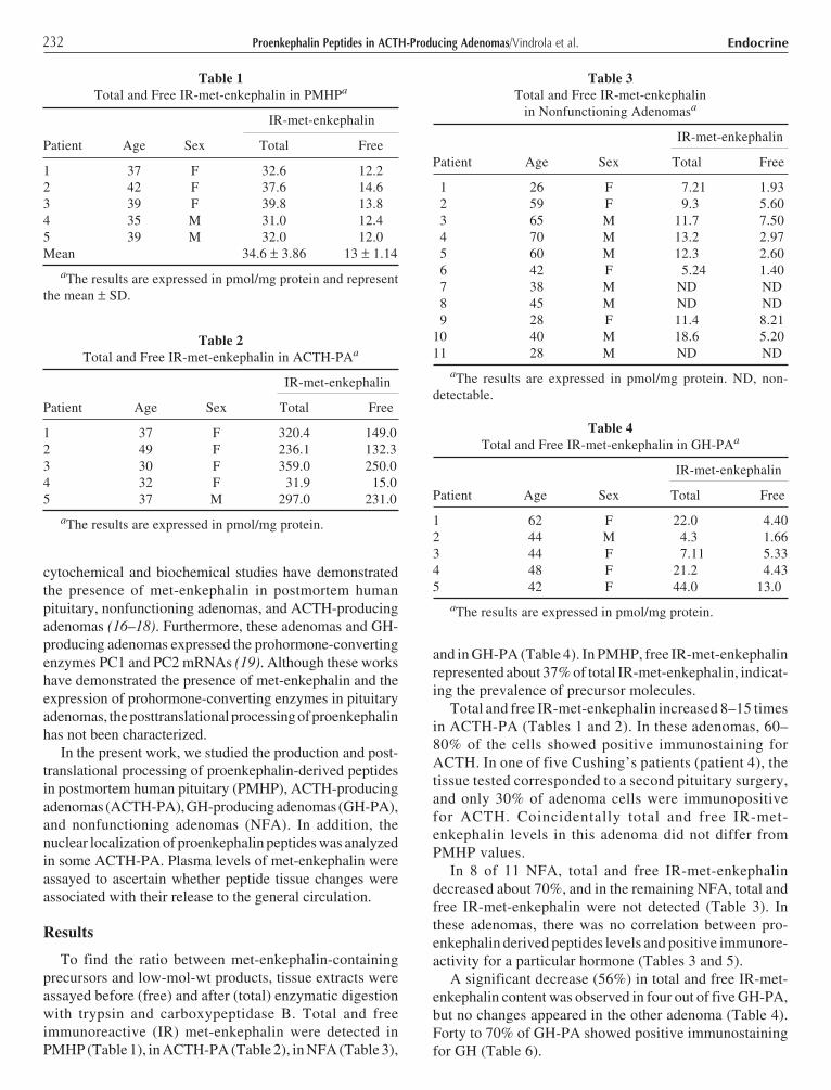
To find the ratio between met-enkephalin-containingprecursors and low-mol-wt products, tissue extracts wereassayed before (free) and after (total) enzymatic digestionwith trypsin and carboxypeptidase B. Total and freeimmunoreactive (IR) met-enkephalin were detected inPMHP (Table 1), in ACTH-PA (Table 2), in NFA (Table 3),
and in GH-PA (Table 4). In PMHP, free IR-met-enkephalinrepresented about 37% of total IR-met-enkephalin, indicat-ing the prevalence of precursor molecules.
Total and free IR-met-enkephalin increased 8–15 timesin ACTH-PA (Tables 1 and 2). In these adenomas, 60–80% of the cells showed positive immunostaining forACTH. In one of five Cushing’s patients (patient 4), thetissue tested corresponded to a second pituitary surgery,and only 30% of adenoma cells were immunopositivefor ACTH. Coincidentally total and free IR-met-enkephalin levels in this adenoma did not differ fromPMHP values.
In 8 of 11 NFA, total and free IR-met-enkephalindecreased about 70%, and in the remaining NFA, total andfree IR-met-enkephalin were not detected (Table 3). Inthese adenomas, there was no correlation between pro-enkephalin derived peptides levels and positive immunore-activity for a particular hormone (Tables 3 and 5).
A significant decrease (56%) in total and free IR-met-enkephalin content was observed in four out of five GH-PA,but no changes appeared in the other adenoma (Table 4).Forty to 70% of GH-PA showed positive immunostainingfor GH (Table 6).
Table 1Total and Free IR-met-enkephalin in PMHPa
IR-met-enkephalin
Patient Age Sex Total Free
1 37 F 32.6 12.22 42 F 37.6 14.63 39 F 39.8 13.84 35 M 31.0 12.45 39 M 32.0 12.0Mean 34.6 ± 3.86 13 ± 1.14
aThe results are expressed in pmol/mg protein and representthe mean ± SD.
Table 2Total and Free IR-met-enkephalin in ACTH-PAa
IR-met-enkephalin
Patient Age Sex Total Free
1 37 F 320.4 149.02 49 F 236.1 132.33 30 F 359.0 250.04 32 F 31.9 15.05 37 M 297.0 231.0
aThe results are expressed in pmol/mg protein.
Table 3Total and Free IR-met-enkephalin
in Nonfunctioning Adenomasa
IR-met-enkephalin
Patient Age Sex Total Free
1 26 F 7.21 1.93 2 59 F 9.3 5.60 3 65 M 11.7 7.50 4 70 M 13.2 2.97 5 60 M 12.3 2.60 6 42 F 5.24 1.40 7 38 M ND ND 8 45 M ND ND 9 28 F 11.4 8.2110 40 M 18.6 5.2011 28 M ND ND
aThe results are expressed in pmol/mg protein. ND, non-detectable.
Table 4Total and Free IR-met-enkephalin in GH-PAa
IR-met-enkephalin
Patient Age Sex Total Free
1 62 F 22.0 4.402 44 M 4.3 1.663 44 F 7.11 5.334 48 F 21.2 4.435 42 F 44.0 13.0
aThe results are expressed in pmol/mg protein.
233Proenkephalin Peptides in ACTH-Producing Adenomas/Vindrola et al.Vol. 8, No. 3
These results showed that total IR-met-enkephalin con-tent was different in PMHP, ACTH-PA, NFA, and GH-PA.To discover the posttranslational processing of pro-enkephalin in these tissues, the extracts were applied togel-filtration chromatography, and fractions were assayedwith met-enkephalin, synenkephalin, and met-enkephalin-Arg-Phe radioimmunoassay.
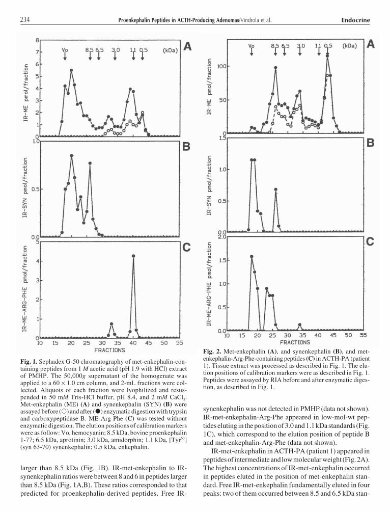
Peptides of high, intermediate, and low-molecularweight displaying IR-met-enkephalin were observed inPMHP (Fig. 1A). Free IR-met-enkephalin appeared in pep-tides smaller than 6.5 kDa (3.5, 2.0, and 0.5 kDa), andthe highest concentrations corresponded to materials elutedin the position of met-enkephalin standard (Fig. 1A).IR-synenkephalin was observed in peptides of 8.5 kDa and
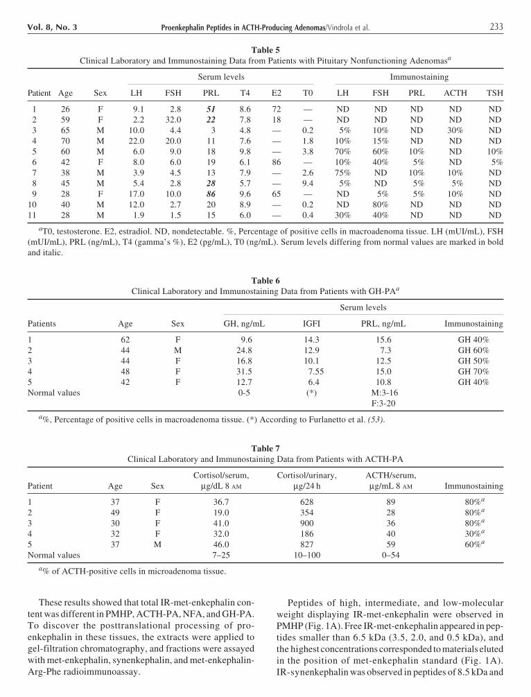
Table 7Clinical Laboratory and Immunostaining Data from Patients with ACTH-PA
Cortisol/serum, Cortisol/urinary, ACTH/serum,Patient Age Sex µg/dL 8 AM µg/24 h µg/mL 8 AM Immunostaining
1 37 F 36.7 628 89 80%a
2 49 F 19.0 354 28 80%a
3 30 F 41.0 900 36 80%a
4 32 F 32.0 186 40 30%a
5 37 M 46.0 827 59 60%a
Normal values 7–25 10–100 0–54
a% of ACTH-positive cells in microadenoma tissue.
Table 5Clinical Laboratory and Immunostaining Data from Patients with Pituitary Nonfunctioning Adenomasa
Serum levels Immunostaining
Patient Age Sex LH FSH PRL T4 E2 T0 LH FSH PRL ACTH TSH
1 26 F 9.1 2.8 51 8.6 72 — ND ND ND ND ND 2 59 F 2.2 32.0 22 7.8 18 — ND ND ND ND ND 3 65 M 10.0 4.4 3 4.8 — 0.2 5% 10% ND 30% ND 4 70 M 22.0 20.0 11 7.6 — 1.8 10% 15% ND ND ND 5 60 M 6.0 9.0 18 9.8 — 3.8 70% 60% 10% ND 10% 6 42 F 8.0 6.0 19 6.1 86 — 10% 40% 5% ND 5% 7 38 M 3.9 4.5 13 7.9 — 2.6 75% ND 10% 10% ND 8 45 M 5.4 2.8 28 5.7 — 9.4 5% ND 5% 5% ND 9 28 F 17.0 10.0 86 9.6 65 — ND 5% 5% 10% ND10 40 M 12.0 2.7 20 8.9 — 0.2 ND 80% ND ND ND11 28 M 1.9 1.5 15 6.0 — 0.4 30% 40% ND ND ND
aT0, testosterone. E2, estradiol. ND, nondetectable. %, Percentage of positive cells in macroadenoma tissue. LH (mUI/mL), FSH(mUI/mL), PRL (ng/mL), T4 (gamma’s %), E2 (pg/mL), T0 (ng/mL). Serum levels differing from normal values are marked in boldand italic.
Table 6Clinical Laboratory and Immunostaining Data from Patients with GH-PAa
Serum levels
Patients Age Sex GH, ng/mL IGFI PRL, ng/mL Immunostaining
1 62 F 9.6 14.3 15.6 GH 40%2 44 M 24.8 12.9 7.3 GH 60%3 44 F 16.8 10.1 12.5 GH 50%4 48 F 31.5 7.55 15.0 GH 70%5 42 F 12.7 6.4 10.8 GH 40%Normal values 0-5 (*) M:3-16
F:3-20
a%, Percentage of positive cells in macroadenoma tissue. (*) According to Furlanetto et al. (53).
234 EndocrineProenkephalin Peptides in ACTH-Producing Adenomas/Vindrola et al.
larger than 8.5 kDa (Fig. 1B). IR-met-enkephalin to IR-synenkephalin ratios were between 8 and 6 in peptides largerthan 8.5 kDa (Fig. 1A,B). These ratios corresponded to thatpredicted for proenkephalin-derived peptides. Free IR-
synenkephalin was not detected in PMHP (data not shown).IR-met-enkephalin-Arg-Phe appeared in low-mol-wt pep-tides eluting in the position of 3.0 and 1.1 kDa standards (Fig.1C), which correspond to the elution position of peptide Band met-enkephalin-Arg-Phe (data not shown).
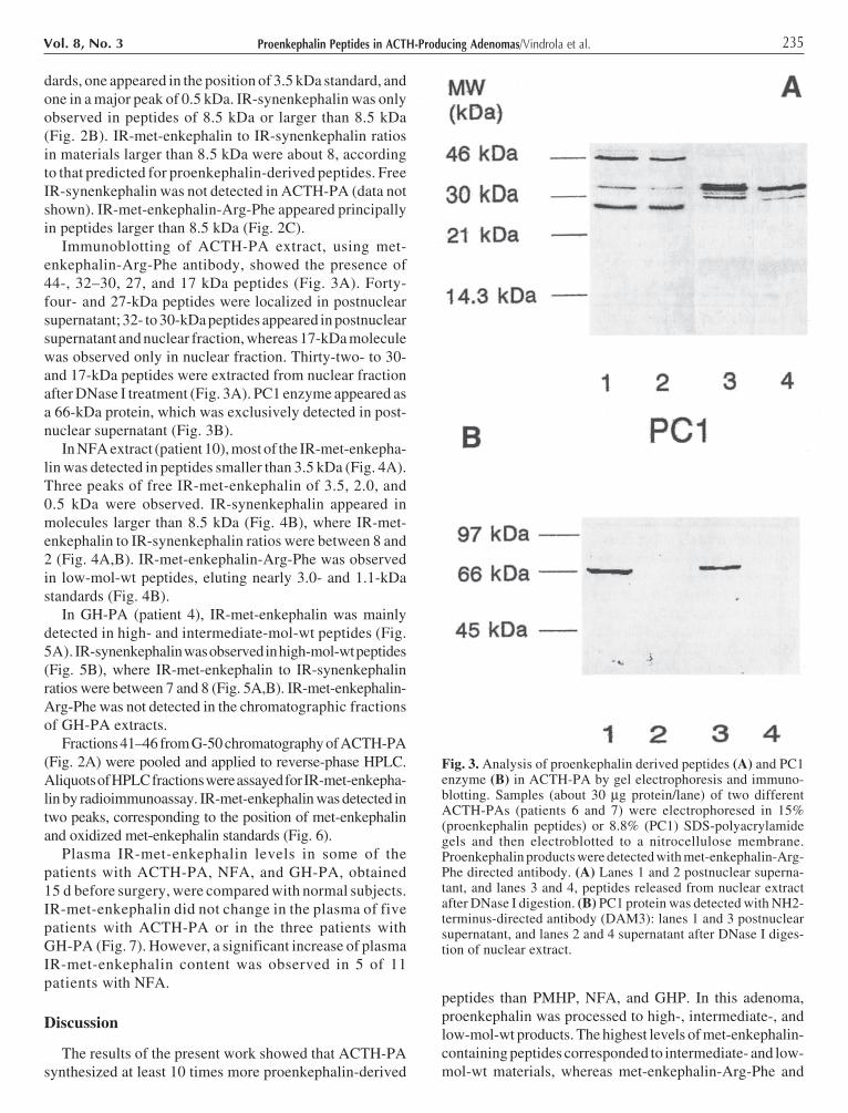
IR-met-enkephalin in ACTH-PA (patient 1) appeared inpeptides of intermediate and low molecular weight (Fig. 2A).The highest concentrations of IR-met-enkephalin occurredin peptides eluted in the position of met-enkephalin stan-dard. Free IR-met-enkephalin fundamentally eluted in fourpeaks: two of them occurred between 8.5 and 6.5 kDa stan-
Fig. 1. Sephadex G-50 chromatography of met-enkephalin-con-taining peptides from 1 M acetic acid (pH 1.9 with HCl) extractof PMHP. The 50,000g supernatant of the homogenate wasapplied to a 60 × 1.0 cm column, and 2-mL fractions were col-lected. Aliquots of each fraction were lyophilized and resus-pended in 50 mM Tris-HCl buffer, pH 8.4, and 2 mM CaCl2.Met-enkephalin (ME) (A) and synenkephalin (SYN) (B) wereassayed before (�) and after (�) enzymatic digestion with trypsinand carboxypeptidase B. ME-Arg-Phe (C) was tested withoutenzymatic digestion. The elution positions of calibration markerswere as follow: Vo, hemocyanin; 8.5 kDa, bovine proenkephalin1-77; 6.5 kDa, aprotinin; 3.0 kDa, amidorphin; 1.1 kDa, [Tyr63](syn 63-70) synenkephalin; 0.5 kDa, enkephalin.
Fig. 2. Met-enkephalin (A), and synenkephalin (B), and met-enkephalin-Arg-Phe-containing peptides (C) in ACTH-PA (patient1). Tissue extract was processed as described in Fig. 1. The elu-tion positions of calibration markers were as described in Fig. 1.Peptides were assayed by RIA before and after enzymatic diges-tion, as described in Fig. 1.
235Proenkephalin Peptides in ACTH-Producing Adenomas/Vindrola et al.Vol. 8, No. 3
dards, one appeared in the position of 3.5 kDa standard, andone in a major peak of 0.5 kDa. IR-synenkephalin was onlyobserved in peptides of 8.5 kDa or larger than 8.5 kDa(Fig. 2B). IR-met-enkephalin to IR-synenkephalin ratiosin materials larger than 8.5 kDa were about 8, accordingto that predicted for proenkephalin-derived peptides. FreeIR-synenkephalin was not detected in ACTH-PA (data notshown). IR-met-enkephalin-Arg-Phe appeared principallyin peptides larger than 8.5 kDa (Fig. 2C).
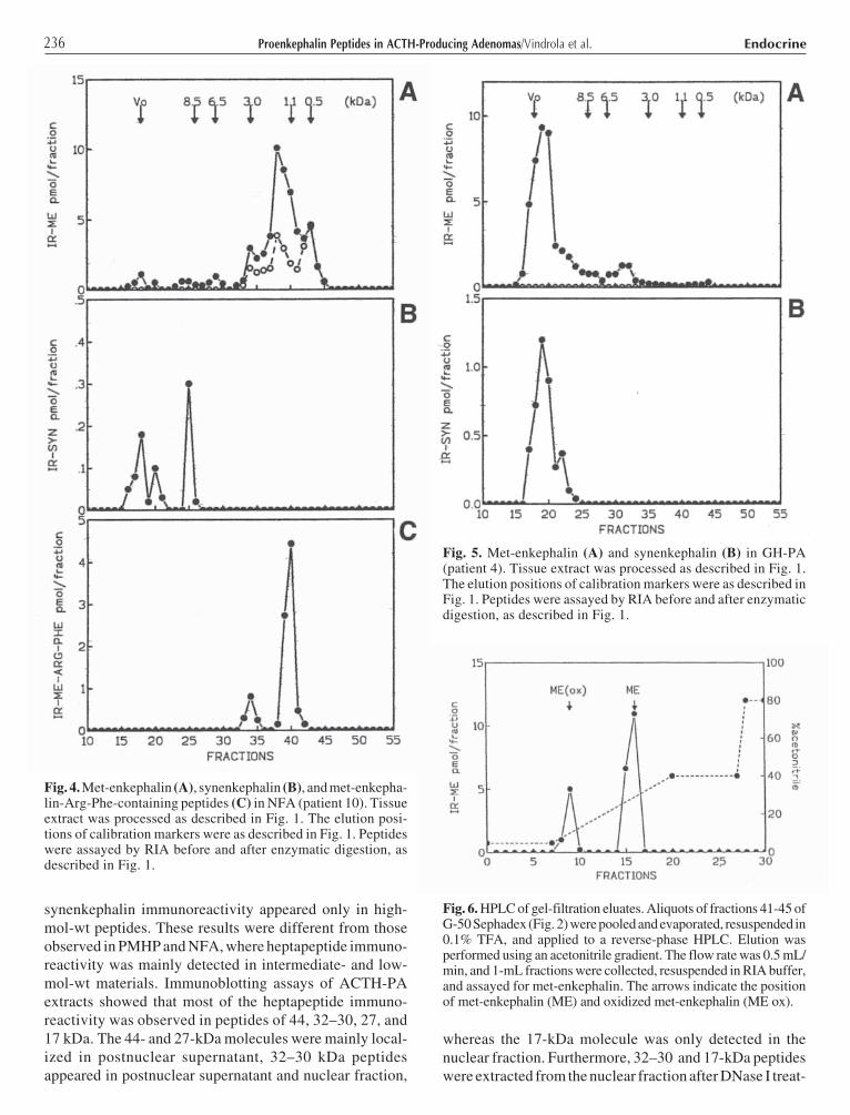
Immunoblotting of ACTH-PA extract, using met-enkephalin-Arg-Phe antibody, showed the presence of44-, 32–30, 27, and 17 kDa peptides (Fig. 3A). Forty-four- and 27-kDa peptides were localized in postnuclearsupernatant; 32- to 30-kDa peptides appeared in postnuclearsupernatant and nuclear fraction, whereas 17-kDa moleculewas observed only in nuclear fraction. Thirty-two- to 30-and 17-kDa peptides were extracted from nuclear fractionafter DNase I treatment (Fig. 3A). PC1 enzyme appeared asa 66-kDa protein, which was exclusively detected in post-nuclear supernatant (Fig. 3B).
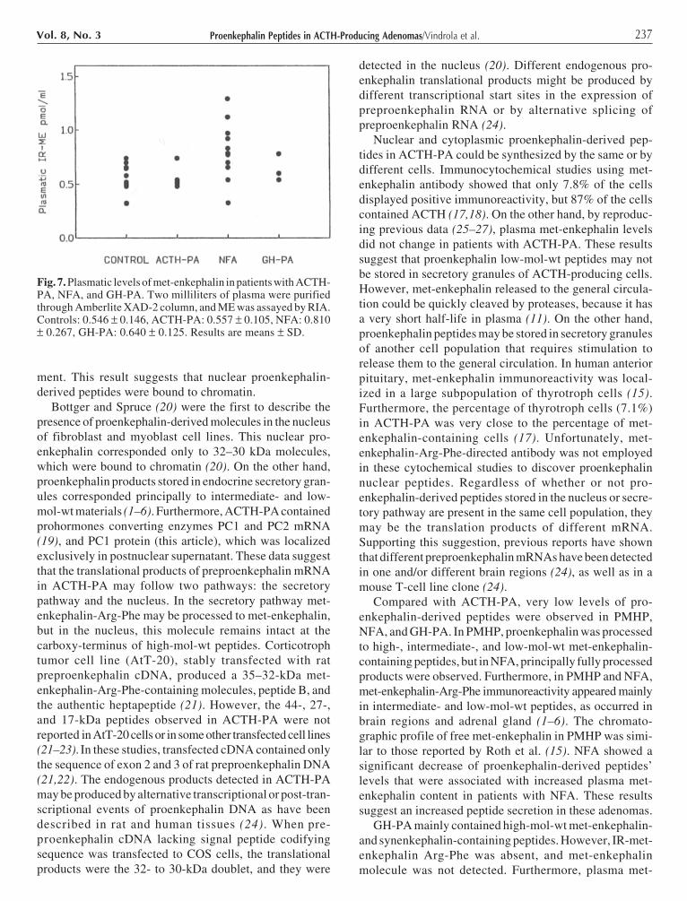
In NFA extract (patient 10), most of the IR-met-enkepha-lin was detected in peptides smaller than 3.5 kDa (Fig. 4A).Three peaks of free IR-met-enkephalin of 3.5, 2.0, and0.5 kDa were observed. IR-synenkephalin appeared inmolecules larger than 8.5 kDa (Fig. 4B), where IR-met-enkephalin to IR-synenkephalin ratios were between 8 and2 (Fig. 4A,B). IR-met-enkephalin-Arg-Phe was observedin low-mol-wt peptides, eluting nearly 3.0- and 1.1-kDastandards (Fig. 4B).
In GH-PA (patient 4), IR-met-enkephalin was mainlydetected in high- and intermediate-mol-wt peptides (Fig.5A). IR-synenkephalin was observed in high-mol-wt peptides(Fig. 5B), where IR-met-enkephalin to IR-synenkephalinratios were between 7 and 8 (Fig. 5A,B). IR-met-enkephalin-Arg-Phe was not detected in the chromatographic fractionsof GH-PA extracts.
Fractions 41–46 from G-50 chromatography of ACTH-PA(Fig. 2A) were pooled and applied to reverse-phase HPLC.Aliquots of HPLC fractions were assayed for IR-met-enkepha-lin by radioimmunoassay. IR-met-enkephalin was detected intwo peaks, corresponding to the position of met-enkephalinand oxidized met-enkephalin standards (Fig. 6).
Plasma IR-met-enkephalin levels in some of thepatients with ACTH-PA, NFA, and GH-PA, obtained15 d before surgery, were compared with normal subjects.IR-met-enkephalin did not change in the plasma of fivepatients with ACTH-PA or in the three patients withGH-PA (Fig. 7). However, a significant increase of plasmaIR-met-enkephalin content was observed in 5 of 11patients with NFA.
Discussion
The results of the present work showed that ACTH-PAsynthesized at least 10 times more proenkephalin-derived
peptides than PMHP, NFA, and GHP. In this adenoma,proenkephalin was processed to high-, intermediate-, andlow-mol-wt products. The highest levels of met-enkephalin-containing peptides corresponded to intermediate- and low-mol-wt materials, whereas met-enkephalin-Arg-Phe and
Fig. 3. Analysis of proenkephalin derived peptides (A) and PC1enzyme (B) in ACTH-PA by gel electrophoresis and immuno-blotting. Samples (about 30 µg protein/lane) of two differentACTH-PAs (patients 6 and 7) were electrophoresed in 15%(proenkephalin peptides) or 8.8% (PC1) SDS-polyacrylamidegels and then electroblotted to a nitrocellulose membrane.Proenkephalin products were detected with met-enkephalin-Arg-Phe directed antibody. (A) Lanes 1 and 2 postnuclear superna-tant, and lanes 3 and 4, peptides released from nuclear extractafter DNase I digestion. (B) PC1 protein was detected with NH2-terminus-directed antibody (DAM3): lanes 1 and 3 postnuclearsupernatant, and lanes 2 and 4 supernatant after DNase I diges-tion of nuclear extract.
236 EndocrineProenkephalin Peptides in ACTH-Producing Adenomas/Vindrola et al.
synenkephalin immunoreactivity appeared only in high-mol-wt peptides. These results were different from thoseobserved in PMHP and NFA, where heptapeptide immuno-reactivity was mainly detected in intermediate- and low-mol-wt materials. Immunoblotting assays of ACTH-PAextracts showed that most of the heptapeptide immuno-reactivity was observed in peptides of 44, 32–30, 27, and17 kDa. The 44- and 27-kDa molecules were mainly local-ized in postnuclear supernatant, 32–30 kDa peptidesappeared in postnuclear supernatant and nuclear fraction,
whereas the 17-kDa molecule was only detected in thenuclear fraction. Furthermore, 32–30 and 17-kDa peptideswere extracted from the nuclear fraction after DNase I treat-
Fig. 4. Met-enkephalin (A), synenkephalin (B), and met-enkepha-lin-Arg-Phe-containing peptides (C) in NFA (patient 10). Tissueextract was processed as described in Fig. 1. The elution posi-tions of calibration markers were as described in Fig. 1. Peptideswere assayed by RIA before and after enzymatic digestion, asdescribed in Fig. 1.
Fig. 5. Met-enkephalin (A) and synenkephalin (B) in GH-PA(patient 4). Tissue extract was processed as described in Fig. 1.The elution positions of calibration markers were as described inFig. 1. Peptides were assayed by RIA before and after enzymaticdigestion, as described in Fig. 1.
Fig. 6. HPLC of gel-filtration eluates. Aliquots of fractions 41-45 ofG-50 Sephadex (Fig. 2) were pooled and evaporated, resuspended in0.1% TFA, and applied to a reverse-phase HPLC. Elution wasperformed using an acetonitrile gradient. The flow rate was 0.5 mL/min, and 1-mL fractions were collected, resuspended in RIA buffer,and assayed for met-enkephalin. The arrows indicate the positionof met-enkephalin (ME) and oxidized met-enkephalin (ME ox).
237Proenkephalin Peptides in ACTH-Producing Adenomas/Vindrola et al.Vol. 8, No. 3
ment. This result suggests that nuclear proenkephalin-derived peptides were bound to chromatin.
Bottger and Spruce (20) were the first to describe thepresence of proenkephalin-derived molecules in the nucleusof fibroblast and myoblast cell lines. This nuclear pro-enkephalin corresponded only to 32–30 kDa molecules,which were bound to chromatin (20). On the other hand,proenkephalin products stored in endocrine secretory gran-ules corresponded principally to intermediate- and low-mol-wt materials (1–6). Furthermore, ACTH-PA containedprohormones converting enzymes PC1 and PC2 mRNA(19), and PC1 protein (this article), which was localizedexclusively in postnuclear supernatant. These data suggestthat the translational products of preproenkephalin mRNAin ACTH-PA may follow two pathways: the secretorypathway and the nucleus. In the secretory pathway met-enkephalin-Arg-Phe may be processed to met-enkephalin,but in the nucleus, this molecule remains intact at thecarboxy-terminus of high-mol-wt peptides. Corticotrophtumor cell line (AtT-20), stably transfected with ratpreproenkephalin cDNA, produced a 35–32-kDa met-enkephalin-Arg-Phe-containing molecules, peptide B, andthe authentic heptapeptide (21). However, the 44-, 27-,and 17-kDa peptides observed in ACTH-PA were notreported in AtT-20 cells or in some other transfected cell lines(21–23). In these studies, transfected cDNA contained onlythe sequence of exon 2 and 3 of rat preproenkephalin DNA(21,22). The endogenous products detected in ACTH-PAmay be produced by alternative transcriptional or post-tran-scriptional events of proenkephalin DNA as have beendescribed in rat and human tissues (24). When pre-proenkephalin cDNA lacking signal peptide codifyingsequence was transfected to COS cells, the translationalproducts were the 32- to 30-kDa doublet, and they were
detected in the nucleus (20). Different endogenous pro-enkephalin translational products might be produced bydifferent transcriptional start sites in the expression ofpreproenkephalin RNA or by alternative splicing ofpreproenkephalin RNA (24).
Nuclear and cytoplasmic proenkephalin-derived pep-tides in ACTH-PA could be synthesized by the same or bydifferent cells. Immunocytochemical studies using met-enkephalin antibody showed that only 7.8% of the cellsdisplayed positive immunoreactivity, but 87% of the cellscontained ACTH (17,18). On the other hand, by reproduc-ing previous data (25–27), plasma met-enkephalin levelsdid not change in patients with ACTH-PA. These resultssuggest that proenkephalin low-mol-wt peptides may notbe stored in secretory granules of ACTH-producing cells.However, met-enkephalin released to the general circula-tion could be quickly cleaved by proteases, because it hasa very short half-life in plasma (11). On the other hand,proenkephalin peptides may be stored in secretory granulesof another cell population that requires stimulation torelease them to the general circulation. In human anteriorpituitary, met-enkephalin immunoreactivity was local-ized in a large subpopulation of thyrotroph cells (15).Furthermore, the percentage of thyrotroph cells (7.1%)in ACTH-PA was very close to the percentage of met-enkephalin-containing cells (17). Unfortunately, met-enkephalin-Arg-Phe-directed antibody was not employedin these cytochemical studies to discover proenkephalinnuclear peptides. Regardless of whether or not pro-enkephalin-derived peptides stored in the nucleus or secre-tory pathway are present in the same cell population, theymay be the translation products of different mRNA.Supporting this suggestion, previous reports have shownthat different preproenkephalin mRNAs have been detectedin one and/or different brain regions (24), as well as in amouse T-cell line clone (24).
Compared with ACTH-PA, very low levels of pro-enkephalin-derived peptides were observed in PMHP,NFA, and GH-PA. In PMHP, proenkephalin was processedto high-, intermediate-, and low-mol-wt met-enkephalin-containing peptides, but in NFA, principally fully processedproducts were observed. Furthermore, in PMHP and NFA,met-enkephalin-Arg-Phe immunoreactivity appeared mainlyin intermediate- and low-mol-wt peptides, as occurred inbrain regions and adrenal gland (1–6). The chromato-graphic profile of free met-enkephalin in PMHP was simi-lar to those reported by Roth et al. (15). NFA showed asignificant decrease of proenkephalin-derived peptides’levels that were associated with increased plasma met-enkephalin content in patients with NFA. These resultssuggest an increased peptide secretion in these adenomas.
GH-PA mainly contained high-mol-wt met-enkephalin-and synenkephalin-containing peptides. However, IR-met-enkephalin Arg-Phe was absent, and met-enkephalinmolecule was not detected. Furthermore, plasma met-
Fig. 7. Plasmatic levels of met-enkephalin in patients with ACTH-PA, NFA, and GH-PA. Two milliliters of plasma were purifiedthrough Amberlite XAD-2 column, and ME was assayed by RIA.Controls: 0.546 ± 0.146, ACTH-PA: 0.557 ± 0.105, NFA: 0.810± 0.267, GH-PA: 0.640 ± 0.125. Results are means ± SD.
238 EndocrineProenkephalin Peptides in ACTH-Producing Adenomas/Vindrola et al.
enkephalin levels did not change in patients with GH-PA.These results indicate that proenkephalin posttranslationalprocessing was different in the three groups of adenomas,suggesting a differential physiological role.
Several reports showed that secreted met-enkephalin-and met-enkephalin-containing peptides participate intumoral activity (28–30); in addition, a nuclear δ opioidreceptor has been described in a tumoral cell line (31). Inmost of the cases, met-enkephalin-containing peptidesinhibited tumoral activity (28–30). In vivo and in vitro stud-ies have shown that met-enkephalin directly inhibitedACTH release in human corticotroph (32–34). However,this effect was abolished in corticotroph cells of patientswith Cushing’s disease, suggesting a decrease of opioidsreceptor levels (32–34). In rat brain, reduction of the num-ber of opioid receptors induced increased expression of theproenkephalin gene (35). We speculate that a decreasednumber of opioid receptors in corticotroph cells mightinduce the increase of met-enkephalin-containing peptides’levels in ACTH-PA. Proenkephalin mRNA and pro-enkephalin-derived peptides are expressed in cells ofmesodermal, ectodermal, and endodermal tissues in earlystage of embryonic development (36–40). In mesodermalcell lines, proenkephalin immunoreactivity has beendetected in the nucleus and associated with chromatin (20).Furthermore, when fibroblasts were transfected withpreproenkephalin DNA antisense, the cell proliferation wasinhibited (41). Taken together these results suggest thatproenkephalin products present in nuclear and secretorypathways may have a physiological role in ACTH-PA.
In conclusion, we demonstrated that only ACTH-PAcontained high levels of nuclear and secretory proenkephalinpeptides. These results suggest a probable adenoma-spe-cific physiological role of proenkephalin products.
Materials and Methods
In this study, 21 patients (12 females and 9 males) betweenthe ages of 26 and 70 were tested. Five patients hadCushing’s disease; 11 patients had NFA, and 5 patients hadGH-PA, showing clinical acromegaly.
The adenomas were classified as ACTH-PA, GH-PA,and NFA, based on preoperative clinical and laboratorydata, and immunocytochemical staining of tissuesremoved at surgery (42–44). Tables 5, 6, and 7 showclinical laboratory and immunocytochemical data.PMHP controls were obtained from individuals who diedfrom severe wounds that did not involve the head.Adenomas and control pituitary glands were stored at–70°C until their use.
Tissue Extracts
Frozen tissues were resuspended in 10 vol of 1 M aceticacid (pH 1.9 with HCl), boiled for 15 min, homogenizedwith a Polytron, and centrifuged at 50,000g for 1 h. Thesupernatant was stored at –70°C.
Nuclei Isolation
Nuclei were isolated as described previously (45).Immediately after the surgery, the ACTH-PA was immersedin ice-cold 0.32 M sucrose. Thirty minutes later, they werehomogenized in 10 vol of 0.5 M sucrose, 10 mM HEPES,pH 7.9, 1.5 mM MgCl2, 10 mM KCl, 10% glycerol, 1 mMEDTA, 1 mM dithiotreitol, 1 mM phenylmethylsulfonylfluoride (PMSF) and 1 µg/mL each of aprotinin, leupeptin,and pepstatin A in Dounce homogenizer, with five strokeswith loose-fitting pestle and five strokes with light-fittingpestle. The homogenate was centrifuged at 4000g for 5 minat 4°C. The supernatant was removed (postnuclear super-natant), and the pellet (nuclei) was processed as describedpreviously by Fey and Penman (46). Briefly, the pellet wasdigested with RNase-free DNase I (250 µg/mL) in diges-tion buffer (10 mM PIPES pH 6.8/50 mM NaCl/300 mMsucrose/3 mM MgCl/0.5% [v/v] Triton X-100, with PMSF,aprotinin, leupeptin, and pepstatin A) for 20 min at roomtemperature. Chromatin-associated proteins were releasedby subsequent 250 mM ammonium sulfate extraction andcentrifuged at 20,000g for 1 h at 4°C. The supernatant wasdiluted in Laemmli buffer (2X final concentration) andelectrophoresed.
Postnuclear supernatant was diluted in acetic acid (finalconcentration 1 N, containing 50 mM HCl), boiled for10 min, homogenized in Polytron, and centrifuged at20,000g for 1 h. The supernatant was concentrated in a10-kDa cutoff Centricon filter, resuspended in 2X Laemmlibuffer, and electrophoresed.
Total and Free Met-enkephalin Assay
Aliquots of tissue extracts were lyophilized and recon-stituted in 50 mM Tris-HCl buffer, pH 8.4, and 2 mM CaCl2.Free and total IR-met-enkephalin and synenkephalin, thatis, previous to and after sequential enzymatic digestion withtrypsin and carboxypeptidase B, were determined by RIAas previously described (47,48).
Enkephalin, Synenkephalin,and Met-enkephalin-Arg-Phe Assays
Met-enkephalin and synenkephalin were measured byRIA as described previously (47–49). Iodinated met-enkephalin and [Tyr63] (syn 63–70) synenkephalin wereused as tracers. We found that met-enkephalin antiserumcrossreactivity was 100% with oxidized met-enkephalin,0.3% with leu-enkephalin, and <0.01% with met-enkepha-lin-Arg, dynorphin (1–13), and α-, γ-, and β-endorphin.Sensitivity of the assay was 9 fmol; IC50 was 130 fmol. Theantiserum to [Tyr63] (syn 63-70) synenkephalin showedcrossreactivity of 1% with a native 8.6-kDa peptide(proenkephalin 1–77), which, when digested with trypsin,displayed a 20% crossreactivity that reached 100% whendigested with trypsin and carboxypeptidase B. No crossreac-tivity was observed with met-enkephalin, met-enkephalin-Arg, met-enkephalin-Arg-Phe, met-enkephalin-Arg-Gly-Leu,
239Proenkephalin Peptides in ACTH-Producing Adenomas/Vindrola et al.Vol. 8, No. 3
leu-enkephalin, peptide E and F, dynorphin B, amidorphin,and metorphamide. Sensitivity was 2 fmol; IC50 was 50 fmol.ME-Arg-Phe was measured as described previously (50).
Gel Filtration
Samples were applied to a Sephadex G-50 superfine (60 ×1.0 cm) column equilibrated with 1 M acetic acid (pH 1.9with HCl). The elution was performed at a flow rate of10 mL/h at room temperature; 2-mL fractions were col-lected, and aliquots were lyophilized and reconstituted in50 mM of Tris-HCl buffer, pH 8.4, and 2 mM CaCl2. Elu-ates were monitored by ultraviolet absorbance at 280 nm.IR-synenkephalin and IR-met-enkephalin were assayedprior to and after enzymatic digestion with trypsin and car-boxypeptidase B. IR-met-enkephalin-Arg-Phe was assayedwithout enzymatic digestion.
HPLC Chromatography
Samples were applied to a reverse-phase column,Ultropac, Lichrosorb RF18, 5 µm, 4 × 250 mm (WatersCorporation, Milford, MA). The column was equili-brated with 10% acetonitrile in TFA 0.1%, and the elutionwas performed in a discontinuous acetonitrile gradient(10–80%). The flow rate was 0.5 mL/min, and 1-mL frac-tions were collected. Aliquots were evaporated in a SpeedVac concentrator, and resuspended in the Tris-HClbuffer. All the chromatographic assays were repeated atleast three times, using pituitary or tumor extracts fromdifferent patients.
Electrophoresis and Immunoblotting
Samples diluted in Laemmli buffer were applied to elec-trophoresis on 15 and 8.8% acrylamide for proenkephalinpeptides and PC1 enzyme, respectively. Blotting protocolwas described previously (51,52). Briefly, proteins weretransferred to nitrocellulose and blocked with 5% milk inTris-buffered saline. Blots were then sliced and incubatedovernight, at 4°C, with gentle rocking, using specific anti-bodies diluted in 5% milk. Proenkephalin peptides were iden-tified with met-enkephalin-Arg-Phe directed antibody,diluted 1:400 in 5% milk, whereas PC1 enzyme was tested withNH2-terminus-directed antibody (DAM3) diluted 1:1000. Theblots were developed as previously described (51,52).
Plasma Met-enkephalin Assay
Blood was put into polypropylene tubes kept on ice,containing 10 µL/mL of a protease inhibitor cocktail(10,000 IUC/aprotinin, 0.23 mM citric acid, and 0.024 mMEDTA) and 5 U of heparin. Blood was centrifuged at 4°C,and plasma was immediately stored at –20°C; 2 mL ofplasma were diluted to 10 mL with 0.1 N HCl and appliedto Amberlite XAD-2 column and processed as describedpreviously (23), except that met-enkephalin was eluted witha mixture of 1 M acetic acid/acetonitrile (50%/50%). Usingthis protocol, the recovery of 3H-met-enkephalin (identi-fied by HPLC) added to the blood was between 80 and 90%.
Acknowledgments
We thank Jean Rossier for the donation of SYN andME-Arg-Phe antibodies; Maria Rosa Padros, Anne Compli-ment, Isabel Perez Monfort, and Phil Arriaga for the valuablecommentaries. This work was supported by grants fromCONACYT (Mexico) (O.V. 3898-N9401) and CONICET(Argentina).
References
1. Akil, H., Watson, S., Young, E., Lewis, M., Khachaturian, H.,and Walker, M. (1984). Ann. Rev. Neurosci. 7, 223–255.
2. Comb, M., Seeburg, H. P., Adelman, J., Eiden, L., and Herbert,E. (1982). Nature 295, 663–666.
3. Douglass, J., Civelli, O., and Herbert, E. (1984). Ann. Rev.Biochem. 53, 665–715.
4. Gubler, U., Seeburg, H. P., Hoffman, B. J., Gage, L. P., andUdenfriend, S. (1982). Nature 295, 206–209.
5. Noda, M., Furutani, Y., Takahashi, H., Toyosato, M., Hirose,T., Inayama, S., et al. (1982). Nature 295, 202–206.
6. Rossier, J. (1993). In: Handbook of Experimental Pharmacol-ogy. Herz, A. (ed.). Berlin: Springer-Verlag.
7. Liston, D., Vanderhaeghen, J. J., and Rossier, J. (1983). Nature302, 62,63.
8. Liston, D. and Rossier, J. (1984). Neurosci. Lett. 48, 211–216.9. Rodriguez Vida, M. I., Kleid, M. C., Ase, A., Finkielman, S.,
Nahmod, V. E., and Vindrola, O. (1994). Dev. Brain Res. 77,151–156.
10. Song, D. D. and Harlan, R. E. (1993). Neuroscience 52,883–909.
11. Clemente-Jones, V. and Besser, G. M. (1983). Br. Med. Bull.39, 95–100.
12. Rossier, J., Battenberg, E., Pittman, Q., Bayon, A., Koda, L.,Miller, R. J., et al. (1979). Nature 277, 653–655.
13. Jingami, H., Nakanishi, S., Imura, H., and Numa, S. (1984).Eur. J. Biochem. 142, 441–445.
14. Pittius, C. W., Kley, N., Loeffler, J. P., and Hollt, V. (1985).EMBO J. 4, 1257–1263.
15. Roth, K. A., Lorenz, R. G., Mckeel, D. W., Leykam, J.,Barchas, J. D., and Tyler, A. N. (1988). J. Clin. Invest. Metab.66, 804–810.
16. Desiderio, D. M., Kusmierz, J. J., Zhu, X., Dass, C., Hilton, D.,and Roberton, J. T. (1993). Biol. Mass Spectrom. 22, 89–97.
17. Wowra, B. and Peiffer, J. (1984). Pathol. Res. Pract. 178,349–354.
18. Hossoun, J., Charpin, C., Jaquet, P., Oliver, C., Lissitzky, J.C., Grisoli, F., et al. (1979). Ann Endocrinol. 40, 559,560.
19. Hashimoto, K., Koga, M., Kouhara, H., Arita, N., Hayakawa,T., Kishimoto, T., et al. (1994). Clin. Endocrinol. 41, 185–191.
20. Bottger, A. and Spruce, B. (1995). J. Cell Biol. 130, 1251–1262.21. Mathis, J. P. and Lindberg, I. (1992). Endocrinology 131,
2287–2296.22. Thomas, G., Thomas, L., Thorne, B. A., and Herbert, E.
(1988). Biochimie 70, 89–87.23. Comb, M., Liston, D., Martin, M., Rosen, H., and Herbert, E.
(1985). EMBO J. 4, 3115–3122.24. Weisinger, G. (1995). Biochem J. 307, 617–629.25. Furui, T., Kuwayama, A., and Kageyama, N. (1980). No To
Shinkei 32, 305–309.26. Kimura, T., Ota, K., Matsui, K., Iitake, K., Shoji, M., Inouse,
M., et al. (1985). Endocrinol. Jpn. 32, 517–527.27. Gaillard, R. C., Grossman, A., Smith, R., Rees, L. H., and
Besser, G. M. (1981). Clin. Endocrinol. 14, 471–478.28. Zagon, I. S. and McLaughlin, P. J. (1989). Brain Res. 490, 14–25.29. Zagon, I. S., McLaughlin, P. J., Goodman, S. R., and Rhodes,
R. E. (1987). J. Natl. Cancer Inst. 79, 1059–1065.
240 EndocrineProenkephalin Peptides in ACTH-Producing Adenomas/Vindrola et al.
30. Zagon, I. S. and Mc Laughlin, P. J. (1988). Life Sci. 43,1313–1318.
31. Belcheva, M. M., Glucker, S., Chuang, D., Clark, W. G.,Jefcoat, L. B., McHale, R. J., et al. (1995). J. Pharm. Exp.Ther. 274, 1513–1523.
32. Lamberts, S. W., Oosterom, R., Bons, E. G., and Uitterlinden,P. (1981). J. Clin. Endocrinol. Metab. 53, 1084–1086.
33. Allolio, B., Winkelmann, W., Hipp, F. X., Kaulen, D., andMies, R. (1982). J. Clin. Endocrinol. Metab. 55, 1–7.
34. Allolio, B., Deuss, U., Kaulen, D., Leonhanrdt, U., Kallabis,D., Hamel, E., et al. (1986). J. Clin. Endocrinol. Metab. 63,1427–1431.
35. Morris, B. J., Hollt, V., and Herz, A. (1988). Neurosci. Lett.89, 80–84.
36. Keshed, E., Polakiewicz, R. D., Itin, A., Ornoy, A., and Rosen,H. (1989). EMBO J. 8, 2917–2923.
37. Kew, D. and Kilpatrick, D. L. (1990). Mol. Endocrinol. 4,337–340.
38. Polakiewicz, R. D. and Rosen, H. (1990). Mol. Cell. Biol. 10,736–742.
39. Rosen, H., Polakiewicz, R. D., and Simantov, R. (1990).Biochem. Biophys. Res. Commun. 171, 722–728.
40. Rosen, H., Polakiewicz, R. D., Benzakine, S., and Bar-Shavit,Z. (1991). Proc. Natl. Acad. Sci. USA 88, 3705–3709.
41. Weisinger, G., Zinder, O., and Simantov, R. (1995). Biochem.Biophys. Res. Commun. 274, 530–537.
42. Asa, S. L. and Kovacs, K. (1983). Clin. Endocrinol. Metab.12, 567–596.
43. Klibanski, A. (1987). Endocrinol. Metab. Clin. North. Am. 16,793–804.
44. Chang-Moraville, B. M. and Jackson, I. M. (1992).Endocrinol. Metab. Clin. North Am. 21, 649–668.
45. Unlap, T. and Jope, R. S. (1994). Mol. Brain Res. 24, 275–282.
46. Fey, E. G. and Penman S. (1988). Proc. Natl. Acad. Sci. 85,101–125.
47. Saravia, F., Ase, A., Aloyz, R., Kleid, M. C., Rodriguez Vida,M. I., Nahmod, V. E., et al. (1993). Endocrinology 132,1431–1437.
48. Vindrola, O., Padros, M. R., Sterin-Prync, A., Ase, A.,Finkielman, S., and Nahmod, V. E. (1990). J. Clin. Invest. 86,531–537.
49. Vindrola, O., Briones, R., Asai, M., and Fernandez Guardiola,A. (1981). Neurosci. Lett. 21, 39–43.
50. Meeters, K. M. and Rossier, J. (1987). J. Neurochem. 49,721–728.
51. Vindrola, O., Mayer, A. M. S., Citera, G., Spitzer, J. A., andEspinoza L. R. (1994). Neuropeptides 27, 235–244.
52. Vindrola, O. and Lindberg, I. (1992). Mol. Endocrinol. 6,1088–1094.
53. Furlanetto, R., Underwood, L., and Van Wyk, J. (1977). J. Clin.Invest. 60, 648–653.
Related Documents