The Journal of Experimental Biology 1752 © 2014. Published by The Company of Biologists Ltd | The Journal of Experimental Biology (2014) 217, 1752-1757 doi:10.1242/jeb.100677 ABSTRACT Low concentrations of endogenous carbon monoxide (CO), generated primarily through degradation of heme from heme- proteins, have been shown to maintain physiological function of organs and to exert cytoprotective effects. However, high concentrations of carboxyhemoglobin (COHb), formed by CO binding to hemoglobin, potentially prevent adequate O 2 delivery to tissues by lowering arterial O 2 content. Elevated heme-protein concentrations, as found in marine mammals, are likely associated with greater heme degradation, more endogenous CO production and, consequently, elevated COHb concentrations. Therefore, we measured COHb in elephant seals, a species with large blood volumes and elevated hemoglobin and myoglobin concentrations. The levels of COHb were positively related to the total hemoglobin concentration. The maximum COHb value was 10.4% of total hemoglobin concentration. The mean (±s.e.m.) value in adult seals was 8.7±0.3% (N=6), while juveniles and pups (with lower heme-protein contents) had lower mean COHb values of 7.6±0.2% and 7.1±0.3%, respectively (N=9 and N=9, respectively). Serial samples over several hours revealed little to no fluctuation in COHb values. This consistent elevation in COHb suggests that the magnitude and/or rate of heme-protein turnover is much higher than in terrestrial mammals. The maximum COHb values from this study decrease total body O 2 stores by 7%, thereby reducing the calculated aerobic dive limit for this species. However, the constant presence of elevated CO in blood may also protect against potential ischemia–reperfusion injury associated with the extreme breath-holds of elephant seals. We suggest the elephant seal represents an ideal model for understanding the potential cytoprotective effects, mechanisms of action and evolutionary adaptation associated with chronically elevated concentrations of endogenously produced CO. KEY WORDS: Calculated aerobic dive limit, Carbon monoxide, Hemoglobin absorption spectra, Marine mammal, Oxygen stores INTRODUCTION Carbon monoxide (CO) is often classified as a strictly toxic gas, depriving the body of oxygen (O 2 ) by binding to heme-proteins such as hemoglobin and forming carboxyhemoglobin (COHb) (Weaver, 2009). Deleterious symptoms (e.g. headache, nausea and shortness of breath) of CO-driven hypoxia are typically seen when COHb values reach ≥20% of total hemoglobin concentration, and death is associated with values of 50–80% (Stewart, 1975; Weaver, 2009). These COHb values drastically reduce blood-O 2 transport and O 2 RESEARCH ARTICLE 1 Center for Marine Biotechnology and Biomedicine, Scripps Institution of Oceanography, University of California San Diego, La Jolla, CA 92093-0204, USA. 2 Department of Biology, Sonoma State University, Rohnert Park, CA 94928, USA. *Author for correspondence ([email protected]) Received 1 December 2013; Accepted 4 February 2014 storage capacity (decreased arterial O 2 content), thus limiting mitochondrial respiration. However, CO is also generated endogenously in low concentrations, and functions in neurotransmission and in protection of tissues and cells against inflammation, apoptosis and ischemia–reperfusion injuries (Snyder et al., 1998; Kevin and Laffey, 2008; Mustafa et al., 2009; Kajimura et al., 2010; Prabhakar, 2012). Therefore, low concentrations of CO can provide beneficial and therapeutic effects up to a specific concentration, at which elevated CO then leads to detrimental effects from reduced O 2 delivery. These relatively recent findings give CO a new functional perspective and emphasize the importance of understanding the biological effects of specific CO concentrations in the body which can be viewed as therapeutic. Ironically, the primary source of endogenous CO comes from the breakdown of heme, which is an essential component of many heme- proteins (e.g. hemoglobin, myoglobin and cytochrome c) that transport O 2 or associate closely with aerobic respiration. The breakdown of heme-proteins releases heme, which is then enzymatically degraded by heme-oxygenase, resulting in equimolar production of free iron, CO and biliverdin (Coburn et al., 1963; Tenhunen et al., 1968). Biliverdin is then reduced to bilirubin via biliverdin reductase. These products (biliverdin and bilirubin) and by- products (CO) of heme degradation have been shown to have a multitude of beneficial effects including: vasodilatation, antioxidative properties, attenuation of ischemia/reperfusion injury, inhibition of apoptosis and downregulation of the inflammatory response (Stocker et al., 1987; Barañano et al., 2002; Mustafa et al., 2009). It is these properties that have stimulated investigation of CO therapy and the use of CO-releasing pharmaceuticals for future clinical applications (i.e. sepsis, organ transplants, heart failure, hypertension, inflammation and cancer) (Motterlini and Otterbein, 2010). Considering that heme-proteins are the primary source of endogenous CO production, marine mammals, which have elevated blood volumes, hemoglobin content and myoglobin concentrations (Ponganis et al., 2011), potentially represent an excellent model for investigating elevated endogenous CO production. For example, in an early study by Pugh, there was an unexpected finding of elevated CO in the blood of Weddell seals (Leptonychotes weddelli) (Pugh, 1959). This study found mean CO levels in Weddell seal blood that were over six times the values seen in the blood of human non- smokers (Pugh, 1959). Bilirubin concentration in Weddell seal plasma was also elevated approximately threefold to fourfold that of human plasma. Similarly, bilirubin and biliverdin have been measured in the blood of adult and juvenile northern elephant seals [Mirounga angustirostris (Gill 1866)], yet the values are in the same range as those seen in healthy humans (Thorson and Le Boeuf, 1994; Dennery et al., 2001; D.E.C., unpublished). Northern elephant seals have some of the highest known mammalian blood volumes (216 ml kg −1 ) and hemoglobin concentrations (25 g dl −1 ) in nature, accounting for over 70% of their total body O 2 store (Ponganis, 2011). This elevated blood O 2 storage Elevated carboxyhemoglobin in a marine mammal, the northern elephant seal Michael S. Tift 1,2, *, Paul J. Ponganis 1 and Daniel E. Crocker 2

Welcome message from author
This document is posted to help you gain knowledge. Please leave a comment to let me know what you think about it! Share it to your friends and learn new things together.
Transcript
The
Jour
nal o
f Exp
erim
enta
l Bio
logy
1752
© 2014. Published by The Company of Biologists Ltd | The Journal of Experimental Biology (2014) 217, 1752-1757 doi:10.1242/jeb.100677
ABSTRACTLow concentrations of endogenous carbon monoxide (CO),generated primarily through degradation of heme from heme-proteins, have been shown to maintain physiological function oforgans and to exert cytoprotective effects. However, highconcentrations of carboxyhemoglobin (COHb), formed by CO bindingto hemoglobin, potentially prevent adequate O2 delivery to tissues bylowering arterial O2 content. Elevated heme-protein concentrations,as found in marine mammals, are likely associated with greater hemedegradation, more endogenous CO production and, consequently,elevated COHb concentrations. Therefore, we measured COHb inelephant seals, a species with large blood volumes and elevatedhemoglobin and myoglobin concentrations. The levels of COHb werepositively related to the total hemoglobin concentration. Themaximum COHb value was 10.4% of total hemoglobin concentration.The mean (±s.e.m.) value in adult seals was 8.7±0.3% (N=6), whilejuveniles and pups (with lower heme-protein contents) had lowermean COHb values of 7.6±0.2% and 7.1±0.3%, respectively (N=9and N=9, respectively). Serial samples over several hours revealedlittle to no fluctuation in COHb values. This consistent elevation inCOHb suggests that the magnitude and/or rate of heme-proteinturnover is much higher than in terrestrial mammals. The maximumCOHb values from this study decrease total body O2 stores by 7%,thereby reducing the calculated aerobic dive limit for this species.However, the constant presence of elevated CO in blood may alsoprotect against potential ischemia–reperfusion injury associated withthe extreme breath-holds of elephant seals. We suggest the elephantseal represents an ideal model for understanding the potentialcytoprotective effects, mechanisms of action and evolutionaryadaptation associated with chronically elevated concentrations ofendogenously produced CO.
KEY WORDS: Calculated aerobic dive limit, Carbon monoxide,Hemoglobin absorption spectra, Marine mammal, Oxygen stores
INTRODUCTIONCarbon monoxide (CO) is often classified as a strictly toxic gas,depriving the body of oxygen (O2) by binding to heme-proteins suchas hemoglobin and forming carboxyhemoglobin (COHb) (Weaver,2009). Deleterious symptoms (e.g. headache, nausea and shortnessof breath) of CO-driven hypoxia are typically seen when COHbvalues reach ≥20% of total hemoglobin concentration, and death isassociated with values of 50–80% (Stewart, 1975; Weaver, 2009).These COHb values drastically reduce blood-O2 transport and O2
RESEARCH ARTICLE
1Center for Marine Biotechnology and Biomedicine, Scripps Institution ofOceanography, University of California San Diego, La Jolla, CA 92093-0204, USA.2Department of Biology, Sonoma State University, Rohnert Park, CA 94928, USA.
*Author for correspondence ([email protected])
Received 1 December 2013; Accepted 4 February 2014
storage capacity (decreased arterial O2 content), thus limitingmitochondrial respiration. However, CO is also generatedendogenously in low concentrations, and functions inneurotransmission and in protection of tissues and cells againstinflammation, apoptosis and ischemia–reperfusion injuries (Snyderet al., 1998; Kevin and Laffey, 2008; Mustafa et al., 2009; Kajimuraet al., 2010; Prabhakar, 2012). Therefore, low concentrations of COcan provide beneficial and therapeutic effects up to a specificconcentration, at which elevated CO then leads to detrimental effectsfrom reduced O2 delivery. These relatively recent findings give COa new functional perspective and emphasize the importance ofunderstanding the biological effects of specific CO concentrationsin the body which can be viewed as therapeutic.
Ironically, the primary source of endogenous CO comes from thebreakdown of heme, which is an essential component of many heme-proteins (e.g. hemoglobin, myoglobin and cytochrome c) thattransport O2 or associate closely with aerobic respiration. Thebreakdown of heme-proteins releases heme, which is thenenzymatically degraded by heme-oxygenase, resulting in equimolarproduction of free iron, CO and biliverdin (Coburn et al., 1963;Tenhunen et al., 1968). Biliverdin is then reduced to bilirubin viabiliverdin reductase. These products (biliverdin and bilirubin) and by-products (CO) of heme degradation have been shown to have amultitude of beneficial effects including: vasodilatation, antioxidativeproperties, attenuation of ischemia/reperfusion injury, inhibition ofapoptosis and downregulation of the inflammatory response (Stockeret al., 1987; Barañano et al., 2002; Mustafa et al., 2009). It is theseproperties that have stimulated investigation of CO therapy and theuse of CO-releasing pharmaceuticals for future clinical applications(i.e. sepsis, organ transplants, heart failure, hypertension,inflammation and cancer) (Motterlini and Otterbein, 2010).
Considering that heme-proteins are the primary source ofendogenous CO production, marine mammals, which have elevatedblood volumes, hemoglobin content and myoglobin concentrations(Ponganis et al., 2011), potentially represent an excellent model forinvestigating elevated endogenous CO production. For example, inan early study by Pugh, there was an unexpected finding of elevatedCO in the blood of Weddell seals (Leptonychotes weddelli) (Pugh,1959). This study found mean CO levels in Weddell seal blood thatwere over six times the values seen in the blood of human non-smokers (Pugh, 1959). Bilirubin concentration in Weddell sealplasma was also elevated approximately threefold to fourfold that ofhuman plasma. Similarly, bilirubin and biliverdin have beenmeasured in the blood of adult and juvenile northern elephant seals[Mirounga angustirostris (Gill 1866)], yet the values are in the samerange as those seen in healthy humans (Thorson and Le Boeuf,1994; Dennery et al., 2001; D.E.C., unpublished).
Northern elephant seals have some of the highest knownmammalian blood volumes (216 ml kg−1) and hemoglobinconcentrations (25 g dl−1) in nature, accounting for over 70% of theirtotal body O2 store (Ponganis, 2011). This elevated blood O2 storage
Elevated carboxyhemoglobin in a marine mammal, the northernelephant sealMichael S. Tift1,2,*, Paul J. Ponganis1 and Daniel E. Crocker2

The
Jour
nal o
f Exp
erim
enta
l Bio
logy
1753
RESEARCH ARTICLE The Journal of Experimental Biology (2014) doi:10.1242/jeb.100677
capacity contributes to their ability to perform repetitive dives of 20to 25 min duration to depths >500 m with only 2–3 min surfaceintervals during foraging trips lasting up to 8 months in duration(Robinson et al., 2012). Additionally, these animals are well knownfor their extended and repeated breath-holds (up to 25 min) duringsleep apnea events on land, which are also usually followed by briefeupneic periods (Blackwell and Le Boeuf, 1993). Breath-holds ofthese seals are accompanied by the dive response [bradycardia andperipheral ischemia (Andrews et al., 1997; Ponganis et al., 2006)]and routine hypoxemia with arterial hemoglobin saturationsreaching as low as 10% (Stockard et al., 2007; Meir et al., 2009).The brief periods of eupnea following breath-holds includehyperventilation, tachycardia and vasodilation, which results inreperfusion of tissues with oxygenated blood, thereby increasing thepotential for exposure of tissues to reactive oxygen species anddamage from oxidative stress. Additionally, a typical elephant sealspends 9–10 months per year at sea with over 90% of its timeunderwater, and when on land will spend a large portion of time insleep apnea (Blackwell and Le Boeuf, 1993; Robinson et al., 2012).These life history behaviors drastically limit the amount of time theanimals spend in eupnea.
Considering endogenous CO is expelled via respiration, theintermittent breathing patterns (sleep apnea and/or diving) of manyair-breathing marine divers introduces a potential limitation forremoval of CO. Therefore, increased endogenous CO productionand delayed removal of CO could elevate COHb content, leading toa decrease in blood O2 stores and therefore potentially limiting theduration for aerobic metabolism during a dive. On the other hand,elevated endogenous CO may provide protection against potentialreperfusion injury associated with these natural breath-holdingbehaviors.
To address the potential for elevated endogenous CO productionin a species with exceptionally large O2 stores, we measured levelsof COHb in the blood of northern elephant seals and compared thesevalues against hemoglobin concentrations. Further, because of anatural ontogenetic increase in both hemoglobin and myoglobin(Thorson and Le Boeuf, 1994; Tift et al., 2013), we investigateddifferences in COHb values associated with age. We hypothesizedthat (1) the amount of COHb would correlate with the totalhemoglobin concentration (tHb), and (2) because of larger bloodvolumes in adults versus younger seals, the amount of COHb wouldbe highest in adults.
RESULTSSpectrophotometric absorption peaks of deoxyhemoglobin (HHb),oxyhemoglobin (O2Hb) and COHb from elephant seals, cattle andsheep were measured to validate the use of the co-oximeter forevaluation of the hemoglobin properties in elephant seals. Allelephant seal hemoglobin varieties had absorption peaks that wereidentical to the cattle, sheep and multiple other mammalian species,including humans (Zijlstra et al., 2000) (Fig. 1).
Similar to results found in a previous study (Thorson and LeBoeuf, 1994), adult elephant seal tHb was significantly higher than
that of pups, but not juveniles (F2,21=6.5, P=0.0066). Mean tHb inadults was 24.0±1.0 g dl−1, while juveniles and pups had tHb valuesof 21.7±0.8 and 19.6±0.8 g dl−1, respectively.
Adults had significantly higher COHb values (8.7±0.3%) thanboth juveniles (7.6±0.2%) and pups (7.1±0.3%; Fig. 2). Together,age and tHb explained 80% of the variance in COHb values(F3,20=30, P<0.0001; Fig. 3). Over the course of several hours,COHb remained elevated and showed no significant variation fromthe mean COHb value (F1,801=0.81, P=0.37; Fig. 4).
DISCUSSIONThe values of COHb found in northern elephant seals from thisstudy are higher than values in humans that smoke ≥40 cigarettesper day (Law et al., 1997) and comparable to the highest recordedendogenous values (9.7% COHb) found in a critically ill humanpatient with hemolytic anemia (Hampson, 2007) (Fig. 2). Themaximum COHb value found in an adult elephant seal (10.4%COHb) was also comparable to 12% COHb values measured in oneof the first human clinical pharmaceutical investigations using
List of abbreviationscADL calculated aerobic dive limitCO carbon monoxideCOHb carboxyhemoglobinHHb deoxyhemoglobinO2 oxygenO2Hb oxyhemoglobintHb total hemoglobin concentration
Wavelength (nm)450 500 550 600 650 700
Abs
orba
nce
0
0.1
0.2
0.3
0.4O2HbHHbCOHb
Fig. 1. Absorption spectra for oxyhemoglobin (O2Hb), deoxyhemoglobin(HHb) and carboxyhemoglobin (COHb) in northern elephant seals.Peaks for O2Hb (542 and 577 nm), HHb (555 nm) and COHb (539 and569 nm) match those of other mammalian species (human, cow, sheep)(from Zijlstra et al., 2000).
0 2 4 6 8 10COHb (%)
Human(non-smoker)
Human (smoker)
Human(hemolytic anemia)
Seal pup
Seal juvenile
Seal adult
Fig. 2. Mean carboxyhemoglobin (COHb) values in the blood of humansmokers and non-smokers and elephant seal adults and pups. Humanvalues of COHb for smoking and non-smoking are from Law et al. (Law et al.,1997). Human COHb values of COHb for hemolytic anemia are fromHampson (Hampson, 2007). Values are means + s.e.m.

The
Jour
nal o
f Exp
erim
enta
l Bio
logy
1754
RESEARCH ARTICLE The Journal of Experimental Biology (2014) doi:10.1242/jeb.100677
inhaled CO as a therapeutic agent (Motterlini and Otterbein, 2010).These values in healthy humans were well tolerated and showed noadverse effects compared with control levels.
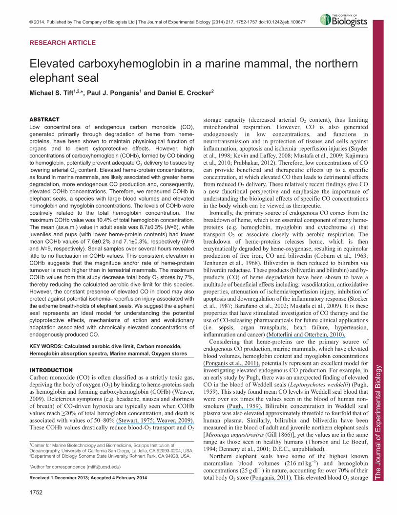
As originally suggested in Weddell seals, we believe that the highendogenous CO values in northern elephant seals from this study canbe attributed primarily to the elevated heme stores associated withincreased myoglobin content, blood volume, hematocrit andhemoglobin concentrations (Pugh, 1959; Ponganis et al., 1993;Thorson and Le Boeuf, 1994). In elephant seals, the molar equivalentsof heme from both hemoglobin and myoglobin stores alone areapproximately four and 16 times greater, respectively, than those inadult humans (Fig. 5). The levels of COHb measured in elephant sealssuggest: (1) more rapid turnover of heme stores (i.e. shorter half-lifeof heme-proteins or erythrocytes), as is found in human patients with
hemolytic anemia (Coburn et al., 1966; Hampson, 2007), (2) a greatermagnitude of heme degradation than has been measured in othermammals, or (3) a combination of both. A greater magnitude of hemedegradation could be associated simply with the elevated hemeconcentrations and/or with an increase in the activity or concentrationof heme-oxygenase, the enzyme responsible for heme degradation andendogenous CO production.
Currently, the life-span of heme-proteins and erythrocytes inmarine mammals is unknown, but an increased heme turnover ratecould elevate the production rate of endogenous CO. Additionally,the natural and repetitive breath-holds both at sea (diving) and onland (sleep apnea) limit the time elephant seals spend in eupnea, andtherefore, decrease or at least delay the exhalation of endogenouslyproduced CO. Between these two breath-hold behaviors, a typicalelephant seal will spend a majority of its life in an apneic state(Andrews et al., 2000) and thus, this natural breath-holding behaviormay also contribute to the build-up of endogenous CO.
Unlike Weddell seals (Pugh, 1959), elephant seals do not have anelevation in the hemoglobin breakdown product bilirubin (D.E.C.,unpublished) to accompany the high blood CO levels seen in theseanimals. This is especially interesting in elephant seal pups, whereneonatal hyperbilirubinemia might be expected because of thecommon occurrence in human infants, which have much lowerheme stores (Dennery et al., 2001). Neonatal hyperbilirubinemia hasalso been found in some neonate marine mammals (Dierauf et al.,1984), yet there are relatively low levels of bilirubin found in all ageclasses of elephant seals (Costa and Ortiz, 1982; Champagne et al.,2013; D.E.C., unpublished). One possibility is that the clearance rateor recycling of bilirubin and biliverdin relative to absolute total bodyheme content and heme turnover in these animals is adequate tomaintain lower levels of those metabolites.
Effect of elevated CO on O2 stores and O2 deliveryThe concepts of increased O2 storage, a low diving metabolic rate, andaerobic diving underlie current interpretations of diving physiology,dive performance and foraging ecology in apex marine predators(Kooyman, 1989; Butler and Jones, 1997; Ponganis et al., 2003). Total
AdultsJuvenilesPups
12
10
8
6
4
214 16 18 20 22 24 26 28
Hemoglobin (g dl–1)
CO
Hb
(%)
Fig. 3. Relationship between the total concentration of hemoglobin andpercent carboxyhemoglobin (COHb) in the blood from three differentage classes of northern elephant seals. The regression line is in red(COHb=–1.31+0.42tHb), and the 95% confidence intervals are in black(F3,20=30.1, P<0.0001, r2=0.80).
8 10 12 14 1602468
10121416
8 12 16 20
CO
Hb
(%)
02468
10121416
Time of day (h)7 9 11 13 15 17
02468
10121416
9 11 13 15 17 1902468
10121416
9 11 13 15 17 1902468
10121416
10 12 14 16 18 2002468
10121416
Fig. 4. Time course ofcarboxyhemoglobin (COHb) valuesfrom six juvenile elephant seals. Thered lines represent the best-fit linearregression line for the data and the blacklines represent 95% confidence intervals.
The
Jour
nal o
f Exp
erim
enta
l Bio
logy
1755
RESEARCH ARTICLE The Journal of Experimental Biology (2014) doi:10.1242/jeb.100677
body O2 stores (lung, blood and muscle) are used to calculate theaerobic dive limit (cADL), which is a prediction of the maximumduration of a dive or breath-hold before a post-dive rise in plasmalactate occurs (Kooyman and Kooyman, 1995; Butler and Jones,1997; Kooyman and Ponganis, 1998; Costa et al., 2001). For thiscalculation, which is performed by dividing total body O2 stores bythe rate of breath-hold O2 consumption, all measured hemoglobin isincluded in the blood O2 store (which can make up 70% of the totalO2 store in some species). However, the present study shows that over10% of total hemoglobin content can be bound to CO in the premierpinniped diver, effectively reducing its total body O2 stores by ~7%.
When these CO-related reductions in O2 stores are taken intoconsideration in the calculation of a cADL of adult elephant seals[~30 min (Hassrick et al., 2010)], there is reduction of ~2 min. Thisreduction might help explain why the majority of dives in femaleelephant seals are actually 22–23 min (Robinson et al., 2012) insteadof the predicted cADL of 30 min. The elevated COHb values seenin elephant seals from this study should heighten awareness of thepotential for endogenous CO to reduce the magnitude of total bodyO2 stores calculated in other species.
Potential therapeutic benefits of elevated CO in marinemammalsThe routine hypoxemia, hypercarbia, reduction in peripheralperfusion, and activation of the sympathetic nervous system during
both diving and sleep apnea in elephant seals potentially increase therisks of systemic and pulmonary hypertension, ischemia–reperfusioninjury, reactive oxygen species formation, and subsequent tissuedamage (Kooyman, 1989; Butler and Jones, 1997; Kooyman andPonganis, 1998; Zenteno-Savín et al., 2002). Adaptations thatpotentially decrease such risks in marine mammals include hypoxicpulmonary vasodilatation and elevations in both antioxidantconcentrations and antioxidant enzyme activities (Zenteno-Savín etal., 2002; Olson et al., 2010; Tift et al., 2011; Vázquez-Medina etal., 2011). We propose that the levels of heme degradation, andassociated by-products such as endogenous CO found in elephantseals, may also contribute to the protection against these conditions.
Ischemia–reperfusion injury is commonly seen during a varietyof clinical scenarios, including heart attacks, strokes and organtransplantation (Carden and Granger, 2000). Deleterious effectsassociated with ischemia–reperfusion injury typically includeapoptosis, intense inflammation and thrombogenesis. However,exposure to low levels of CO significantly decreases the risk ofthese injuries during ischemia–reperfusion events, and is currentlybeing applied in clinical investigations (Ozaki et al., 2012).Therefore, the elevated levels of endogenous CO found in elephantseals could provide protection against potential ischemia–reperfusion injury, associated with the marked changes in peripheralperfusion associated with breath-holds (Ponganis et al., 2008).Additionally, low concentrations of CO are known to promotevasodilation and decrease hypertension in several species. Forexample, in the llama (Lama glama) at high altitude, elevatedpulmonary CO production is associated with decreased pulmonaryhypertension (Herrera et al., 2008). And lastly, although smokingduring pregnancy is associated with numerous adverse outcomesand low birth weights (Cnattingius and Lambe, 2002), there hasbeen shown to be an associated decreased risk of hypertension andpre-eclampsia (Wikström et al., 2010). Thus, elevated CO may alsocontribute to vascular regulation for coping with hypoxemia andtissue vasoconstriction during breath-holding in seals (Ponganis etal., 2008; Meir et al., 2009).
While the mechanism is still unknown, placental blood flow ismaintained during extreme peripheral vasoconstriction associatedwith the dive response in a phocid seal (Liggins et al., 1980).Recently demonstrated in the pregnant mouse (Venditti et al., 2013),the elevated CO exposure in seals may help optimize uterine bloodflow and placental growth during the at-sea pregnancies ofcontinuously diving female elephant seals. However, complicatingthis suggestion is the negative relationship between smoking (andpotentially CO exposure) and endothelial nitric oxide synthaseactivity, which could reduce vasodilatory capacity in the fetus(Andersen et al., 2009).
Exposure to other heme-protein breakdown products (bilirubinand biliverdin) are also associated with a wide array of potentialtherapeutic effects in the avoidance of re-perfusion injury andoxidative stress. These include anti-inflammatory, anti-apoptosis andanti-oxidative responses (Wu and Wang, 2005; Ryter et al., 2007;Motterlini and Otterbein, 2010). Thus, although CO decreases O2
storage, it has the potential to offer several other beneficial effectsin diving animals and other animals exposed to inflammation,apoptosis and oxidative stress.
We conclude with the suggestion that the elevated endogenousCO and COHb concentrations found in deep-diving phocid sealsrepresent an excellent opportunity to study and understand thefundamentals and extremes of this unique physiological system(Krogh, 1929). With the growing knowledge on the therapeuticpotential of carbon monoxide, deep-diving seals may become a
A
B
10
8
6
4
2
0
0.0014
0.0012
0.0010
0.0008
0.0006
0.0004
0.0002
0
Elephant seal Human
Mas
s-sp
ecifi
c m
olar
equ
ival
ents
of
hem
e fro
m h
emog
lobi
n (m
ol k
g–1)
Mas
s-sp
ecifi
c m
olar
equ
ival
ents
of
hem
e fro
m m
yogl
obin
(mol
kg–1
)
Elephant seal Human
Fig. 5. Mass-specific molar equivalents of heme calculated from (A)blood (hemoglobin) and (B) muscle (myoglobin) oxygen stores innorthern elephant seals and humans. Molar heme equivalents werecalculated using a hemoglobin molecular weight of 65,000 g mol–1 andmyoglobin molecular weight of 17,000 g mol–1. Data for species-specificblood volumes, muscle mass, hemoglobin and myoglobin concentrations arefrom previous studies (Kendrew et al., 1958; Hill et al., 1962; Åkeson et al.,1968; Oscai et al., 1968; Simpson et al., 1970; Bryden, 1972; Möller andSylven, 1981; Hassrick et al., 2010; Ponganis et al., 2011).

The
Jour
nal o
f Exp
erim
enta
l Bio
logy
1756
RESEARCH ARTICLE The Journal of Experimental Biology (2014) doi:10.1242/jeb.100677
valuable model to investigate the potential cytoprotective effects,mechanisms of action and evolutionary adaptation from long-termexposure to elevated concentrations of this endogenously producedgasotransmitter.
MATERIALS AND METHODSElephant seal COHb absorption spectrumTo validate the use of a Bayer Rapidlab 845 blood gas analyzer with co-oximeter (Siemens Medical Diagnostics, Bayer, Tarrytown, NY, USA) forthe determination of elephant seal hemoglobin values, we confirmed that thehemoglobin absorption spectra of elephant seals were similar to those ofhumans and other terrestrial animals. Hemoglobin from the blood of healthyjuvenile northern elephant seals (n=2, see blood collection technique,below), sheep (n=1) and cattle (n=1) (sheep and cattle blood obtained fromHemostat Laboratories, Dixon, CA, USA) were spectrophotometricallycompared with hemoglobin absorption peak values for human, cattle andsheep from Zijlstra et al. (Zijlstra et al., 2000). Isolated hemoglobinabsorption peaks for oxygenated hemoglobin (O2Hb), deoxygenatedhemoglobin (HHb) and COHb were measured and compared using methodssimilar to that described previously (Kreutzer et al., 1993; Zijlstra et al.,2000). Briefly, blood was collected into chilled vacutainers containing158 USP units (equivalent to 158 IU) of sodium heparin (BD, FranklinLakes, NJ, USA). Plasma was discarded and erythrocytes were washed threetimes with 0.9% NaCl solution. Erythrocytes were then lysed with 1 volumeof distilled H2O and 0.4 volumes of toluene and left to sit at 4°C for up to16 h. This resulted in a top layer of toluene, a middle layer of erythrocytestromata and a bottom layer of hemoglobin lysate. The bottom layer wascollected and centrifuged at 8000 g for 20 min, and the erythrolysate wasthen filtered through sterile gauze and diluted with deionized water (Milli-Q, Millipore, Billerica, MA, USA). The diluted erythrolysate was thenplaced inside a tonometer where respective gases were introduced. Thedifferent hemoglobin forms (HHb, O2Hb and COHb) were prepared byintroducing 100% nitrogen (N2), O2 and CO, respectively, into the tonometerfor up to 2 h at a rate of 30–40 ml min−1. For O2Hb and COHb, polystyrenespectrophotometer cuvettes (light-path=1.0 cm) covered with Parafilm® Mwere filled with 2.5 ml of erythrolysate collected from the tonometer usingan airtight blood-gas syringe, thereby minimizing exposure to the ambientenvironment. For HHb, an addition of 3 mg of sodium hydrosulfite(Na2S2O4) per 2 ml of buffered erythrolysate was included for tonometry toprevent O2 or CO from binding. Once the lysate was transferred, cuvetteswere then immediately capped, sealed with Parafilm® M and immediatelyanalyzed.
Absorbance measurements were made at room temperature (20–25°C)with a Cary 60 UV-Vis spectrophotometer (Agilent Technologies, SantaClara, CA, USA) in the spectral range of 450–700 nm.
Study site and subjectsWe sampled 24 northern elephant seals throughout the 2009 winter breedingseason (December–April) from Año Nuevo State Reserve, San MateoCounty, CA, USA. Every year, a subset of pups at the reserve are markedwith flipper-tags (Jumbo Rototag, Dalton, UK) following weaning, whichenables reference to the animals’ ages. Subjects were divided into three ageclasses: pups (<3 months old; n=9), juveniles (3 months–3 years old; n=9)and adults (˃3 years old; n=6). Adult animals were all female, and weresampled from January to February at the beginning of their breeding season.Pups were sampled in February–March and juveniles were sampledApril–May.
Animals were chemically immobilized with an intramuscular injection(1 mg kg−1) of Telazol (tiletamine/zolazepam HCl) and subsequentintravenous ketamine injections (0.5 mg kg−1) as necessary to maintainimmobilization for the intravenous catheterization of the extradural vein (alldrugs from Fort Dodge Labs, Fort Dodge, IA, USA). Venous blood samples(~3 ml) were collected into lithium heparinized blood tubes (BDVacutainer®, Fisher Scientific, Franklin Lakes, NJ, USA) and chilled overice until analysis (<3 h). Standard blood gas analysis was performed usinga Bayer Rapidlab 845 blood gas analyzer with co-oximeter (Siemens
Medical Diagnostics, Bayer, Tarrytown, NY, USA) to obtain fractions ofCOHb and tHb concentrations.
To determine the variability of COHb over time and between voluntaryperiods of apnea and eupnea, we opportunistically sampled six of the ninejuvenile elephant seals for up to 12 h. Once sedated, the animals were placedin a holding cage and the extradural vein was percutaneously catheterizedwith a long-term polyurethane catheter (MILA; 16 gauge × 25 cm), whichwas affixed to the fur using Loctite glue (Henkel Corporation, Westlake,OH, USA). An extension tube (76 cm) with a three-way stopcock systemwas attached to the catheter to allow blood collection during periods ofvoluntary sleep apnea and eupnea, with minimal disturbance. Over thecourse of 8–12 h, we collected a total of 806 blood samples ranging from 36to 134 samples per individual.
This work was conducted through the National Marine Fisheries Servicepermit no. 14636 and was approved by the Institutional Animal Care andUse Committee at Sonoma State University.
Statistical analysisData were analyzed using the statistical program JMP 11.0. We examinedthe effects of age class (adult, juvenile and pup) and tHb concentration onthe percentage of COHb in blood samples using a multiple regressionanalysis. Changes in COHb over the course of several hours were evaluatedusing a repeated-measures linear mixed model, with individual ID as therandom subject effect. All mean values are reported ±s.e.m. Significancewas determined at P<0.05.
AcknowledgementsWe thank the rangers and docents at Año Nuevo State Reserve for their logisticalsupport. E. Ranalli, A. Dallara, V. Farnham, J. Jelincic, J. Vázquez-Medina, C.Champagne and M. Fowler provided valuable assistance in the field. Advice fromT. Jue and V. Agarwal on absorption spectra techniques and use of B. Moore’sspectropohotometer are greatly appreciated. We also thank G. Kooyman, J.Maresh and B. McDonald for manuscript review, and J. West, who introducedP.J.P. to Pugh’s 1959 findings and stimulated this research.
Competing interestsThe authors declare no competing financial interests.
Author contributionsM.S.T., P.J.P. and D.E.C. designed the study. Field work, execution of experimentsand data analysis were conducted by M.S.T. and D.E.C. Manuscript preparationand interpretation of results were conducted by M.S.T., P.J.P. and D.E.C.
FundingFunding for this project was provided by the Office of Naval Research[N000141010514 to P.J.P.]. Graduate student support for M.S.T. was provided byNational Institutes of Health [NHLBI R01-HL091767 to R.M.O.] and the NationalScience Foundation [09-44220 to P.J.P.]. Deposited in PMC for release after 12months.
ReferencesÅkeson, A., Biörck, G. and Simon, R. (1968). On the content of myoglobin in human
muscles. Acta Med. Scand. 183, 307-316. Andersen, M. R., Simonsen, U., Uldbjerg, N., Aalkjaer, C. and Stender, S. (2009).
Smoking cessation early in pregnancy and birth weight, length, head circumference,and endothelial nitric oxide synthase activity in umbilical and chorionic vessels: anobservational study of healthy singleton pregnancies. Circulation 119, 857-864.
Andrews, R. D., Jones, D. R., Williams, J. D., Thorson, P. H., Oliver, G. W., Costa,D. P. and Le Boeuf, B. J. (1997). Heart rates of northern elephant seals diving atsea and resting on the beach. J. Exp. Biol. 200, 2083-2095.
Andrews, R. D., Costa, D. P., Le Boeuf, B. J. and Jones, D. R. (2000). Breathingfrequencies of northern elephant seals at sea and on land revealed by heart ratespectral analysis. Respir. Physiol. 123, 71-85.
Barañano, D. E., Rao, M., Ferris, C. D. and Snyder, S. H. (2002). Biliverdinreductase: a major physiologic cytoprotectant. Proc. Natl. Acad. Sci. USA 99, 16093-16098.
Blackwell, S. B. and Le Boeuf, B. (1993). Developmental aspects of sleep apnoea innorthern elephant seals, Mirounga angustirostris. J. Zool. 231, 437-447.
Bryden, M. (1972). Body size and composition of elephant seals (Mirounga leonine):absolute measurements and estimates from bone dimensions. J. Zool. 167, 265-276.
Butler, P. J. and Jones, D. R. (1997). Physiology of diving of birds and mammals.Physiol. Rev. 77, 837-899.
Carden, D. L. and Granger, D. N. (2000). Pathophysiology of ischaemia-reperfusioninjury. J. Pathol. 190, 255-266.
The
Jour
nal o
f Exp
erim
enta
l Bio
logy
1757
RESEARCH ARTICLE The Journal of Experimental Biology (2014) doi:10.1242/jeb.100677
Champagne, C. D., Boaz, S. M., Fowler, M. A., Houser, D. S., Costa, D. P. andCrocker, D. E. (2013). A profile of carbohydrate metabolites in the fasting northernelephant seal. Comp. Biochem. Physiol. 8D, 141-151.
Cnattingius, S. and Lambe, M. (2002). Trends in smoking and overweight duringpregnancy: prevalence, risks of pregnancy complications, and adverse pregnancyoutcomes. Semin. Perinatol. 26, 286-295.
Coburn, R. F., Blakemore, W. S. and Forster, R. E. (1963). Endogenous carbonmonoxide production in man. J. Clin. Invest. 42, 1172-1178.
Coburn, R. F., Williams, W. J. and Kahn, S. B. (1966). Endogenous carbon monoxideproduction in patients with hemolytic anemia. J. Clin. Invest. 45, 460-468.
Costa, D. P. and Ortiz, C. L. (1982). Blood chemistry homeostasis during prolongedfasting in the northern elephant seal. Am. J. Physiol. 242, R591-R595.
Costa, D. P., Gales, N. J. and Goebel, M. E. (2001). Aerobic dive limit: how oftendoes it occur in nature? Comp. Biochem. Physiol. 129A, 771-783.
Dennery, P. A., Seidman, D. S. and Stevenson, D. K. (2001). Neonatalhyperbilirubinemia. N. Engl. J. Med. 344, 581-590.
Dierauf, L., Dougherty, S. and Baker, B. (1984). Neonatal hyperbilirubinemia inharbor seals (Phoca vitulina richardsi). J. Zoo Animal Med. 15, 55-59.
Hampson, N. B. (2007). Carboxyhemoglobin elevation due to hemolytic anemia. J.Emerg. Med. 33, 17-19.
Hassrick, J. L., Crocker, D. E., Teutschel, N. M., McDonald, B. I., Robinson, P. W.,Simmons, S. E. and Costa, D. P. (2010). Condition and mass impact oxygen storesand dive duration in adult female northern elephant seals. J. Exp. Biol. 213, 585-592.
Herrera, E. A., Reyes, R. V., Giussani, D. A., Riquelme, R. A., Sanhueza, E. M.,Ebensperger, G., Casanello, P., Méndez, N., Ebensperger, R., Sepúlveda-Kattan, E. et al. (2008). Carbon monoxide: a novel pulmonary artery vasodilator inneonatal llamas of the Andean altiplano. Cardiovasc. Res. 77, 197-201.
Hill, R. J., Konigsberg, W., Guidotti, G. and Craig, L. C. (1962). The structure ofhuman hemoglobin. I. The separation of the α and β chains and their amino acidcomposition. J. Biol. Chem. 237, 1549-1554.
Kajimura, M., Fukuda, R., Bateman, R. M., Yamamoto, T. and Suematsu, M. (2010).Interactions of multiple gas-transducing systems: hallmarks and uncertainties of CO,NO, and H2S gas biology. Antioxid. Redox Signal. 13, 157-192.
Kendrew, J. C., Bodo, G., Dintzis, H. M., Parrish, R. G., Wyckoff, H. and Phillips,D. C. (1958). A three-dimensional model of the myoglobin molecule obtained by x-ray analysis. Nature 181, 662-666.
Kevin, L. G. and Laffey, J. G. (2008). Carbon monoxide: from poison to therapy forcardiopulmonary bypass-induced lung injury? Anesthesiology 108, 977-978.
Kooyman, G. L. (1989). Diverse Divers: Physiology and Behaviour. Berlin: Springer-Verlag.
Kooyman, G. and Kooyman, T. (1995). Diving behavior of emperor penguinsnurturing chicks at Coulman Island, Antarctica. Condor 97, 536-549.
Kooyman, G. L. and Ponganis, P. J. (1998). The physiological basis of diving todepth: birds and mammals. Annu. Rev. Physiol. 60, 19-32.
Kreutzer, U., Chung, Y., Butler, D. and Jue, T. (1993). 1H-NMR characterization of thehuman myocardium myoglobin and erythrocyte hemoglobin signals. Biochim.Biophys. Acta 1161, 33-37.
Krogh, A. (1929). The progress of physiology. J. Physiol. 90, 243-251. Law, M. R., Morris, J. K., Watt, H. C. and Wald, N. J. (1997). The dose–response
relationship between cigarette consumption, biochemical markers and risk of lungcancer. Br. J. Cancer 75, 1690-1693.
Liggins, G. C., Qvist, J., Hochachka, P. W., Murphy, B. J., Creasy, R. K.,Schneider, R. C., Snider, M. T. and Zapol, W. M. (1980). Fetal cardiovascular andmetabolic responses to simulated diving in the Weddell seal. J. Appl. Physiol. 49,424-430.
Meir, J. U., Champagne, C. D., Costa, D. P., Williams, C. L. and Ponganis, P. J.(2009). Extreme hypoxemic tolerance and blood oxygen depletion in diving elephantseals. Am. J. Physiol. 297, R927-R939.
Möller, P. and Sylvén, C. (1981). Myoglobin in human skeletal muscle. Scand. J. Clin.Lab. Invest. 41, 479-482.
Motterlini, R. and Otterbein, L. E. (2010). The therapeutic potential of carbonmonoxide. Nat. Rev. Drug Discov. 9, 728-743.
Mustafa, A. K., Gadalla, M. M. and Snyder, S. H. (2009). Signaling bygasotransmitters. Sci. Signal. 2, re2.
Olson, K. R., Whitfield, N. L., Bearden, S. E., St Leger, J., Nilson, E., Gao, Y. andMadden, J. A. (2010). Hypoxic pulmonary vasodilation: a paradigm shift with ahydrogen sulfide mechanism. Am. J. Physiol. 298, R51-R60.
Oscai, L. B., Williams, B. T. and Hertig, B. A. (1968). Effect of exercise on bloodvolume. J. Appl. Physiol. 24, 622-624.
Ozaki, K. S., Kimura, S. and Murase, N. (2012). Use of carbon monoxide inminimizing ischemia/reperfusion injury in transplantation. Transplant. Rev. 26, 125-139.
Ponganis, P. J. (2011). Diving mammals. Compr. Physiol. 1, 447-465.
Ponganis, P. J., Kooyman, G. L. and Castellini, M. A. (1993). Determinants of theaerobic dive limit of weddell seals: analysis of diving metabolic rates, postdive endtidal PO2’s, and blood and muscle oxygen stores. Physiol. Zool. 66, 732-749.
Ponganis, P. J., Kooyman, G. L. and Ridgway, S. H. (2003). Comparative divingphysiology. In Bennett and Elliott’s Physiology and Medicine of Diving, 5th. edn. (A.O. Brubakk and T. S. Neuman), pp. 211-226. Edinburgh: Saunders Ltd.
Ponganis, P. J., Stockard, T. K., Levenson, D. H., Berg, L. and Baranov, E. A.(2006). Cardiac output and muscle blood flow during rest-associated apneas ofelephant seals. Comp. Biochem. Physiol. 144A, 105-111.
Ponganis, P. J., Kreutzer, U., Stockard, T. K., Lin, P. C., Sailasuta, N., Tran, T. K.,Hurd, R. and Jue, T. (2008). Blood flow and metabolic regulation in seal muscleduring apnea. J. Exp. Biol. 211, 3323-3332.
Ponganis, P. J., Meir, J. U. and Williams, C. L. (2011). In pursuit of Irving andScholander: a review of oxygen store management in seals and penguins. J. Exp.Biol. 214, 3325-3339.
Prabhakar, N. R. (2012). Carbon monoxide (CO) and hydrogen sulfide (H2S) inhypoxic sensing by the carotid body. Respir. Physiol. Neurobiol. 184, 165-169.
Pugh, L. G. (1959). Carbon monoxide content of the blood and other observations onWeddell seals. Nature 183, 74-76.
Robinson, P. W., Costa, D. P., Crocker, D. E., Gallo-Reynoso, J. P., Champagne, C.D., Fowler, M. A., Goetsch, C., Goetz, K. T., Hassrick, J. L., Hückstädt, L. A. etal. (2012). Foraging behavior and success of a mesopelagic predator in thenortheast Pacific Ocean: insights from a data-rich species, the northern elephantseal. PLoS ONE 7, e36728.
Ryter, S. W., Morse, D. and Choi, A. M. (2007). Carbon monoxide and bilirubin:potential therapies for pulmonary/vascular injury and disease. Am. J. Respir. CellMol. Biol. 36, 175-182.
Simpson, J. G., Gilmartin, W. G. and Ridgway, S. H. (1970). Blood Volume andOther Hematologic Values in Young Elephant Seals (Mirounga angustirostris).Belvoir, VA: Defense Technical Information Center.
Snyder, S. H., Jaffrey, S. R. and Zakhary, R. (1998). Nitric oxide and carbonmonoxide: parallel roles as neural messengers. Brain Res. Brain Res. Rev. 26, 167-175.
Stewart, R. D. (1975). The effect of carbon monoxide on humans. Annu. Rev.Pharmacol. 15, 409-423.
Stockard, T. K., Levenson, D. H., Berg, L., Fransioli, J. R., Baranov, E. A. andPonganis, P. J. (2007). Blood oxygen depletion during rest-associated apneas ofnorthern elephant seals (Mirounga angustirostris). J. Exp. Biol. 210, 2607-2617.
Stocker, R., Glazer, A. N. and Ames, B. N. (1987). Antioxidant activity of albumin-bound bilirubin. Proc. Natl. Acad. Sci. USA 84, 5918-5922.
Tenhunen, R., Marver, H. S. and Schmid, R. (1968). The enzymatic conversion ofheme to bilirubin by microsomal heme oxygenase. Proc. Natl. Acad. Sci. USA 61,748-755.
Thorson, P. H. and Le Boeuf, B. (1994). Developmental aspects of diving in northernelephant seal pups. In Elephant Seals: Population Ecology, Behavior, andPhysiology (ed. B. J. Le Boeuf and R. M. Laws), pp. 271-289. Berkeley, CA:University of California Press.
Tift, M. S., Houser, D. S. and Crocker, D. E. (2011). High-density lipoprotein remainselevated despite reductions in total cholesterol in fasting adult male elephant seals(Mirounga angustirostris). Comp. Biochem. Physiol. 159B, 214-219.
Tift, M. S., Ranalli, E. C., Houser, D. S., Ortiz, R. M. and Crocker, D. E. (2013).Development enhances hypometabolism in northern elephant seal pups (Miroungaangustirostris). Funct. Ecol. 27, 1155-1165.
Vázquez-Medina, J. P., Zenteno-Savín, T., Tift, M. S., Forman, H. J., Crocker, D. E.and Ortiz, R. M. (2011). Apnea stimulates the adaptive response to oxidative stressin elephant seal pups. J. Exp. Biol. 214, 4193-4200.
Venditti, C. C., Casselman, R., Murphy, M. S., Adamson, S. L., Sled, J. G. andSmith, G. N. (2013). Chronic carbon monoxide inhalation during pregnancyaugments uterine artery blood flow and uteroplacental vascular growth in mice. Am.J. Physiol. 305, R939-R948.
Weaver, L. K. (2009). Clinical practice. Carbon monoxide poisoning. N. Engl. J. Med.360, 1217-1225.
Wikström, A.-K., Stephansson, O. and Cnattingius, S. (2010). Tobacco use duringpregnancy and preeclampsia risk: effects of cigarette smoking and snuff.Hypertension 55, 1254-1259.
Wu, L. and Wang, R. (2005). Carbon monoxide: endogenous production, physiologicalfunctions, and pharmacological applications. Pharmacol. Rev. 57, 585-630.
Zenteno-Savín, T., Clayton-Hernández, E. and Elsner, R. (2002). Diving seals: arethey a model for coping with oxidative stress? Comp. Biochem. Physiol. 133C, 527-536.
Zijlstra, W. G., Buursma, A. and van Assendelft, O. W. (2000). Visible and NearInfrared Absorption Spectra of Human and Animal Haemoglobin: Determination andApplication. Leiden, The Netherlands: VSP BV.
Related Documents