Electrostatics Control Actin Filament Nucleation and Elongation Kinetics * □ S Received for publication, February 2, 2013, and in revised form, March 11, 2013 Published, JBC Papers in Press, March 13, 2013, DOI 10.1074/jbc.M113.456327 Alvaro H. Crevenna ‡§1 , Nikolaus Naredi-Rainer § , Andre ´ Scho ¨ nichen ¶ , Joachim Dzubiella , Diane L. Barber ¶ , Don C. Lamb § ** ‡‡2 , and Roland Wedlich-So ¨ ldner ‡3 From ‡ AG Cellular Dynamics and Cell Patterning, Max Planck Institute of Biochemistry, Am Klopferspitz 18, 82152 Martinsried, Germany, the § Department of Chemistry and Biochemistry and Center for Nanoscience, Ludwig-Maximilians-Universita ¨tMu ¨nchen, Butenandtstrasse 5-13, Haus E, 81377 Mu ¨nchen, Germany, the ¶ Department of Cell and Tissue Biology, University of California, San Francisco, California 94143, Soft Matter and Functional Materials, Helmholtz-Zentrum at Berlin, Hahn-Meitner-Platz 1, 14109 Berlin, Germany, the Institut fu ¨r Physik, Humboldt University at Berlin, Newtonstrasse 15, 12489 Berlin, Germany, the **Munich Center for Integrated Protein Science, Ludwig-Maximilians-Universita ¨tMu ¨nchen, Butenandtstrasse 11, 81377 Mu ¨nchen, Germany, and the ‡‡ Department of Physics, University of Illinois at Urbana-Champaign, Urbana, Illinois 61801 Background: pH plays important roles in cellular morphogenesis, but how actin polymer dynamics are affected by pH is not well understood. Results: Actin filament nucleation and elongation are strongly enhanced at acidic pH due to a reduction of charge repulsion. Conclusion: Actin filament dynamics in vitro and in vivo are strongly influenced by electrostatics. Significance: Cellular pH homeostasis will impact directly on actin dynamics. The actin cytoskeleton is a central mediator of cellular mor- phogenesis, and rapid actin reorganization drives essential pro- cesses such as cell migration and cell division. Whereas several actin-binding proteins are known to be regulated by changes in intracellular pH, detailed information regarding the effect of pH on the actin dynamics itself is still lacking. Here, we combine bulk assays, total internal reflection fluorescence microscopy, fluorescence fluctuation spectroscopy techniques, and theory to comprehensively characterize the effect of pH on actin poly- merization. We show that both nucleation and elongation are strongly enhanced at acidic pH, with a maximum close to the pI of actin. Monomer association rates are similarly affected by pH at both ends, although dissociation rates are differentially affected. This indicates that electrostatics control the diffu- sional encounter but not the dissociation rate, which is critical for the establishment of actin filament asymmetry. A generic model of protein-protein interaction, including electrostatics, explains the observed pH sensitivity as a consequence of charge repulsion. The observed pH effect on actin in vitro agrees with measurements of Listeria propulsion in pH-controlled cells. pH regulation should therefore be considered as a modulator of actin dynamics in a cellular environment. The actin cytoskeleton plays a crucial role in many funda- mental cellular processes such as adhesion, polarization, migra- tion, division, and differentiation. This pivotal function of actin is largely due to its capacity to self-assemble into linear poly- mers with a directed polarity, a process that generates local mechanical forces (1–3). Arrays and networks of actin fila- ments in turn provide cells with a skeleton that determines shape and helps to resist and sense mechanical deformation (4, 5). An essential feature of the actin cytoskeleton is its dynamic nature, known to be regulated by a multitude of signaling path- ways. However, one ubiquitous parameter that has been shown to affect actin polymerization (21, 22) has been overlooked in most studies, i.e. pH. The physiological role of increasing pH in actin polymerization has already been observed in early studies on fertilization of sea urchin eggs (6) and on the acrosomal reaction of echinoderm sperms (7). More recently, molecular mechanisms contributing to pH regulation of actin turnover (8) and force generation (9) have been identified. In this respect, the major actin severing and depolymerizing factor cofilin has been shown to be inhibited by low pH (8). Similarly, activity of talin, which links actin to integrins in focal adhesion and helps traction force generation, has been shown to be sensitive to local pH (9). The major regulator of intracellular pH (pH i ) in mammalian cells, the Na/H exchanger 1 (NHE1), 4 is localized at the leading edge, where continuous polymerization and turn- * This work was supported, in whole or in part, by National Institutes of Health Grant GM58642. This work was also supported by Deutsche Forschungs- gemeinschaft Grants SFB 863 and SPP 1464 and Emmy-Noether Programm (to J. D.), the Ludwig-Maximilians-Universita ¨tMu ¨ nchen (LMUInnovativBio- Imaging Network), the Nanosystems Initiative Munich, and the Max Planck Society. □ S This article contains supplemental Figs. S1–S3 and an additional reference. 1 To whom correspondence may be addressed: Physical Chemistry, Dept. of Chemistry and Biochemistry and Center for Nanoscience, Ludwig- Maximilians-Universita ¨ t Mu ¨ nchen, Butenandtstrasse 5-13, Haus E, 81377, Mu ¨ nchen, Germany. Tel.: 49-89-2180-77516; E-mail: alvaro.crevenna@ cup.uni-muenchen.de. 2 To whom correspondence may be addressed: Physical Chemistry, Dept. of Chemistry and Biochemistry and Center for Nanoscience, Ludwig-Maximil- ians-Universita ¨t Mu ¨ nchen, Butenandtstrasse 5-13, Haus E, 81377, Mu ¨ nchen, Germany. Tel.: 49-89-2180-77564; E-mail: don.lamb@cup. uni-muenchen.de. 3 To whom correspondence may be addressed: AG Cellular Dynamics and Cell Patterning, Max Planck Institute of Biochemistry, Am Klopferspitz 18, 82152 Martinsried, Germany. Tel.: 49-89-8578-3410; E-mail: wedlich@ biochem.mpg.de. 4 The abbreviations used are: NHE1, sodium proton exchanger 1; TIRF, total internal reflection fluorescence; TIRFM, TIRF microscopy; FFS, fluorescence fluctuation spectroscopy; RFP, red fluorescent protein; PCH, photon- counting histogram. THE JOURNAL OF BIOLOGICAL CHEMISTRY VOL. 288, NO. 17, pp. 12102–12113, April 26, 2013 © 2013 by The American Society for Biochemistry and Molecular Biology, Inc. Published in the U.S.A. 12102 JOURNAL OF BIOLOGICAL CHEMISTRY VOLUME 288 • NUMBER 17 • APRIL 26, 2013

Welcome message from author
This document is posted to help you gain knowledge. Please leave a comment to let me know what you think about it! Share it to your friends and learn new things together.
Transcript

Electrostatics Control Actin Filament Nucleation andElongation Kinetics*□S
Received for publication, February 2, 2013, and in revised form, March 11, 2013 Published, JBC Papers in Press, March 13, 2013, DOI 10.1074/jbc.M113.456327
Alvaro H. Crevenna‡§1, Nikolaus Naredi-Rainer§, Andre Schonichen¶, Joachim Dzubiella�, Diane L. Barber¶,Don C. Lamb§**‡‡2, and Roland Wedlich-Soldner‡3
From ‡AG Cellular Dynamics and Cell Patterning, Max Planck Institute of Biochemistry, Am Klopferspitz 18, 82152 Martinsried,Germany, the §Department of Chemistry and Biochemistry and Center for Nanoscience, Ludwig-Maximilians-Universitat Munchen,Butenandtstrasse 5-13, Haus E, 81377 Munchen, Germany, the ¶Department of Cell and Tissue Biology, University of California,San Francisco, California 94143, �Soft Matter and Functional Materials, Helmholtz-Zentrum at Berlin, Hahn-Meitner-Platz 1,14109 Berlin, Germany, the Institut fur Physik, Humboldt University at Berlin, Newtonstrasse 15, 12489 Berlin, Germany, the**Munich Center for Integrated Protein Science, Ludwig-Maximilians-Universitat Munchen, Butenandtstrasse 11, 81377 Munchen,Germany, and the ‡‡Department of Physics, University of Illinois at Urbana-Champaign, Urbana, Illinois 61801
Background: pHplays important roles in cellularmorphogenesis, but how actin polymer dynamics are affected by pH is notwell understood.Results: Actin filament nucleation and elongation are strongly enhanced at acidic pH due to a reduction of charge repulsion.Conclusion: Actin filament dynamics in vitro and in vivo are strongly influenced by electrostatics.Significance: Cellular pH homeostasis will impact directly on actin dynamics.
The actin cytoskeleton is a central mediator of cellular mor-phogenesis, and rapid actin reorganization drives essential pro-cesses such as cell migration and cell division. Whereas severalactin-binding proteins are known to be regulated by changes inintracellular pH, detailed information regarding the effect of pHon the actin dynamics itself is still lacking. Here, we combinebulk assays, total internal reflection fluorescence microscopy,fluorescence fluctuation spectroscopy techniques, and theory tocomprehensively characterize the effect of pH on actin poly-merization. We show that both nucleation and elongation arestrongly enhanced at acidic pH, with a maximum close to the pIof actin.Monomer association rates are similarly affected by pHat both ends, although dissociation rates are differentiallyaffected. This indicates that electrostatics control the diffu-sional encounter but not the dissociation rate, which is criticalfor the establishment of actin filament asymmetry. A genericmodel of protein-protein interaction, including electrostatics,explains the observed pH sensitivity as a consequence of charge
repulsion. The observed pH effect on actin in vitro agrees withmeasurements of Listeria propulsion in pH-controlled cells. pHregulation should therefore be considered as a modulator ofactin dynamics in a cellular environment.
The actin cytoskeleton plays a crucial role in many funda-mental cellular processes such as adhesion, polarization,migra-tion, division, and differentiation. This pivotal function of actinis largely due to its capacity to self-assemble into linear poly-mers with a directed polarity, a process that generates localmechanical forces (1–3). Arrays and networks of actin fila-ments in turn provide cells with a skeleton that determinesshape and helps to resist and sense mechanical deformation (4,5). An essential feature of the actin cytoskeleton is its dynamicnature, known to be regulated by a multitude of signaling path-ways. However, one ubiquitous parameter that has been shownto affect actin polymerization (21, 22) has been overlooked inmost studies, i.e. pH. The physiological role of increasing pH inactin polymerization has already been observed in early studieson fertilization of sea urchin eggs (6) and on the acrosomalreaction of echinoderm sperms (7). More recently, molecularmechanisms contributing to pH regulation of actin turnover (8)and force generation (9) have been identified. In this respect,the major actin severing and depolymerizing factor cofilin hasbeen shown to be inhibited by low pH (8). Similarly, activity oftalin, which links actin to integrins in focal adhesion and helpstraction force generation, has been shown to be sensitive tolocal pH (9). The major regulator of intracellular pH (pHi) inmammalian cells, the Na/H� exchanger 1 (NHE1),4 is localizedat the leading edge, where continuous polymerization and turn-
* This work was supported, in whole or in part, by National Institutes of HealthGrant GM58642. This work was also supported by Deutsche Forschungs-gemeinschaft Grants SFB 863 and SPP 1464 and Emmy-Noether Programm(to J. D.), the Ludwig-Maximilians-Universitat Munchen (LMUInnovativBio-Imaging Network), the Nanosystems Initiative Munich, and the Max PlanckSociety.
□S This article contains supplemental Figs. S1–S3 and an additional reference.1 To whom correspondence may be addressed: Physical Chemistry, Dept.
of Chemistry and Biochemistry and Center for Nanoscience, Ludwig-Maximilians-Universitat Munchen, Butenandtstrasse 5-13, Haus E, 81377,Munchen, Germany. Tel.: 49-89-2180-77516; E-mail: [email protected].
2 To whom correspondence may be addressed: Physical Chemistry, Dept. ofChemistry and Biochemistry and Center for Nanoscience, Ludwig-Maximil-ians-Universitat Munchen, Butenandtstrasse 5-13, Haus E, 81377,Munchen, Germany. Tel.: 49-89-2180-77564; E-mail: [email protected].
3 To whom correspondence may be addressed: AG Cellular Dynamics and CellPatterning, Max Planck Institute of Biochemistry, Am Klopferspitz 18,82152 Martinsried, Germany. Tel.: 49-89-8578-3410; E-mail: [email protected].
4 The abbreviations used are: NHE1, sodium proton exchanger 1; TIRF, totalinternal reflection fluorescence; TIRFM, TIRF microscopy; FFS, fluorescencefluctuation spectroscopy; RFP, red fluorescent protein; PCH, photon-counting histogram.
THE JOURNAL OF BIOLOGICAL CHEMISTRY VOL. 288, NO. 17, pp. 12102–12113, April 26, 2013© 2013 by The American Society for Biochemistry and Molecular Biology, Inc. Published in the U.S.A.
12102 JOURNAL OF BIOLOGICAL CHEMISTRY VOLUME 288 • NUMBER 17 • APRIL 26, 2013

over of actin occurs during cell migration (10). NHE1 has beenshown to directly affect cell migration by locally modulatingintracellular pHi (11, 12). Interestingly, the localization ofNHE1 itself is mediated by the actin cytoskeleton (13). pHi ishighly regulated to prevent disruption of basic cell functionssuch as enzyme activity, membrane permeability, cellularmetabolism, ATP maintenance, cell proliferation, and apopto-tic mechanisms (14–17). Not surprisingly, dynamic changes inpH are linked to pathogenic conditions, with many cancer cellsexhibiting elevated pHi (18–20). Although there have beenintense efforts to understand the molecular basis of how pHiregulates cellular actin dynamics (20), many fundamental ques-tions remain unresolved.In contrast to complex cellular actin turnover (6, 7), which
depends on many regulatory actin-binding proteins, polymeri-zation of pure actin in vitro is paradoxically increased at acidicpH (21, 22). In general, pH modulates protein electrostatics byaltering the protonation state of amino acid side chains, there-fore influencing protein-protein interactions (e.g. actin/actin oractin/actin-binding protein). In this way, electrostatics canenhance or retard reaction rates of complex formation that arelimited by diffusion (23, 24), such as barbed-end actin filamentelongation (25). It was suggested, using Brownian dynamic sim-ulations, that electrostatics could establish the actin filamentasymmetry (26), although this prediction has so far not beentested experimentally.Here, we use biochemical assays, TIRFmicroscopy (TIRFM),
and advanced fluorescence fluctuation methods to explore theeffects of electrostatics on actin nucleation and elongation. Ageneric theoretical model provides a framework for ourobservations.
EXPERIMENTAL PROCEDURES
Proteins—G-actin was purified from rabbit or chicken skele-tal muscle (27, 28) and stored at 4 °C for up to 2 weeks. Myosinwas purified and chemically inactivatedwithN-ethylmaleimideaccording to Ref. 29. Actin was labeled in random surfacelysines with atto488 or atto532, or it was purchased fromHypermol (Bielefeld, Germany).Bulk Assays—Polymerization of actin was induced by addi-
tion of one-tenth of the final volume of 10� KMEI buffer (1�contained the following: 50mMKCl, 1mMMgCl2, 1mMEGTA,and 10 mM imidazole HCl, pH 7.0) and incubation for �1 h atroom temperature. For the polymerization assays, 20% pyrene-labeled actin was used (30, 31). Ca2� to Mg2� exchange wasdone by adding one-tenth of the final volume of 10�MEbuffer(50 �M MgCl2, 0.2 mM EGTA) for 5 min. Polymerization waspromoted by addition of one-tenth of the final volume 10�KMEI buffer. The final volumewas 100 �l. Pyrene fluorescencewas monitored by excitation at 365 nm and emission at 407 nmwith a Cary Varian spectrofluorometer. Lag times wereextracted as the intercept from the slope at the inflection pointof the polymerization curve. Depolymerization was carried outby diluting 1 �M of 100% pyrene-actin (polymerized for 2 h atRT in the desired pH) in F-buffer, pH 7.1, to 10 nM. Data fromZimmerle and Frieden (22) was digitized using the freely avail-able program grabit in MATLAB.
Circular Dichroism and Electron Microscopy—CD spectrafrom 195 to 260 nm were acquired with a Jasco J-715 spectro-polarimeter (Jasco). A protein concentration of 10�M in P-buf-fer at the desired pH value was used. The measurements wereperformed at a bandwidth of 5 nm, a response time of 2 s, and ascan rate of 0.1 nm�s�1. For electronmicroscopy, a 3-�l drop ofpolymerized actin was incubated for 90 s on a copper grid pre-viously plasma-cleaned for 30 s and stained with 3% uracil ace-tate for 30 s.Fluorescence Fluctuation Spectroscopy—Photon counting
histogram experiments (32) with pulsed interleaved excitation(33) were performed on a homebuilt confocal multiparameterfluorescence detection setup (33). Themultiparameter fluores-cence detection setup uses a polarizing beam splitter in thedetection path to separate fluorescence photonswith respect totheir polarization, followed by spectral separation. Hence, mul-ticolors can be detected, and the anisotropy can be determinedin addition to the fluorescence lifetime information. The sys-tem is built about a Nikon TE2000 microscope. As picosecondexcitation sources, we used an amplified frequency-doubleddiode laser (PicoTA, PicoQuant, Berlin) at 530 nm and a diodelaser at 636 nm (LDH-P-C-635b PicoQuant) for fluorophoreexcitation. The laser was coupled into a single-mode fiber(AMS Technologies, Martinsried, Germany), collimated(Schafter&Kirchhoff,Hamburg,Germany), and focused on thesample by a �60 1.2NA water immersion objective (Plan ApoVC �60 WI, Nikon, Germany). Average excitation powers of5–10 microwatts were used and measured before the dichroicmirror (DualLine z532/635, AHF Analysentechnik, Munich,Germany) separating excitation and emission beam paths. Thecollected fluorescence was focused on a 50-�m pinhole (Thor-labs, Dachau, Germany) via the microscope tube lens. A polar-izing beam splitter cube (AHF Analysentechnik) separated thetwo polarization planes followed by two identical emission fil-ters (BrightLine HC 582/75, AHF Analysetechnik, Tubingen,Germany) for each polarization. Fluorescence was collected bytwo single-photon-counting avalanche photodiodes (�2SPQR-16, PerkinElmer Life Sciences) and registered by twoindependent TCSPC data collection cards (SPC-154,Becker&Hickl, Berlin, Germany). The lasers and the data col-lection cards were synchronized by the laser controller (Sepia,Picoquant, Berlin, Germany). For the measurements, labeledactin was diluted to the designated concentration in G-bufferfor nonpolymerizing conditions and F-buffer for polymerizingconditions, respectively, and measured in a Teflon sampleholder with a glass coverslip. The photon-counting histogramdata (32, 34) were analyzed using a bin width of 37.5 �s in acustom programwritten inMATLAB. For PCH analysis, 60 nMof atto532-labeled actin monomers were measured either in G-or F-buffer for 30 min. The solutions were preincubated for 60min before the measurements were started to allow equilibra-tion of the sample.TIRF Microscopy and Analysis—Flow cells were made as a
sandwich of a coverslip (20 � 20 mm), parafilm, and a coverslide. Flow cells were passivated by incubating them with 10%BSA in PBS for 10min. Flow cells were washed three times with90 �l of G-buffer. The tethering protein was then applied for 5min and washed again three times with 90 �l of G-buffer. Actin
Electrostatic Control of Actin Polymerization
APRIL 26, 2013 • VOLUME 288 • NUMBER 17 JOURNAL OF BIOLOGICAL CHEMISTRY 12103
was incubated for 5 min on ice with one-tenth volume of 10�ME buffer (MgCl2 and EGTA) to exchange Ca2� for Mg2�.Actin-containing solution was then mixed with imaging buffer(catalase, �-mercaptoethanol, glucose oxidase, D-glucose,0.25% (w/v) methylcellulose, and one-tenth 10� KMEI buffer,with a final pH of 7.1) and introduced into the flow cell. TIRFmicrocopy was performed using a TILL photonics invertedmicroscope. Kymographs were made using Metamorph, andfurther analysis was done in MATLAB.For length distribution measurements, actin was polymer-
ized for 30 min, stabilized with Alexa488-phalloidin, anddiluted 100� for imaging individual filaments on a poly-L-ly-sine-coated coverslip using an epifluorescence Zeiss micro-scope. Length measurements were done manually usingImageJ.Rate Calculations—In general, protein-protein (e.g. A and B)
complex formation (AB) is described by Equation 1,
A � B -|0kD
k�D
A � B -|0kC
k�C
AB (Eq. 1)
where A�B is the transient complex defined with a separationand orientation close to the native complex but where shortrange interactions still have to be formed. kD and k�D are thediffusion-limited rate constant for transient complex formationand the reverse process, respectively. kC and k�C are the rateconstants from the transient complex to tight native complexthrough conformational changes and the reverse process,respectively. The association rate constant, kon, is then definedas kDkC/k�D � kC. The dissociation rate constant, i.e. the offrate, koff, is given by k�DkC/k�D � kC. If the conformationalrearrangement is very fast upon transient complex formation, itis said that the reaction is diffusion-limited or diffusion-con-trolled (i.e. when kC �� k�D).To rationalize the experimental trends in the pH and salt
concentration dependence of the rate constants, we make useof a simple generic theory. We assume the reaction is com-pletely diffusion-controlled and isotropic, and the two reactiveconstituents A and B, say, two actin proteins or one dimer andone actin protein, can be effectively modeled as two sphereswith size (diameter) �i � 2Ri, with i � A, B, where R is equal to2.5 nm. The reaction size is then given by �AB � (�A � �B)/2.We further assume that the two spheres interact with an isotro-pic pair potentialVAB(r) for center-to-center distances r� �AB.The steady-state result for the diffusion-controlled rate con-stant kD is then given by Equation 2 (35, 36),
kD�1 �
1
4�D0�
�AB
�
drexp��VAB�r��
r2 (Eq. 2)
where D0 is the mutual diffusion constant between the reac-tants and is assumed to be given by Stokes relationship shownin Equation 3,
D0 � �i kBT/6��Ri (Eq. 3)
in a solvent with viscosity �, and � � kBT. As the proteins carrya (pH-dependent) net charge, the leading order interaction
between the spheres can be modeled by the so-called DLVOpotential (37) shown in Equation 4,
VAB�r� � VDLVO�r� � Vdisp�r� � Vdl�r� (Eq. 4)
which is the sum of attractive dispersion interactions (Vdisp)and the repulsive electrostatic double-layer interaction (Vdl).Higher order multipoles are neglected in our approach but canbe included in principle in Equation 2 by resolving angulardegrees of freedom in the integral. To a good approximation forshort distances (r �AB),
Vdisp � �H
24
�A�B
�AB�r � �AB�(Eq. 5)
whereH is amaterial constant called theHamaker constant andis typically between 3 and 10 kBT values for proteins. We usedH � 5 kBT. The double layer interaction between two chargedspheres in a solution with salt concentration c is given by Equa-tion 6,
�Vdl � Zi Zj B
exp�RA�
1 � RA
exp�RB�
1 � RB
exp��r�
r(Eq. 6)
where B � e2/4��0 kBT � 0.71 nm is the Bjerrum length inwater, � �8��Bc the inverse (Debye-Huckel) screeninglength, and Zi the valency of reactant i. In our case, Zi � nizi(pH), where ni is the degree of polymerized actin of reactant i,for instance ni � 2 for a dimer. The actin net charge zi (pH) hasbeen estimated by the Scripps APBS software for the actin pro-tein structure (Protein Data Bank code 1J6Z) and is shown inFig. 5. Actin has an isoelectric point at a pH � 5.4. This analysisis very generic, and the trends with pH and salt should hold forevery step during polymerization, i.e. dimerization, trimeriza-tion, etc., where the reactive size �AB has to be effectivelyadjusted. Quantitatively, the analysis ignores a few details suchas anisotropic reactivity (the “patchiness”), hydrodynamic cor-rections to the diffusion constant, and rotational diffusion.These extensions will only slow down the reaction. Otherrefinements, such as a more accurate approximation of the dis-persion interactions (Equation 3) or inclusion of multipoles,will also not change the calculated trends. At higher salt con-centrations (above 0.1 M), nontrivial ion-specific effects mayoccur (38) as well as a reduction of any favorable electrostati-cally mediated interactions that stabilize filament formation(39). The observedmaximal association rates at a pHof 6.3 (Fig.5), which are higher than the predicted pI of actin at 5.4, may bedue to the anisotropic reactivity of themonomer to a particularfilament end.Cell Line Construction and Listeria Infection—Lentiviral
constructs of Lifeact-mEGFP were generated according to theprotocol of Untergasser, using the ViraPowerTM II LentiviralGateway� expression system (Invitrogen). Cellular pHi wasdetermined using the fluorescent H�-sensitive dye 2,7-biscar-boxyethyl-5(6)-carboxyfluorescein (Invitrogen) as describedpreviously (40, 41). Listeria monocytogenes 10403S strainexpresses red fluorescent protein (RFP) under the actA pro-moter (pactA-RFP). Upon entry of the bacteria into the cytosolof the host cell, transcription of RFP is up-regulated 200-fold
Electrostatic Control of Actin Polymerization
12104 JOURNAL OF BIOLOGICAL CHEMISTRY VOLUME 288 • NUMBER 17 • APRIL 26, 2013

(42). Hence, the expression of RFP in this strain correlates withentry of bacteria into the cytoplasm. Before infection with L.monocytogenes, cells were plated in MatTek dishes containingan inserted coverslip, maintained in growthmedium for 24–48h, then washed three times with PBS, and incubated withDMEM containing 10% FBS without antibiotics. A culture of L.monocytogenes incubated overnight at 30 °C was diluted 1:100in PBS, and 5�l of this dilutionwas added to theMatTek dishesand incubated for 8–14 h to achieve 90% infection. Just beforeimaging, the cells were washed two times with PBS and main-tained in Hepes buffer (25 mMHepes, 140mMNaCl, 5 mMKCl,10mM glucose, 1mMKPi buffer, 1 mMMgSO4, 2mMCaCl2, pH7.4) for 2–3 min during imaging.Imaging and Image Analysis of Listeria Motility—The pH-
dependent motility of L. monocytogenes was tested using PSNand E266I fibroblasts cells. Cells were imaged at 37 °C using a�100 objective on an inverted microscope system (NikonEclipseTE2000Perfect Focus System;Nikon Instruments,Mel-ville, NY), equipped with a spinning disk confocal scanner unit(CSU10; Yokogawa, Newnan, GA), 488-nm solid-state laser(LMM5; Spectral Applied Research, Richmond Hill, Canada),561-nm solid state laser (MPB VFL-P-series), multipoint stage(MS-2000; Applied Scientific Instruments, Eugene, OR), a
CoolSnap HQ2 cooled charge-coupled device (CCD) camera(Photometrics, Tucson, AZ), and camera-triggered electronicshutters controlled with NIS-Elements Imaging Software(Nikon). A single confocal plane of cells was imaged for 3min, 1image/s, with an exposure time from 50 to 100 ms for both 488and 561 nm excitation. Forty six L. monocytogenes trajectoriesper cell line were tracked by the manual tracking command inthe NIS elements software.
RESULTS
pH Affects Actin Polymerization—We studied the effect ofpH on actin polymerization by measuring the increased fluo-rescence of pyrene-labeled actin. In agreement with previousreports (21, 22), we found that polymerization kinetics weresensitive to solution pH for both muscle (Fig. 1A) and non-muscle actin (Fig. 1A, inset). The duration of the lag phaseunder standard polymerization conditions (i.e. 50 mM KCl and2 mM MgCl2) exhibited a nonlinear relationship with pH (Fig.1B), suggesting an effect on nucleation. Previous data in thepresence of 2 mM MgCl2 only (22) showed a similar power lawdependence of the lag phase for a narrow pH range (Fig. 1B).The filament length distribution, observed 30 min after thestart of polymerization, revealed an increase in number and
FIGURE 1. pH dependence of actin polymerization. A, polymerization kinetics of 2 �M muscle actin (20% pyrene-labeled) at different pH values. Inset,non-muscle actin (2 �M total) with 1% muscle actin pyrene-labeled. B, estimated lag time versus pH for 2 and 5 �M actin. Red and black lines are power law fitswith exponents 0.9 (black) and 1.1 (red) and comparison for previously reported data (filled squares) (22). The filled squared data points are taken from Zimmerleand Frieden (22) who measured 10 �M actin in 1.8 mM Mg2� and 200 �M Ca2�, in contrast to our values of 50 mM KCl and 2 mM MgCl2. C, individual actinfilaments polymerized at the indicated pH value. They were stabilized and labeled with Alexa488-phalloidin after polymerization and visualized on a 0.05%polylysine-coated coverslip. D, normalized probability of filament length for actin polymerized at various pH values (�1000 filaments were measured undereach condition). Inset, filament number (Nf) measured as a function of pH. Error bars represent S.E. and are within symbol size. E, far-UV circular dichroismmeasurements with 5 �M actin in P-buffer with pH values between 4 and 9 (see under “Experimental Procedures”. No significant structural change wasobserved above pH 5. F, 85% pyrene-labeled muscle actin depolymerized in pH 7 buffer after 2 h of polymerization at pH 7.1 (red) or pH 5.5 (purple). G, electronmicroscopy of actin filaments polymerized at the indicated pH values. a.u., arbitrary units. Scale bar, 100 nm.
Electrostatic Control of Actin Polymerization
APRIL 26, 2013 • VOLUME 288 • NUMBER 17 JOURNAL OF BIOLOGICAL CHEMISTRY 12105

decrease in length of filaments at lower pH (Fig. 1,C andD, andinset ofD). This suggests an effect of pH on nucleation, as fasternucleation would be expected to lead to more and shorter fila-ments as monomers are consumed faster.One possible explanation for the observed effects is that low
pH, especially at values close to the pI of actin, promotes irre-versible actin denaturation and aggregation instead of reversi-ble actin polymerization. However, when varying the pHbetween 5 and 9, we found no significant effects on the overallfold and secondary structure of monomeric actin asmeasuredby circular dichroism (Fig. 1E). In addition, filaments grownat pH 5 or 7 for 2 h exhibited no significant difference indepolymerization assays (Fig. 1F), suggesting that pH doesnot induce structural changes during filament formation.Finally, individual actin filaments grown at pH 5–7 did notshow obvious differences when visualized by negative stainelectron microscopy, whereas acid-unfolded actin did notform any filaments (Fig. 1G). Hence, we concluded that thepH effects on actin polymerization between pH 5 and 9 didnot originate from structural changes in actin or actin fila-ments, and thus we investigated the effects of pH on thereaction kinetics.
Spontaneous Nucleation of Actin Filaments Increases withDecreasing pH—Actin nucleation and its regulation havemostly been studied using bulk assays such as pyrene fluores-cence (Fig. 1,A, C, andD), which only provide an indirect mea-sure of polymer mass without resolving individual species. Toobtain a more direct readout of actin nucleation, we appliedvarious fluorescence fluctuation spectroscopy (FFS) methodol-ogies. FFS techniques have single molecule sensitivity, hightemporal resolution, and can be performed with dyes that, incontrast to pyrene, are pH-insensitive (Fig. 2C) (22). FFS tech-niques such as fluorescence correlation spectroscopy (FCS) uti-lize a very small observation volume (1 fl in a diffraction-limited focus of a confocal microscope (Fig. 3A)) to detectindividual and interacting single molecules (43, 44). A practicallimitation in the use of fluorescently labeled actin is its overalllower polymerization capacity when labeled at Cys-375 (45).We therefore used lysine-labeled actin (46). Labeling of theactin did not affect polymerization kinetics or filament struc-ture (Fig. 2, A and B).The fluorescence of a sample containing 400 nM of atto488-
labeled actin wasmonitored over time after the addition of saltsto initiate polymerization (Fig. 3B). After a period of time,
FIGURE 2. Photophysical characterization and polymerization capacity of atto488-labeled actin. A, 2 �M total actin, of which 5% is pyrene-labeled actin,was polymerized under standard conditions in the presence of 95% unlabeled actin (control) or 100% atto488-labeled actin (atto488-actin). B, negative-stainof 100% atto488-actin was polymerized for 1 h and then applied to an EM grid. C, normalized photon count of 400 nM atto488-labeled actin in F-buffer at pH6.3 (yellow), 7.1 (red), and 8.3 (black). The fluorescence lifetimes are 3.97 � 0.01, 4.32 � 0.02, and 4.39 � 0.02 ns, respectively. Inset, brightness of atto488measured at 10 microwatts determined using PCH (32). Bars represent S.E. of three independent measurements. D, there is a linear relationship between theactin concentration and the mean count rate; upper inset, the mean number of molecules in the observation volume �N (as determined by PCH or FCS) versusactin concentration are shown. Lower inset, the laser power and the mean count rate at 100 nM atto488-actin. a.u., arbitrary units.
Electrostatic Control of Actin Polymerization
12106 JOURNAL OF BIOLOGICAL CHEMISTRY VOLUME 288 • NUMBER 17 • APRIL 26, 2013

spikes appeared due to fluorescently labeled actin filaments dif-fusing through the focal volume. Without filaments, the fluo-rescence signal fluctuated due to thermodynamic fluctuationsin the number of monomers within the observation volume,which is given by �N (Fig. 3C). For a Poisson distribution, �N isproportional to �N� , where N� is the average number of mole-cules in the confocal spot (47), and themode of the count rate is
related to the monomer concentration in solution (Fig. 2D).When a filament is diffused through the observation volume,the fluorescence intensity increased well beyond what wasobserved due to the statistical fluctuations in monomer con-centration (Fig. 3C) andwas detected as a spike (Fig. 3,B andD).With time, spikes became more frequent and increased inamplitude (Fig. 3B). The initial delay from the start of polymer-ization to the appearance of the first spikes resembled the lagphase observed in pyrene assays. To compare our results withthose from standard pyrene assays, we defined the spikiness ofthe measurement as the percentage of counts with valueshigher than three standard deviations beyond the mode of thesignal (Fig. 3D). Curves of spikiness over time closely resembledthe sigmoidal shape of pyrene assays (Fig. 3E). The beginning ofthe experiment was spike-free, followed by the appearances ofspikes that increased in number and size over time (Fig. 3,B andC), while the mode (i.e. monomer concentration) decreasedwith time (Fig. 3F). The monomer concentration continued todecrease until it reached the critical concentration, making itpossible to directly determine the critical concentration in asingle measurement.Visual inspection of the raw data for several pH values
already revealed a strong effect of pH on actin polymerization(supplemental Fig. S1, A–C). There was a faster appearance ofspikes at lower pH (Fig. 3G). At pH 6.3, there was practically nolag phase observed while, at pH 8.3, the lag phase lasted formore than an hour (Fig. 3G). Monitoring monomer concentra-tion revealed the existence of lag phase at all pH values mea-sured (Fig. 3H). The duration of the lag phase in monomerconcentration was also shorter with lower pH (Fig. 3H). It isinteresting to note that the lag phase, as estimated from bothpolymer mass and monomer concentration, was not devoid offilaments. For example, at pH 7, the first filaments wereobserved around 500 s, whereas the lag phase lasted for 1000s (Fig. 3, E–H, and supplemental Fig. S1, A–C).
To further investigate the origin of the faster kinetics at lowerpH, we performed fluorescence cross-correlation spectroscopywith a mixture of atto488- and atto647-labeled actin. The pres-ence of dual-color-labeled complexes reveals the existence ofactin filament nuclei and short oligomers. Measurements wereperformed at 80 nM of each labeled species of actin (total actinconcentration of 160 nM), which is just above the critical con-centration of actin at pH 7 (150 nM). Spikes appeared coinci-dently in the two different detection channels as the filamentsassembled and incorporated actinmonomers with the differentlabels (Fig. 4). At a pH of 5.8, larger multiple two-color coinci-dent spikes were observed withinminutes after the initiation ofpolymerization (Fig. 4A). In contrast, fewer coincident spikeswere observed at pH 7.1 (Fig. 4B). Cross-correlation analysis ofthe data collected using pulsed-interleaved excitation (33) dur-ing the very early times (spike-free) of the lag phase revealedlarger amounts of double-labeled complexes prior to filamentemergence at lower pH values (Fig. 4C). A rough estimate of theamount of double-labeled complexes suggests that there is atmost one complex out of 100 molecules at the highest cross-correlation amplitude observed, at pH 5.8. The amount of fila-ments formed (i.e. the number of spikes detected) within thefirst 15min after the start of polymerization is shown in Fig. 4D
FIGURE 3. Monitoring actin polymerization dynamics with fluorescencefluctuation spectroscopy. A, schematic of the experiment. Actin molecules,each labeled with a single atto488 fluorophore (i.e. 95% labeled), are free insolution as monomers and filaments. The fluorescence intensity will fluctuateas the particles diffuse through the observation volume. B, fluorescenceintensity arising from the sample as a function of time after polymerization.Filaments form as a function of time after the start of polymerization. Asfilaments contain multiple-labeled actin molecules, they will be observed as aspike in fluorescence intensity. 400 nM atto488-labeled actin was polymerizedby the addition of one-tenth volume of 10� KMEI at pH 7.1. C, zoom in of theblue box from B. At 1 s temporal binning, the fluorescence signal is absent ofspikes and is well described by a Gaussian distribution of intensities with astandard deviation �. D, zoom in of the red box from B showing the first twospikes. Each individual spike has associated an arrival time T, a duration time,and a number of photons. E, spikiness (i.e. percentage of counts �3�) versustime for 200, 400, and 600 nM. Shown on the right axis is the pyrene fluores-cence increase for a similar concentration of actin. F, mode of counts versustime showing the decrease in monomer concentration upon filament forma-tion. G, spikiness of polymerization at three different pH values. H, decrease ofthe mode versus pH (data from supplemental Fig. S1, A–C). Inset, time that haselapsed between beginning of the experiment and appearance of the first 50spikes in seconds as a function of pH.
Electrostatic Control of Actin Polymerization
APRIL 26, 2013 • VOLUME 288 • NUMBER 17 JOURNAL OF BIOLOGICAL CHEMISTRY 12107

as a function of pH and has a maximum at pH 5.8. Therefore,pHmodulates the kinetics of nuclei formation, which gives riseto filaments (Fig. 4, A and B).Barbed-end and Pointed-end Elongation Rates Are Sensitive
to pH—To determine whether pH also affects actin elongation,we visualized individually growing actin filaments by TIRFM.At 1 �M actin and at pH 7.1, we observed that one filament endelongated at 11 sub�s�1 whereas the other end grew with arate of1 sub�s�1. Therefore, we designated the faster growingend as the barbed end and the slower one as the pointed end.We found that pH strongly affected the elongation rates (Fig. 5,A and B). Filaments grew faster at both ends with decreasingpH, until they reached a maximal extension rate at pH 6.3(Fig. 5C). Below this value, elongation again became slower(Fig. 5 C).Next, we determined association and dissociation rates at
both filament ends by measuring elongation rates at differentactin concentrations (Fig. 5, C–E). The slope of the elongationversus concentration curve corresponds to the association ratekon in sub��M�1�s�1 (Fig. 5, F and I); the y axis intercept corre-sponds to koff in sub�s�1 (Fig. 5,G and J), and the x axis interceptcorresponds to the critical concentration (Cc) (Fig. 5,H and K),where there is neither growth nor disassembly. The associationrates for barbed ends mimicked the pH dependence of overallelongation (Fig. 5, C and F) with a maximal rate around pH 6.3.In contrast, the dissociation rate increased with pH (Fig. 5G)leading to higher critical concentrations with increasing pH(Fig. 5H).Interestingly, a close look at the pointed end elongation rates
revealed no significant growth or shrinkage at actin concentra-tions below 600 nM (see also Ref. 45). This pausing of depo-lymerization has been linked to light-induced effects (48), and
hence, we decided to exclude data points below 600 nM fromour analysis. An analysis including all data points did not yieldqualitatively different results and is shown in supplemental Fig.S2 for comparison. At concentrations above 600 nM, both theassociation (Fig. 5I) and dissociation rates (Fig. 5J) for thepointed end followed the overall elongation rate (Fig. 5C),whereas the critical concentration was pH-independent andremained at 600 nM (Fig. 5K). In summary, our results implythat, in contrast to previous predictions that electrostaticsdetermine filament asymmetry by differentially influencingmonomer association at filament ends (26), pH rather exhibits adifferential influence on monomer dissociation rates. Electro-statics contribute, but do not determine, actin filamentasymmetry.pH-dependent Electrostatics Control Actin Polymer Forma-
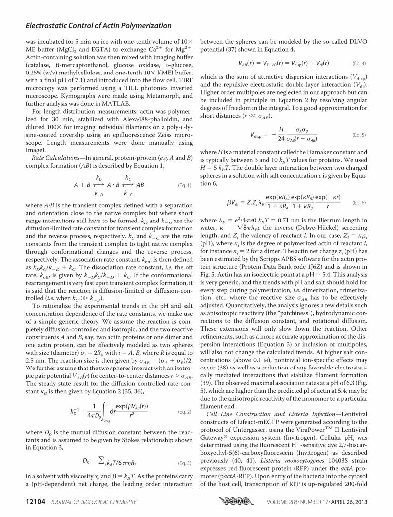
tion—Next,wewantedtoknowwhether thesensitivityofmono-mer association rates to pH is consistent with the general pre-dictions for typical diffusion-controlled reactions. The netcharge of a protein is determined directly by the pH of thesolution (Fig. 6A). At low salt concentrations (G-buffer), theelectrostatic potential reduces monomer-monomer contactformation (Fig. 6B). With increasing salt concentration, theelectrostatic potential is better screened, and polymerization isfacilitated (Fig. 6B). A generic model of protein-protein associ-ation provides a way of rationalizing the observed pH effects.Assuming isoreactive spheres (5 nm in size), an attractivepotential of 5 kBT values, and a homogeneous charge distribu-tion, the diffusion-limited on-rates can be calculated as a func-tion of pH and salt concentration (Fig. 6C). With this simplemodel, two trends are visible. First, the diffusion-limited kon hasa maximum at the isoelectric point where charge repulsioncompletely vanishes. Second, as the salt concentration in-creases, the charge repulsion (Equation 6) is more efficientlyscreened, and the reaction rate increases considerably even forpH values far from the isoelectric point. At high salt concentra-tions (c � 0.03 M and above), the pH dependence flattens out,and actin polymerization becomes practically pH-independent.This generic model already reproduces the overall trendobserved for monomer association rates as a function of pH(Fig. 5). Also, it predicted that even in the absence of salt,polymerization can occur at pH values close to the pI value ofactin.To further explore our model, we performed a PCH (32, 34)
analysis of our fluorescence fluctuation spectroscopy data (Fig.6, D–F). A histogram of photon counts (with 1-ms bin size)directly revealed the effect of pH on the polymerization process(Fig. 6F). The PCH results at pH 6.3 agreed with the data deter-mined from bulk and TIRFmeasurements (Figs. 1 and 4). How-ever, in contrast to pyrene assays, we could measure at pH val-ues below 6, and in contrast to the surface-restrictedpolymerization in TIRFM assays (Fig. 5C), maximal polymermass generation in solution was observable at pH 5.4 (Fig. 6G),close to the calculated pI of actin. In the absence of salts (G-buf-fer), we did not observe any polymer above pH 5.5 (Fig. 6H).The predicted trend of association rates from our model there-fore closely coincided with the observed polymer formed insolution at different salt concentrations (Fig. 6I). In conclusion,
FIGURE 4. Actin nucleation is pH-sensitive. 80 nM atto488- and atto647-la-beled actin (95 and 50% labeled, respectively) were mixed, and polymerizationwas started by addition of one-tenth v/v of 10� KMEI buffer. A and B, count ratefor the first 1800 s of polymerization at pH 5.8 (A) and 7.1 (B) are shown. Blueindicates the signal from the blue detection channel, and red indicates thesignal from the red channel. C, cross-correlation curves from the first 150 s ofthe above measurements. Solid lines are fits to a single diffusion species in athree-dimensional Gaussian volume to guide the eye. A control sample wasmeasured using G-buffer, where no polymerization occurs. D, number ofspikes versus pH during the first 15 min after initiation of polymerization. Theerror bars are the S.D. from three independent measurements.
Electrostatic Control of Actin Polymerization
12108 JOURNAL OF BIOLOGICAL CHEMISTRY VOLUME 288 • NUMBER 17 • APRIL 26, 2013

our results therefore suggest that polymer formation can becontrolled by electrostatics.pHi Modulates Actin-driven Pathogen Motility in Vivo—Fi-
nally, we asked whether the observed effects in vitro could pro-vide insight into processes observed under more physiologicalconditions. To this end, we tested the cellular pH dependenceof L. monocytogenes motility. Listeria is a bacterial pathogenthat hijacks the host actin cytoskeleton to move through thecytoplasm and escapes immune response by spreading from
cell to cell without extracellular exposure. Listeria cells expressActA, an activator of theArp2/3 complex, which then nucleatesactin filaments at the surface of the bacteria, resulting in ‘comettails’ composed of actin filaments that propel the bacteria for-ward (51). The intracellular speed of Listeria correlates directlywith the rate of actin polymerization in cells (3), making it adirect readout of effects on actin polymerization. We deter-mined velocities of moving Listeria in clonal fibroblasts withdifferent pHi values as follows: NHE1-deficient fibroblasts
FIGURE 5. Dependence of actin filament elongation kinetics on pH. A, individual filaments (30% atto488-labeled) monitored with time using TIRFM at pH8.3, 7.1, and 6.3. Barbed ends are marked by a blue plus sign, and pointed ends are marked by a yellow asterisk. All scale bars are 5 �m. B, change of filament lengthversus time for different filaments measured at the indicated pH values. C, elongation rate of barbed end and pointed end at 1 �M versus pH. Error bars are S.E.D, barbed-end elongation rate versus actin concentration for various pH values. E, pointed-end elongation rate versus actin concentration for various pH values.Only concentration points above 600 nM were used to estimate rate constants (see supplemental material for fits using all data points). F–H, rate constants ofbarbed-end elongation versus pH as follows: association rate (F), dissociation rate (G), and critical concentration (H). I–K, rate constants of pointed-endelongation versus pH as follows: association rate (I), dissociation rate (J), and critical concentration (K). F–K, error bars were determined from the propagation oferrors from the linear fits of the data presented in D and E. At least 25 filaments were measured per condition.
Electrostatic Control of Actin Polymerization
APRIL 26, 2013 • VOLUME 288 • NUMBER 17 JOURNAL OF BIOLOGICAL CHEMISTRY 12109
engineered to stably express either wild-type NHE1 (PSN) witha resulting cytosolic pH of 7.4 or an inactive NHE1 mutant(E266I) resulting in a lower pHi value of 7.1 (Fig. 7A) (11). Inaddition, both cell types stably expressed the actinmarker Life-act-GFP (31) for visualization of the Listeria-induced actincomet tails.Listeria internalizationwas visualized byActA-RFPexpression once they entered the cytosol (Fig. 7B). We mea-sured velocities for individual trajectories in both cell lines (Fig.7,B–D). The average transport velocities increased from0.31�0.03 �m�s�1 in PSN control cells to 0.42 � 0.04 �m�s�1 forE266I cells (mean � S.E., n � 46 each, p 0.01, Fig. 7E). Thischange, of20%, of Listeria actin-drivenmotility in fibroblastsagrees well with the change in elongation measured in vitro,also of 20% (Fig. 5).
DISCUSSION
We performed an extensive evaluation of the direct pHeffects on the polymerization of actin and actin assemblies,independent of actin regulatory proteins. By using a combina-tion of bulk fluorimetricmeasurements, single filament TIRFManalysis, and FFS methods, we were able to reveal mechanisticdetails on actin filament nucleation and elongation and explainthe observed pH effects with a simple model of electrostaticprotein-protein interaction.
Nucleation—Here, we have introduced the use of FFS tech-niques to study actin nucleation. In its simplest application (i.e.spikiness), FFS data provide an equivalent readout to the estab-lished pyrene method with the typical sigmoidal polymeriza-tion kinetics. In addition, FFS provides direct information onmonomer concentration (i.e. the mode of the count rate) andcan be applied over long time scales and a wide range of pHvalues. Using FFS techniques, we found a strong effect of pH onnucleation. The high sensitivity of FFS clearly showed thatshort filaments already form during the lag phase of polymeri-zation. This is predicted by the kinetic model for actin poly-merization (52, 53) but had not been detected experimentally.The early existence of such rapidly elongating filaments leads toa direct competitionwith newly forming nuclei, a critical aspectof the nucleation-elongation model (53). Using fluorescencecross-correlation spectroscopy, we were able to detect actincomplexes formed prior to filament appearance, suggestingthat these species represent the actual polymerization nuclei.Our findings that nuclei are present preceding filament emer-gence agree with and support the expectations based on pyreneexperiments (52, 53). Finally, something that also became obvi-ous from using ultrasensitive FFS techniques (in particular atlow molecule concentrations) is that extensive actin polymeri-zation dynamics occur below the critical concentration. Hence,
FIGURE 6. pH-dependent actin electrostatics control polymer formation. A, protein net charge prediction from the atomic structure of muscle actin (ProteinData Bank code 1J6Z) as a function of pH using the protein calculator (see The Scripts website). B, electrostatic map made with the Adaptative Poisson-Boltzmann Solver visualized with PyMOL. Isosurfaces displayed are 2 kT/e in blue and �2 kT/e in red. C, diffusion-controlled kon for dimer formation with actin’spH-defined net charge determined using isoreactive spheres as an approximation for the actin interactions. The orange-shaded region indicates the physio-logical pH range (see text). D, fluorescence intensity trace for 60 nM atto532-actin in F-buffer at pH 7.1. E, fluorescence intensity trace for 60 nM atto532-actin(80% labeled) in F-buffer at pH 6.3. F, photon-counting histogram analysis (with 1-ms bin size) of the traces shown in D and E. G and H, photon countinghistogram versus pH in F-buffer (G) and G-buffer (H). I, difference of the maximum number of photons per bin detected during a measurement at given pH withrespect to the measurement at pH 8.3. The light solid curves are taken from C for the same salt concentrations for comparison with its association rate axis onthe right.
Electrostatic Control of Actin Polymerization
12110 JOURNAL OF BIOLOGICAL CHEMISTRY VOLUME 288 • NUMBER 17 • APRIL 26, 2013

FFS opens up new possibilities for elucidating mechanisms ofnucleation.Elongation—Using TIRFM on single elongating actin fila-
ments, we determined the effect of pH on the association anddissociation rates at both filament ends. Barbed-end elongationis known to be a diffusion-controlled reaction (25). Consistentwith this idea, we find that net charge modulation by pH deter-mines barbed-end growth kinetics. In contrast to a previoussuggestion that the removal of electrostatic forceswould lead toan inversion of filament asymmetry (26), we observe thatbarbed-end and pointed-end growth kinetics are affected sim-ilarly by pH (Fig. 5, F, G, I, and J). This is largely due to effects onmonomer association rates, which are strongly regulated by pH(implying a role of surface charge). In contrast, we find thatdissociation ofmonomers is differentially affected by pH.At thebarbed end, koff increases with pH, whereas the dissociationrate at the pointed end follows the overall elongation as a func-tion of pH.If elongation is faster at lower pH, why then are filaments at
pH 6.3 shorter than those at pH 7.8 (Fig. 1, C andD)? To betterunderstand this effect, we performed simulations using ourexperimentally obtained kinetic parameters (supplemental Fig.S3). At early times, the faster elongation kinetics at low pHdominates and leads to the expected longer filaments. How-ever, monomers are depleted faster. At later time points, oncethe monomer pool has been depleted, dissociation and frag-mentation become relevant, leading to a reduced average fila-ment length (supplemental Fig. S3). Therefore, the observedlength distributions (Fig. 1,C andD) reflect themeasured trendin dissociation rates and not only the change in nucleation.
Our results indicate that electrostatics do contribute to fila-ment asymmetry but not via monomer association rates as sug-gested by Sept et al. (26). In addition, we observed very littlepointed-end growth or shrinkage at low actin concentrations.This could be due to a “closed” conformation as suggested by arecent structural study (54). Future workwill elucidate whetherthis closed state is due to the presence of lattice-binding pro-teins, such as N-ethylmaleimide-myosin (29), a light-inducedeffect (48), or is indeed related to a conformational transition(54). In summary, we find that electrostatics regulates the dif-fusional encounter to both filament ends, with monomer bind-ing at the pointed end likely involving an extra step (25).Electrostatics—The strong pH dependence of polymeriza-
tion suggests an important role of electrostatic forces in fila-ment formation. Consistent with this hypothesis, we found thatscreening of charges by the addition of salt increased the asso-ciation rate for actin (Fig. 6). A similar explanation for the effectof salt on actinwas proposedmore than 50 years ago (55, 56). Byusing a generic theoretical model, we were now able to quanti-tatively correlate effects of pH and salt. Hence, polymerizationof actin can either occur in the presence of sufficient amounts ofscreening salts or if the net charge is reduced, i.e. close to the pIof actin. Interestingly, salt has been reported to have a non-monotonic effect on actin polymerization; at high salt concen-trations, the elongation rate decreases monotonically withincreasing salt (25). This is possibly due to a reduction of intra-strand contacts that mostly consist of salt bridges (57). Con-versely, at low salt concentrations, screening allows inter-strand contacts to be formed, which are largely based onhydrophobic interactions (57). Altogether, our results explain
FIGURE 7. Effect of pHi on actin polymerization-driven motility of L. monocytogenes. A, schematic of NHE1 and the point mutation that blocks iontranslocation of NHE1 while maintaining its ability to interact with adaptor proteins through its long cytoplasmic tail and thereby bind to the cytoskeleton. PMrefers to the plasma membrane, Out to the extracellular space, and In to the cytosol. B, single bacteria moving in either a PSN cell (red) (upper panels) or an E266Icell (lower panels). Scale bar, 5 �m. C, representative trajectories of bacterial transport in PSN cells and E266I cells (blue). D, traveled distance as a function of timefor the trajectories shown in C. E, boxplot of L. monocytogenes speeds in PSN or E266I cells. n � 46 for each condition, *, p � 0.009.
Electrostatic Control of Actin Polymerization
APRIL 26, 2013 • VOLUME 288 • NUMBER 17 JOURNAL OF BIOLOGICAL CHEMISTRY 12111

the overall polymerization response to pH within a generalframework of electrostatic forces and provide a basis to under-stand the nonspecific effects of salt on actin polymerization.Implications within a Cellular Environment—We performed
an extensive analysis of pH effects on polymerization of pureactin. However, actin in cells always functions within complexassemblies that are regulated by a vast number of modulatingfactors. Along this line, we have recently found that themechanical properties of actin networks cross-linked by eithercortexillin or filamin can be tuned by pH (49). Interestingly, thepH sensitivities of various cross-linkers are very different fromeach other (49). This diversity could help to regulate mechani-cal properties of local cellular environments such as stressfibers, lamellipodia, or the isotropic actin cortex. In this way, acell could modulate its mechanical properties and contractilityby locally changing its pHi values (50) in time scales that areonly limited by proton diffusion within the cytoplasm. Fordynamic processes that are determined by the rate of actinpolymerization, such as cell migration or bacteria movingthrough the cytosol, changes in pHi could be expected to have adirect effect. Indeed, we found that velocities of Listeria infibroblast were higher at lower pHi values (Fig. 7), which agreeswith the increased polymerization rates of actin measured invitro. In contrast, the increasedmotility andmetastatic capacityof cancer cells with an increased pHi (20) are likely dominatedby pH-sensitive actin regulators, such as cofilin (8). Carefulevaluation of all involved regulators will therefore be requiredto fully understand the effects of pH on the complexmachinerygenerating cellular motion and how it is harnessed by cancercells. So far, our results suggest that actin polymerization-driven cellular processes will be sensitive to the pHi and suggestthat pH modulation can be used to finely tune the intracellularenvironment to promote and assist the vast variety of cellularbehaviors.
REFERENCES1. Footer, M. J., Kerssemakers, J.W., Theriot, J. A., and Dogterom,M. (2007)
Direct measurement of force generation by actin filament polymerizationusing an optical trap. Proc. Natl. Acad. Sci. U.S.A. 104, 2181–2186
2. Kovar, D. R., and Pollard, T. D. (2004) Insertional assembly of actin fila-ment barbed ends in association with formins produces piconewtonforces. Proc. Natl. Acad. Sci. U.S.A. 101, 14725–14730
3. Theriot, J. A., Mitchison, T. J., Tilney, L. G., and Portnoy, D. A. (1992) Therate of actin-based motility of intracellular Listeria monocytogenes equalsthe rate of actin polymerization. Nature 357, 257–260
4. Albiges-Rizo, C., Destaing, O., Fourcade, B., Planus, E., and Block, M. R.(2009) Actin machinery and mechanosensitivity in invadopodia, podo-somes, and focal adhesions. J. Cell Sci. 122, 3037–3049
5. Schwarz, U. S., and Gardel, M. L. (2012) United we stand: integrating theactin cytoskeleton and cell-matrix adhesions in cellular mechanotrans-duction. J. Cell Sci. 125, 3051–3060
6. Begg, D. A., and Rebhun, L. I. (1979) pH regulates the polymerization ofactin in the sea urchin egg cortex. J. Cell Biol. 83, 241–248
7. Tilney, L. G., Kiehart, D. P., Sardet, C., and Tilney, M. (1978) Polymeriza-tion of actin. IV. Role of Ca2� and H� in the assembly of actin and inmembrane fusion in the acrosomal reaction of echinoderm sperm. J. CellBiol. 77, 536–550
8. Frantz, C., Barreiro, G., Dominguez, L., Chen, X., Eddy, R., Condeelis, J.,Kelly, M. J., Jacobson,M. P., and Barber, D. L. (2008) Cofilin is a pH sensorfor actin free barbed end formation: role of phosphoinositide binding.J. Cell Biol. 183, 865–879
9. Srivastava, J., Barreiro, G., Groscurth, S., Gingras, A. R., Goult, B. T.,
Critchley, D. R., Kelly, M. J., Jacobson, M. P., and Barber, D. L. (2008)Structural model and functional significance of pH-dependent talin-actinbinding for focal adhesion remodeling. Proc. Natl. Acad. Sci. U.S.A. 105,14436–14441
10. Ridley, A. J. (2011) Life at the leading edge. Cell 145, 1012–102211. Denker, S. P., and Barber, D. L. (2002) Cell migration requires both ion
translocation and cytoskeletal anchoring by the Na-H exchanger NHE1.J. Cell Biol. 159, 1087–1096
12. Stock, C., and Schwab, A. (2006) Role of theNa/H exchanger NHE1 in cellmigration. Acta Physiol. 187, 149–157
13. Denker, S. P., and Barber, D. L. (2002) Ion transport proteins anchor andregulate the cytoskeleton. Curr. Opin. Cell Biol. 14, 214–220
14. Brooks, C., Ketsawatsomkron, P., Sui, Y., Wang, J., Wang, C. Y., Yu, F. S.,and Dong, Z. (2005) Acidic pH inhibits ATP depletion-induced tubularcell apoptosis by blocking caspase-9 activation in apoptosome. Am. J.Physiol. Renal Physiol. 289, F410–F419
15. Pouyssegur, J., Franchi, A., L’Allemain, G., and Paris, S. (1985) Cytoplas-mic pH, a key determinant of growth factor-induced DNA synthesis inquiescent fibroblasts. FEBS Lett. 190, 115–119
16. Pouyssegur, J., Sardet, C., Franchi, A., L’Allemain,G., and Paris, S. (1984)Aspecific mutation abolishing Na�/H� antiport activity in hamster fibro-blasts precludes growth at neutral and acidic pH. Proc. Natl. Acad. Sci.U.S.A. 81, 4833–4837
17. Roos, A., and Boron, W. F. (1981) Intracellular pH. Physiol. Rev. 61,296–434
18. Cardone, R. A., Casavola, V., andReshkin, S. J. (2005) The role of disturbedpH dynamics and the Na�/H� exchanger in metastasis. Nat. Rev. Cancer5, 786–795
19. Gillies, R. J., Raghunand, N., Karczmar, G. S., and Bhujwalla, Z. M. (2002)MRI of the tumor microenvironment. J. Magn. Reson. Imaging 16,430–450
20. Webb, B. A., Chimenti, M., Jacobson, M. P., and Barber, D. L. (2011)Dysregulated pH: a perfect storm for cancer progression.Nat. Rev. Cancer11, 671–677
21. Wang, F., Sampogna, R. V., andWare, B. R. (1989) pH dependence of actinself-assembly. Biophys. J. 55, 293–298
22. Zimmerle, C. T., and Frieden, C. (1988) Effect of pH on the mechanism ofactin polymerization. Biochemistry 27, 7766–7772
23. Qin, S., Pang, X., and Zhou, H. X. (2011) Automated prediction of proteinassociation rate constants. Structure 19, 1744–1751
24. Zhou, H. X. (2010) Rate theories for biologists. Q. Rev. Biophys. 43,219–293
25. Drenckhahn, D., and Pollard, T. D. (1986) Elongation of actin filaments isa diffusion-limited reaction at the barbed end and is accelerated by inertmacromolecules. J. Biol. Chem. 261, 12754–12758
26. Sept, D., Elcock, A. H., and McCammon, J. A. (1999) Computer simula-tions of actin polymerization can explain the barbed-pointed end asym-metry. J. Mol. Biol. 294, 1181–1189
27. Spudich, J. A., andWatt, S. (1971) The regulation of rabbit skeletal musclecontraction. I. Biochemical studies of the interaction of the tropomyosin-troponin complex with actin and the proteolytic fragments of myosin.J. Biol. Chem. 246, 4866–4871
28. MacLean-Fletcher, S., and Pollard, T. D. (1980) Identification of a factor inconventional muscle actin preparations which inhibits actin filament self-association. Biochem. Biophys. Res. Commun. 96, 18–27
29. Breitsprecher, D., Kiesewetter, A. K., Linkner, J., and Faix, J. (2009) Anal-ysis of actin assembly by in vitro TIRF microscopy. Methods Mol. Biol.571, 401–415
30. Cooper, J. A., Walker, S. B., and Pollard, T. D. (1983) Pyrene actin: docu-mentation of the validity of a sensitive assay for actin polymerization.J. Muscle Res. Cell Motil. 4, 253–262
31. Riedl, J., Crevenna, A. H., Kessenbrock, K., Yu, J. H., Neukirchen, D., Bista,M., Bradke, F., Jenne, D., Holak, T. A., Werb, Z., Sixt, M., and Wedlich-Soldner, R. (2008) Lifeact: a versatile marker to visualize F-actin. Nat.Methods 5, 605–607
32. Chen, Y., Muller, J. D., So, P. T., andGratton, E. (1999) The photon count-ing histogram in fluorescence fluctuation spectroscopy. Biophys. J. 77,553–567
Electrostatic Control of Actin Polymerization
12112 JOURNAL OF BIOLOGICAL CHEMISTRY VOLUME 288 • NUMBER 17 • APRIL 26, 2013

33. Muller, B. K., Zaychikov, E., Brauchle, C., and Lamb, D. C. (2005) Pulsedinterleaved excitation. Biophys. J. 89, 3508–3522
34. Muller, J. D., Chen, Y., and Gratton, E. (2000) Resolving heterogeneity onthe singlemolecular level with the photon-counting histogram. Biophys. J.78, 474–486
35. Debye, P. (1942) Reaction rates in ionic solutions.Trans. Electrochem. Soc.82, 265–272
36. von Smoluchowski, M. (1916) Drei vortrage uber diffusion, brownschemolekularbewegung und koagulation von kolloidteilchen. Phys. Z. 17,557–571; 585–599
37. Barrat, J.-L., and Hansen, J.-P. (2003) Basic Concepts for Simple and Com-plex Liquids, Cambridge University Press, New York
38. Kang, H., Bradley, M. J., McCullough, B. R., Pierre, A., Grintsevich, E. E.,Reisler, E., and De La Cruz, E. M. (2012) Identification of cation-bindingsites on actin that drive polymerization and modulate bending stiffness.Proc. Natl. Acad. Sci. U.S.A. 109, 16923–16927
39. Oda, T., Iwasa,M., Aihara, T.,Maeda, Y., andNarita, A. (2009) The natureof the globular- to fibrous-actin transition. Nature 457, 441–445
40. Tominaga, T., Ishizaki, T., Narumiya, S., and Barber, D. L. (1998)p160ROCK mediates RhoA activation of Na-H exchange. EMBO J. 17,4712–4722
41. Voyno-Yasenetskaya, T., Conklin, B. R., Gilbert, R. L., Hooley, R., Bourne,H. R., and Barber, D. L. (1994) G 13 stimulates Na-H exchange. J. Biol.Chem. 269, 4721–4724
42. Shetron-Rama, L.M.,Marquis, H., Bouwer, H.G., and Freitag, N. E. (2002)Intracellular induction of Listeria monocytogenes actA expression. Infect.Immun. 70, 1087–1096
43. Elson, E. L. (2011) Fluorescence correlation spectroscopy: past, present,future. Biophys. J. 101, 2855–2870
44. Ivanchenko, S., and Lamb, D. C. (2011) in Fluorescence Correlation Spec-troscopy: Principles and Developments Supramolecular Structure andFunction 10 (Brnjas-Kraljevic, J., and Pifat-Mrzljak, G., eds) pp. 1–30,Springer, Netherlands
45. Kuhn, J. R., and Pollard, T. D. (2005) Real-time measurements of actinfilament polymerization by total internal reflection fluorescence micros-
copy. Biophys. J. 88, 1387–140246. Jegou, A., Niedermayer, T., Orban, J., Didry, D., Lipowsky, R., Carlier,
M. F., and Romet-Lemonne, G. (2011) Individual actin filaments in a mi-crofluidic flow reveal the mechanism of ATP hydrolysis and give insightinto the properties of profilin. PLoS Biol. 9, e1001161
47. Krichevsky, O., and Bonnet, G. (2002) Fluorescence correlation spectros-copy: the technique and its applications. Reports on Progress in Physics 65,251
48. Niedermayer, T., Jegou, A., Chieze, L., Guichard, B., Helfer, E., Romet-Lemonne, G., Carlier, M. F., and Lipowsky, R. (2012) Intermittent depo-lymerization of actin filaments is caused by photo-induced dimerizationof actin protomers. Proc. Natl. Acad. Sci. U.S.A. 109, 10769–10774
49. Schmoller, K. M., Kohler, S., Crevenna, A. H., Wedlich-Soldner, R., andBausch, A. R. (2012) Resolving heterogeneity on the single molecular levelwith the photon-counting histogram. Soft Matter 8, 9685–9690
50. Kohler, S., Schmoller, K. M., Crevenna, A. H., and Bausch, A. R. (2012)Regulating contractility of the actomyosin cytoskeleton by pH. Cell Re-ports 2, 433–439
51. Haglund, C. M., and Welch, M. D. (2011) Pathogens and polymers: mi-crobe-host interactions illuminate the cytoskeleton. J. Cell Biol. 195, 7–17
52. Frieden, C. (1983) Polymerization of actin: mechanism of the Mg2�-in-duced process at pH 8 and 20°C. Proc. Natl. Acad. Sci. U.S.A. 80,6513–6517
53. Sept, D., and McCammon, J. A. (2001) Thermodynamics and kinetics ofactin filament nucleation. Biophys. J. 81, 667–674
54. Narita, A., Oda, T., and Maeda, Y. (2011) Structural basis for the slowdynamics of the actin filament pointed end. EMBO J. 30, 1230–1237
55. Steiner, R. F., Laki, K., and Spicer, S. (1952) Light scattering studies onsome muscle proteins. J. Polymer Sci. 8, 23–33
56. Oosawa, F., Asakura, S., Hotta, K., Imai, N., and Ooi, T. (1959) G-F trans-formation of actin as a fibrous condensation. J. Polymer Sci. 37, 323–336
57. Oda, T., Makino, K., Yamashita, I., Namba, K., and Maeda, Y. (2001) Dis-tinct structural changes detected by x-ray fiber diffraction in stabilizationof F-actin by lowering pH and increasing ionic strength. Biophys. J. 80,841–851
Electrostatic Control of Actin Polymerization
APRIL 26, 2013 • VOLUME 288 • NUMBER 17 JOURNAL OF BIOLOGICAL CHEMISTRY 12113
Related Documents