Electrophysiology (Conduction System of Heart) Dr. Mohammed Sharique Ahmed Quadri Assistant Prof. physiology Al maarefa college م ي ح ر ل ا ن م ح ر ل ه ا ل ل م ا س ب1

Electrophysiology (Conduction System of Heart) Dr. Mohammed Sharique Ahmed Quadri Assistant Prof. physiology Al maarefa college 1.
Dec 14, 2015
Welcome message from author
This document is posted to help you gain knowledge. Please leave a comment to let me know what you think about it! Share it to your friends and learn new things together.
Transcript

Electrophysiology (Conduction System of Heart)
Dr. Mohammed Sharique Ahmed Quadri Assistant Prof. physiology
Al maarefa college
بسم الله الرحمن الرحيم
1

2
Objectives • Identify the components of conducting system
of heart .• Know the sequence of conduction of impulse
in the heart • Recognize the concept associated with
pacemaker • Appreciate the role of ANS in controlling rate
of generation and conduction of impulse • Recognize the difference between A.P of SA
Node and ventricular muscle fiber
3
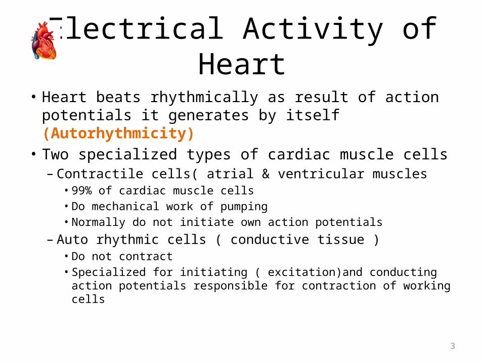
Electrical Activity of Heart
• Heart beats rhythmically as result of action potentials it generates by itself (Autorhythmicity)
• Two specialized types of cardiac muscle cells– Contractile cells( atrial & ventricular muscles
• 99% of cardiac muscle cells• Do mechanical work of pumping• Normally do not initiate own action potentials
– Auto rhythmic cells ( conductive tissue )• Do not contract• Specialized for initiating ( excitation)and conducting action
potentials responsible for contraction of working cells
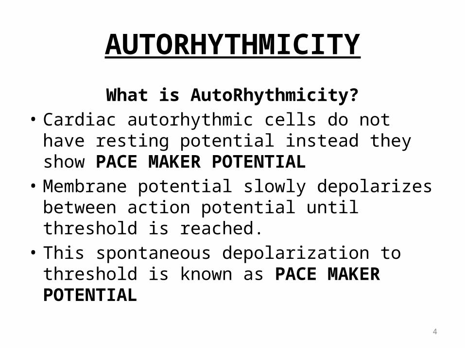
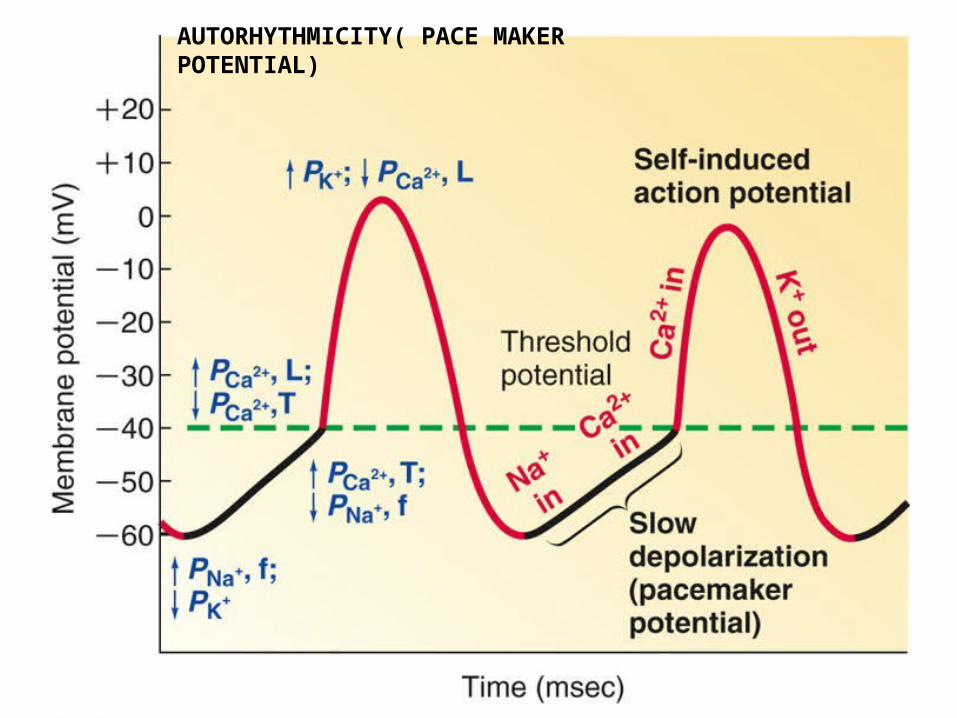
AUTORHYTHMICITY
What is AutoRhythmicity?• Cardiac autorhythmic cells do not have resting
potential instead they show PACE MAKER POTENTIAL
• Membrane potential slowly depolarizes between action potential until threshold is reached.
• This spontaneous depolarization to threshold is known as PACE MAKER POTENTIAL
4
AUTORHYTHMICITY( PACE MAKER POTENTIAL)
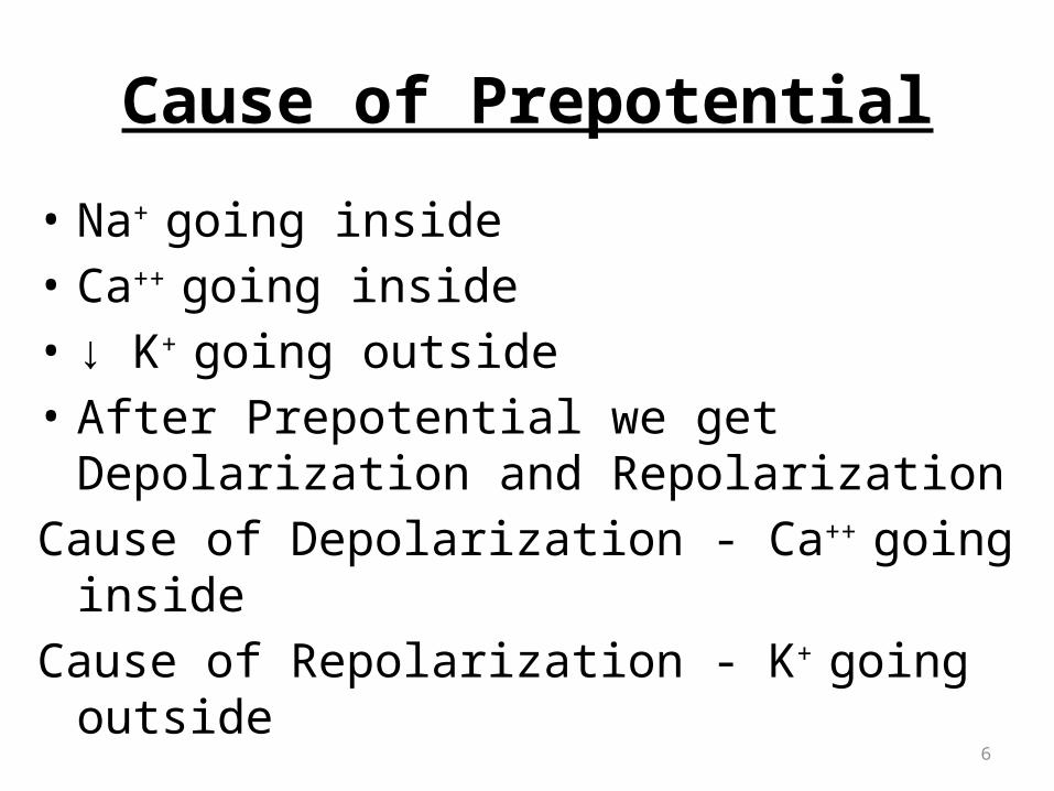
Cause of Prepotential
• Na+ going inside• Ca++ going inside• ↓ K+ going outside• After Prepotential we get Depolarization and
RepolarizationCause of Depolarization - Ca++ going insideCause of Repolarization - K+ going outside
6

S A NODE POTENTIAL
7
PHASE 4 = Prepotential
PHASE 0 =Depolarization
PHASE 3 =Repolarization

Conducting Tissues of the Heart (autorhythmic cells)
• APs spread through myocardial cells through gap junctions.
• Impulses cannot spread to ventricles directly because of fibrous tissue.
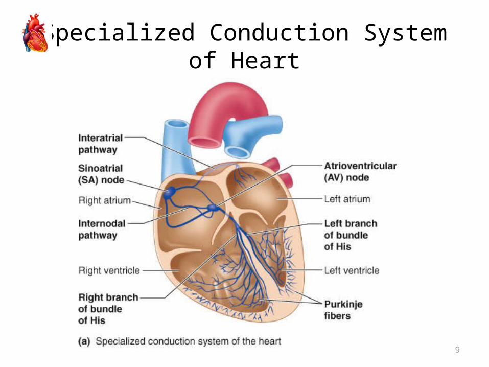
• Conduction pathway:– SA node.– AV node.– Bundle of His.– Purkinje fibers.
8
9
Specialized Conduction System of Heart

10
Conducting Tissues of the Heart– Sinoatrial Node (SA node)
• Specialized region in right atrial wall near opening of superior vena cava
• Pacemaker of the heart
– INTERNODAL FIBERS• Internodal Fibers – Anterior, Middle and
Posterior [Bachman, Wenchkeback, Thorel].
– Atrioventricular Node (AV node)• Small bundle of specialized cardiac cells located
at base of right atrium near inter atrial septum.
11
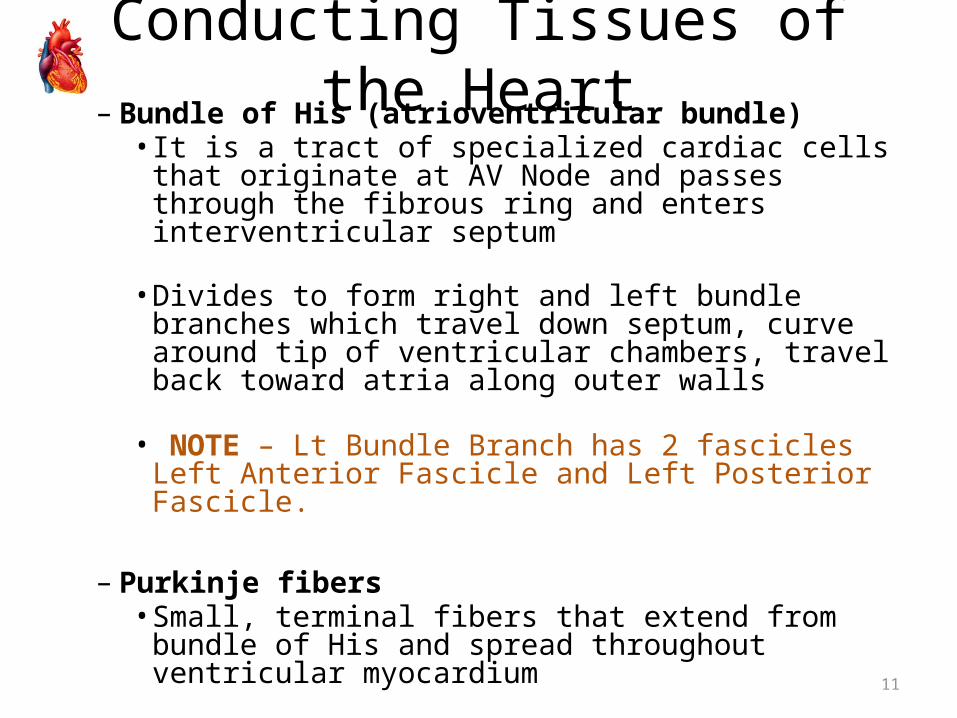
Conducting Tissues of the Heart– Bundle of His (atrioventricular bundle)
• It is a tract of specialized cardiac cells that originate at AV Node and passes through the fibrous ring and enters interventricular septum
• Divides to form right and left bundle branches which travel down septum, curve around tip of ventricular chambers, travel back toward atria along outer walls
• NOTE – Lt Bundle Branch has 2 fascicles Left Anterior Fascicle and Left Posterior Fascicle.
– Purkinje fibers
• Small, terminal fibers that extend from bundle of His and spread throughout ventricular myocardium

12
13
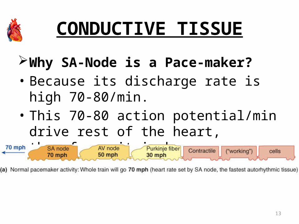
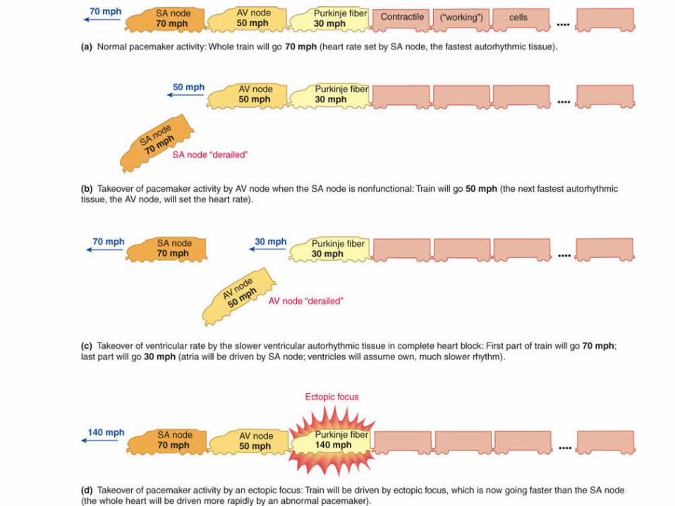
CONDUCTIVE TISSUE
Why SA-Node is a Pace-maker?• Because its discharge rate is high 70-80/min.• This 70-80 action potential/min drive rest of
the heart, therefore, it is known as pace-maker of the heart.
14
CONDUCTIVE TISSUE
• Other auto - rhythmic tissue are firing at slow rate.• They can work as pace-maker, if SA-Node is not
functioning e.g. if AV Node takes over as pace-maker, heart rate will be about 50/min.
• Any pace-maker other than SA-Node is called ‘Ectopic Pace-maker’. ( associated with organic heart disease or lack of sleep, anxiety, excess caffeine, nicotine)
15
16
APPLIED – HEART BLOCKS
• There are three types of heart blocks: FIRST DEGREE HEART BLOCK – Every impulse is
conducted but very slowly, therefore, there is increase in conduction time [we can see on ECG].
SECOND DEGREE HEART BLOCK – Some impulses are conducted and other are not conducted.
17
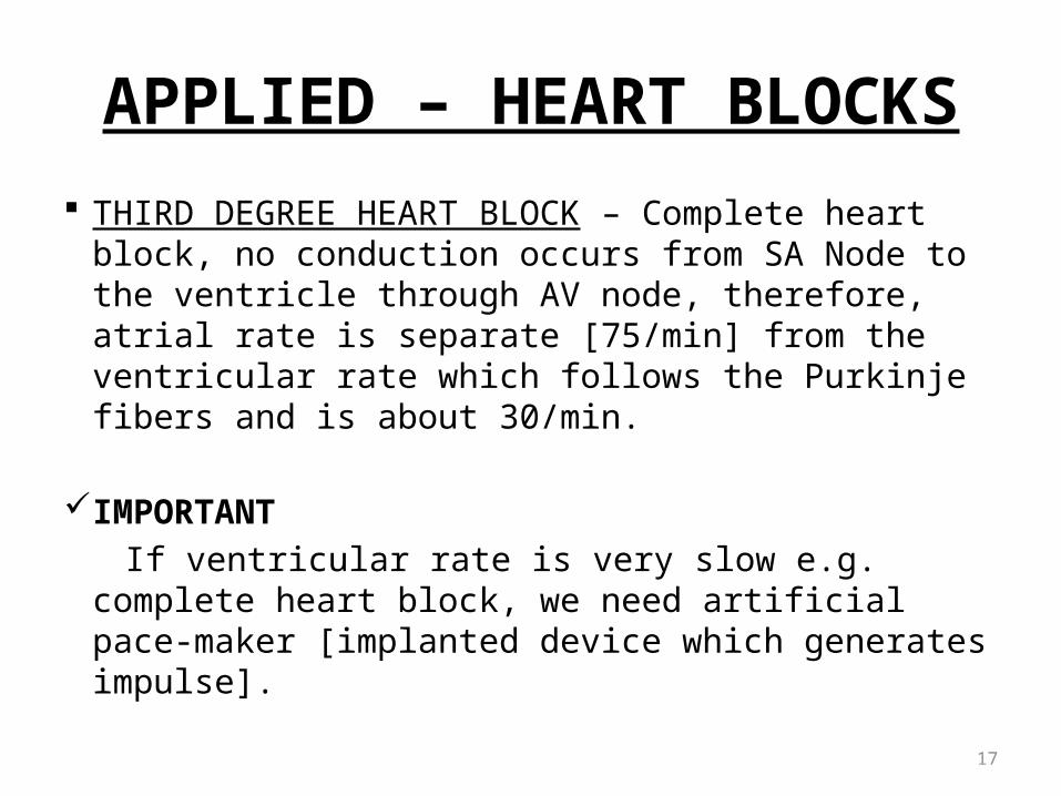
APPLIED – HEART BLOCKS
THIRD DEGREE HEART BLOCK – Complete heart block, no conduction occurs from SA Node to the ventricle through AV node, therefore, atrial rate is separate [75/min] from the ventricular rate which follows the Purkinje fibers and is about 30/min.
IMPORTANT If ventricular rate is very slow e.g. complete heart
block, we need artificial pace-maker [implanted device which generates impulse].

18
Control of Excitability by ANS
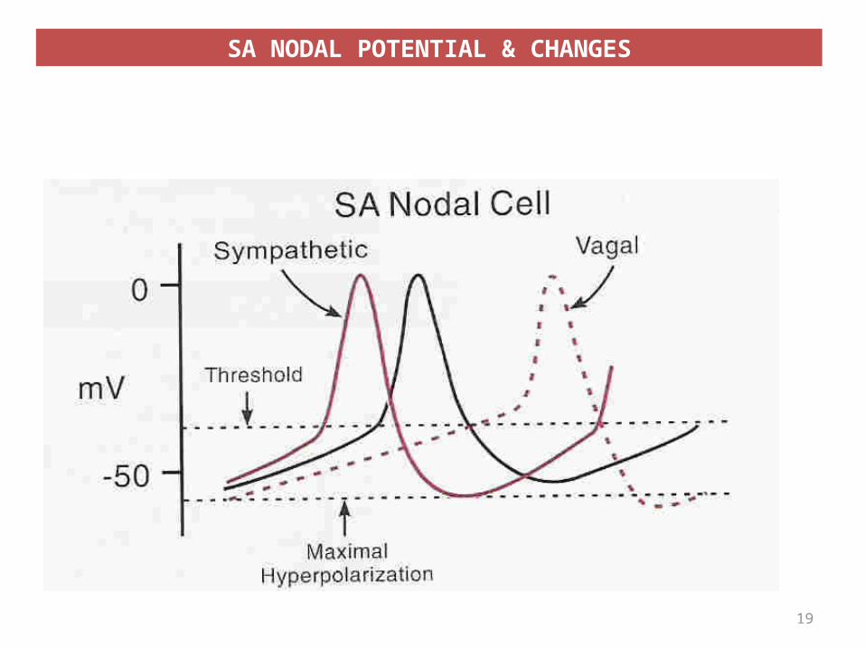
SA NODAL POTENTIAL & CHANGES
19

Effect 0f Sympathetic and parasympathetic Stimulation on Prepotential
(Pace Maker Potential)
• Epinephrine & Norepinephrine (Adrenaline and Noradrenaline) causes
Prepotential to occur faster therefore increase the heart rate
• Acetylcholine causes Prepotential to occur at slow rate therefore decrease the heart rate
20

Effect 0f Sympathetic Stimulation on Prepotential
Why Sympathetic Stimulation causesPrepotential to occur faster?• Because Sympathetic Stimulation causes - more Na+ influx [entry] - more Ca2+ influx [entry] - decreased K+ efflux [going outside]• Therefore, membrane potential changes quickly
from -60mV to -40mV [increases the slope of Prepotential] and when it reaches the threshold level, AP starts.
21
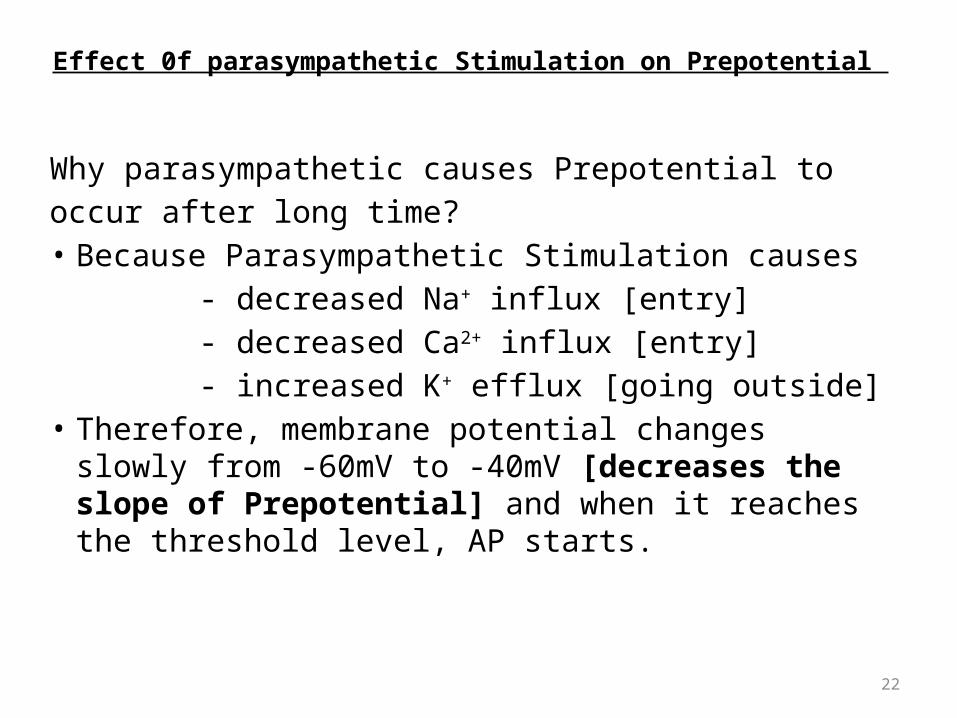
Effect 0f parasympathetic Stimulation on Prepotential
Why parasympathetic causes Prepotential tooccur after long time?• Because Parasympathetic Stimulation causes - decreased Na+ influx [entry] - decreased Ca2+ influx [entry] - increased K+ efflux [going outside]• Therefore, membrane potential changes slowly from -
60mV to -40mV [decreases the slope of Prepotential] and when it reaches the threshold level, AP starts.
22

– Heart rate is determined by balance between Inhibition of SA node by vagus(parasympathetic) & stimulation by sympathetic
– Under resting condition parasympathetic discharge dominates
Control of heart rate:

24
POINT TO PONDER
In Transplanted Heart, where there is no sympathetic and parasympathetic nerve supply, what will be the rate of SA Node discharge [Heart Rate] ?

25
SPREAD OF CARDIAC EXCITATION
• Cardiac impulse originates at SA node and spread to the atria [via gap junction] – Atrial Syncytium, therefore, both atria depolarize same time.
• Impulse [AP] goes to AV-Node by Internodal pathway.
• AV-Node is the only point of electrical contact between atria and ventricle [as atria and ventricle are separated by fibrous ring which is non-conductive].

26
SPREAD OF CARDIAC EXCITATION
AV – Node• At AV-Node, there is delay of 0.1 sec [100
milli- sec].• This delay is important to allow complete
ventricular filling – because it allows the atria to contract and empty
their blood into the ventricle, before impulse reaches the ventricle and causes ventricular depolarization and contraction

27
SPREAD OF CARDIAC EXCITATION
Ventricular Excitation• After AV delay of 0.1sec, impulse [AP] travels quickly
via Right Bundle Branch and Left Bundle Branch [branches of Bundle of His] to Purkinje Fibers to the ventricles.
• Both ventricle depolarize, than contract at same time.
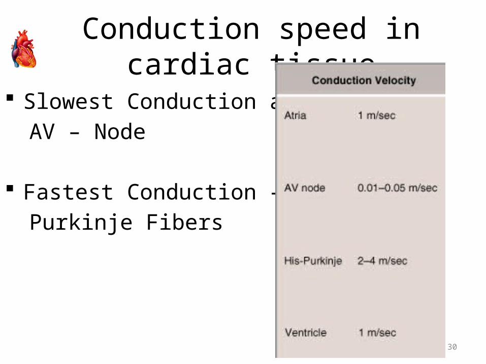
• Conduction in Purkinje Fiber is fastest 2-4 meter/sec, therefore, both ventricle depolarize
quickly and at the same time.

28
Spread of Cardiac Excitation
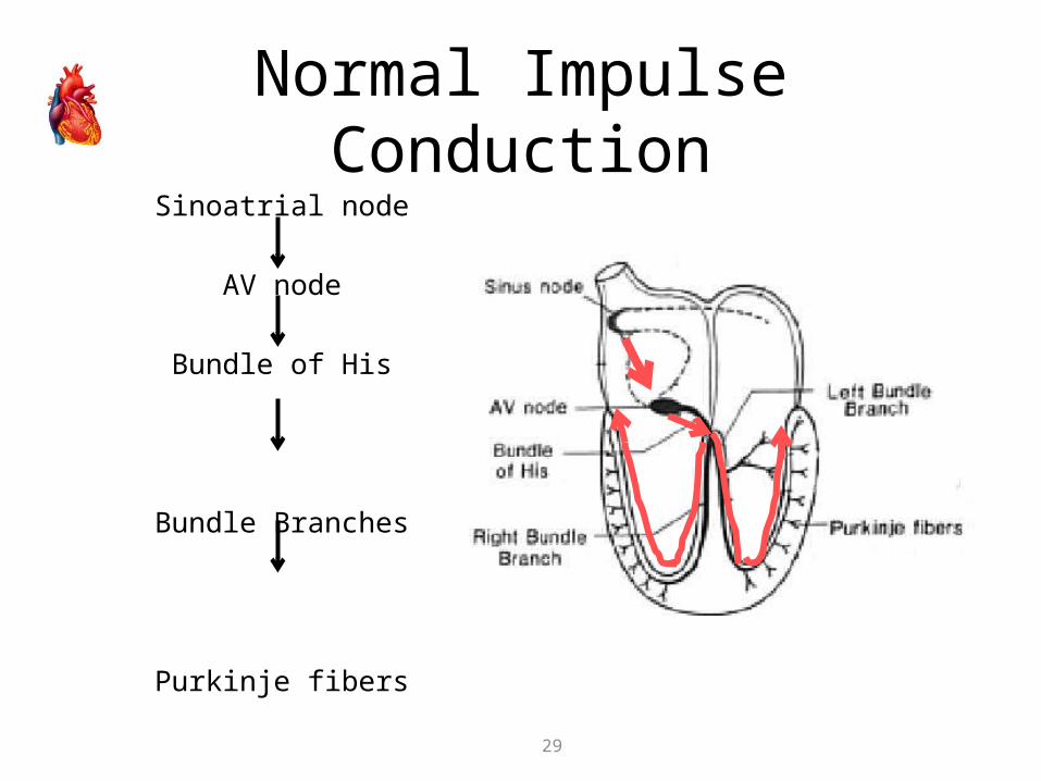
Normal Impulse ConductionSinoatrial node
AV node
Bundle of His
Bundle Branches
Purkinje fibers
29
30
Conduction speed in cardiac tissue
Slowest Conduction at AV – Node
Fastest Conduction – Purkinje Fibers
31
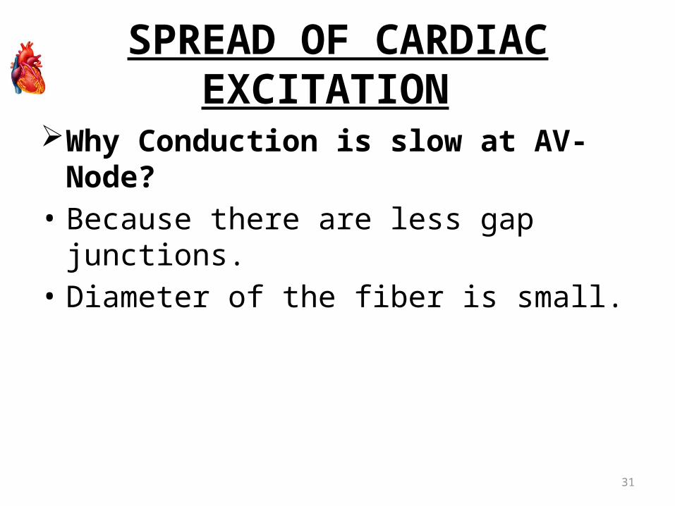
SPREAD OF CARDIAC EXCITATION
Why Conduction is slow at AV-Node?• Because there are less gap junctions.• Diameter of the fiber is small.

Myocardial Action Potential ( Excitability )
32
•Ventricular Muscle membrane has resting membrane potential of -90mV.•Action Potential of ventricular muscle fiber has four phases 0, 1, 2, 3 ,4.
• Once myocardial cells are stimulated by action potential originating in SA node, it produces its own action potential

Ventricular action potential • Rapid depolarization (Phase
0) – due to Na+ influx• Rapid Repolarization (Phase
1) - Due to closure of Na+ channels
• Slow depolarization (Phase 2) - this is called Plateau phase and is maintained for 200 – 300 ms – due to Ca++ influx
• Repolarization (Phase 3) – due to K+ efflux
• Resting Membrane Potential (Phase 4)
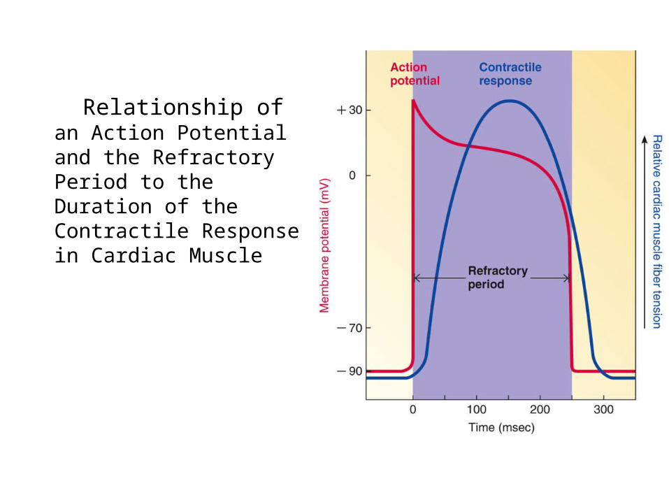
Relationship of an Action Potential and the Refractory Period to the Duration of the Contractile Response in Cardiac Muscle
Electrical Activity of Heart
• Because long refractory period occurs in conjunction with prolonged plateau phase, summati on and tetanus of cardiac muscle is impossible– Ensures alternate periods of contraction and
relaxation which are essential for pumping blood

References
• Human physiology by Lauralee Sherwood, seventh edition
• Text book physiology by Guyton &Hall,11th edition
• Text book of physiology by Linda .s contanzo,third edition
Related Documents