Electrophysiological studies on oxindole, a neurodepressant tryptophan metabolite 1 Guido Mannaioni, 1 Raaella Carpenedo, 1 Anna Maria Pugliese, 1 Renato Corradetti & 1,2 Flavio Moroni 1 Department of Pharmacology, University of Florence, Viale Morgagni 65, 50134, Firenze, Italy 1 The aim of the present work was to investigate the electrophysiological eects of oxindole, a tryptophan metabolite present in rat blood and brain, and recently proposed as a contributing factor in the pathogenesis of hepatic encephalopathy. 2 Using rat hippocampal slices in vitro and extra- or intracellular recordings, we evaluated oxindole eects on the neurotransmission of the CA1 region following orthodromic stimulation of the Schaer collaterals. 3 Oxindole (0.3 – 3 mM) decreased the amplitude of population spikes extracellularly recorded at the somatic level and of the fEPSPs recorded at the dendritic level. In intracellular recordings, oxindole (0.1 – 3 mM) did not aect the resting membrane potential or the neuronal input resistance, but reduced the probability of firing action potentials upon either synaptic or direct activation of the pyramidal cells. 4 Oxindole (0.3 – 3 mM) increased the threshold and the latency of firing action potentials elicited by depolarizing steps without changing the duration or the peak amplitude of the spikes. It also significantly increased the spike frequency adaptation induced by long lasting (400 ms) depolarizing stimuli. 5 In separate experiments, performed by measuring AMPA or NMDA-induced responses in cortical slices, oxindole (1 – 3 mM) did not modify glutamate receptor agonist responses. 6 Our results show that concentrations of oxindole which may be reached in pathological conditions, significantly decrease neuronal excitability by modifying the threshold of action potential generation. Keywords: Oxindole; hippocampus; Na + channels; synaptic potentials; tryptophan metabolism; CA1 Introduction In the course of experiments performed to study the neurochemical eects of inhibitors of kynurenine metabolism (Chiarugi et al., 1995; Carpenedo et al., 1994) we obtained data suggesting that tryptophan may be metabolized into oxindole and that oxindole is physiologically present in blood and other biological fluids in concentrations ranging from 0.1 – 1 mM (Carpenedo et al., 1997). Systemic administration of oxindole to rats, dogs or humans has been shown to cause profound sedation, decrease in blood pressure, decrease in muscular tone and loss of consciousness (Orcutt et al., 1964). Since rats treated with oxindole or having acute liver impairment share several common signs and since it is widely accepted that tryptophan metabolites are involved in the neurological signs associated to liver damage (Bengtsson & Bergqvist, 1996; Bengtsson et al., 1991), we measured brain oxindole content in rat models of acute liver impairment. As expected, we found a very large increase (10 – 100 fold) in blood and brain oxindole content (Moroni et al., 1997; Carpenedo et al., 1998) thus suggesting that the compound could play an important role in the neurological symptoms associated with liver failure. Large increases in oxindole concentration have also been found in urine samples obtained from a subpopulation of patients aected by phenylchetonuria, a hereditary disorder of amino acid metabolism. Although the biochemical mechan- isms responsible for such increases have not been studied, it has been suggested that the compound could have a role in the pathophysiology of the syndrome (Kochen et al., 1972). It seems, therefore, that oxindole is a neuroactive metabolite which should be added to the already long list of compounds originating from tryptophan and able to aect brain function in physiology or in pathology (Allegri Filippini et al., 1997; Stone, 1993). The mechanism of the neurodepressant action of oxindole has not been previously studied. In prior experiments, we found that it does not displace the binding of [ 3 H]-GABA to either GABA A or GABA B recognition sites in brain membranes, [ 3 H]-flunitrazepam to the benzodiazepine recogni- tion sites and [ 3 H]-glycine or [ 3 H]-glutamate to the NMDA receptor complex (Moroni et al., 1997). In the present study, we attempted to obtain quantitative information on the concentrations of brain oxindole which are able to cause sedation or coma and to investigate the mechanism(s) of its actions. In pursuing this goal, we attempted to correlate, in rats, brain oxindole concentration with overall behaviour, and we studied oxindole eects on: (i) the electrically evoked synaptic potentials and cell excitability in the CA1 region of rat hippocampal slices using both extracellular and intracellular recordings and (ii) the responses to ionotrophic glutamate receptor agonists in cortical wedge preparations (Harrison & Simmonds, 1985). Methods Extraction and quantification of oxindole Details on mass-spectrometric identification of oxindole have been previously published (Carpenedo et al., 1997). In most of 2 Author for correspondence at: Dipartmento di Farmacologia Preclinica e Clinica ‘‘Mario Aiazzi-Mancini’’, Universita` di Firenze, Viale G.B. Morgagni 65, 50134 Firenze, Italy. British Journal of Pharmacology (1998) 125, 1751 – 1760 ª 1998 Stockton Press All rights reserved 0007 – 1188/98 $12.00 http://www.stockton-press.co.uk/bjp

Welcome message from author
This document is posted to help you gain knowledge. Please leave a comment to let me know what you think about it! Share it to your friends and learn new things together.
Transcript
Electrophysiological studies on oxindole, a neurodepressanttryptophan metabolite
1Guido Mannaioni, 1Ra�aella Carpenedo, 1Anna Maria Pugliese, 1Renato Corradetti &1,2Flavio Moroni
1Department of Pharmacology, University of Florence, Viale Morgagni 65, 50134, Firenze, Italy
1 The aim of the present work was to investigate the electrophysiological e�ects of oxindole, atryptophan metabolite present in rat blood and brain, and recently proposed as a contributing factor inthe pathogenesis of hepatic encephalopathy.
2 Using rat hippocampal slices in vitro and extra- or intracellular recordings, we evaluated oxindolee�ects on the neurotransmission of the CA1 region following orthodromic stimulation of the Scha�ercollaterals.
3 Oxindole (0.3 ± 3 mM) decreased the amplitude of population spikes extracellularly recorded at thesomatic level and of the fEPSPs recorded at the dendritic level. In intracellular recordings, oxindole(0.1 ± 3 mM) did not a�ect the resting membrane potential or the neuronal input resistance, but reducedthe probability of ®ring action potentials upon either synaptic or direct activation of the pyramidal cells.
4 Oxindole (0.3 ± 3 mM) increased the threshold and the latency of ®ring action potentials elicited bydepolarizing steps without changing the duration or the peak amplitude of the spikes. It also signi®cantlyincreased the spike frequency adaptation induced by long lasting (400 ms) depolarizing stimuli.
5 In separate experiments, performed by measuring AMPA or NMDA-induced responses in corticalslices, oxindole (1 ± 3 mM) did not modify glutamate receptor agonist responses.
6 Our results show that concentrations of oxindole which may be reached in pathological conditions,signi®cantly decrease neuronal excitability by modifying the threshold of action potential generation.
Keywords: Oxindole; hippocampus; Na+ channels; synaptic potentials; tryptophan metabolism; CA1
Introduction
In the course of experiments performed to study theneurochemical e�ects of inhibitors of kynurenine metabolism(Chiarugi et al., 1995; Carpenedo et al., 1994) we obtained datasuggesting that tryptophan may be metabolized into oxindole
and that oxindole is physiologically present in blood and otherbiological ¯uids in concentrations ranging from 0.1 ± 1 mM(Carpenedo et al., 1997).
Systemic administration of oxindole to rats, dogs orhumans has been shown to cause profound sedation, decreasein blood pressure, decrease in muscular tone and loss of
consciousness (Orcutt et al., 1964). Since rats treated withoxindole or having acute liver impairment share severalcommon signs and since it is widely accepted that tryptophan
metabolites are involved in the neurological signs associated toliver damage (Bengtsson & Bergqvist, 1996; Bengtsson et al.,1991), we measured brain oxindole content in rat models ofacute liver impairment. As expected, we found a very large
increase (10 ± 100 fold) in blood and brain oxindole content(Moroni et al., 1997; Carpenedo et al., 1998) thus suggestingthat the compound could play an important role in the
neurological symptoms associated with liver failure.Large increases in oxindole concentration have also been
found in urine samples obtained from a subpopulation of
patients a�ected by phenylchetonuria, a hereditary disorder ofamino acid metabolism. Although the biochemical mechan-isms responsible for such increases have not been studied, it
has been suggested that the compound could have a role in the
pathophysiology of the syndrome (Kochen et al., 1972). Itseems, therefore, that oxindole is a neuroactive metabolitewhich should be added to the already long list of compoundsoriginating from tryptophan and able to a�ect brain function
in physiology or in pathology (Allegri Filippini et al., 1997;Stone, 1993).
The mechanism of the neurodepressant action of oxindole
has not been previously studied. In prior experiments, wefound that it does not displace the binding of [3H]-GABA toeither GABAA or GABAB recognition sites in brain
membranes, [3H]-¯unitrazepam to the benzodiazepine recogni-tion sites and [3H]-glycine or [3H]-glutamate to the NMDAreceptor complex (Moroni et al., 1997).
In the present study, we attempted to obtain quantitativeinformation on the concentrations of brain oxindole which areable to cause sedation or coma and to investigate themechanism(s) of its actions. In pursuing this goal, we
attempted to correlate, in rats, brain oxindole concentrationwith overall behaviour, and we studied oxindole e�ects on: (i)the electrically evoked synaptic potentials and cell excitability
in the CA1 region of rat hippocampal slices using bothextracellular and intracellular recordings and (ii) the responsesto ionotrophic glutamate receptor agonists in cortical wedge
preparations (Harrison & Simmonds, 1985).
Methods
Extraction and quanti®cation of oxindole
Details on mass-spectrometric identi®cation of oxindole havebeen previously published (Carpenedo et al., 1997). In most of
2Author for correspondence at: Dipartmento di FarmacologiaPreclinica e Clinica ``Mario Aiazzi-Mancini'', UniversitaÁ di Firenze,Viale G.B. Morgagni 65, 50134 Firenze, Italy.
British Journal of Pharmacology (1998) 125, 1751 ± 1760 ã 1998 Stockton Press All rights reserved 0007 ± 1188/98 $12.00
http://www.stockton-press.co.uk/bjp
the present experiments, approximately 1 g of tissue (or 1 mlof blood) was homogenized in 2 vol of 0.4 N HClO4 (or 1 NHClO4 for the blood). The mixture was centrifuged at
18,0006g for 20 min and the supernatant was collected. Theprocedure was repeated twice. The supernatants were thenmixed with 8 ml of chloroform and agitated for 5 min. Thechloroform layers were collected and evaporated under a
stream of nitrogen. The residues were resuspended in 200 ml of0.4 N HClO4 and aliquots were injected into the HPLCapparatus which consisted of a Perkin-Elmer LC pump (Model
250), a syringe-loading sample injection valve (RheodyneModel 7125), a C18 reverse-phase precolumn ®lter (0.5 cmlong; Waters, Milford, MA, U.S.A.), and a 25-cm reverse-
phase 18 SpheriSorb ODS-2 10 U column (Alltech, Deer®eld,IL, U.S.A.). The detection was performed with a spectro-photometer (Perkin-Elmer Model LC90UV). The mobile
phases were: 0.05 M acetate bu�er (pH 3.24) and acetonitrile(15%). The recovery of a known amount of oxindole passedthrough the entire procedure was 75+2% (mean+s.e.mean often determinations).
Evaluation of the spontaneous locomotor activity
Spontaneous locomotor activity of rats was evaluated using anAnimex activity meter (mod. LKB FARAD). The number ofinterruptions of photocells was monitored by individually
placing the rats in clean cages for 20 min.
Preparation of hippocampal slices
Experiments were carried out using in vitro hippocampal slicesas previously described (Pugliese et al., 1996). Male Wistar rats(100 ± 200 g body weight, Charles River, Como, Italy) were
anaesthetized with ether and decapitated. The hippocampiwere rapidly removed and placed in ice cold oxygenated (95%O2, 5% CO2) arti®cial cerebrospinal ¯uid (aCSF) of the
following composition (mM): NaCl 124, KCl 3, NaH2PO4
1.25, MgSO4 1.4, CaCl2 2, NaHCO3 25, D-glucose 10 (pH7.4). Slices (400 mm thick) were cut from the hippocampus
using a McIlwain (Gomshall, U.K.) tissue chopper and kept inoxygenated aCSF for at least 1 h at room temperature (20 ±238C). A single slice was then placed on nylon mesh andcompletely submerged in a small chamber and perfused with
oxygenated aCSF (30 ± 328C) at a constant ¯ow rate of 2 ±3 ml min71. Drugs were administered through a three-way tapand a complete exchange of the chamber volume occurred in
1 min.
Extracellular recordings
Test pulses (80 ± 110 ms duration; 0.017 ± 0.05 Hz) weredelivered through bipolar nichrome electrodes positioned in
the CA1 stratum radiatum. Evoked extracellular potentialswere recorded with glass microelectrodes (2 ± 10 MO) ®lledwith 3 M NaCl, either from the pyramidal cell layer of CA1(population spike, PS) or from stratum radiatum (®eld
excitatory postsynaptic potential, fEPSP) in the CA1 area.Responses were ampli®ed (Neurolog NL 104, Digitimer Ltd),displayed on a computer video, digitized (sample rate 33 kHz),
and stored on ¯oppy disks for later analysis using the softwarepClamp 6 (Axon Instruments Ltd.).
Stimulus-response relationships were investigated at the
beginning of each experiment, and at de®ned times during theexperiment by gradually increasing the stimulus strengthwhen required. The test stimulus pulse was adjusted toproduce a population spike whose amplitude was 40 ± 50%
(2 ± 3 mV) of the maximum and, unless otherwise stated, waskept constant throughout the experiment. After 30 min ofequilibration, a control period (10 ± 15 min) was recorded and
used to generate baseline values before experimental tests.In the recordings from the pyramidal cell layer, the
population spike of the evoked responses was measured asthe peak-to-peak amplitude of the ®rst negative phase of the
population spike, whereas the slope of the positive-going phaseof the somatic response was used to estimate excitatorysynaptic input (Pugliese et al., 1994). In recordings from the
stratum radiatum, the amplitude of both fEPSP and a�erentvolley were measured. The initial slope of fEPSP was alsomeasured but since the changes induced by drugs were
qualitatively consistent, only the measurements of theamplitude are here reported. Parameters were evaluatedbefore, during and after drug treatment. Oxindole was applied
for 10 min.
Intracellular recordings
CA1 pyramidal neurones were recorded in current-clampmode with 3 M KCl-®lled electrodes (35 ± 50 MO). Electricalsignals were ampli®ed with an Axoclamp 2A (Axon
Instruments, Foster City, U.S.A.) and displayed on anoscilloscope and chart recorder (2800 Gould, Valley View,U.S.A.). Traces were stored on a digital tape (DTR 1200,
BioLogic, Claix, France; sampling frequency 48 kHz) and on acomputer using pClamp6 software (Axon Instruments) for o�-line analysis. Several criteria were used to select cells for the
experiments: stable resting membrane potential (r.m.p.) of atleast 760 mV and no spontaneous ®ring of action potentials;no sudden drops in the neuronal input resistance (Rin),indicating cell damage; constant amplitude of the action
potential (480 mV), obtained by direct activation of the cell.Modi®cations in synaptic e�cacy by drugs were evaluated bymeasuring the changes in amplitude of the evoked EPSP and in
probability of ®ring an action potential upon stimulation ofa�erent pathway. Stimulus response curves were constructedat the beginning and during the experiment by gradually
increasing the stimulus strength.To evaluate the e�ects of oxindole on EPSPs not
contaminated by the early phase of GABA-mediated IPSPs,most of the experiments were carried out in the presence of a
GABAA receptor blocker (bicuculline 10 mM or picrotoxin25 mM). To keep neurones below ®ring threshold wheninvestigating stimulus response relationships, cells were
hyperpolarized relative to their r.m.p. by injecting a constantnegative current (range from 7 50 to 7200 pA) through therecording electrode. Since preliminary experiments (n=3) and
post-hoc analysis of results indicated that the presence ofGABAA receptor blockers and/or hyperpolarization of theneurones did not change cell responses to oxindole, data
obtained in the presence of GABAA receptor blockers werepooled with those in aCSF.
Probability of ®ring (p.f.) upon synaptic or directdepolarization of the neurone was calculated on ®ve or ten
responses. To study the e�ects of oxindole on cell discharge,direct activation of the impaled neurone was obtained byinjection of short (40 ms, to elicit a probability of ®ring from
0.8 ± 1 in control conditions) or long (400 ms, for actionpotential frequency adaptation) depolarizing current stepsthrough the recording electrode. The intensity of the
depolarizing current step was kept constant during theexperiment. The slow afterhyperpolarization (AHP) whichfollows repetitive ®ring elicited by injection of depolarizingcurrents (from +300 to +500 pA, 400 ms) through the
Oxindole and CA1 hippocampal neurotransmission1752 G. Mannaioni et al
recording electrode was measured either at the peak (peakAHP) or 150 ms (late AHP) after cessation of the depolarizingstep. A series of current steps (400 ms, range from 7900 to
+500 pA) were injected to monitor cell Rin and to constructI/V plots.
Preparation of cortical wedges
The cortical wedge preparation described by Harrison andSimmonds (Harrison & Simmonds, 1985) and modi®ed by
Burton et al. (Burton et al., 1988) was used as previouslydescribed (CarlaÁ & Moroni, 1992). Brie¯y, wedges obtainedfrom white swiss mice (male 15/25 g) were placed in a two-
compartment bath and silicone grease was placed betweenthe two portions of the bath. The wedges were incubated atroom temperature and perfused with Krebs solution (mM:
NaCl 135; CaCl2 2.4; KH2PO4 1.3; MgCl2 1.2; NaHCO3 16.3and glucose 7.7), gassed with 95% O2 and 5% CO2 at a¯ow rate of 2 ml min71. After stabilization, the gray matterwas perfused with a Mg2+ free medium. NMDA and
AMPA were repeatedly applied for 2 min every 15 minwhereas the other compounds between the two compart-ments were monitored via Ag/AgCl electrodes and displayed
on a chart recorder. The preparations were initiallystabilized by repeated application of 10 mM NMDA, aconcentration which gave a sub-maximal response but which
did not signi®cantly reduce the response of subsequentapplications of the agonist (Moroni et al., 1995; Mannaioniet al., 1994).
Materials
Oxindole, obtained from Aldrich Sigma (Milan, Italy), was
dissolved in DMSO. The maximal concentrations of DMSOused in electrophysiological studies was 0.003% (V/V) and hadno measurable e�ects on evoked potentials. Picrotoxin was
from RBI (Natick, MA, U.S.A.); (7)bicuculline methiodidewas from Sigma-Aldrich (Milan, Italy); NMDA and AMPAwere from Tocris (Bristol, U.K.). Solvents (acetonitrile and
chloroform) were from Merck (Darmstadt, Germany).
Data analysis
All numberical data are expressed as means+s.e.mean.Data were analysed statistically by Student's t-test or byone-way analysis of variance (ANOVA) followed by
Dunnett's t-test. A value of P50.05 was consideredstatistically signi®cant.
Results
Brain oxidole content and behaviour
We previously showed that rat brain oxindole content is
0.05+0.01 nmol g71 (Carpenedo et al., 1997). One h afterintraperitoneal administration of the compound at 10 or100 mg kg71, oxindole brain content was 8.1+1.7 or
103+15 nmol g71, respectively. When the lower dose wasadministered (10 mg kg71, i.p.), the rats were calm, easy tomanage and had reduced locomotor activity for at least
30 min. After the larger dose (100 mg kg71, i.p.), the animalswent into coma, the righting re¯ex was absent, muscular tonewas clearly reduced (Table 1) and most of them diedapproximately 2 h later.
Extracellular recordings in rat hippocampal slices
In a ®rst series of experiments, we tested the e�ects of several
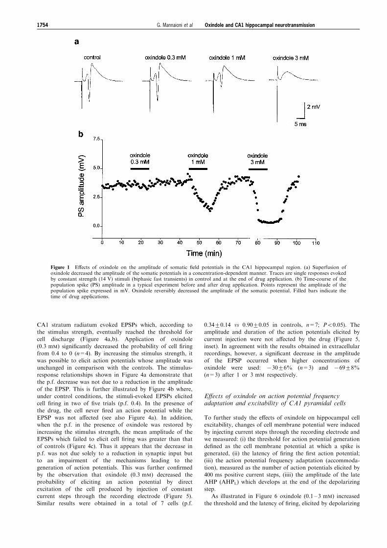
concentrations (0.3, 1 and 3 mM, 10 min application) ofoxindole on evoked synaptic potentials recorded extracellu-larly from the CA1 region. Oxindole decreased the amplitudeof somatic potentials in a concentration-dependent manner.
The time-course of the e�ects of the drug in a typicalexperiment in which three concentrations of oxindole weretested is shown in Figure 1. The decrease in amplitude of
synaptic responses had a rapid onset (52 min) and wascompletely reversible within 15 min of drug washout (seeFigures 1b and 3a). In most of the experiments, a transient
rebound increase in the amplitude of the population spikewas observed during washout (see Figures 1b and 3a). Asshown in Figure 1a, the application of oxindole a�ected
mainly the population spike component of evoked synapticresponses (i.e. the compound action potential generated bysynchronous discharge of CA1 pyramidal cells). Whenoxindole was used at 1 mM, the population spike amplitude
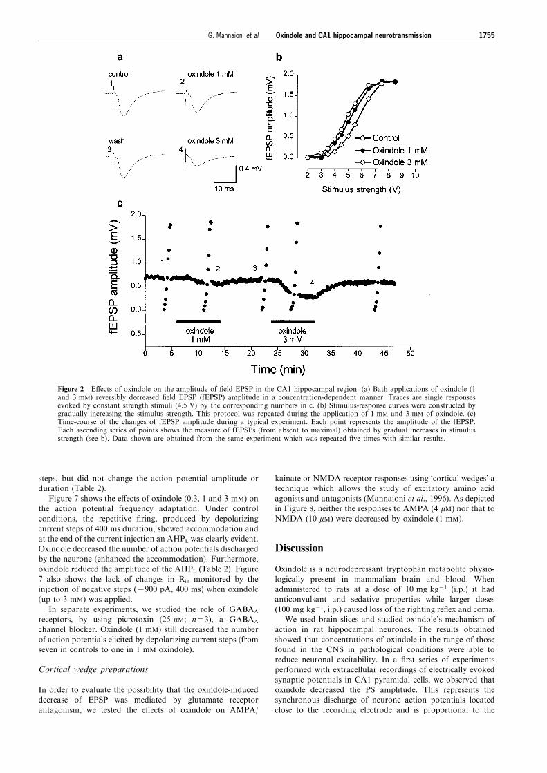
decreased by 40+10% (n=5, P50.05) and at 3 mM,decreased by 85+9% (n=5, P50.05). Figure 2 shows theresults of a typical experiment in which the evoked fEPSP
was recorded in the dendritic region of CA1 pyramidal cells.In these experiments, the oxindole-induced decrease in thefEPSP amplitude was statistically signi®cant only at 3 mM, a
concentration which also decreased the a�erent volleyamplitude (from 117+45 to 80+37 mV; P50.05). When thee�ects of oxindole (1 and 3 mM) were evaluated in thestimulus-response relationship, the curves were displaced
towards the right, but the maximal responses were notdecreased (see Figure 2b).
Intracellular recording from pyramidal CA1 neurones
Intracellular recordings were carried out in 18 slices taken
from 18 rats. Neurones (n=18) had a mean r.m.p. of764+1 mV, Rin of 45+6 MO and an amplitude of actionpotentials greater than 80 mV (89+2 mV).
We studied the e�ects of oxindole (0.1 ± 3 mM) on r.m.p.,Rin, synaptic responses and cell excitability of pyramidal cells.Oxindole concentrations of up to 3 mM did not signi®cantlya�ect cell r.m.p. or Rin (see Table 2).
E�ects of oxindole on EPSPs and probability of ®ringan action potential upon synaptic stimulation
Figure 4 shows the e�ects of oxindole on EPSP amplitudeand on the probability of ®ring an action potential upon
synaptic activation of pyramidal cells. Stimulation of the
Table 1 E�ects of oxindole administration on locomotoractivity and brain oxindole content
Brain Locomotorconcentration activity Righting(nmol g±1) (counts/ 20 min) re¯ex
SalineOxindole 10 mg kg±1
Oxindole 100 mg kg±1
0.05+0.018.1+1.7103+15
580+15387+42*5+2**
normalnormalabsent
In order to measure locomotor activity, oxindole wasinjected 20 min before placing the animals in clean cages.The animals were killed 1 h after treatment. Doses are mgkg±1 i.p. Values are means+s.e.mean of at least six animals;*P<0.05 vs saline; **P<0.01 vs saline (ANOVA andDunnett's t-test).
Oxindole and CA1 hippocampal neurotransmission 1753G. Mannaioni et al
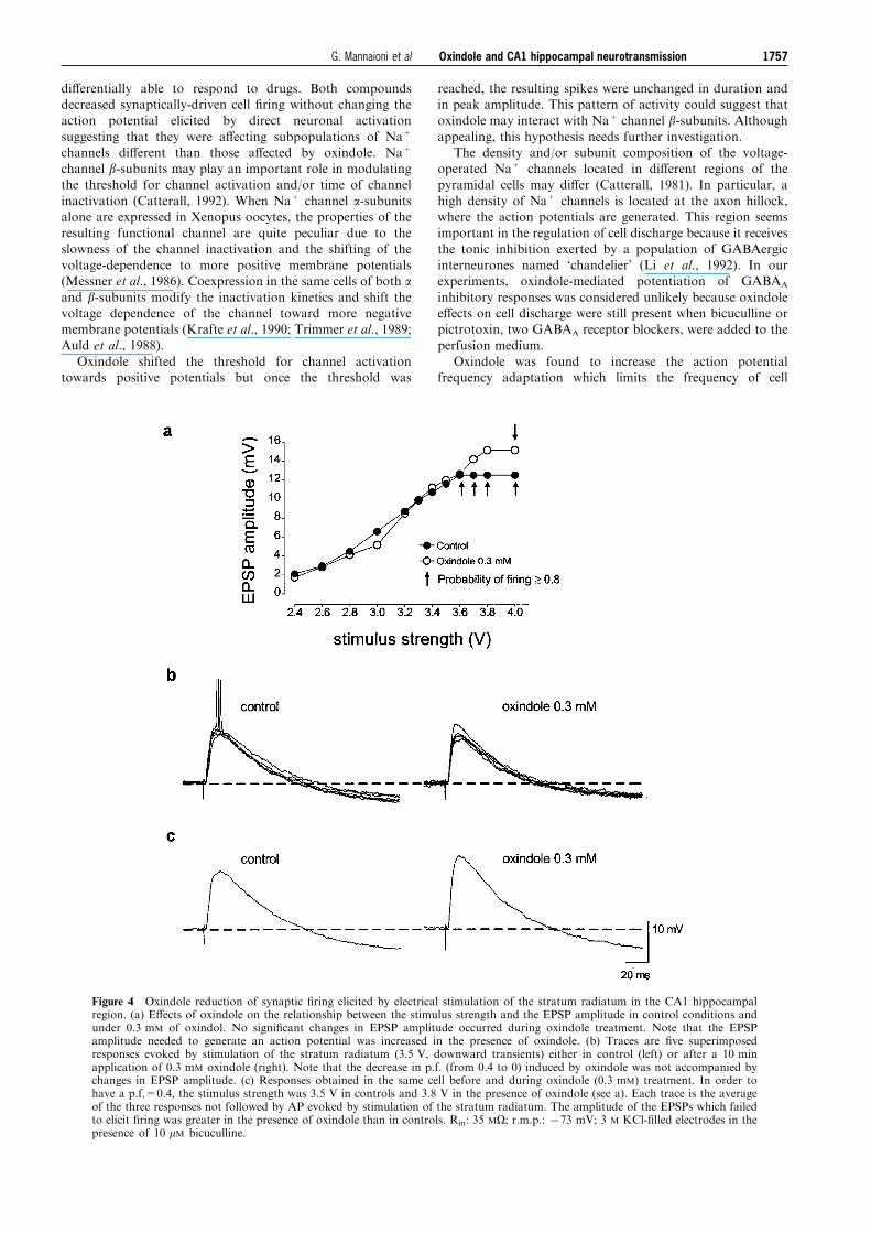
CA1 stratum radiatum evoked EPSPs which, according tothe stimulus strength, eventually reached the threshold for
cell discharge (Figure 4a,b). Application of oxindole(0.3 mM) signi®cantly decreased the probability of cell ®ringfrom 0.4 to 0 (n=4). By increasing the stimulus strength, it
was possible to elicit action potentials whose amplitude wasunchanged in comparison with the controls. The stimulus-response relationships shown in Figure 4a demonstrate thatthe p.f. decrease was not due to a reduction in the amplitude
of the EPSP. This is further illustrated by Figure 4b where,under control conditions, the stimuli-evoked EPSPs elicitedcell ®ring in two of ®ve trials (p.f. 0.4). In the presence of
the drug, the cell never ®red an action potential while theEPSP was not a�ected (see also Figure 4a). In addition,when the p.f. in the presence of oxindole was restored by
increasing the stimulus strength, the mean amplitude of theEPSPs which failed to elicit cell ®ring was greater than thatof controls (Figure 4c). Thus it appears that the decrease in
p.f. was not due solely to a reduction in synaptic input butto an impairment of the mechanisms leading to thegeneration of action potentials. This was further con®rmedby the observation that oxindole (0.3 mM) decreased the
probability of eliciting an action potential by directexcitation of the cell produced by injection of constantcurrent steps through the recording electrode (Figure 5).
Similar results were obtained in a total of 7 cells (p.f.
0.34+0.14 vs 0.90+0.05 in controls, n=7; P50.05). Theamplitude and duration of the action potentials elicited by
current injection were not a�ected by the drug (Figure 5,inset). In agreement with the results obtained in extracellularrecordings, however, a signi®cant decrease in the amplitude
of the EPSP occurred when higher concentrations ofoxindole were used: 730+6% (n=3) and 769+8%(n=3) after 1 or 3 mM respectively.
E�ects of oxindole on action potential frequencyadaptation and excitability of CA1 pyramidal cells
To further study the e�ects of oxindole on hippocampal cellexcitability, changes of cell membrane potential were induced
by injecting current steps through the recording electrode andwe measured: (i) the threshold for action potential generationde®ned as the cell membrane potential at which a spike is
generated, (ii) the latency of ®ring the ®rst action potential;(iii) the action potential frequency adaptation (accommoda-tion), measured as the number of action potentials elicited by400 ms positive current steps, (iiii) the amplitude of the late
AHP (AHPL) which develops at the end of the depolarizingstep.
As illustrated in Figure 6 oxindole (0.1 ± 3 mM) increased
the threshold and the latency of ®ring, elicited by depolarizing
Figure 1 E�ects of oxindole on the amplitude of somatic ®eld potentials in the CA1 hippocampal region. (a) Superfusion ofoxindole decreased the amplitude of the somatic potentials in a concentration-dependent manner. Traces are single responses evokedby constant strength (14 V) stimuli (biphasic fast transients) in control and at the end of drug application. (b) Time-course of thepopulation spike (PS) amplitude in a typical experiment before and after drug application. Points represent the amplitude of thepopulation spike expressed in mV. Oxindole reversibly decreased the amplitude of the somatic potential. Filled bars indicate thetime of drug applications.
Oxindole and CA1 hippocampal neurotransmission1754 G. Mannaioni et al
steps, but did not change the action potential amplitude orduration (Table 2).
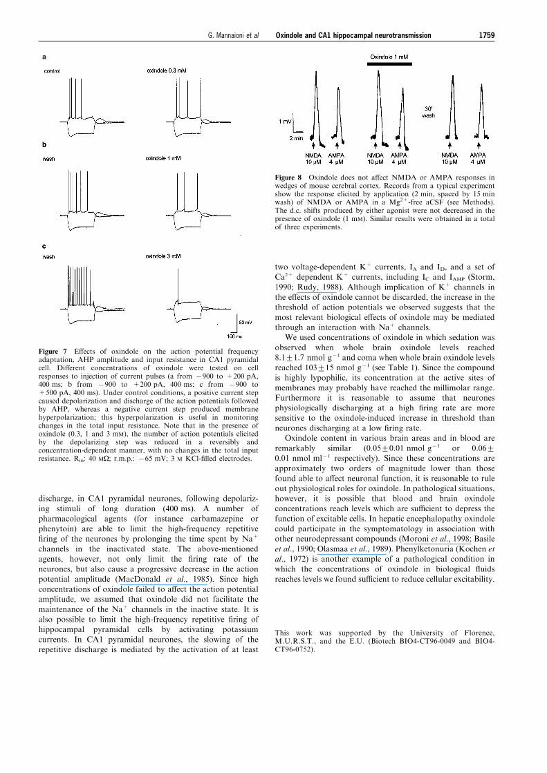
Figure 7 shows the e�ects of oxindole (0.3, 1 and 3 mM) on
the action potential frequency adaptation. Under controlconditions, the repetitive ®ring, produced by depolarizingcurrent steps of 400 ms duration, showed accommodation andat the end of the current injection an AHPL was clearly evident.
Oxindole decreased the number of action potentials dischargedby the neurone (enhanced the accommodation). Furthermore,oxindole reduced the amplitude of the AHPL (Table 2). Figure
7 also shows the lack of changes in Rin monitored by theinjection of negative steps (7900 pA, 400 ms) when oxindole(up to 3 mM) was applied.
In separate experiments, we studied the role of GABAA
receptors, by using picrotoxin (25 mM; n=3), a GABAA
channel blocker. Oxindole (1 mM) still decreased the number
of action potentials elicited by depolarizing current steps (fromseven in controls to one in 1 mM oxindole).
Cortical wedge preparations
In order to evaluate the possibility that the oxindole-induceddecrease of EPSP was mediated by glutamate receptor
antagonism, we tested the e�ects of oxindole on AMPA/
kainate or NMDA receptor responses using `cortical wedges' atechnique which allows the study of excitatory amino acidagonists and antagonists (Mannaioni et al., 1996). As depicted
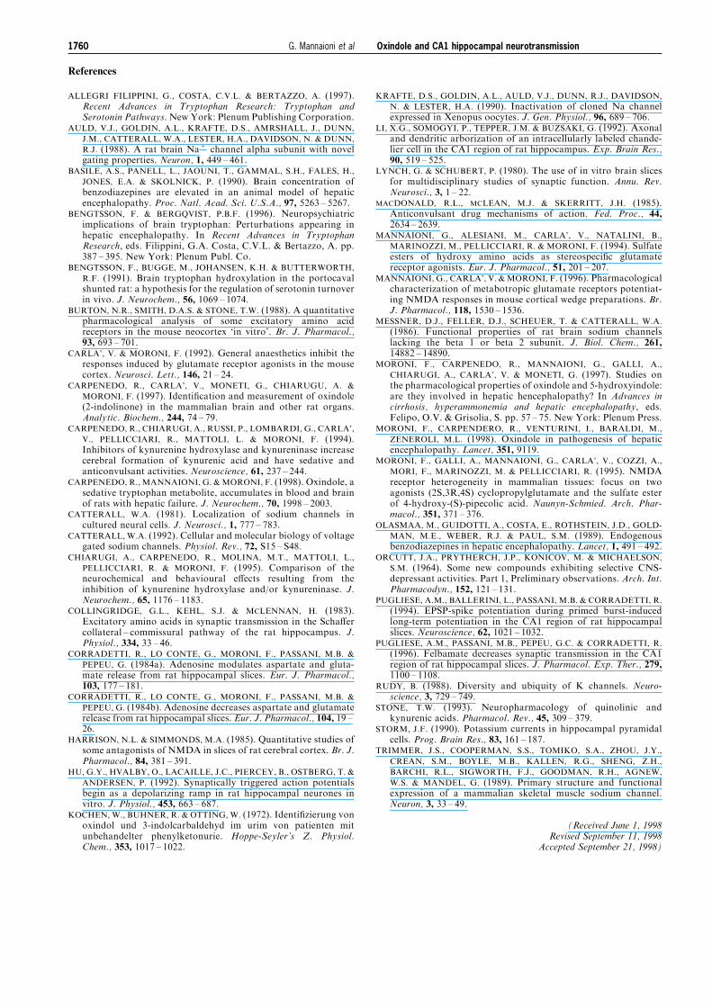
in Figure 8, neither the responses to AMPA (4 mM) nor that toNMDA (10 mM) were decreased by oxindole (1 mM).
Discussion
Oxindole is a neurodepressant tryptophan metabolite physio-
logically present in mammalian brain and blood. Whenadministered to rats at a dose of 10 mg kg71 (i.p.) it hadanticonvulsant and sedative properties while larger doses
(100 mg kg71, i.p.) caused loss of the righting re¯ex and coma.We used brain slices and studied oxindole's mechanism of
action in rat hippocampal neurones. The results obtained
showed that concentrations of oxindole in the range of thosefound in the CNS in pathological conditions were able toreduce neuronal excitability. In a ®rst series of experimentsperformed with extracellular recordings of electrically evoked
synaptic potentials in CA1 pyramidal cells, we observed thatoxindole decreased the PS amplitude. This represents thesynchronous discharge of neurone action potentials located
close to the recording electrode and is proportional to the
Figure 2 E�ects of oxindole on the amplitude of ®eld EPSP in the CA1 hippocampal region. (a) Bath applications of oxindole (1and 3 mM) reversibly decreased ®eld EPSP (fEPSP) amplitude in a concentration-dependent manner. Traces are single responsesevoked by constant strength stimuli (4.5 V) by the corresponding numbers in c. (b) Stimulus-response curves were constructed bygradually increasing the stimulus strength. This protocol was repeated during the application of 1 mM and 3 mM of oxindole. (c)Time-course of the changes of fEPSP amplitude during a typical experiment. Each point represents the amplitude of the fEPSP.Each ascending series of points shows the measure of fEPSPs (from absent to maximal) obtained by gradual increases in stimulusstrength (see b). Data shown are obtained from the same experiment which was repeated ®ve times with similar results.
Oxindole and CA1 hippocampal neurotransmission 1755G. Mannaioni et al
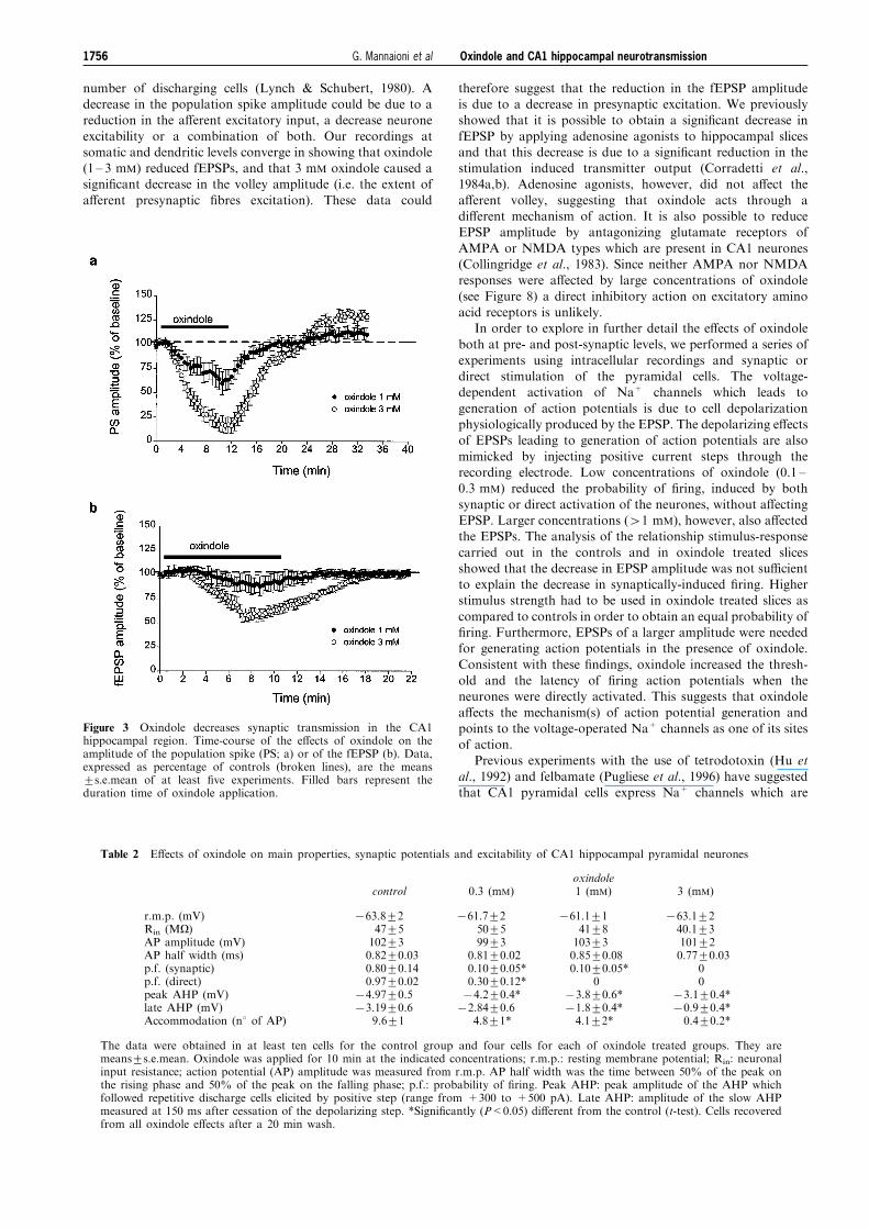
number of discharging cells (Lynch & Schubert, 1980). Adecrease in the population spike amplitude could be due to areduction in the a�erent excitatory input, a decrease neurone
excitability or a combination of both. Our recordings atsomatic and dendritic levels converge in showing that oxindole(1 ± 3 mM) reduced fEPSPs, and that 3 mM oxindole caused asigni®cant decrease in the volley amplitude (i.e. the extent of
a�erent presynaptic ®bres excitation). These data could
therefore suggest that the reduction in the fEPSP amplitudeis due to a decrease in presynaptic excitation. We previouslyshowed that it is possible to obtain a signi®cant decrease in
fEPSP by applying adenosine agonists to hippocampal slicesand that this decrease is due to a signi®cant reduction in thestimulation induced transmitter output (Corradetti et al.,1984a,b). Adenosine agonists, however, did not a�ect the
a�erent volley, suggesting that oxindole acts through adi�erent mechanism of action. It is also possible to reduceEPSP amplitude by antagonizing glutamate receptors of
AMPA or NMDA types which are present in CA1 neurones(Collingridge et al., 1983). Since neither AMPA nor NMDAresponses were a�ected by large concentrations of oxindole
(see Figure 8) a direct inhibitory action on excitatory aminoacid receptors is unlikely.
In order to explore in further detail the e�ects of oxindole
both at pre- and post-synaptic levels, we performed a series ofexperiments using intracellular recordings and synaptic ordirect stimulation of the pyramidal cells. The voltage-dependent activation of Na+ channels which leads to
generation of action potentials is due to cell depolarizationphysiologically produced by the EPSP. The depolarizing e�ectsof EPSPs leading to generation of action potentials are also
mimicked by injecting positive current steps through therecording electrode. Low concentrations of oxindole (0.1 ±0.3 mM) reduced the probability of ®ring, induced by both
synaptic or direct activation of the neurones, without a�ectingEPSP. Larger concentrations (41 mM), however, also a�ectedthe EPSPs. The analysis of the relationship stimulus-response
carried out in the controls and in oxindole treated slicesshowed that the decrease in EPSP amplitude was not su�cientto explain the decrease in synaptically-induced ®ring. Higherstimulus strength had to be used in oxindole treated slices as
compared to controls in order to obtain an equal probability of®ring. Furthermore, EPSPs of a larger amplitude were neededfor generating action potentials in the presence of oxindole.
Consistent with these ®ndings, oxindole increased the thresh-old and the latency of ®ring action potentials when theneurones were directly activated. This suggests that oxindole
a�ects the mechanism(s) of action potential generation andpoints to the voltage-operated Na+ channels as one of its sitesof action.
Previous experiments with the use of tetrodotoxin (Hu et
al., 1992) and felbamate (Pugliese et al., 1996) have suggestedthat CA1 pyramidal cells express Na+ channels which are
Figure 3 Oxindole decreases synaptic transmission in the CA1hippocampal region. Time-course of the e�ects of oxindole on theamplitude of the population spike (PS; a) or of the fEPSP (b). Data,expressed as percentage of controls (broken lines), are the means+s.e.mean of at least ®ve experiments. Filled bars represent theduration time of oxindole application.
Table 2 E�ects of oxindole on main properties, synaptic potentials and excitability of CA1 hippocampal pyramidal neurones
oxindolecontrol 0.3 (mM) 1 (mM) 3 (mM)
r.m.p. (mV)Rin (MO)AP amplitude (mV)AP half width (ms)p.f. (synaptic)p.f. (direct)peak AHP (mV)late AHP (mV)Accommodation (n8 of AP)
763.8+247+5102+30.82+0.030.80+0.140.97+0.02
74.97+0.573.19+0.6
9.6+1
761.7+250+599+3
0.81+0.020.10+0.05*0.30+0.12*74.2+0.4*72.84+0.6
4.8+1*
761.1+141+8103+30.85+0.080.10+0.05*
073.8+0.6*71.8+0.4*4.1+2*
763.1+240.1+3101+20.77+0.03
00
73.1+0.4*70.9+0.4*0.4+0.2*
The data were obtained in at least ten cells for the control group and four cells for each of oxindole treated groups. They aremeans+s.e.mean. Oxindole was applied for 10 min at the indicated concentrations; r.m.p.: resting membrane potential; Rin: neuronalinput resistance; action potential (AP) amplitude was measured from r.m.p. AP half width was the time between 50% of the peak onthe rising phase and 50% of the peak on the falling phase; p.f.: probability of ®ring. Peak AHP: peak amplitude of the AHP whichfollowed repetitive discharge cells elicited by positive step (range from +300 to +500 pA). Late AHP: amplitude of the slow AHPmeasured at 150 ms after cessation of the depolarizing step. *Signi®cantly (P<0.05) di�erent from the control (t-test). Cells recoveredfrom all oxindole e�ects after a 20 min wash.
Oxindole and CA1 hippocampal neurotransmission1756 G. Mannaioni et al
di�erentially able to respond to drugs. Both compoundsdecreased synaptically-driven cell ®ring without changing theaction potential elicited by direct neuronal activation
suggesting that they were a�ecting subpopulations of Na+
channels di�erent than those a�ected by oxindole. Na+
channel b-subunits may play an important role in modulatingthe threshold for channel activation and/or time of channel
inactivation (Catterall, 1992). When Na+ channel a-subunitsalone are expressed in Xenopus oocytes, the properties of theresulting functional channel are quite peculiar due to the
slowness of the channel inactivation and the shifting of thevoltage-dependence to more positive membrane potentials(Messner et al., 1986). Coexpression in the same cells of both aand b-subunits modify the inactivation kinetics and shift thevoltage dependence of the channel toward more negativemembrane potentials (Krafte et al., 1990; Trimmer et al., 1989;
Auld et al., 1988).Oxindole shifted the threshold for channel activation
towards positive potentials but once the threshold was
reached, the resulting spikes were unchanged in duration andin peak amplitude. This pattern of activity could suggest thatoxindole may interact with Na+ channel b-subunits. Althoughappealing, this hypothesis needs further investigation.
The density and/or subunit composition of the voltage-operated Na+ channels located in di�erent regions of thepyramidal cells may di�er (Catterall, 1981). In particular, a
high density of Na+ channels is located at the axon hillock,where the action potentials are generated. This region seemsimportant in the regulation of cell discharge because it receives
the tonic inhibition exerted by a population of GABAergicinterneurones named `chandelier' (Li et al., 1992). In ourexperiments, oxindole-mediated potentiation of GABAA
inhibitory responses was considered unlikely because oxindolee�ects on cell discharge were still present when bicuculline orpictrotoxin, two GABAA receptor blockers, were added to the
perfusion medium.Oxindole was found to increase the action potential
frequency adaptation which limits the frequency of cell
Figure 4 Oxindole reduction of synaptic ®ring elicited by electrical stimulation of the stratum radiatum in the CA1 hippocampalregion. (a) E�ects of oxindole on the relationship between the stimulus strength and the EPSP amplitude in control conditions andunder 0.3 mM of oxindol. No signi®cant changes in EPSP amplitude occurred during oxindole treatment. Note that the EPSPamplitude needed to generate an action potential was increased in the presence of oxindole. (b) Traces are ®ve superimposedresponses evoked by stimulation of the stratum radiatum (3.5 V, downward transients) either in control (left) or after a 10 minapplication of 0.3 mM oxindole (right). Note that the decrease in p.f. (from 0.4 to 0) induced by oxindole was not accompanied bychanges in EPSP amplitude. (c) Responses obtained in the same cell before and during oxindole (0.3 mM) treatment. In order tohave a p.f.=0.4, the stimulus strength was 3.5 V in controls and 3.8 V in the presence of oxindole (see a). Each trace is the averageof the three responses not followed by AP evoked by stimulation of the stratum radiatum. The amplitude of the EPSPs which failedto elicit ®ring was greater in the presence of oxindole than in controls. Rin: 35 MO; r.m.p.: 773 mV; 3 M KCl-®lled electrodes in thepresence of 10 mM bicuculline.
Oxindole and CA1 hippocampal neurotransmission 1757G. Mannaioni et al
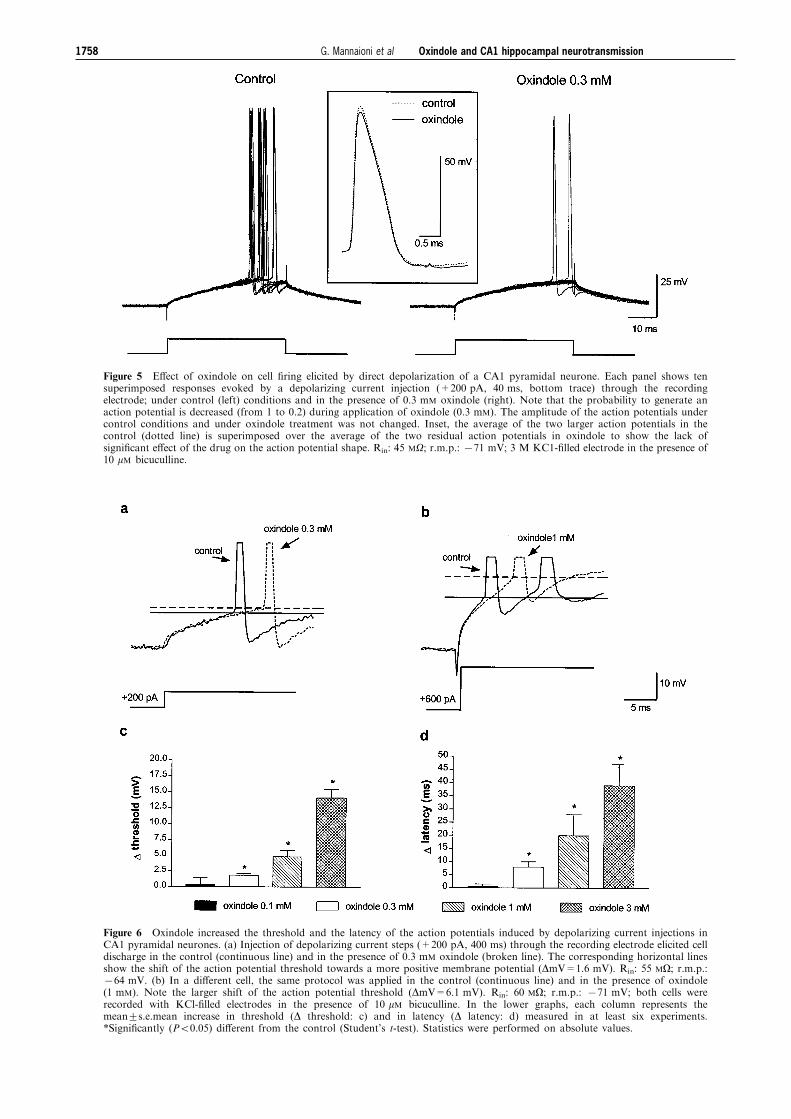
Figure 5 E�ect of oxindole on cell ®ring elicited by direct depolarization of a CA1 pyramidal neurone. Each panel shows tensuperimposed responses evoked by a depolarizing current injection (+200 pA, 40 ms, bottom trace) through the recordingelectrode; under control (left) conditions and in the presence of 0.3 mM oxindole (right). Note that the probability to generate anaction potential is decreased (from 1 to 0.2) during application of oxindole (0.3 mM). The amplitude of the action potentials undercontrol conditions and under oxindole treatment was not changed. Inset, the average of the two larger action potentials in thecontrol (dotted line) is superimposed over the average of the two residual action potentials in oxindole to show the lack ofsigni®cant e�ect of the drug on the action potential shape. Rin: 45 MO; r.m.p.: 771 mV; 3 M KC1-®lled electrode in the presence of10 mM bicuculline.
Figure 6 Oxindole increased the threshold and the latency of the action potentials induced by depolarizing current injections inCA1 pyramidal neurones. (a) Injection of depolarizing current steps (+200 pA, 400 ms) through the recording electrode elicited celldischarge in the control (continuous line) and in the presence of 0.3 mM oxindole (broken line). The corresponding horizontal linesshow the shift of the action potential threshold towards a more positive membrane potential (DmV=1.6 mV). Rin: 55 MO; r.m.p.:764 mV. (b) In a di�erent cell, the same protocol was applied in the control (continuous line) and in the presence of oxindole(1 mM). Note the larger shift of the action potential threshold (DmV=6.1 mV). Rin: 60 MO; r.m.p.: 771 mV; both cells wererecorded with KCl-®lled electrodes in the presence of 10 mM bicuculline. In the lower graphs, each column represents themean+s.e.mean increase in threshold (D threshold: c) and in latency (D latency: d) measured in at least six experiments.*Signi®cantly (P50.05) di�erent from the control (Student's t-test). Statistics were performed on absolute values.
Oxindole and CA1 hippocampal neurotransmission1758 G. Mannaioni et al
discharge, in CA1 pyramidal neurones, following depolariz-
ing stimuli of long duration (400 ms). A number ofpharmacological agents (for instance carbamazepine orphenytoin) are able to limit the high-frequency repetitive
®ring of the neurones by prolonging the time spent by Na+
channels in the inactivated state. The above-mentionedagents, however, not only limit the ®ring rate of theneurones, but also cause a progressive decrease in the action
potential amplitude (MacDonald et al., 1985). Since highconcentrations of oxindole failed to a�ect the action potentialamplitude, we assumed that oxindole did not facilitate the
maintenance of the Na+ channels in the inactive state. It isalso possible to limit the high-frequency repetitive ®ring ofhippocampal pyramidal cells by activating potassium
currents. In CA1 pyramidal neurones, the slowing of therepetitive discharge is mediated by the activation of at least
two voltage-dependent K+ currents, IA and ID, and a set ofCa2+ dependent K+ currents, including IC and IAHP (Storm,
1990; Rudy, 1988). Although implication of K+ channels inthe e�ects of oxindole cannot be discarded, the increase in thethreshold of action potentials we observed suggests that the
most relevant biological e�ects of oxindole may be mediatedthrough an interaction with Na+ channels.
We used concentrations of oxindole in which sedation was
observed when whole brain oxindole levels reached8.1+1.7 nmol g71 and coma when whole brain oxindole levelsreached 103+15 nmol g71 (see Table 1). Since the compound
is highly lypophilic, its concentration at the active sites ofmembranes may probably have reached the millimolar range.Furthermore it is reasonable to assume that neuronesphysiologically discharging at a high ®ring rate are more
sensitive to the oxindole-induced increase in threshold thanneurones discharging at a low ®ring rate.
Oxindole content in various brain areas and in blood are
remarkably similar (0.05+0.01 nmol g71 or 0.06+0.01 nmol ml71 respectively). Since these concentrations areapproximately two orders of magnitude lower than those
found able to a�ect neuronal function, it is reasonable to ruleout physiological roles for oxindole. In pathological situations,however, it is possible that blood and brain oxindoleconcentrations reach levels which are su�cient to depress the
function of excitable cells. In hepatic encephalopathy oxindolecould participate in the symptomatology in association withother neurodepressant compounds (Moroni et al., 1998; Basile
et al., 1990; Olasmaa et al., 1989). Phenylketonuria (Kochen etal., 1972) is another example of a pathological condition inwhich the concentrations of oxindole in biological ¯uids
reaches levels we found su�cient to reduce cellular excitability.
This work was supported by the University of Florence,M.U.R.S.T., and the E.U. (Biotech BIO4-CT96-0049 and BIO4-CT96-0752).
a
Figure 7 E�ects of oxindole on the action potential frequencyadaptation, AHP amplitude and input resistance in CA1 pyramidalcell. Di�erent concentrations of oxindole were tested on cellresponses to injection of current pulses (a from 7900 to +200 pA,400 ms; b from 7900 to +200 pA, 400 ms; c from 7900 to+500 pA, 400 ms). Under control conditions, a positive current stepcaused depolarization and discharge of the action potentials followedby AHP, whereas a negative current step produced membranehyperpolarization; this hyperpolarization is useful in monitoringchanges in the total input resistance. Note that in the presence ofoxindole (0.3, 1 and 3 mM), the number of action potentials elicitedby the depolarizing step was reduced in a reversibly andconcentration-dependent manner, with no changes in the total inputresistance. Rin: 40 MO; r.m.p.: 765 mV; 3 M KCl-®lled electrodes.
Figure 8 Oxindole does not a�ect NMDA or AMPA responses inwedges of mouse cerebral cortex. Records from a typical experimentshow the response elicited by application (2 min, spaced by 15 minwash) of NMDA or AMPA in a Mg2+-free aCSF (see Methods).The d.c. shifts produced by either agonist were not decreased in thepresence of oxindole (1 mM). Similar results were obtained in a totalof three experiments.
Oxindole and CA1 hippocampal neurotransmission 1759G. Mannaioni et al
References
ALLEGRI FILIPPINI, G., COSTA, C.V.L. & BERTAZZO, A. (1997).Recent Advances in Tryptophan Research: Tryptophan andSerotonin Pathways.New York: Plenum Publishing Corporation.
AULD, V.J., GOLDIN, A.L., KRAFTE, D.S., AMRSHALL, J., DUNN,
J.M., CATTERALL, W.A., LESTER, H.A., DAVIDSON, N. & DUNN,
R.J. (1988). A rat brain Na+ channel alpha subunit with novelgating properties. Neuron, 1, 449 ± 461.
BASILE, A.S., PANELL, L., JAOUNI, T., GAMMAL, S.H., FALES, H.,
JONES, E.A. & SKOLNICK, P. (1990). Brain concentration ofbenzodiazepines are elevated in an animal model of hepaticencephalopathy. Proc. Natl. Acad. Sci. U.S.A., 97, 5263 ± 5267.
BENGTSSON, F. & BERGQVIST, P.B.F. (1996). Neuropsychiatricimplications of brain tryptophan: Perturbations appearing inhepatic encephalopathy. In Recent Advances in TryptophanResearch, eds. Filippini, G.A. Costa, C.V.L. & Bertazzo, A. pp.387 ± 395. New York: Plenum Publ. Co.
BENGTSSON, F., BUGGE, M., JOHANSEN, K.H. & BUTTERWORTH,
R.F. (1991). Brain tryptophan hydroxylation in the portocavalshunted rat: a hypothesis for the regulation of serotonin turnoverin vivo. J. Neurochem., 56, 1069 ± 1074.
BURTON, N.R., SMITH, D.A.S. & STONE, T.W. (1988). A quantitativepharmacological analysis of some excitatory amino acidreceptors in the mouse neocortex `in vitro'. Br. J. Pharmacol.,93, 693 ± 701.
CARLA', V. & MORONI, F. (1992). General anaesthetics inhibit theresponses induced by glutamate receptor agonists in the mousecortex. Neurosci. Lett., 146, 21 ± 24.
CARPENEDO, R., CARLA', V., MONETI, G., CHIARUGU, A. &
MORONI, F. (1997). Identi®cation and measurement of oxindole(2-indolinone) in the mammalian brain and other rat organs.Analytic. Biochem., 244, 74 ± 79.
CARPENEDO, R., CHIARUGI, A., RUSSI, P., LOMBARDI, G., CARLA',
V., PELLICCIARI, R., MATTOLI, L. & MORONI, F. (1994).Inhibitors of kynurenine hydroxylase and kynureninase increasecerebral formation of kynurenic acid and have sedative andanticonvulsant activities. Neuroscience, 61, 237 ± 244.
CARPENEDO, R., MANNAIONI, G. &MORONI, F. (1998). Oxindole, asedative tryptophan metabolite, accumulates in blood and brainof rats with hepatic failure. J. Neurochem., 70, 1998 ± 2003.
CATTERALL, W.A. (1981). Localization of sodium channels incultured neural cells. J. Neurosci., 1, 777 ± 783.
CATTERALL, W.A. (1992). Cellular and molecular biology of voltagegated sodium channels. Physiol. Rev., 72, S15 ± S48.
CHIARUGI, A., CARPENEDO, R., MOLINA, M.T., MATTOLI, L.,
PELLICCIARI, R. & MORONI, F. (1995). Comparison of theneurochemical and behavioural e�ects resulting from theinhibition of kynurenine hydroxylase and/or kynureninase. J.Neurochem., 65, 1176 ± 1183.
COLLINGRIDGE, G.L., KEHL, S.J. & MCLENNAN, H. (1983).Excitatory amino acids in synaptic transmission in the Scha�ercollateral ± commissural pathway of the rat hippocampus. J.Physiol., 334, 33 ± 46.
CORRADETTI, R., LO CONTE, G., MORONI, F., PASSANI, M.B. &
PEPEU, G. (1984a). Adenosine modulates aspartate and gluta-mate release from rat hippocampal slices. Eur. J. Pharmacol.,103, 177 ± 181.
CORRADETTI, R., LO CONTE, G., MORONI, F., PASSANI, M.B. &
PEPEU, G. (1984b). Adenosine decreases aspartate and glutamaterelease from rat hippocampal slices. Eur. J. Pharmacol., 104, 19 ±26.
HARRISON, N.L. & SIMMONDS, M.A. (1985). Quantitative studies ofsome antagonists of NMDA in slices of rat cerebral cortex. Br. J.Pharmacol., 84, 381 ± 391.
HU, G.Y., HVALBY, O., LACAILLE, J.C., PIERCEY, B., OSTBERG, T. &
ANDERSEN, P. (1992). Synaptically triggered action potentialsbegin as a depolarizing ramp in rat hippocampal neurones invitro. J. Physiol., 453, 663 ± 687.
KOCHEN, W., BUHNER, R. & OTTING, W. (1972). Identi®zierung vonoxindol und 3-indolcarbaldehyd im urim von patienten mitunbehandelter phenylketonurie. Hoppe-Seyler's Z. Physiol.Chem., 353, 1017 ± 1022.
KRAFTE, D.S., GOLDIN, A.L., AULD, V.J., DUNN, R.J., DAVIDSON,
N. & LESTER, H.A. (1990). Inactivation of cloned Na channelexpressed in Xenopus oocytes. J. Gen. Physiol., 96, 689 ± 706.
LI, X.G., SOMOGYI, P., TEPPER, J.M. & BUZSAKI, G. (1992). Axonaland dendritic arborization of an intracellularly labeled chande-lier cell in the CA1 region of rat hippocampus. Exp. Brain Res.,90, 519 ± 525.
LYNCH, G. & SCHUBERT, P. (1980). The use of in vitro brain slicesfor multidisciplinary studies of synaptic function. Annu. Rev.Neurosci., 3, 1 ± 22.
MACDONALD, R.L., MCLEAN, M.J. & SKERRITT, J.H. (1985).Anticonvulsant drug mechanisms of action. Fed. Proc., 44,2634 ± 2639.
MANNAIONI, G., ALESIANI, M., CARLA', V., NATALINI, B.,
MARINOZZI, M., PELLICCIARI, R. & MORONI, F. (1994). Sulfateesters of hydroxy amino acids as stereospeci®c glutamatereceptor agonists. Eur. J. Pharmacol., 51, 201 ± 207.
MANNAIONI, G., CARLA', V. & MORONI, F. (1996). Pharmacologicalcharacterization of metabotropic glutamate receptors potentiat-ing NMDA responses in mouse cortical wedge preparations. Br.J. Pharmacol., 118, 1530 ± 1536.
MESSNER, D.J., FELLER, D.J., SCHEUER, T. & CATTERALL, W.A.
(1986). Functional properties of rat brain sodium channelslacking the beta 1 or beta 2 subunit. J. Biol. Chem., 261,14882 ± 14890.
MORONI, F., CARPENEDO, R., MANNAIONI, G., GALLI, A.,
CHIARUGI, A., CARLA', V. & MONETI, G. (1997). Studies onthe pharmacological properties of oxindole and 5-hydroxyindole:are they involved in hepatic hencephalopathy? In Advances incirrhosis, hyperammonemia and hepatic encephalopathy, eds.Felipo, O.V. & Grisolia, S. pp. 57 ± 75. New York: Plenum Press.
MORONI, F., CARPENDERO, R., VENTURINI, I., BARALDI, M.,
ZENEROLI, M.L. (1998). Oxindole in pathogenesis of hepaticencephalopathy. Lancet, 351, 9119.
MORONI, F., GALLI, A., MANNAIONI, G., CARLA', V., COZZI, A.,
MORI, F., MARINOZZI, M. & PELLICCIARI, R. (1995). NMDAreceptor heterogeneity in mammalian tissues: focus on twoagonists (2S,3R,4S) cyclopropylglutamate and the sulfate esterof 4-hydroxy-(S)-pipecolic acid. Naunyn-Schmied. Arch. Phar-macol., 351, 371 ± 376.
OLASMAA, M., GUIDOTTI, A., COSTA, E., ROTHSTEIN, J.D., GOLD-
MAN, M.E., WEBER, R.J. & PAUL, S.M. (1989). Endogenousbenzodiazepines in hepatic encephalopathy. Lancet, 1, 491 ± 492.
ORCUTT, J.A., PRYTHERCH, J.P., KONICOV, M. & MICHAELSON,
S.M. (1964). Some new compounds exhibiting selective CNS-depressant activities. Part 1, Preliminary observations. Arch. Int.Pharmacodyn., 152, 121 ± 131.
PUGLIESE, A.M., BALLERINI, L., PASSANI, M.B. & CORRADETTI, R.
(1994). EPSP-spike potentiation during primed burst-inducedlong-term potentiation in the CA1 region of rat hippocampalslices. Neuroscience, 62, 1021 ± 1032.
PUGLIESE, A.M., PASSANI, M.B., PEPEU, G.C. & CORRADETTI, R.
(1996). Felbamate decreases synaptic transmission in the CA1region of rat hippocampal slices. J. Pharmacol. Exp. Ther., 279,1100 ± 1108.
RUDY, B. (1988). Diversity and ubiquity of K channels. Neuro-science, 3, 729 ± 749.
STONE, T.W. (1993). Neuropharmacology of quinolinic andkynurenic acids. Pharmacol. Rev., 45, 309 ± 379.
STORM, J.F. (1990). Potassium currents in hippocampal pyramidalcells. Prog. Brain Res., 83, 161 ± 187.
TRIMMER, J.S., COOPERMAN, S.S., TOMIKO, S.A., ZHOU, J.Y.,
CREAN, S.M., BOYLE, M.B., KALLEN, R.G., SHENG, Z.H.,
BARCHI, R.L., SIGWORTH, F.J., GOODMAN, R.H., AGNEW,
W.S. & MANDEL, G. (1989). Primary structure and functionalexpression of a mammalian skeletal muscle sodium channel.Neuron, 3, 33 ± 49.
(Received June 1, 1998Revised September 11, 1998
Accepted September 21, 1998)
Oxindole and CA1 hippocampal neurotransmission1760 G. Mannaioni et al
Related Documents


