Electronic Structure, Dielectric Response, and Surface Charge Distribution of RGD (1FUV) Peptide Puja Adhikari 1 , Amy M. Wen 2 , Roger H. French 3,4,6 , V. Adrian Parsegian 5 , Nicole F. Steinmetz 2,3,6,7 , Rudolf Podgornik 5,8,9 & Wai-Yim Ching 1 1 Department of Physics and Astronomy, University of Missouri-Kansas City, Kansas City, MO, 64110, USA, 2 Department of Biomedical Engineering, Case Western Reserve University, 10900 Euclid Avenue, Cleveland, OH 44106, USA, 3 Department of Materials Science and Engineering, Case Western Reserve University, 10900 Euclid Avenue, Cleveland, OH 44106, USA, 4 Department of Physics, Case Western Reserve University, 10900 Euclid Avenue, Cleveland, OH 44106, USA, 5 Department of Physics, University of Massachusetts, Amherst, Massachusetts 01003, USA, 6 Department of Radiology, Case Western Reserve University, 10900 Euclid Avenue, Cleveland, OH 44106, USA, 7 Department of Macromolecular Science and Engineering, Case Western Reserve University, 10900 Euclid Avenue, Cleveland, OH 44106, USA, 8 Department of Theoretical Physics, J. Stefan Institute, SI-1000 Ljubljana, Slovenia, 9 Department of Physics, Faculty of Mathematics and Physics, University of Ljubljana, SI-1000 Ljubljana, Slovenia. Long and short range molecular interactions govern molecular recognition and self-assembly of biological macromolecules. Microscopic parameters in the theories of these molecular interactions are either phenomenological or need to be calculated within a microscopic theory. We report a unified methodology for the ab initio quantum mechanical (QM) calculation that yields all the microscopic parameters, namely the partial charges as well as the frequency-dependent dielectric response function, that can then be taken as input for macroscopic theories of electrostatic, polar, and van der Waals-London dispersion intermolecular forces. We apply this methodology to obtain the electronic structure of the cyclic tripeptide RGD-4C (1FUV). This ab initio unified methodology yields the relevant parameters entering the long range interactions of biological macromolecules, providing accurate data for the partial charge distribution and the frequency-dependent dielectric response function of this peptide. These microscopic parameters determine the range and strength of the intricate intermolecular interactions between potential docking sites of the RGD-4C ligand and its integrin receptor. N ature has evolved many sophisticated bio-specific recognition systems that play a crucial role in cell signaling and orchestration of self-assembly of molecules, cells, and entire organisms. Fundamental understanding of the molecular machinery governing these bio-specific interactions is expected to have impact on the basic sciences, materials science, and translational approaches where bio-recognition systems are being exploited and further developed to yield novel functional materials with various properties such as self- assembly, stimuli-response, and/or self-healing. Interactions between biological macromolecules can be deduced from standard principles of colloid and nanoscale stability theory 1 that identify different types of direct long range interactions as well as different types of short-range solvent-mediated interactions, together governing the molecular recognition and self-assembly of biological macromolecules 2 . The former include electrostatic interactions 3 , depending on the specific nature of molecular charges and the net charge on a body, polar interactions 4 arising from dipolar and higher order charge multipoles, and van der Waals-London dispersion (vdW) interactions 4 , that in their turn depend on the details of the dielectric response properties of the molecular materials. The short-range solvent-mediated components of the overall intermolecular forces can be classified as hydration interactions 5 , due to hydrophilic moieties, and hydrophobic interactions 6 , engendered by the hydrophobic moieties, both along the solvent-exposed surfaces. Theoretical modeling of the long range components of the molecular interactions can be decomposed con- ceptually and methodologically into a microscopic and a macroscopic part 2 . While the two methodological levels are coupled in principle, the standard assumption is that this coupling is weak and that the microscopic calcula- tion yields parameters for isolated molecules that then enter the macroscopic theory of interactions between the molecules. For electrostatic and vdW interactions, the microscopic part follows from ab initio QM calculations, OPEN SUBJECT AREAS: COMPUTATIONAL BIOPHYSICS NANOSCALE MATERIALS BIOMATERIALS Received 31 March 2014 Accepted 6 June 2014 Published 8 July 2014 Correspondence and requests for materials should be addressed to R.H.F. (roger.french@ case.edu) SCIENTIFIC REPORTS | 4 : 5605 | DOI: 10.1038/srep05605 1

Welcome message from author
This document is posted to help you gain knowledge. Please leave a comment to let me know what you think about it! Share it to your friends and learn new things together.
Transcript
Electronic Structure, Dielectric Response,and Surface Charge Distribution of RGD(1FUV) PeptidePuja Adhikari1, Amy M. Wen2, Roger H. French3,4,6, V. Adrian Parsegian5, Nicole F. Steinmetz2,3,6,7,Rudolf Podgornik5,8,9 & Wai-Yim Ching1
1Department of Physics and Astronomy, University of Missouri-Kansas City, Kansas City, MO, 64110, USA, 2Department ofBiomedical Engineering, Case Western Reserve University, 10900 Euclid Avenue, Cleveland, OH 44106, USA, 3Department ofMaterials Science and Engineering, Case Western Reserve University, 10900 Euclid Avenue, Cleveland, OH 44106, USA,4Department of Physics, Case Western Reserve University, 10900 Euclid Avenue, Cleveland, OH 44106, USA, 5Department ofPhysics, University of Massachusetts, Amherst, Massachusetts 01003, USA, 6Department of Radiology, Case Western ReserveUniversity, 10900 Euclid Avenue, Cleveland, OH 44106, USA, 7Department of Macromolecular Science and Engineering, CaseWestern Reserve University, 10900 Euclid Avenue, Cleveland, OH 44106, USA, 8Department of Theoretical Physics, J. StefanInstitute, SI-1000 Ljubljana, Slovenia, 9Department of Physics, Faculty of Mathematics and Physics, University of Ljubljana, SI-1000Ljubljana, Slovenia.
Long and short range molecular interactions govern molecular recognition and self-assembly of biologicalmacromolecules. Microscopic parameters in the theories of these molecular interactions are eitherphenomenological or need to be calculated within a microscopic theory. We report a unified methodologyfor the ab initio quantum mechanical (QM) calculation that yields all the microscopic parameters, namelythe partial charges as well as the frequency-dependent dielectric response function, that can then be taken asinput for macroscopic theories of electrostatic, polar, and van der Waals-London dispersion intermolecularforces. We apply this methodology to obtain the electronic structure of the cyclic tripeptide RGD-4C(1FUV). This ab initio unified methodology yields the relevant parameters entering the long rangeinteractions of biological macromolecules, providing accurate data for the partial charge distribution andthe frequency-dependent dielectric response function of this peptide. These microscopic parametersdetermine the range and strength of the intricate intermolecular interactions between potential dockingsites of the RGD-4C ligand and its integrin receptor.
Nature has evolved many sophisticated bio-specific recognition systems that play a crucial role in cellsignaling and orchestration of self-assembly of molecules, cells, and entire organisms. Fundamentalunderstanding of the molecular machinery governing these bio-specific interactions is expected to have
impact on the basic sciences, materials science, and translational approaches where bio-recognition systems arebeing exploited and further developed to yield novel functional materials with various properties such as self-assembly, stimuli-response, and/or self-healing.
Interactions between biological macromolecules can be deduced from standard principles of colloid andnanoscale stability theory1 that identify different types of direct long range interactions as well as different typesof short-range solvent-mediated interactions, together governing the molecular recognition and self-assembly ofbiological macromolecules2. The former include electrostatic interactions3, depending on the specific nature ofmolecular charges and the net charge on a body, polar interactions4 arising from dipolar and higher order chargemultipoles, and van der Waals-London dispersion (vdW) interactions4, that in their turn depend on the details ofthe dielectric response properties of the molecular materials. The short-range solvent-mediated components ofthe overall intermolecular forces can be classified as hydration interactions5, due to hydrophilic moieties, andhydrophobic interactions6, engendered by the hydrophobic moieties, both along the solvent-exposed surfaces.
Theoretical modeling of the long range components of the molecular interactions can be decomposed con-ceptually and methodologically into a microscopic and a macroscopic part2. While the two methodological levelsare coupled in principle, the standard assumption is that this coupling is weak and that the microscopic calcula-tion yields parameters for isolated molecules that then enter the macroscopic theory of interactions between themolecules. For electrostatic and vdW interactions, the microscopic part follows from ab initio QM calculations,
OPEN
SUBJECT AREAS:COMPUTATIONAL
BIOPHYSICS
NANOSCALE MATERIALS
BIOMATERIALS
Received31 March 2014
Accepted6 June 2014
Published8 July 2014
Correspondence andrequests for materials
should be addressed toR.H.F. (roger.french@
case.edu)
SCIENTIFIC REPORTS | 4 : 5605 | DOI: 10.1038/srep05605 1
that are in general of different types, focused specifically on theelectronic structure calculations, which ideally yield the partialcharges of all the atoms composing the interacting molecules, orindeed on the frequency-dependent dielectric function (the opticalproperties) of the whole molecule, respectively7. These microscopicparameters then enter the macroscopic theories, viz. the Poisson-Boltzmann (PB) theory and/or its derivatives for electrostatic8 andpolar interactions9, and the Lifshitz theory for vdW interactions4,
that provide the separation and angular dependence of the long rangepart of the interaction between the molecules10–12, governing importantaspects of the recognition and assembly processes of macromolecules.
As a model for a biomolecular recognition system we chose aligand-receptor couple, specifically, the well-known and extensivelystudied RGD peptide ligand-integrin receptor pair. However, oneshould note here, that these studies are available only in the settingof biochemistry, nanotechnology, and medicine, while no quantitat-
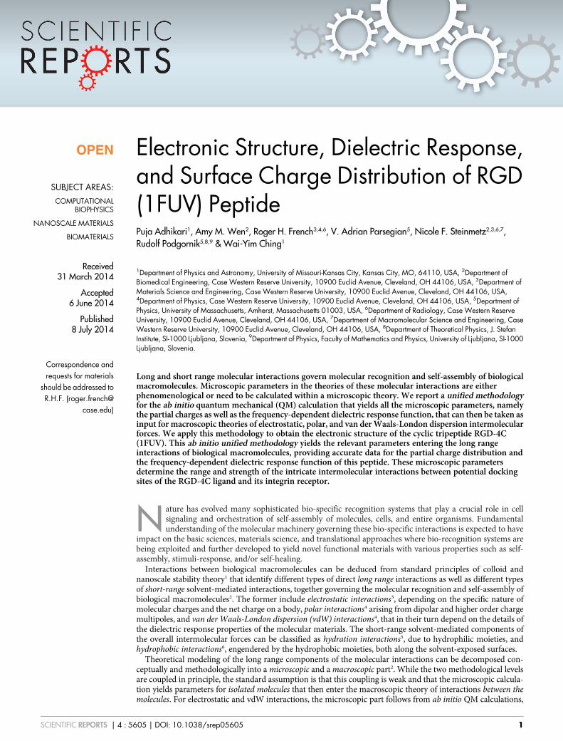
Figure 1 | (a) Molecular structure of the peptide RGD-A. Each amino acid is enclosed by dashed line and marked with ARG, GLY and ASP in larger
font since they represent the R,G,D respectively in the peptide; (b) same as (a) with 90 degree orientation; (c) Gross sketch of the structure of RGD-A
showing two-looped structure and the connecting atoms in each amino acid.
www.nature.com/scientificreports
SCIENTIFIC REPORTS | 4 : 5605 | DOI: 10.1038/srep05605 2
ive assessment of its electronic structure, optical properties, andpartial charge has been undertaken. The tripeptide RGD plays apivotal role in cell signaling. It should be noted that out of 24 knownintegrins, a third recognize and bind to the RGD tripeptide13.Receptor specificity and affinity is dependent on peptide conforma-tion and flanking amino acids14. RGD-containing biomolecularmaterials have become popular as scaffolds for bone, tissue, andcartilage synthesis15; they are also candidates for radiotracers inimaging16–18 and targeted drug delivery19–21. The broader opportun-ities represented by molecular recognition systems, such as theligand-receptor pairs of the RGD-integrin type, include also the abil-ity to architecturally predefine building blocks that can then establishbonds between complementary surface moieties. Other bio-specificrecognition systems, for example, are orthogonal pairs of coiled-coilpeptides, which are being investigated for self-induced assembly ofnanoparticles and hierarchical structures22,23. This type of remotecontrol of nanoscale and mesoscale assembly and architecture isone of the opportunities recently identified in a mesoscale sciencereport24, where programmable molecular recognition could be easilyexploited for electrical energy storage, lighting, photovoltaic devices,and electronics.
In this paper, we report an ab initio unified methodology for theQM calculation of one representative isomer of RGD-4C (Figure 1)that yields its electronic structure, its partial charges, as well as itsfrequency-dependent dielectric response function, in a singlescalable calculation that could then be taken as input for the mac-roscopic PB-vdW theories of intermolecular forces. RGD-4C isstructurally characterized as a peptide with the amino acid sequenceACDCRGDCFCG consisting of alanine (A), cysteine (C), asparagine(D), glycine (G), phenylanine (F), and arginine (R). This peptide,identified through the phage library display25, is probably one of themost utilized RGD variants. There are four cysteines in RGD-4C,which would allow for three possible fully disulfide-bonded forms.Others have reported that only two forms, specifically the 1–4; 2–3and 1–3; 2–4 bonded arrangements, are detected in the sponta-neously cyclized peptide26. The 1–4; 2–3 bonded peptide, isomerRGD-A, is a stronger avb3 integrin binder. It thus seemed appropri-ate to consider RGD-A as a paradigm for our calculations. To the bestof our knowledge, there have been no fundamental studies on RGDpeptides of this particular type.
While the ab initio unified methodology of our single scalablecalculation yields accurate data for the partial charge distributionand the frequency-dependent imaginary part of the dielectric res-ponse function of this important peptide, this fundamental under-standing in turn provides novel and helpful information for
elucidating the nature of intricate intermolecular interactionsbetween potential docking sites. In recent years, such calculationshave started to emerge on various levels, with varied methodo-logies27, and are expected to make a major impact in biomedicalengineering28–32, energy, and broadly in mesoscale science ingeneral24.
ResultsThe structure for RGD-A (1FUV) was obtained from the RCSBprotein data bank (PDB) (http://www.rcsb.org/pdb/explore.do?structureId51FUV, date accessed 5/23/2014) based on data fromnuclear magnetic resonance (NMR) measurements. There are a totalof 19 such models with the same number of atoms, molecular for-mula, molecular composition, and molecular weight but with aslightly different molecular volume. We selected model 1 for theelectronic structure calculations. It has 11 amino acids that consistof a single ALA, ARG and PHE, two ASP and GLY, and 4 CYS AAs(altogether 135 atoms). The atomic positions of the amino acids ofthe RGD-A peptide were kept in a super-cell of size 26A 3 25A 3
20A, which is sufficiently large to avoid any interaction with the samemolecule in the adjacent cells. This model structure was then furtherrelaxed using Vienna ab-initio simulation package (VASP) (seeMethods). There was no significant change in the structure fromPDB data, signifying the integrity and accuracy of the structuredeposited in PDB. Figures 1 (a) and (b) show the molecular structure
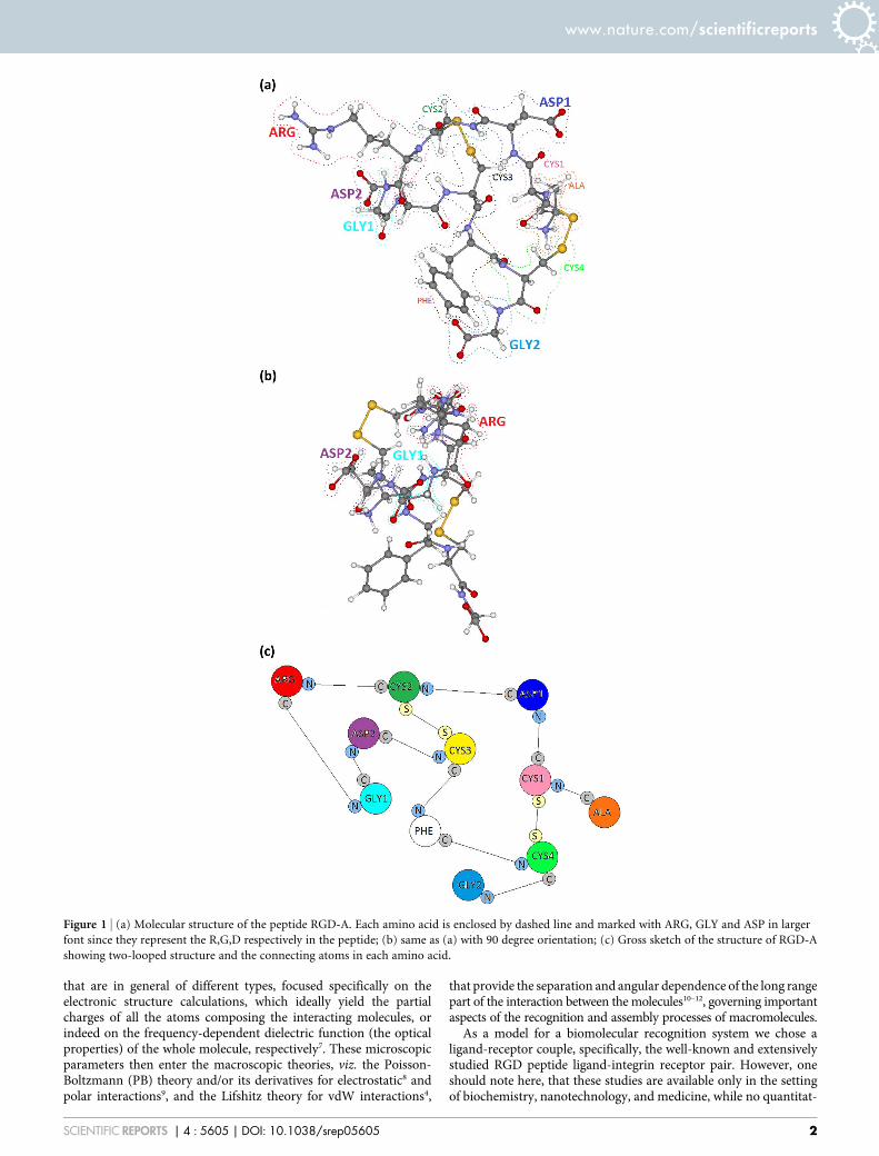
Figure 2 | The calculated total density of states (TDOS) of 1FUV (RGD-A) with each energy state slightly broadened; the energy of HOMOstate is set at 0.0 eV.
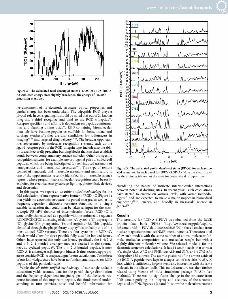
Figure 3 | The calculated partial density of states (PDOS) for each aminoacid as marked in each panel for 1FUV (RGD-A). Note the Y-axis scales
for the amino acids are not the same for better visual juxtaposition.
www.nature.com/scientificreports
SCIENTIFIC REPORTS | 4 : 5605 | DOI: 10.1038/srep05605 3
of the peptide with the constituting 11 amino acids delimited bydotted lines. Figure 1 (c) is a simplified symbolic sketch of the mole-cule showing two topological loops on the graph of chemical bondingfor this peptide.
Figure 2 shows the calculated total density of states (TDOS) or thenumber of electron states per unit of energy of RGD-A (1FUV). TheHOMO-LUMO gap of 1FUV is about 3.38 eV and the range ofthe occupied valence band region is 22.9 eV. The TDOS is resolvedinto 11 partial DOS (PDOS) in Figure 3 for the six different types of
amino acids in the sequence. The integrated area of the PDOS up toHOMO corresponds to the total number of electrons in the aminoacid in the RGD molecule. The HOMO state (defined as the top of thevalence band at 0.0 eV) originates from ASP1; the LUMO state isfrom ARG. An interesting feature in the PDOS is the four differentCYS amino acids that have somewhat different features indicatingthe difference in their connectivity (1–4; 2–3) and in their localenvironments. The same can be said about the two different GLY(GLY1 and GLY2) and the two different ASP (ASP1 and ASP2) (seeFigure 3). These differences for the same amino acid types are usuallyobserved in the form of a shift of their peak positions in the PDOSspectra and their relative heights. Such differences reflect the abilityof ab initio calculations in delineating the electron structures andlocal geometries of individual amino acids.
Figure 4 displays the calculated partial charge DQ* on each of the135 atoms in RGD-A (1FUV) for the relaxed structure (open symbol)and the original structure from PDB (closed symbol). Partial chargeDQ* 5 Q0 2 Q* is the deviation between the charge on the neutralatom Q0 and the effective charge of the same atom in the presentcalculation in accordance with the Mulliken scheme33 using the min-imal basis in the orthogonalized linear combination of atomic orbitals(OLCAO) method7 (see Methods section). There is no significantchange in the partial charge of the atoms before and after relaxation,indicating that the changes due to relaxation are minor. Nitrogen andoxygen atoms are all negatively charged, whereas all hydrogen atomsare positively charged and sulfur atoms are only slightly positive.However, the carbon atoms can be both positively and negativelycharged according to their local coordination and bonding withindifferent amino acids. The most positive DQ* value is 0.553 e whichcomes from a C atom in ASP1 and the most negative DQ* value for Cis 20.640 e from a C atom in ALA. The most negative DQ* value is
Figure 4 | Calculated partial charge DQ* vs. atom number in 1FUV(RGD-A). Solid symbols are the values obtained from the calculation using
the unrelaxed structure from PDB and the open symbols are from the
VASP-relaxed structure (R).
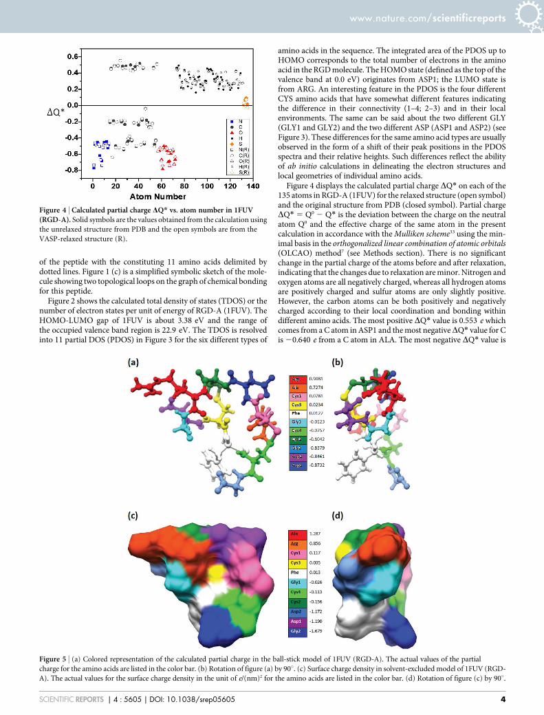
Figure 5 | (a) Colored representation of the calculated partial charge in the ball-stick model of 1FUV (RGD-A). The actual values of the partial
charge for the amino acids are listed in the color bar. (b) Rotation of figure (a) by 90u. (c) Surface charge density in solvent-excluded model of 1FUV (RGD-
A). The actual values for the surface charge density in the unit of e/(nm)2 for the amino acids are listed in the color bar. (d) Rotation of figure (c) by 90u.
www.nature.com/scientificreports
SCIENTIFIC REPORTS | 4 : 5605 | DOI: 10.1038/srep05605 4
20.741 e from the N atom of ARG and the O atom of ASP2(20.742 e). By adding the DQ* values of all the atoms within eachamino acid, we obtain the partial charges for each amino acid.
Figure 5 (a) and (b) presents a ball and stick model in two differentorientations in which the amino acids are differentiated by coloraccording to their partial charge. ASP has the most negative chargeof 20.87 e (dark blue) whereas ARG has the most positive charge of10.91 e (dark red). The variations in partial charge among aminoacids (hence difference in color) exemplify the different chemicalbonding between atoms within it. For instance, there are fourcysteines, and their partial charges are 20.076 e, 10.023 e,20.104 e, and 10.078 e for CYS4 (10), CYS3 (8), CYS2 (4), andCYS1 (2) respectively, where the integer in the parentheses indicatestheir location in the sequence.
At physiological pH 7.4, one standardly considers the followingisolated amino acids as charged: aspartic acid (ASP) and glutamicacid (GLU) carrying a charge of 21.0 e originating from the depro-tonated carboxylate on the side chains of aspartic and glutamic acid,lysine (LYS) and arginine (ARG) carrying a charge of 11.0 e origin-ating in the protonated amine group of arginine and lysine, andhistidine (HIS) carrying a fractional charge of 10.1 e originatingin the protonated state of the secondary amine of histidine.Cysteine (CYS) is usually not considered to be an acid because thethiol group is often reactive and can form disulfide bonds34. The abinitio result that shows the most positive charge 10.91 e on ARG isin agreement with the above standard value of 11.0 e. Also in agree-ment, ASP with 20.87 e emerges as the most negatively-chargedamino acid and compares well with 21.0 e assumed in the aqueoussolvent at physiological conditions. The other amino acids usuallyassumed to be charged at physiological conditions are GLU withpartial charge of 21.0 e, and LYS and HIS, the former with a partialcharge of 11.0 e and the latter with 10.1 e. However, these aminoacids are not part of the RGD peptide studied in this paper. On theother hand, the ab initio partial charges on ALA with 10.72 e andGLY with 20.84 e are quite substantial, while they are usuallyassumed to be neutral at physiological conditions. These differencesbetween the ab initio and the standard values of partial charges implythat effects of the solvent and local microenvironment, such as prox-imity of other charged groups, geometry of the folded polypeptide,solution ions, dielectric permittivities of the various peptide moietiesetc. all strongly affect the local environment of the dissociable aminoacid groups in the polypeptide. They can have a dominating effect onits effective charge (for details see Ref. 35).
Apart from the assignment of partial charges, what is important inlarge macromolecules are their surface charge densities that deter-mine the electrostatic potential and the long range polar interac-tions. Different amino acids have different effective volumes andsurface areas based on the standard solvent excluded layer.Figure 5 (c) and (d) shows the distribution of the surface chargedensity in units of e/(nm)2 obtained by using the surface chargedensity for each amino acid normalized by its solvent excluded sur-face area. The surface charge density map shows the spectacularpolar nature of the RGD-A (1FUV) peptide, which controls dock-ing to its binding site (the integrin receptor) and its movementswithin the aqueous solution. The effective surface charge density isdisplayed in two orientations as in Figure 5 (a) and (b). Because of thelocal geometry of the folded peptide chain, the highest negativecharge density is now exhibited by GLY2 (21.679 e/(nm)2), withthe highest positive charge density found on ALA (11.287 e/(nm)2). The region close to PHE is more or less neutral (white color).The RGD motif exhibits the following effective surface charges: ARGwith 10.856 e/(nm)2, GLY1 20.026 e/(nm)2, and ASP2 21.172 e/(nm)2.
The values of the surface charge density are not far from the valuestypically assumed for polypeptides (,0.6 e/(nm)2) and are smallerthan that for DNA (,1 e/(nm)2) or many lipid bilayers (,0.1 to
,1 e/(nm)2) considered within idealized cylindrical or planar effec-tive shape models. In fact, one should note that the charge densitycalculated here pertains to a certain choice of the area normalization,i.e., solvent excluded in this case. In case one wants to use an effectivegeometric model of the molecule, e.g. spherical, cylindrical, prolateor oblate idealized forms, due provision must be taken into accountfor additional surface area normalization36. Nevertheless, the effec-tive surface charge density shown in Figure 5 (c) and (d), rigorouslycalculated by QM means, does provide a quantitative measure for thecharge multipoles associated with the molecule. The dipole is themost prominent, as it clearly shows the lacunae of the positive(and/or negative) charges that would provide molecular interactionhandles to position the molecule on approach to another molecule.
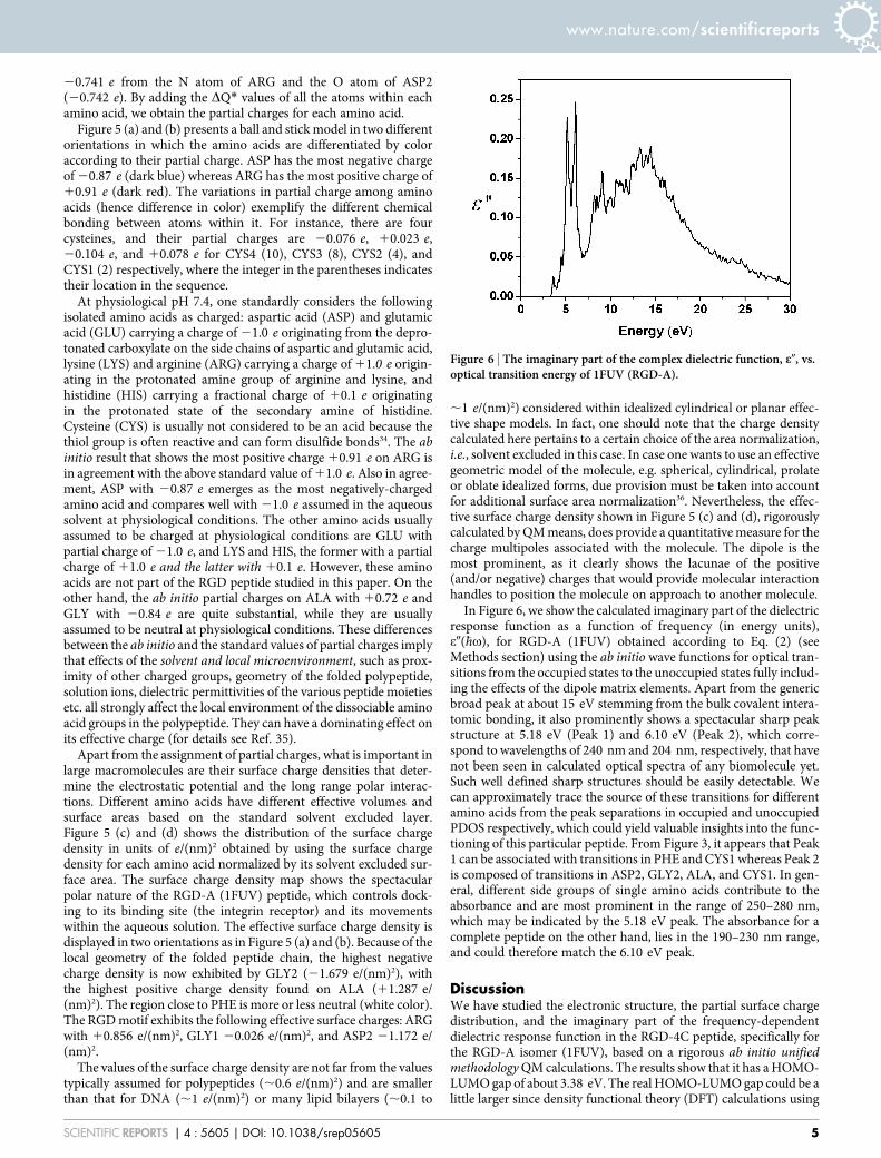
In Figure 6, we show the calculated imaginary part of the dielectricresponse function as a function of frequency (in energy units),e0(�hv), for RGD-A (1FUV) obtained according to Eq. (2) (seeMethods section) using the ab initio wave functions for optical tran-sitions from the occupied states to the unoccupied states fully includ-ing the effects of the dipole matrix elements. Apart from the genericbroad peak at about 15 eV stemming from the bulk covalent intera-tomic bonding, it also prominently shows a spectacular sharp peakstructure at 5.18 eV (Peak 1) and 6.10 eV (Peak 2), which corre-spond to wavelengths of 240 nm and 204 nm, respectively, that havenot been seen in calculated optical spectra of any biomolecule yet.Such well defined sharp structures should be easily detectable. Wecan approximately trace the source of these transitions for differentamino acids from the peak separations in occupied and unoccupiedPDOS respectively, which could yield valuable insights into the func-tioning of this particular peptide. From Figure 3, it appears that Peak1 can be associated with transitions in PHE and CYS1 whereas Peak 2is composed of transitions in ASP2, GLY2, ALA, and CYS1. In gen-eral, different side groups of single amino acids contribute to theabsorbance and are most prominent in the range of 250–280 nm,which may be indicated by the 5.18 eV peak. The absorbance for acomplete peptide on the other hand, lies in the 190–230 nm range,and could therefore match the 6.10 eV peak.
DiscussionWe have studied the electronic structure, the partial surface chargedistribution, and the imaginary part of the frequency-dependentdielectric response function in the RGD-4C peptide, specifically forthe RGD-A isomer (1FUV), based on a rigorous ab initio unifiedmethodology QM calculations. The results show that it has a HOMO-LUMO gap of about 3.38 eV. The real HOMO-LUMO gap could be alittle larger since density functional theory (DFT) calculations using
Figure 6 | The imaginary part of the complex dielectric function, e0, vs.optical transition energy of 1FUV (RGD-A).
www.nature.com/scientificreports
SCIENTIFIC REPORTS | 4 : 5605 | DOI: 10.1038/srep05605 5
LDA approximation generally underestimate the band gap, as is wellknown and discussed in Ref. 7. In principle, one can get a slightlylarger gap by using a different exchange-correlation potential inDFT, such as PAW-PBE or GGA, meta-GGA, or a hybrid potentialsuch as B3LYP etc., depending on the system under investigation andon the basis set used in a particular method. LDA is currently imple-mented in the OLCAO method that we use. Since no calculation ofthe RGD electronic structure is available, it seems important to us toobtain a solid estimate on the HOMO-LUMO gap that can later beimproved, if it turns out to be necessary. One thing is, nevertheless,certain: the HOMO-LUMO gap does not in any way affect the partialcharge calculations which are derived from the occupied states welldescribed by DFT. It may shift the two prominent peaks in theimaginary part of the dielectric response function, Figure 6, to aslightly higher frequency, though.
By resolving the total density of states of the peptide into compo-nents from individual amino acids, detailed interactions betweenthem at an atomic level can be further elaborated. The surface chargedensity distribution obtained from partial charge calculations showsthe molecule to be highly polar, thus able to promote interactionswith its avb3 integrin receptor either via long range polar comple-mentarity or short-range hydrogen bonding.
We have also calculated the optical properties of this peptide.These show a striking feature of two sharp absorption peaks apartfrom the bulk broad interatomic bonding peak. The optical spectra ofnano-scale objects, such as single wall carbon nanotubes (SWCNT)with different chirality, have already been used to estimate the long-range vdW interaction either in vacuum or in the solvent throughtheir connection with Hamaker coefficients that quantify thestrength of this interaction10–12. It is conceivable that the uniqueoptical spectrum of RGD peptide could result in a strong vdWattractive force that could influence its interaction with othermolecular moieties and even modify its mobility in aqueoussolutions.
The ab initio unified methodology QM calculations used in thispaper, which yield different microscopic parameters in a single scal-able calculation, are the first for the RGD peptide to allow for areliable estimate of the partial charges and the frequency dependentdielectric response function directly from the wave functions (see e.g.Eq. (1)) and can be further refined and applied to other substantiallylarger and significantly more complex biomolecules and proteins.They can also be used to refine the crucial parameters in moleculardynamic simulations vastly used in biophysics community. In viewof the paucity of theoretical studies of partial charge distribution anddielectric response properties for the RGD peptide, we believe ourseminal study will be helpful making simulation force field para-meters more accurate and useful.
Future investigations should include in particular the investiga-tion of solvent effects by adding explicit water molecules with andwithout salts and observing their influence on the electronic struc-ture, surface charge distribution, and dielectric response, as well asusing the present results to estimate the electrostatic and polar inter-actions between RGD and other biomolecules or peptides. Oneshould nevertheless note, that such full scale quantum calculationsare a non-trivial task, and even calculations with no explicit watermolecules for this polypeptide have not been available before. Inaddition to the explicit solvent, one would also need to implementthe microenvironment of the various dissociable groups on the solv-ent exposed surface of the polypeptide, making hopes for a fullquantum mechanical treatment very remote.
MethodsWe used the orthogonalized linear combination of atomic orbitals (OLCAO)method7. The fundamental quantities discussed here are total density of states(TDOS), partial density of states (PDOS), and effective charge Qa* (for details seeRef. 7):
Q�!~X
i
Xn occ
Xj,b
Cm�ia Cm
jbSia,jb ð1Þ
Here, the Sia, jb are the overlap integrals between the ith orbital in the ath atom and jth
orbital in the bth atom and Cmjb are the eigenvector coefficients of the mth band, jth
orbital in the bth atom7. Basically, OLCAO is an electronic structure method based onDFT with local density approximation originally designed for crystalline solids, but itworks equally well for complex biomolecules using the supercell. The use of atomicorbitals in the basis expansion is particularly appropriate for such calculations. Detailsof the method can be found in Ref. 7. Vienna ab-initio simulation package (VASP)37,38
is also a DFT-based method used solely to relax the structure of the biomolecule. Ituses plane waves as the basis set and is very efficient for geometry optimization andstructural relaxation. In the present work, we used the PAW-PBE potential for theexchange-correlation functional39. The combination of these two methods enables usto investigate materials of very complex structures27.
The same OLCAO method is also used to calculate the imaginary part of thefrequency-dependent dielectric response function e0(�hv) within the random phaseapproximation (RPA) of the one-electron theory according to the following equation(for details see Ref. 7):
e’’ �hvð Þ~ e2
pmv2
Xn,l
Ynh ~rð Þj j{i�h ~+� �
Yl ~rð Þij j2d En{El{�hvð Þ: ð2Þ
Ynh ~rð Þj j{i�h ~+� �
Yl ~rð Þij j is the dipolar matrix element of electron transition from
the occupied molecular orbital state Yl to the state Yn with �hv being the transitionenergy. e0(�hv) or its Kramers-Kronig transform enters the Lifshitz theory of vdWinteractions10.
1. Leckband, D. & Israelachvili, J. Intermolecular forces in biology. Q. Rev. Biophys.34, 105–267 (2001).
2. French, R. H. et al. Long range interactions in nanoscale science. Rev. Mod. Phys.82, 1887–1944 (2010).
3. Dean, D. S., Dobnikar, J., Naji, A. & Podgornik, R. Electrostatics of Soft andDisordered Matter. (Pan Stanford Publishing, 2014).
4. Parsegian, V. A. Van der Waals Forces: A Handbook for Biologists, Chemists,Engineers, and Physicists. (Cambridge University Press, 2006).
5. Parsegian, V. A. & Zemb, T. Hydration forces: Observations, explanations,expectations, questions. Curr. Opin. Colloid Interface Sci. 16, 618–624 (2011).
6. Chandler, D. Interfaces and the driving force of hydrophobic assembly. Nature437, 640–647 (2005).
7. Ching, W.-Y. & Rulis, P. M. Electronic Structure Methods for Complex Materials:The Orthogonalized Linear Combination of Atomic Orbitals, 1st edition. (OxfordUniversity Press, Oxford, 2012).
8. Naji, A., Kanduc, M., Forsman, J. & Podgornik, R. Perspective: Coulomb fluids–Weak coupling, strong coupling, in between and beyond. J. Chem. Phys. 139,150901 (2013).
9. van Oss, C. J. Interfacial Forces in Aqueous Media, 1st edition. (CRC Press, 1994).10. Rajter, R. F., French, R. H., Ching, W. Y., Podgornik, R. & Parsegian, V. A.
Chirality-dependent properties of carbon nanotubes: electronic structure, opticaldispersion properties, Hamaker coefficients and van der Waals-Londondispersion interactions. R. Soc. Chem. Adv. 3, 823–842 (2013).
11. Siber, A. et al. Dispersion interactions between optically anisotropic cylinders atall separations: Retardation effects for insulating and semiconducting single-wallcarbon nanotubes. Phys. Rev. B 80, 165414 (2009).
12. Rajter, R. F., Podgornik, R., Parsegian, V. A., French, R. H. & Ching, W. Y. van derWaals–London dispersion interactions for optically anisotropic cylinders:Metallic and semiconducting single-wall carbon nanotubes. Phys. Rev. B 76,045417 (2007).
13. Barczyk, M., Carracedo, S. & Gullberg, D. Integrins. Cell Tissue Res. 339, 269–280(2010).
14. Ruoslahti, E. RGD and other recognition sequences for integrins. Annu. Rev. CellDev. Biol. 12, 697–715 (1996).
15. Hersel, U., Dahmen, C. & Kessler, H. RGD modified polymers: biomaterials forstimulated cell adhesion and beyond. Biomaterials 24, 4385–4415 (2003).
16. Dobrucki, L. W. & Sinusas, A. J. PET and SPECT in cardiovascular molecularimaging. Nat. Rev. Cardiol. 7, 38–47 (2010).
17. Liu, S. Radiolabeled multimeric cyclic RGD peptides as integrin alphavbeta3targeted radiotracers for tumor imaging. Mol. Pharm. 3, 472–487 (2006).
18. Temming, K., Schiffelers, R. M., Molema, G. & Kok, R. J. RGD-based strategies forselective delivery of therapeutics and imaging agents to the tumour vasculature.Drug Resist. Updat. 8, 381–402 (2005).
19. Arap, W., Pasqualini, R. & Ruoslahti, E. Cancer treatment by targeted drugdelivery to tumor vasculature in a mouse model. Science 279, 377–380 (1998).
20. Chen, K. & Chen, X. Integrin targeted delivery of chemotherapeutics. Theranostics1, 189–200 (2011).
21. Murphy, E. A. et al. Nanoparticle-mediated drug delivery to tumor vasculaturesuppresses metastasis. Proc. Natl. Acad. Sci. U.S.A. 105, 9343–9348 (2008).
22. Wagner, J. et al. Mind the bend: cerebral activations associated with mentalimagery of walking along a curved path. Exp. Brain Res. 191, 247–255 (2008).
23. Stevens, M. M., Flynn, N. T., Wang, C., Tirrell, D. A. & Langer, R. Coiled-coilpeptide-based assembly of gold nanoparticles. Adv. Mater. 16, 915–918 (2004).
www.nature.com/scientificreports
SCIENTIFIC REPORTS | 4 : 5605 | DOI: 10.1038/srep05605 6
24. Hemminger, J. US Department of Energy Basic Energy Sciences AdvisoryCommittee. From Quanta to the Continuum: Opportunities for Mesoscale Science(2012); available via http://science.energy.gov/,/media/bes/pdf/reports/files/OFMS_rpt.pdf, accessed 5/26/2014.
25. Pasqualini, R., Koivunen, E. & Ruoslahti, E. Alpha v integrins as receptors fortumor targeting by circulating ligands. Nat. Biotechnol. 15, 542–546 (1997).
26. Assa-Munt, N., Jia, X., Laakkonen, P. & Ruoslahti, E. Solution structures andintegrin binding activities of an RGD peptide with two isomers. Biochemistry 40,2373–2378 (2001).
27. Eifler, J., Rulis, P., Tai, R. & Ching, W.-Y. Computational Study of aHeterostructural Model of Type I Collagen and Implementation of an AminoAcid Potential Method Applicable to Large Proteins. Polymers 6, 491–514 (2014).
28. De Vivo, M. Bridging quantum mechanics and structure-based drug design.Front. Biosci. 16, 1619–1633 (2011).
29. Mucs, D. & Bryce, R. A. The application of quantum mechanics in structure-baseddrug design. Expert Opin. Drug Discov. 8, 263–276 (2013).
30. Peters, M. B., Raha, K. & Merz, K. M., Jr. Quantum mechanics in structure-baseddrug design. Curr. Opin. Drug Discov. Devel. 9, 370–379 (2006).
31. Raha, K. et al. The role of quantum mechanics in structure-based drug design.Drug Discov. Today 12, 725–731 (2007).
32. Zhou, T., Huang, D. & Caflisch, A. Quantum mechanical methods for drug design.Curr. Top. Med. Chem. 10, 33–45 (2010).
33. Mulliken, R. S. Electronic Population Analysis on LCAO–MO Molecular WaveFunctions. I. J. Chem. Phys. 23, 1833–1840 (1955).
34. Betts, M. J. & Russell, R. B. Amino Acid Properties and Consequences ofSubstitutions. In Bioinformatics for Geneticists. Edited by Barnes, M. R. & Gray,I. C. (Wiley, 2003).
35. Nap, R., Gong, P. & Szleifer, I. Weak polyelectrolytes tethered to surfaces: effect ofgeometry, acid–base equilibrium and electrical permittivity. J. Polym. Sci. Part BPolym. Phys. 44, 2638–2662 (2006).
36. Lipfert, J., Chu, V. B., Bai, Y., Herschlag, D. & Doniach, S. Low-resolution modelsfor nucleic acids from small-angle X-ray scattering with applications toelectrostatic modeling. J. Appl. Crystallogr. 40, s229–s234 (2007).
37. Kresse, G. & Furthmuller, J. Efficiency of ab-initio total energy calculations formetals and semiconductors using a plane-wave basis set. Comput. Mater. Sci. 6,15–50 (1996).
38. Kresse, G. & Hafner, J. Ab initio molecular dynamics for open-shell transitionmetals. Phys. Rev. B 48, 13115–13118 (1993).
39. Perdew, J. P., Burke, K. & Ernzerhof, M. Generalized gradient approximationmade simple. Phys. Rev. Lett 77, 3865–3868 (1996).
AcknowledgmentsThis work is supported by US DOE-Office of BES, Division of Materials Science andEngineering under the grant DE-SC008176 and DE-SC008068. This research used the highperformance computing resources of NERSC supported by the Office of Science of DOEunder contract No. DE-AC03-76SF00098.
Author contributionsP.A. and W.Y.C. performed ab initio quantum mechanical calculations. N.F.S. suggestedthe system for study. P.A., A.W.M., R.H.F., V.A.P., N.F.S., R.P. and W.Y.C. contributed tothe analysis and discussion of the paper, and edited the manuscript.
Additional informationCompeting financial interests: The authors declare no competing financial interests.
How to cite this article: Adhikari, P. et al. Electronic Structure, Dielectric Response, andSurface Charge Distribution of RGD (1FUV) Peptide. Sci. Rep. 4, 5605; DOI:10.1038/srep05605 (2014).
This work is licensed under a Creative Commons Attribution 4.0 InternationalLicense. The images or other third party material in this article are included in thearticle’s Creative Commons license, unless indicated otherwise in the credit line; ifthe material is not included under the Creative Commons license, users will needto obtain permission from the license holder in order to reproduce the material. Toview a copy of this license, visit http://creativecommons.org/licenses/by/4.0/
www.nature.com/scientificreports
SCIENTIFIC REPORTS | 4 : 5605 | DOI: 10.1038/srep05605 7
Related Documents