REVISED Basrani—Endodontic Radiology YU Endodontic Radiology, Second Edition. Edited by Bettina Basrani. © 2012 John Wiley & Sons, Inc. Published 2012 by John Wiley & Sons, Inc. 218 14 Electronic Apex Locators and Conventional Radiograph in Working Length Measurement Gevik Malkhassian, Andres Plazas, and Yosef Nahmias Introduction The success of endodontic treatment is highly dependent on the adequate three-dimensional cleaning, shaping, disinfection, and obturation of the root canal system. It is universally accepted that the correct determination of the working length (WL) is one of the crucial steps in the process of a successful treatment. It is believed that root canal preparation and filling should be kept inside the root canal system to prevent damage to the periradicular tissues. On the other hand, selecting a point shorter than apical constriction may leave infected tissue apically, which may cause the persistence of the disease (Schilder, 1967; Seltzer et al., 1969). The glossary of endodontic terminology of the American Association of Endodontists (American Association of Endodontists 2003) defines the WL as “the distance from a coronal reference point to the point at which canal preparation and obtura- tion should terminate.” In order to define an apical end point during a course of root canal therapy, it is imperative to know the anatomy of the apical portion of the root. Several anatomical landmarks exist at the apical segment of each root (Figure 14.1; Table 14.1). Traditionally, radiographic images were exten- sively used to help locate the apical end of the roots. The radiographic apex, which was believed to commonly coincide with apical foramen and was easy to detect radiographically, was consid- ered as the end of the root canal. However, several investigators (Green, 1956, 1960; Kuttler, 1955; Pineda and Kuttler, 1972) have shown that less than 50% of the time, the apical foramen coincides with the anatomical apex (Figure 14.1). Such varia- tions are not easily detectable in two-dimensional radiography, even with minimum distortion. Therefore, considering the radiographic apex as the terminus seems not ideal. Although two dif- ferent anatomical entities, traditionally, the apical constriction is known as the cementodentinal junc- tion (CDJ) (Grove, 1928, 1930; Kuttler, 1958). However; the location of the apical constriction that coincides with CDJ is known to be variable (Dummer et al., 1984). The apical constriction is easily detectable in histological sections. It is a challenge to detect it clinically or radiologically. Moreover, the apical constriction varies in its Basrani_8490_c14_main.indd 218 6/14/2012 7:06:46 PM

Welcome message from author
This document is posted to help you gain knowledge. Please leave a comment to let me know what you think about it! Share it to your friends and learn new things together.
Transcript
Re
vi s
ed
Basrani—Endodontic Radiology
YU Endodontic Radiology, Second Edition. Edited by Bettina Basrani.© 2012 John Wiley & Sons, Inc. Published 2012 by John Wiley & Sons, Inc.
218
14 Electronic Apex Locators and Conventional Radiograph in Working Length Measurement
Gevik Malkhassian, Andres Plazas, and Yosef Nahmias
Introduction
The success of endodontic treatment is highly dependent on the adequate three-dimensional cleaning, shaping, disinfection, and obturation of the root canal system. It is universally accepted that the correct determination of the working length (WL) is one of the crucial steps in the process of a successful treatment.
It is believed that root canal preparation and filling should be kept inside the root canal system to prevent damage to the periradicular tissues. On the other hand, selecting a point shorter than apical constriction may leave infected tissue apically, which may cause the persistence of the disease (Schilder, 1967; Seltzer et al., 1969).
The glossary of endodontic terminology of the American Association of Endodontists (American Association of Endodontists 2003) defines the WL as “the distance from a coronal reference point to the point at which canal preparation and obtura-tion should terminate.”
In order to define an apical end point during a course of root canal therapy, it is imperative to know the anatomy of the apical portion of the root.
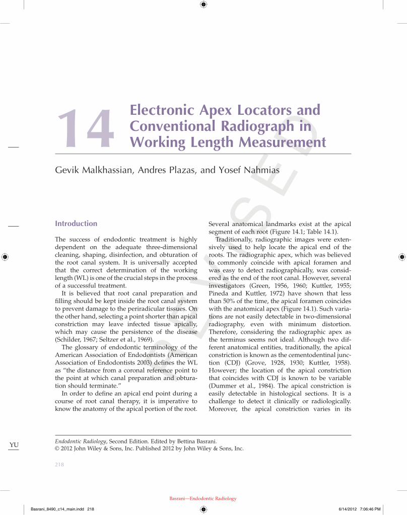
Several anatomical landmarks exist at the apical segment of each root (Figure 14.1; Table 14.1).
Traditionally, radiographic images were exten-sively used to help locate the apical end of the roots. The radiographic apex, which was believed to commonly coincide with apical foramen and was easy to detect radiographically, was consid-ered as the end of the root canal. However, several investigators (Green, 1956, 1960; Kuttler, 1955; Pineda and Kuttler, 1972) have shown that less than 50% of the time, the apical foramen coincides with the anatomical apex (Figure 14.1). Such varia-tions are not easily detectable in two-dimensional radiography, even with minimum distortion. Therefore, considering the radiographic apex as the terminus seems not ideal. Although two dif-ferent anatomical entities, traditionally, the apical constriction is known as the cementodentinal junc-tion (CDJ) (Grove, 1928, 1930; Kuttler, 1958). However; the location of the apical constriction that coincides with CDJ is known to be variable (Dummer et al., 1984). The apical constriction is easily detectable in histological sections. It is a challenge to detect it clinically or radiologically. Moreover, the apical constriction varies in its
Basrani_8490_c14_main.indd 218 6/14/2012 7:06:46 PM
Re
vi s
ed
Basrani—Endodontic Radiology
E1
Electronic Apex Locators and Conventional Radiograph in Working Length Measurement 219
presence within root canal space as an anatomical reference.
The majority of the endodontists and clinicians would agree that the apical constriction or minor foramen is where the apical end of the root canal preparation and filling should terminate. The ratio-nale is that minor foramen is the narrowest section of the canal close to the apex with the minimum blood supply where the pulp tissue and the peri-odontal ligament (PDL) meet and that during root canal treatment, it provides the smallest wound site which is the most favorable for healing (Ricucci and Langeland, 1998).
WL measurement methods
Several methods have been used to determine the WL: (1) knowledge of anatomy and average root canal lengths, (2) apical sensitivity reported by patient when the instrument passes through the apical foramen, (3) tactile sensation of the apical constriction with endodontic file, (4) bleeding point as detected by the use of paper points showing bleeding in the most apical portion of the canal, and (5) radiographic technique by using an endodontic instrument within the root canal.
Most of these methods have some limitations and are not sufficiently reliable to be considered as the main measuring technique. Teeth lengths are variable. Apical sensitivity may be absent due to the use of local anesthetic. The tactile sensation of the apical constriction depends on countless factors (Palmer et al., 1971); for example, it might be only helpful in detection of the apical constriction in less than 60% of the cases (Seidberg et al., 1975). An incorrect radiographic technique can cause major distortion of the image (Vande Voorde and Bjorn-dahl, 1969).
Radiographic technique
Grove explained that using radiographic technique in the determination of WL is achieved by insertion of an endodontic instrument, usually a file, into the canal to a predetermined length, using a table showing the average length of each of the teeth and taking a radiograph and then adjusting the length as required (Grove, 1928, 1930). This method also
Figure 14.1 Anatomy of the apex of the root.
Table 14.1 Some definitions from Glossary of Endodontic Terms (2003).
Anatomy of the apical section Definition
Anatomic apex The tip or end of the root as determined morphologically
Apical foramen or major foramen
The main apical opening of the root canal.
Apical constriction or minor foramen (minor apical diameter, minor diameter)
The apical portion of the root canal having the narrowest diameter; position may vary but is usually 0.5–1.0 mm short of the center of the apical foramen.
Cementodentinal junction (CDJ)
The region at which the dentin and cementum are united commonly. Its position can range from 0.5 to 3.0 mm from the anatomic apex.
Radiographic apex The tip or end of the root as determined radiographically; its location can vary from the anatomical apex due to root morphology and distortion of the radiographic image.
topography. It has been classified into simple, diverging, multiple, and parallel constrictions (Dummer et al., 1984). According to these findings it may be concluded that the constriction and its position differ not only in form but also in their
Basrani_8490_c14_main.indd 219 6/14/2012 7:06:46 PM
Re
vi s
ed
Basrani—Endodontic Radiology
E1
220 Sequence of Endodontic Treatment
The rationale for using this technique is based on the suggestion that the apical constriction may be located at 1 mm short of the radiographic apex. However; it is not all inclusive, and in some cases, the apical foramen can even be located as far as 3 mm short of the radiographic apex (Kuttler, 1955; Pineda and Kuttler, 1972).
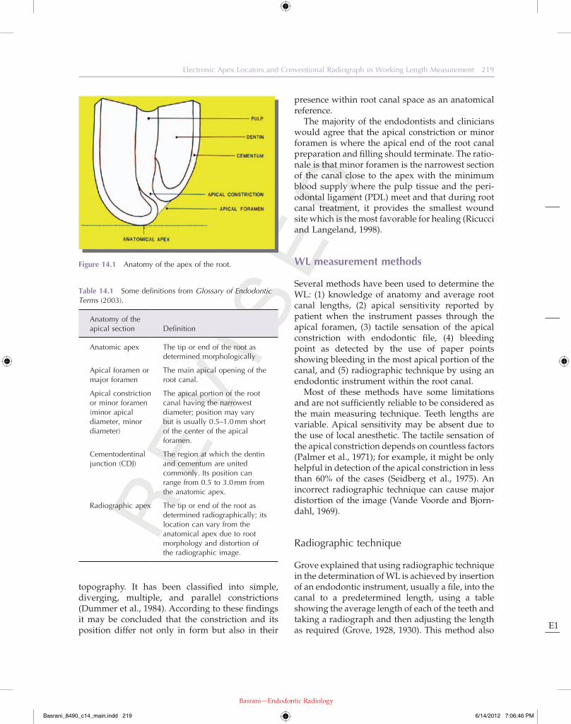
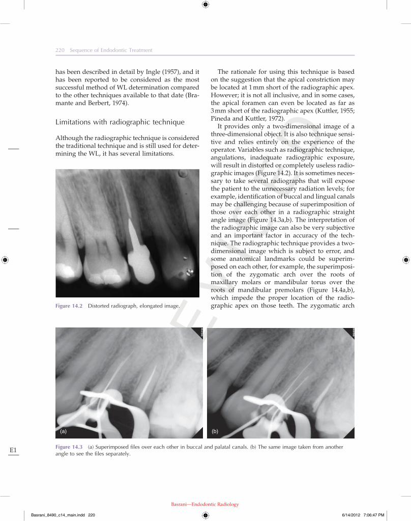
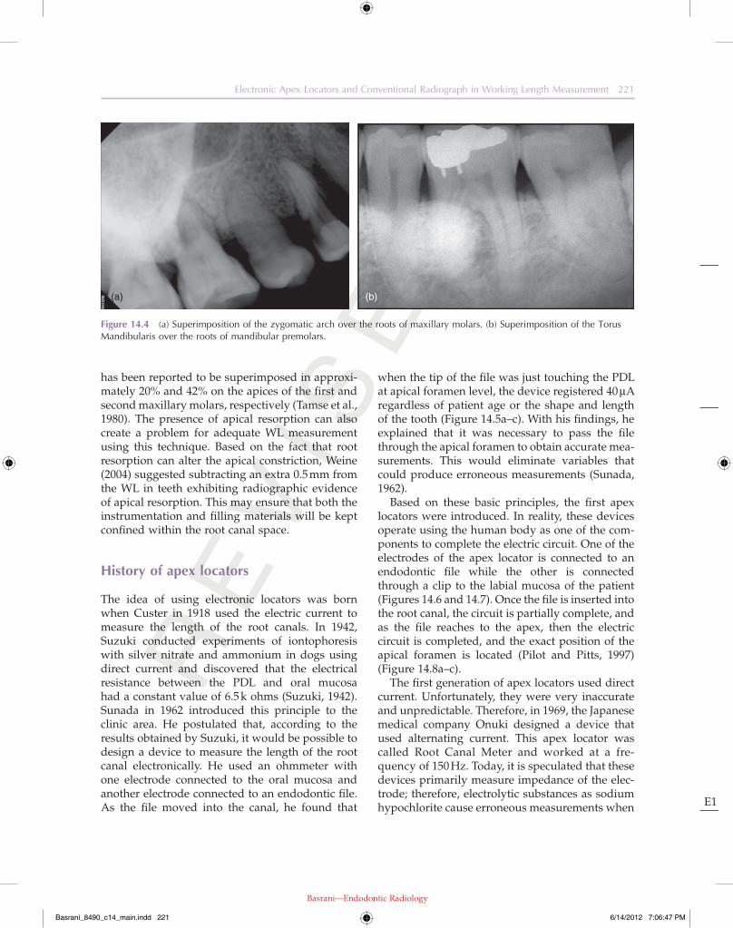
It provides only a two-dimensional image of a three-dimensional object. It is also technique sensi-tive and relies entirely on the experience of the operator. Variables such as radiographic technique, angulations, inadequate radiographic exposure, will result in distorted or completely useless radio-graphic images (Figure 14.2). It is sometimes neces-sary to take several radiographs that will expose the patient to the unnecessary radiation levels; for example, identification of buccal and lingual canals may be challenging because of superimposition of those over each other in a radiographic straight angle image (Figure 14.3a,b). The interpretation of the radiographic image can also be very subjective and an important factor in accuracy of the tech-nique. The radiographic technique provides a two-dimensional image which is subject to error, and some anatomical landmarks could be superim-posed on each other, for example, the superimposi-tion of the zygomatic arch over the roots of maxillary molars or mandibular torus over the roots of mandibular premolars (Figure 14.4a,b), which impede the proper location of the radio-graphic apex on those teeth. The zygomatic arch
has been described in detail by Ingle (1957), and it has been reported to be considered as the most successful method of WL determination compared to the other techniques available to that date (Bra-mante and Berbert, 1974).
Limitations with radiographic technique
Although the radiographic technique is considered the traditional technique and is still used for deter-mining the WL, it has several limitations.
Figure 14.2 Distorted radiograph, elongated image.
Figure 14.3 (a) Superimposed files over each other in buccal and palatal canals. (b) The same image taken from another angle to see the files separately.
(a) (b)
Basrani_8490_c14_main.indd 220 6/14/2012 7:06:47 PM
Re
vi s
ed
Basrani—Endodontic Radiology
E1
Electronic Apex Locators and Conventional Radiograph in Working Length Measurement 221
Figure 14.4 (a) Superimposition of the zygomatic arch over the roots of maxillary molars. (b) Superimposition of the Torus Mandibularis over the roots of mandibular premolars.
(a) (b)
has been reported to be superimposed in approxi-mately 20% and 42% on the apices of the first and second maxillary molars, respectively (Tamse et al., 1980). The presence of apical resorption can also create a problem for adequate WL measurement using this technique. Based on the fact that root resorption can alter the apical constriction, Weine (2004) suggested subtracting an extra 0.5 mm from the WL in teeth exhibiting radiographic evidence of apical resorption. This may ensure that both the instrumentation and filling materials will be kept confined within the root canal space.
History of apex locators
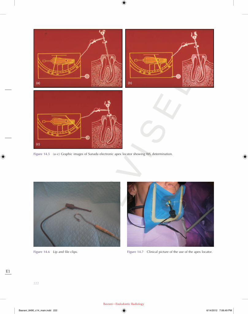
The idea of using electronic locators was born when Custer in 1918 used the electric current to measure the length of the root canals. In 1942, Suzuki conducted experiments of iontophoresis with silver nitrate and ammonium in dogs using direct current and discovered that the electrical resistance between the PDL and oral mucosa had a constant value of 6.5 k ohms (Suzuki, 1942). Sunada in 1962 introduced this principle to the clinic area. He postulated that, according to the results obtained by Suzuki, it would be possible to design a device to measure the length of the root canal electronically. He used an ohmmeter with one electrode connected to the oral mucosa and another electrode connected to an endodontic file. As the file moved into the canal, he found that
when the tip of the file was just touching the PDL at apical foramen level, the device registered 40 µA regardless of patient age or the shape and length of the tooth (Figure 14.5a–c). With his findings, he explained that it was necessary to pass the file through the apical foramen to obtain accurate mea-surements. This would eliminate variables that could produce erroneous measurements (Sunada, 1962).
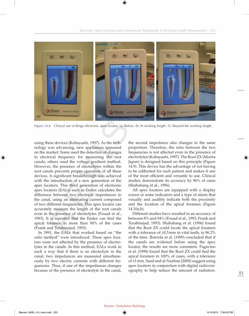
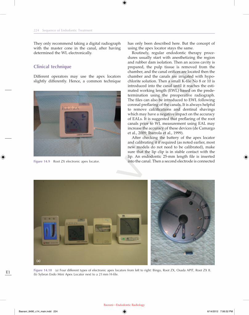
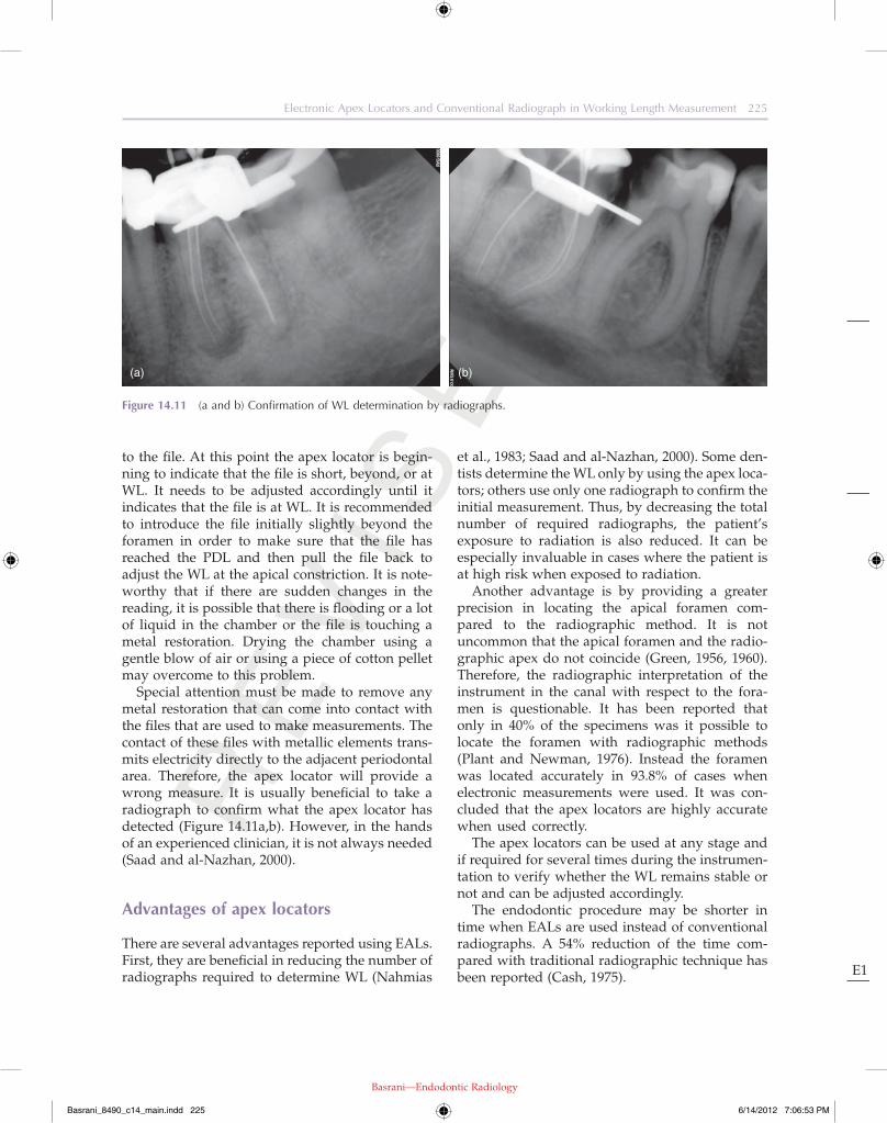
Based on these basic principles, the first apex locators were introduced. In reality, these devices operate using the human body as one of the com-ponents to complete the electric circuit. One of the electrodes of the apex locator is connected to an endodontic file while the other is connected through a clip to the labial mucosa of the patient (Figures 14.6 and 14.7). Once the file is inserted into the root canal, the circuit is partially complete, and as the file reaches to the apex, then the electric circuit is completed, and the exact position of the apical foramen is located (Pilot and Pitts, 1997) (Figure 14.8a–c).
The first generation of apex locators used direct current. Unfortunately, they were very inaccurate and unpredictable. Therefore, in 1969, the Japanese medical company Onuki designed a device that used alternating current. This apex locator was called Root Canal Meter and worked at a fre-quency of 150 Hz. Today, it is speculated that these devices primarily measure impedance of the elec-trode; therefore, electrolytic substances as sodium hypochlorite cause erroneous measurements when
Basrani_8490_c14_main.indd 221 6/14/2012 7:06:47 PM
Re
vi s
ed
Basrani—Endodontic Radiology
E1
Figure 14.6 Lip and file clips. Figure 14.7 Clinical picture of the use of the apex locator.
Figure 14.5 (a–c) Graphic images of Sunada electronic apex locator showing WL determination.
(a)
(c)
(b)
222
Basrani_8490_c14_main.indd 222 6/14/2012 7:06:49 PM
Re
vi s
ed
Basrani—Endodontic Radiology
E1
Electronic Apex Locators and Conventional Radiograph in Working Length Measurement 223
the second impedance also changes in the same proportion. Therefore, the ratio between the two frequencies is not affected even in the presence of electrolytes (Kobayashi, 1997). The Root ZX (Morita Japan) is designed based on this principle (Figure 14.9). This device has the advantage of not having to be calibrated for each patient and makes it one of the most efficient and versatile to use. Clinical studies demonstrate its accuracy by 96% of cases (Shabahang et al., 1996).
All apex locators are equipped with a display screen or some indicators and a type of alarm that visually and audibly indicate both the proximity and the location of the apical foramen (Figure 14.10a,b).
Different studies have resulted in an accuracy of between 8% and 94% (Fouad et al., 1993; Frank and Torabinejad, 1993). Shabahang et al. (1996) found that the Root ZX could locate the apical foramen with a tolerance of ±0.5 mm in vital teeth, in 96.2% of the time. Ibarrola et al. (1999) concluded that if the canals are widened before using the apex locator, the results are more consistent. Pagavino et al. (1998) found that the Root ZX could find the apical foramen in 100% of cases, with a tolerance of ±1 mm. Saad and al-Nazhan (2000) suggest using apex locators in conjunction with digital radiovisi-ography to help reduce the amount of radiation.
using these devices (Kobayashi, 1997). As the tech-nology was advancing, new appliances appeared on the market. Some used the detection of changes in electrical frequency for measuring the root canals; others used the voltage gradient method. However, the presence of electrolytes within the root canals prevents proper operation of all these devices. A significant breakthrough was achieved with the introduction of a new generation of the apex locators. This third generation of electronic apex locators (EALs) such as Endex calculates the difference between two electrical impedances in the canal, using an alternating current composed of two different frequencies. This apex locator can accurately measure the length of the root canals even in the presence of electrolytes (Fouad et al., 1993). It is reported that the Endex can find the apical foramen in more than 90% of the cases (Frank and Torabinejad, 1993).
In 1991, the EALs that worked based on “the ratio method” were introduced. These apex loca-tors were not affected by the presence of electro-lytes in the canals. In this method, EALs work in such a way that if there is an electrolyte in the canal, two impedances are measured simultane-ously by two electric currents with different fre-quencies. Thus, if one of the impedances changes because of the presence of electrolyte in the canal,
Figure 14.8 Clinical use of Bingo electronic apex locator. (a) Before. (b) At working length. (C) Beyond the working length.
(a) (b) (c)
Basrani_8490_c14_main.indd 223 6/14/2012 7:06:50 PM
Re
vi s
ed
Basrani—Endodontic Radiology
E1
224 Sequence of Endodontic Treatment
has only been described here. But the concept of using the apex locator stays the same.
Routinely, regular endodontic therapy proce-dures usually start with anesthetizing the region and rubber dam isolation. Then an access cavity is prepared, the pulp tissue is removed from the chamber, and the canal orifices are located then the chamber and the canals are irrigated with hypo-chlorite solution. Then a small K-file No 8 or 10 is introduced into the canal until it reaches the esti-mated working length (EWL) based on the prede-termination using the preoperative radiograph. The files can also be introduced to EWL following coronal preflaring of the canals. It is always helpful to remove calcifications and dentinal shavings which may have a negative impact on the accuracy of EALs. It is suggested that preflaring of the root canals prior to WL measurement using EAL may increase the accuracy of these devices (de Camargo et al., 2009; Ibarrola et al., 1999).
After checking the battery of the apex locator and calibrating it if required (as noted earlier, most new models do not need to be calibrated), make sure that the lip clip is in stable contact with the lip. An endodontic 25-mm length file is inserted into the canal. Then a second electrode is connected
They only recommend taking a digital radiograph with the master cone in the canal, after having determined the WL electronically.
Clinical technique
Different operators may use the apex locators slightly differently. Hence, a common technique
Figure 14.9 Root ZX electronic apex locator.
Figure 14.10 (a) Four different types of electronic apex locators from left to right: Bingo, Root ZX, Osada APIT, Root ZX II. (b) Sybron Endo Mini Apex Locator next to a 21 mm H-file.
(a) (b)
Basrani_8490_c14_main.indd 224 6/14/2012 7:06:52 PM
Re
vi s
ed
Basrani—Endodontic Radiology
E1
Electronic Apex Locators and Conventional Radiograph in Working Length Measurement 225
et al., 1983; Saad and al-Nazhan, 2000). Some den-tists determine the WL only by using the apex loca-tors; others use only one radiograph to confirm the initial measurement. Thus, by decreasing the total number of required radiographs, the patient’s exposure to radiation is also reduced. It can be especially invaluable in cases where the patient is at high risk when exposed to radiation.
Another advantage is by providing a greater precision in locating the apical foramen com-pared to the radiographic method. It is not uncommon that the apical foramen and the radio-graphic apex do not coincide (Green, 1956, 1960). Therefore, the radiographic interpretation of the instrument in the canal with respect to the fora-men is questionable. It has been reported that only in 40% of the specimens was it possible to locate the foramen with radiographic methods (Plant and Newman, 1976). Instead the foramen was located accurately in 93.8% of cases when electronic measurements were used. It was con-cluded that the apex locators are highly accurate when used correctly.
The apex locators can be used at any stage and if required for several times during the instrumen-tation to verify whether the WL remains stable or not and can be adjusted accordingly.
The endodontic procedure may be shorter in time when EALs are used instead of conventional radiographs. A 54% reduction of the time com-pared with traditional radiographic technique has been reported (Cash, 1975).
to the file. At this point the apex locator is begin-ning to indicate that the file is short, beyond, or at WL. It needs to be adjusted accordingly until it indicates that the file is at WL. It is recommended to introduce the file initially slightly beyond the foramen in order to make sure that the file has reached the PDL and then pull the file back to adjust the WL at the apical constriction. It is note-worthy that if there are sudden changes in the reading, it is possible that there is flooding or a lot of liquid in the chamber or the file is touching a metal restoration. Drying the chamber using a gentle blow of air or using a piece of cotton pellet may overcome to this problem.
Special attention must be made to remove any metal restoration that can come into contact with the files that are used to make measurements. The contact of these files with metallic elements trans-mits electricity directly to the adjacent periodontal area. Therefore, the apex locator will provide a wrong measure. It is usually beneficial to take a radiograph to confirm what the apex locator has detected (Figure 14.11a,b). However, in the hands of an experienced clinician, it is not always needed (Saad and al-Nazhan, 2000).
Advantages of apex locators
There are several advantages reported using EALs. First, they are beneficial in reducing the number of radiographs required to determine WL (Nahmias
Figure 14.11 (a and b) Confirmation of WL determination by radiographs.
(a) (b)
Basrani_8490_c14_main.indd 225 6/14/2012 7:06:53 PM
Re
vi s
ed
Basrani—Endodontic Radiology
E1
226 Sequence of Endodontic Treatment
5. The technique requires the operator to be familiar with the equipment and learn how to interpret it. Like any new instrument, the apex locators require a period of learning to use them effectively.
Comparing EAL to radiographs
Apex locators and conventional radiographs
Accurate WL determination is a crucial part of successful root canal treatment (Ricucci, 1998). The apical constriction is regarded as an ideal apical end point for instrumentation and obtura-tion in root canal therapy (Dummer et al., 1984); unfortunately, the location and shape of the apical constriction are variable and are not radiographi-cally detectable (ElAyouti et al., 2001; Ricucci and Langeland, 1998). In addition to radiographic measurements, electronic WL determination has become increasingly important. The electronic method reduces many of the problems associated with radiographic measurements. Its most impor-tant advantage over radiography is that it can measure the length of the root canal to the apical foramen, not to the radiographic apex (Kobayashi, 1995).
Based on a study on extracted teeth using Root ZX apex locator, it was reported that Radiographic WL determination alone resulted in overestimation in 51% of the root canals while the percentage of overestimation was reduced to 21% by using EAL
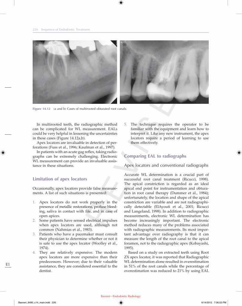
In multirooted teeth, the radiographic method can be complicated for WL measurement. EALs could be very helpful in lessening the uncertainties in these cases (Figure 14.12a,b).
Apex locators are invaluable in detection of per-forations (Fuss et al., 1996; Kaufman et al., 1997).
In patients with an acute gag reflex, taking radio-graphs can be extremely challenging. Electronic WL measurement can provide an invaluable assis-tance in these situations.
Limitation of apex locators
Occasionally, apex locators provide false measure-ments. A list of such situations is presented:
1. Apex locators do not work properly in the presence of metallic restorations, profuse bleed-ing, saliva in contact with file, and in case of open apices.
2. Some patients have sensed electrical impulses when apex locators are used, although not common (Nahmias et al., 1983).
3. Patients who have a pacemaker must consult their physician to determine whether or not it is safe to use the apex locator (Woolley et al., 1974).
4. They are relatively expensive. The modern apex locators are more expensive than their predecessors. However; due to their valuable assistance, they are considered essential to the dentist.
Figure 14.12 (a and b) Cases of multirooted obturated root canals.
(a) (b)
Basrani_8490_c14_main.indd 226 6/14/2012 7:06:53 PM
Re
vi s
ed
Basrani—Endodontic Radiology
E1
Electronic Apex Locators and Conventional Radiograph in Working Length Measurement 227
radiography, the apex locators showed to be more accurate than digital radiography in determining the WLs (Cianconi et al., 2010; Real et al., 2011).
As a result, it seems logical to conclude that EALs are more accurate than radiographic tech-niques alone in determination of WL during root canal treatments.
Apex locators and digital radiography
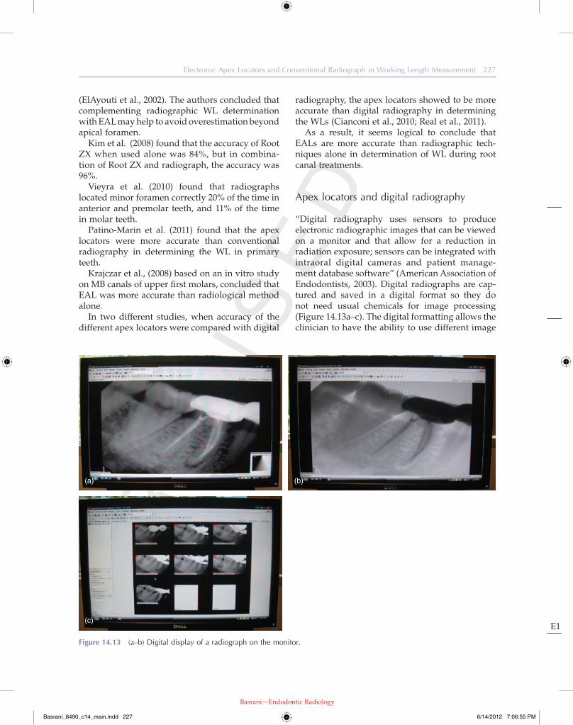
“Digital radiography uses sensors to produce electronic radiographic images that can be viewed on a monitor and that allow for a reduction in radiation exposure; sensors can be integrated with intraoral digital cameras and patient manage-ment database software” (American Association of Endodontists, 2003). Digital radiographs are cap-tured and saved in a digital format so they do not need usual chemicals for image processing (Figure 14.13a–c). The digital formatting allows the clinician to have the ability to use different image
(ElAyouti et al., 2002). The authors concluded that complementing radiographic WL determination with EAL may help to avoid overestimation beyond apical foramen.
Kim et al. (2008) found that the accuracy of Root ZX when used alone was 84%, but in combina-tion of Root ZX and radiograph, the accuracy was 96%.
Vieyra et al. (2010) found that radiographs located minor foramen correctly 20% of the time in anterior and premolar teeth, and 11% of the time in molar teeth.
Patino-Marin et al. (2011) found that the apex locators were more accurate than conventional radiography in determining the WL in primary teeth.
Krajczar et al., (2008) based on an in vitro study on MB canals of upper first molars, concluded that EAL was more accurate than radiological method alone.
In two different studies, when accuracy of the different apex locators were compared with digital
Figure 14.13 (a–b) Digital display of a radiograph on the monitor.
(a)
(c)
(b)
Basrani_8490_c14_main.indd 227 6/14/2012 7:06:55 PM
Re
vi s
ed
Basrani—Endodontic Radiology
E1
228 Sequence of Endodontic Treatment
exposure dosage to the patient. As a result, in order to reduce the radiation dose, the use of digital radiographs instead of conventional radiographs in combination with EAL’s is advised for WL deter-mination (Saad and al-Nazhan, 2000).
Apex locator’s employment in the detection of perforations
Perforations are one of the significant factors affect-ing the prognosis of root canal treatment (de Chev-igny et al., 2008). They can happen due to pathological factors like resorption or caries or be caused during regular root canal treatment or during post space preparation (Fuss and Trope, 1996). They may occur at the floor or wall of the
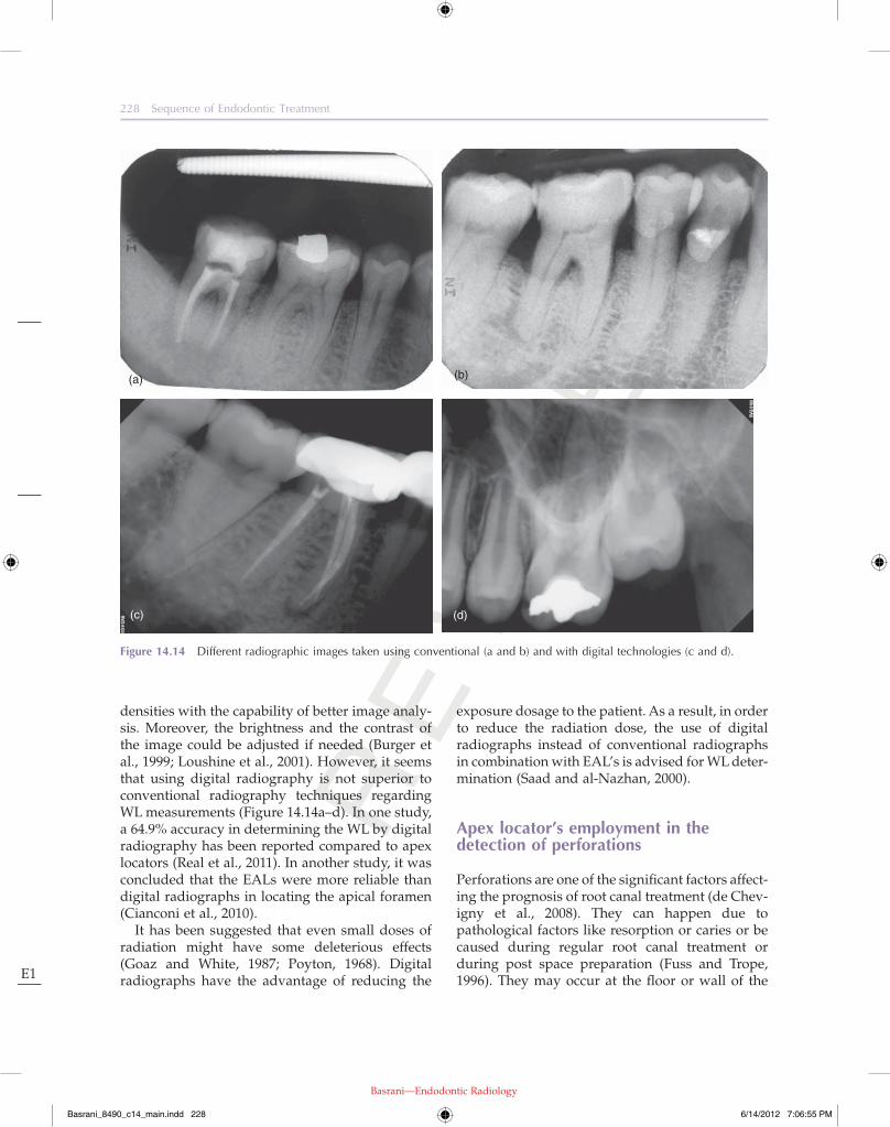
densities with the capability of better image analy-sis. Moreover, the brightness and the contrast of the image could be adjusted if needed (Burger et al., 1999; Loushine et al., 2001). However, it seems that using digital radiography is not superior to conventional radiography techniques regarding WL measurements (Figure 14.14a–d). In one study, a 64.9% accuracy in determining the WL by digital radiography has been reported compared to apex locators (Real et al., 2011). In another study, it was concluded that the EALs were more reliable than digital radiographs in locating the apical foramen (Cianconi et al., 2010).
It has been suggested that even small doses of radiation might have some deleterious effects (Goaz and White, 1987; Poyton, 1968). Digital radiographs have the advantage of reducing the
Figure 14.14 Different radiographic images taken using conventional (a and b) and with digital technologies (c and d).
(a)
(c) (d)
(b)
Basrani_8490_c14_main.indd 228 6/14/2012 7:06:55 PM
Re
vi s
ed
Basrani—Endodontic Radiology
E1
Electronic Apex Locators and Conventional Radiograph in Working Length Measurement 229
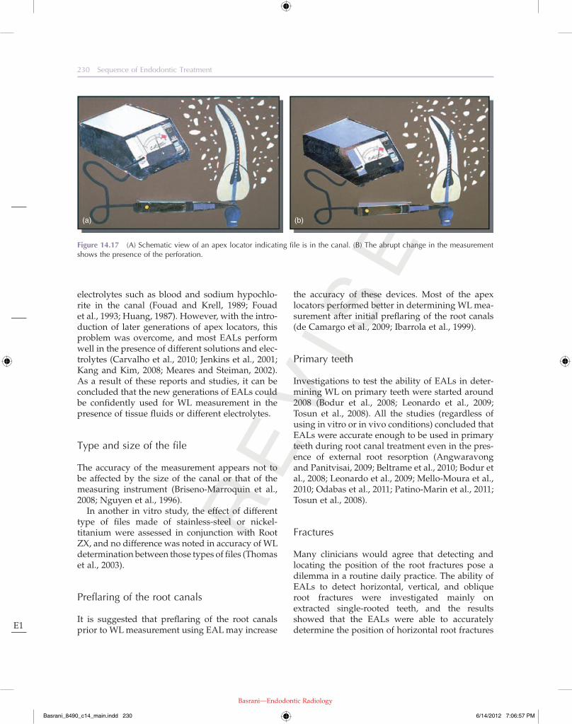
In order to detect a perforation, a simple technique has been described. A small file could be inserted into the suspected area and connected to the apex locator. If the apex locator indicates immediately that it has detected the apex which is usually not there, it probably means that a perforation was created. Commonly, if the apex locator indicator moves slowly while introducing the file, it is more likely that the file is inside the canal rather than in a perforation (Figure 14.17a,b). In a study using different models of EALs, it was found that radio-graphs were less reliable than apex locators in the identification of perforation locations (Fuss et al., 1996). In another in vitro study using three differ-ent type of EALs, it was reported that EALs were acceptable tools for the detection of root perfora-tions clinically (Kaufman et al., 1997).
The effect of different parameters on performance of EALs
Moisture, different irrigants, or solutions in the canal
WL measurements using the first two generation models of EALs were considered to be inaccurate in the presence of conductive fluids, moisture and
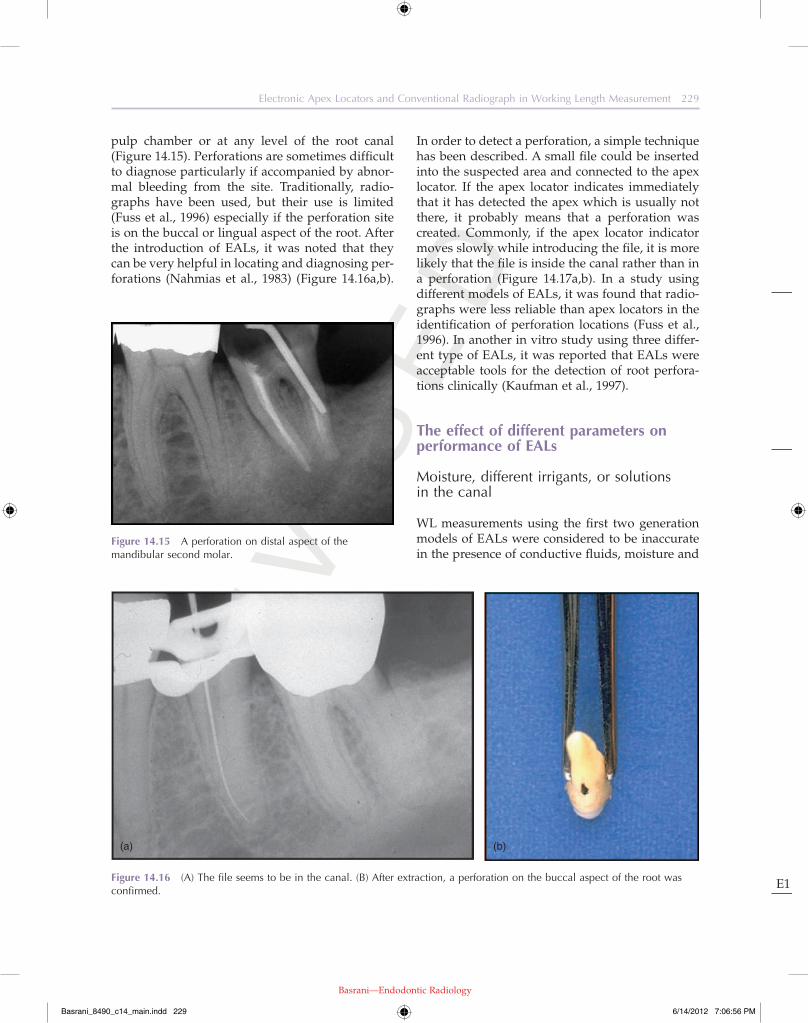
pulp chamber or at any level of the root canal (Figure 14.15). Perforations are sometimes difficult to diagnose particularly if accompanied by abnor-mal bleeding from the site. Traditionally, radio-graphs have been used, but their use is limited (Fuss et al., 1996) especially if the perforation site is on the buccal or lingual aspect of the root. After the introduction of EALs, it was noted that they can be very helpful in locating and diagnosing per-forations (Nahmias et al., 1983) (Figure 14.16a,b).
Figure 14.15 A perforation on distal aspect of the mandibular second molar.
Figure 14.16 (A) The file seems to be in the canal. (B) After extraction, a perforation on the buccal aspect of the root was confirmed.
(a) (b)
Basrani_8490_c14_main.indd 229 6/14/2012 7:06:56 PM
Re
vi s
ed
Basrani—Endodontic Radiology
E1
230 Sequence of Endodontic Treatment
the accuracy of these devices. Most of the apex locators performed better in determining WL mea-surement after initial preflaring of the root canals (de Camargo et al., 2009; Ibarrola et al., 1999).
Primary teeth
Investigations to test the ability of EALs in deter-mining WL on primary teeth were started around 2008 (Bodur et al., 2008; Leonardo et al., 2009; Tosun et al., 2008). All the studies (regardless of using in vitro or in vivo conditions) concluded that EALs were accurate enough to be used in primary teeth during root canal treatment even in the pres-ence of external root resorption (Angwaravong and Panitvisai, 2009; Beltrame et al., 2010; Bodur et al., 2008; Leonardo et al., 2009; Mello-Moura et al., 2010; Odabas et al., 2011; Patino-Marin et al., 2011; Tosun et al., 2008).
Fractures
Many clinicians would agree that detecting and locating the position of the root fractures pose a dilemma in a routine daily practice. The ability of EALs to detect horizontal, vertical, and oblique root fractures were investigated mainly on extracted single-rooted teeth, and the results showed that the EALs were able to accurately determine the position of horizontal root fractures
electrolytes such as blood and sodium hypochlo-rite in the canal (Fouad and Krell, 1989; Fouad et al., 1993; Huang, 1987). However, with the intro-duction of later generations of apex locators, this problem was overcome, and most EALs perform well in the presence of different solutions and elec-trolytes (Carvalho et al., 2010; Jenkins et al., 2001; Kang and Kim, 2008; Meares and Steiman, 2002). As a result of these reports and studies, it can be concluded that the new generations of EALs could be confidently used for WL measurement in the presence of tissue fluids or different electrolytes.
Type and size of the file
The accuracy of the measurement appears not to be affected by the size of the canal or that of the measuring instrument (Briseno-Marroquin et al., 2008; Nguyen et al., 1996).
In another in vitro study, the effect of different type of files made of stainless-steel or nickel-titanium were assessed in conjunction with Root ZX, and no difference was noted in accuracy of WL determination between those types of files (Thomas et al., 2003).
Preflaring of the root canals
It is suggested that preflaring of the root canals prior to WL measurement using EAL may increase
Figure 14.17 (A) Schematic view of an apex locator indicating file is in the canal. (B) The abrupt change in the measurement shows the presence of the perforation.
(a) (b)
Basrani_8490_c14_main.indd 230 6/14/2012 7:06:57 PM
Re
vi s
ed
Basrani—Endodontic Radiology
E1
Electronic Apex Locators and Conventional Radiograph in Working Length Measurement 231
introduction and evolvement of electronic WL measurement devices have provided another reli-able means to help practitioners in their routine daily root canal treatment procedures. Although both techniques have their own limitations, the great advantages of EALs have made them an important part of a daily endodontic practice. It has been suggested that more predictable results can be achieved by combination of EALs and radiographic techniques in WL measurement.
References
Aggarwal, V., Singla, M., and Kabi, D. (2010) An in vitro evaluation of performance of two electronic root canal length measurement devices during retreatment of different obturating materials. Journal of Endodontics, 36, 1526–1530.
Akisue, E., Gavini, G., and De Figueiredo, J.A.P. (2007) Influence of pulp vitality on length determination by using the Elements Diagnostic Unit and Apex Locator. Oral Surgery, Oral Medicine, Oral Pathology, Oral Radiol-ogy, and Endodontics, 104, e129–e132.
American Association of Endodontists (AAE). (2003) Glossary of Endodontic Terms, 7th ed. American Asso-ciation of Endodontists, Chicago, IL.
Angwaravong, O. and Panitvisai, P. (2009) Accuracy of an electronic apex locator in primary teeth with root resorption. International Endodontic Journal, 42, 115–121.
Azabal, M., Garcia-Otero, D., and de la Macorra, J.C. (2004) Accuracy of the Justy II Apex locator in deter-mining working length in simulated horizontal and vertical fractures. International Endodontic Journal, 37, 174–177.
Beltrame, A.P.C.A., Triches, T.C., Sartori, N., and Bolan, M. (2010) Electronic determination of root canal working length in primary molar teeth: an in vivo and ex vivo study. International Endodontic Journal, 44, 402–406.
Bodur, H., Odabas, M., Tulunoglu, O., and Tinaz, A.C. (2008) Accuracy of two different apex locators in primary teeth with and without root resorption. Clini-cal Oral Investigations, 12, 137–141.
Bramante, C.M. and Berbert, A. (1974) A critical evalua-tion of some methods of determining tooth length. Oral Surgery, Oral Medicine, and Oral Pathology, 37, 463–473.
Briseno-Marroquin, B., Frajlich, S., Goldberg, F., and Wil-lershausen, B. (2008) Influence of instrument size on the accuracy of different apex locators: an in vitro study. Journal of Endodontics, 34, 698–702.
but were unreliable to detect the position of verti-cal root fractures (Azabal et al., 2004; Ebrahim et al., 2006a; Goldberg et al., 2008).
Pulp status (vitality)
The presence of vital or necrotic tissue in the root canals does not affect the reliability of the EALs when tested in vivo (Akisue et al., 2007; Dunlap et al., 1998).
Apical foramen size
In the presence of a large apical foramen either enlarged inadvertently during a course of root canal treatment, or due to the presence of external root resorption at the apex, or because of partially developed apical root segment, EALs can be valu-able tools to establish a reasonable WL (Goldberg et al., 2002; Kang and Kim, 2008; Nguyen et al., 1996). However, some limitations have been reported on the performance of EALs in these situ-ations. For example, some initial studies showed that some apex locators may be more reliable than the other ones (Fouad et al., 1993). Using small files in widened foramens may affect the reliability of the measurements (Ebrahim et al., 2006b). The diameter of apical constriction may affect the accu-racy of the EALs, and it was reported that the WL measurements of root canals with apical foramens larger than a size 100 K-file were not reliable (Herrera et al., 2007).
Retreatment of previously treated teeth
The results of two studies also indicated that EALs are useful in determining the WL of root canals during retreatment of previously treated root canals (Aggarwal et al., 2010; Goldberg et al., 2005).
Conclusion
Radiographic techniques were used convention-ally as the main tools by practitioners during end-odontic WL measurements for many years. The
Basrani_8490_c14_main.indd 231 6/14/2012 7:06:57 PM
Re
vi s
ed
Basrani—Endodontic Radiology
E1
232 Sequence of Endodontic Treatment
Fouad, A.F. and Krell, K.V. (1989) An in vitro comparison of five root canal length measuring instruments. Journal of Endodontics, 15, 573–577.
Fouad, A.F., Rivera, E.M., and Krell, K.V. (1993) Accuracy of the Endex with variations in canal irrigants and foramen size. Journal of Endodontics, 19, 63–67.
Frank, A.L. and Torabinejad, M. (1993) An in vivo evalu-ation of Endex electronic apex locator. Journal of End-odontics, 19, 177–179.
Fuss, Z. and Trope, M. (1996) Root perforations: classifi-cation and treatment choices based on prognostic factors. Endodontics & Dental Traumatology, 12, 255–264.
Fuss, Z., Assooline, L.S., and Kaufman, A.Y. (1996) Determination of location of root perforations by electronic apex locators. Oral Surgery, Oral Medicine, Oral Pathology, Oral Radiology, and Endodontics, 82, 324–329.
Goaz, P.W. and White, S.C. (1987) Radiation Biology. Oral Radiology: Principles and Interpretation, 2nd ed. Mosby, St. Louis, MO.
Goldberg, F., De Silvio, A.C., Manfre, S., and Nastri, N. (2002) In vitro measurement accuracy of an electronic apex locator in teeth with simulated apical root resorp-tion. Journal of Endodontics, 28, 461–463.
Goldberg, F., Marroquin, B.B., Frajlich, S., and Dreyer, C. (2005) In vitro evaluation of the ability of three apex locators to determine the working length during retreatment. Journal of Endodontics, 31, 676–678.
Goldberg, F., Frajlich, S., Kuttler, S., Manzur, E., and Briseno-Marroquin, B. (2008) The evaluation of four electronic apex locators in teeth with simulated hori-zontal oblique root fractures. Journal of Endodontics, 34, 1497–1499.
Green, D. (1956) A stereomicroscopic study of the root apices of 400 maxillary and mandibular anterior teeth. Oral Surgery, Oral Medicine, and Oral Pathology, 9, 1224–1232.
Green, D. (1960) Stereomicroscopic study of 700 root apices of maxillary and mandibular posterior teeth. Oral Surgery, Oral Medicine, and Oral Pathology, 13, 728–733.
Grove, C.J. (1928) A new simple standardized technique producing perfect fitting impermeable root canal fill-ings extending to the dento-cemento junction. Dental Items of Interest, 50, 855–857.
Grove, C.J. (1930) Further evidence that root canals can be filled to the dentinocemental junction. The Journal of the American Dental Association, 17, 293–296.
Herrera, M., Abalos, C., Planas, A.J., and Llamas, R. (2007) Influence of apical constriction diameter on Root ZX apex locator precision. Journal of Endodontics, 33, 995–998.
Burger, C.L., Mork, T.O., Hutter, J.W., and Nicoll, B. (1999) Direct digital radiography versus conventional radiography for estimation of canal length in curved canals. Journal of Endodontics, 25, 260–263.
Carvalho, A.L.P., Moura-Netto, C., Moura, A.A.M.D., Marques, M.M., and Davidowicz, H. (2010) Accuracy of three electronic apex locators in the presence of different irrigating solutions. Brazilian Oral Research, 24, 394–398.
Cash, P.W. (1975) Letter: endo research challenged. Journal of the American Dental Association (1939), 91, 1135–1136.
Cianconi, L., Angotti, V., Felici, R., Conte, G., and Mancini, M. (2010) Accuracy of three electronic apex locators compared with digital radiography: an ex vivo study. Journal of Endodontics, 36, 2003–2007.
de Camargo, E.J., Zapata, R.O., Medeiros, P.L., Bramante, C.M., Bernardineli, N., Garcia, R.B., de Moraes, I.G., and Duarte, M.A.H. (2009) Influence of preflaring on the accuracy of length determination with four electronic apex locators. Journal of Endodontics, 35, 1300–1302.
de Chevigny, C., Dao, T.T., Basrani, B.R., Marquis, V., Farzaneh, M., Abitbol, S., and Friedman, S. (2008) Treatment outcome in endodontics: the Toronto study—phases 3 and 4: orthograde retreatment. Journal of Endodontics, 34, 131–137.
Dummer, P.M., Mcginn, J.H., and Rees, D.G. (1984) The position and topography of the apical canal constric-tion and apical foramen. International Endodontic Journal, 17, 192–198.
Dunlap, C.A., Remeikis, N.A., Begole, E.A., and Rauschenberger, C.R. (1998) An in vivo evaluation of an electronic apex locator that uses the ratio method in vital and necrotic canals. Journal of Endodontics, 24, 48–50.
Ebrahim, A.K., Wadachi, R., and Suda, H. (2006a) Accuracy of three different electronic apex locators in detecting simulated horizontal and vertical root fractures. Australian Endodontic Journal: The Journal of the Australian Society of Endodontology Inc, 32, 64–69.
Ebrahim, A.K., Yoshioka, T., Kobayashi, C., and Suda, H. (2006b) The effects of file size, sodium hypochlorite and blood on the accuracy of Root ZX apex locator in enlarged root canals: an in vitro study. Australian Dental Journal, 51, 153–157.
ElAyouti, A., Weiger, R., and Lost, C. (2001) Frequency of overinstrumentation with an acceptable radiographic working length. Journal of Endodontics, 27, 49–52.
ElAyouti, A., Weiger, R., and Lost, C. (2002) The ability of root ZX apex locator to reduce the frequency of overestimated radiographic working length. Journal of Endodontics, 28, 116–119.
Basrani_8490_c14_main.indd 232 6/14/2012 7:06:57 PM
Re
vi s
ed
Basrani—Endodontic Radiology
E1
Electronic Apex Locators and Conventional Radiograph in Working Length Measurement 233
Meares, W.A. and Steiman, H.R. (2002) The influence of sodium hypochlorite irrigation on the accuracy of the Root ZX electronic apex locator. Journal of Endodontics, 28, 595–598.
Mello-Moura, A.C.V., Moura-Netto, C., Araki, A.T., Guedes-Pinto, A.C., and Mendes, F.M. (2010) Ex vivo performance of five methods for root canal length determination in primary anterior teeth. International Endodontic Journal, 43, 142–147.
Nahmias, Y., Aurelio, J.A., and Gerstein, H. (1983) Expanded use of the electronic canal length measur-ing devices. Journal of Endodontics, 9, 347–349.
Nguyen, H.Q., Kaufman, A.Y., Komorowski, R.C., and Friedman, S. (1996) Electronic length measurement using small and large files in enlarged canals. Interna-tional Endodontic Journal, 29, 359–364.
Odabas, M.E., Bodur, H., Tulunoglu, O., and Alacam, A. (2011) Accuracy of an electronic apex locator: a clinical evaluation in primary molars with and without resorption. The Journal of Clinical Pediatric Dentistry, 35, 255–258.
Pagavino, G., Pace, R., and Baccetti, T. (1998) A SEM study of in vivo accuracy of the Root ZX electronic apex locator. Journal of Endodontics, 24, 438–441.
Palmer, M.J., Weine, F.S., and Healey, H.J. (1971) Position of the apical foramen in relation to endo-dontic therapy. Journal of the Canadian Dental Associa-tion, 37, 305–308.
Patino-Marin, N., Zavala-Alonso, N.V., Martinez-Castanon, G.A., Sanchez-Benavides, N., Villanueva-Gordillo, M., Loyola-Rodriguez, J.P., and Medina-Solis, C.E. (2011) Clinical evaluation of the accuracy of con-ventional radiography and apex locators in primary teeth. Pediatric Dentistry, 33, 19–22.
Pilot, T.F. and Pitts, D.L. (1997) Determination of imped-ance changes at varying frequencies in relation to root canal file position and irrigant. Journal of Endodontics, 23, 719–724.
Pineda, F. and Kuttler, Y. (1972) Mesiodistal and bucco-lingual roentgenographic investigation of 7275 root canals. Oral Surgery, Oral Medicine, and Oral Pathology, 33, 101–110.
Plant, J.J. and Newman, R.F. (1976) Clinical evaluation of the Sono-Explorer. Journal of Endodontics, 2, 215–216.
Poyton, H.G. (1968) The effects of radiation on teeth. Oral Surgery, Oral Medicine, and Oral Pathology, 26, 639–646.
Real, D.G., Davidowicz, H., Moura-Netto, C., Zenkner, C.D.L.L., Pagliarin, C.M.L., Barletta, F.B., and de Moura, A.A.M. (2011) Accuracy of working length determination using 3 electronic apex locators and direct digital radiography. Oral Surgery, Oral Medicine, Oral Pathology, Oral Radiology, and Endodontics, 111, e44–e49.
Huang, L. (1987) An experimental study of the principle of electronic root canal measurement. Journal of End-odontics, 13, 60–64.
Ibarrola, J.L., Chapman, B.L., Howard, J.H., Knowles, K.I., and Ludlow, M.O. (1999) Effect of preflaring on Root ZX apex locators. Journal of Endodontics, 25, 625–626.
Ingle, J.I. (1957) Endodontic instruments and instru-mentation. Dental Clinics of North America, 1, 805–822.
Jenkins, J.A., Walker, W.A., Schindler, W.G., and Flores, C.M. (2001) An in vitro evaluation of the accuracy of the root ZX in the presence of various irrigants. Journal of Endodontics, 27, 209–211.
Kang, J.-A. and Kim, S.K. (2008) Accuracies of seven dif-ferent apex locators under various conditions. Oral Surgery, Oral Medicine, Oral Pathology, Oral Radiology, and Endodontics, 106, e57–e62.
Kaufman, A.Y., Fuss, Z., Keila, S., and Waxenberg, S. (1997) Reliability of different electronic apex locators to detect root perforations in vitro. International End-odontic Journal, 30, 403–407.
Kim, E., Marmo, M., Lee, C.-Y., Oh, N.-S., and Kim, I.-K. (2008) An in vivo comparison of working length deter-mination by only root-ZX apex locator versus combin-ing root-ZX apex locator with radiographs using a new impression technique. Oral Surgery, Oral Medi-cine, Oral Pathology, Oral Radiology, and Endodontics, 105, e79–e83.
Kobayashi, C. (1995) Electronic canal length measure-ment. Oral Surgery, Oral Medicine, Oral Pathology, Oral Radiology, and Endodontics, 79, 226–231.
Kobayashi, C. (1997) The evolution of apex-locating devices. The Alpha Omegan, 90, 21–27.
Krajczar, K., Marada, G., Gyulai, G., and Toth, V. (2008) Comparison of radiographic and electronical working length determination on palatal and mesio-buccal root canals of extracted upper molars. Oral Surgery, Oral Medicine, Oral Pathology, Oral Radiology, and Endodon-tics, 106, e90–e93.
Kuttler, Y. (1955) Microscopic investigation of root apexes. Journal of the American Dental Association (1939), 50, 544–552.
Kuttler, Y. (1958) A precision and biologic root canal filling technic. Journal of the American Dental Associa-tion (1939), 56, 38–50.
Leonardo, M.R., da Silva, L.A.B., Nelson-Filho, P., da Silva, R.A.B., and Lucisano, M.P. (2009) Ex vivo accu-racy of an apex locator using digital signal processing in primary teeth. Pediatric Dentistry, 31, 320–322.
Loushine, R.J., Weller, R.N., Kimbrough, W.F., and Potter, B.J. (2001) Measurement of endodontic file lengths: calibrated versus uncalibrated digital images. Journal of Endodontics, 27, 779–781.
Basrani_8490_c14_main.indd 233 6/14/2012 7:06:57 PM
Re
vi s
ed
Basrani—Endodontic Radiology
E1
234 Sequence of Endodontic Treatment
Suzuki, K. (1942) Experimental study on iontophoresis. Journal of Japanese Stomatology, 16, 411–429.
Tamse, A., Kaffe, I., and Fishel, D. (1980) Zygomatic arch interference with correct radiographic diagnosis in maxillary molar endodontics. Oral Surgery, Oral Medi-cine, and Oral Pathology, 50, 563–566.
Thomas, A.S., Hartwell, G.R., and Moon, P.C. (2003) The accuracy of the Root ZX electronic apex locator using stainless-steel and nickel-titanium files. Journal of End-odontics, 29, 662–663.
Tosun, G., Erdemir, A., Eldeniz, A.U., Sermet, U., and Sener, Y. (2008) Accuracy of two electronic apex loca-tors in primary teeth with and without apical resorp-tion: a laboratory study. International Endodontic Journal, 41, 436–441.
Vande Voorde, H.E. and Bjorndahl, A.M. (1969) Estimat-ing endodontic “working length” with paralleling radiographs. Oral Surgery, Oral Medicine, and Oral Pathology, 27, 106–110.
Vieyra, J.P., Acosta, J., and Mondaca, J.M. (2010) Com-parison of working length determination with radio-graphs and two electronic apex locators. International Endodontic Journal, 43, 16–20.
Weine, F.S. (2004) Endodontic Therapy. Mosby, St. Louis, MO.
Woolley, L.H., Woodworth, J., and Dobbs, J.L. (1974) A preliminary evaluation of the effects of electrical pulp testers on dogs with artificial pacemakers. Journal of the American Dental Association (1939), 89, 1099–1101.
Ricucci, D. (1998) Apical limit of root canal instrumenta-tion and obturation: Part 1. Literature review. Interna-tional Endodontic Journal, 31, 384–393.
Ricucci, D. and Langeland, K. (1998) Apical limit of root canal instrumentation and obturation: Part 2. A histological study. International Endodontic Journal, 31, 394–409.
Saad, A.Y. and al-Nazhan, S. (2000) Radiation dose reduc-tion during endodontic therapy: a new technique combining an apex locator (Root ZX) and a digital imaging system (RadioVisioGraphy). Journal of End-odontics, 26, 144–147.
Schilder, H. (1967) Filling root canals in three dimen-sions. Dental Clinics of North America, 11, 723–744.
Seidberg, B.H., Alibrandi, B.V., Fine, H., and Logue, B. (1975) Clinical investigation of measuring working lengths of root canals with an electronic device and with digital-tactile sense. Journal of the American Dental Association (1939), 90, 379–387.
Seltzer, S., Soltanoff, W., Sinai, I., and Smith, J. (1969) Biologic aspects of endodontics: IV. Periapical tissue reactions to root-filled teeth whose canals had been instrumented short of their apices. Oral Surgery, Oral Medicine, and Oral Pathology, 28, 724–738.
Shabahang, S., Goon, W.W., and Gluskin, A.H. (1996) An in vivo evaluation of Root ZX electronic apex locator. Journal of Endodontics, 22, 616–618.
Sunada, I. (1962) New method for measuring the length of the root canal. Journal of Dental Research, 41, 375–387.
Basrani_8490_c14_main.indd 234 6/14/2012 7:06:57 PM
Related Documents