Electrochemistry Communications 10 (2008) 977-979 Contents lists available at ScienceDirect Electrochemistry Communications journal homepage: www.elsevier.com/locate/elecom ELSEVIER • Camphoric carbon nanobeads - A new electrode material for supercapacitors b D. Kalpana a . , K. Karthikeyan b , N.G. Renganathan b , Y.S. Lee C '* a The Research Institute for Catalysis, Chonnam National University. Gwanju 500-757, Republic of Korea bCentral Electrochemical Research Institute, Karaikudi 630006, India C Faculty of Applied Chemical Engineering, Chonnam National University, Gwanju 500-757, Republic of Korea ARTICLE INFO ABSTRACT For the first time activated carbon nanobeads have been synthesized for supercapacitor applications by a simple pyrolysis technique. TEM analysis suggests the size of the carbon nanobeads are around 40 nm and XRD measurement reveals an amorphous structure. The average surface area of the nanobeads by BET studies is measured to be 79.6 m 2 /g. which is higher than the previously reported values. This cell shows an excellent cycleability successfully cycled more than 100.000 cycles and with minimum lR drop. It is suggested that this carbon nanobeads may be used as an active electrode material for supercapacitor applications. © 2008 Elsevier B.V. All rights reserved. Article history: Received 20 March 2008 Received in revised form 9 April 2008 Accepted 17 April 2008 Available online 22 April 2008 Keywords: Supercapacitor camphor Carbon nanobeads Cyclic voltammetry Specific capacitance 1. Introduction Supercapacitors provide high power during several seconds and good cycleability, which makes them useful in power electronic systems [1,2]. Carbon nanotubes (CNT) [3.4], carbon aerogel [5], and activated carbon [6,7] are some of the well-known carbon materials used for supercapacitor electrodes. Among the different forms of activated carbon, carbon nanobeads of different size find application in energy storage systems due to its large surface area, lightweight and ease of intercalation with host ions [8]. Recently, the major challenge for the supercapacitor community is to devel- op new electrode materials from natural source (coconut shell, bamboo, coal, pitch, polyfurfuryl alcohol, etc) that are significantly cheaper and excellent in their electrochemical performances [91. We have chosen camphor as it has a hexagonal ring structure with Sp3 hybridization which breaks into C -H, C=O, or C -C bonds of pentagonal ring during burning. After breaking into pentagonal ring structure, carbon atoms are twisted in the hexagonal ring such that three carbon atoms lie in one plane and other three in another plane [10]. The formation of hexagonal unit continues one after an- other, thus forming carbon nanobeads and many carbon beads combined to form fibrous structure on the surface. Recently Sharon et al. prepared carbon nanobeads of two sizes around 500-800 nm and 250 nm [10) and Zhou et al. [11] prepared interconnected nan- obeads (30-100 nm) by the catalytic arc discharge of graphite. For the first time, we synthesized carbon nanobeads (40 nm) by using • Corresponding author. Tel.: +82 62 530 1904; fax: +82 62 530 1909. E-mail address:[email protected] (V.S. Lee). 1388-2481/$ - see front matter © 2008 Elsevier B.V. All rights reserved. doi: 10.1 016/j.elecom.2008.04.027 simple pyrolysis technique which presented excellent electro- chemical properties as supercapacitor electrode. 2. Experimental Camphor black was well agitated in a stainless steel beaker with KOH and H 2 0 (in the 1: 1: 1 weight ratio). After drying at 130 DC for 24 h, the carbon black was heated at a rate of 10 DC/min to 900 DC for 10 h in nitrogen atmosphere at 3 dm 3 /min flow rate. The acti- vated carbon was then cooled to room temperature and washed with deionized water. Then the sample was stirred for 1 h in 0.1 M HCl and washed with hot deionized water until pH was about 6-7. Finally they were dried in a vacuum oven at room temperature for 24 h. The pellet made from this carbon showed a resistivity of about 4.7 ohm m- I suggesting that the carbon black obtained from camphor is a good conductor. The carbon beads were structurally investigated by X-ray diffraction (X'per PRO, PANalytical, The Netherlands) with Cu KCl radiation (A = 1.54056 A), Fourier Trans- form Infra Red spectroscopy (NEXUS 670, Thermo Electron Corpora- tion, USA). elemental analysis (VarioEL III), scanning electron microscope (S-3000H, HITACHI. japan) and transmission electron microscope (2000FX-I1. jEaL, japan). Elemental analysis (EA) revealed a C/H molar ratio of 4.2 (EA result C: 92.81%. H: 1.75%). Surface area of the synthesized powders was determined by BET adsorption method using low temperature nitrogen adsorption (Quanta Chrome Nova 1000. USA). The specific surface area was found to be 79.6 m 2 g-I which is higher than the reported value. 16 m 2 g-I [10]. The carbon was made into a paste by using N-methyl pyrrolidone along with a binder. polyvinylidene fluoride

Welcome message from author
This document is posted to help you gain knowledge. Please leave a comment to let me know what you think about it! Share it to your friends and learn new things together.
Transcript

II
I
Electrochemistry Communications 10 (2008) 977-979
Contents lists available at ScienceDirect
Electrochemistry Communications
journal homepage: www.elsevier.com/locate/elecomELSEVIER •
Camphoric carbon nanobeads - A new electrode material for supercapacitors bD. Kalpana a. , K. Karthikeyan b, N.G. Renganathan b, Y.S. Lee C
'*
a The Research Institute for Catalysis, Chonnam National University. Gwanju 500-757, Republic of Korea bCentral Electrochemical Research Institute, Karaikudi 630006, India C Faculty of Applied Chemical Engineering, Chonnam National University, Gwanju 500-757, Republic of Korea
ARTICLE INFO ABSTRACT
For the first time activated carbon nanobeads have been synthesized for supercapacitor applications by a simple pyrolysis technique. TEM analysis suggests the size of the carbon nanobeads are around 40 nm and XRD measurement reveals an amorphous structure. The average surface area of the nanobeads by BET studies is measured to be 79.6 m2/g. which is higher than the previously reported values. This cell shows an excellent cycleability successfully cycled more than 100.000 cycles and with minimum lR drop. It is suggested that this carbon nanobeads may be used as an active electrode material for supercapacitor applications.
© 2008 Elsevier B.V. All rights reserved.
Article history: Received 20 March 2008 Received in revised form 9 April 2008 Accepted 17 April 2008 Available online 22 April 2008
Keywords: Supercapacitor camphor Carbon nanobeads Cyclic voltammetry Specific capacitance
1. Introduction
Supercapacitors provide high power during several seconds and good cycleability, which makes them useful in power electronic systems [1,2]. Carbon nanotubes (CNT) [3.4], carbon aerogel [5], and activated carbon [6,7] are some of the well-known carbon materials used for supercapacitor electrodes. Among the different forms of activated carbon, carbon nanobeads of different size find application in energy storage systems due to its large surface area, lightweight and ease of intercalation with host ions [8]. Recently, the major challenge for the supercapacitor community is to develop new electrode materials from natural source (coconut shell, bamboo, coal, pitch, polyfurfuryl alcohol, etc) that are significantly cheaper and excellent in their electrochemical performances [91.
We have chosen camphor as it has a hexagonal ring structure with Sp3 hybridization which breaks into C-H, C=O, or C-C bonds of pentagonal ring during burning. After breaking into pentagonal ring structure, carbon atoms are twisted in the hexagonal ring such that three carbon atoms lie in one plane and other three in another plane [10]. The formation of hexagonal unit continues one after another, thus forming carbon nanobeads and many carbon beads combined to form fibrous structure on the surface. Recently Sharon et al. prepared carbon nanobeads of two sizes around 500-800 nm and 250 nm [10) and Zhou et al. [11] prepared interconnected nanobeads (30-100 nm) by the catalytic arc discharge of graphite. For the first time, we synthesized carbon nanobeads (40 nm) by using
• Corresponding author. Tel.: +82 62 530 1904; fax: +82 62 530 1909. E-mail address:[email protected] (V.S. Lee).
1388-2481/$ - see front matter © 2008 Elsevier B.V. All rights reserved. doi: 10.1 016/j.elecom.2008.04.027
simple pyrolysis technique which presented excellent electrochemical properties as supercapacitor electrode.
2. Experimental
Camphor black was well agitated in a stainless steel beaker with KOH and H20 (in the 1: 1:1 weight ratio). After drying at 130 DC for 24 h, the carbon black was heated at a rate of 10 DC/min to 900 DC for 10 h in nitrogen atmosphere at 3 dm3/min flow rate. The activated carbon was then cooled to room temperature and washed with deionized water. Then the sample was stirred for 1 h in 0.1 M HCl and washed with hot deionized water until pH was about 6-7. Finally they were dried in a vacuum oven at room temperature for 24 h. The pellet made from this carbon showed a resistivity of about 4.7 ohm m-I suggesting that the carbon black obtained from camphor is a good conductor. The carbon beads were structurally investigated by X-ray diffraction (X'per PRO, PANalytical, The Netherlands) with Cu KCl radiation (A = 1.54056 A), Fourier Transform Infra Red spectroscopy (NEXUS 670, Thermo Electron Corporation, USA). elemental analysis (VarioEL III), scanning electron microscope (S-3000H, HITACHI. japan) and transmission electron microscope (2000FX-I1. jEaL, japan). Elemental analysis (EA) revealed a C/H molar ratio of 4.2 (EA result C: 92.81%. H: 1.75%). Surface area of the synthesized powders was determined by BET adsorption method using low temperature nitrogen adsorption (Quanta Chrome Nova 1000. USA). The specific surface area was found to be 79.6 m2 g-I which is higher than the reported value. 16 m2 g-I [10]. The carbon was made into a paste by using N-methyl pyrrolidone along with a binder. polyvinylidene fluoride
1
978
1
D. Kalpana et 01) Electrochemistry Communications 10 (2008) 977-979
(PVDF) in the ratio of90: 10 and the slurry was pasted on a stainless steel current collector (geometrical area = 1 cm2
) and dried at 200 0
( for 30 min. The thickness of the electrode was 0.4 mm. The
Fig. 1. (a) SEM and (b) TEM pictures of camphoric carbon nanobeads.
capacitor cell has been assembled with two symmetric carbon black electrodes sandwiched between a polypropylene separator and characterized by using cyclic voltammetry (1 OOB, Bioanalytical System, USA), impedance (2263, PARSTAT Impedance Analyzer, USA)and cycle tests (3000, WonA-Tech battery cycler, I<orea).
3. Results and discussions
3.1. Powder analysis
Fig. 1a and b show a large number of homogenous nanobeads of about 40-50 nm diameter, concentric and spread like a chain. The camphoric carbon beads grow like a chain during pyrolysis which clearly shows the graphitic nature of the beads. The oxygen present in camphor readily oxidizes the amorphous carbon in situ which is responsible for getting the carbon nanobeads of uniform diameter particles. The inner parts of the beads are non-graphitic while outer parts in graphitic nature. The data obtained from FTIR also confirms the formation of Sp3 carbon clusters.
3.2. Spectroscopic analysis
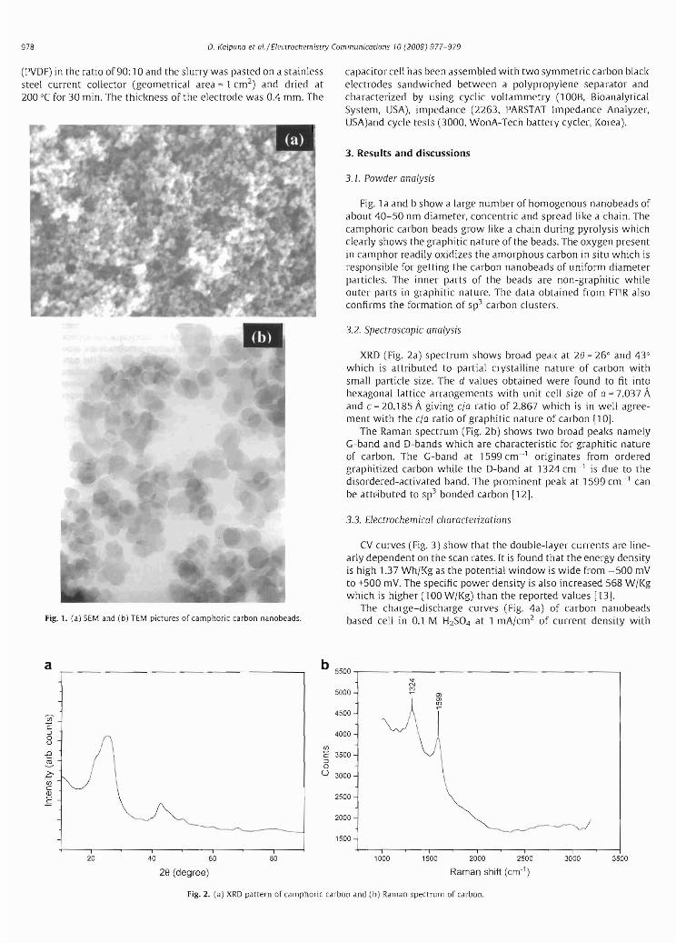
XRD (Fig. 2a) spectrum shows broad peak at 20 = 26° and 43° which is attributed to partial crystalline nature of carbon with small particle size. The d values obtained were found to fit into hexagonal lattice arrangements with unit cell size of a = 7.037 A and c = 20.185 A giving cIa ratio of 2.867 which is in well agreement with the cIa ratio of graphitic nature of carbon [1 OJ.
The Raman spectrum (Fig. 2b) shows two broad peaks namely G-band and D-bands which are characteristic for graphitic nature of carbon. The G-band at 1599 cm-1 originates from ordered graphitized carbon while the D-band at 1324 cm- 1 is due to the disordered-activated band. The prominent peak at 1599 cm- 1 can be attr-ibuted to Sp3 bonded carbon [12J.
3.3. Electrochemical characterizations
CV curves (Fig. 3) show that the double-layer currents are linearly dependent on the scan rates. It is found that the energy density is high 1.37 Wh/I<g as the potential window is wide from -500 mV to +500 mY. The specific power density is also increased 568 W/I<g which is higher (100 W/I<g) than the reported values [131.
The charge-discharge curves (Fig. 4a) of carbon nanobeads based cell in 0.1 M H2S04 at 1 mA/cm2 of current density with
A cI (r
20 40 60 80
28 (degree)
b 5500
5000
4500
4000
(fJ
C 3500 :::J 0 U 3000
2500
2000
1500
1000 1500 2000 2500 3000 3500
Raman shift (cm'1)
Fig. 2. (a) XRD pattern of camphoric carbon and (b) Raman spectrum of carbon. F' cu

-------
979 o Kalpana et al./ Electrochemistry Communications 10 (2008) 977-979
1 Vcut-off voltage are linear during both charging and discharging. As there is no charge-transfer reaction occurred due to electrochemical double layer capacitance, it shows very good cycleability (more than 100,000 cycles have been achieved). The specific capac
--25mV/s0.004 -- 50mV/s
......... 75mV/s --100mV/s
0.002 .~' --------,
~. 0.000 c
~ ::J
U -0002
-0004
-0.006 +--~-r-~-.--~--r-~---,r-~-,----r-----i
-600 -400 -200 0 200 400 600
Potential (mV)
Fig. 3. Cyclic voltammogram of camphoric carbon at various scan rates.
a 1.0
0.8
~ 0.6 III OJ
l'J g 04
0.2
0.0
0 250 500 750 1000
Time(s)
b 1m 90
E 700 88 (") .c oo o
c,Seoo 86 3
0"2J (j'c m~fro
84 ~'w Ql / ro
/ ::J0:: ()400
OJ 82 :::.
2 c
~ c 300
80
--0--0--0200
20000 40000 60000 80000 100000
Cycle numbers
Fig. 4. (a) Charge-discharge profile of camphoric carbon nanobeads at 1 mA/cm2
current density and (b) discharge profiles of carbon nanobeads.
itance can be calculated from charge-discharge curve by using the following equation:
(= 1x tlV x m (Fig)
where 1 is the current density (A/cm2 ), t is the discharge
time (s), Vis the voltage (V) and m is the mass of electrode material (g).
Hence, a maximum specific capacitance of 77 F/g was obtained at 1 mA/cm2 current density. For carbons, it is more noticeable that the volumetric conductivity decreases as the porosity and surface area increases, since the number of conductive pathways can also be expected to decrease [141. It can be explained that carbon with low/high surface area or porosity does not necessarily have the same physical or even chemical structure. The variations in carbon precursor influences more the resistivity of the compacted carbon powders than variations in surface area or porosity alone. If the equivalent series resistance (ESR) is signiRcant, the maximum electric energy stored in supercapcitors will be consumed during discharge which results in poor conductivity as well as poor diffusion of solvated ions with the micropores of carbons [15.16]. Though the surface area of the carbon nanobeads is low, the ESR at 1 kHz is 1.17 n. This low value of ESR is due to high electronic conductivity of camphoric carbon electrode materials and low ionic resistance of the electrolyte with the pores of carbons during charge-discharge tests. It has high coloumbic efficiency and good cyclic stability with minimum IR drop (shown in Fig. 4b) even after 100,000 cycles. The surface chemistry of the camphoric carbon nanobeads electrodes contributes the superior electrochemical performance of this symmetric supercapacitor.
4. Conclusions
The results imply that this camphor based carbon nanobeads can be used as a promising electrode material for high power supercapacitors. This will provide a new base material for possible high power and high energy supercapacitor.
Acknowledgement
This work was supported by the Division of Advanced Batteries in NGE Program (Project No. = 10028357). One of the authors D. Kalpana, is thankful to Prof. AX Shukla, Director, (ECRI and CSIR for granting leave to avail post doctoral fellowship at Chonnam National University, Korea.
References
[11 B.E. Conway. Electrochemical Supercapacitors: Scientific Fundamentals and Technological Applications. Klumer Academic Plenum Publishers, New York, 1959.
[21 R. Kotz, M. Carlen, Electrochim. Acta 45 (2000) 2483. [31 AG. Pandolfo. A.F. Hollenkamp,]. Power Sources 157 (2006) 1. [41 Qiao-Ling Chen, Kuan-Hong Xue, Wei Shen, Fei-Fei Tao. Shou-Yin, Electrochim.
Acta 49 (2004) 4157. 15[ jun Li. Xianyou Wang. Qjnghua Huang, Sergio Gamboa. P.j. Sebastian.]. Power
Sources 158 (2006) 784. 161 TA Centeno, F. Stoeckli. Electrochim. Acta 52 (2006) 560. [71 Feng-Chin Wu, Ru-Ling Tseng. Chi-Chang Hu, Chen-Ching Wang. J. Power
Sources 159 (2006) 1532. [81 M. Sharon, W.I<' Hsu,]. Power Sources 104 (2002) 148. [91 M. Endo, Chemtech 18 (1988) 568.
[101 Maheshwar Sharon. Kingsuk Mukhopadhyay. Kiyosh Vase, Sumio lijima, Yoshinori Ando. Xinluo Zhao, Carbon 36 (1998) 507.
[111 D. Zhou, in: Proceedings 52nd Annual Meeting of Microscopy Society of America, 1994. p. 770.
[12J AC. Ferrari.]. Robertson, Phys. Rev. 8 61 (2000) 14095. [13[ Cesar Merino, Pablo Soto, Eduardo Vilaplana-Ortego,jose M. Gomez de Salazar.
Fernando Pico, Jose M. Rojo, Carbon 43 (2005) 551. [141 X. Andrieu, L. josser. Proc. Electrochem. Soc 181 (1996) 95. [151 X. Andrieu. Energy storage sysr. Electron: New trends Electrochem. Techno!. 1
(2000) 521. [161 M.F. Rose, in: Proceeding of the 33rd International Power Sources Symposium.
Pennington, Nj. 1988, p. 572.
Related Documents