Citation: Gusev, I.; Ferreira, M.; Versace, D.-L.; Abbad-Andaloussi, S.; Pluczyk-Malek, S.; Erfurt, K.; Duda, A.; Data, P.; Blacha-Grzechnik, A. Electrochemically Deposited Zinc (Tetraamino)phthalocyanine as a Light-activated Antimicrobial Coating Effective against S. aureus. Materials 2022, 15, 975. https:// doi.org/10.3390/ma15030975 Academic Editor: Karla A. Batista Received: 14 December 2021 Accepted: 23 January 2022 Published: 27 January 2022 Publisher’s Note: MDPI stays neutral with regard to jurisdictional claims in published maps and institutional affil- iations. Copyright: © 2022 by the authors. Licensee MDPI, Basel, Switzerland. This article is an open access article distributed under the terms and conditions of the Creative Commons Attribution (CC BY) license (https:// creativecommons.org/licenses/by/ 4.0/). materials Article Electrochemically Deposited Zinc (Tetraamino)phthalocyanine as a Light-activated Antimicrobial Coating Effective against S. aureus Ivan Gusev 1,2 , Marli Ferreira 2 , Davy-Louis Versace 3, *, Samir Abbad-Andaloussi 4 , Sandra Pluczyk-Malek 1,2 , Karol Erfurt 1 , Alicja Duda 1 , Przemyslaw Data 2 and Agata Blacha-Grzechnik 1,2, * 1 Faculty of Chemistry, Silesian University of Technology, Strzody 9, 44-100 Gliwice, Poland; [email protected] (I.G.); [email protected] (S.P.-M.); [email protected] (K.E.); [email protected] (A.D.) 2 Centre for Organic and Nanohybrid Electronics, Silesian University of Technology, Konarskiego 22b, 44-100 Gliwice, Poland; [email protected] (M.F.); [email protected] (P.D.) 3 Institut de Chimie et des Matériaux Paris-Est (ICMPE, UMR-CNRS 7182-UPEC), 2-8 Rue Henri Dunant, 94320 Thiais, France 4 Laboratoire Eau, Environnement, Systèmes Urbains (LEESU), UMR-MA 102, Université Paris-Est Créteil (UPEC), 61 Avenue Général de Gaulle, 94010 Créteil Cedex, France; [email protected] * Correspondence: [email protected] (D.-L.V.); [email protected] (A.B.-G.) Abstract: Light-activated antimicrobial coatings are currently considered to be a promising approach for the prevention of nosocomial infections. In this work, we present a straightforward strategy for the deposition of a photoactive biocidal organic layer of zinc (tetraamino)phthalocyanine (ZnPcNH 2 ) in an electrochemical oxidative process. The chemical structure and morphology of the resulting layer are widely characterized by microscopic and spectroscopic techniques, while its ability to photogen- erate reactive oxygen species (ROS) is investigated in situ by UV–Vis spectroscopy with α-terpinene or 1,3-diphenylisobenzofuran as a chemical trap. It is shown that the ZnPcNH 2 photosensitizer retained its photoactivity after immobilization, and that the reported light-activated coating exhibits promising antimicrobial properties towards Staphyloccocus aureus (S. aureus). Keywords: electrochemical deposition; light-activated antimicrobial layer; reactive oxygen species (ROS); phthalocyanines; photosensitizers 1. Introduction In recent years, thin layers of organic and/or inorganic photosensitizers have gained wide research interest for application as antimicrobial coatings deposited on the surfaces of objects that are used daily. The main advantage of such structures is their remarkable biocidal efficiency against bacteria, viruses, and fungi. This makes them an attractive alternative to classical antimicrobial coatings containing poly(ethylene glycol) chains, silver or copper nanoparticles, quaternary ammonium salts or cations, fluorinated polymers, etc. [1,2]. The antimicrobial action of photoactive layers is based on the formation of so-called reactive oxygen species (ROS), including superoxides, peroxides, and singlet oxygen [3]. ROS act in a highly effective and non-selective manner (e.g. via the oxidation of enzymes, by increasing ions’ permeability by the cell wall) against microorganisms, which, in turn, strongly reduces the possibility of the development of resistance by microbes [4–6]. Until now, light-activated antimicrobial coatings containing, e.g., phenothiazine, porphyrin, or fullerene photosensitizers have been successfully applied against Staphylococcus aureus, Escherichia coli, Clostridium difficile, Candida albicans, and Pseudomonas aeruginosa [7–14]. However, the practical usage of such photoactive layers is quite limited, and intense research is conducted in order to overcome key constraints, such as (photo)stability, long- term action, or the ease and accessibility of a deposition procedure. Materials 2022, 15, 975. https://doi.org/10.3390/ma15030975 https://www.mdpi.com/journal/materials

Welcome message from author
This document is posted to help you gain knowledge. Please leave a comment to let me know what you think about it! Share it to your friends and learn new things together.
Transcript
�����������������
Citation: Gusev, I.; Ferreira, M.;
Versace, D.-L.; Abbad-Andaloussi, S.;
Pluczyk-Małek, S.; Erfurt, K.;
Duda, A.; Data, P.; Blacha-Grzechnik,
A. Electrochemically Deposited Zinc
(Tetraamino)phthalocyanine as a
Light-activated Antimicrobial
Coating Effective against S. aureus.
Materials 2022, 15, 975. https://
doi.org/10.3390/ma15030975
Academic Editor: Karla A. Batista
Received: 14 December 2021
Accepted: 23 January 2022
Published: 27 January 2022
Publisher’s Note: MDPI stays neutral
with regard to jurisdictional claims in
published maps and institutional affil-
iations.
Copyright: © 2022 by the authors.
Licensee MDPI, Basel, Switzerland.
This article is an open access article
distributed under the terms and
conditions of the Creative Commons
Attribution (CC BY) license (https://
creativecommons.org/licenses/by/
4.0/).
materials
Article
Electrochemically Deposited Zinc (Tetraamino)phthalocyanineas a Light-activated Antimicrobial Coating Effective againstS. aureusIvan Gusev 1,2 , Marli Ferreira 2, Davy-Louis Versace 3,*, Samir Abbad-Andaloussi 4 , Sandra Pluczyk-Małek 1,2,Karol Erfurt 1 , Alicja Duda 1 , Przemysław Data 2 and Agata Blacha-Grzechnik 1,2,*
1 Faculty of Chemistry, Silesian University of Technology, Strzody 9, 44-100 Gliwice, Poland;[email protected] (I.G.); [email protected] (S.P.-M.); [email protected] (K.E.);[email protected] (A.D.)
2 Centre for Organic and Nanohybrid Electronics, Silesian University of Technology, Konarskiego 22b,44-100 Gliwice, Poland; [email protected] (M.F.); [email protected] (P.D.)
3 Institut de Chimie et des Matériaux Paris-Est (ICMPE, UMR-CNRS 7182-UPEC), 2-8 Rue Henri Dunant,94320 Thiais, France
4 Laboratoire Eau, Environnement, Systèmes Urbains (LEESU), UMR-MA 102, Université Paris-EstCréteil (UPEC), 61 Avenue Général de Gaulle, 94010 Créteil Cedex, France; [email protected]
* Correspondence: [email protected] (D.-L.V.); [email protected] (A.B.-G.)
Abstract: Light-activated antimicrobial coatings are currently considered to be a promising approachfor the prevention of nosocomial infections. In this work, we present a straightforward strategy forthe deposition of a photoactive biocidal organic layer of zinc (tetraamino)phthalocyanine (ZnPcNH2)in an electrochemical oxidative process. The chemical structure and morphology of the resulting layerare widely characterized by microscopic and spectroscopic techniques, while its ability to photogen-erate reactive oxygen species (ROS) is investigated in situ by UV–Vis spectroscopy with α-terpineneor 1,3-diphenylisobenzofuran as a chemical trap. It is shown that the ZnPcNH2 photosensitizerretained its photoactivity after immobilization, and that the reported light-activated coating exhibitspromising antimicrobial properties towards Staphyloccocus aureus (S. aureus).
Keywords: electrochemical deposition; light-activated antimicrobial layer; reactive oxygen species(ROS); phthalocyanines; photosensitizers
1. Introduction
In recent years, thin layers of organic and/or inorganic photosensitizers have gainedwide research interest for application as antimicrobial coatings deposited on the surfacesof objects that are used daily. The main advantage of such structures is their remarkablebiocidal efficiency against bacteria, viruses, and fungi. This makes them an attractivealternative to classical antimicrobial coatings containing poly(ethylene glycol) chains, silveror copper nanoparticles, quaternary ammonium salts or cations, fluorinated polymers,etc. [1,2]. The antimicrobial action of photoactive layers is based on the formation ofso-called reactive oxygen species (ROS), including superoxides, peroxides, and singletoxygen [3]. ROS act in a highly effective and non-selective manner (e.g. via the oxidation ofenzymes, by increasing ions’ permeability by the cell wall) against microorganisms, which,in turn, strongly reduces the possibility of the development of resistance by microbes [4–6].Until now, light-activated antimicrobial coatings containing, e.g., phenothiazine, porphyrin,or fullerene photosensitizers have been successfully applied against Staphylococcus aureus,Escherichia coli, Clostridium difficile, Candida albicans, and Pseudomonas aeruginosa [7–14].However, the practical usage of such photoactive layers is quite limited, and intenseresearch is conducted in order to overcome key constraints, such as (photo)stability, long-term action, or the ease and accessibility of a deposition procedure.
Materials 2022, 15, 975. https://doi.org/10.3390/ma15030975 https://www.mdpi.com/journal/materials
Materials 2022, 15, 975 2 of 9
For many years, phthalocyanines (Pcs) have been mainly investigated as promisingcandidates for application in (opto)electronics. Until recently, Pcs in a free-base formor with a central metal have been used in organic photovoltaic devices (OPVs) [15–17],organic light-emitting diodes (OLEDs) [18,19], or gas sensors [20–22]. Their effectivenessas photoinitiating systems [23,24] and photosensitizers [25,26] has also been reported.Moreover, it has been shown that the photosensitizing abilities of Pcs can be tuned bychanging the central metal atom and/or by the introduction of outer functional groups.
Taking into account the above, in this work, we aimed to investigate the possibil-ity of applying an electrodeposited Pc-based photoactive layer as a light-activated an-timicrobial coating. Thus, zinc (tetraamino)phthalocyanine (ZnPcNH2) was selected asa primary photosensitizer molecule. The choice was first made based on the fact thatZn-containing Pcs show considerable efficiency of ROS production [27,28] and high an-timicrobial properties [12,29]. Secondly, as shown in our previous work, the outer primaryamino groups can be used in the electrochemically driven deposition of Pcs [30]. Thepresented novel, straightforward strategy, consisting of an electrochemical oxidation of pri-mary amino groups present in the ZnPcNH2 structure, may be beneficial for the formationof a layer on conductive substrates. The deposited coating is widely characterized by meansof spectroscopic and microscopic techniques. It is shown that the immobilized ZnPcNH2photosensitizer is able to produce ROS under red laser or white light illumination and thusshows a light antimicrobial response against Staphyloccocus aureus (S. aureus).
2. Materials and Methods2.1. Materials
Zinc (tetraamino)phthalocyanine (ZnPcNH2) was synthesized based on previousworks [31,32]. The synthesis procedure and the identification of the obtained product aredescribed in the Supporting Information.
Dimethylformamide (DMF; ≥99.8%, Sigma Aldrich, St. Louis, MO, USA) containingtetrabutylammonium tetrafluoroborate (TBABF4; 99%, Sigma Aldrich, St. Louis, MO, USA)was used as an electrolyte solution for the electrochemical deposition of (ZnPcNH2)layer.Indium tin oxide/borosilicate glass (ITO) purchased from Präzisions Glas & Optik GmbH(PGO, Iserlohn, Germany) was used as a support. ROS photogeneration was investigatedin α-terpinene (TCI; purity > 90%)–acetonitrile (Sigma Aldrich, St. Louis, MO, USA) or1,3-diphenylisobenzofuran (DPBF; Acros Organics, Geel, Belgium, purity > 97%)–methanol(Acros Organics, Geel, Belgium, 99.9%) systems.
2.2. Formation and Characterization of (ZnPcNH2)layer
(ZnPcNH2)layer was formed on ITO or a platinum plate from 0.1 mM solution ofZnPcNH2 in 0.1 M TBABF4/DMF. The starting solution was homogenized with an ul-trasonic mixer and purged with argon for 15 minutes to remove oxygen. The process ofelectrochemical deposition was conducted using a cyclic voltammetry (CV) technique anda CHI 660C electrochemical workstation (CH Instruments Inc., Austin, TX, USA). A three-electrode setup was used: ITO or Pt as a working electrode, Ag wire as a pseudo-referenceelectrode, and glassy carbon (GC) as a counter electrode. The electrodes were copiouslyrinsed with DMF and mounted in a Teflon holder prior to use. The electrochemical de-position was conducted by means of cyclic voltammetry (CV) with the following processparameters: the potential range (−1.8; 1.4) V, 10 scan cycles, and a scan rate of 0.1 V/s.Ferrocene (Fc/Fc+) was used as a reference for the potential calibration.
The morphology of the layer was investigated using scanning electron microscopy(SEM) and atomic force microscopy (AFM). SEM images were acquired using PhenomProX with an accelerating voltage equal to 15 kV and a magnification between 15,000×and 20,000×. The samples were sputter coated with 10 nm of gold film (Q150R QuorumTechnologies, Laughton, East Sussex, UK) before SEM imaging. The images were analyzedwith Phenom software. AFM investigations were conducted using Nanosurf CoreAFMworking in a contact mode with the standard contact-mode AFM HQ:CSC17/Al BS (Mikro-
Materials 2022, 15, 975 3 of 9
Masch, Tallinn, Estonia) probe (resonance frequency 13 kHz; force constant 0.18 Nm−1).The images were processed with the use of Gwyddion SPM (Brno University of Technology,Brno, Czech Republic).
UV–Vis spectra of the ZnPcNH2 solution in DMF and (ZnPcNH2)layer supported onITO were acquired with a Hewlett Packard 8452A UV–Vis spectrometer. IR spectra ofZnPcNH2 powder and (ZnPcNH2)layer deposited on a platinum plate were recorded withinthe 3500–700 cm−1 range in attenuated total reflectance (ATR) mode using a Perkin ElmerSpectrum Two IR spectrometer (Hopkinton, MA, USA).
2.3. Reactive Oxygen Species (ROS) Photogeneration and Microbiological Analysis
The process of ROS photogeneration was investigated either under 638 nm diodelaser (Oxxius LBX-638-150-ELL-PP, Lannion, France, power reduced to 20 mW) or whitelight (Fiber-Coupled Xenon Light Source, Newton, NJ, USA, SLS205, 75 W, Thorlabs)illumination. In the first case, a DPBF trap was used with 0.05 mM solution in methanol,while in the second case, the trap was used with 0.05 mM solution of α-terpinene inacetonitrile. The experimental setup was arranged in this way so that a photoactive layersupported on a glass plate was put in a quartz cuvette (Hellma Analytics, Müllheim,Germany, 10 × 4 mm). It was illuminated with a light source, while UV–Vis spectra of thechemical traps were collected in situ with a Hewlett Packard 8452A UV–Vis spectrometer(Palo Alto, CA, USA) along a direction perpendicular to the layer’s illumination. Thecourse of ROS production was observed by a drop of DPBF or α-terpinene absorbance at410 nm or 266 nm, respectively.
The antibacterial properties of the ZnPcNH2-derived coatings, glass, and ITO sur-faces were evaluated against S. aureus ATCC12000 according to previously describedprocedures [13,33,34]. The corresponding coatings were immersed in a bacterial solutionfor 24 h prior to visible-light activation, in order to maximize bacterial adhesion on surfaces.Then, half of the samples were kept in the dark while half of them were illuminated for1 h on each side under a solar emission lamp. Following adhesion, all the defined squaresamples were rinsed seven times with sterile NaCl solution (0.9% w/v) to remove thenon-adherent or dead bacteria from each surface. Then, samples were immersed in 3 mL ofsterile saline solution and sonicated for 5 min in order to detach the viable bacteria fromthe surface of the studied samples. This solution was serially diluted by 10 to 105 factors.An amount of 100 µL of each diluted and detached microorganism solution was thenintroduced on the surface of a Plat Count agar plate. This process was repeated as manytimes as there were dilutions. The total amount of viable bacteria was determined bycounting the colony-forming units, after 48 h of incubation of the agar plates at 37 ◦ C (foreach dilution), and levels of adhesion were given as numbers of counted bacteria/cm2 (cm2
corresponds to the surface of the defined samples). Four experiments were conducted oneach sample.
3. Results and Discussion3.1. Formation and Spectroscopic and Microscopic Characterization of (ZnPcNH2)layer
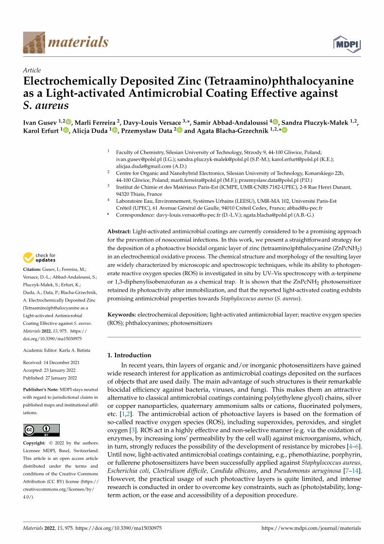
Figure 1 presents CV curves recorded during continuous scanning in an electrolytesolution containing ZnPcNH2. In the first anodic scan, an irreversible oxidation can beobserved at ca. 0.75 V vs. Ag, which can be assigned to the oxidation of the primaryamino group of ZnPcNH2 to a radical cation, similar to the electro-oxidation of the anilinemonomer [35]. A steady increase in the registered current in the broad potential rangeduring continuous scanning can be observed, confirming the formation of the electroactivelayer on the surface of the ITO electrode. The newly occurring redox couples at ca. −0.75 Vand −1.25 V arise from the electrochemical activity of the ZnPc layer [36–39]. In the courseof the 10 scan cycles of the electrodeposition process, a uniform blue-green (ZnPcNH2)layerwas synthesized on the ITO surface (Figure 1 inset).
Materials 2022, 15, 975 4 of 9Materials 2022, 14, x FOR PEER REVIEW 4 of 9
Figure 1. CV curves recorded for the ITO working electrode in 0.1 mM ZnPcNH2 electrolyte solution (0.1 M TBABF4/DMF). Inset: photography of (ZnPcNH2)layer electrodeposited on ITO.
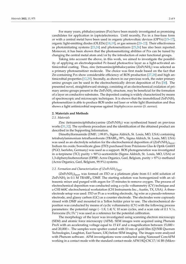
The morphology of (ZnPcNH2)layer on ITO was investigated using SEM and AFM techniques. An SEM image of ITO covered with ZnPcNH2 is shown in Figure 2a. Spherical crystallites, typical for solution-deposited ZnPc films [40,41], sized within the 0.6–1.3 µm range, can be observed. Further, AFM studies confirmed the continuous coverage of the ITO surface, as shown in the exemplary AFM topography 31 × 31 µm2 image shown in Figure 2b. An AFM image shown in Figure 2c also confirms the presence of rather vertically oriented crystallites. This is in contrast to ZnPc films deposited on a heated substrate with the PVD technique which have a rather ribbon-like structure [42]. The root mean square roughness (RMS) value for the presented area of the film was 100.9 nm.
Figure 2. (a) SEM and (b,c) AFM images of (ZnPcNH2)layer electrodeposited on ITO.
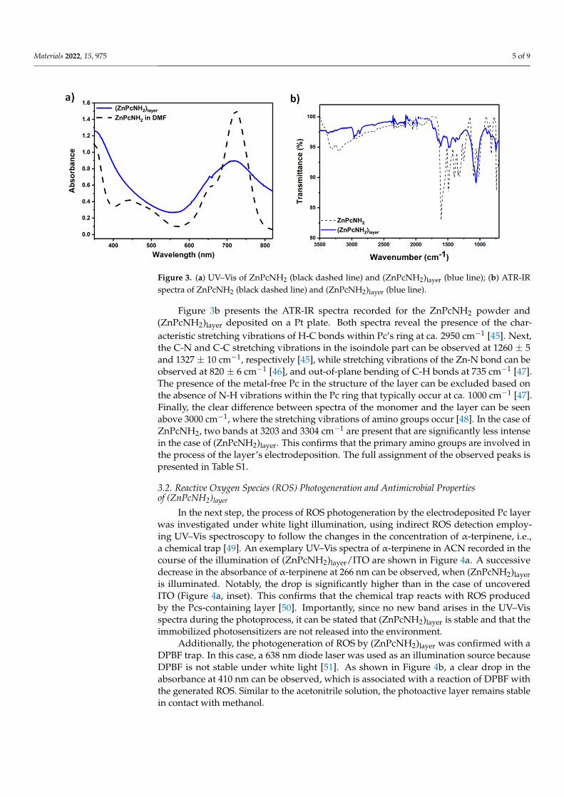
The optical properties and the chemical structure of (ZnPcNH2)layer were character-ized by UV–Vis and IR spectroscopies, respectively. UV–Vis spectra of the solution of ZnPcNH2 in DMF and (ZnPcNH2)layer deposited on ITO are presented in Figure 3a. The characteristic B (Soret band) and Q bands (π → π* transition band) for ZnPcNH2, both in a solution phase and in the form of a layer, can be observed at ca. 350 nm and 720 nm, respectively. The latter is significantly broadened in the case of the layer, possibly due to the aggregation of ZnPc molecules [36,43,44]. Notably, the deposited Pc layer exhibits
Figure 1. CV curves recorded for the ITO working electrode in 0.1 mM ZnPcNH2 electrolyte solution(0.1 M TBABF4/DMF). Inset: photography of (ZnPcNH2)layer electrodeposited on ITO.
The morphology of (ZnPcNH2)layer on ITO was investigated using SEM and AFMtechniques. An SEM image of ITO covered with ZnPcNH2 is shown in Figure 2a. Sphericalcrystallites, typical for solution-deposited ZnPc films [40,41], sized within the 0.6–1.3 µmrange, can be observed. Further, AFM studies confirmed the continuous coverage of theITO surface, as shown in the exemplary AFM topography 31 × 31 µm2 image shown inFigure 2b. An AFM image shown in Figure 2c also confirms the presence of rather verticallyoriented crystallites. This is in contrast to ZnPc films deposited on a heated substrate withthe PVD technique which have a rather ribbon-like structure [42]. The root mean squareroughness (RMS) value for the presented area of the film was 100.9 nm.
Materials 2022, 14, x FOR PEER REVIEW 4 of 9
Figure 1. CV curves recorded for the ITO working electrode in 0.1 mM ZnPcNH2 electrolyte solution (0.1 M TBABF4/DMF). Inset: photography of (ZnPcNH2)layer electrodeposited on ITO.
The morphology of (ZnPcNH2)layer on ITO was investigated using SEM and AFM techniques. An SEM image of ITO covered with ZnPcNH2 is shown in Figure 2a. Spherical crystallites, typical for solution-deposited ZnPc films [40,41], sized within the 0.6–1.3 µm range, can be observed. Further, AFM studies confirmed the continuous coverage of the ITO surface, as shown in the exemplary AFM topography 31 × 31 µm2 image shown in Figure 2b. An AFM image shown in Figure 2c also confirms the presence of rather vertically oriented crystallites. This is in contrast to ZnPc films deposited on a heated substrate with the PVD technique which have a rather ribbon-like structure [42]. The root mean square roughness (RMS) value for the presented area of the film was 100.9 nm.
Figure 2. (a) SEM and (b,c) AFM images of (ZnPcNH2)layer electrodeposited on ITO.
The optical properties and the chemical structure of (ZnPcNH2)layer were character-ized by UV–Vis and IR spectroscopies, respectively. UV–Vis spectra of the solution of ZnPcNH2 in DMF and (ZnPcNH2)layer deposited on ITO are presented in Figure 3a. The characteristic B (Soret band) and Q bands (π → π* transition band) for ZnPcNH2, both in a solution phase and in the form of a layer, can be observed at ca. 350 nm and 720 nm, respectively. The latter is significantly broadened in the case of the layer, possibly due to the aggregation of ZnPc molecules [36,43,44]. Notably, the deposited Pc layer exhibits
Figure 2. (a) SEM and (b,c) AFM images of (ZnPcNH2)layer electrodeposited on ITO.
The optical properties and the chemical structure of (ZnPcNH2)layer were characterizedby UV–Vis and IR spectroscopies, respectively. UV–Vis spectra of the solution of ZnPcNH2in DMF and (ZnPcNH2)layer deposited on ITO are presented in Figure 3a. The characteristicB (Soret band) and Q bands (π→ π* transition band) for ZnPcNH2, both in a solution phaseand in the form of a layer, can be observed at ca. 350 nm and 720 nm, respectively. Thelatter is significantly broadened in the case of the layer, possibly due to the aggregationof ZnPc molecules [36,43,44]. Notably, the deposited Pc layer exhibits strong and broadabsorption in the visible range, which should be advantageous for a white light-drivenphotosensitization process.
Materials 2022, 15, 975 5 of 9
Materials 2022, 14, x FOR PEER REVIEW 5 of 9
strong and broad absorption in the visible range, which should be advantageous for a white light-driven photosensitization process.
Figure 3. (a) UV–Vis of ZnPcNH2 (black dashed line) and (ZnPcNH2)layer (blue line); (b) ATR-IR spectra of ZnPcNH2 (black dashed line) and (ZnPcNH2)layer (blue line).
Figure 3b presents the ATR-IR spectra recorded for the ZnPcNH2 powder and (ZnPcNH2)layer deposited on a Pt plate. Both spectra reveal the presence of the characteris-tic stretching vibrations of H-C bonds within Pc’s ring at ca. 2950 cm−1 [45]. Next, the C-N and C-C stretching vibrations in the isoindole part can be observed at 1260 ± 5 and 1327 ± 10 cm−1, respectively [45], while stretching vibrations of the Zn-N bond can be observed at 820 ± 6 cm-1 [46], and out-of-plane bending of C-H bonds at 735 cm−1 [47]. The presence of the metal-free Pc in the structure of the layer can be excluded based on the absence of N-H vibrations within the Pc ring that typically occur at ca. 1000 cm−1 [47]. Finally, the clear difference between spectra of the monomer and the layer can be seen above 3000 cm−1, where the stretching vibrations of amino groups occur [48]. In the case of ZnPcNH2, two bands at 3203 and 3304 cm−1 are present that are significantly less intense in the case of (ZnPcNH2)layer. This confirms that the primary amino groups are involved in the process of the layer’s electrodeposition. The full assignment of the observed peaks is presented in Table S1.
3.2. Reactive Oxygen Species (ROS) photogeneration and antimicrobial properties of (ZnPcNH2)layer
In the next step, the process of ROS photogeneration by the electrodeposited Pc layer was investigated under white light illumination, using indirect ROS detection employing UV–Vis spectroscopy to follow the changes in the concentration of α-terpinene, i.e., a chemical trap [49]. An exemplary UV–Vis spectra of α-terpinene in ACN recorded in the course of the illumination of (ZnPcNH2)layer/ITO are shown in Figure 4a. A successive de-crease in the absorbance of α-terpinene at 266 nm can be observed, when (ZnPcNH2)layer is illuminated. Notably, the drop is significantly higher than in the case of uncovered ITO (Figure 4a, inset). This confirms that the chemical trap reacts with ROS produced by the Pcs-con-taining layer [50]. Importantly, since no new band arises in the UV–Vis spectra during the photoprocess, it can be stated that (ZnPcNH2)layer is stable and that the immobilized pho-tosensitizers are not released into the environment.
Additionally, the photogeneration of ROS by (ZnPcNH2)layer was confirmed with a DPBF trap. In this case, a 638 nm diode laser was used as an illumination source because DPBF is not stable under white light [51]. As shown in Figure 4b, a clear drop in the ab-sorbance at 410 nm can be observed, which is associated with a reaction of DPBF with the
Figure 3. (a) UV–Vis of ZnPcNH2 (black dashed line) and (ZnPcNH2)layer (blue line); (b) ATR-IRspectra of ZnPcNH2 (black dashed line) and (ZnPcNH2)layer (blue line).
Figure 3b presents the ATR-IR spectra recorded for the ZnPcNH2 powder and(ZnPcNH2)layer deposited on a Pt plate. Both spectra reveal the presence of the char-acteristic stretching vibrations of H-C bonds within Pc’s ring at ca. 2950 cm−1 [45]. Next,the C-N and C-C stretching vibrations in the isoindole part can be observed at 1260 ± 5and 1327 ± 10 cm−1, respectively [45], while stretching vibrations of the Zn-N bond can beobserved at 820 ± 6 cm−1 [46], and out-of-plane bending of C-H bonds at 735 cm−1 [47].The presence of the metal-free Pc in the structure of the layer can be excluded based onthe absence of N-H vibrations within the Pc ring that typically occur at ca. 1000 cm−1 [47].Finally, the clear difference between spectra of the monomer and the layer can be seenabove 3000 cm−1, where the stretching vibrations of amino groups occur [48]. In the case ofZnPcNH2, two bands at 3203 and 3304 cm−1 are present that are significantly less intensein the case of (ZnPcNH2)layer. This confirms that the primary amino groups are involved inthe process of the layer’s electrodeposition. The full assignment of the observed peaks ispresented in Table S1.
3.2. Reactive Oxygen Species (ROS) Photogeneration and Antimicrobial Propertiesof (ZnPcNH2)layer
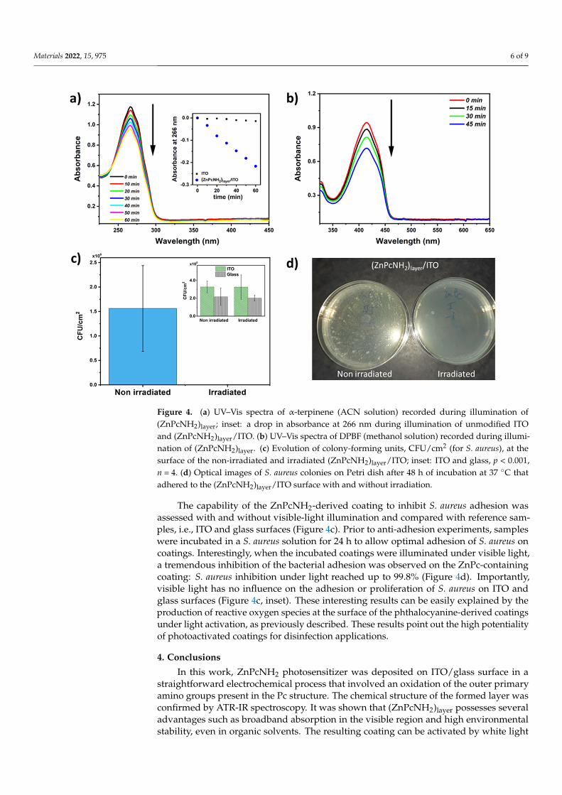
In the next step, the process of ROS photogeneration by the electrodeposited Pc layerwas investigated under white light illumination, using indirect ROS detection employ-ing UV–Vis spectroscopy to follow the changes in the concentration of α-terpinene, i.e.,a chemical trap [49]. An exemplary UV–Vis spectra of α-terpinene in ACN recorded in thecourse of the illumination of (ZnPcNH2)layer/ITO are shown in Figure 4a. A successivedecrease in the absorbance of α-terpinene at 266 nm can be observed, when (ZnPcNH2)layeris illuminated. Notably, the drop is significantly higher than in the case of uncoveredITO (Figure 4a, inset). This confirms that the chemical trap reacts with ROS producedby the Pcs-containing layer [50]. Importantly, since no new band arises in the UV–Visspectra during the photoprocess, it can be stated that (ZnPcNH2)layer is stable and that theimmobilized photosensitizers are not released into the environment.
Additionally, the photogeneration of ROS by (ZnPcNH2)layer was confirmed with aDPBF trap. In this case, a 638 nm diode laser was used as an illumination source becauseDPBF is not stable under white light [51]. As shown in Figure 4b, a clear drop in theabsorbance at 410 nm can be observed, which is associated with a reaction of DPBF withthe generated ROS. Similar to the acetonitrile solution, the photoactive layer remains stablein contact with methanol.
Materials 2022, 15, 975 6 of 9
Materials 2022, 14, x FOR PEER REVIEW 6 of 9
generated ROS. Similar to the acetonitrile solution, the photoactive layer remains stable in contact with methanol.
Figure 4. (a) UV–Vis spectra of α-terpinene (ACN solution) recorded during illumination of (ZnPcNH2)layer; inset: a drop in absorbance at 266 nm during illumination of unmodified ITO and (ZnPcNH2)layer/ITO. (b) UV–Vis spectra of DPBF (methanol solution) recorded during illumination of (ZnPcNH2)layer. (c) Evolution of colony-forming units, CFU/cm2 (for S. aureus), at the surface of the non-irradiated and irradiated (ZnPcNH2)layer/ITO; inset: ITO and glass, p < 0.001, n = 4. (d) Optical images of S. aureus colonies on Petri dish after 48 h of incubation at 37 °C that adhered to the (ZnPcNH2)layer/ITO surface with and without irradiation.
The capability of the ZnPcNH2-derived coating to inhibit S. aureus adhesion was as-sessed with and without visible-light illumination and compared with reference samples, i.e., ITO and glass surfaces (Figure 4c). Prior to anti-adhesion experiments, samples were incubated in a S. aureus solution for 24 h to allow optimal adhesion of S. aureus on coatings. Interestingly, when the incubated coatings were illuminated under visible light, a tremen-dous inhibition of the bacterial adhesion was observed on the ZnPc-containing coating: S. aureus inhibition under light reached up to 99.8% (Figure 4d). Importantly, visible light has no influence on the adhesion or proliferation of S. aureus on ITO and glass surfaces (Figure 4c, inset). These interesting results can be easily explained by the production of reactive oxygen species at the surface of the phthalocyanine-derived coatings under light activation, as previously described. These results point out the high potentiality of photo-activated coatings for disinfection applications.
4. Conclusions In this work, ZnPcNH2 photosensitizer was deposited on ITO/glass surface in a
straightforward electrochemical process that involved an oxidation of the outer primary
Figure 4. (a) UV–Vis spectra of α-terpinene (ACN solution) recorded during illumination of(ZnPcNH2)layer; inset: a drop in absorbance at 266 nm during illumination of unmodified ITOand (ZnPcNH2)layer/ITO. (b) UV–Vis spectra of DPBF (methanol solution) recorded during illumi-nation of (ZnPcNH2)layer. (c) Evolution of colony-forming units, CFU/cm2 (for S. aureus), at thesurface of the non-irradiated and irradiated (ZnPcNH2)layer/ITO; inset: ITO and glass, p < 0.001,n = 4. (d) Optical images of S. aureus colonies on Petri dish after 48 h of incubation at 37 ◦C thatadhered to the (ZnPcNH2)layer/ITO surface with and without irradiation.
The capability of the ZnPcNH2-derived coating to inhibit S. aureus adhesion wasassessed with and without visible-light illumination and compared with reference sam-ples, i.e., ITO and glass surfaces (Figure 4c). Prior to anti-adhesion experiments, sampleswere incubated in a S. aureus solution for 24 h to allow optimal adhesion of S. aureus oncoatings. Interestingly, when the incubated coatings were illuminated under visible light,a tremendous inhibition of the bacterial adhesion was observed on the ZnPc-containingcoating: S. aureus inhibition under light reached up to 99.8% (Figure 4d). Importantly,visible light has no influence on the adhesion or proliferation of S. aureus on ITO andglass surfaces (Figure 4c, inset). These interesting results can be easily explained by theproduction of reactive oxygen species at the surface of the phthalocyanine-derived coatingsunder light activation, as previously described. These results point out the high potentialityof photoactivated coatings for disinfection applications.
4. Conclusions
In this work, ZnPcNH2 photosensitizer was deposited on ITO/glass surface in astraightforward electrochemical process that involved an oxidation of the outer primaryamino groups present in the Pc structure. The chemical structure of the formed layer wasconfirmed by ATR-IR spectroscopy. It was shown that (ZnPcNH2)layer possesses severaladvantages such as broadband absorption in the visible region and high environmentalstability, even in organic solvents. The resulting coating can be activated by white light

Materials 2022, 15, 975 7 of 9
illumination to effectively produce reactive oxygen species. This proves that immobilizedZnPcNH2 retains its photosensitizing properties. Finally, it was shown that (ZnPcNH2)layeris active against S. aureus, there was a 99.8% light-associated decrease in the number of theadhered bacteria. To sum up, the presented results confirm that with a proper design of aphotosensitizer molecule’s structure, an efficient light-activated antimicrobial layer can bedeposited in a simple electrodeposition process. In this work, ITO/glass or platinum wasused as a support. However, it is believed that, since the deposition process is conductedin an oxygen-free organic environment under a rather low oxidative potential, it may beattractive for covering various (semi)conductive materials, irrespective of size or shape.
Supplementary Materials: The following are available online at https://www.mdpi.com/article/10.3390/ma15030975/s1: Synthesis and characterization of ZnPcNH2 monomer, Table S1: Assign-ment of IR signals for ZnPcNH2 monomer and (ZnPcNH2)layer.
Author Contributions: Conceptualization, A.B.-G.; methodology, M.F., D.-L.V. and A.B.-G.; valida-tion, M.F., D.-L.V. and A.B.-G.; formal analysis, M.F., D.-L.V., S.P-M. and A.B.-G.; investigation, I.G.,M.F., D.-L.V., S.A.-A., S.P.-M., K.E., A.D. and A.B.-G.; writing—original draft preparation, M.F., D.-L.V.and A.B.-G.; writing—review and editing, A.B.-G.; visualization, M.F., D.-L.V., S.P.-M. and A.B.-G.;supervision, P.D. and A.B.-G.; project administration, P.D. and A.B.-G.; funding acquisition, I.G., P.D.and A.B.-G. All authors have read and agreed to the published version of the manuscript.
Funding: This work was financially supported by the First Team program of the Foundation forPolish Science co-financed by the European Union under the European Regional Development Fund(project number: First TEAM POIR.04.04.00-00-4668/17-00). I.G. and A.B.-G. kindly acknowledgethe support received from the Polish Budget Funds for Scientific Research in 2021 as core fundingfor R&D activities in the Silesian University of Technology—funding for young researchers (grantnumbers: 04/040/BKM21/0160 and 04/040/BKM21/0171, respectively). The authors are grateful tothe training actions funded by the European Union’s Horizon 2020 research and innovation programunder grant agreement no. 952008.
Institutional Review Board Statement: Not applicable.
Informed Consent Statement: Not applicable.
Data Availability Statement: Data are presented in the article and Supplementary Materials.
Conflicts of Interest: The authors declare no conflict of interest.
References1. Sautrot-Ba, P.; Malval, J.-P.; Weiss-Maurin, M.; Paul, J.; Blacha-Grzechnik, A.; Tomane, S.; Mazeran, P.-E.; Lalevée, J.; Langlois, V.;
Versace, D.-L. Paprika, Gallic Acid, and Visible Light: The Green Combination for the Synthesis of Biocide Coatings. ACS Sustain.Chem. Eng. 2018, 6, 104–109. [CrossRef]
2. Hwang, G.B.; Allan, E.; Parkin, I.P. White Light-Activated Antimicrobial Paint using Crystal Violet. ACS Appl. Mater. Interfaces2015, 8, 15033–15039. [CrossRef] [PubMed]
3. Walker, T.; Canales, M.; Noimark, S.; Page, K.; Parkin, I.; Faull, J.; Bhatti, M.; Ciric, L. A Light-Activated Antimicrobial Surface IsActive Against Bacterial, Viral and Fungal Organisms. Sci. Rep. 2017, 7, 15298. [CrossRef] [PubMed]
4. Wainwright, M.; Crossley, K.B. Photosensitising agents—circumventing resistance and breaking down biofilms: A review.Int. Biodeterior. Biodegrad. 2004, 53, 119–126. [CrossRef]
5. Spagnul, C.; Turner, L.C.; Boyle, R.W. Immobilized photosensitizers for antimicrobial applications. J. Photochem. Photobiol. B Biol.2015, 150, 11–30. [CrossRef]
6. Dahl, T.; RobertMiddenand, W.; Hartman, P. Pure singlet oxygen cytotoxicity for bacteria. Photochem. Photobiol. 1987, 46, 345–352.[CrossRef]
7. Peveler, W.J.; Noimark, S.; Al-Azawi, H.; Hwang, G.B.; Crick, C.R.; Allan, E.; Edel, J.B.; Ivanov, A.; MacRobert, A.J.; Parkin, I.P.Covalently Attached Antimicrobial Surfaces Using BODIPY: Improving Efficiency and Effectiveness. ACS Appl. Mater. Interfaces2018, 10, 98–104. [CrossRef]
8. Piccirillo, C.; Perni, S.; Gil-Thomas, J.; Prokopovich, P.; Wilson, M.; Pratten, J.; Parkin, I.P. Antimicrobial activity of methylene blueand toluidine blue O covalently bound to a modified silicone polymer surface. J. Mater. Chem. 2009, 19, 6167–6171. [CrossRef]
9. Decraene, V.; Pratten, J.; Wilson, M. Novel Light-Activated Antimicrobial Coatings Are Effective Against Surface-DepositedStaphylococcus aureus. Curr. Microbiol. 2008, 57, 269–273. [CrossRef]
Materials 2022, 15, 975 8 of 9
10. Ballatore, M.B.; Durantini, J.; Gsponer, N.S.; Suarez, M.B.; Gervaldo, M.; Otero, L.; Spesia, M.; Milanesio, M.E.; Durantini, E.N.Photodynamic Inactivation of Bacteria Using Novel Electrogenerated Porphyrin-Fullerene C60 Polymeric Films. Environ. Sci.Technol. 2015, 49, 7456–7463. [CrossRef]
11. Noimark, S.; Dunnill, C.; Parkin, I. Shining light on materials—A self-sterilising revolution. Adv. Drug Deliv. Rev. 2013, 65,570–580. [CrossRef] [PubMed]
12. Grammatikova, N.E.; George, L.; Ahmed, Z.; Candeias, N.R.; Durandin, N.A.; Efimov, A. Zinc phthalocyanine activated byconventional indoor light makes a highly efficient antimicrobial material from regular cellulose. J. Mater. Chem. B 2019, 7,4379–4384. [CrossRef]
13. Condat, M.; Mazeran, P.-E.; Malval, J.-P.; Lalevée, J.; Morlet-Savary, F.; Renard, E.; Langlois, V.; Andalloussi, S.A.; Versace, D.-L.Photoinduced curcumin derivative-coatings with antibacterial properties. RSC Adv. 2015, 5, 85214–85224. [CrossRef]
14. Sautrot-Ba, P.; Jockusch, S.; Nguyen, T.-T.-T.; Grande, D.; Chiapionne, A.; Abbad-Andaloussi, S.; Pan, M.; Méallet-Renault, R.;Versace, D.-L. Photoinduced synthesis of antibacterial hydrogel from aqueous photoinitiating system. Eur. Polym. J. 2020,138, 109936. [CrossRef]
15. Urbani, M.; Ragoussi, M.-E.; Nazeeruddin, M.K.; Torres, T. Phthalocyanines for dye-sensitized solar cells. Co-Ord. Chem. Rev.2019, 381, 1–64. [CrossRef]
16. Tunç, G.; Güzel, E.; Sisman, I.; Ahsen, V.; Cárdenas-Jirón, G.; Gürek, A.G. Effect of new asymmetrical Zn(ii) phthalocyanines onthe photovoltaic performance of a dye-sensitized solar cell. New J. Chem. 2019, 43, 14390–14401. [CrossRef]
17. Suzuki, A.; Okumura, H.; Yamasaki, Y.; Oku, T. Fabrication and characterization of perovskite type solar cells using phthalocyaninecomplexes. Appl. Surf. Sci. 2019, 488, 586–592. [CrossRef]
18. Hamui, L.; Sánchez-Vergara, M.E.; Díaz-Ortega, N.; Salcedo, R. Comparative Study of Conduction Mechanisms in DisodiumPhthalocyanine-Based Organic Diodes for Flexible Electronics. Molecules 2020, 25, 3687. [CrossRef]
19. Rai, V.; Gerhard, L.; Sun, Q.; Holzer, C.; Repän, T.; Krstic, M.; Yang, L.; Wegener, M.; Rockstuhl, C.; Wulfhekel, W. Boosting LightEmission from Single Hydrogen Phthalocyanine Molecules by Charging. Nano Lett. 2020, 20, 7600–7605. [CrossRef]
20. Bohrer, F.I.; Colesniuc, C.N.; Park, J.; Ruidiaz, M.E.; Schuller, I.K.; Kummel, A.C.; Trogler, W.C. Comparative Gas Sensing inCobalt, Nickel, Copper, Zinc, and Metal-Free Phthalocyanine Chemiresistors. J. Am. Chem. Soc. 2008, 131, 478–485. [CrossRef]
21. Demir, F.; Yenilmez, H.Y.; Koca, A.; Bayır, Z.A. Metallo-phthalocyanines containing thiazole moieties: Synthesis, characterization,electrochemical and spectroelectrochemical properties and sensor applications. J. Electroanal. Chem. 2019, 832, 254–265. [CrossRef]
22. Kaya, E.N.; Senocak, A.; Klyamer, D.D.; Demirbas, E.; Basova, T.V.; Durmus, M. Ammonia sensing performance of thin films ofcobalt(II) phthalocyanine bearing fluorinated substituents. J. Mater. Sci. Mater. Electron. 2019, 30, 7543–7551. [CrossRef]
23. Breloy, L.; Brezová, V.; Blacha-Grzechnik, A.; Presset, M.; Yildirim, M.S.; Yilmaz, I.; Yagci, Y.; Versace, D.-L. Visible LightAnthraquinone Functional Phthalocyanine Photoinitiator for Free-Radical and Cationic Polymerizations. Macromolecules 2020, 53,112–124. [CrossRef]
24. Breloy, L.; Alcay, Y.; Yilmaz, I.; Breza, M.; Bourgon, J.; Brezová, V.; Yagci, Y.; Versace, D.-L. Dimethyl amino phenyl substitutedsilver phthalocyanine as a UV- and visible-light absorbing photoinitiator: In situ preparation of silver/polymer nanocomposites.Polym. Chem. 2021, 12, 1273–1285. [CrossRef]
25. Ishii, K. Functional singlet oxygen generators based on phthalocyanines. Coord. Chem. Rev. 2012, 256, 1556–1568. [CrossRef]26. Ogunbayo, T.B.; Nyokong, T. Photophysical and photochemical properties of Ni(II), Pd(II) and Pt(II) aryloxo and alkylthio
derivatised phthalocyanine. J. Mol. Struct. 2010, 973, 96–103. [CrossRef]27. Ke, M.-R.; Eastel, J.M.; Ngai, K.L.K.; Cheung, Y.-Y.; Chan, P.K.S.; Hui, M.; Ng, D.K.P.; Lo, P.-C. Oligolysine-Conjugated Zinc(II)
Phthalocyanines as Efficient Photosensitizers for Antimicrobial Photodynamic Therapy. Chem.-Asian J. 2014, 9, 1868–1875.[CrossRef]
28. Wan, Y.; Liang, Q.; Cong, T.; Wang, X.; Tao, Y.; Sun, M.; Li, Z.; Xu, S. Novel catalyst of zinc tetraamino-phthalocyanine supportedby multi-walled carbon nanotubes with enhanced visible-light photocatalytic activity. RSC Adv. 2015, 5, 66286–66293. [CrossRef]
29. Mapukata, S.; Sen, P.; Osifeko, O.L.; Nyokong, T. The antibacterial and antifungal properties of neutral, octacationic andhexadecacationic Zn phthalocyanines when conjugated to silver nanoparticles. Photodiagnosis Photodyn. Ther. 2021, 35, 102361.[CrossRef]
30. Krzywiecki, M.; Pluczyk-Małek, S.; Powroznik, P.; Slusarczyk, C.; Król-Molenda, W.; Smykała, S.; Kurek, J.; Kopton, P.;Łapkowski, M.; Blacha-Grzechnik, A. Chemical and Electronic Structure Characterization of Electrochemically Deposited NickelTetraamino-phthalocyanine: A Step toward More Efficient Deposition Techniques for Organic Electronics Application. J. Phys.Chem. C 2021, 125, 13542–13550. [CrossRef]
31. Jia, H.; Yao, Y.; Zhao, J.; Gao, Y.; Luo, Z.; Du, P. A novel two-dimensional nickel phthalocyanine-based metal–organic frameworkfor highly efficient water oxidation catalysis. J. Mater. Chem. A 2018, 6, 1188–1195. [CrossRef]
32. Yüksel, F.; Gürek, A.G.; Lebrun, C.; Ahsen, V. Synthesis and solvent effects on the spectroscopic properties of octatosylamidophthalocyanines. New J. Chem. 2005, 29, 726–732. [CrossRef]
33. Sautrot-Ba, P.; Jockusch, S.; Malval, J.-P.; Brezová, V.; Rivard, M.; Abbad-Andaloussi, S.; Blacha-Grzechnik, A.; Versace, D.-L.Quinizarin Derivatives as Photoinitiators for Free-Radical and Cationic Photopolymerizations in the Visible Spectral Range.Macromolecules 2020, 53, 1129–1141. [CrossRef]
Materials 2022, 15, 975 9 of 9
34. Condat, M.; Babinot, J.; Tomane, S.; Malval, J.-P.; Kang, I.-K.; Spillebout, F.; Mazeran, P.-E.; Lalevée, J.; Andalloussi, S.A.;Versace, D.-L. Development of photoactivable glycerol-based coatings containing quercetin for antibacterial applications. RSC Adv.2016, 6, 18235–18245. [CrossRef]
35. Lokesh, K.S.; Adriaens, A. Electropolymerization of palladium tetraaminephthalocyanine: Characterization and supercapacitancebehavior. Dye. Pigment. 2015, 112, 192–200. [CrossRef]
36. Koca, A.; Özkaya, A.R.; Selçukoglu, M.; Hamuryudan, E. Electrochemical and spectroelectrochemical characterization of thephthalocyanines with pentafluorobenzyloxy substituents. Electrochim. Acta 2007, 52, 2683–2690. [CrossRef]
37. Kalkan, A.; Koca, A.; Bayır, Z.A. Unsymmetrical phthalocyanines with alkynyl substituents. Polyhedron 2004, 23, 3155–3162.[CrossRef]
38. Demirbas, Ü.; Kobak, R.Z.U.; AKÇAY, H.T.; Ünlüer, D.; Koca, A.; Çelik, F.; Kantekin, H. Synthesis, characterization, electrochemicaland spectroelectrochemical properties of novel peripherally tetra-1,2,4-triazole substituted phthalocyanines. Synth. Met. 2016,215, 68–76. [CrossRef]
39. Manivannan, V.; Nevin, W.A.; Leznoff, C.C.; Lever, A.B.P. Electrochemistry and Spectroelectrochemistry of Polynuclear ZincPhthalocyanines: Formation of Mixed Valence Cation Radical Species. J. Coord. Chem. 1988, 19, 139–158. [CrossRef]
40. Roy, D.; Das, N.M.; Shakti, N.; Gupta, P.S. Comparative study of optical, structural and electrical properties of zinc phthalocyanineLangmuir–Blodgett thin film on annealing. RSC Adv. 2014, 4, 42514–42522. [CrossRef]
41. Zhai, Z.; Xu, M. All-solution-processed small-molecule solar cells by stripping-transfer method. J. Mater. Sci. Mater. Electron. 2020,31, 5789–5793. [CrossRef]
42. Cranston, R.R.; Lessard, B.H. Metal phthalocyanines: Thin-film formation, microstructure, and physical properties. RSC Adv.2021, 11, 21716–21737. [CrossRef]
43. Topal, S.Z.; Isci, Ü.; Kumru, U.; Atilla, D.; Gürek, A.G.; Hirel, C.; Durmus, M.; Tommasino, J.-B.; Luneau, D.; Berber, S.; et al.Modulation of the electronic and spectroscopic properties of Zn(ii) phthalocyanines by their substitution pattern. Dalton Trans.2014, 43, 6897–6908. [CrossRef] [PubMed]
44. Socol, M.; Preda, N.; Costas, A.; Breazu, C.; Stanculescu, A.; Rasoga, O.; Popescu-Pelin, G.; Mihailescu, A.; Socol, G. Hybridorganic-inorganic thin films based on zinc phthalocyanine and zinc oxide deposited by MAPLE. Appl. Surf. Sci. 2020, 503, 144317.[CrossRef]
45. Seoudi, R.; El-Bahy, G.; El Sayed, Z. FTIR, TGA and DC electrical conductivity studies of phthalocyanine and its complexes.J. Mol. Struct. 2005, 753, 119–126. [CrossRef]
46. Saini, G.S.S.; Singh, S.; Kaur, S.; Kumar, R.; Sathe, V.; Tripathi, S.K. Zinc phthalocyanine thin film and chemical analyte interactionstudies by density functional theory and vibrational techniques. J. Phys. Condens. Matter 2009, 21, 225006. [CrossRef]
47. Verma, D.; Dash, R.; Katti, K.S.; Schulz, D.L.; Caruso, A.N. Role of coordinated metal ions on the orientation of phthalocyaninebased coatings. Spectrochim. Acta Part A Mol. Biomol. Spectrosc. 2008, 70, 1180–1186. [CrossRef]
48. Lee, J.U.; Kim, Y.D.; Jo, J.W.; Kim, J.P.; Jo, W.H. Efficiency enhancement of P3HT/PCBM bulk heterojunction solar cells byattaching zinc phthalocyanine to the chain-end of P3HT. J. Mater. Chem. 2011, 21, 17209–17218. [CrossRef]
49. Nyga, A.; Motyka, R.; Bussetti, G.; Calloni, A.; Jagadeesh, M.S.; Fijak, S.; Pluczyk-Malek, S.; Data, P.; Blacha-Grzechnik, A.Electrochemically deposited poly(selenophene)-fullerene photoactive layer: Tuning of the spectroscopic properties towardsvisible light-driven generation of singlet oxygen. Appl. Surf. Sci. 2020, 525, 146594. [CrossRef]
50. Ronzani, F.; Costarramone, N.; Blanc, S.; Benabbou, A.K.; LE Bechec, M.; Pigot, T.; Oelgemöller, M.; Lacombe, S. Visible-lightphotosensitized oxidation of α-terpinene using novel silica-supported sensitizers: Photooxygenation vs. photodehydrogenation.J. Catal. 2013, 303, 164–174. [CrossRef]
51. Entradas, T.; Waldron, S.; Volk, M. The detection sensitivity of commonly used singlet oxygen probes in aqueous environments.J. Photochem. Photobiol. B Biol. 2020, 204, 111787. [CrossRef] [PubMed]
Related Documents