SAGE-Hindawi Access to Research International Journal of Electrochemistry Volume 2011, Article ID 619782, 7 pages doi:10.4061/2011/619782 Research Article Electrochemical Detection of Sequence-Specific DNA with the Amplification of Gold Nanoparticles Yuzhong Zhang, Zhen Wang, Yuehong Wang, Lei Huang, Wei Jiang, and Mingzhu Wang College of Chemistry and Materials Science, Anhui Key Laboratory of Chemo-Biosensing, Anhui Normal University, Wuhu 241000, China Correspondence should be addressed to Yuzhong Zhang, [email protected] Received 6 December 2010; Accepted 28 March 2011 Academic Editor: Farnoush Faridbod Copyright © 2011 Yuzhong Zhang et al. This is an open access article distributed under the Creative Commons Attribution License, which permits unrestricted use, distribution, and reproduction in any medium, provided the original work is properly cited. A sensitive electrochemical DNA biosensor was prepared based on mercaptoacetic acid (MAA)/gold nanoparticles (AuNPs) modified electrode. Probe DNA (NH 2 -DNA) was covalently linked to the carboxyl group of MAA in the presence of 1-ethyl-3-(3- dimethylaminopropyl) carbodiimide hydrochloride (EDC) and N-hydroxyl-succinimide (NHS). Scanning electron microscopy (SEM) and electrochemical impedance spectra (EIS) were used to investigate the film assembly process. The DNA hybridization events were monitored by differential pulse voltammetry (DPV), and adriamycin was used as the electrochemical indicator. Also the factors influencing the performance of the DNA hybridization were investigated in detail. Under the optimal conditions, the signal was linearly changed with target DNA concentration increased from 5.0 × 10 −13 to 1.0 × 10 −9 M and had a detection limit of 1.7 × 10 −13 M (signal/noise ratio of 3). In addition, the DNA biosensor showed good reproducibility and stability during DNA assay. 1. Introduction Nowadays, specific sequences DNA detection has become a most important research field due to its application in disease diagnosis, drug screening, epidemic prevention, and environmental protection [1–3]. Many methods have been used for DNA detection including optics [4, 5], piezoelec- tricity [6], surface plasmon resonance spectroscopy [7], and electrochemistry [8–11]. Among them, it should be noted that electrochemical DNA sensor is a promising candidate because of its simple, rapid, inexpensive, high sensitivity and selectivity. AuNPs are well-known low-dimensional functional materials with large surface-to-volume ratios and biocom- patibility with biosystem. So it is often used for DNA biosensor material. Zhang et al. [12] and Zhang et al. [13, 14] have fabricated some AuNPs-based electrochemical DNA sensors. Wang and his coworkers have invented a DNA biosensor based on amplified voltammetric detection of DNA hybridization via oxidation of ferrocene caps on gold nanoparticle/streptavidin conjugates [15]. Abouzar and his coworkers have developed a label-free electrical detection of DNA hybridization functionalized with AuNPs [16]. Our groups have fabricated several DNA biosensor based on AuNPs amplification [17–19]. Casta˜ neda et al. have a review on electrochemical sensing of DNA using AuNPs [20]. In the present paper, we fabricate an electrochemical DNA biosensor for detection of E. coli sequence with the amplification of gold nanoparticles. AuNPs were firstly electrodeposited on the surface of the gold electrode. And the modified AuNPs were used to increase the electrode surface area for more binding amount of MAA so as to enhance the immobilization amount of probe DNA. DPV was used to monitor DNA hybridization event by measurement of the intercalated adriamycin. This DNA biosensor shows a higher sensitivity and selectivity.

Welcome message from author
This document is posted to help you gain knowledge. Please leave a comment to let me know what you think about it! Share it to your friends and learn new things together.
Transcript

SAGE-Hindawi Access to ResearchInternational Journal of ElectrochemistryVolume 2011, Article ID 619782, 7 pagesdoi:10.4061/2011/619782
Research Article
Electrochemical Detection of Sequence-Specific DNA withthe Amplification of Gold Nanoparticles
Yuzhong Zhang, Zhen Wang, Yuehong Wang, Lei Huang, Wei Jiang, and Mingzhu Wang
College of Chemistry and Materials Science, Anhui Key Laboratory of Chemo-Biosensing, Anhui Normal University,Wuhu 241000, China
Correspondence should be addressed to Yuzhong Zhang, [email protected]
Received 6 December 2010; Accepted 28 March 2011
Academic Editor: Farnoush Faridbod
Copyright © 2011 Yuzhong Zhang et al. This is an open access article distributed under the Creative Commons AttributionLicense, which permits unrestricted use, distribution, and reproduction in any medium, provided the original work is properlycited.
A sensitive electrochemical DNA biosensor was prepared based on mercaptoacetic acid (MAA)/gold nanoparticles (AuNPs)modified electrode. Probe DNA (NH2-DNA) was covalently linked to the carboxyl group of MAA in the presence of 1-ethyl-3-(3-dimethylaminopropyl) carbodiimide hydrochloride (EDC) and N-hydroxyl-succinimide (NHS). Scanning electron microscopy(SEM) and electrochemical impedance spectra (EIS) were used to investigate the film assembly process. The DNA hybridizationevents were monitored by differential pulse voltammetry (DPV), and adriamycin was used as the electrochemical indicator. Alsothe factors influencing the performance of the DNA hybridization were investigated in detail. Under the optimal conditions, thesignal was linearly changed with target DNA concentration increased from 5.0 × 10⁻¹³ to 1.0 × 10⁻⁹ M and had a detection limitof 1.7 × 10⁻¹³ M (signal/noise ratio of 3). In addition, the DNA biosensor showed good reproducibility and stability during DNAassay.
1. Introduction
Nowadays, specific sequences DNA detection has becomea most important research field due to its application indisease diagnosis, drug screening, epidemic prevention, andenvironmental protection [1–3]. Many methods have beenused for DNA detection including optics [4, 5], piezoelec-tricity [6], surface plasmon resonance spectroscopy [7], andelectrochemistry [8–11]. Among them, it should be notedthat electrochemical DNA sensor is a promising candidatebecause of its simple, rapid, inexpensive, high sensitivity andselectivity.
AuNPs are well-known low-dimensional functionalmaterials with large surface-to-volume ratios and biocom-patibility with biosystem. So it is often used for DNAbiosensor material. Zhang et al. [12] and Zhang et al. [13, 14]have fabricated some AuNPs-based electrochemical DNAsensors. Wang and his coworkers have invented a DNA
biosensor based on amplified voltammetric detection ofDNA hybridization via oxidation of ferrocene caps on goldnanoparticle/streptavidin conjugates [15]. Abouzar and hiscoworkers have developed a label-free electrical detectionof DNA hybridization functionalized with AuNPs [16]. Ourgroups have fabricated several DNA biosensor based onAuNPs amplification [17–19]. Castaneda et al. have a reviewon electrochemical sensing of DNA using AuNPs [20].
In the present paper, we fabricate an electrochemicalDNA biosensor for detection of E. coli sequence withthe amplification of gold nanoparticles. AuNPs were firstlyelectrodeposited on the surface of the gold electrode. And themodified AuNPs were used to increase the electrode surfacearea for more binding amount of MAA so as to enhancethe immobilization amount of probe DNA. DPV was usedto monitor DNA hybridization event by measurement of theintercalated adriamycin. This DNA biosensor shows a highersensitivity and selectivity.

2 International Journal of Electrochemistry
2. Experiment
2.1. Materials. Adriamycin, MAA, sodium dodecyl sulfate(SDS), EDC, and NHS were purchased from Alfa Aesar(Tianjing, China). HAuCl4·4H2O was obtained from Shang-hai Chemical Reagent Co., Ltd. Sodium hydroxide andPhosphate were obtained from Nanjing Chemical Reagent(Nanjing, China). Various oligonucleotides were purchasedfrom Shanghai Sangon Bioengineering Technology & Ser-vices Co., Ltd. (Shanghai, China). And their sequences areas follows.
Probe Sequence. 5′-NH2-GAG CGG CGC AAC ATT TCAGGT CGA-3′.
Complementary Sequence. 5′-TCG ACC TGA AAT GTTGCG CCG CTC-3′.
Noncomplementary Sequence. 5′-AGC TGG ACT TTA CAACGC GGC GAG-3′.
Single-Base Mismatched Sequence. 5′-TCG ACC TGA AACGTT GCG CCG CTC-3′.
Stock solutions of oligonucleotides were prepared with0.01 M phosphate buffer solution (PBS, pH 7.40) and storedin a freezer. The following buffer solutions were used:hybridization buffer solution (0.1 M NaCl + 0.01 M PBS,pH 7.40) and electrochemical test solution (0.01 M PBS,pH 7.40). All chemicals were of analytical grade and usedwithout further purification. All solutions were preparedwith twice-quartz-distilled water.
2.2. Apparatus. All electrochemical measurements suchas electrochemical impedance spectroscopy (EIS), cyclicvoltammetry (CV), and DPV were performed on a CHI 660Aelectrochemical workstation (Shanghai Chenhua Instru-ments Co., China). The three-electrode system was usedin the experiment with bare gold electrode or modifiedelectrode as working electrode, a saturated calomel electrode(SCE) as reference electrode and platinum wire as counterelectrode. EIS was performed in 0.1 M KCl solution con-taining 5.0 mM K4Fe(CN)6/K3Fe(CN)6 (pH 7.40) with thefrequency range between 0.1 and 100 kHz at the formalpotential of 0.115 V. CV and DPV were carried out in a10 mL electrochemical cell with 5 mL solutions, from whichoxygen was removed by purging with high-purity nitrogenfor 20 min, and a blanket of nitrogen was maintained overthe solution during the measurements. The morphology ofAuNPs was obtained by scanning electron microscopy (SEM)using a JEOLJSM-6700F microscope (Hitachi, Japan).
2.3. Preparation of ssDNA/MAA/AuNPs Modified Electrode.Prior to modification, the bare gold electrode was firstlypolished to a mirror-like surface with gamma aluminasuspensions (1.0 µm, 0.25 µm and 0.05 µm, resp.). Then itwas rinsed with twice-quartz-distilled water and cleanedultrasonically sequentially in water and 95% ethanol for3 min. Finally, the electrode was electrochemically cleanedbetween −0.3 and +1.55 V in 0.5 M H2SO4 until a stable CVwas obtained.
AuNPs electrochemical deposition was performed in 2.0× 10⁻³ M HAuCl4/0.2 M KNO3 solution and electrodeposi-tion time is 50 s at −250 mV (versus SCE). The electrode wasdenoted as AuNPs/Au. Then the AuNPs/Au electrode wasimmersed into a 10.0 mM MAA for 4 h to form MAA filmthrough Au–S bond. The obtained electrode was denoted asMAA/AuNPs/Au.
After the MAA/AuNPs/Au modified electrode wasimmersed in a mixture of 5.0 mM EDC and 8.0 mM NHS(pH 7.40) for 45 min, 8 µL of 1.0 × 10⁻⁵ M probe DNAwas dropped on the surface of MAA/AuNPs/Au and keptit for 12 h at the room temperature. Finally, the probemodified electrode was immersed into 0.1% SDS solution for10 min to remove the unbound probe DNA. Thus the probeDNA modified electrode was prepared, and it was denotedssDNA/MAA/AuNPs/Au.
2.4. Hybridization and Electrochemical Detection. The hy-bridization experiment was carried out by immersing theprobe modified electrode into hybridization buffer solutioncontaining different concentrations of target DNA for 25min at 37◦C. The hybridized electrode was then rinsed withPBS containing 0.1% SDS to remove the unhybridized targetDNA. After that, it was incubated in 1.0× 10⁻⁶ M adriamycinfor 20 min, followed by rinsing with water and 0.01 M PBSfor three times to remove physically absorbed adriamycin.
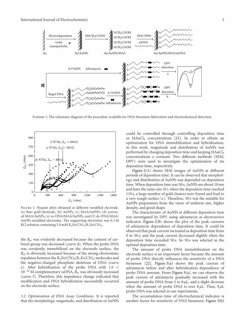
The DNA hybridization was assessed with the DPV peakcurrent of intercalated adriamycin in pH 7.0 PBS, and theconcentration of complementary DNA was quantified bypeak current of adriamycin (ΔI), which was subtractedfrom the reduction peak current generated at the ssDNA/MAA/AuNPs modified electrode (ΔI = Ids-DNA − Iss-DNA).DPV parameters were as follows: potential range: −0.45∼−0.80 V; amplitude: 0.05 V; pulse width: 0.05 s; samplewidth: 0.0167 s; pulse period: 0.2 s; quiet time: 2 s. Theschematic diagram of the DNA biosensor fabrication andelectrochemical detection is illustrated in Scheme 1.
3. Results and Discussion
3.1. Electrochemical Impedance Spectroscopy at DifferentModified Electrode. Impedance spectroscopy is an effectivemethod for probing the surface features of the modifiedelectrode. In EIS, the semicircle part at higher frequenciescorresponds to the electron transfer limited process or theelectron transfer resistance (Ret). The linear sect at lower fre-quencies shows a controlled diffusion process. Figure 1 com-pares the Nyquist plots of 5.0 mM K4Fe(CN)6/K3Fe(CN)6
at the different modified electrodes. When AuNPs wereelectrodeposited on the surface of the gold electrode (curveb), the Ret was decreased in contrast to the bare gold electrode(curve a). The results could be rationalized by improvedelectron transfer kinetics of redox probe on AuNPs modifiedelectrode. The assembly of MAA monolayer on electrodesurface induced a larger Ret (curve c) as compared with thatof AuNPs electrode due to the electrostatic repulsion betweenthe negative MAA and K4Fe(CN)6/K3Fe(CN)6. When theMAA/AuNPs/Au surface was activated with EDC and NHS,
International Journal of Electrochemistry 3
Au
Electrodeposition
Gold nanoparticles
HSCH2COOH EDC/NHS
ssDNA
Adriamycin
DPV detection
aa
Target DNA
Adriamycin
-S
-S
-S
-S
DPV detection
b
b
-S
-S
-S
-S
Au/AuNPs Au/AuNPs/MAA/ssDNAAu/AuNPs/MAA
0.1%SDS
0.1%SDS
-S
-S
-S
-S
-S
-S
-S
-S
-SCH2COOH
-SCH2COOH
-SCH2COOH
-SCH2COOH
Scheme 1: The schematic diagram of the procedure available for DNA biosensor fabrication and electrochemical detection.
0 300 600 900 1200 1500 18000
150
300
450
600
750
900
f
e
d
c
b a
Zim
(oh
m)
Zre (ohm)
f: 97 Hz, Zre = 684Ω
d: 97 Hz, Zre = 406Ω
e: 97 Hz, Zre = 583Ω
Figure 1: Nyquist plots obtained at different modified electrode.(a) Bare gold electrode, (b) AuNPs, (c) MAA/AuNPs, (d) activat-ed MAA/AuNPs, (e) ss-DNA/MAA/AuNPs, and (f) ds-DNA/MAA/AuNPs modified electrodes. The supporting electrolyte was 0.1 MKCl solution containing 5.0 mM K4Fe(CN)6/K3Fe(CN)6.
the Ret was evidently decreased because the content of car-boxyl group was decreased (curve d). When the probe DNAwas covalently immobilized on the electrode surface, theRet is obviously increased because of the strong electrostaticrepulsion between the K4Fe(CN)6/K3Fe(CN)6 molecules andthe negative-charged phosphate skeletons of DNA (curvee). After hybridization of the probe DNA with 1.0 ×10⁻¹⁰ M complementary ssDNA, Ret was obviously increased(curve f). Therefore, this impedance change indicated thatmodification and DNA hybridization successfully occurredon the electrode surface.
3.2. Optimization of DNA Assay Conditions. It is reportedthat the morphology, magnitude, and distribution of AuNPs
could be controlled through controlling deposition timeor HAuCl4 concentrations [21]. In order to obtain anoptimization for DNA immobilization and hybridization,in this work, magnitude and distribution of AuNPs wasperformed by changing deposition time and keeping HAuCl4concentrations a constant. Two different methods (SEM,DPV) were used to investigate the optimization of Audeposition time, respectively.
Figure 2(A) shows SEM images of AuNPs at differentperiods of deposition time. It can be observed that morphol-ogy and distribution of AuNPs was depended on depositiontime. When deposition time was 50 s, AuNPs are about 10 nmand have the same size (b), when the deposition time reached110 s, a large number of gold clusters were found and lead toa very rough surface (c). Therefore, 50 s was the suitable forAuNPs preparation from the views of uniform size, higherdensity, and good shape.
The characteristic of AuNPs at different deposition timewas investigated by DPV using adriamycin as electroactiveindicator. Figure 2(B) shows the plot of the peak currentsof adriamycin dependence of deposition time. It could beobserved that peak current increased as deposition time from0 to 50 s, and the peak current decreased slightly when thedeposition time exceeded 50 s. So 50 s was selected as theoptimal deposition time.
The amount of probe DNA immobilization on theelectrode surface is an important factor because the amountof probe DNA directly influences the sensitivity of a DNAbiosensor [22]. Figure 3(a) shows the peak current ofadriamycin before and after hybridization dependence ofprobe DNA amount. From Figure 3(a), we can observe thepeak current of adriamycin gradually increased with theamount of probe DNA from 3 to 8 µL, and a slight decreasewhen the amount of probe DNA is over 8 µL. Thus, 8 µLprobe DNA was selected in our experiments.
The accumulation time of electrochemical indicator isanother factor for sensitivity of DNA biosensor. Figure 3(b)

4 International Journal of Electrochemistry
(a)
(b)
(c)
(A)
100 nm
100 nm
100 nm
0 10 20 30 40 50 60 70−2
−3
−4
−5
−6
−7
−8
−9
×10−7
Cu
rren
t(A
)
Time (s)
(B)
Figure 2: (A) SEM images of AuNPs at various deposition time. (a) 0, (b) 50 s, (c) 110 s, Deposition potential is −250 mV (versus SCE). (B)DPV responses of adriamycin dependence of deposition time. Electrochemical deposition was performed in 3.0 mM HAuCl4 at various times(t = 0, 8, 16, 24, 32, 40, 48, 56, and 64 s). DPV measurement was performed in 0.01 M PBS (pH 7.40) containing 1.0 × 10⁻⁶ M adriamycin.DPV parameters were as follows: potential range: −0.45∼−0.80 V; amplitude: 0.05 V; pulse width: 0.05 s; sample width: 0.0167 s; pulseperiod: 0.2 s; quiet time: 2 s.
shows the reduction peak currents of adriamycin dependenceof the accumulation time. It could be seen that the peak cur-rent increased significantly with the increasing accumulationtime from 5 to 20 min. When the accumulation time is higherthan 20 min, the peak current of adriamycin keeps constantrelatively. Therefore, 20 min was selected as the optimumaccumulation time.
Figure 3(c) shows the peak current of adriamycin depen-dence of the hybridization time. From Figure 3(c), it couldbe seen that the reduction peak currents of adriamycinincreased significantly as the hybridization time increasedfrom 5 to 25 min and keep constant after 25 min. This indi-cated that the hybridization reaction was completed after25 min. From a view of the sensitivity and assay time, 25 minwas generally used for hybridization time.
3.3. Amplification of AuNPs. Could the application of thegold nanoparticles enhance the immobilization capacity ofprobe DNA and amplify signal of DNA hybridization? In thispaper, we fabricated two different sensors: one is a sensor
containing AuNPs, and another containing no AuNPs. Theelectrochemical investigation was performed as experimentSection 2.4, and the results were shown in Figure 4. It couldbe observed that the sensor containing AuNPs has a largesignal (Figure 4(A): 10.16 × 10⁻⁷ A) compared with thesensor containing no AuNPs (Figure 4(B): 2.29 × 10⁻⁷ A).These results suggested that the immobilization capacity ofprobe DNA and hybridization signal improved greatly in thepresence of AuNPs.
3.4. Selectivity of the DNA Biosensor. Differentiation of singlemismatches is of significant interest for a variety of impor-tant applications. In this paper, the selectivity of the DNAbiosensor was also evaluated by using single-base mismatch,no complementary and complementary DNA sequences.As is shown in Figure 5, it could be observed that theresponse of single-base mismatch sequence has a significantdifference with that of complementary sequence. Theseresults indicated that this DNA biosensor can distinguishsingle-base mismatch DNA sequence.

International Journal of Electrochemistry 5
2 3 4 5 6 7 8 9 10 11
−5
−6
−7
−8
−9
−10
−11
ΔI
(A)
×10−7
V (µL)
(a)
×10−7
Cu
rren
t(A
)
5 10 15 20 25
−4
−6
−8
−10
−12
−14
Time (min)
(b)
×10−7
Cu
rren
t(A
)
−4
−6
−8
−10
−12
−14
5 10 15 20 25
Time (min)
30−2
(c)
Figure 3: (a) Peak current of adriamycin (ΔI) dependence of amounts of probe DNA. The concentration of complementary target DNA is1.0 × 10−10 M; DPV parameters were the same as in Figure 2(B). (b) Effect of accumulation time on the DPV signals of adriamycin. Theconcentration of adriamycin is 1.0 × 10⁻⁶ M; DPV parameters were the same as in Figure 2(B). (c) DPV signals of adriamycin dependenceon DNA hybridization time. The concentration of adriamycin is 1.0 × 10⁻⁶ M; the concentration of complementary target DNA is 1.0 ×10⁻¹⁰ M. DPV parameters were the same as in Figure 2(B).
3.5. Analytical Performance. Under the optimal conditions,the analytical performance of the DNA biosensor wasinvestigated using the immobilized probe DNA to hybridizewith the different concentrations of the complementarysequence. Figure 6 shows the DPVs records of intercalatedadriamycin at various complementary oligonucleotides con-centrations. It could be observed that the peak currents ofadriamycin increased with increasing the concentration ofcomplementary DNA, and the peak currents difference (ΔI)was linear with the logarithmic value of the concentrationof the complementary DNA in the range from 5.0 × 10⁻¹³
to 1.0 × 10⁻⁹ M. The regression equation was ΔI (10−7 A)= 3.5518 logCDNA + 45.3301 (unit of C is M), and the
regression coefficient (R) of the linear curve was 0.9979. Thedetection limit is 1.7 × 10⁻¹³ M (S/N = 3). Compared withreported sensors [23–25], this DNA biosensor showed highersensitivity.
3.6. Reproducibility, Reusability, and Stability of the DNABiosensor. The reproducibility of biosensor is a very impor-tant factor for their application. Four DNA sensors werefabricated independently under the same conditions andused to detect 1.0 × 10⁻¹⁰ M complementary DNA. Andthe reduction peak currents of adriamycin were 1.217 ×10⁻⁶ A, 1.258 × 10⁻⁶ A, 1.259 × 10⁻⁶ A, and 1.227 × 10⁻⁶ A,

6 International Journal of Electrochemistry
−0.8 −0.7 −0.6 −0.5−24
−21
−18
−15
−12
−9
−6
−3
Cu
rren
t(A
)
Potential (V)
a
b
×10−7
ΔI(1)
(A)
ΔI(2)
−24
−21
−18
−15
−12
−9
−6
−3
Cu
rren
t(A
)
×10−7
−0.8 −0.7 −0.6 −0.5
Potential (V)
c
d
(B)
Figure 4: (A) DPV responses obtained for ss-DNA/MAA/AuNPs/Au (a), hybridized with 1.0 × 10⁻¹⁰ M complementary DNA (b). (B)DPV responses obtained for the ss-DNA/MAA/Au (c), hybridized with 1.0 × 10⁻¹⁰ M complementary DNA (d). Experiment condition:accumulation time of adriamycin: 20 min; hybridization time: 25 min. DPV parameters were the same as in Figure 2(B).
A B C0
−2
−4
−6
−8
−10
ΔI
(A)
×10−7
−0.8 −0.7 −0.6 −0.5−24−21−18−15−12−9−6−3
Cu
rren
t(A
)
Potential (V)
×10−7
a
bc
d
Figure 5: The histograms of the peak current difference (ΔI) ofadriamycin versus different DNA sequences (1.0 × 10⁻¹⁰ M): (A)noncomplementary sequence, (B) single-base mismatch sequence,(C) complementary sequence. Inset: DPV responses of adriamycinrecorded at probe modified electrode (a) and after hybridizationwith 1.0× 10⁻¹⁰ M noncomplementary sequence (b); 1.0× 10⁻¹⁰ Msingle-base mismatch sequence (c); 1.0 × 10⁻¹⁰ M complementarysequence (d). Accumulation time of adriamycin: 20 min; Hybridiza-tion time: 25 min. DPV parameters were the same as in Figure 2(B).
respectively. The average value was 1.2402× 10⁻⁶ A, and therelative standard deviation (RSD) was 2.15%.
The reusability of the DNA biosensor was investigatedby immersing hybridized electrodes in hot water (80◦C) for5–10 min to make ds-DNA into ss-DNA via thermal denat-uration. The renew sensor was used to test 1.0 × 10⁻¹⁰ M
ab
c
d
e
f
g
−0.8 −0.75 −0.7 −0.65 −0.6 −0.55 −0.5 −0.45
−24
−22
−20
−18
−16
−14
−12
−10
−8
−6
−4
×10−7
Cu
rren
t(A
)
Potential (V)
−12.75 −11.25 −9.750
−2−4−6−8−10−12−14
ΔI
(A)
log(CDNA)
×10−7
Figure 6: DPV response of the intercalated adriamycin recordedfor the probe DNA modified electrode that hybridized with variousconcentrations of complementary DNA: (a) 0 M; (b) 5.0× 10⁻¹³ M;(c) 1.0 × 10⁻¹² M; (d) 5.0 × 10⁻¹² M; (e) 1.0 × 10⁻¹¹ M; (f)1.0 × 10⁻¹⁰ M; (g) 1.0 × 10⁻⁹ M. Inset: Peak current (ΔI) ofthe intercalated adriamycin versus logarithm of concentration ofcomplementary DNA. Error bars show the standard deviationsof measurements taken from three independent experiments.Accumulation time of adriamycin: 20 min; hybridization time:25 min. The DPV parameters were the same as in Figure 2(B).
complementary DNA and found that the fifth regeneratedsensor has 84.7% response of the initial sensor (initial: 1.24× 10⁻⁶ A, final: 1.05 × 10⁻⁶ A). This result indicated theproposed DNA biosensor had a good reusability.

International Journal of Electrochemistry 7
The stability of the biosensor was also examined. TheDNA sensor was stored in a freezer at 4◦C for two weeks, andno apparent change about the peak current of adriamycinwas observed.
4. Conclusions
A simple and sensitive electrochemical DNA biosensor basedon ss-DNA/MAA/AuNPs modified electrode was fabricated.AuNPs could enhance the immobilization capacity of probeDNA and amplify electrochemical signal. This DNA biosen-sor exhibited excellent selectivity and stability.
Acknowledgment
The authors gratefully acknowledge the National Nature Sci-ence Foundation of China (no. 20675002), which financiallysupported this work.
References
[1] F. Patolsky, G. Zheng, and C. M. Lieber, “Nanowire sensors formedicine and the life sciences,” Nanomedicine, vol. 1, no. 1, pp.51–65, 2006.
[2] D. Ivnitski, D. J. O’Neil, A. Gattuso, R. Schlicht, M. Calidonna,and R. Fisher, “Nucleic acid approaches for detection andidentification of biological warfare and infectious diseaseagents,” BioTechniques, vol. 35, no. 4, pp. 862–869, 2003.
[3] F. Lucarelli, I. Palchetti, G. Marrazza, and M. Mascini,“Electrochemical DNA biosensor as a screening tool for thedetection of toxicants in water and wastewater samples,”Talanta, vol. 56, no. 5, pp. 949–957, 2002.
[4] V. Benoit, A. Steel, M. Torres, Y. Y. Yu, H. Yang, and J.Cooper, “Evaluation of three-dimensional microchannel glassbiochips for multiplexed nucleic acid fluorescence hybridiza-tion assays,” Analytical Chemistry, vol. 73, no. 11, pp. 2412–2420, 2001.
[5] R. Y. Wang, M. N. Ji, R. Wang, and J. Shi, “Stopped-flowkinetic fluorimetric studies of the interaction of Ru(II) com-plex with DNA and its analytical application,” SpectrochimicaActa Part A, vol. 71, no. 3, pp. 1042–1048, 2008.
[6] Q. Wang, X. Yang, and K. Wang, “Enhanced surface plasmonresonance for detection of DNA hybridization based on layer-by-layer assembly films,” Sensors and Actuators B, vol. 123, no.1, pp. 227–232, 2007.
[7] X. Yang, Q. Wang, K. Wang, W. Tan, and H. Li, “Enhancedsurface plasmon resonance with the modified catalytic growthof Au nanoparticles,” Biosensors and Bioelectronics, vol. 22, no.6, pp. 1106–1110, 2007.
[8] J. Zhang, R. Lao, S. Song, Z. Yan, and C. Fan, “Designof an oligonucleotide-incorporated nonfouling surface andits application in electrochemical DNA sensors for highlysensitive and sequence-specific detection of target DNA,”Analytical Chemistry, vol. 80, no. 23, pp. 9029–9033, 2008.
[9] C. Ding, F. Zhao, M. Zhang, and S. Zhang, “Hybridizationbiosensor using 2,9-dimethyl-1,10-phenantroline cobalt aselectrochemical indicator for detection of hepatitis B virusDNA,” Bioelectrochemistry, vol. 72, no. 1, pp. 28–33, 2008.
[10] M. Li, S. Huang, P. Zhu, L. Kong, B. Peng, and H.Gao, “A novel DNA biosensor based on ssDNA/Cyt c/l-Cys/GNPs/Chits/GCE,” Electrochimica Acta, vol. 54, no. 8, pp.2284–2289, 2009.
[11] T. G. Drummond, M. G. Hill, and J. K. Barton, “Electrochem-ical DNA sensors,” Nature Biotechnology, vol. 21, no. 10, pp.1192–1199, 2003.
[12] J. Zhang, S. Song, L. Zhang et al., “Sequence-specific detectionof femtomolar DNA via a chronocoulometric DNA sensor(CDS): effects of nanoparticle-mediated amplification andnanoscale control of DNA assembly at electrodes,” Journal ofthe American Chemical Society, vol. 128, no. 26, pp. 8575–8580,2006.
[13] H. Zhong, X. Lei, X. Hun, and S. Zhang, “Design of one-to-one recognition triple Au nanoparticles DNA probe and itsapplication in the electrochemical DNA biosensor,” ChemicalCommunications, no. 45, pp. 6958–6960, 2009.
[14] S. Zhang, J. Xia, and X. Li, “Electrochemical biosensor fordetection of adenosine based on structure-switching aptamerand amplification with reporter probe DNA modified Aunanoparticles,” Analytical Chemistry, vol. 80, no. 22, pp. 8382–8388, 2008.
[15] J. Wang, J. Li, A. J. Baca et al., “Amplified voltammetricdetection of DNA hybridization via oxidation of ferrocenecaps on gold nanoparticle/streptavidin conjugates,” AnalyticalChemistry, vol. 75, no. 15, pp. 3941–3945, 2003.
[16] M. H. Abouzar, A. Poghossian, A. M. Pedraza et al., “An arrayof field-effect nanoplate SOI capacitors for (bio-)chemicalsensing,” Biosensors and Bioelectronics, vol. 26, no. 6, pp. 3023–3028, 2011.
[17] K. Zhang, H. Ma, L. Zhang, and Y. Zhang, “Fabrication of asensitive impedance biosensor of DNA hybridization based ongold nanoparticles modified gold electrode,” Electroanalysis,vol. 20, no. 19, pp. 2127–2133, 2008.
[18] H. Ma, L. Zhang, Y. Pan, K. Zhang, and Y. Zhang, “A novelelectrochemical DNA biosensor fabricated with layer-by-layercovalent attachment of multiwalled carbon nanotubes andgold nanoparticles,” Electroanalysis, vol. 20, no. 11, pp. 1220–1226, 2008.
[19] Y. Zhang, H. Ma, K. Zhang, S. Zhang, and J. Wang, “Animproved DNA biosensor built by layer-by-layer covalentattachment of multi-walled carbon nanotubes and goldnanoparticles,” Electrochimica Acta, vol. 54, no. 8, pp. 2385–2391, 2009.
[20] M. T. Castaneda, S. Alegret, and A. Merkoci, “Electrochemicalsensing of DNA using gold nanoparticles,” Electroanalysis, vol.19, no. 7-8, pp. 743–753, 2007.
[21] X. Zhang, F. Shi, X. Yu et al., “Polyelectrolyte multilayer asmatrix for electrochemical deposition of gold clusters: towardsuper-hydrophobic surface,” Journal of the American ChemicalSociety, vol. 126, no. 10, pp. 3064–3065, 2004.
[22] A. W. Peterson, R. J. Heaton, and R. M. Georgiadis, “The effectof surface probe density on DNA hybridization,” Nucleic AcidsResearch, vol. 29, no. 24, pp. 5163–5168, 2001.
[23] S. F. Liu, Y. F. Li, J. R. Li, and L. Jiang, “Enhancement of DNAimmobilization and hybridization on gold electrode modifiedby nanogold aggregates,” Biosensors and Bioelectronics, vol. 21,no. 5, pp. 789–795, 2005.
[24] H. Cai, C. Xu, P. He, and Y. Fang, “Colloid Au-enhanced DNAimmobilization for the electrochemical detection of sequence-specific DNA,” Journal of Electroanalytical Chemistry, vol. 510,no. 1-2, pp. 78–85, 2001.
[25] J. Kang, X. Li, G. Wu, Z. Wang, and X. Lu, “A new schemeof hybridization based on the Aunano-DNA modified glassycarbon electrode,” Analytical Biochemistry, vol. 364, no. 2, pp.165–170, 2007.

Submit your manuscripts athttp://www.hindawi.com
Hindawi Publishing Corporationhttp://www.hindawi.com Volume 2014
Inorganic ChemistryInternational Journal of
Hindawi Publishing Corporation http://www.hindawi.com Volume 2014
International Journal ofPhotoenergy
Hindawi Publishing Corporationhttp://www.hindawi.com Volume 2014
Carbohydrate Chemistry
International Journal of
Hindawi Publishing Corporationhttp://www.hindawi.com Volume 2014
Journal of
Chemistry
Hindawi Publishing Corporationhttp://www.hindawi.com Volume 2014
Advances in
Physical Chemistry
Hindawi Publishing Corporationhttp://www.hindawi.com
Analytical Methods in Chemistry
Journal of
Volume 2014
Bioinorganic Chemistry and ApplicationsHindawi Publishing Corporationhttp://www.hindawi.com Volume 2014
SpectroscopyInternational Journal of
Hindawi Publishing Corporationhttp://www.hindawi.com Volume 2014
The Scientific World JournalHindawi Publishing Corporation http://www.hindawi.com Volume 2014
Medicinal ChemistryInternational Journal of
Hindawi Publishing Corporationhttp://www.hindawi.com Volume 2014
Chromatography Research International
Hindawi Publishing Corporationhttp://www.hindawi.com Volume 2014
Applied ChemistryJournal of
Hindawi Publishing Corporationhttp://www.hindawi.com Volume 2014
Hindawi Publishing Corporationhttp://www.hindawi.com Volume 2014
Theoretical ChemistryJournal of
Hindawi Publishing Corporationhttp://www.hindawi.com Volume 2014
Journal of
Spectroscopy
Analytical ChemistryInternational Journal of
Hindawi Publishing Corporationhttp://www.hindawi.com Volume 2014
Journal of
Hindawi Publishing Corporationhttp://www.hindawi.com Volume 2014
Quantum Chemistry
Hindawi Publishing Corporationhttp://www.hindawi.com Volume 2014
Organic Chemistry International
ElectrochemistryInternational Journal of
Hindawi Publishing Corporation http://www.hindawi.com Volume 2014
Hindawi Publishing Corporationhttp://www.hindawi.com Volume 2014
CatalystsJournal of
Related Documents