Electrochimica Acta 85 (2012) 588–593 Contents lists available at SciVerse ScienceDirect Electrochimica Acta jou rn al h om epa ge: www.elsevier.com/locate/electacta Electrochemical investigation of interactions between quinone derivatives and single stranded DNA Qidong Zhang a , Benoît Piro a , Sophia Ramsay b , Vincent Noël a , Steeve Reisberg a , Minh-Chau Pham a,∗ a Univ. Paris Diderot, Sorbonne Paris Cité, ITODYS, UMR 7086, CNRS, F-75205 Paris, France b Bangor University, Bangor, Gwynedd LL57 2DG, United Kingdom a r t i c l e i n f o Article history: Received 7 May 2012 Received in revised form 4 July 2012 Accepted 5 August 2012 Available online 6 September 2012 Keywords: Quinone redox marker Nucleobases DNA Interactions with DNA Hydrogen bonds Cyclic voltammetry a b s t r a c t The interactions of 5-hydroxy-1,4-naphthoquinone with three nucleobases (thymine, cytosine, ade- nine) and one nucleoside (guanosine), investigated by cyclic voltammetry in aprotic solvent, showed significant change in the redox behavior of the quinone group. Then, the interactions between 5- hydroxy-1,4-naphthoquinone and single-stranded oligonucleotides were studied in phosphate buffer saline solution using a random sequence and homo-oligonucleotides (polyA 20 , polyT 20 , polyC 20 and polyG 20 ). Finally, the interactions of 1,4-benzoquinone and 1,4-naphthoquinone were studied to compare with 5-hydroxy-1,4-naphthoquinone and to propose different interaction modes. The results help to elu- cidate the transduction mechanism involved in label-free quinone-based electrochemical DNA sensors. © 2012 Elsevier Ltd. All rights reserved. 1. Introduction Quinones, present in several living organisms, participate to var- ious biological processes. For instance, their redox system allows them to work as electron carriers to transport electrons between macromolecular complexes embedded in the membrane of the mitochondria in eukaryotic cells. Quinones can also interact with DNA, which has been demonstrated in many works. Some quinones can interact with nucleobases from DNA and inhibit replication [1], or bind to double-stranded DNA (dsDNA) by intercalation [2,3] or groove binding [4]. These characteristics make quinoid compounds attractive for anti-cancer drugs [5,6]. Besides, redox compounds which can interact with single or double stranded DNA can be used as electrochemical hybridization indicators in DNA sensors [7]. Such sensors take advantage of interactions between the target in solution, the recognition layer and a redox indicator, whether present in solution or immobilized on the sensor surface. Among these redox indicators, quinone derivatives have been investigated, mostly anthraquinones [8–11]. In our group, a quinone deriva- tive, 5-hydroxy-1,4-naphthoquinone (commonly called juglone), has been advantageously used as transducer for direct and label- free electrochemical DNA detection [12–15]. In some cases [14,15], the transduction step was achieved by monitoring the change in ∗ Corresponding author. Tel.: +33 0157277223. E-mail address: [email protected] (M.-C. Pham). interactions between oligonucleotides (ODN) and juglone. In these models, ODN probes and juglone were grafted together within a thin organic layer. The conformational change of ODN probes upon hybridization led to variation of interactions with juglone, which is finally expressed by change in juglone electroactivity. ODN/juglone interactions were assumed to be either hydrogen bonding (for DNA, the capability of hydrogen bonding comes mainly from nucle- obases) or local pH changes (ODN have acidic character). In the literature, the interaction mechanisms between nucleobases and quinone derivatives have been already studied by physical [16–18] and electrochemical methods [7,19,20]. However, no work relative to the influence of ODN strand on juglone electroactivity in aque- ous medium has been done yet, which can help to elucidate the transduction mechanisms involved in such juglone-based sensors. This is the aim of this work. We have first studied the interactions between nucleobases and juglone in aprotic solvent (DMF), using cyclic voltammetry. Significant variations of juglone electroactivity were observed in the presence of nucleobases. It is also shown that these changes depend on the type of nucleobase (G, C, T or A). Then the interac- tions between juglone and a single-stranded ODN (ssODN) have been studied in phosphate buffer saline solution (PBS). Obvi- ous changes of both peak current (I p ) and peak potential (E p ) of juglone were observed and interpreted. Finally, we investigated the interactions between ssODN and two other quinone deriva- tives (1,4-benzoquinone and 1,4-naphthoquinone) in PBS, in order to compare with juglone. 0013-4686/$ – see front matter © 2012 Elsevier Ltd. All rights reserved. http://dx.doi.org/10.1016/j.electacta.2012.08.017

Welcome message from author
This document is posted to help you gain knowledge. Please leave a comment to let me know what you think about it! Share it to your friends and learn new things together.
Transcript
Es
Qa
b
a
ARRAA
KQNDIHC
1
itmmDcogawu[iptmthft
0h
Electrochimica Acta 85 (2012) 588– 593
Contents lists available at SciVerse ScienceDirect
Electrochimica Acta
jou rn al h om epa ge: www.elsev ier .com/ locate /e lec tac ta
lectrochemical investigation of interactions between quinone derivatives andingle stranded DNA
idong Zhanga, Benoît Piroa, Sophia Ramsayb, Vincent Noëla, Steeve Reisberga, Minh-Chau Phama,∗
Univ. Paris Diderot, Sorbonne Paris Cité, ITODYS, UMR 7086, CNRS, F-75205 Paris, FranceBangor University, Bangor, Gwynedd LL57 2DG, United Kingdom
r t i c l e i n f o
rticle history:eceived 7 May 2012eceived in revised form 4 July 2012ccepted 5 August 2012vailable online 6 September 2012
a b s t r a c t
The interactions of 5-hydroxy-1,4-naphthoquinone with three nucleobases (thymine, cytosine, ade-nine) and one nucleoside (guanosine), investigated by cyclic voltammetry in aprotic solvent, showedsignificant change in the redox behavior of the quinone group. Then, the interactions between 5-hydroxy-1,4-naphthoquinone and single-stranded oligonucleotides were studied in phosphate buffersaline solution using a random sequence and homo-oligonucleotides (polyA20, polyT20, polyC20 andpolyG20). Finally, the interactions of 1,4-benzoquinone and 1,4-naphthoquinone were studied to compare
eywords:uinone redox markerucleobasesNA
nteractions with DNAydrogen bondsyclic voltammetry
with 5-hydroxy-1,4-naphthoquinone and to propose different interaction modes. The results help to elu-cidate the transduction mechanism involved in label-free quinone-based electrochemical DNA sensors.
© 2012 Elsevier Ltd. All rights reserved.
. Introduction
Quinones, present in several living organisms, participate to var-ous biological processes. For instance, their redox system allowshem to work as electron carriers to transport electrons between
acromolecular complexes embedded in the membrane of theitochondria in eukaryotic cells. Quinones can also interact withNA, which has been demonstrated in many works. Some quinonesan interact with nucleobases from DNA and inhibit replication [1],r bind to double-stranded DNA (dsDNA) by intercalation [2,3] orroove binding [4]. These characteristics make quinoid compoundsttractive for anti-cancer drugs [5,6]. Besides, redox compoundshich can interact with single or double stranded DNA can besed as electrochemical hybridization indicators in DNA sensors7]. Such sensors take advantage of interactions between the targetn solution, the recognition layer and a redox indicator, whetherresent in solution or immobilized on the sensor surface. Amonghese redox indicators, quinone derivatives have been investigated,
ostly anthraquinones [8–11]. In our group, a quinone deriva-ive, 5-hydroxy-1,4-naphthoquinone (commonly called juglone),
as been advantageously used as transducer for direct and label-ree electrochemical DNA detection [12–15]. In some cases [14,15],he transduction step was achieved by monitoring the change in
∗ Corresponding author. Tel.: +33 0157277223.E-mail address: [email protected] (M.-C. Pham).
013-4686/$ – see front matter © 2012 Elsevier Ltd. All rights reserved.ttp://dx.doi.org/10.1016/j.electacta.2012.08.017
interactions between oligonucleotides (ODN) and juglone. In thesemodels, ODN probes and juglone were grafted together within athin organic layer. The conformational change of ODN probes uponhybridization led to variation of interactions with juglone, which isfinally expressed by change in juglone electroactivity. ODN/jugloneinteractions were assumed to be either hydrogen bonding (forDNA, the capability of hydrogen bonding comes mainly from nucle-obases) or local pH changes (ODN have acidic character). In theliterature, the interaction mechanisms between nucleobases andquinone derivatives have been already studied by physical [16–18]and electrochemical methods [7,19,20]. However, no work relativeto the influence of ODN strand on juglone electroactivity in aque-ous medium has been done yet, which can help to elucidate thetransduction mechanisms involved in such juglone-based sensors.This is the aim of this work.
We have first studied the interactions between nucleobasesand juglone in aprotic solvent (DMF), using cyclic voltammetry.Significant variations of juglone electroactivity were observed inthe presence of nucleobases. It is also shown that these changesdepend on the type of nucleobase (G, C, T or A). Then the interac-tions between juglone and a single-stranded ODN (ssODN) havebeen studied in phosphate buffer saline solution (PBS). Obvi-ous changes of both peak current (Ip) and peak potential (Ep) of
juglone were observed and interpreted. Finally, we investigatedthe interactions between ssODN and two other quinone deriva-tives (1,4-benzoquinone and 1,4-naphthoquinone) in PBS, in orderto compare with juglone.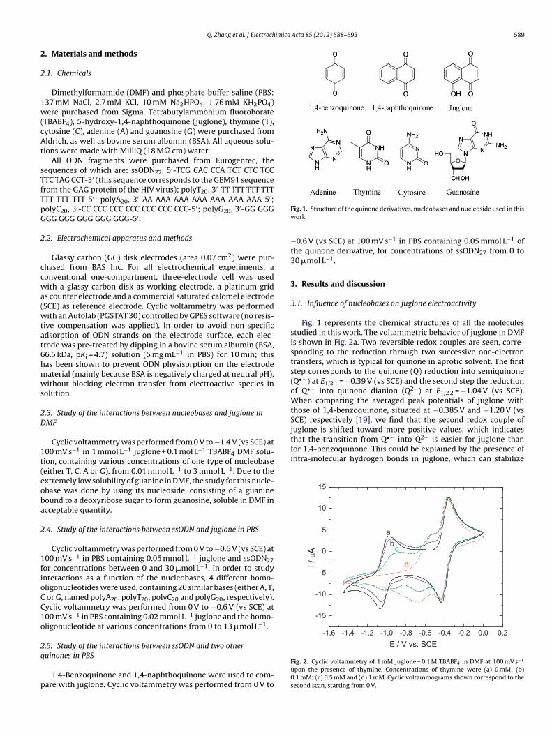
imica Acta 85 (2012) 588– 593 589
2
2
1w(cAt
sTfTpG
2
ccwa(wtat6hmws
2D
1t(eoba
2
1fioCC1o
2q
p
that the transition from Q•− into Q2− is easier for juglone thanfor 1,4-benzoquinone. This could be explained by the presence ofintra-molecular hydrogen bonds in juglone, which can stabilize
0,20,0-0,2-0,4-0,6-0,8-1,0-1,2-1,4-1,6
-15
-10
-5
0
5
10
15
d
cb
a
I /A
E / V vs. SCE
Q. Zhang et al. / Electroch
. Materials and methods
.1. Chemicals
Dimethylformamide (DMF) and phosphate buffer saline (PBS:37 mM NaCl, 2.7 mM KCl, 10 mM Na2HPO4, 1.76 mM KH2PO4)ere purchased from Sigma. Tetrabutylammonium fluoroborate
TBABF4), 5-hydroxy-1,4-naphthoquinone (juglone), thymine (T),ytosine (C), adenine (A) and guanosine (G) were purchased fromldrich, as well as bovine serum albumin (BSA). All aqueous solu-
ions were made with MilliQ (18 M� cm) water.All ODN fragments were purchased from Eurogentec, the
equences of which are: ssODN27, 5′-TCG CAC CCA TCT CTC TCCTC TAG CCT-3′ (this sequence corresponds to the GEM91 sequencerom the GAG protein of the HIV virus); polyT20, 3′-TT TTT TTT TTTTT TTT TTT-5′; polyA20, 3′-AA AAA AAA AAA AAA AAA AAA-5′;olyC20, 3′-CC CCC CCC CCC CCC CCC CCC-5′; polyG20, 3′-GG GGGGG GGG GGG GGG GGG-5′.
.2. Electrochemical apparatus and methods
Glassy carbon (GC) disk electrodes (area 0.07 cm2) were pur-hased from BAS Inc. For all electrochemical experiments, aonventional one-compartment, three-electrode cell was usedith a glassy carbon disk as working electrode, a platinum grid
s counter electrode and a commercial saturated calomel electrodeSCE) as reference electrode. Cyclic voltammetry was performedith an Autolab (PGSTAT 30) controlled by GPES software (no resis-
ive compensation was applied). In order to avoid non-specificdsorption of ODN strands on the electrode surface, each elec-rode was pre-treated by dipping in a bovine serum albumin (BSA,6.5 kDa, pKi = 4.7) solution (5 mg mL−1 in PBS) for 10 min; thisas been shown to prevent ODN physisorption on the electrodeaterial (mainly because BSA is negatively charged at neutral pH),ithout blocking electron transfer from electroactive species in
olution.
.3. Study of the interactions between nucleobases and juglone inMF
Cyclic voltammetry was performed from 0 V to −1.4 V (vs SCE) at00 mV s−1 in 1 mmol L−1 juglone + 0.1 mol L−1 TBABF4 DMF solu-ion, containing various concentrations of one type of nucleobaseeither T, C, A or G), from 0.01 mmol L−1 to 3 mmol L−1. Due to thextremely low solubility of guanine in DMF, the study for this nucle-base was done by using its nucleoside, consisting of a guanineound to a deoxyribose sugar to form guanosine, soluble in DMF incceptable quantity.
.4. Study of the interactions between ssODN and juglone in PBS
Cyclic voltammetry was performed from 0 V to −0.6 V (vs SCE) at00 mV s−1 in PBS containing 0.05 mmol L−1 juglone and ssODN27or concentrations between 0 and 30 �mol L−1. In order to studynteractions as a function of the nucleobases, 4 different homo-ligonucleotides were used, containing 20 similar bases (either A, T,
or G, named polyA20, polyT20, polyC20 and polyG20, respectively).yclic voltammetry was performed from 0 V to −0.6 V (vs SCE) at00 mV s−1 in PBS containing 0.02 mmol L−1 juglone and the homo-ligonucleotide at various concentrations from 0 to 13 �mol L−1.
.5. Study of the interactions between ssODN and two other
uinones in PBS1,4-Benzoquinone and 1,4-naphthoquinone were used to com-are with juglone. Cyclic voltammetry was performed from 0 V to
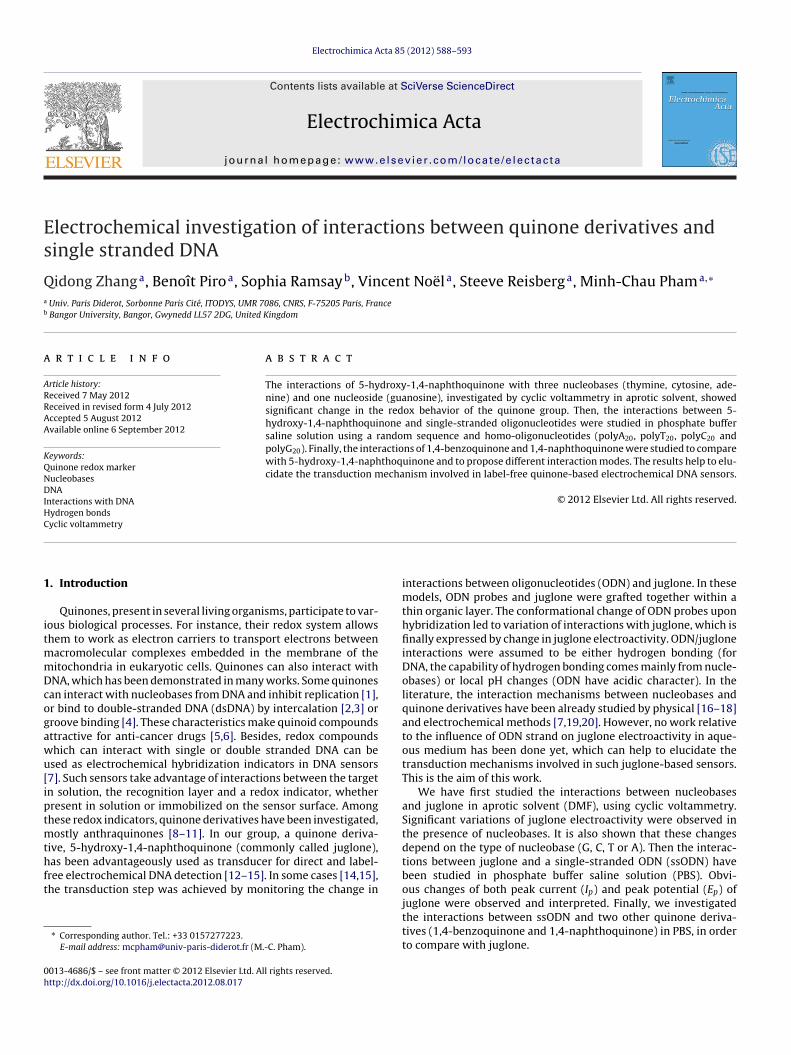
Fig. 1. Structure of the quinone derivatives, nucleobases and nucleoside used in thiswork.
−0.6 V (vs SCE) at 100 mV s−1 in PBS containing 0.05 mmol L−1 ofthe quinone derivative, for concentrations of ssODN27 from 0 to30 �mol L−1.
3. Results and discussion
3.1. Influence of nucleobases on juglone electroactivity
Fig. 1 represents the chemical structures of all the moleculesstudied in this work. The voltammetric behavior of juglone in DMFis shown in Fig. 2a. Two reversible redox couples are seen, corre-sponding to the reduction through two successive one-electrontransfers, which is typical for quinone in aprotic solvent. The firststep corresponds to the quinone (Q) reduction into semiquinone(Q•−) at E1/2 1 = −0.39 V (vs SCE) and the second step the reductionof Q•− into quinone dianion (Q2−) at E1/2 2 = −1.04 V (vs SCE).When comparing the averaged peak potentials of juglone withthose of 1,4-benzoquinone, situated at −0.385 V and −1.20 V (vsSCE) respectively [19], we find that the second redox couple ofjuglone is shifted toward more positive values, which indicates
Fig. 2. Cyclic voltammetry of 1 mM juglone + 0.1 M TBABF4 in DMF at 100 mV s−1
upon the presence of thymine. Concentrations of thymine were (a) 0 mM; (b)0.1 mM; (c) 0.5 mM and (d) 1 mM. Cyclic voltammograms shown correspond to thesecond scan, starting from 0 V.
5 imica Acta 85 (2012) 588– 593
tm
crc−a
rwtlaStilbtnHn
a1tvrorctfETtfb
3
sstiooha
3
Dwtbatppsms
27As shown, interactions with the oxidized form of juglone are
predominant. This suggests that interactions with nucleobasesmake the oxidized form of juglone more stable, so that more energy
Table 1K1/K2 values as a function of ssODN27 concentration. K1 and K2 are the bindingconstants for the oxidized and reduced form of juglone, respectively.
CssODN27 (mM) K1/K2
90 Q. Zhang et al. / Electroch
he reduced species and delocalize the negative charges on theolecule [21,22].The variation of juglone electroactivity as a function of thymine
oncentration is shown in Fig. 2, curves b–d. One can see that theedox couple located at −0.39 V does not vary with thymine con-entration. However, the second redox couple, initially situated at1.04 V, decreases and disappears while a redox couple appearsnd increases at about −0.6 V.
The stability of the first redox couple indicates that the firsteduction step is not influenced by the presence of thymine,hereas the second reduction step from Q•− into Q2− is sensi-
ive to thymine. Interaction with thymine makes the semiquinoneess negative and, eventually, more easily reduced, explaining why
redox couple appears at more positive potentials (−0.6 V vsCE). According to the study of Salas et al. [19] on the interac-ions between thymine derivatives and 1,4-benzoquinone, the QH−
ntermediate is not stable when the concentration of thymine isow, but it can be stabilized by hydrogen bonding interactions. Thisehavior can also indicate that a strong interaction exists betweenhymine and the Q2− dianion. This could explain why we obtainew redox peaks only for a thymine/juglone ratio greater than 2.owever, this behavior can also be explained by a local pH change,ucleobases having an acidic character.
The same experiment has been done for cytosine, adeninend guanosine. Unlike the results obtained by Salas et al. for,4-benzoquinone, which demonstrated very different electroac-ivities with different nucleobases [19,20], herein we got changesery similar to those obtained with thymine. The first quinoneeduction was not significantly changed by addition of A, Gr C; however, the second redox couple decreases and a newedox couple appears between the two former redox ones withoncentration increase (data not shown). The averaged peak poten-ials corresponding to these new redox couples are differentrom one base to another (E1/2 JUG/T = −0.60 V; E1/2 JUG/A = −0.45 V;1/2 JUG/G = −0.41 V; E1/2 JUG/C = −0.81 V; all potentials stated vs SCE).his probably reflects the different hydrogen bonding behavior ofhese nucleobases. Although C is a better proton donor than T, weound a smaller potential change. This seems to indicate that theehavior is not only due to local pH change.
.2. Interactions between ssODN and juglone in PBS
The above results indicate that the juglone electroactivity istrongly influenced by nucleobases in aprotic solvent. In aqueousolution, these interactions are more difficult to characterize dueo the protic character of the solvent. To go further, we investigatednteractions in PBS between juglone and oligonucleotides insteadf individual bases. For this, we studied interactions with an hetero-ligonucleotide of 27 bases (ssODN27) then after, with four differentomo-oligonucleotides of 20 bases each (polyA20, polyT20, polyG20nd polyC20).
.2.1. Interactions with ssODN27In PBS, the redox behavior of juglone is different from that in
MF. It shows only one redox couple at E1/2 = −0.25 V (vs SCE),hich corresponds to a two-electron transfer in one step. Addi-
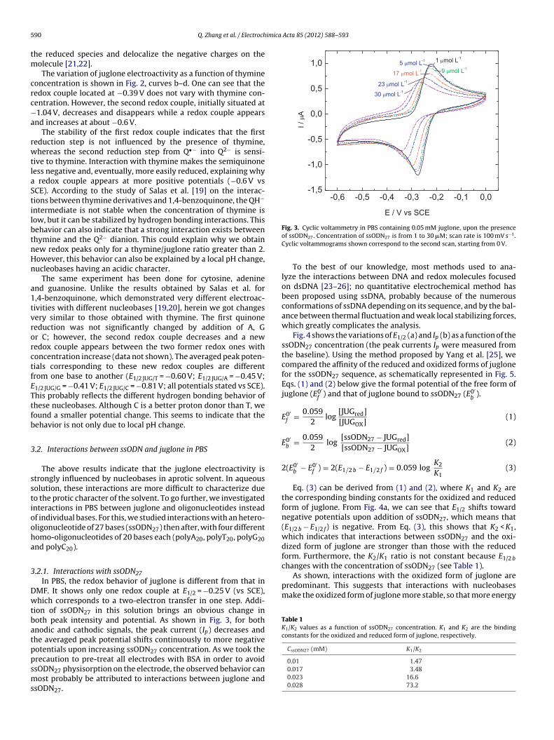
ion of ssODN27 in this solution brings an obvious change inoth peak intensity and potential. As shown in Fig. 3, for bothnodic and cathodic signals, the peak current (Ip) decreases andhe averaged peak potential shifts continuously to more negativeotentials upon increasing ssODN27 concentration. As we took the
recaution to pre-treat all electrodes with BSA in order to avoidsODN27 physisorption on the electrode, the observed behavior canost probably be attributed to interactions between juglone andsODN27.
Fig. 3. Cyclic voltammetry in PBS containing 0.05 mM juglone, upon the presenceof ssODN27. Concentration of ssODN27 is from 1 to 30 �M; scan rate is 100 mV s−1.Cyclic voltammograms shown correspond to the second scan, starting from 0 V.
To the best of our knowledge, most methods used to ana-lyze the interactions between DNA and redox molecules focusedon dsDNA [23–26]; no quantitative electrochemical method hasbeen proposed using ssDNA, probably because of the numerousconformations of ssDNA depending on its sequence, and by the bal-ance between thermal fluctuation and weak local stabilizing forces,which greatly complicates the analysis.
Fig. 4 shows the variations of E1/2 (a) and Ip (b) as a function of thessODN27 concentration (the peak currents Ip were measured fromthe baseline). Using the method proposed by Yang et al. [25], wecompared the affinity of the reduced and oxidized forms of juglonefor the ssODN27 sequence, as schematically represented in Fig. 5.Eqs. (1) and (2) below give the formal potential of the free form ofjuglone (E0′
f) and that of juglone bound to ssODN27 (E0′
b).
E0′f = 0.059
2log
[JUGred][JUGOX]
(1)
E0′b = 0.059
2log
[ssODN27 − JUGred][ssODN27 − JUGOX]
(2)
2(E0′b − E0′
f ) = 2(E1/2 b − E1/2 f ) = 0.059 logK2
K1(3)
Eq. (3) can be derived from (1) and (2), where K1 and K2 arethe corresponding binding constants for the oxidized and reducedform of juglone. From Fig. 4a, we can see that E1/2 shifts towardnegative potentials upon addition of ssODN27, which means that(E1/2 b − E1/2 f) is negative. From Eq. (3), this shows that K2 < K1,which indicates that interactions between ssODN27 and the oxi-dized form of juglone are stronger than those with the reducedform. Furthermore, the K2/K1 ratio is not constant because E1/2 bchanges with the concentration of ssODN (see Table 1).
0.01 1.470.017 3.480.023 16.60.028 73.2
Q. Zhang et al. / Electrochimica Acta 85 (2012) 588– 593 591
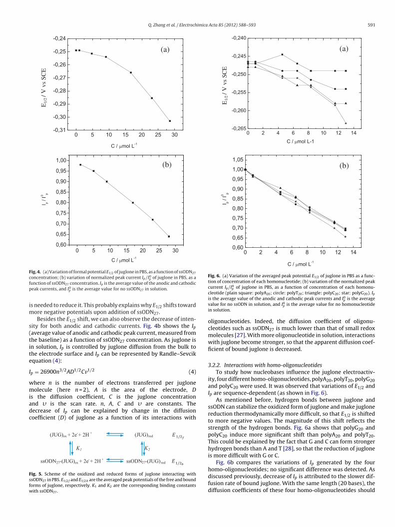
Fig. 4. (a) Variation of formal potential E1/2 of juglone in PBS, as a function of ssODN27
cfp
im
s(tite
I
wmiadc
Fsfw
Fig. 6. (a) Variation of the averaged peak potential E1/2 of juglone in PBS as a func-tion of concentration of each homonucleotide; (b) variation of the normalized peakcurrent Ip/I0
p of juglone in PBS, as a function of concentration of each homonu-
oncentration; (b) variation of normalized peak current Ip/I0p of juglone in PBS, as a
unction of ssODN27 concentration. Ip is the average value of the anodic and cathodiceak currents, and I0
p is the average value for no ssODN27 in solution.
s needed to reduce it. This probably explains why E1/2 shifts towardore negative potentials upon addition of ssODN27.Besides the E1/2 shift, we can also observe the decrease of inten-
ity for both anodic and cathodic currents. Fig. 4b shows the Ipaverage value of anodic and cathodic peak current, measured fromhe baseline) as a function of ssODN27 concentration. As juglone isn solution, Ip is controlled by juglone diffusion from the bulk tohe electrode surface and Ip can be represented by Randle–Sevcikquation (4):
p = 26900n3/2AD1/2Cv1/2 (4)
here n is the number of electrons transferred per jugloneolecule (here n = 2), A is the area of the electrode, D
s the diffusion coefficient, C is the juglone concentrationnd � is the scan rate. n, A, C and � are constants. Theecrease of Ip can be explained by change in the diffusionoefficient (D) of juglone as a function of its interactions with
ig. 5. Scheme of the oxidized and reduced forms of juglone interacting withsODN27 in PBS. E1/2 f and E1/2 b are the averaged peak potentials of the free and boundorms of juglone, respectively. K1 and K2 are the corresponding binding constantsith ssODN27.
cleotide (plain square: polyA20; circle: polyT20; triangle: polyC20; star: polyG20). Ipis the average value of the anodic and cathodic peak currents and I0
p is the averagevalue for no ssODN in solution, and I0
p is the average value for no homonucleotidein solution.
oligonucleotides. Indeed, the diffusion coefficient of oligonu-cleotides such as ssODN27 is much lower than that of small redoxmolecules [27]. With more oligonucleotide in solution, interactionswith juglone become stronger, so that the apparent diffusion coef-ficient of bound juglone is decreased.
3.2.2. Interactions with homo-oligonucleotidesTo study how nucleobases influence the juglone electroactiv-
ity, four different homo-oligonucleotides, polyA20, polyT20, polyG20and polyC20 were used. It was observed that variations of E1/2 andIp are sequence-dependent (as shown in Fig. 6).
As mentioned before, hydrogen bonds between juglone andssODN can stabilize the oxidized form of juglone and make juglonereduction thermodynamically more difficult, so that E1/2 is shiftedto more negative values. The magnitude of this shift reflects thestrength of the hydrogen bonds. Fig. 6a shows that polyG20 andpolyC20 induce more significant shift than polyA20 and polyT20.This could be explained by the fact that G and C can form strongerhydrogen bonds than A and T [28], so that the reduction of jugloneis more difficult with G or C.
Fig. 6b compares the variations of Ip generated by the four
homo-oligonucleotides; no significant difference was detected. Asdiscussed previously, decrease of Ip is attributed to the slower dif-fusion rate of bound juglone. With the same length (20 bases), thediffusion coefficients of these four homo-oligonucleotides should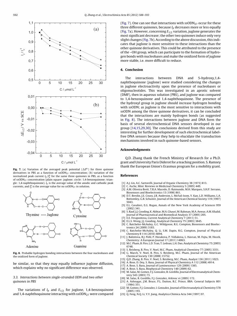
592 Q. Zhang et al. / Electrochimica
Fig. 7. (a) Variation of the averaged peak potential (�E0 ′) for three quinonederivatives in PBS as a function of ssODN27 concentration; (b) variation of thenormalized peak current Ip/I0
p for the same three quinones in PBS, as a functionof ssODN27 concentration (plain square: juglone; circle: 1,4-benzoquinone; trian-gle: 1,4-naphthoquinone). Ip is the average value of the anodic and cathodic peakcurrents, and I0
p is the average value for no ssODN27 in solution.
Ft
bw
3q
a
[
[
[
[[
[[[[[
[
ig. 8. Probable hydrogen bonding interactions between the four nucleobases andhe oxidized form of juglone.
e similar, so that they may equally influence juglone diffusion,hich explains why no significant difference was observed.
.3. Interactions between single-stranded ODN and two other
uinones in PBSThe variations of Ip and E1/2 for juglone, 1,4-benzoquinonend 1,4-naphthoquinone interacting with ssODN27 were compared
[
[
[
Acta 85 (2012) 588– 593
(Fig. 7). One can see that interactions with ssODN27 occur for thesethree different quinones, because Ip decreases more or less equally(Fig. 7a). However, concerning E1/2 variation, juglone generates themost significant decrease; the other two quinones induce only veryslight changes (Fig. 7b). According to the above discussion, this indi-cates that juglone is more sensitive to these interactions than theother quinone derivatives. This could be attributed to the presenceof the –OH group, which can participate to the formation of hydro-gen bonds with nucleobases and make the oxidized form of juglonemore stable, i.e. more difficult to reduce.
4. Conclusion
The interactions between DNA and 5-hydroxy,1,4-naphthoquinone (juglone) were studied considering the changesin juglone electroactivity upon the presence of nucleobases oroligonucleotides. This was investigated in an aprotic solvent(DMF), then in aqueous solution (PBS), and juglone was comparedto 1,4-benzoquinone and 1,4-naphthoquinone. The presence ofthe hydroxyl group in juglone should increase hydrogen bondingwith ssODN; as juglone is the most sensitive to interactions withssODN among the three quinone derivatives, it can be concludedthat the interactions are mainly hydrogen bonds (as suggestedin Fig. 8). The interactions between juglone and DNA form thebasis of several electrochemical DNA sensors developed in ourgroup [14,15,29,30]. The conclusions derived from this study areinteresting for further development of such electrochemical label-free DNA sensors because they help to elucidate the transductionmechanisms involved in such quinone-based sensors.
Acknowledgments
Q.D. Zhang thank the French Ministry of Research for a Ph.D.grant and University Paris Diderot for a teaching position. S. Ramseythank the European Union’s Erasmus program for a mobility grant.
References
[1] A.J. Lin, A.C. Sartorelli, Journal of Organic Chemistry 38 (1973) 813.[2] C. Asche, Mini: Reviews in Medicinal Chemistry 5 (2005) 449.[3] A.M. Oliveira Brett, T.R.A. Macedo, D. Raimundo, M.H. Marques, S.H.P. Serrano,
Biosensors and Bioelectronics 13 (1998) 861.[4] D.T. Breslin, J.E. Coury, J.R. Anderson, L. McFail-Isom, Y. Kan, L.D. Williams, L.A.
Bottomley, G.B. Schuster, Journal of the American Chemical Society 119 (1997)5043.
[5] E.L. Cavalieri, E.G. Rogan, Annals of the New York Academy of Sciences 959(2002) 341.
[6] S. Rauf, J.J. Gooding, K. Akhtar, M.A. Ghauri, M. Rahman, M.A. Anwar, A.M. Khalid,Journal of Pharmaceutical and Biomedical Analysis 37 (2005) 205.
[7] E.E. Ferapontova, Current Analytical Chemistry 7 (2011) 51.[8] E.L.S. Wong, J.J. Gooding, Analytical Chemistry 75 (2003) 3845.[9] C. Batchelor-McAuley, G.C. Wildgoose, R.G. Compton, Biosensors and Bioelec-
tronics 24 (2009) 3183.10] C. Batchelor-McAuley, Q. Li, S.M. Dapin, R.G. Compton, Journal of Physical
Chemistry B 114 (2010) 4094.11] J. Balintova, R.J. Pohl, P. Horakova, P. Vidlakova, L. Havran, M. Fojta, M. Hocek,
Chemistry: A European Journal 17 (2011) 14063.12] M.C. Pham, B. Piro, L.D. Tran, T. Ledoan, L.H. Dao, Analytical Chemistry 75 (2003)
6748.13] S. Reisberg, B. Piro, V. Noel, M.C. Pham, Analytical Chemistry 77 (2005) 3351.14] G. March, V. Noel, B. Piro, S. Reisberg, M.C. Pham, Journal of the American
Chemical Society 130 (2008) 15752.15] Q.D. Zhang, B. Piro, V. Noel, S. Reisberg, M.C. Pham, Analyst 136 (2011) 1023.16] A. Bose, D. Dey, S. Basu, Journal of Physical Chemistry A 112 (2008) 4914.17] A. Bose, S. Basu, Journal of Luminescence 129 (2009) 1385.18] A. Bose, S. Basu, Biophysical Chemistry 140 (2009) 62.19] M. Salas, M. Gomez, F.J. Gonzalez, B. Gordillo, Journal of Electroanalytical Chem-
istry 543 (2003) 73.20] M. Salas, B. Gordillo, F.J. Gonzalez, Arkivoc vi (2005) 172.
21] A. Ashnagar, J.M. Bruce, P.L. Dutton, R.C. Prince, BBA: General Subjects 801(1984) 351.22] M. Gomez, F.J. Gonzalez, I. Gonzalez, Journal of Electroanalytical Chemistry 578
(2005) 193.23] Q. Feng, N.Q. Li, Y.Y. Jiang, Analytica Chimica Acta 344 (1997) 97.

imica
[[
[[
[
Q. Zhang et al. / Electroch
24] Y. Wu, X. Ji, S. Hu, Bioelectrochemistry 64 (2004) 91.
25] Z.S. Yang, D.P. Zhang, H.Y. Long, G.C. Zhao, Electroanalysis 19 (2007)2577.26] H. Heli, S.Z. Bathaie, M.F. Mousavi, Electrochimica Acta 51 (2005) 1108.27] R.M. Robertson, Proceedings of the National Academy of Sciences of the United
States of America 103 (2006) 7310.
[
[
Acta 85 (2012) 588– 593 593
28] J. Grunenberg, Journal of the American Chemical Society 126 (2004) 16310.
29] G. March, S. Reisberg, B. Piro, M.C. Pham, C. Fave, V. Noel, Analytical Chemistry82 (2010) 3523.30] Q.D. Zhang, G. March, V. Noel, B. Piro, S. Reisberg, L.D. Tran, L.H. Hai, E. Aba-
dia, P.E. Nielsen, C. Sola, M.C. Pham, Biosensors and Bioelectronics 32 (2012)163.
Related Documents