Electrochemical Impedance Spectroscopy to Assess Vascular Oxidative Stress Fei Yu 1 , Rongsong Li 1 , Lisong Ai 1 , Collin Edington 1 , Hongyu Yu 2 , Mark Barr 3 , E. S. Kim 4 , and Tzung K. Hsiai 1 1 Department of Biomedical Engineering and Cardiovascular Medicine, University of Southern California, Los Angeles, CA 90089, USA 2 School of Earth & Space Exploration, Arizona State University, Tempe, AZ 85287-8406, USA 3 Department of Cardiothoracic Surgery, University of Southern California, Los Angeles, CA, USA 4 Department of Electrical Engineering and Electrophysics, University of Southern California, Los Angeles, CA, USA Abstract Vascular inflammatory responses are intimately linked with oxidative stress, favoring the development of pre-atherosclerotic lesions. We proposed that oxidized low density lipoprotein (oxLDL) and foam cell infiltrates in the subendothelial layer engendered distinct electrochemical properties that could be measured in terms of the electrochemical impedance spectroscopy (EIS). Concentric bipolar microelectrodes were applied to interrogate EIS of aortas isolated from fat-fed New Zealand White (NZW) rabbits and explants of human aortas. Frequency-dependent EIS measurements were assessed between 10 kHz and 100 kHz, and were significantly elevated in the pre-atherosclerotic lesions in which oxLDL and macrophage infiltrates were prevalent (At 100 kHz: aortic arch lesion = 26.7 ± 2.7 kΩ vs. control = 15.8 ± 2.4 kΩ; at 10 kHz: lesions = 49.2 ± 7.3 kΩ vs. control = 27.6 ± 2.7 kΩ, n = 10, p<0.001). Similarly, EIS measurements were significantly elevated in the human descending aorta where pre-atherosclerotic lesions or fatty streaks were prominent. EIS measurements remained unchanged in spite of various depths of electrode submersion or orientation of the specimens. Hence, the concentric bipolar microelectrodes provided a reliable means to measure endoluminal electrochemical modifications in regions of pro-inflammatory with high spatial resolution and reproducibility albeit uneven lesion topography and non-uniform current distribution. Keywords Electrochemical impedance spectroscopy; Oxidative stress; Atherosclerosis; oxLDL; Endothelium INTRODUCTION Oxidized low density lipoprotein (oxLDL) and macrophage infiltrates contribute to pro- inflammatory states relevant to the initiation of atherosclerotic lesions. 37 These lesions can be classified as stable or unstable plaque. The latter is often non-obstructive by conventional X-ray angiography or intravascular ultrasound (IVUS), and is prone to rupture, leading to © 2010 Biomedical Engineering Society Address correspondence to Tzung K. Hsiai, Department of Biomedical Engineering and Cardiovascular Medicine, University of Southern California, Los Angeles, CA 90089, USA. [email protected], [email protected]. NIH Public Access Author Manuscript Ann Biomed Eng. Author manuscript; available in PMC 2012 April 10. Published in final edited form as: Ann Biomed Eng. 2011 January ; 39(1): 287–296. doi:10.1007/s10439-010-0127-y. NIH-PA Author Manuscript NIH-PA Author Manuscript NIH-PA Author Manuscript

Welcome message from author
This document is posted to help you gain knowledge. Please leave a comment to let me know what you think about it! Share it to your friends and learn new things together.
Transcript
Electrochemical Impedance Spectroscopy to Assess VascularOxidative Stress
Fei Yu1, Rongsong Li1, Lisong Ai1, Collin Edington1, Hongyu Yu2, Mark Barr3, E. S. Kim4,and Tzung K. Hsiai11Department of Biomedical Engineering and Cardiovascular Medicine, University of SouthernCalifornia, Los Angeles, CA 90089, USA2School of Earth & Space Exploration, Arizona State University, Tempe, AZ 85287-8406, USA3Department of Cardiothoracic Surgery, University of Southern California, Los Angeles, CA, USA4Department of Electrical Engineering and Electrophysics, University of Southern California, LosAngeles, CA, USA
AbstractVascular inflammatory responses are intimately linked with oxidative stress, favoring thedevelopment of pre-atherosclerotic lesions. We proposed that oxidized low density lipoprotein(oxLDL) and foam cell infiltrates in the subendothelial layer engendered distinct electrochemicalproperties that could be measured in terms of the electrochemical impedance spectroscopy (EIS).Concentric bipolar microelectrodes were applied to interrogate EIS of aortas isolated from fat-fedNew Zealand White (NZW) rabbits and explants of human aortas. Frequency-dependent EISmeasurements were assessed between 10 kHz and 100 kHz, and were significantly elevated in thepre-atherosclerotic lesions in which oxLDL and macrophage infiltrates were prevalent (At 100kHz: aortic arch lesion = 26.7 ± 2.7 kΩ vs. control = 15.8 ± 2.4 kΩ; at 10 kHz: lesions = 49.2 ± 7.3kΩ vs. control = 27.6 ± 2.7 kΩ, n = 10, p<0.001). Similarly, EIS measurements were significantlyelevated in the human descending aorta where pre-atherosclerotic lesions or fatty streaks wereprominent. EIS measurements remained unchanged in spite of various depths of electrodesubmersion or orientation of the specimens. Hence, the concentric bipolar microelectrodesprovided a reliable means to measure endoluminal electrochemical modifications in regions ofpro-inflammatory with high spatial resolution and reproducibility albeit uneven lesion topographyand non-uniform current distribution.
KeywordsElectrochemical impedance spectroscopy; Oxidative stress; Atherosclerosis; oxLDL; Endothelium
INTRODUCTIONOxidized low density lipoprotein (oxLDL) and macrophage infiltrates contribute to pro-inflammatory states relevant to the initiation of atherosclerotic lesions.37 These lesions canbe classified as stable or unstable plaque. The latter is often non-obstructive by conventionalX-ray angiography or intravascular ultrasound (IVUS), and is prone to rupture, leading to
© 2010 Biomedical Engineering SocietyAddress correspondence to Tzung K. Hsiai, Department of Biomedical Engineering and Cardiovascular Medicine, University ofSouthern California, Los Angeles, CA 90089, USA. [email protected], [email protected].
NIH Public AccessAuthor ManuscriptAnn Biomed Eng. Author manuscript; available in PMC 2012 April 10.
Published in final edited form as:Ann Biomed Eng. 2011 January ; 39(1): 287–296. doi:10.1007/s10439-010-0127-y.
NIH
-PA Author Manuscript
NIH
-PA Author Manuscript
NIH
-PA Author Manuscript
acute coronary syndromes or stroke.2,11,21 Unstable plaque or otherwise known as thin-capfibroatheroma represents a transitional plaque that is characterized by a thin fibrous cap (<65μm), a large necrotic core, an abundance of macrophages, and limited luminal narrowing. 37
However, detection and diagnosing the non-obstructive, albeit pro-inflammatory, lesionsduring catheterization remains a clinical challenge.
Up to date, there are no diagnostic tools to detect intravascular oxidative stress and pro-inflammatory states when patients undergo angiograms. Serum biomarkers have beenproposed for predicting pro-inflammatory states. Examples include C-reactive protein,matrix metalloproteinases, CD40 ligand, Lp-PLA2 (lipoprotein-associated phospholipaseA2) and myelo-peroxidase. Specifically, circulating Lp-PLA2 is bound predominantly tolow-density lipoprotein (LDL) (~80%) which transmigrated to the subendothelial space ofatherosclerosis-prone regions.28 Imaging modalities including magnetic resonance imaging(MRI) and electron beam computed tomography (EBCT) have also been proposed to predictunstable plaque.7,29 However, assessing stable vs. unstable atherosclerotic lesions duringcatheterization remains an unmet clinical need.
Atherosclerotic lesions display distinct electrochemical properties.34,35 Pre-atheroscleroticlesions harbor pro-inflammatory substrates; namely, oxLDL and macrophage-derived foamcells infiltrates, which engender distinct endoluminal electrochemical impedance orotherwise quantified as electrochemical impedance spectroscopy (EIS).19,34 Previously,changes in bulk resistance were prevalent in the atherosclerotic lesions as measured by thelinear 4-point probe.19,34,35 Electric impedance (Z) develops as a function of frequency inresponse to the applied alternative current (AC) to the biological tissue. Recently, a catheter-based linear 4-point microelectrode was reported to assess EIS in New Zealand White(NZW) rabbits.19,34,35 In our study, we introduced concentric bipolar microelectrodes tofurther address the non-uniform and complex tissue current distribution, unevenendoluminal topography and non-uniform current distribution, in both rabbit aortas andhuman descending aortas. Our findings indicate the potential application of concentricbipolar microelectrodes to measure electrochemical impedance in regions of pro-inflammatory states with high spatial resolution.
MATERIALS AND METHODSRabbit Aortas and Explants of Human Descending Aortas
Two male NZW rabbits (10-week-old) were acquired from a local breeder (Irish Farms,Norco, CA) and maintained by the USC vivaria in accordance with the National Institutes ofHealth guidelines. After a 7-day quarantine period, they were fed with a high-fat, high-cholesterol diet (Newco® 1.5% cholesterol and 6% peanut oil) for 8 weeks. The rabbitswere then euthanized with an overdose of intramuscular injection of ketamine (Fort DodgeLaboratories, Inc) combined with 1 mg/kg Acepromazine (Aveco Co.). The hearts and aortawere resected for ex vivo study and immunohistochemistry. All experimental procedureswere performed in compliance with the Institutional Animal Care and Use Committee in theHeart Institute of the Good Samaritan Hospital, Los Angeles, which is accredited by theAmerican Association for Accreditation for Laboratory Animal Care.
Also, six explants of human descending aortas and common carotid arteries were collectedfrom cardiac transplant patients or National Disease Research Interchange (NDRI) for studyin compliance with the University Institutional Review Board.
Operating Principle and Equivalent Circuit ModelElectrical impedance (Z) is a measure of the opposition to electrical flow through asubstance, a complex quantity combining resistance and reactance as a function of frequency
Yu et al. Page 2
Ann Biomed Eng. Author manuscript; available in PMC 2012 April 10.
NIH
-PA Author Manuscript
NIH
-PA Author Manuscript
NIH
-PA Author Manuscript
when an AC was applied. The value of impedance is conventionally represented as acomplex number (Z = R + iXc), consisting of the real number R for the resistance and thecomplex number iXc for the reactance.3 Blood vessels harbor resistance and store charges,exhibiting complex electric impedances as a function of frequency. Hence, we were able todetermine the lesions’ frequency-dependent electrical and dielectrical behavior by recordingthe electric impedance of a tissue over a frequency range.20
Biological tissue impedance measured by metallic electrodes entailed electrode–electrolyteinterface. Investigations on the electrochemical properties at the interface can be dated to asearly as 1871,13 and various equivalent circuit models have been proposed. To date, asimple yet efficient model for the electrochemical impedance in tissue is represented as aparallel interface capacitance impedance, CP, shunted by a charge transfer resistance, RP, inseries with the tissue resistance, RS (Fig. 1a).3 A more sophisticated model replaces CP witha constant phase angle impedance ZCPA (Fig. 1b), a measurement for the non-Faradayimpedance arising from the interface capacitance and non-homogeneities,10 which can beexpressed by the empirical relation:
(1)
with n being a constant (0<n<1) for the nonhomogeneities of the surface and ω beingangular frequency equal to 2πf. If n = 1, the Q is equal to CP, and ZCPA represents a purelycapacitive impedance element corresponding to the interface capacitance. 10,23 In the case ofconcentric bipolar electrode experiments (Fig. 1c), the RS is mainly dependent on the tissuecomposition and the geometric dimension of the tissue between the inner working electrodeand the outer counter electrode, whereas the RP and ZCPA are predominantly dependent onthe tissue dielectric and conductive properties as well as the geometric dimension betweenthe two electrodes.
Ex vivo Electrochemical Impedance Spectroscopy MeasurementsThe rabbit aorta was flushed with physiological saline solution and resected longitudinallyto expose the inner lumen. Tissue specimens at approximately 2 cm in length were isolatedfrom the aortic arch, thoracic aorta, and abdominal aorta, respectively. Gross histology ofatherosclerotic lesions was identified for the individual specimens (Fig. 2). For the humanartery specimens, en face arteries were sectioned longitudinally to reveal endoluminalsurface. Two specimens were observed to have mild to prevalent fatty streaks (Fig. 3). Thetissues were maintained in the phosphate buffered saline (PBS).
Endoluminal EIS measurements were performed at multiple sites associated with the plaquelesions and compared to the healthy arterial lumens and to PBS. More than three replicateswere performed at each site. The concentric bipolar microelectrodes (FHC Co., ME, USA)consisted of working and reference electrodes; the former was the inner pole made of theplatinum at 75 μm in diameter, and the latter was the stainless steel outer shell made of a at300 μm in diameter. An Ag/AgCl electrode immersed in the PBS solution was used as areference electrode. EIS measurements were performed by using a Gamry Series G 300potentiostat (Gamry Instruments, PA) installed in a Dell desktop computer. An input of 10mV peak-to-peak AC voltage and a frequency decay ranging from 300 kHz to 100 Hz weredelivered to the sites. The magnitudes and phases of the impedance were acquired at 20 datapoints per frequency decade. To test whether the depth of PBS solution and orientation ofcontact with the specimens would interfere with the EIS recordings, we mounted theconcentric bipolar microelectrode on a micro-manipulator (World Precision InstrumentsInc., FL, USA). EIS measurements were performed at the identical point of interest for the
Yu et al. Page 3
Ann Biomed Eng. Author manuscript; available in PMC 2012 April 10.
NIH
-PA Author Manuscript
NIH
-PA Author Manuscript
NIH
-PA Author Manuscript
electrodes submersed in four various depths in PBS solution while in contact with thespecimen, or the specimens were rotated around the electrodes by 90°.
ImmunohistochemistryVascular rings or stripes were cut from the rabbit aorta or human artery immediately afterthe specimens were collected and measured, and immersed in 4% paraformaldehyde for 24h. They were then frozen in optical cutting temperature compound (Sakura Finetek,Torrance, CA) for histopathological analysis. Serial 5-μm cryosections were cut.Immunostaining was performed with standard techniques in frozen vascular tissue usingbiotinylated secondary antibodies and streptavidin-conjugated horse radish peroxidase(HRP). Chromogen Diaminobenzidine (DAB) was used as the substrate of HRP, and thesections were counterstained with hematoxylin for the visualization of intima, media,smooth muscle cells, and adventitia. Plaque macrophages, or foam cells, were identified.Tissue sections were viewed with a microscope (Leica DM LB2, Leica Microsystems,Germany), and images were captured with a CCD digital camera (Spot RT-KE, DiagnosticInstruments, MI, USA). Evaluation of the plaque histology was performed according to themodified AHA classification of atherosclerotic lesions.32–34,39 For further evaluation of thelocal oxidation stress, foam cells were identified by Sudan black stain,15,16,38 macrophageswith anti-CD68 antibody, and oxLDL with mAb4E6.15 H&E and von Kossa staining wereused to demonstrate calcification. A color intensity threshold mask for immunostaining wasdefined to detect foam cells and oxLDL.
Statistical AnalysisHistological plaque characteristics were qualitatively ranked in four groups (no/minor/moderate/severe oxidative stress). Statistical analyses were performed using two-tailed T-test for two groups of data, or one-way ANOVA for multi-group comparison with thestatistical software package SPSS 15.0 (SPSS Inc., Chicago, IL). p values of less than 0.05were considered statistically significant. Power analysis determined sample size (N) forrabbits.
RESULTSEndoluminal EIS Measurements at Various Microelectrode Submersion and TissueOrientation
En face segments of rabbit aorta revealed prevalent pre-atherosclerotic lesions. After 8weeks of high-fat diet, pre-atherosclerotic lesions were prominent in the aortic arch in whichdisturbed flow occurred (Fig. 2a), whereas the segment distal to the aortic arch was sparedof visible lesions (Fig. 2b). Next, EIS measurements were performed in relation to the depthof microelectrode submersion and orientation of the specimens. Both EIS and phasemeasurements were identical as the electrode was positioned from 0 mm to 4 mm belowPBS solution surface (Fig. 4a). At 300 kHz, the lowest impedance was measured at ~4000Ω, corresponding to a phase value of about −15°, indicating near-ohmic resistance.Furthermore, impedance was inversely proportional to the frequencies, indicating the effectof double layer capacitance at the electrode/electrolyte interface. Moreover, EISmeasurements were compared at two different sites of the endoluminal surface (Fig. 4b).Both EIS and phase measurements remained unchanged despite rotation of the specimens at90°. Hence, the impedance readings acquired by the concentric bipolar microelectrodes wereunaffected by the depth of electrode submersion and orientation of specimens.
Yu et al. Page 4
Ann Biomed Eng. Author manuscript; available in PMC 2012 April 10.
NIH
-PA Author Manuscript
NIH
-PA Author Manuscript
NIH
-PA Author Manuscript
Endoluminal EIS Measurements between Healthy and Atherosclerotic Lesions in RabbitsElectrochemical impedance spectroscopy measurements were compared between the healthyand atherosclerotic tissues from the two rabbits. Five representative healthy sites andatherosclerotic sites were assessed (from Fig. 2a), and significant differences in EISmeasurements were observed between 10 kHz and 100 kHz in the aortic arch (Fig. 5a). Bargraphs further corroborated statistically significant differences in EIS values (p<0.01, n =10) (Fig. 5b). The impedances of the lesions were nearly 2-fold higher than those of thehealthy sites. Hence, concentric bipolar microelectrodes offered a reliable entry point toassess endoluminal EIS for plaque diagnosis.
Linking Electrochemical Impedance with ImmunohistochemistryWe demonstrated EIS measurements in the non-obstructive, albeit pro-inflammatory, pre-atherosclerotic lesions in both aortic arch and descending aortas isolated from the fat-fedrabbits. The pre-atherosclerotic lesions, occupying less than 20% of the luminal diameter,were notable for both intimal thickening and intimal xanthoma (Fig. 6), which correspondedto the EIS measurements as illustrated in Fig. 4. Intimal thickening consisted of mainlysmooth muscle cells in a proteoglycan-rich matrix, whereas intimal xanthoma or otherwiseknown as a fatty streak primarily contained macrophage-derived foam cells (Fig. 6c), Tlymphocytes (not stained), and varying degrees of smooth muscle cells (SMC).39 Intimalthickening further revealed positive staining for oxLDL (Fig. 6d). Calcification was absentas evidenced by the negative von Kossa staining. These lesions possessing minor to mediumoxidative stress were considered clinically silent and were previously reported to have nosignificantly different impedance spectrum compared to healthy aorta.35 However,concentric bipolar micro-electrodes enabled the detection of endoluminal impedancespectrum changes in the presence of oxLDL and macrophage/foam cell infiltrates.
Linking Electrochemical Impedance with Fatty Streaks in Human Descending AortasFatty streak or otherwise known as early atheromas harbor oxLDL and microphages/foamcell infiltrates.39 En face segments of human descending aorta and carotid arteries revealednumerous fatty streaks for which endoluminal EIS measurements were performed (Fig. 3).Significant differences in EIS measurements were observed between 10 kHz and 100 kHz inthe descending aortas (Figs. 7a and 8a). Bar graphs further corroborated statisticallysignificant differences in EIS values (Figs. 7b and 8b) (p<0.05, n = 3, and p<0.01, n = 6,respectively). The impedances of the fatty streaks were nearly 1.5-fold higher than those ofthe healthy sites. Immunohistochemistry analysis revealed presence of oxLDL (Figs. 8c and8d) in the lesion region. Hence, endoluminal EIS measurements provided distinctelectrochemical impedance in regions properties of pre-atheromas from explants of humanarteries.
DISCUSSIONTo better characterize the biochemical properties of non-obstructive, albeit pro-inflammatory, lesions, we demonstrated the application of endoluminal EIS. EIS measuresthe dielectric properties of a specimen as a function of frequency.5 It is based on theinteraction of an external field with the electric dipole moment of the samples, oftenexpressed by permittivity.26 The novelty of this study was to address biological tissues thatharbor both energy storing and dissipation properties. The configuration of concentricbipolar microelectrodes allowed for consistent and reliable electrical impedancemeasurement despite the non-homogeneous composition and non-uniform currentdistribution in the lesions.
Yu et al. Page 5
Ann Biomed Eng. Author manuscript; available in PMC 2012 April 10.
NIH
-PA Author Manuscript
NIH
-PA Author Manuscript
NIH
-PA Author Manuscript
We showed that EIS measurements were significantly elevated in the pre-atheroscleroticlesions or otherwise known as fatty streaks or deposition in which oxLDL and macrophage/foam cell infiltrates were prevalent. Thus, linking endoluminal EIS measurements with pre-atherosclerotic lesions holds promise to develop intravascular-based sensors and to predictpro-inflammatory lesions when the patients are undergoing catheterization.
The use of a linear array of four microelectrodes based on microfibrication andsemiconductor technology was recently reported.34 A balloon catheter system with anintegrated polyiminde-based micro-electrode structure was introduced into the aorta, and theimpedance was measured at frequency range 1–10 kHz. Then the correspondinghistomorphometric data of the aortic segments were compared.35 The investigators providedthe first catheter-based four-point electrodes arranged axially with a diameter of 100 μm anda spacing of 333 μm apart for impedance measurement in the NZW rabbits on high-fat diet.
Unlike the linear array of electrodes, concentric configuration further allowed for EISmeasurement independent of the depth of the PBS submersion and orientation of the tissues.Electrical currents flow preferably via the least resistive pathways; that is, the shortestconducting paths between the central electrode and the outer shell of the concentric bipolarmicroelectrodes. Owing to the micro-scale of the concentric electrodes, the impedancemeasurement is mainly sensitive to the electrochemical properties of the tissue at closeproximity, and changes in volume of saline solution would not alter the impedance reading.Our ex vivo investigation can be extrapolated to perform in vivo investigation in which theimpedance measurements will be independent of lumen diameters, blood volumes, and flowrates as long as the contact is made between microelectrodes and endoluminal surface.
Up to date, little is known about the numerous variables affecting assessment ofatherosclerotic lesions. Investigations have been centered around atherosclerotic lesion’slipid pool content, calcification, fibrous cap, and intima/media thickness.11 The emergingvascular oxidative hypothesis of unstable plaque is that atherosclerotic lesion is deemed adynamic process.8,14 Reactive oxygen species via NADPH oxidase enzyme system and therelease of matrix metalloproteinase (MMP) by the inflammatory cells contribute to thevulnerability of a rupture-prone plaque.12 These macrophage-derived foam cells are trappedby interaction with oxLDL and can be mobilized by dynamic exposure in response to keyantioxidants such as apocynin, N-acetylecysteine (NAC), as well as resveratrol, apolyphenolic compound in grapes and red wine.27
In this study, we focused on the specific content of oxidative stress; namely, oxLDL andfoam cell infiltrates, and assessed both aortas of NZW rabbits and explants of humanarteries. The ability to obtain reliable EIS measurements with high special resolutionprovides a basis to further deploy the catheter-based concentric bipolar microelectrodes forendoluminal EIS assessment in vivo. Ideally, EIS sensors can be incorporated onto asteerable catheter accompanied with intravascular ultrasound (IVUS) to scan thecircumferential profile of the atherosclerotic.24 In fact, EIS measurement can be performedat multiple sites for a single lesion to generate a contour map containing both topographicaland electrochemical information. In addition, EIS measurements can be potentiallyincorporated with intracardiac echocardiogram, 22 optical coherence tomography(OCT),17,40 and/or micro-thermal sensors1 to further enhance the sensitivity and specificityfor the assessment of pro-inflammatory states or unstable plaque.
Distinct from the linear four-point electrode arrays, our methodology employed theconcentric bipolar electrodes (FHC®), allowing for reproducible assessment for vascularregions harboring vascular oxidative stress in terms of oxLDL and foam cells.18 The uniquefeature of the concentric electrodes included the constant and symmetric displacement
Yu et al. Page 6
Ann Biomed Eng. Author manuscript; available in PMC 2012 April 10.
NIH
-PA Author Manuscript
NIH
-PA Author Manuscript
NIH
-PA Author Manuscript
between the working and counter electrodes. Similar to the previously reported EIS,34,35
specimens that harbor oxidative stress generated distinctly higher EIS values compared tothe healthy tissues over a range of frequency from 10 kHz to 100 kHz. Moreover, the EISmeasurements were independent of the surrounding medium and orientation of thespecimens.
Severe stenosis reduces arterial blood flow and causes high tensile and compressive stress inthe stenotic plaque.36 Oxidative stress and pro-inflammatory states modulate mechanicalvulnerability of plaque.30 Oxidative stress is involved in the oxidation/modification of low-density lipoprotein,4,6,41 which occurs at high levels in the atherogenic prone regions ofaorta and plays a critical role in pro-inflammatory states.25 Mounting evidence supports thatoxLDL and macrophage/foam cell-rich shoulder areas are more prone to disruption, leadingto thrombus formation and embolic events.9 Hence, the application of EIS to assess pre-atherosclerotic lesions will likely provide a means to measure electrochemical properties inregions of pro-inflammatory states that harbor both oxLDL and foam cells. Furthercharacterization of EIS in relation to histology is warranted to stratify the extent of oxidativestress and pro-inflammatory states to identify patients in whom selective intervention maybe indicated.
In summary, we demonstrated a link between electrochemical properties and vascularoxidative stress in the pre-atherosclerotic lesions in the aortas of NZW rabbits and explantsof human descending aortas. These distinct electrochemical properties of the lesionscorrelated with the substrates of vascular oxidative stress; namely, oxLDL and macrophage/foam cell infiltrates. It has been demonstrated that the fat present in the plaques has highspecific electrical resistivity compared to other possible plaque compounds.31 Atheromas atvarious stages contain differential levels of lipid-rich components, rendering a differentiallevel of oxidative stress that can be potentially identified by the magnitude of tissueimpedance.
AcknowledgmentsThis study was supported by AHA Pre-Doctoral Fellowship 0615063Y (L. A.), NHLBI HL 83015 (T. K. H.), andNHLBI HL091302 (T. K. H.). We thank Dr. P. Holvoet of the Catholic University of Leuven, Belgium forproviding us the oxLDL antibody.
References1. Ai L, Yu H, Dai W, Hale SL, Kloner RA, Hsiai TK. Real-time intravascular shear stress in the
rabbit abdominal aorta. IEEE Trans Biomed Eng. 2009; 56(6):1755–1764. [PubMed: 19527952]2. Ambrose JA, Tannenbaum MA, Alexopoulos D, Hjemdahl-Monsen CE, Leavy J, Weiss M, Borrico
S, Gorlin R, Fuster V. Angiographic progression of coronary artery disease and the development ofmyocardial infarction. Lead Article. 1988; 12:184–202.
3. Aroom KR, Harting MT, Cox CS, Radharkrishnan RS, Smith C, Gill BS. Bioimpedance analysis: aguide to simple design and implementation. J Surg Res. 2009; 153(1):23–30. [PubMed: 18805550]
4. Asatryan L, Hamilton R, Mario Isas J, Hwang J, Kayed R, Sevanian A. LDL phospholipidhydrolysis produces modified electronegative particles with an unfolded apoB-100 protein. J LipidRes. 2005; 46:115–122. [PubMed: 15489541]
5. Barsoukov, E.; Macdonald, JR. Impedance spectroscopy: theory, experiment, and applications.Malden, MA: Wiley-Interscience; 2005.
6. Berliner JA, Heinecke JW. The role of oxidized lipoprotein in atherogenesis. Free Radic Biol Med.1996; 20(5):707–727. [PubMed: 8721615]
7. Choudhury RP, Fuster V, Badimon JJ, Fisher EA, Fayad ZA. MRI and characterization ofatherosclerotic plaque: emerging applications and molecular imaging. Arterioscler Thromb VascBiol. 2002; 22(7):1065–1074. [PubMed: 12117718]
Yu et al. Page 7
Ann Biomed Eng. Author manuscript; available in PMC 2012 April 10.
NIH
-PA Author Manuscript
NIH
-PA Author Manuscript
NIH
-PA Author Manuscript
8. Curtiss LK. Reversing atherosclerosis. N Engl J Med. 2009; 360:1144–1146. [PubMed: 19279347]9. Ehara S, Ueda M, Naruko T, Haze K, Itoh A, Otsuka M, Komatsu R, Matsuo T, Itabe H, Takano T,
Tsukamoto Y, Yoshiyama M, Takeuchi K, Yoshikawa J, Becker AE. Elevated levels of oxidizedlow density lipoprotein show a positive relationship with the severity of acute coronary syndromes.Circulation. 2001; 103(15):1955–1960. [PubMed: 11306523]
10. Franks W, Schenker I, Schmutz P, Hierlemann A. Impedance characterization and modeling ofelectrodes for biomedical applications. IEEE Trans Biomed Eng. 2005; 52(7):1295–1302.[PubMed: 16041993]
11. Fuster V, Badimon L, Badimon JJ, Chesebro JH. The pathogenesis of coronary artery disease andthe acute coronary syndromes. N Eng J Med. 1992; 326(4):242–250.
12. Galis ZS. Vulnerable plaque: the devil is in the details. Circulation. 2004; 110(3):244–246.[PubMed: 15262853]
13. Geddes LA. Historical evolution of circuit models for the electrode-electrolyte interface. AnnBiomed Eng. 1997; 25(1):1–14. [PubMed: 9124725]
14. Heistad DD. Unstable coronary-artery plaques. N Eng J Med. 2003; 349(24):2285–2287.15. Holvoet P, Vanhaecke J, Janssens S, Van de Werf F, Collen D. Oxidized LDL and
malondialdehyde-modified LDL in patients with acute coronary syndromes and stable coronaryartery disease. Circulation. 1998; 98(15):1487–1494. [PubMed: 9769301]
16. Holvoet P, Mertens A, Verhamme P, Bogaerts K, Beyens G, Verhaeghe R, Collen D, Muls E, Vande Werf F. Circulating oxidized LDL is a useful marker for identifying patients with coronaryartery disease. Arterioscler Thromb Vasc Biol. 2001; 21(5):844–848. [PubMed: 11348884]
17. Jang IK, Bouma BE, Kang DH, Park SJ, Park SW, Seung KB, Choi KB, Shishkov M, SchlendorfK, Pomerantsev E. Visualization of coronary atherosclerotic plaques in patients using opticalcoherence tomography: comparison with intravascular ultrasound. J Am Coll Cardiol. 2002; 39(4):604–609. [PubMed: 11849858]
18. Kolodgie FD, Katocs ASJ, Largis EE, Wrenn SM, Cornhill JF, Herderick EE, Lee SJ, Virmani R.Hypercholesterolemia in the rabbit induced by feeding graded amounts of low-level cholesterol.Methodological considerations regarding individual variability in response to dietary cholesteroland development of lesion type. Arterioscler Thromb Vasc Biol. 1996; 16(12):1454–1464.[PubMed: 8977449]
19. Konings MK, Mali W, Viergever MA. Development of an intravascular impedance catheter fordetection of fatty lesions in arteries. IEEE Trans Med Imaging. 1997; 16(4):439–446. [PubMed:9263001]
20. Foster KR, Schwan HP. Dielectric properties of tissues. Handbook of biological effects ofelectromagnetic fields. 1996:25–102.
21. Little WC, Constantinescu M, Applegate RJ, Kutcher MA, Burrows MT, Kahl FR, Santamore WP.Can coronary angiography predict the site of a subsequent myocardial infarction in patients withmild-to-moderate coronary artery disease? Circulation. 1988; 78(5):1157–1166. [PubMed:3180375]
22. Marrouche NF, Martin DO, Wazni O, Gillinov AM, Klein A, Bhargava M, Saad E, Bash D,Yamada H, Jaber W. Phased-array intracardiac echocardiography monitoring during pulmonaryvein isolation in patients with atrial fibrillation: impact on outcome and complications. Circulation.2003; 107(21):2710–2716. [PubMed: 12756153]
23. McAdams ET, Lackermeier A, McLaughlin JA, Macken D, Jossinet J. The linear and non-linearelectrical properties of the electrode-electrolyte interface. Biosens Bioelectron. 1995; 10(1–2):67–74.
24. Nair A, Kuban BD, Tuzcu EM, Schoenhagen P, Nissen SE, Vince DG. Coronary plaqueclassification with intravascular ultrasound radiofrequency data analysis. Circulation. 2002;106(17):2200–2206. [PubMed: 12390948]
25. Navab M, Berliner JA, Watson AD, Hama SY, Territo MC, Lusis AJ, Shih DM, Van Lenten BJ,Frank JS, Demer LL, Edwards PA, Fogelman AM. The Yin and Yang of oxidation in thedevelopment of the fatty streak. A review based on the 1994 George Lyman Duff MemorialLecture. Arterioscler Thromb Vasc Biol. 1996; 16(7):831–842. [PubMed: 8673557]
26. Orazem, ME.; Tribollet, B. Electrochemical Impedance Spectroscopy. 2008.
Yu et al. Page 8
Ann Biomed Eng. Author manuscript; available in PMC 2012 April 10.
NIH
-PA Author Manuscript
NIH
-PA Author Manuscript
NIH
-PA Author Manuscript
27. Park YM, Febbraio M, Silverstein RL. CD36 modulates migration of mouse and humanmacrophages in response to oxidized LDL and may contribute to macrophage trapping in thearterial intima. J Clin Invest. 2009; 119(1):136–145. [PubMed: 19065049]
28. Ross R. Atherosclerosis is an inflammatory disease. Am Heart J. 1999; 138(5):S419–S420.[PubMed: 10539839]
29. Schmermund A, Erbel R. Unstable coronary plaque and its relation to coronary calcium.Circulation. 2001; 104(14):1682–1687. [PubMed: 11581149]
30. Schwartz SM, Galis ZS, Rosenfeld ME, Falk E. Plaque rupture in humans and mice. ArteriosclerThromb Vasc Biol. 2007; 27(4):705–713. [PubMed: 17332493]
31. Slager CJ, Phaff AC, Essed CE, Bom N, Schuurbiers JCH, Serruys PW. Electrical impedance oflayered atherosclerotic plaques on human aortas. IEEE Trans Biomed Eng. 1992; 39(4):411–419.[PubMed: 1592407]
32. Stary HC. Natural history and histological classification of atherosclerotic lesions: an update.Arterioscler Thromb Vasc Biol. 2000; 20(5):1177–1178. [PubMed: 10807728]
33. Stary HC, Chandler AB, Dinsmore RE, Fuster V, Glagov S, Insull WJ, Rosenfeld ME, SchwartzCJ, Wagner WD, Wissler RW. A definition of advanced types of atherosclerotic lesions and ahistological classification of atherosclerosis. Arterioscler Thromb Vasc Biol. 1995; 15(9):1512–1531. [PubMed: 7670967]
34. Streitner I, Goldhofer M, Cho S, Thielecke H, Kinscherf R, Streitner F, Metz J, Haase KK,Borggrefe M, Suselbeck T. Electric impedance spectroscopy of human atherosclerotic lesions.Atherosclerosis. 2009; 206(2):464–468. [PubMed: 19419719]
35. Suselbeck T, Thielecke H, Kochlin J, Cho S, Weinschenk I, Metz J, Borggrefe M, Haase KK.Intravascular electric impedance spectroscopy of atherosclerotic lesions using a new impedancecatheter system. Basic Res Cardiol. 2005; 100(5):446–452. [PubMed: 15795794]
36. Tang D, Yang C, Kobayashi S, Zheng J, Vito RP. Effect of stenosis asymmetry on blood flow andartery compression: a three-dimensional fluid-structure interaction model. Ann Biomed Eng. 2003;31(10):1182–1193. [PubMed: 14649492]
37. Vengrenyuk Y, Carlier S, Xanthos S, Cardoso L, Ganatos P, Virmani R, Einav S, Gilchrist L,Gilchrist L, Gilchrist L, Weinbaum S. A hypothesis for vulnerable plaque rupture due to stress-induced debonding around cellular microcalcifications in thin fibrous caps. Proc Natl Acad SciUSA. 2006; 103(40):14678–14683. [PubMed: 17003118]
38. Verhamme P, Quarck R, Hao H, Knaapen M, Dymarkowski S, Bernar H, Van Cleemput J,Janssens S, Vermylen J, Gabbiani G, Kockx M, Holvoet P. Dietary cholesterol withdrawal reducesvascular inflammation and induces coronary plaque stabilization in miniature pigs. CardiovascRes. 2002; 56(1):135–144. [PubMed: 12237174]
39. Virmani R, Kolodgie FD, Burke AP, Farb A, Schwartz SM. Lessons from sudden coronary death:a comprehensive morphological classification scheme for atherosclerotic lesions. ArteriosclerThromb Vasc Biol. 2000; 20(5):1262–1275. [PubMed: 10807742]
40. Yabushita H, Bouma BE, Houser SL, Aretz HT, Jang IK, Schlendorf KH, Kauffman CR, ShishkovM, Kang DH, Halpern EF. Characterization of human atherosclerosis by optical coherencetomography. Circulation. 2002; 106(13):1640–1645. [PubMed: 12270856]
41. Yamaguchi Y, Haginaka J, Morimoto S, Fujioka Y, Kunitomo M. Facilitated nitration andoxidation of LDL in cigarette smokers. Eur J Clin Invest. 2005; 35(3):186–193. [PubMed:15733073]
Yu et al. Page 9
Ann Biomed Eng. Author manuscript; available in PMC 2012 April 10.
NIH
-PA Author Manuscript
NIH
-PA Author Manuscript
NIH
-PA Author Manuscript
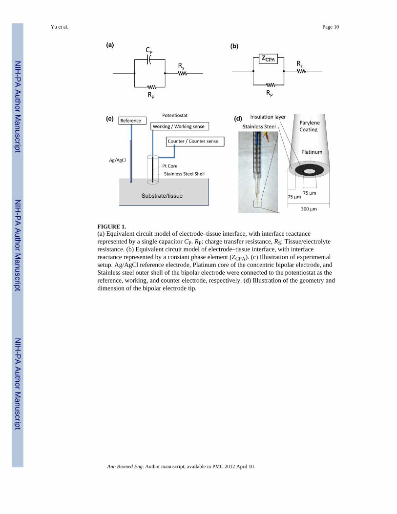
FIGURE 1.(a) Equivalent circuit model of electrode–tissue interface, with interface reactancerepresented by a single capacitor CP. RP: charge transfer resistance, RS: Tissue/electrolyteresistance. (b) Equivalent circuit model of electrode–tissue interface, with interfacereactance represented by a constant phase element (ZCPA). (c) Illustration of experimentalsetup. Ag/AgCl reference electrode, Platinum core of the concentric bipolar electrode, andStainless steel outer shell of the bipolar electrode were connected to the potentiostat as thereference, working, and counter electrode, respectively. (d) Illustration of the geometry anddimension of the bipolar electrode tip.
Yu et al. Page 10
Ann Biomed Eng. Author manuscript; available in PMC 2012 April 10.
NIH
-PA Author Manuscript
NIH
-PA Author Manuscript
NIH
-PA Author Manuscript
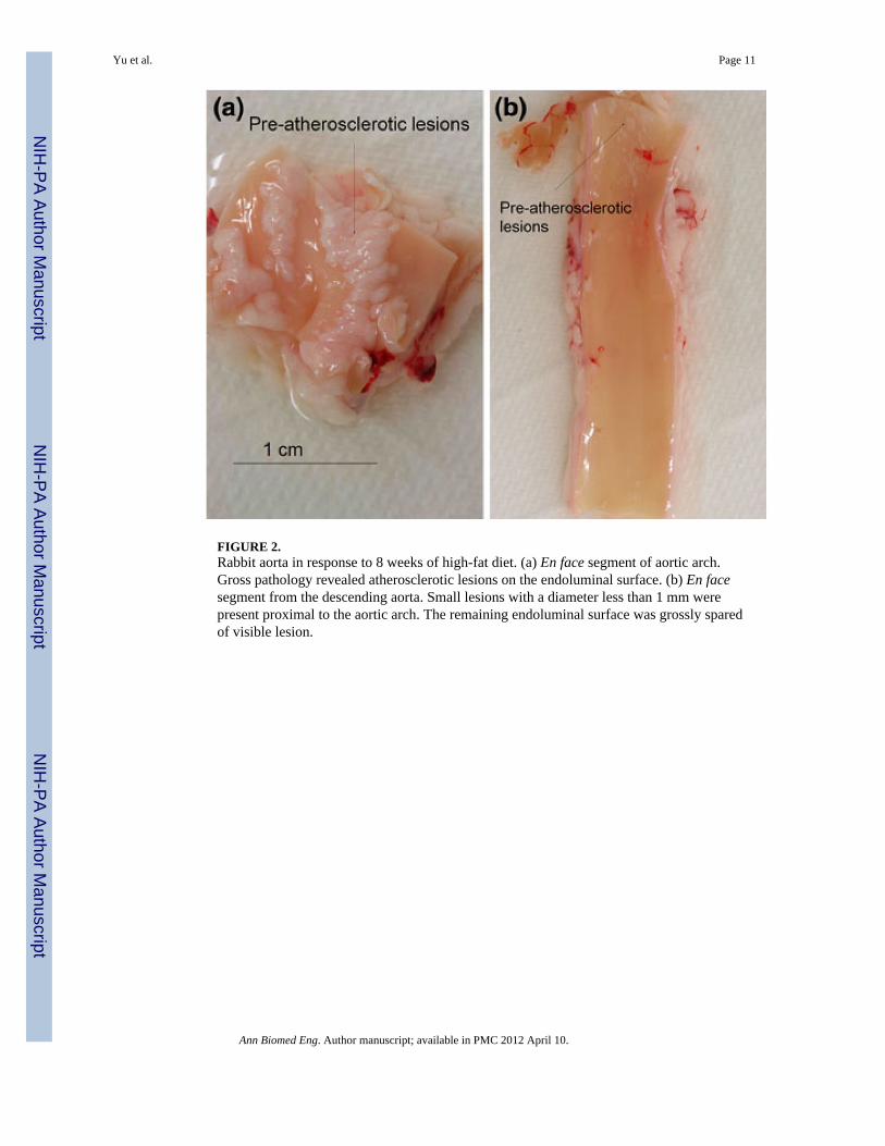
FIGURE 2.Rabbit aorta in response to 8 weeks of high-fat diet. (a) En face segment of aortic arch.Gross pathology revealed atherosclerotic lesions on the endoluminal surface. (b) En facesegment from the descending aorta. Small lesions with a diameter less than 1 mm werepresent proximal to the aortic arch. The remaining endoluminal surface was grossly sparedof visible lesion.
Yu et al. Page 11
Ann Biomed Eng. Author manuscript; available in PMC 2012 April 10.
NIH
-PA Author Manuscript
NIH
-PA Author Manuscript
NIH
-PA Author Manuscript
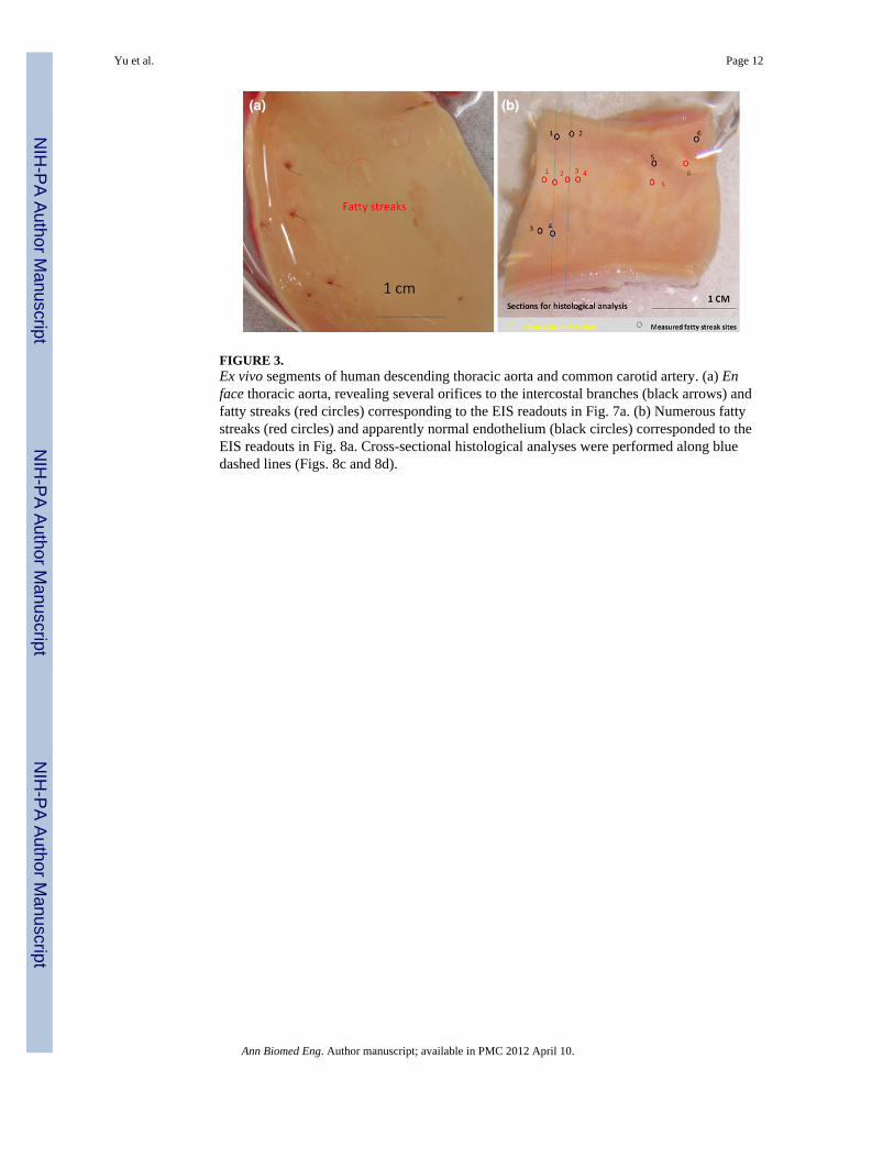
FIGURE 3.Ex vivo segments of human descending thoracic aorta and common carotid artery. (a) Enface thoracic aorta, revealing several orifices to the intercostal branches (black arrows) andfatty streaks (red circles) corresponding to the EIS readouts in Fig. 7a. (b) Numerous fattystreaks (red circles) and apparently normal endothelium (black circles) corresponded to theEIS readouts in Fig. 8a. Cross-sectional histological analyses were performed along bluedashed lines (Figs. 8c and 8d).
Yu et al. Page 12
Ann Biomed Eng. Author manuscript; available in PMC 2012 April 10.
NIH
-PA Author Manuscript
NIH
-PA Author Manuscript
NIH
-PA Author Manuscript
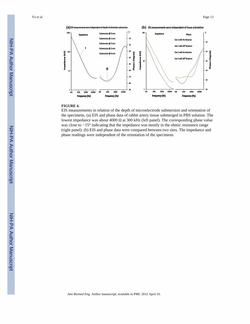
FIGURE 4.EIS measurements in relation of the depth of microelectrode submersion and orientation ofthe specimens. (a) EIS and phase data of rabbit artery tissue submerged in PBS solution. Thelowest impedance was about 4000 Ω at 300 kHz (left panel). The corresponding phase valuewas close to −15° indicating that the impedance was mostly in the ohmic resistance range(right panel). (b) EIS and phase data were compared between two sites. The impedance andphase readings were independent of the orientation of the specimens.
Yu et al. Page 13
Ann Biomed Eng. Author manuscript; available in PMC 2012 April 10.
NIH
-PA Author Manuscript
NIH
-PA Author Manuscript
NIH
-PA Author Manuscript
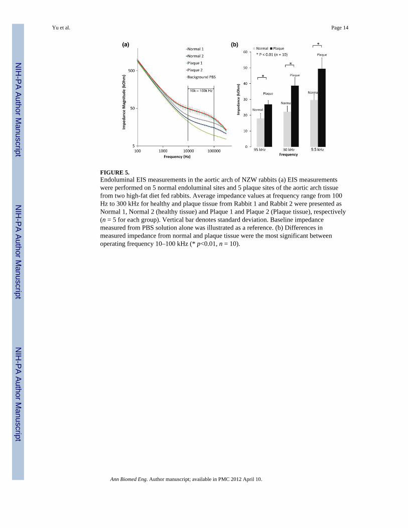
FIGURE 5.Endoluminal EIS measurements in the aortic arch of NZW rabbits (a) EIS measurementswere performed on 5 normal endoluminal sites and 5 plaque sites of the aortic arch tissuefrom two high-fat diet fed rabbits. Average impedance values at frequency range from 100Hz to 300 kHz for healthy and plaque tissue from Rabbit 1 and Rabbit 2 were presented asNormal 1, Normal 2 (healthy tissue) and Plaque 1 and Plaque 2 (Plaque tissue), respectively(n = 5 for each group). Vertical bar denotes standard deviation. Baseline impedancemeasured from PBS solution alone was illustrated as a reference. (b) Differences inmeasured impedance from normal and plaque tissue were the most significant betweenoperating frequency 10–100 kHz (* p<0.01, n = 10).
Yu et al. Page 14
Ann Biomed Eng. Author manuscript; available in PMC 2012 April 10.
NIH
-PA Author Manuscript
NIH
-PA Author Manuscript
NIH
-PA Author Manuscript
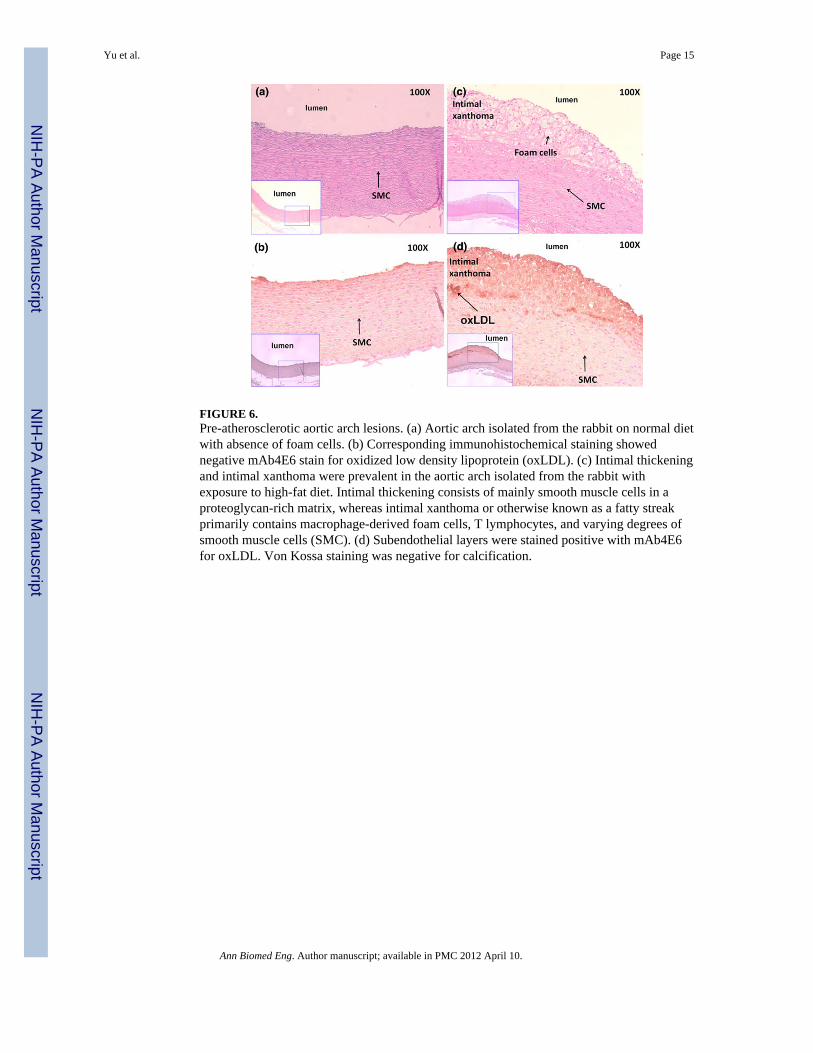
FIGURE 6.Pre-atherosclerotic aortic arch lesions. (a) Aortic arch isolated from the rabbit on normal dietwith absence of foam cells. (b) Corresponding immunohistochemical staining showednegative mAb4E6 stain for oxidized low density lipoprotein (oxLDL). (c) Intimal thickeningand intimal xanthoma were prevalent in the aortic arch isolated from the rabbit withexposure to high-fat diet. Intimal thickening consists of mainly smooth muscle cells in aproteoglycan-rich matrix, whereas intimal xanthoma or otherwise known as a fatty streakprimarily contains macrophage-derived foam cells, T lymphocytes, and varying degrees ofsmooth muscle cells (SMC). (d) Subendothelial layers were stained positive with mAb4E6for oxLDL. Von Kossa staining was negative for calcification.
Yu et al. Page 15
Ann Biomed Eng. Author manuscript; available in PMC 2012 April 10.
NIH
-PA Author Manuscript
NIH
-PA Author Manuscript
NIH
-PA Author Manuscript
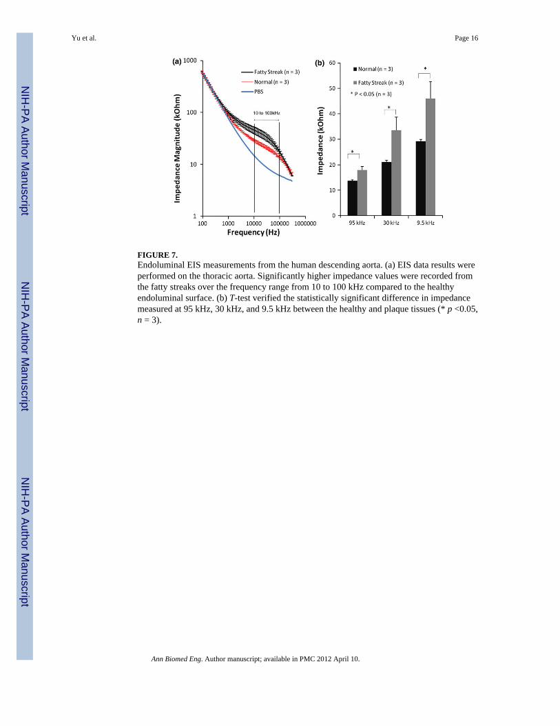
FIGURE 7.Endoluminal EIS measurements from the human descending aorta. (a) EIS data results wereperformed on the thoracic aorta. Significantly higher impedance values were recorded fromthe fatty streaks over the frequency range from 10 to 100 kHz compared to the healthyendoluminal surface. (b) T-test verified the statistically significant difference in impedancemeasured at 95 kHz, 30 kHz, and 9.5 kHz between the healthy and plaque tissues (* p <0.05,n = 3).
Yu et al. Page 16
Ann Biomed Eng. Author manuscript; available in PMC 2012 April 10.
NIH
-PA Author Manuscript
NIH
-PA Author Manuscript
NIH
-PA Author Manuscript
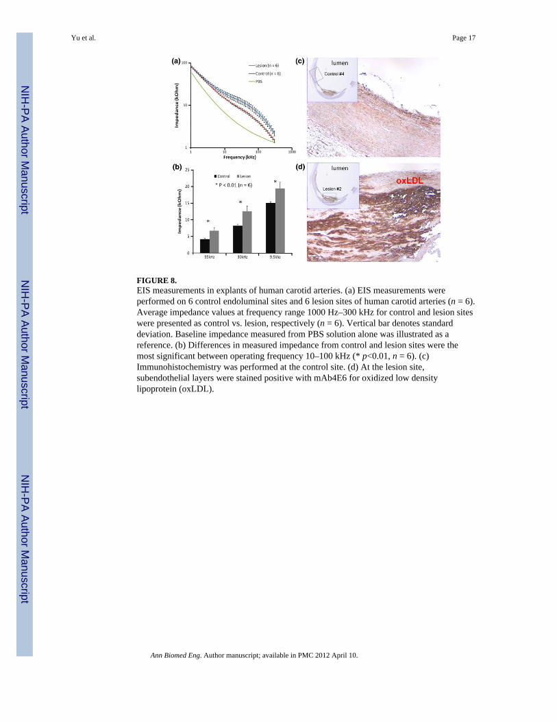
FIGURE 8.EIS measurements in explants of human carotid arteries. (a) EIS measurements wereperformed on 6 control endoluminal sites and 6 lesion sites of human carotid arteries (n = 6).Average impedance values at frequency range 1000 Hz–300 kHz for control and lesion siteswere presented as control vs. lesion, respectively (n = 6). Vertical bar denotes standarddeviation. Baseline impedance measured from PBS solution alone was illustrated as areference. (b) Differences in measured impedance from control and lesion sites were themost significant between operating frequency 10–100 kHz (* p<0.01, n = 6). (c)Immunohistochemistry was performed at the control site. (d) At the lesion site,subendothelial layers were stained positive with mAb4E6 for oxidized low densitylipoprotein (oxLDL).
Yu et al. Page 17
Ann Biomed Eng. Author manuscript; available in PMC 2012 April 10.
NIH
-PA Author Manuscript
NIH
-PA Author Manuscript
NIH
-PA Author Manuscript
Related Documents