Sensors 2014, 14, 15714-15728; doi:10.3390/s140915714 sensors ISSN 1424-8220 www.mdpi.com/journal/sensors Article An Immunosensor Based on Antibody Binding Fragments Attached to Gold Nanoparticles for the Detection of Peptides Derived from Avian Influenza Hemagglutinin H5 Urszula Jarocka 1 , Róża Sawicka 2 , Anna Góra-Sochacka 2 , Agnieszka Sirko 2 , Włodzimierz Zagórski-Ostoja 2 , Jerzy Radecki 1 and Hanna Radecka 1, * 1 Institute of Animal Reproduction and Food Research of Polish Academy of Sciences, Tuwima 10, 10-748 Olsztyn, Poland; E-Mails: [email protected] (U.J.); [email protected] (J.R.) 2 Institute of Biochemistry and Biophysics, Polish Academy of Sciences, Pawińskiego 5A, 02-106 Warsaw, Poland; E-Mails: [email protected] (R.S.); [email protected] (A.G.-S.); [email protected] (A.S.); [email protected] (W.Z.-O.) * Author to whom correspondence should be addressed; E-Mail: [email protected]; Tel.: +48-89-523-4636; Fax: +48-89-524-0124. Received: 19 May 2014; in revised form: 10 July 2014 / Accepted: 11 August 2014 / Published: 25 August 2014 Abstract: This paper concerns the development of an immunosensor for detection of peptides derived from avian influenza hemagglutinin H5. Its preparation consists of successive gold electrode modification steps: (i) modification with 1,6-hexanedithiol and gold colloidal nanoparticles; (ii) immobilization of antibody-binding fragments (Fab’) of anti-hemagglutinin H5 monoclonal antibodies Mab 6-9-1 via S-Au covalent bonds; and (iii) covering the remaining free space on the electrode surfaces with bovine serum albumin. The interactions between Fab’ fragments and hemagglutinin (HA) variants have been explored with electrochemical impedance spectroscopy (EIS) in the presence of [Fe(CN) 6 ] 3−/4− as an electroactive marker. The immunosensor was able to recognize three different His-tagged variants of recombinant hemagglutinin from H5N1 viruses: H1 subunit (17–340 residues) of A/swan/Poland/305-135V08/2006, the long HA (17–530 residues) A/Bar-headed Goose/Qinghai/12/2005 and H1 subunit (1–345 residues) of A/Vietnam/1194/2004. The strongest response has been observed for the long variant with detection limit of 2.2 pg/mL and dynamic range from 4.0 to 20.0 pg/mL. OPEN ACCESS

Welcome message from author
This document is posted to help you gain knowledge. Please leave a comment to let me know what you think about it! Share it to your friends and learn new things together.
Transcript
Sensors 2014, 14, 15714-15728; doi:10.3390/s140915714
sensors ISSN 1424-8220
www.mdpi.com/journal/sensors
Article
An Immunosensor Based on Antibody Binding Fragments Attached to Gold Nanoparticles for the Detection of Peptides Derived from Avian Influenza Hemagglutinin H5
Urszula Jarocka 1, Róża Sawicka 2, Anna Góra-Sochacka 2, Agnieszka Sirko 2,
Włodzimierz Zagórski-Ostoja 2, Jerzy Radecki 1 and Hanna Radecka 1,*
1 Institute of Animal Reproduction and Food Research of Polish Academy of Sciences, Tuwima 10,
10-748 Olsztyn, Poland; E-Mails: [email protected] (U.J.); [email protected] (J.R.) 2 Institute of Biochemistry and Biophysics, Polish Academy of Sciences, Pawińskiego 5A,
02-106 Warsaw, Poland; E-Mails: [email protected] (R.S.); [email protected] (A.G.-S.);
[email protected] (A.S.); [email protected] (W.Z.-O.)
* Author to whom correspondence should be addressed; E-Mail: [email protected];
Tel.: +48-89-523-4636; Fax: +48-89-524-0124.
Received: 19 May 2014; in revised form: 10 July 2014 / Accepted: 11 August 2014 /
Published: 25 August 2014
Abstract: This paper concerns the development of an immunosensor for detection of
peptides derived from avian influenza hemagglutinin H5. Its preparation consists of
successive gold electrode modification steps: (i) modification with 1,6-hexanedithiol and
gold colloidal nanoparticles; (ii) immobilization of antibody-binding fragments (Fab’) of
anti-hemagglutinin H5 monoclonal antibodies Mab 6-9-1 via S-Au covalent bonds; and
(iii) covering the remaining free space on the electrode surfaces with bovine serum
albumin. The interactions between Fab’ fragments and hemagglutinin (HA) variants have
been explored with electrochemical impedance spectroscopy (EIS) in the presence of
[Fe(CN)6]3−/4− as an electroactive marker. The immunosensor was able to recognize three
different His-tagged variants of recombinant hemagglutinin from H5N1 viruses:
H1 subunit (17–340 residues) of A/swan/Poland/305-135V08/2006, the long HA
(17–530 residues) A/Bar-headed Goose/Qinghai/12/2005 and H1 subunit (1–345 residues)
of A/Vietnam/1194/2004. The strongest response has been observed for the long variant
with detection limit of 2.2 pg/mL and dynamic range from 4.0 to 20.0 pg/mL.
OPEN ACCESS
Sensors 2014, 14 15715
Keywords: avian influenza virus; Fab’ fragments; gold electrodes; electrochemical
immunosensor; electrochemical impedance spectroscopy
1. Introduction
Avian influenza (AI) can be an extremely contagious disease of birds caused by type A influenza
virus, a member of the family Orthomyxoviridae. It is an enveloped RNA virus about 100 nm in
diameter with an eight-segmented, single-stranded and negative-sense genome. Two glycoproteins,
hemagglutinin (HA) and neuraminidase (NA), are present on the surface and play important roles in
infecting a host cell. Sixteen HA (H1–H16) and nine NA (N1–N9) subtypes of the influenza virus are
found [1,2].
Influenza A is the only virus type that can infect birds, and almost all wild life birds and domestic
poultry can be infected. Highly pathogenic variants of avian influenza (HPAI) virus often cause
systemic infections in poultry that result in huge economic losses around the world [3]. The H5N1
HPAIV virus has been shown to spread incessantly in many regions all over the world [4]. Most of
these outbreaks were confined to poultry, but the virus was reported to be transmitted to humans in a
few countries and this often lead to deaths in infected humans [5]. Consequently, rapid and sensitive
technology for detection of H5N1 is urgently needed to help containing the spread of H5N1.
The traditional methods for avian influenza virus diagnostic are Enzyme Linked Immunosorbent
Assay (ELISA) [2,6]. Reverse Transcriptase Polymerase Chain Reaction (RT–PCR) [7–9] and
real-time electrical detection [10]. These methods are expensive and require highly trained laboratory
staff. Immunosensors provide a promising alternative to currently used detection systems [11–13].
They are analytical devices consisting of antibodies or their fragments coupled to a transducer and they
generate an analytical response related to analyte concentration in a sample [14,15].
Antibodies are considered to be the first choice as molecular recognition elements due to their target
specificity and affinity, which make them excellent probes in biosensor development. These biological
molecules are special kinds of proteins made by cells of the immune system, B-lymphocytes.
The antibody molecule shaped as “Y” consists of two identical polypeptide chains (Fab’ fragments)
responsible for antigen binding, and one Fc domain. The Fc fragment could be removed by enzyme
digestions [16–18]. The prepared F(ab’)2 or Fab’ fragments could be self-assembled on the gold
surface or other functionalized group due to disulfide or thiol group from the hinge region of the
antibody (immunoglobulin G). The covalent immobilization is stronger in comparison to electrostatic
interactions between antibodies and sensing platform. In addition, covalent bonds enable appropriate
orientation and accessibility of antibody binding sites [19–21].
Immunosensors have found widespread applications in food industry, environmental and pollutants,
biotechnology, pharmaceutical chemistry and clinical diagnostics [22–27]. Several types of
immunosensors, such as surface plasmon resonance (SPR) [3,28,29] and quartz crystal microbalance
(QCM) [30–34], optical interferometric [35,36], label-free microcantilever [37] and fluorescent [38]
have been researched as alternatives to the conventional influenza virus detection methods. These
biosensors have shown analytical potential, but they lack subtype specificity. Many of them are also
Sensors 2014, 14 15716
not practical for use in the field. Compared to these devices electrochemical biosensors [39,40] appear
very promising because they require minimal instrumentation and are readily integrated with
microelectronics. Among electrochemical immunosensors, electrochemical impedance spectroscopy
(EIS)-based label-free sensing has gained much attention [11,13,19,25,41–45]. Impedance
immunosensors have advantages over traditional influenza virus detection methods due to their simple
design and relatively low cost. EIS is a very powerful non-destructive method and can be employed to
study a biological interaction. In this method data points are generated using a small perturbation in
that reduces the matrix interference coming from the composition of biological samples. Several
successful applications of EIS for detection of different biomolecules such as antibodies [46],
His-tagged proteins [19,25], viruses [47–49] and Aβ peptide [50,51] have been reported by our group.
Here, we present a sensitive immunosensor for the detection of peptides derived from avian
influenza hemagglutinin H5 based on the recognition of the epitope localized in the N-terminus of HA
by polypeptide Fab’ from monoclonic Mab 6-9-1. Specific exposure of thiol groups by the Fab’
fragment allows for random dense and precise saturation of the sensor surface with antibodies. This
simplifies the preparation of electrode surfaces and sensing of the HA antigen composition. Specific
interactions between oriented Fab’ fragments immobilized on the gold electrode surface and three
different His-tagged variants of recombinant hemagglutinin: H1 subunit (17–340 residues) of
A/swan/Poland/305-135V08/2006 (Clade 2.2), H1 subunit (1–345 residues) of A/Vietnam/1194/2004
(Clade 1) and the truncated HA (without signal peptide and the transmembrane domain; 17–530
residues) A/Bar-headed Goose/Qinghai/12/2005 (Clade 2.2) were observed using EIS in the presence
of [Fe(CN)6]3−/4− as an electroactive marker.
2. Experimental Section
2.1. Chemicals and Preparations
Gold colloidal nanoparticles (17–23 nm in diameter), 1,6-hexanedithiol (1,6-HDT), potassium
ferro- and ferricyanides, phosphate buffer saline (PBS) components (137 mM NaCl, 2.7 mM KCl,
1.8 mM Na2HPO4, 10 mM KH2PO4) were supplied by Sigma-Aldrich (Poznań, Poland). Alumina
slurry 0.3 and 0.05 μm was purchased from Buehler (Lake Bluff, IL, USA). Sulphuric acid, potassium
hydroxide, ethanol and methanol were obtained from POCh (Gliwice, Poland). Bovine serum albumin
(BSA) was purchased from Invitrogen Life Technologies (Darmstadt, Germany). Hybridoma
producing anti-hemagglutinin H5 monoclonal antibodies (Mab 6-9-1) was from the Institute of
Biotechnology and Antibiotics (Warsaw, Poland). The Mab 6-9-1 antibodies were purified using the
NAb Protein G Spin Kit according to manufacturer protocol (Pierce, Rockford, IL, USA) and the Fab’
fragments were digested by papain agarose from Papaya latex (Sigma Aldrich, P4406-25UN) with the
1:2 (weight to weight) ratio of papain agarose to Mab 6-9-1. Efficiency of digestion was verified by
western blot analysis where Fab’ fragments were detected by goat anti mouse IgG Fab’ specific
antibody. Three His-tagged recombinant hemagglutinin variants of H5N1 virus were used in this
work: (i) HA/Nde, (ii) Qinghai and (iii) Vietnam. The “HA/Nde” protein is based on the sequence of
A/swan/Poland/305-135V08/2006 (EpiFlu Database Acc no. EPI156789) and covers region of 17–340
residues (corresponding to the H1 subunit). The “HA/Nde” protein was produced in Escherichia coli
Sensors 2014, 14 15717
cells in fusion with 6xHis-tag and affinity purified. The “Qinghai” protein (based on the sequence of
A/bar headed goose/Qinghai/12/2005; 17–530 residues) and the “Vietnam” protein (based on the
sequence of A/Vietnam/1194/2004; 1–345 residues) were produced in mammalian cells and purchased
from Immune Technology (New York, NY, USA).
All aqueous solutions were prepared using Milli-Q water, resistivity 18.2 MΩ·cm (Millipore,
Darmstadt, Germany). Reagents and solvents were of analytical grade and were used without further
purification. Experiments were carried out at room temperature unless stated otherwise.
2.2. Preparation of Immunosensor
The gold disk electrodes (2 mm diameter) were obtained from Bioanalytical System (BAS, West
Lafayette, IN, USA). Electrodes after washing with methanol and Milli-Q water were polished in
alumina slurries (Alpha and Gamma Micropolish, Buehler) with particles size of 0.3 and 0.05 μm on
microcloth polishing pads (BAS) for 5 min each. Afterwards they were carefully washed with Milli-Q
water. Then, electrochemical cleaning was performed by cyclic voltammetry (CV). At first they were
dipped in 0.5 M potassium hydroxide solution and swept with a potential between −0.4 V and −1.2 V
against the silver chloride reference electrode (Ag/AgCl) and the platinum wire counter electrode with
a scan rate of 100 mV/s, number of cycles: 3, 50 and 10. Next, the electrodes were cleaned in 0.5 M
sulphuric acid solution in the potential window between −0.3 V and +1.5 V, number of cycles: 3, 10
and 3. Before modification, the surfaces of electrodes were refreshed in 0.5 M potassium hydroxide
solution for 10 cycles. After finishing the electrochemical cleaning, each electrode was rinsed with
Mili-Q water and stored in water (for several minutes, until the next step) to avoid contaminations
from air. All solutions were deoxygenated by purging with nitrogen (ultra pure 6.0, Air Products,
Warszawa, Poland) for 10 min.
The clean gold electrodes were washed repeatedly with water and ethanol. Then, they were
immersed for 20 h in 10 mM 1,6-hexanedithiol (1,6-HDT) solution in ethanol. The tubes containing
electrodes and 1,6-HDT solution were sealed with Teflon tape and Parafilm to avoid solvent
evaporation. Subsequently electrodes were rinsed with ethanol and water. Electrodes with formed
1,6-HDT self-assembled monolayer (SAM) were fixed upside down and a 10 µL droplets of gold
colloidal nanoparticles (GCP) solution were spotted on each gold surface. The tubes containing
electrodes were sealed with Parafilm and stored in +4 °C for 18 h. After incubation, electrodes were
rinsed with water and 0.1 M phosphate buffer saline pH 7.4. Next 10 µL droplets of 1 µg/mL Fab’ 6-9-1
in PBS buffer were aliquoted onto the surface of each electrode. The tubes with electrodes were again
sealed with parafilm and incubated in +4 °C for 20 h. Then, electrodes were carefully rinsed with PBS
buffer. Bovine serum albumin (BSA) solution (in 0.1 M PBS pH 7.4) in concentration of 0.5%
(mass/volume) was used for blocking of unspecific binding. As in prior steps, a 10 µL droplets were
spotted on each electrode and stored in +4 °C for 2 h. Finally, electrodes were rinsed with 0.1 M PBS.
Fully modified electrodes were kept in refrigerator (+4 °C) in 0.1 M PBS buffer pH 7.4 until use, no
longer than one day.

Sensors 2014, 14 15718
2.3. Electrochemical Measurements
All electrochemical measurements were performed at room temperature with an AutoLab
potentiostat-galvanostat (Eco Chemie, Utrecht, The Netherlands) using a three-electrode configuration.
Working electrodes were polycrystalline gold discs 2 mm diameter (BioAnalytical System). All
potentials were measured versus an Ag/AgCl reference electrode and platinum wire was used as the
counter electrode. Cyclic voltammetry (CV) and electrochemical impedance spectroscopy (EIS) were
performed in solution comprised of 0.1 M PBS pH 7.4 and K3[Fe(CN)6]/K4[Fe(CN)6] (0.5 mM each).
In the CV potential were cycled from 0.6 to −0.2 V with scan rate 0.1 V/s. The EIS were recorded
within the frequency range of 0.1 Hz to 10 kHz at formal potential of the redox couple [Fe(CN)6]3−/4−
(0.17 V) with ac amplitude of 10 mV. Obtained spectra were fitted by the AutoLab software in order to
obtain values of electron transfer resistance (Ri). The electrode responses were expressed as:
(Ri−R0)/R0 where R0 means electron transfer resistance of fully modified electrode measured in pure
PBS buffer before His-tagged recombinant proteins detection, Ri means electron transfer resistance of
fully modified electrode measured in PBS containing particular concentration of the detected proteins.
3. Results and Discussion
3.1. Immunosensor Preparation and Its Electrochemical Characterization
The process of immunosensor fabrication is shown in Figure 1. After cleaning gold electrodes were
coated with 1,6-hexanedithiol. Next, a colloidal gold nanoparticles layer is formed via covalent Au-S
bonds. Fab’ 6-9-1 was deposited onto the colloidal gold layer through covalent bonds between the Au
atoms of the gold colloidal nanoparticles and the SH group of Fab’ fragments. Bovine serum albumin
was used to block the remaining spaces on the gold layer.
Figure 1. Schematic illustration of the process of immobilization of Fab’ fragments onto
the gold electrode surface.
The cyclic voltammograms of each steps of modification were shown in Figure 2. Bare gold
electrode in 1 mM K3[Fe(CN)6]/K4[Fe(CN)6] as redox probe in PBS showed a couple of redox peaks

Sensors 2014, 14 15719
with ΔEp = 75 mV (Figure 2, Curve a). After the covalent attachment of 1,6-HDT, shape of CV
changed dramatically. The 1,6-HDT formed a self-assembled monolayer on the gold electrode surface.
This results in blocking of the surface for the redox marker as can be seen in the CV with no apparent
peaks (Figure 2, Curve b). The reversibility of the system was restored after immobilization of
colloidal gold on the layer of 1,6-HDT with peak separation ΔEp = 166 mV (Figure 2, Curve c). The
colloidal gold particles (GCP) size (17–23 nm) have been selected and characterized by Atomic Force
Microscopy in our previous study [49]. GCP increased the conduction pathways and promote the
electron transfer between the redox marker and electrode surface [19,25,47,49–51]. The
immobilization of Fab’ fragments on the colloidal gold layer, formed the insulating layer and
decreased the accessibility of [Fe(CN)6]3−/4−. This caused an increase of the CV peak separation to
ΔEp = 328 mV (Figure 2, Curve d). Further decreasing of faradaic current was observed upon
immobilization of BSA onto the remaining sites on the gold layer (Figure 2, Curve e).
Figure 2. The typical cyclic voltammograms of: (a) bare gold electrode; (b) 1,6-HDT
modified electrode; (c) GCP/1,6-HDT modified electrode; (d) Fab’/GCP/1,6-HDT
modified electrode; (e) BSA/Fab’/GCP/1,6-HDT modified electrode. Solution composition:
1 mM K3[Fe(CN)6]/K4[Fe(CN)6], 0.1 M PBS pH 7.4. The measuring conditions: three
electrode configurations—Au working electrode, Ag/AgCl reference electrode, and
Pt counter electrode; scan rate 100 mV/s.
The each step of the gold electrode modification was also controlled using EIS. Impedance spectra are
shown as Nyquist plots of real (Zre) vs. imaginary (Zim) impedance (Figure 3). The bare gold electrode
exhibited an almost straight line in the Nyquist plot, which is characteristic of a diffusion-limited
electrochemical process (Figure 3, Curve a). The 1,6-HDT modified electrode showed a semicircular
plot at higher frequencies (Figure 3, Curve b). This indicated that the electrode redox processes were
limited by electron transfer (Ret = 1359.3 kΩ). The chemisorptions of GCP on the 1,6-HDT layer
decreased the electron transfer resistance to 112.4 kΩ (Figure 3, Curve c). The immobilized Fab’
fragments were covalently linked to the colloidal gold layer through Au-S bonds. This caused an
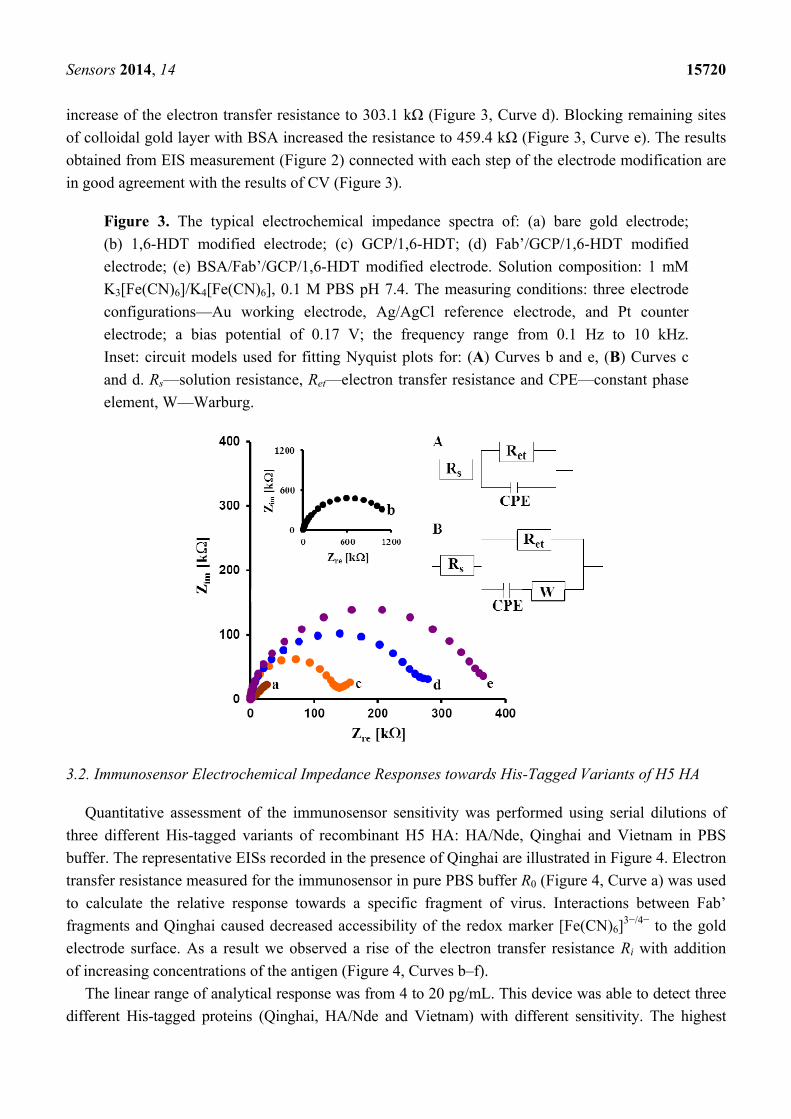
Sensors 2014, 14 15720
increase of the electron transfer resistance to 303.1 kΩ (Figure 3, Curve d). Blocking remaining sites
of colloidal gold layer with BSA increased the resistance to 459.4 kΩ (Figure 3, Curve e). The results
obtained from EIS measurement (Figure 2) connected with each step of the electrode modification are
in good agreement with the results of CV (Figure 3).
Figure 3. The typical electrochemical impedance spectra of: (a) bare gold electrode;
(b) 1,6-HDT modified electrode; (c) GCP/1,6-HDT; (d) Fab’/GCP/1,6-HDT modified
electrode; (e) BSA/Fab’/GCP/1,6-HDT modified electrode. Solution composition: 1 mM
K3[Fe(CN)6]/K4[Fe(CN)6], 0.1 M PBS pH 7.4. The measuring conditions: three electrode
configurations—Au working electrode, Ag/AgCl reference electrode, and Pt counter
electrode; a bias potential of 0.17 V; the frequency range from 0.1 Hz to 10 kHz.
Inset: circuit models used for fitting Nyquist plots for: (A) Curves b and e, (B) Curves c
and d. Rs—solution resistance, Ret—electron transfer resistance and CPE—constant phase
element, W—Warburg.
3.2. Immunosensor Electrochemical Impedance Responses towards His-Tagged Variants of H5 HA
Quantitative assessment of the immunosensor sensitivity was performed using serial dilutions of
three different His-tagged variants of recombinant H5 HA: HA/Nde, Qinghai and Vietnam in PBS
buffer. The representative EISs recorded in the presence of Qinghai are illustrated in Figure 4. Electron
transfer resistance measured for the immunosensor in pure PBS buffer R0 (Figure 4, Curve a) was used
to calculate the relative response towards a specific fragment of virus. Interactions between Fab’
fragments and Qinghai caused decreased accessibility of the redox marker [Fe(CN)6]3−/4− to the gold
electrode surface. As a result we observed a rise of the electron transfer resistance Ri with addition
of increasing concentrations of the antigen (Figure 4, Curves b–f).
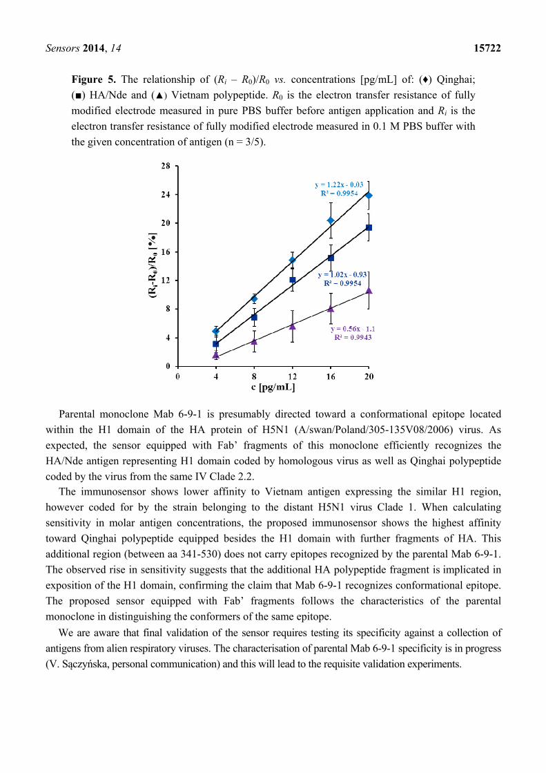
The linear range of analytical response was from 4 to 20 pg/mL. This device was able to detect three
different His-tagged proteins (Qinghai, HA/Nde and Vietnam) with different sensitivity. The highest

Sensors 2014, 14 15721
concentration of antigen (20 pg/mL) caused a significant increase of the electron transfer resistance to
23.9 ± 2.0% for Qinghai, 19.4 ± 1.9% for HA/Nde and 10.3 ± 2.5% for Vietnam (Figure 5).
Figure 4. The typical electrochemical impedance spectra of BSA/Fab’/GCP/1,6-HDT
modified electrode (a) in buffer solution; and after applying (b) 4 pg/mL; (c) 8 pg/mL;
(d) 12 pg/mL; (e) 16 pg/mL; (f) 20 pg/mL of Qinghai polypeptide. Solution composition:
1 mM K3[Fe(CN)6]/K4[Fe(CN)6], 0.1 M PBS (pH 7.4). The measuring conditions: three
electrode configurations—Au working electrode, Ag/AgCl reference electrode, and Pt
counter electrode; a bias potential of 0.17 V; the frequency range: from 0.1 Hz to 10 kHz.
Circuit model used for fitting Nyquist plots in inset: Rs—solution resistance, Ret—electron
transfer resistance and CPE—constant phase element.
Limits of detection (LOD) were calculated based on the standard deviation of the response and the
slope of the calibration curve:
LOD = 3.3σ/S (1)
where σ is the standard deviation of the response and S is the slope of the calibration curve [52]. Limits of
detection were 2.2 pg/mL for Qinghai, 4.0 pg/mL for HA/Nde and 3.5 pg/mL for Vietnam. Sensitivities of
the proposed biosensor were 1.2%[(Ri − R0)/R0c] pg/mL for Qinghai, 1.0%[(Ri − R0)/R0c] pg/mL for
HA/Nde and 0.6%[(Ri − R0)/R0c] pg/mL for Vietnam. The immunosensor presented in this paper thus
has good sensitivity. This is mainly because of the immobilization strategy used. The covalent bonds
between Au atoms of GCP and SH group of Fab’ fragments enable stable and appropriate orientation
for antigen binding.
Sensors 2014, 14 15722
Figure 5. The relationship of (Ri – R0)/R0 vs. concentrations [pg/mL] of: (♦) Qinghai;
() HA/Nde and () Vietnam polypeptide. R0 is the electron transfer resistance of fully
modified electrode measured in pure PBS buffer before antigen application and Ri is the
electron transfer resistance of fully modified electrode measured in 0.1 M PBS buffer with
the given concentration of antigen (n = 3/5).
Parental monoclone Mab 6-9-1 is presumably directed toward a conformational epitope located
within the H1 domain of the HA protein of H5N1 (A/swan/Poland/305-135V08/2006) virus. As
expected, the sensor equipped with Fab’ fragments of this monoclone efficiently recognizes the
HA/Nde antigen representing H1 domain coded by homologous virus as well as Qinghai polypeptide
coded by the virus from the same IV Clade 2.2. The immunosensor shows lower affinity to Vietnam antigen expressing the similar H1 region,
however coded for by the strain belonging to the distant H5N1 virus Clade 1. When calculating
sensitivity in molar antigen concentrations, the proposed immunosensor shows the highest affinity
toward Qinghai polypeptide equipped besides the H1 domain with further fragments of HA. This
additional region (between aa 341-530) does not carry epitopes recognized by the parental Mab 6-9-1.
The observed rise in sensitivity suggests that the additional HA polypeptide fragment is implicated in
exposition of the H1 domain, confirming the claim that Mab 6-9-1 recognizes conformational epitope.
The proposed sensor equipped with Fab’ fragments follows the characteristics of the parental
monoclone in distinguishing the conformers of the same epitope.
We are aware that final validation of the sensor requires testing its specificity against a collection of
antigens from alien respiratory viruses. The characterisation of parental Mab 6-9-1 specificity is in progress
(V. Sączyńska, personal communication) and this will lead to the requisite validation experiments.
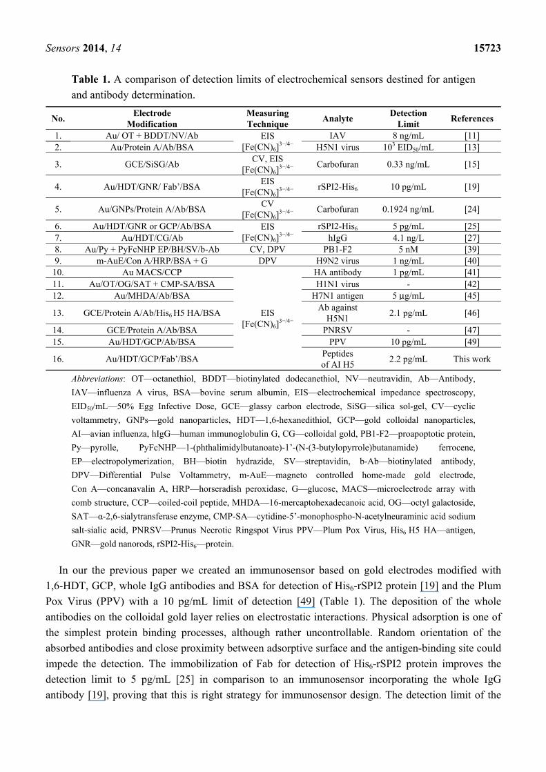
Sensors 2014, 14 15723
Table 1. A comparison of detection limits of electrochemical sensors destined for antigen
and antibody determination.
No. Electrode
Modification Measuring Technique
Analyte Detection
Limit References
1. Au/ OT + BDDT/NV/Ab EIS [Fe(CN)6]
3−/4− IAV 8 ng/mL [11]
2. Au/Protein A/Ab/BSA H5N1 virus 103 EID50/mL [13]
3. GCE/SiSG/Ab CV, EIS
[Fe(CN)6]3−/4−
Carbofuran 0.33 ng/mL [15]
4. Au/HDT/GNR/ Fab’/BSA EIS
[Fe(CN)6]3−/4−
rSPI2-His6 10 pg/mL [19]
5. Au/GNPs/Protein A/Ab/BSA CV
[Fe(CN)6]3−/4−
Carbofuran 0.1924 ng/mL [24]
6. Au/HDT/GNR or GCP/Ab/BSA EIS [Fe(CN)6]
3−/4− rSPI2-His6 5 pg/mL [25]
7. Au/HDT/CG/Ab hIgG 4.1 ng/L [27] 8. Au/Py + PyFcNHP EP/BH/SV/b-Ab CV, DPV PB1-F2 5 nM [39] 9. m-AuE/Con A/HRP/BSA + G DPV H9N2 virus 1 ng/mL [40]
10. Au MACS/CCP
EIS [Fe(CN)6]
3−/4−
HA antibody 1 pg/mL [41] 11. Au/OT/OG/SAT + CMP-SA/BSA H1N1 virus - [42] 12. Au/MHDA/Ab/BSA H7N1 antigen 5 μg/mL [45]
13. GCE/Protein A/Ab/His6 H5 HA/BSA Ab against
H5N1 2.1 pg/mL [46]
14. GCE/Protein A/Ab/BSA PNRSV - [47] 15. Au/HDT/GCP/Ab/BSA PPV 10 pg/mL [49]
16. Au/HDT/GCP/Fab’/BSA Peptides of AI H5
2.2 pg/mL This work
Abbreviations: OT—octanethiol, BDDT—biotinylated dodecanethiol, NV—neutravidin, Ab—Antibody,
IAV—influenza A virus, BSA—bovine serum albumin, EIS—electrochemical impedance spectroscopy,
EID50/mL—50% Egg Infective Dose, GCE—glassy carbon electrode, SiSG—silica sol-gel, CV—cyclic
voltammetry, GNPs—gold nanoparticles, HDT—1,6-hexanedithiol, GCP—gold colloidal nanoparticles,
AI—avian influenza, hIgG—human immunoglobulin G, CG—colloidal gold, PB1-F2—proapoptotic protein,
Py—pyrolle, PyFcNHP—1-(phthalimidylbutanoate)-1’-(N-(3-butylopyrrole)butanamide) ferrocene,
EP—electropolymerization, BH—biotin hydrazide, SV—streptavidin, b-Ab—biotinylated antibody,
DPV—Differential Pulse Voltammetry, m-AuE—magneto controlled home-made gold electrode,
Con A—concanavalin A, HRP—horseradish peroxidase, G—glucose, MACS—microelectrode array with
comb structure, CCP—coiled-coil peptide, MHDA—16-mercaptohexadecanoic acid, OG—octyl galactoside,
SAT—α-2,6-sialytransferase enzyme, CMP-SA—cytidine-5’-monophospho-N-acetylneuraminic acid sodium
salt-sialic acid, PNRSV—Prunus Necrotic Ringspot Virus PPV—Plum Pox Virus, His6 H5 HA—antigen,
GNR—gold nanorods, rSPI2-His6—protein.
In our the previous paper we created an immunosensor based on gold electrodes modified with
1,6-HDT, GCP, whole IgG antibodies and BSA for detection of His6-rSPI2 protein [19] and the Plum
Pox Virus (PPV) with a 10 pg/mL limit of detection [49] (Table 1). The deposition of the whole
antibodies on the colloidal gold layer relies on electrostatic interactions. Physical adsorption is one of
the simplest protein binding processes, although rather uncontrollable. Random orientation of the
absorbed antibodies and close proximity between adsorptive surface and the antigen-binding site could
impede the detection. The immobilization of Fab for detection of His6-rSPI2 protein improves the
detection limit to 5 pg/mL [25] in comparison to an immunosensor incorporating the whole IgG
antibody [19], proving that this is right strategy for immunosensor design. The detection limit of the
Sensors 2014, 14 15724
presented immunosensor in the range of 2.2 pg/mL is better in comparison to those already
published [11,15,19,24,25,27,39,45] or at least at the similar level [40,41,46] (Table 1).
It is worth emphasizeing that the main advantages of the immunosensor proposed here are very
small sample demand, good sensitivity and simple fabrication, with the possibility for miniaturization.
The detection limits for nanoscale biosensors are mainly governed by analyte transport limitation
towards sensing layers, not a signal transduction [53]. The sensor shape, analyte diffusing ability, as
well as the appropriate analyte accumulation time are important parameters for nano-sensor design and
will be taken into account in our future research.
4. Conclusions and Outlook
Concluding, a sensitive and selective impedimetric immunosensor for the detection of peptides
derived from avian influenza hemagglutinin H5 using Fab’ immobilized on a gold electrode surface
via colloidal gold nanoparticles was developed. This device is able to recognize three different
His-tagged fragments of HA: Qinghai, HA/Nde and Vietnam. The strongest response was observed for
Qinghai, with a detection limit of 2.2 pg/mL and a dynamic range from 4.0 pg/mL to 20.0 pg/mL. The
presented research shows that gold colloidal nanoparticles may be used for the creation of a very good
underlayer for Fab’ oriented immobilization. They allow fragments of antibodies to retain their activity
and constitute a good electron conductive layer for electrochemical sensors. Considering its good
selectivity and sensitivity in the pg/mL range, the proposed immunosensor was superior in comparison
to others already reported, therefore, it could be recommended for the rapid, simple and direct
electrochemical detection of avian influenza virus H5N1.
Acknowledgments
This work was supported by Innovative Economy Program, No. WND-POIG.01.01.02-00-007/08
and Institute of Animal Reproduction and Food Research of Polish Academy of Sciences, Olsztyn,
Poland. The work was performed in frame of Vaccine Cluster Consortium (VCC): Institute of
Biochemistry and Biophysics, Polish Academy of Sciences, Warsaw, Poland (IBB); Institute of
Biotechnology and Antibiotics, Warsaw, Poland (IBA); Department of Recombinant Vaccines,
Intercollegiate Faculty of Biotechnology, University of Gdansk and Medical University of Gdańsk,
Gdańsk, Poland (UG); Kucharczyk TE sp. z o.o., Warsaw, Poland (KTE); Institute of Animal
Reproduction and Food Research, Polish Academy of Sciences, Olsztyn, Poland; Department of
Poultry Diseases, National Veterinary Research Institute, Puławy, Poland (PIWet). The authors are
grateful to Anna Porębska, Violetta Cecuda-Adamczewska, Grażyna Płucienniczak and Violetta
Sączyńska from Institute of Biotechnology and Antibiotics (Warsaw, Poland) for the hybridoma
culture producing Mab 6-9-1 and to Bogusław Szewczyk from University of Gdańsk and Medical
University of Gdańsk (Poland) for H5 HA used for hybridoma production.
Author Contributions
Urszula Jarocka—sensors preparation, electrochemical measurements, interpretation of the
electrochemical and analytical results, preparation of the manuscript;
Sensors 2014, 14 15725
Róża Sawicka—preparation and serological verification of antigens and antibody fragments;
Anna Góra-Sochacka—verification of antigens and antibody fragments;
Agnieszka Sirko—design of the biological material, biological interpretation of the results;
Włodzimierz Zagórski-Ostoja—biological interpretation of the results;
Jerzy Radecki—design of the experiments, interpretation of the electrochemical and analytical
results, preparation of the manuscript;
Hanna Radecka—design of the experiments, interpretation of the electrochemical and analytical
results, preparation of the manuscript.
Conflicts of Interest
The authors declare no conflict of interest.
References
1. Kukol, A.; Li, P.; Estrela, P.; Ko-Ferrigno, P.; Migliorato, P. Label-free electrical detection of
DNA hybridization for the example of influenza virus gene sequences. Anal. Biochem. 2008, 374,
143–153.
2. He, F.; Soejoedono, R.D.; Murtini, S.; Goutama, M.; Kwang, J. Complementary monoclonal
antibody-based dot ELISA for universal detection of H5 avian influenza virus. BMC Microbiol.
2010, 10, 330–338.
3. Bai, H.; Wang, R.; Hargis, B.; Lu, H.; Li, Y. A SPR aptasensor for detection of avian influenza
virus H5N1. Sensors 2012, 12, 12506–12518.
4. Peiris, J.S.; de Jong, M.D.; Guan, Y. Avian influenza virus (H5N1): A threat to human health.
Clin. Microbiol. Rev. 2007, 20, 243–267.
5. Chou, C.-C.; Huang, Y.-H. Nucleic acid sandwich hybridization assay with quantum dot-induced
fluorescence resonance energy transfer for pathogen detection. Sensors 2012, 12, 16660–16672.
6. Luo, Q.; Huang, H.; Zou, W.; Dan, H.; Guo, X.; Zhang, A.; Yu, Z.; Chen, H.; Jin, M. An indirect
sandwich ELISA for the detection of avian influenza H5 subtype viruses using anti-hemagglutinin
protein monoclonal antibody. Vet. Micrbiol. 2009, 137, 24–30.
7. Boivin, G.; Côté, S.; Déry, P.; de Serres, G.; Bergeron, M.G. Multiplex Real-Time PCR assay for
detection of influenza and human respiratory syncytial viruses. J. Clin. Microbiol. 2004, 42, 45–51.
8. Trani, L.D.; Bedini, B.; Donatelli, I.; Campitelli, L.; Chiappini, B.; de Marco, M.A.; Delogu, M.;
Buonavoglia, C.; Vaccari, G. A sensitive one-step real-time PCR for detection of avian influenza
viruses using a MGB probe and an internal positive control. BMC Infect. Dis. 2006, 6, 87–94.
9. Das, A.; Spackman, E.; Pantin-Jackwood, M.J.; Suarez, D.L. Removal of real-time Reverse
Transcription Polymerase Chain Reaction (RT-PCR) inhibitors associated with cloacal swab
samples and tissues for improved diagnosis of avian influenza virus by RT-PCR. J. Vet. Diagn.
Invest. 2009, 21, 771–778.
10. Patolsky, F.; Zheng, G.; Hayden, O.; Lakadamyali, M.; Zhuang, X.; Lieber, C.M. Electrical
detection of single viruses. Proc. Natl. Acad. Sci. USA 2004, 101, 14017–14022.
Sensors 2014, 14 15726
11. Hassen, W.M.; Duplan, V.; Frost, E.; Dubowski J.J. Quantitation of influenza A virus in the
presence of extraneous protein using electrochemical impedance spectroscopy. Electrochim. Acta
2011, 56, 8325–8328.
12. Miller, S.A.; Hiatt, L.A.; Keil, R.G.; Wright, D.W.; Cliffel, D.E. Multifunctional nanoparticles as
simulants for a gravimetric immunoassay. Anal. Bioanal. Chem. 2011, 399, 1021–1029.
13. Wang, R.; Wang, Y.; Lassiter, K.; Li, Y.; Hargis, B.; Tung, S.; Berghman, L.; Bottje, W.
Interdigitated array microelectrode based impedance immunosensor for detection of avian
influenza virus H5N1. Talanta 2009, 79, 159–164.
14. Soler, M.; Estevez, M.-C.; Alvarez, M.; Otte, M.A.; Sepulveda, B.; Lechuga, L.M. Direct
detection of protein biomarkers in human fluids using site-specific antibody immobilization
strategies. Sensors 2014, 14, 2239–2258.
15. Sun, X.; Du, S.; Wang, X.; Zhao, W.; Li, Q. A label-free electrochemical immunosensor for
carbofuran detection based on a sol-gel entrapped antibody. Sensors 2011, 11, 9520–9531.
16. Kittipongwarakarn, S.; Hawe, A.; Tantipolphan, R.; Limsuwun, K.; Khomvilai, S.;
Puttipipatkhachorn, S.; Jiskoot, W. New method to produce equine antirabies immunoglobulin F(ab’)2
fragments from crude plasma in high quality and yield. Eur. J. Pharm. Biopharm. 2011, 78, 189–195.
17. Zhao, Y.; Gutshall, L.; Jiang, H.; Baker, A.; Beil, E.; Obmolova, G.; Carton, J.; Taudte, S.;
Amegadzie, B. Two routes for production and purification of Fab fragments in biopharmaceutical
discovery research: Papain digestion of mAb and transient expression in mammalian cells.
Protein Expr. Purif. 2009, 67, 182–189.
18. Coleman, L.; Mahler, S. Purification of Fab fragments from a monoclonal antibody papain digest
by Gradiflow electrophoresis. Protein Expr. Purif. 2003, 32, 246–251.
19. Wąsowicz, M.; Milner, M.; Radecka, D.; Grzelak, K.; Radecka, H. Immunosensor incorporating
Anti-His (C-term) IgG F(ab’) fragments attached to gold nanorods for detection of his-tagged
proteins in culture medium. Sensors 2010, 10, 5409–5424.
20. Saerens, D.; Huang, L.; Bonroy, K.; Muyldermans, S. Antibody fragments as probe in biosensor
development. Sensors 2008, 8, 4669–4686.
21. Bonroy, K.; Frederix, F.; Reekmans, G.; Dewolf, E.; De Palma, R.; Borghs, G.; Declerck, P.;
Goddeeris, B. Comparison of random and oriented immobilisation of antibody fragments on
mixed self-assembled monolayers. J. Immunol. Meth. 2006, 312, 167–181.
22. Wu, H.; Zuo, Y.; Cui, C.; Yang, W.; Ma, H.; Wang, X. Rapid quantitative detection of brucella
melitensis by a label-free impedance immunosensor based on a gold nanoparticle-modified
screen-printed carbon electrode. Sensors 2013, 13, 8551–8563.
23. Fonseca, R.A.S.; Ramos-Jesus, J.; Kubota, L.T.; Dutra, R.F. A nanostructured piezoelectric
immunosensor for detection of human cardiac troponin T. Sensors 2011, 11, 10785–10797.
24. Sun, X.; Zhu, Y.; Wang, X. Amperometric immunosensor based on a protein A / deposited gold
nanocrystals modified electrode for carbofuran detection. Sensors 2011, 11, 11679–11691.
25. Wąsowicz, M.; Subramanian, V.; Dvornyk, A.; Grzelak, K.; Kłudkiewicz, B.; Radecka, H.
Comparison of electrochemical immunosensors based on gold nanomaterials and immunoblot
techniques for detection of histidine-tagged proteins in culture medium. Biosens. Bioelectron.
2008, 24, 284–289.
Sensors 2014, 14 15727
26. Byrne, B.; Stack, E.; Gilmartin, N.; O’Kennedy, R. Antibody-based sensors: Principles, problems
and potential for detection of pathogens and associated toxins. Sensors 2009, 9, 4407–4445.
27. Chen, H.; Jiang, J.-H.; Huang, Y.; Deng, T.; Li, J.-S.; Shen, G.-L.; Yu, R.-Q. An electrochemical
impedance immunosensor with signal amplification based on Au-colloid labeled antibody
complex. Sens. Actuators B Chem. 2006, 117, 211–218.
28. Nilsson, C.E.; Abbas, S.; Bennemo, M.; Larsson, A.; Hämäläinen, M.D.; Frostell-Karlsson, Å.
A novel assay for influenza virus quantification using surface plasmon resonance. Vaccine 2010,
28, 759–766.
29. Diltemiz, S.E.; Ersöz, A.; Hür, D.; Keçili, R.; Say, R. 4-Aminophenyl boronic acid modified gold
platforms for influenza diagnosis. Mater. Sci. Eng. 2013, 33, 824–830.
30. Hewa, T.M.P.; Tannock, G.A.; Mainwaring, D.E.; Harrison, S.; Fecondo, J.V. The detection of
influenza A and B viruses in clinical specimens using a quartz crystal microbalance. J. Virol.
Meth. 2009, 162, 14–21.
31. Brockman, L.; Wang, R.; Lum, J.; Li, Y. QCM aptasensor for rapid and specific detection of
avian influenza virus. Open J. Appl. Biosens. 2013, 2, 97–103.
32. Wang, R.; Li, Y. Hydrogel based QCM aptasensor for detection of avian influenza virus. Biosens.
Bioelectron. 2013, 42, 148–155.
33. Li, D.; Wang, J.; Wang, R.; Li, Y.; Abi-Ghanem, D.; Berghman, L.; Hargis, B.; Lu, H.
A nanobeads amplified QCM immunosensor for the detection of avian influenza virus H5N1.
Biosens. Bioelectron. 2011, 26, 4146–4154.
34. Owen, T.W.; Al-Kaysi, R.O.; Bardeen, C.J.; Cheng, Q. Microgravimetric immunosensor for direct
detection of aerosolized influenza A virus particles. Sens. Actuators B 2007, 126, 691–699.
35. Xu, J.; Suarez, D.; Gottfried, D.S. Detection of avian influenza virus using an interferometric
biosensor. Anal. Bioanal. Chem. 2007, 389, 1193–1199.
36. Farris, L.R.; Wu, N.; Wang, W.; Clarizia, L.-J.A.; Wang, X.; McDonald, M.J.
Immuno-interferometric sensor for the detection of influenza A nucleoprotein. Anal. Bioanal.
Chem. 2010, 396, 667–674.
37. Xu, D.; Liu, L.; Guan, J.; Xu, J.; Wang, T.; Qin, A.; Hu, X.; Wang, C. Label-free
microcantilever-based immunosensors for highly sensitive determination of avian influenza
virus H9. Microchim. Acta 2014, 181, 403–410.
38. Li, Y.; Hong, M.; Qiu, B.; Lin, Z.; Chen, Y.; Cai, Z.; Chen, G. Highly sensitive fluorescent
immunosensor for detection of influenza virus based on Agauto catalysis. Biosens. Bioelectron.
2014, 54, 358–364.
39. Miodek, A.; Sauriat-Dorizon, H.; Chevalier, C.; Delmas, B.; Vidic, J.; Korri-Youssoufi, H. Direct
electrochemical detection of PB1-F2 protein of influenza A virus in infected cells. Biosens.
Bioelectron. 2014, 59, 6–13.
40. Zhou, C.-H.; Long, Y.-M.; Qi, B.-P.; Pang, D.-W.; Zhang, Z.-L. A magnetic bead-based
bienzymatic electrochemical immunosensor for determination of H9N2 avian influenza virus.
Electrochem. Commun. 2013, 31, 129–132.
41. Arya, S.K.; Kongsuphol, P.; Wong, C.C.; Polla, L.J.; Park, M.K. Label free biosensor for sensitive
human influenza virus hemagglutinin specific antibody detection using coiled-coil peptide
modified microelectrode array based platform. Sens. Actuators B Chem. 2014, 194, 127–133.
Sensors 2014, 14 15728
42. Wicklein, B.; del Burgo, M.Á.M; Yuste, M.; Carregal-Romero, E.; Llobera, A.; Darder, M.; Aranda, P.;
Ortín, J.; del Real, G.; Fernández-Sánchez, C.; Ruiz-Hitzky, E. Biomimetic architectures for the
impedimetric discrimination of influenza virus phenotypes. Adv. Funct. Mater. 2013, 23, 254–262.
43. Lum, J.; Wang, R.; Lassiter, K.; Srinivasan, B.; Abi-Ghanem, D.; Berghman, L.; Hargis, B.; Tung, S.;
Lu, H.; Li, Y. Rapid detection of avian influenza H5N1 virus using impedance measurement of
immuno-reaction coupled with RBC amplification. Biosens. Bioelectron. 2012, 38, 67–73.
44. Wang, R.; Jianhan, L.; Lassiter, K.; Srinivasan, B.; Lin, L.; Lu, H.; Tung, S.; Hargis, B.; Bottje, W.;
Berghman, L.; Li, Y. Evaluation study of a portable impedance biosensor for detection of avian
influenza virus. J. Virol. Meth. 2011, 178, 52–58.
45. Diouani, M.F.; Helali, S.; Hafaid, I.; Hassen, W.M.; Snoussi, M.A.; Ghram, A.; Jaffrezic-Renault, N.;
Abdelghani, A. Miniaturized biosensor for avian influenza virus detection. Mater. Sci. Eng. C
2008, 2, 580–583.
46. Jarocka, U.; Sawicka, R.; Góra-Sochacka, A.; Sirko, A.; Zagórski-Ostoja, W.; Radecki, J.;
Radecka, H. Electrochemical immunosensor for detection of antibodies against influenza A virus
H5N1 in hen serum. Biosens. Bioelectron. 2014, 55, 301–306.
47. Jarocka, U.; Radecka, H.; Malinowski, T.; Michalczuk, L.; Radecki, J. Detection of Prunus
Necrotic Ringspot Virus in plant extracts with impedimetric immunosensor based on glassy
carbon electrode. Electroanalysis 2013, 25, 433–438.
48. Malecka, K.; Grabowska, I.; Radecki, J.; Stachyra, A.; Góra-Sochacka, A.; Sirko, A.; Radecka, H.
Electrochemical detection of avian influenza virus genotype using amino-ssDNA probe modified
gold electrodes. Electroanalysis 2013, 25, 1871–1878.
49. Jarocka, U.; Wąsowicz, M.; Radecka, H.; Malinowski, T.; Michalczuk, L.; Radecki, J.
Impedimetric immunosensor for detection of Plum Pox Virus in plant extracts. Electroanalysis
2011, 23, 2197–22204.
50. Grabowska, I.; Radecka, H.; Burza, A.; Radecki, J.; Kaliszan, M.; Kaliszan, R. Association
constants of pyridine and piperidine alkaloids to amyloid β peptide determined by electrochemical
impedance spectroscopy. Curr. Alzheimer Res. 2010, 7, 165–172.
51. Szymańska, I.; Radecka, H.; Radecki, J.; Kaliszan, R. Electrochemical impedance spectroscopy
for study of amyloid β-peptide interactions with (-) nicotine ditartrate and (-) cotinine. Biosens.
Bioelectron. 2007, 22, 1955–1960.
52. Swartz, M.E.; Krull, I.S. Handbook of Analytical Validation, 3rd ed.; CRC Press: Boca Raton, FL,
USA, 2012; p. 70.
53. Sheehan, P.E.; Whitman, L.J. Detection limits for nanoscale biosensors. Nano Lett. 2005, 5, 803–807.
© 2014 by the authors; licensee MDPI, Basel, Switzerland. This article is an open access article
distributed under the terms and conditions of the Creative Commons Attribution license
(http://creativecommons.org/licenses/by/3.0/).
Related Documents


![Electrochemical miRNA Biosensors: The Benefits of ...€¦ · electrochemical nanobiosensors [6, 7]. The electrochemical nanobiosensors are pulling together the advantages of electrochemical](https://static.cupdf.com/doc/110x72/5f5dab2fa5702b13b4580399/electrochemical-mirna-biosensors-the-benefits-of-electrochemical-nanobiosensors.jpg)
