Leading Opinion Electro-spinning of pure collagen nano-fibres e Just an expensive way to make gelatin? * Dimitrios I. Zeugolis a,b,c,1 , Shih T. Khew d , Elijah S.Y. Yew b , Andrew K. Ekaputra b , Yen W. Tong b,d , Lin-Yue L. Yung d , Dietmar W. Hutmacher b,e,2 , Colin Sheppard b , Michael Raghunath a,b,f, * a Tissue Modulation Laboratory, National University of Singapore (NUS), 117510 Singapore, Singapore b Division of Bioengineering, Faculty of Engineering, NUS, 117576 Singapore, Singapore c Immunology Programme, Department of Microbiology, Yong Loo Lin School of Medicine, NUS, 117456 Singapore, Singapore d Department of Chemical and Biomolecular Engineering, Faculty of Engineering, NUS, 117576 Singapore, Singapore e Department of Orthopaedic Surgery, Yong Loo Lin School of Medicine, NUS, 117597 Singapore, Singapore f Department of Biochemistry, Yong Loo Lin School of Medicine, NUS, 117597 Singapore, Singapore Received 6 November 2007; accepted 7 February 2008 Available online 3 March 2008 Abstract Scaffolds manufactured from biological materials promise better clinical functionality, providing that characteristic features are preserved. Collagen, a prominent biopolymer, is used extensively for tissue engineering applications, because its signature biological and physico-chemical properties are retained in in vitro preparations. We show here for the first time that the very properties that have established collagen as the leading natural biomaterial are lost when it is electro-spun into nano-fibres out of fluoroalcohols such as 1,1,1,3,3,3-hexafluoro-2-propanol or 2,2,2-trifluoroethanol. We further identify the use of fluoroalcohols as the major culprit in the process. The resultant nano-scaffolds lack the unique ultra-structural axial periodicity that confirms quarter-staggered supramolecular assemblies and the capacity to generate second har- monic signals, representing the typical crystalline triple-helical structure. They were also characterised by low denaturation temperatures, similar to those obtained from gelatin preparations ( p > 0.05). Likewise, circular dichroism spectra revealed extensive denaturation of the electro-spun collagen. Using pepsin digestion in combination with quantitative SDS-PAGE, we corroborate great losses of up to 99% of triple-helical collagen. In conclusion, electro-spinning of collagen out of fluoroalcohols effectively denatures this biopolymer, and thus appears to defeat its purpose, namely to create biomimetic scaffolds emulating the collagen structure and function of the extracellular matrix. Ó 2008 Elsevier Ltd. All rights reserved. Keywords: Collagen denaturation; Gelatin; Denaturation temperature; Second harmonic generation; Transmission electron microscopy; Circular dichroism * Editor’s Note: Leading Opinions: This paper is one of a newly instituted series of scientific articles that provide evidence-based scientific opinions on topical and important issues in biomaterials science. They have some features of an invited editorial but are based on scientific facts, and some features of a review paper, without attempting to be comprehensive. These papers have been commissioned by the Editor-in-Chief and reviewed for factual, scientific content by referees. * Corresponding author. Tissue Modulation Laboratory, National University of Singapore (NUS), 117510 Singapore, Singapore. Tel.: þ65 6516 5307; fax: þ65 6776 5322. E-mail address: [email protected] (M. Raghunath). 1 Present address: Department of Mechanical and Biomedical Engineering and National Centre for Biomedical Engineering Science, National University of Ireland Galway, Galway, Ireland. 2 Present address: Division of Regenerative Medicine, Institute of Health and Biomedical Innovation, Queensland University of Technology, QLD 4059, Australia. 0142-9612/$ - see front matter Ó 2008 Elsevier Ltd. All rights reserved. doi:10.1016/j.biomaterials.2008.02.009 Available online at www.sciencedirect.com Biomaterials 29 (2008) 2293e2305 www.elsevier.com/locate/biomaterials

Welcome message from author
This document is posted to help you gain knowledge. Please leave a comment to let me know what you think about it! Share it to your friends and learn new things together.
Transcript
Available online at www.sciencedirect.com
Biomaterials 29 (2008) 2293e2305www.elsevier.com/locate/biomaterials
Leading Opinion
Electro-spinning of pure collagen nano-fibres e Just an expensiveway to make gelatin?*
Dimitrios I. Zeugolis a,b,c,1, Shih T. Khew d, Elijah S.Y. Yew b, Andrew K. Ekaputra b,Yen W. Tong b,d, Lin-Yue L. Yung d, Dietmar W. Hutmacher b,e,2,
Colin Sheppard b, Michael Raghunath a,b,f,*
a Tissue Modulation Laboratory, National University of Singapore (NUS), 117510 Singapore, Singaporeb Division of Bioengineering, Faculty of Engineering, NUS, 117576 Singapore, Singapore
c Immunology Programme, Department of Microbiology, Yong Loo Lin School of Medicine, NUS, 117456 Singapore, Singapored Department of Chemical and Biomolecular Engineering, Faculty of Engineering, NUS, 117576 Singapore, Singapore
e Department of Orthopaedic Surgery, Yong Loo Lin School of Medicine, NUS, 117597 Singapore, Singaporef Department of Biochemistry, Yong Loo Lin School of Medicine, NUS, 117597 Singapore, Singapore
Received 6 November 2007; accepted 7 February 2008
Available online 3 March 2008
Abstract
Scaffolds manufactured from biological materials promise better clinical functionality, providing that characteristic features are preserved.Collagen, a prominent biopolymer, is used extensively for tissue engineering applications, because its signature biological and physico-chemicalproperties are retained in in vitro preparations. We show here for the first time that the very properties that have established collagen as theleading natural biomaterial are lost when it is electro-spun into nano-fibres out of fluoroalcohols such as 1,1,1,3,3,3-hexafluoro-2-propanolor 2,2,2-trifluoroethanol. We further identify the use of fluoroalcohols as the major culprit in the process. The resultant nano-scaffolds lackthe unique ultra-structural axial periodicity that confirms quarter-staggered supramolecular assemblies and the capacity to generate second har-monic signals, representing the typical crystalline triple-helical structure. They were also characterised by low denaturation temperatures, similarto those obtained from gelatin preparations ( p> 0.05). Likewise, circular dichroism spectra revealed extensive denaturation of the electro-spuncollagen. Using pepsin digestion in combination with quantitative SDS-PAGE, we corroborate great losses of up to 99% of triple-helicalcollagen. In conclusion, electro-spinning of collagen out of fluoroalcohols effectively denatures this biopolymer, and thus appears to defeatits purpose, namely to create biomimetic scaffolds emulating the collagen structure and function of the extracellular matrix.� 2008 Elsevier Ltd. All rights reserved.
Keywords: Collagen denaturation; Gelatin; Denaturation temperature; Second harmonic generation; Transmission electron microscopy; Circular dichroism
* Editor’s Note: Leading Opinions: This paper is one of a newly instituted series of scientific articles that provide evidence-based scientific opinions on topic
and important issues in biomaterials science. They have some features of an invited editorial but are based on scientific facts, and some features of a review pape
without attempting to be comprehensive. These papers have been commissioned by the Editor-in-Chief and reviewed for factual, scientific content by referee
* Corresponding author. Tissue Modulation Laboratory, National University of Singapore (NUS), 117510 Singapore, Singapore. Tel.: þ65 6516 5307; fax: þ6
6776 5322.
E-mail address: [email protected] (M. Raghunath).1 Present address: Department of Mechanical and Biomedical Engineering and National Centre for Biomedical Engineering Science, National University
Ireland Galway, Galway, Ireland.2 Present address: Division of Regenerative Medicine, Institute of Health and Biomedical Innovation, Queensland University of Technology, QLD 405
Australia.
0142-9612/$ - see front matter � 2008 Elsevier Ltd. All rights reserved.
doi:10.1016/j.biomaterials.2008.02.009
al
r,
s.
5
of
9,
2294 D.I. Zeugolis et al. / Biomaterials 29 (2008) 2293e2305
1. Introduction
Collagen type I accounts for up to 70e90% of the collagenfound in the body and it is present in the form of elongatedfibres in various tissues. Individual fibrils can be greater than500 mm in length and 500 nm in diameter [1,2]. These buildingblocks are rod-like triple helices that are stabilised by intra-molecular hydrogen bonds between Gly and Hyp in adjacentchains [3e6]. Tissues rich in fibrous collagen such as skinand tendon are generally used to extract collagen. Dilute acidicsolvents are used to break intermolecular cross-links of the al-dimine type, whilst proteolytic enzymes, such as pepsin, areused to cleave the more stable cross-links of the keto-iminetype. Pepsin cleaves only the non-triple-helical C- and N-telopeptides, leaving the triple-helical molecule intact [7e9].Extracted collagen from either of the above preparations isfavoured for biomedical applications, since in vitro, underappropriate conditions, will spontaneously self-assemble toform biodegradable and biocompatible insoluble fibrils ofhigh mechanical strength, low immunogenicity and with a D-periodicity indistinguishable from that of native fibres [10e14].
Electro-spinning has been recently introduced as the mostpromising technique to manufacture in vitro fibrous scaffoldsfor tissue engineering application with fibre diameter rangingfrom a few microns to less than 100 nm. Such materials aimto mimic extracellular matrix components, such as collagenfibrils whose diameter in vivo range from 20 nm to 40 mm[15e17]. Currently, the most widely adopted method involvesthe electro-spinning of pure collagen or collagen-poly(3-caprolactone) blends out of highly volatile fluoroalcoholssuch as 1,1,1,3,3,3-hexafluoro-2-propanol (HFP) [18e25] or2,2,2-trifluoroethanol (TFE) [24,26]. However, it has beenshown earlier with non-collagenous proteins that fluoroalco-hols not only denaturate the native structure, but also lowerthe denaturation temperature [27e29]. Moreover, in a recentpublication, it was shown that 45% of collagen was apparentlylost during electro-spinning [30]. Additionally, electro-spinning of collagen using either HFP or TFE has been re-ported repeatedly to yield collagen nano-fibres that do notswell when in aqueous media like other collagenous structures[31e33], but instead are readily soluble in water, tissue fluidsor blood [20,22,24,34e36]. Since gelatin is the water-solubledegradation product of the originally water-insoluble collagenfibril [37], the observed water solubility of the electro-spuncollagen scaffolds might point to an extensive conformationalchange. Given, the above, our hypothesis is that through theelectro-spinning process, denaturation of collagen takes placeand gelatin is created.
To verify our hypothesis, we conducted a series of specificexperiments that distinguish collagen from gelatin. Collagen isa crystalline [38e41] (second harmonic generation experimen-tation), triple-helical molecule [2e6,31] (circular dichroismexperimentation), whilst gelatin is characterised by destroyeda-chains, disrupted triple-helical and fibrillar structure andlacking internal structure or configurational order [37]. More-over, the collagen fibrils possess a high degree of axial align-ment and exhibit a characteristic D banding (the finger print of
fibrous collagens), which results from alternating overlap andgap zones, produced by the specific packing arrangement ofthe 300 nm long and 1.5 nm wide collagen molecules. Thisproduces an average periodicity of 67 nm in the native hydratedstate [1,3,4,12,14,31,33,42e45], although dehydration andshrinkage during conventional sample preparation for transmissionelectron microscopy results in lower values of around 55e65 nm[1,44] (transmission electron microscopy experimentation).Furthermore, the denaturation temperature of collagen is higherthan the denaturation temperature of gelatin [31,46e52](differential Scanning calorimetry experimentation). Most im-portantly, the tight triple-helical structure of the collagen mole-cule makes it resistant to pepsin or trypsin, unless its folding islocally compromised by either point mutations or heat denatur-ation [53] (pepsin digestion and SDS-PAGE experimentation).Such molecules are unstable at physiological temperaturesand they are degraded intra-cellularly [14]. Based on all theabove, we demonstrate for first time that the electro-spun colla-gen scaffolds are not crystalline; are not triple-helical; are notquarter-staggered arranged; have denaturation temperaturelower than or similar to gelatin; and are pepsin susceptible.Freeze-dried collagen dissolved in HFP and freeze-dried again(HFP-recovered collagen) also exhibited similar properties withthose obtained from gelatin preparations, clearly indicating thatfluoroalcohols are the major cause of denaturation. Taken to-gether, this builds up the strongest evidence that electro-spin-ning of collagen or co-spinning of collagen-syntheticpolymers out of fluoroalcohols results in the creation of gelatin,a protein derived from denatured collagen and is characterisedby destroyed a-chains, disrupted triple-helical and fibrillarstructure and lacking internal structure or configurational order[37].
2. Materials and methods
2.1. Materials
Porcine skin type A and bovine type B gelatin were obtained from Sigmae
Aldrich (Singapore). Purified type I freeze-dried bovine dermal atelocollagens
were obtained from Koken Co. (Japan) and Symatese Biomateriaux (France).
In-house type I atelocollagen from porcine Achilles tendon was extracted as
has been described previously [54]. Medical grade poly(3-caprolactone)
(mPCL) was purchased from Birmingham Polymers Inc (USA). Rat-tail
tendons and normal human skin were used as representatives of native assem-
blies. Unless noted otherwise, all chemicals and reagents were purchased from
SigmaeAldrich (Singapore).
2.2. Nano-fibre fabrication through electro-spinning
Typical protocols for the electro-spinning were used based on previous
publications [18,19,21,24]. The following preparations were investigated: (a)
in-house, Koken and Symatese collagens and Sigma gelatin type A and B
dissolved in HFP at 50 mg/ml concentration; (b) Koken collagen dissolved
in HFP and TFE at 180 mg/ml concentration; and (c) in order to investigate
whether blending and consequent co-spinning of collagen with mPCL could
prohibit or restrict the denaturation of collagen mPCLeSymatese collagen
and mPCLeSigma gelatin type A and B blends (5:1 ratio) and mPCL dis-
solved in HFP at 125 mg/ml concentration. Either of the above solutions
was loaded into a syringe pump (KD-Scientific, USA), which was set at
0.75e1.2 ml/h. Upon application of high voltage (10e15 kV; applied current
was below 1 mA) (Gamma High Voltage Research, USA) between the syringe
2295D.I. Zeugolis et al. / Biomaterials 29 (2008) 2293e2305
needle (internal diameter 27G1/2) and the aluminium collector (12e15 cm
distance), the solvent was evaporated and the nano-fibres were deposited on
the collector. All experiments were carried out at room temperature (RT:
22e26 �C) and 55e73% relative humidity.
2.3. HFP-recovered collagen
In order to assess the effect of the fluoroalcohols alone on collagen struc-
ture, freeze-dried in-house, Koken and Symatese type I collagen preparations
were dissolved in HFP (50 mg/ml concentration) and freeze-dried (Advantage
ES-53, VirTis, SP Industries, Inc., USA). The material obtained is referred to
as HFP-recovered collagen.
2.4. Micro-fibre fabrication through extrusion
The procedure for fibre formation has been described in detail previously
[54] based on previous publications [55,56]. Briefly, a solution of in-house col-
lagen (6 mg/ml in 0.5 M acetic acid) was extruded into the fibre formation
buffer (FFB) comprised of 118 mM phosphate buffer and 20% polyethylene
glycol Mw 8000 at pH 7.55 and 37 �C at a flow rate of 0.4 ml/min. Resultant
fibres were allowed to remain in this buffer for a maximum period of 10 min,
followed by further 10 min incubation in 6.0 mM phosphate buffer and 75 mM
NaCl at pH 7.10 and 37 �C and further 10 min incubation in distilled water at
RT. Finally the fibres air-dried under the tension of their own weight at RT.
2.5. Self-assembly of collagen
For the self-assembly experiments, freeze-dried collagens from in-house,
Koken and Symatese preparations and their HFP-recovered counterparts
were dissolved in 0.5 M acetic acid at 1 mg/ml concentration. FFB (see above)
was warmed for 30 min at 35 �C and was mixed with either of the collagen
preparations (ratio of FFB to collagen solution: 3:2). The mixture was incu-
bated for 48 h at 35 �C.
2.6. Scanning electron microscopy (SEM)
The morphology of the produced scaffolds was evaluated using a QuanTA
200F Jeol Scanning Electron Microscope (FEI Company, Hillsbora, Oregon-
USA) after gold sputtering with a Jeol JFC-1600 Auto Fine Coater (Tokyo,
Japan).
2.7. Transmission electron microscopy (TEM)
Rat-tail tendon, extruded collagen fibres and electro-spun scaffolds were
fixed in 2.5% glutaraldehyde in 0.1 M phosphate buffer for 16 h at RT. After
osmication and dehydration through a series of ascending aqueous ethanol
concentrations, samples were embedded in araldite using appropriate interme-
diate infiltration steps. Ultra-thin sections were obtained using a Reichert Ul-
tracut E ultra-microtome (Leica Microsystems Ltd, Germany), collected on
Formvar coated copper grids and were viewed with a JEM-1220 Transmission
Electron Microscope (JEOL Ltd, Japan) at 80 kV, after were contrasted with
1% phosphotungstic acid pH 7.4 for 10 s and with aqueous 2% uranyl acetate
solution for 10 min. Collagen self-assemblies were analysed without fixation
and embedding. The assembly solution (20 ml) was transferred onto Formvar
coated grids blotted against filter paper to remove excess water, dried for
16 h at RT and contrasted as described above prior to viewing.
2.8. Second harmonic generation (SHG)
Second harmonic microscopy was performed by coupling a Titanium-
Sapphire laser (Mira 900, Coherent, USA) to an upright microscope (IX71,
Olympus, Japan). The laser was tuned to 838 nm (<200 fs, 76 MHz) and scan-
ning was done with the commercial FV300 (Olympus, Japan). The beam was
such that it slightly under-filled the back-aperture of the 20� objective and the
input power was <100 mW. Collection of the SHG signals was done in trans-
mission with an oil immersion condenser and spectral filtering of the signal
was carried out using a band-pass filter (BG40, Schott, Germany) and
a short-pass filter passing below 450 nm (FES450, Thorlabs, USA). Imaging
was controlled with the Fluoview 5.0b software (Olympus, Japan) and images
were scanned at 512� 512 pixels and averaged over four images depending on
the noise levels. Nano-scaffolds, freeze-dried materials and 5 mm thick cryo-
sections (CM3050S, Leica Microsystems Ltd, Germany) of rat-tail tendon,
normal human skin and extruded collagen fibres were mounted on glass-slides
using polyvinyl-alcohol-DABCO medium underneath glass-coverslips to re-
tain the materials wet prior to SHG investigation.
2.9. Differential scanning calorimetry (DSC)
The denaturation temperature was determined using an 822e Mettler-
Toledo differential scanning calorimeter (Mettler-Toledo International Inc.,
Singapore). Fifty milligrams of either preparation was hydrated in 1500 ml
of PBS. The following day, the samples were blotted with filter paper to re-
move excess water. Wet samples (5e15 mg) of every preparation were her-
metically sealed in 50 ml aluminium pans and heated at a constant
temperature of 5 �C/min in the temperature range of 15e90 �C. An empty al-
uminium pan was used as reference probe. Thermal denaturation, the endo-
thermic transition, was recorded as a typical peak, and two characteristic
temperatures were measured corresponding to the peak (temperature of max-
imum power absorption during denaturation) and onset (temperature at which
the tangent to the initial power versus temperature line crosses the baseline)
temperatures.
2.10. Sodium dodecyl sulfate-polyacrylamide gelelectrophoresis (SDS-PAGE)
Freeze-dried materials (original and HFP-recovered), extruded collagen
micro-fibres and electro-spun nano-fibres were subjected to acid-solubilisation
in 0.5 M acetic acid or pepsin-digestion in 0.1 mg pepsin per milliliter of 0.5 M
acetic acid (1 mg/ml concentration in either case). Obtained suspensions were
centrifuged for 15 min at 13,326g at 4 �C (Biofuge, Fresco Heraeus Instru-
ments, Germany) and the supernatants were analysed using SDS-PAGE.
Protein bands were visualised using silver-staining (SilverQuest� Kit, Invi-
trogen, Singapore) and quantitated using a GS-800 densitometer (BioRad,
USA).
2.11. Circular dichroism (CD)
CD measurements of extracted material (see above) were performed using
a Jasco Model J-810 spectropolarimeter (Jasco, UK) using a quartz cylindrical
cuvette (Hellma, Germany) with a path length of 0.1 mm. The cuvette was
filled with 150 ml of sample for each measurement. CD spectra were obtained
by continuous wavelength scans (average of three scans) from 180 to 260 nm
at a scan-speed of 50 nm/min. The samples were equilibrated for 1 h at RT be-
fore the CD spectra were acquired.
2.12. Statistical analysis
Numerical data are expressed as mean� SD. Analysis was performed us-
ing statistical software (MINITAB� version 13.1, Minitab, Inc.). One way
analysis of variance (ANOVA) for multiple comparisons and 2-sample t-
test for pair wise comparisons were employed after confirming the following
assumptions: (a) the distribution from which each of the samples was derived
was normal (AndersoneDarling normality test); and (b) the variances of the
population of the samples were equal to one another (Bartlett’s and Levene’s
tests for homogenicity of variance). Non-parametric statistics were utilised
when either or both of the above assumptions were violated and conse-
quently KruskaleWallis test for multiple comparisons or Mann-Whitney
test for two-samples were carried out. Statistical significance was accepted
at p< 0.05.
2296 D.I. Zeugolis et al. / Biomaterials 29 (2008) 2293e2305
3. Results
3.1. Scanning electron microscopy
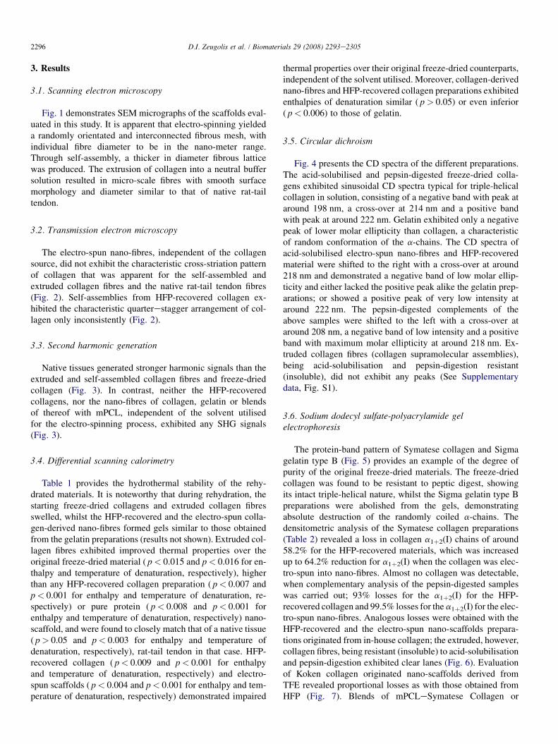
Fig. 1 demonstrates SEM micrographs of the scaffolds eval-uated in this study. It is apparent that electro-spinning yieldeda randomly orientated and interconnected fibrous mesh, withindividual fibre diameter to be in the nano-meter range.Through self-assembly, a thicker in diameter fibrous latticewas produced. The extrusion of collagen into a neutral buffersolution resulted in micro-scale fibres with smooth surfacemorphology and diameter similar to that of native rat-tailtendon.
3.2. Transmission electron microscopy
The electro-spun nano-fibres, independent of the collagensource, did not exhibit the characteristic cross-striation patternof collagen that was apparent for the self-assembled andextruded collagen fibres and the native rat-tail tendon fibres(Fig. 2). Self-assemblies from HFP-recovered collagen ex-hibited the characteristic quarterestagger arrangement of col-lagen only inconsistently (Fig. 2).
3.3. Second harmonic generation
Native tissues generated stronger harmonic signals than theextruded and self-assembled collagen fibres and freeze-driedcollagen (Fig. 3). In contrast, neither the HFP-recoveredcollagens, nor the nano-fibres of collagen, gelatin or blendsof thereof with mPCL, independent of the solvent utilisedfor the electro-spinning process, exhibited any SHG signals(Fig. 3).
3.4. Differential scanning calorimetry
Table 1 provides the hydrothermal stability of the rehy-drated materials. It is noteworthy that during rehydration, thestarting freeze-dried collagens and extruded collagen fibresswelled, whilst the HFP-recovered and the electro-spun colla-gen-derived nano-fibres formed gels similar to those obtainedfrom the gelatin preparations (results not shown). Extruded col-lagen fibres exhibited improved thermal properties over theoriginal freeze-dried material ( p< 0.015 and p< 0.016 for en-thalpy and temperature of denaturation, respectively), higherthan any HFP-recovered collagen preparation ( p< 0.007 andp< 0.001 for enthalpy and temperature of denaturation, re-spectively) or pure protein ( p< 0.008 and p< 0.001 forenthalpy and temperature of denaturation, respectively) nano-scaffold, and were found to closely match that of a native tissue( p> 0.05 and p< 0.003 for enthalpy and temperature ofdenaturation, respectively), rat-tail tendon in that case. HFP-recovered collagen ( p< 0.009 and p< 0.001 for enthalpyand temperature of denaturation, respectively) and electro-spun scaffolds ( p< 0.004 and p< 0.001 for enthalpy and tem-perature of denaturation, respectively) demonstrated impaired
thermal properties over their original freeze-dried counterparts,independent of the solvent utilised. Moreover, collagen-derivednano-fibres and HFP-recovered collagen preparations exhibitedenthalpies of denaturation similar ( p> 0.05) or even inferior( p< 0.006) to those of gelatin.
3.5. Circular dichroism
Fig. 4 presents the CD spectra of the different preparations.The acid-solubilised and pepsin-digested freeze-dried colla-gens exhibited sinusoidal CD spectra typical for triple-helicalcollagen in solution, consisting of a negative band with peak ataround 198 nm, a cross-over at 214 nm and a positive bandwith peak at around 222 nm. Gelatin exhibited only a negativepeak of lower molar ellipticity than collagen, a characteristicof random conformation of the a-chains. The CD spectra ofacid-solubilised electro-spun nano-fibres and HFP-recoveredmaterial were shifted to the right with a cross-over at around218 nm and demonstrated a negative band of low molar ellip-ticity and either lacked the positive peak alike the gelatin prep-arations; or showed a positive peak of very low intensity ataround 222 nm. The pepsin-digested complements of theabove samples were shifted to the left with a cross-over ataround 208 nm, a negative band of low intensity and a positiveband with maximum molar ellipticity at around 218 nm. Ex-truded collagen fibres (collagen supramolecular assemblies),being acid-solubilisation and pepsin-digestion resistant(insoluble), did not exhibit any peaks (See Supplementarydata, Fig. S1).
3.6. Sodium dodecyl sulfate-polyacrylamide gelelectrophoresis
The protein-band pattern of Symatese collagen and Sigmagelatin type B (Fig. 5) provides an example of the degree ofpurity of the original freeze-dried materials. The freeze-driedcollagen was found to be resistant to peptic digest, showingits intact triple-helical nature, whilst the Sigma gelatin type Bpreparations were abolished from the gels, demonstratingabsolute destruction of the randomly coiled a-chains. Thedensitometric analysis of the Symatese collagen preparations(Table 2) revealed a loss in collagen a1þ2(I) chains of around58.2% for the HFP-recovered materials, which was increasedup to 64.2% reduction for a1þ2(I) when the collagen was elec-tro-spun into nano-fibres. Almost no collagen was detectable,when complementary analysis of the pepsin-digested sampleswas carried out; 93% losses for the a1þ2(I) for the HFP-recovered collagen and 99.5% losses for the a1þ2(I) for the elec-tro-spun nano-fibres. Analogous losses were obtained with theHFP-recovered and the electro-spun nano-scaffolds prepara-tions originated from in-house collagen; the extruded, however,collagen fibres, being resistant (insoluble) to acid-solubilisationand pepsin-digestion exhibited clear lanes (Fig. 6). Evaluationof Koken collagen originated nano-scaffolds derived fromTFE revealed proportional losses as with those obtained fromHFP (Fig. 7). Blends of mPCLeSymatese Collagen or
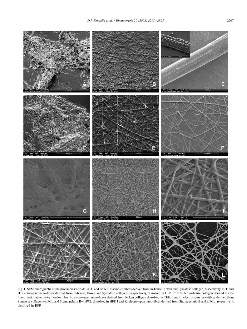
Fig. 1. SEM micrographs of the produced scaffolds. A, D and G: self-assembled fibres derived from in-house, Koken and Symatese collagen, respectively. B, E and
H: electro-spun nano-fibres derived from in-house, Koken and Symatese collagens, respectively, dissolved in HFP. C: extruded in-house collagen derived micro-
fibre; inset: native rat-tail tendon fibre. F: electro-spun nano-fibres derived from Koken collagen dissolved in TFE. I and L: electro-spun nano-fibres derived from
Symatese collagenemPCL and Sigma gelatin BemPCL dissolved in HFP. J and K: electro-spun nano-fibres derived from Sigma gelatin B and mPCL, respectively,
dissolved in HFP.
2297D.I. Zeugolis et al. / Biomaterials 29 (2008) 2293e2305
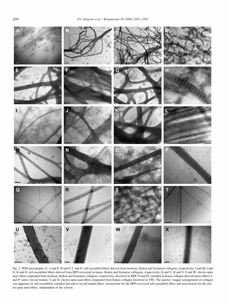
Fig. 2. TEM micrographs of: A and E; B and F; C and G: self-assembled fibres derived from in-house, Koken and Symatese collagens, respectively; I and M, J and
N, K and O: self-assembled fibres derived from HFP-recovered in-house, Koken and Symatese collagens, respectively; Q and U, R and V, S and W: electro-spun
nano-fibres originated from in-house, Koken and Symatese collagens, respectively, dissolved in HFP; D and H: extruded in-house collagen derived micro-fibres; L
and P: native rat-tail tendon; T and X: electro-spun nano-fibres originated from Koken collagen dissolved in TFE. The quarterestagger arrangement of collagen
was apparent for self-assembled, extruded and native rat-tail tendon fibres, inconsistent for the HFP-recovered self-assembled fibres and non-existent for the elec-
tro-spun nano-fibres, independent of the solvent.
2298 D.I. Zeugolis et al. / Biomaterials 29 (2008) 2293e2305
Fig. 3. Bright-field and corresponding second harmonic generation images for: (a and e) human normal skin; (b and f) rat-tail tendon; (c and g) in-house freeze-
dried collagen; (d and h) extruded in-house collagen micro-fibres; (i and m) in-house HFP-recovered freeze-dried collagen; (j and n) electro-spun nano-fibres de-
rived from Koken collagen using HFP; (k and o) electro-spun nano-fibres derived from Koken collagen using TFE; and (l and p) electro-spun nano-fibres derived
from blend of Symatese collagen and mPCL using HFP. Electro-spun nano-fibres failed to exhibit SHG signals, in contrast to any other collagenous structure.
2299D.I. Zeugolis et al. / Biomaterials 29 (2008) 2293e2305
mPCLeSigma gelatin type A showed a comparable degree ofprotein denaturation after electro-spinning (Fig. 8).
4. Discussion
Since electro-spinning is currently the prime method toconstruct nano-fibrous scaffolds and collagen is a superior,
clinically approved biopolymer, it appeared logical to combineboth technology and biomaterial to fabricate submicron scaf-folds for tissue engineering applications. After Huang et al.(2001) [57] showed the feasibility to electro-spin collagen,this approach has become popular in the biomaterials and tis-sue engineering field, as reflected in a recent steep rise of pub-lications. However, every single article testifies that collagen
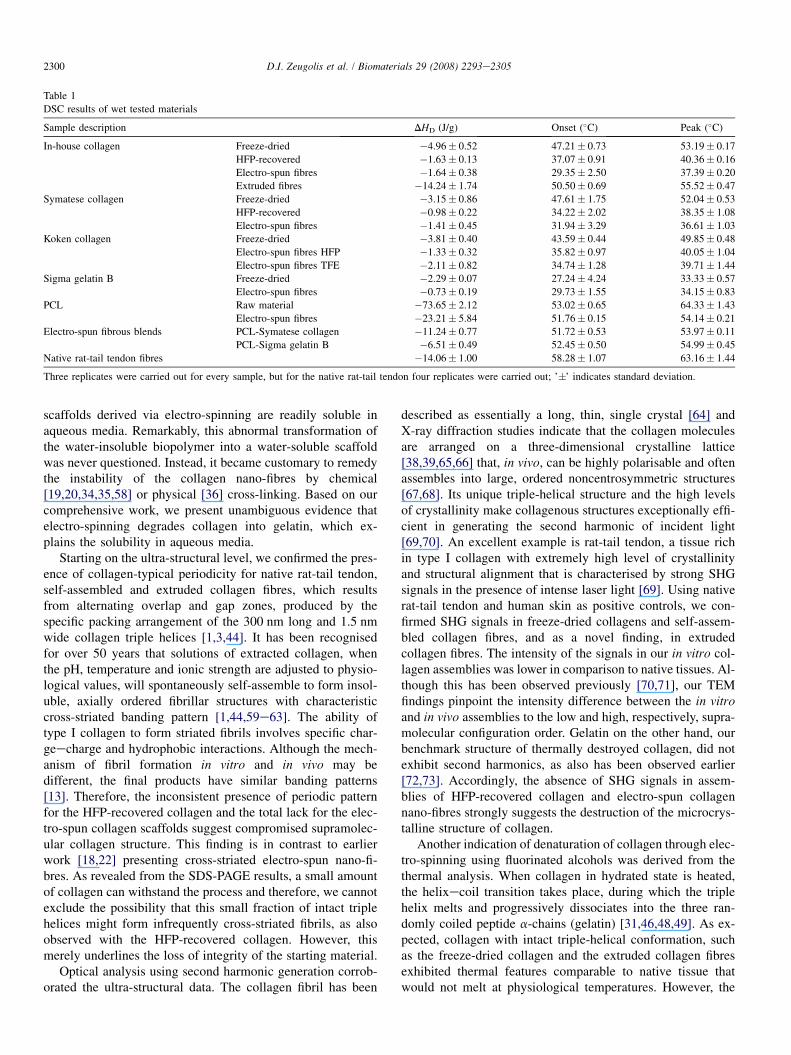
Table 1
DSC results of wet tested materials
Sample description DHD (J/g) Onset (�C) Peak (�C)
In-house collagen Freeze-dried �4.96� 0.52 47.21� 0.73 53.19� 0.17
HFP-recovered �1.63� 0.13 37.07� 0.91 40.36� 0.16
Electro-spun fibres �1.64� 0.38 29.35� 2.50 37.39� 0.20
Extruded fibres �14.24� 1.74 50.50� 0.69 55.52� 0.47
Symatese collagen Freeze-dried �3.15� 0.86 47.61� 1.75 52.04� 0.53
HFP-recovered �0.98� 0.22 34.22� 2.02 38.35� 1.08
Electro-spun fibres �1.41� 0.45 31.94� 3.29 36.61� 1.03
Koken collagen Freeze-dried �3.81� 0.40 43.59� 0.44 49.85� 0.48
Electro-spun fibres HFP �1.33� 0.32 35.82� 0.97 40.05� 1.04
Electro-spun fibres TFE �2.11� 0.82 34.74� 1.28 39.71� 1.44
Sigma gelatin B Freeze-dried �2.29� 0.07 27.24� 4.24 33.33� 0.57
Electro-spun fibres �0.73� 0.19 29.73� 1.55 34.15� 0.83
PCL Raw material �73.65� 2.12 53.02� 0.65 64.33� 1.43
Electro-spun fibres �23.21� 5.84 51.76� 0.15 54.14� 0.21
Electro-spun fibrous blends PCL-Symatese collagen �11.24� 0.77 51.72� 0.53 53.97� 0.11
PCL-Sigma gelatin B �6.51� 0.49 52.45� 0.50 54.99� 0.45
Native rat-tail tendon fibres �14.06� 1.00 58.28� 1.07 63.16� 1.44
Three replicates were carried out for every sample, but for the native rat-tail tendon four replicates were carried out; ’�’ indicates standard deviation.
2300 D.I. Zeugolis et al. / Biomaterials 29 (2008) 2293e2305
scaffolds derived via electro-spinning are readily soluble inaqueous media. Remarkably, this abnormal transformation ofthe water-insoluble biopolymer into a water-soluble scaffoldwas never questioned. Instead, it became customary to remedythe instability of the collagen nano-fibres by chemical[19,20,34,35,58] or physical [36] cross-linking. Based on ourcomprehensive work, we present unambiguous evidence thatelectro-spinning degrades collagen into gelatin, which ex-plains the solubility in aqueous media.
Starting on the ultra-structural level, we confirmed the pres-ence of collagen-typical periodicity for native rat-tail tendon,self-assembled and extruded collagen fibres, which resultsfrom alternating overlap and gap zones, produced by thespecific packing arrangement of the 300 nm long and 1.5 nmwide collagen triple helices [1,3,44]. It has been recognisedfor over 50 years that solutions of extracted collagen, whenthe pH, temperature and ionic strength are adjusted to physio-logical values, will spontaneously self-assemble to form insol-uble, axially ordered fibrillar structures with characteristiccross-striated banding pattern [1,44,59e63]. The ability oftype I collagen to form striated fibrils involves specific char-geecharge and hydrophobic interactions. Although the mech-anism of fibril formation in vitro and in vivo may bedifferent, the final products have similar banding patterns[13]. Therefore, the inconsistent presence of periodic patternfor the HFP-recovered collagen and the total lack for the elec-tro-spun collagen scaffolds suggest compromised supramolec-ular collagen structure. This finding is in contrast to earlierwork [18,22] presenting cross-striated electro-spun nano-fi-bres. As revealed from the SDS-PAGE results, a small amountof collagen can withstand the process and therefore, we cannotexclude the possibility that this small fraction of intact triplehelices might form infrequently cross-striated fibrils, as alsoobserved with the HFP-recovered collagen. However, thismerely underlines the loss of integrity of the starting material.
Optical analysis using second harmonic generation corrob-orated the ultra-structural data. The collagen fibril has been
described as essentially a long, thin, single crystal [64] andX-ray diffraction studies indicate that the collagen moleculesare arranged on a three-dimensional crystalline lattice[38,39,65,66] that, in vivo, can be highly polarisable and oftenassembles into large, ordered noncentrosymmetric structures[67,68]. Its unique triple-helical structure and the high levelsof crystallinity make collagenous structures exceptionally effi-cient in generating the second harmonic of incident light[69,70]. An excellent example is rat-tail tendon, a tissue richin type I collagen with extremely high level of crystallinityand structural alignment that is characterised by strong SHGsignals in the presence of intense laser light [69]. Using nativerat-tail tendon and human skin as positive controls, we con-firmed SHG signals in freeze-dried collagens and self-assem-bled collagen fibres, and as a novel finding, in extrudedcollagen fibres. The intensity of the signals in our in vitro col-lagen assemblies was lower in comparison to native tissues. Al-though this has been observed previously [70,71], our TEMfindings pinpoint the intensity difference between the in vitroand in vivo assemblies to the low and high, respectively, supra-molecular configuration order. Gelatin on the other hand, ourbenchmark structure of thermally destroyed collagen, did notexhibit second harmonics, as also has been observed earlier[72,73]. Accordingly, the absence of SHG signals in assem-blies of HFP-recovered collagen and electro-spun collagennano-fibres strongly suggests the destruction of the microcrys-talline structure of collagen.
Another indication of denaturation of collagen through elec-tro-spinning using fluorinated alcohols was derived from thethermal analysis. When collagen in hydrated state is heated,the helixecoil transition takes place, during which the triplehelix melts and progressively dissociates into the three ran-domly coiled peptide a-chains (gelatin) [31,46,48,49]. As ex-pected, collagen with intact triple-helical conformation, suchas the freeze-dried collagen and the extruded collagen fibresexhibited thermal features comparable to native tissue thatwould not melt at physiological temperatures. However, the
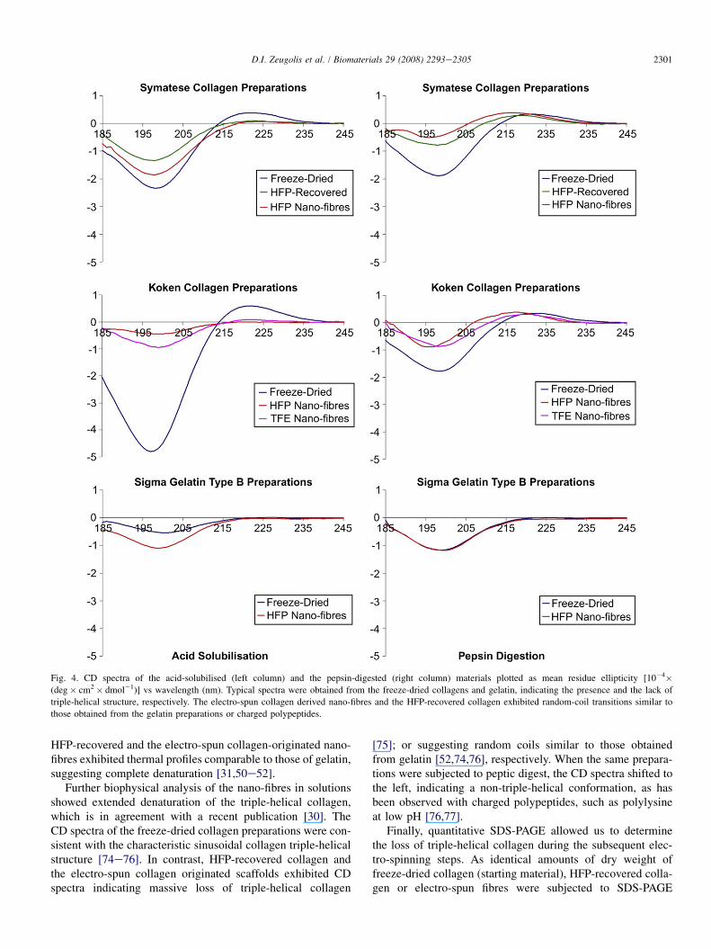
Fig. 4. CD spectra of the acid-solubilised (left column) and the pepsin-digested (right column) materials plotted as mean residue ellipticity [10�4�(deg� cm2� dmol�1)] vs wavelength (nm). Typical spectra were obtained from the freeze-dried collagens and gelatin, indicating the presence and the lack of
triple-helical structure, respectively. The electro-spun collagen derived nano-fibres and the HFP-recovered collagen exhibited random-coil transitions similar to
those obtained from the gelatin preparations or charged polypeptides.
2301D.I. Zeugolis et al. / Biomaterials 29 (2008) 2293e2305
HFP-recovered and the electro-spun collagen-originated nano-fibres exhibited thermal profiles comparable to those of gelatin,suggesting complete denaturation [31,50e52].
Further biophysical analysis of the nano-fibres in solutionsshowed extended denaturation of the triple-helical collagen,which is in agreement with a recent publication [30]. TheCD spectra of the freeze-dried collagen preparations were con-sistent with the characteristic sinusoidal collagen triple-helicalstructure [74e76]. In contrast, HFP-recovered collagen andthe electro-spun collagen originated scaffolds exhibited CDspectra indicating massive loss of triple-helical collagen
[75]; or suggesting random coils similar to those obtainedfrom gelatin [52,74,76], respectively. When the same prepara-tions were subjected to peptic digest, the CD spectra shifted tothe left, indicating a non-triple-helical conformation, as hasbeen observed with charged polypeptides, such as polylysineat low pH [76,77].
Finally, quantitative SDS-PAGE allowed us to determinethe loss of triple-helical collagen during the subsequent elec-tro-spinning steps. As identical amounts of dry weight offreeze-dried collagen (starting material), HFP-recovered colla-gen or electro-spun fibres were subjected to SDS-PAGE
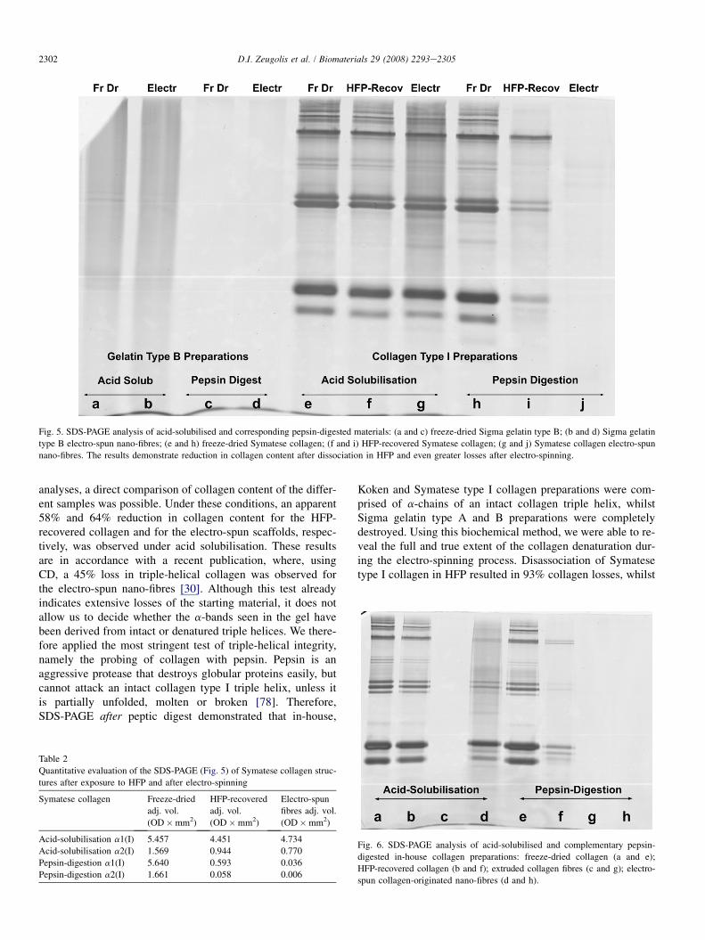
Fig. 5. SDS-PAGE analysis of acid-solubilised and corresponding pepsin-digested materials: (a and c) freeze-dried Sigma gelatin type B; (b and d) Sigma gelatin
type B electro-spun nano-fibres; (e and h) freeze-dried Symatese collagen; (f and i) HFP-recovered Symatese collagen; (g and j) Symatese collagen electro-spun
nano-fibres. The results demonstrate reduction in collagen content after dissociation in HFP and even greater losses after electro-spinning.
2302 D.I. Zeugolis et al. / Biomaterials 29 (2008) 2293e2305
analyses, a direct comparison of collagen content of the differ-ent samples was possible. Under these conditions, an apparent58% and 64% reduction in collagen content for the HFP-recovered collagen and for the electro-spun scaffolds, respec-tively, was observed under acid solubilisation. These resultsare in accordance with a recent publication, where, usingCD, a 45% loss in triple-helical collagen was observed forthe electro-spun nano-fibres [30]. Although this test alreadyindicates extensive losses of the starting material, it does notallow us to decide whether the a-bands seen in the gel havebeen derived from intact or denatured triple helices. We there-fore applied the most stringent test of triple-helical integrity,namely the probing of collagen with pepsin. Pepsin is anaggressive protease that destroys globular proteins easily, butcannot attack an intact collagen type I triple helix, unless itis partially unfolded, molten or broken [78]. Therefore,SDS-PAGE after peptic digest demonstrated that in-house,
Table 2
Quantitative evaluation of the SDS-PAGE (Fig. 5) of Symatese collagen struc-
tures after exposure to HFP and after electro-spinning
Symatese collagen Freeze-dried
adj. vol.
(OD�mm2)
HFP-recovered
adj. vol.
(OD�mm2)
Electro-spun
fibres adj. vol.
(OD�mm2)
Acid-solubilisation a1(I) 5.457 4.451 4.734
Acid-solubilisation a2(I) 1.569 0.944 0.770
Pepsin-digestion a1(I) 5.640 0.593 0.036
Pepsin-digestion a2(I) 1.661 0.058 0.006
Koken and Symatese type I collagen preparations were com-prised of a-chains of an intact collagen triple helix, whilstSigma gelatin type A and B preparations were completelydestroyed. Using this biochemical method, we were able to re-veal the full and true extent of the collagen denaturation dur-ing the electro-spinning process. Disassociation of Symatesetype I collagen in HFP resulted in 93% collagen losses, whilst
Fig. 6. SDS-PAGE analysis of acid-solubilised and complementary pepsin-
digested in-house collagen preparations: freeze-dried collagen (a and e);
HFP-recovered collagen (b and f); extruded collagen fibres (c and g); electro-
spun collagen-originated nano-fibres (d and h).
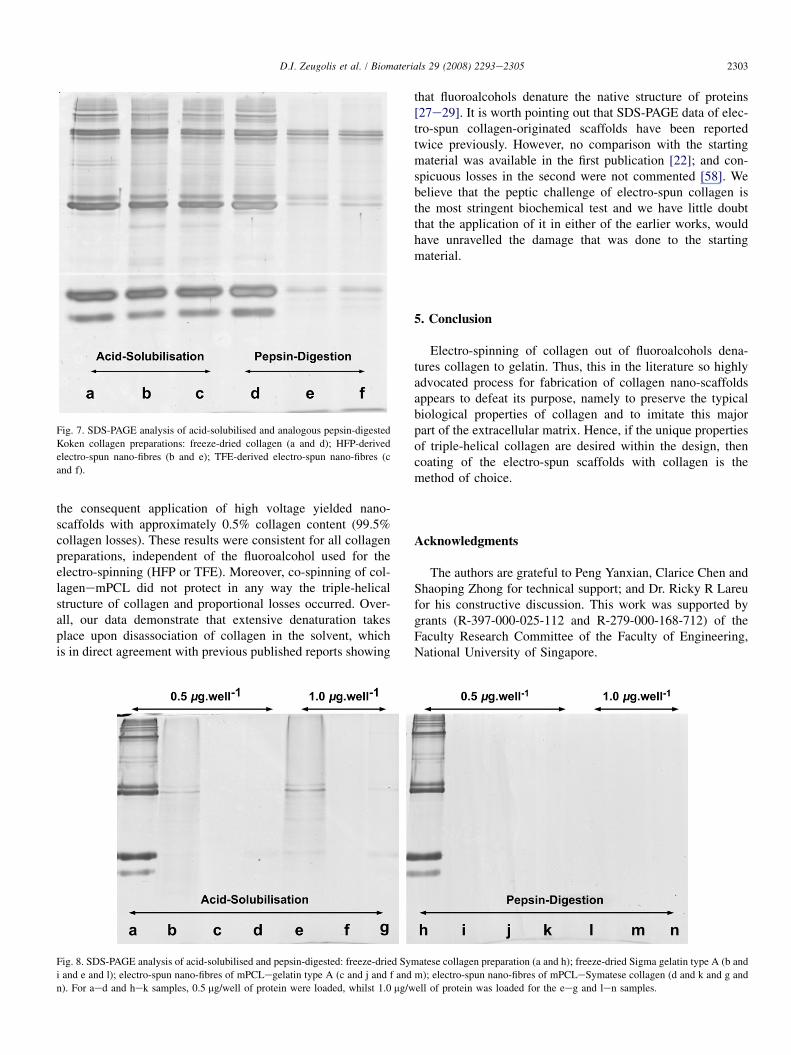
Fig. 7. SDS-PAGE analysis of acid-solubilised and analogous pepsin-digested
Koken collagen preparations: freeze-dried collagen (a and d); HFP-derived
electro-spun nano-fibres (b and e); TFE-derived electro-spun nano-fibres (c
and f).
2303D.I. Zeugolis et al. / Biomaterials 29 (2008) 2293e2305
the consequent application of high voltage yielded nano-scaffolds with approximately 0.5% collagen content (99.5%collagen losses). These results were consistent for all collagenpreparations, independent of the fluoroalcohol used for theelectro-spinning (HFP or TFE). Moreover, co-spinning of col-lagenemPCL did not protect in any way the triple-helicalstructure of collagen and proportional losses occurred. Over-all, our data demonstrate that extensive denaturation takesplace upon disassociation of collagen in the solvent, whichis in direct agreement with previous published reports showing
Fig. 8. SDS-PAGE analysis of acid-solubilised and pepsin-digested: freeze-dried Sym
i and e and l); electro-spun nano-fibres of mPCLegelatin type A (c and j and f and
n). For aed and hek samples, 0.5 mg/well of protein were loaded, whilst 1.0 mg/w
that fluoroalcohols denature the native structure of proteins[27e29]. It is worth pointing out that SDS-PAGE data of elec-tro-spun collagen-originated scaffolds have been reportedtwice previously. However, no comparison with the startingmaterial was available in the first publication [22]; and con-spicuous losses in the second were not commented [58]. Webelieve that the peptic challenge of electro-spun collagen isthe most stringent biochemical test and we have little doubtthat the application of it in either of the earlier works, wouldhave unravelled the damage that was done to the startingmaterial.
5. Conclusion
Electro-spinning of collagen out of fluoroalcohols dena-tures collagen to gelatin. Thus, this in the literature so highlyadvocated process for fabrication of collagen nano-scaffoldsappears to defeat its purpose, namely to preserve the typicalbiological properties of collagen and to imitate this majorpart of the extracellular matrix. Hence, if the unique propertiesof triple-helical collagen are desired within the design, thencoating of the electro-spun scaffolds with collagen is themethod of choice.
Acknowledgments
The authors are grateful to Peng Yanxian, Clarice Chen andShaoping Zhong for technical support; and Dr. Ricky R Lareufor his constructive discussion. This work was supported bygrants (R-397-000-025-112 and R-279-000-168-712) of theFaculty Research Committee of the Faculty of Engineering,National University of Singapore.
atese collagen preparation (a and h); freeze-dried Sigma gelatin type A (b and
m); electro-spun nano-fibres of mPCLeSymatese collagen (d and k and g and
ell of protein was loaded for the eeg and len samples.
2304 D.I. Zeugolis et al. / Biomaterials 29 (2008) 2293e2305
Appendix. Supplementary data
Supplementary data associated with this article can befound in the online version, at doi:10.1016/j.biomaterials.2008.02.009.
References
[1] Kielty CM, Grant ME. The collagen family: structure, assembly and
organization in the extracellular matrix. In: Royce PM, Steinmann B, editors.
Connective tissue and its heritable disorders: molecular, genetic and medical
aspects. 2nd ed. New York: John Wiley Inc; 2002. p. 159e221.
[2] Ramshaw JAM, Werkmeister JA, Glattauer V. Collagen-based biomate-
rials. Biotechnology and Genetic Engineering Reviews 1995;13:335e82.
[3] van der Rest M, Garrone R, Herbage D. Collagen: a family of proteins
with many facets. In: Kleinman HK, editor. Advances in molecular and
cell biology: JAI Press Inc; 1993. p. 1e67.
[4] Hulmes DJS, Miller A, Parry DAD, Piez KA, Woodhead-Galloway J.
Analysis of the primary structure of collagen for the origins of molecular
packing. Journal of Molecular Biology 1973;79:137e48.
[5] Paul RG, Bailey AJ. Chemical stabilisation of collagen as a biomimetic.
The Scientific World Journal 2003;3:138e55.
[6] Bailey AJ, Paul RG. Collagen: a not so simple protein. Journal of the
Society of Leather Technologists and Chemists 1998;82:104e10.
[7] Light ND. Collagen in skin: preparation and analysis. In: Skerrow D,
Skerrow CJ, editors. Methods in skin research. John Wiley and Sons
Ltd; 1985. p. 559e86.
[8] Gelman RA, Poppke DC, Piez KA. Collagen fibril formation in vitro.
The role of the nonhelical terminal regions. Journal of Biological Chem-
istry 1979;254:11741e5.
[9] Friess W. Collagen e biomaterial for drug delivery. European Journal of
Pharmaceutics and Biopharmaceutics 1998;45:113e36.
[10] Lynn AK, Yannas IV, Bonfield W. Antigenicity and immunogenicity of
collagen. Journal of Biomedical Materials Research Part B e Applied
Biomaterials 2004;71B:343e54.
[11] Hsu S, Jamieson AM, Blackwell J. Viscoelastic studies of extracellular
matrix interactions in a model native collagen gel system. Biorheology
1994;31:21e36.
[12] Holmes DF, Graham HK, Trotter JA, Kadler KE. STEM/TEM studies of
collagen fibril assembly. Micron 2001;32:273e85.
[13] Silver FH, Birk DE. Molecular structure of collagen in solution: compar-
ison of types I, II, III and V. International Journal of Biological Macro-
molecules 1984;6:125e32.
[14] Hulmes DJS. The collagen superfamily e diverse structures and assem-
blies. Essays in Biochemistry 1992;27:49e67.
[15] Jarvinen T, Jarvinen T, Kannus P, Jozsa L, Jarvinen M. Collagen fibres of
the spontaneously ruptured human tendons display decrease thickness
and crimp angle. Journal of Orthopaedic Research 2004;22:1303e9.
[16] Huang Y, Meek KM, Ho M-W, Paterson CA. Anaylsis of birefringence
during wound healing and remodeling following alkali burns in rabbit
cornea. Experimental Eye Research 2001;73:521e32.
[17] Silver FH, Freeman JW, Seehra GP. Collagen self-assembly and the de-
velopment of tendon mechanical properties. Journal of Biomechanics
2003;36:1529e53.
[18] Matthews JA, Wnek GE, Simpson DG, Bowlin GL. Electrospinning of
collagen nanofibers. Biomacromolecules 2002;3:232e8.
[19] Li M, Mondrinos MJ, Gandhi MR, Ko FK, Weiss AS, Lelkes PI. Electro-
spun protein fibers as matrices for tissue engineering. Biomaterials
2005;26:5999e6008.
[20] Rho KS, Jeong L, Lee G, Seo B-M, Park YJ, Hong S-D, et al.
Electrospinning of collagen nanofibers: effects on the behavior of normal
human keratinocytes and early-stage wound healing. Biomaterials
2006;27:1452e61.
[21] Shih Y-RV, Chen C-N, Tsai S-W, Wang YJ, Lee OK. Growth of mesen-
chymal stem cells on electrospun type I collagen nanofibers. Stem Cells
2006;24:2391e7.
[22] Telemeco TA, Ayres C, Bowlin GL, Wnek GE, Boland ED, Cohen N,
et al. Regulation of cellular infiltration into tissue engineering scaffolds
composed of submicron diameter fibrils produced by electrospinning.
Acta Biomaterialia 2005;1:377e85.
[23] Zhong S, Teo WE, Zhu X, Beuerman RW, Ramakrishna S, Yung LYL.
An aligned nanofibrous collagen scaffold by electrospinning and its
effects on in vitro fibroblast culture. Journal of Biomedical Materials
Research Part A 2006;79A:456e63.
[24] Zhang YZ, Venugopal J, Huang ZM, Lim CT, Ramakrishna S. Character-
ization of the surface biocompatibility of the electrospun PCLecollagen
nanofibers using fibroblasts. Biomacromolecules 2005;6:2583e9.
[25] Kwon IK, Matsuda T. Co-electrospun nanofiber fabrics of poly(L-lactide-
co-3-caprolactone) with type I collagen or heparin. Biomacromolecules
2005;6:2096e105.
[26] Zhong S, Teo WE, Zhu X, Beuerman R, Ramakrishna S, Yung LY. For-
mation of collagen-glycosaminoglycan blended nanofibrous scaffolds
and their biological properties. Biomacromolecules 2005;6:2998e3004.
[27] Hong D-P, Hoshino M, Kuboi R, Goto Y. Clustering of fluorine-
substituted alcohols as a factor responsible for their marked effects on
proteins and peptides. J Am Chem Soc 1999;121:8427e33.
[28] Cort JR, Andersen NH. Formation of a molten-globule-like state of myo-
globin in aqueous hexafluoroisopropanol. Biochemical and Biophysical
Research Communications 1997;233:687e91.
[29] Kundu A, Kishore N. 1,1,1,3,3,3-Hexafluoroisopropanol induced thermal
unfolding and molten globule state of bovine alpha-lactalbumin: calori-
metric and spectroscopic studies. Biopolymers 2004;73:405e20.
[30] Yang L, Fitie CFC, van der Werf KO, Bennink ML, Dijkstra PJ, Feijen J.
Mechanical properties of single electrospun collagen type I fibers.
Biomaterials 2008;29:955e62.
[31] Bailey AJ. Procter memorial lecture collagen e nature’s framework in
the medical, food and leather industries. Journal of the Society of Leather
Technologists and Chemists 1992;76:111e27.
[32] Finch A, Gardner PJ, Ledward DA, Menashi S. The thermal denaturation
of collagen fibres swollen in aqueous solutions of urea, hexamethylene-
tetramine, p-benzoquinone and tetra-alkylammonium salts. Biochimica
et Biophysica Acta (BBA) e Protein Structure 1974;365:400e4.
[33] Friess W, Lee G. Basic thermoanalytical studies of insoluble collagen
matrices. Biomaterials 1996;17:2289e94.
[34] Buttafoco L, Kolkman NG, Engbers-Buijtenhuijs P, Poot AA,
Dijkstra PJ, Vermes I, et al. Electrospinning of collagen and elastin for
tissue engineering applications. Biomaterials 2006;27:724e34.
[35] Zhong SP, Teo WE, Zhu X, Beuerman R, Ramakrishna S, Yung LYL.
Development of a novel collageneGAG nanofibrous scaffold via electro-
spinning. Materials Science and Engineering C 2007;27:262e6.
[36] Kidoaki S, Kwon IKIK, Matsuda T. Mesoscopic spatial designs of nano-
and microfiber meshes for tissue-engineering matrix and scaffold based
on newly devised multilayering and mixing electrospinning techniques.
Biomaterials 2005;26:37e46.
[37] Veis A, Anesey J, Cohen J. The long range reorganization of gelatin to
the collagen structure. Archives of Biochemistry and Biophysics
1961;94:20e31.
[38] Hulmes DJS, Miller A. Quasi-hexagonal molecular packing in collagen
fibrils. Nature 1979;282:878e80.
[39] Hulmes DJS, Holmes DF, Cummings C. Crystalline regions in collagen
fibrils. Journal of Molecular Biology 1985;184:473e7.
[40] Wess TJ, Hammersley AP, Wess L, Miller A. Molecular packing of type I
collagen in tendon. Journal of Molecular Biology 1998;275:255e67.
[41] Prockop DJ, Fertala A. The collagen fibril: the almost crystalline struc-
ture. Journal of Structural Biology 1998;122:111e8.
[42] Gross J, Highberger JH, Schmitt FO. Collagen structures considered as
states of aggregation of a kinetic unit. The tropocollagen particle. Pro-
ceedings of the National Academy of Sciences of the United States of
America 1954;40:679e88.
[43] Kadler KE, Holmes DF, Trotter JA, Chapman JA. Collagen fibril forma-
tion. Biochemical Journal 1996;316:1e11.
[44] Chapman JA, Tzaphlidou M, Meek KM, Kadler KE. The collagen
fibril e a model system for studying the staining and fixation of a protein.
Electron Microscopy Reviews 1990;3:143e82.
2305D.I. Zeugolis et al. / Biomaterials 29 (2008) 2293e2305
[45] Birk DE, Fitch JM, Babiarz JP, Doane KJ, Linsenmayer TF. Collagen fi-
brillogenesis in vitro: interaction of types I and V collagen regulates fibril
diameter. Journal of Cell Science 1990;95:649e57.
[46] Danielsen CC. Thermal stability of reconstituted collagen fibrils. Shrink-
age characteristics upon in vitro maturation. Mechanisms of Ageing and
Development 1981;15:269e78.
[47] Mentink CJAL, Hendriks M, Levels AAG, Wolffenbuttel BHR. Glucose-
mediated cross-linking of collagen in rat tendon and skin. Clinica
Chimica Acta 2002;321:69e76.
[48] Hormann H, Schlebusch H. Reversible and irreversible denaturation of
collagen fibers. Biochemistry 1971;10:932e7.
[49] Kopp J, Bonnet M, Renou JP. Effect of collagen crosslinking on collagenewater interactions (a DSC investigation). Matrix (Stuttgart, Germany)
1989;9:443e50.
[50] Bigi A, Panzavolta S, Rubini K. Relationship between triple-helix
content and mechanical properties of gelatin films. Biomaterials
2004;25:5675e80.
[51] Tsereteli GI, Smirnova OI. Calometric study of the melting of gelatin
gels. Polymer Science USSR 1991;33:2112e8.
[52] Zhang Z, Li G, Shi B. Physicochemical properties of collagen, gelatin,
and collagen hydrolysate derived from bovine limed split wastes.
Journal of the Society of Leather Technologists and Chemists 2006;
90:23e8.
[53] Raghunath M, Bruckner P, Steinmann B. Delayed triple helix formation
of mutant collagen from patient with osteogenesis imperfecta. Journal of
Molecular Biology 1994;236:940e9.
[54] Zeugolis DI, Paul RG, Attenburrow G. Factors influencing the properties
of reconstituted collagen fibres prior to self assembly: animal species and
collagen extraction method. Journal of Biomedical Materials Research
Part A, <http://www3.interscience.wiley.com/cgi-bin/abstract/117351552/
Abstract>; 27 Nov 2007.
[55] Cavallaro JF, Kemp PD, Kraus KH. Collagen fabrics as biomaterials.
Biotechnology and Bioengineering 1994;43:781e91.
[56] Wang M-C, Pins GD, Silver FH. Collagen fibres with improved
strength for the repair of soft tissue injuries. Biomaterials 1994;15:
507e12.
[57] Huang L, Nagapudi K, Apkarian RP, Chaikof EL. Engineered collagen e
PEO nanofibers and fabrics. Journal of Biomaterials Science Polymer
Edition 2001;12:979e93.
[58] Barnes CP, Pemble CW, Brand DD, Simpson DG, Bowlin GL.
Cross-linking electrospun type II collagen tissue engineering scaf-
folds with carbodiimide in ethanol. Tissue Engineering 2007;13:
1593e605.
[59] Highberger JH, Gross J, Schmitt FO. The interaction of mucoprotein with
soluble collagen: an electron microscope study. Proceedings of the
National Academy of Sciences of the United States of America
1951;37:286e91.
[60] Gross J, Highberger JH, Schmitt FO. Extraction of collagen from connec-
tive tissue by neutral salt solutions. Proceedings of the National Academy
of Sciences of the United States of America 1955;41:1e7.
[61] Comper WD, Veis A. The mechanism of nucleation for in vitro collagen
fibril formation. Biopolymers 1977;16:2113e31.
[62] Williams BR, Gelman RA, Poppke DC, Piez KA. Collagen fibril forma-
tion. Optimal in vitro conditions and preliminary kinetic results. Journal
of Biological Chemistry 1978;253:6578e85.
[63] Holmes DF, Capaldi MJ, Chapman JA. Reconstitution of collagen fibrils
in vitro; the assembly process depends on the initiating procedure. Inter-
national Journal of Biological Macromolecules 1986;8:161e6.
[64] Bear RS. The structure of collagen fibrils. Advanced Protein Research
1952;7:69e160.
[65] Brodsky Doyle B, Hukins DWL, Hulmes DJS, Miller A, White S, Wood-
head Galloway J. Low angle X-ray diffraction studies on stained rat tail
tendons. Biochimica et Biophysica Acta (BBA) e Protein Structure
1978;535:25e32.
[66] Hulmes DJS, Wess TJ, Prockop DJ, Fratzl P. Radial packing, order and
disorder in collagen fibrils. Biophysical Journal 1995;68:1661e70.
[67] Campagnola PJ, Loew LM. Second-harmonic imaging microscopy for
visualizing biomolecular arrays in cells, tissues and organisms. Nature
Biotechnology 2003;21:1356e60.
[68] Williams RM, Zipfel WR, Webb WW. Interpreting second-harmonic
generation images of collagen I fibrils. Biophysical Journal
2005;88:1377e86.
[69] Cox G, Kable E, Jones A, Fraser I, Manconi F, Gorrell MD. 3-Dimen-
sional imaging of collagen using second harmonic generation. Journal
of Structural Biology 2003;141:53e62.
[70] Cox G, Xu P, Sheppard C, Ramshaw J. Characterization of the second
harmonic signal from collagen. In: Periasamy A, So PT, editors. Multi-
photon microscopy in the biomedical sciences III: proceedings of
SPIE; 2003.
[71] Stoller P, Reiser KM, Celliers PM, Rubenchik AM. Polarization-
modulated second harmonic generation in collagen. Biophysical Journal
2002;82:3330e42.
[72] Theodossiou TA, Thrasivoulou C, Ekwobi C, Becker DL. Second har-
monic generation confocal microscopy of collagen type I from rat tendon
cryosections. Biophysical Journal 2006;91:4665e77.
[73] Kim B-M, Eichler J, Reiser KM, Rubenchik AM, Da Silva LB. Collagen
structure and nonlinear susceptibility: effects of heat, glycation, and en-
zymatic cleavage on second harmonic signal intensity. Lasers in Surgery
and Medicine 2000;27:329e35.
[74] Usha R, Ramasami T. The effects of urea and n-propanol on collagen de-
naturation: using DSC, circular dichroism and viscosity. Thermochimica
Acta 2004;409:201e6.
[75] Chu FH, Lukton A. Collagenase induced changes in the circular dichro-
ism spectrum of collagen. Biopolymers 1974;13:1427e34.
[76] Brodsky-Doyle B, Leonard KR, Reid KBM. Circular-dichroism and
electron-microscopy studies of human subcomponent C1q before and
after limited proteolysis by pepsin. Biochemical Journal 1976;
159:279e86.
[77] Jenness DD, Sprecher C, Johnson JWC. Circular dichroism of collagen,
gelatin, and poly(proline) II in the vacuum ultraviolet. Biopolymers
1976;15:513e21.
[78] Bruckner P, Prockop DJ. Proteolytic enzymes as probes for the triple-
helical conformation of procollagen. Analytical Biochemistry
1981;110:360e8.
Related Documents