Elderberry flavonoids bind to and prevent H1N1 infection in vitro Bill Roschek Jr. a , Ryan C. Fink b , Matthew D. McMichael a , Dan Li c , Randall S. Alberte a, * a HerbalScience Group LLC, 1004 Collier Center Way, Suite 200, Naples, FL 34110, USA b Leonard M. Miller School of Medicine, University of Miami, Miami, FL 33136, USA c HerbalScience Singapore, Pte. Ltd., 1 Science Park Road, Capricorn, Science Park II, Singapore 117528, Singapore article info Article history: Received 9 September 2007 Received in revised form 18 May 2009 Available online 12 August 2009 Keywords: Sambucus nigra L. Caprifoliaceae Elderberry Anti-viral flavonoids DART TOF-MS Flavonoids Influenza abstract A ionization technique in mass spectrometry called Direct Analysis in Real Time Mass Spectrometry (DART TOF-MS) coupled with a Direct Binding Assay was used to identify and characterize anti-viral com- ponents of an elderberry fruit (Sambucus nigra L.) extract without either derivatization or separation by standard chromatographic techniques. The elderberry extract inhibited Human Influenza A (H1N1) infec- tion in vitro with an IC 50 value of 252 ± 34 lg/mL. The Direct Binding Assay established that flavonoids from the elderberry extract bind to H1N1 virions and, when bound, block the ability of the viruses to infect host cells. Two compounds were identified, 5,7,3 0 ,4 0 -tetra-O-methylquercetin (1) and 5,7-dihy- droxy-4-oxo-2-(3,4,5-trihydroxyphenyl)chroman-3-yl-3,4,5-trihydroxycyclohexanecarboxylate (2), as H1N1-bound chemical species. Compound 1 and dihydromyricetin (3), the corresponding 3-hydroxyfl- avonone of 2, were synthesized and shown to inhibit H1N1 infection in vitro by binding to H1N1 virions, blocking host cell entry and/or recognition. Compound 1 gave an IC 50 of 0.13 lg/mL (0.36 lM) for H1N1 infection inhibition, while dihydromyricetin (3) achieved an IC 50 of 2.8 lg/mL (8.7 lM). The H1N1 inhi- bition activities of the elderberry flavonoids compare favorably to the known anti-influenza activities of Oseltamivir (Tamiflu Ò ; 0.32 lM) and Amantadine (27 lM). Ó 2009 Elsevier Ltd. All rights reserved. 1. Introduction The chemical complexity of botanical extracts has made mass spectrometric characterization of whole extracts difficult due to the lack of reliable extraction methodologies that yield optimized extracts with dose-to-dose reliable chemical compositions (Schmidt et al., 2007). A relatively new ionization source in mass spectrometry, termed DART (Direct Analysis in Real Time) (Cody et al., 2005), is coupled to a time-of-flight mass spectrometer, mak- ing it possible to rapidly and accurately identify the chemical com- ponents in botanicals and extracts at atmospheric pressure, typically with no sample preparation or processing requirements. The DART ion source utilizes electronic excited-state species, such as metastable helium and nitrogen atoms, as plasmas. These ex- cited atoms ionize samples directly for mass spectrometric analysis. The most common ions produced during DART analysis are the [M+H] + cations and the [M+NH 4 ] + adducts (observed if ammonium hydroxide is present near the DART source); however metal, cation adducts are never observed (Cody et al., 2005). DART is capable of analyzing surface materials without direct exposure of the samples to elevated temperatures and/or electrical potentials as occurs dur- ing atmospheric pressure chemical ionization (Sciex, 1992) and elec- trospray ionization (Pramanik et al., 2002) mass spectrometric techniques. Fragmentation of the samples during DART ionization can be induced by adjusting the mass spectrometer voltages, allow- ing for more detailed structural information (Cody et al., 2005). Re- cently, DART TOF-MS was used to determine the molecular formulae and structures of toxoid compounds in cell cultures of Taxus wallichiana (Banerjee et al., 2008), and alkaloids expressed in the hairy roots of Rauvolfia serpentine (Madhusudanan et al., 2008). The combination of enhanced super critical CO 2 extraction tech- nologies and affinity chromatography has enabled the production of optimized and dose-reliable botanical extracts from variable feedstocks that possess a defined bioactive profile (Alberte et al., 2007). These extraction technologies were employed herein to generate reproducible extracts of elderberry (Sambucus nigra L.) fruits for both chemical characterization and assessment of biolog- ical activity. Elderberries are known to be rich in phenolic com- pounds, including phenolic acids, flavonoids, catechins, and proanthocyanidins (de Pascual-Teresa et al., 2000; Hakkinen et al., 1999), as well as possessing a variety of anti-oxidant proper- ties (Abuja et al., 1998; Rice-Evans et al., 1996; Seeram and Nair, 2002; Wang et al., 1997), and enhancing the immune response (Barak et al., 2001; Zakay-Rones et al., 1995). In addition, elder- berry extracts have shown anti-influenza activity in human clinical trials (Zakay-Rones et al., 2004). 0031-9422/$ - see front matter Ó 2009 Elsevier Ltd. All rights reserved. doi:10.1016/j.phytochem.2009.06.003 * Corresponding author. Tel.: +1 239 597 8822; fax: +1 239 597 8001. E-mail addresses: [email protected], [email protected] (R.S. Alberte). Phytochemistry 70 (2009) 1255–1261 Contents lists available at ScienceDirect Phytochemistry journal homepage: www.elsevier.com/locate/phytochem

Welcome message from author
This document is posted to help you gain knowledge. Please leave a comment to let me know what you think about it! Share it to your friends and learn new things together.
Transcript
Phytochemistry 70 (2009) 1255–1261
Contents lists available at ScienceDirect
Phytochemistry
journal homepage: www.elsevier .com/locate /phytochem
Elderberry flavonoids bind to and prevent H1N1 infection in vitro
Bill Roschek Jr. a, Ryan C. Fink b, Matthew D. McMichael a, Dan Li c, Randall S. Alberte a,*
a HerbalScience Group LLC, 1004 Collier Center Way, Suite 200, Naples, FL 34110, USAb Leonard M. Miller School of Medicine, University of Miami, Miami, FL 33136, USAc HerbalScience Singapore, Pte. Ltd., 1 Science Park Road, Capricorn, Science Park II, Singapore 117528, Singapore
a r t i c l e i n f o
Article history:Received 9 September 2007Received in revised form 18 May 2009Available online 12 August 2009
Keywords:Sambucus nigra L. CaprifoliaceaeElderberryAnti-viral flavonoidsDART TOF-MSFlavonoidsInfluenza
0031-9422/$ - see front matter � 2009 Elsevier Ltd. Adoi:10.1016/j.phytochem.2009.06.003
* Corresponding author. Tel.: +1 239 597 8822; faxE-mail addresses: [email protected]
Alberte).
a b s t r a c t
A ionization technique in mass spectrometry called Direct Analysis in Real Time Mass Spectrometry(DART TOF-MS) coupled with a Direct Binding Assay was used to identify and characterize anti-viral com-ponents of an elderberry fruit (Sambucus nigra L.) extract without either derivatization or separation bystandard chromatographic techniques. The elderberry extract inhibited Human Influenza A (H1N1) infec-tion in vitro with an IC50 value of 252 ± 34 lg/mL. The Direct Binding Assay established that flavonoidsfrom the elderberry extract bind to H1N1 virions and, when bound, block the ability of the viruses toinfect host cells. Two compounds were identified, 5,7,30,40-tetra-O-methylquercetin (1) and 5,7-dihy-droxy-4-oxo-2-(3,4,5-trihydroxyphenyl)chroman-3-yl-3,4,5-trihydroxycyclohexanecarboxylate (2), asH1N1-bound chemical species. Compound 1 and dihydromyricetin (3), the corresponding 3-hydroxyfl-avonone of 2, were synthesized and shown to inhibit H1N1 infection in vitro by binding to H1N1 virions,blocking host cell entry and/or recognition. Compound 1 gave an IC50 of 0.13 lg/mL (0.36 lM) for H1N1infection inhibition, while dihydromyricetin (3) achieved an IC50 of 2.8 lg/mL (8.7 lM). The H1N1 inhi-bition activities of the elderberry flavonoids compare favorably to the known anti-influenza activities ofOseltamivir (Tamiflu�; 0.32 lM) and Amantadine (27 lM).
� 2009 Elsevier Ltd. All rights reserved.
1. Introduction
The chemical complexity of botanical extracts has made massspectrometric characterization of whole extracts difficult due tothe lack of reliable extraction methodologies that yield optimizedextracts with dose-to-dose reliable chemical compositions(Schmidt et al., 2007). A relatively new ionization source in massspectrometry, termed DART (Direct Analysis in Real Time) (Codyet al., 2005), is coupled to a time-of-flight mass spectrometer, mak-ing it possible to rapidly and accurately identify the chemical com-ponents in botanicals and extracts at atmospheric pressure,typically with no sample preparation or processing requirements.
The DART ion source utilizes electronic excited-state species,such as metastable helium and nitrogen atoms, as plasmas. These ex-cited atoms ionize samples directly for mass spectrometric analysis.The most common ions produced during DART analysis are the[M+H]+ cations and the [M+NH4]+ adducts (observed if ammoniumhydroxide is present near the DART source); however metal, cationadducts are never observed (Cody et al., 2005). DART is capable ofanalyzing surface materials without direct exposure of the samplesto elevated temperatures and/or electrical potentials as occurs dur-
ll rights reserved.
: +1 239 597 8001.m, [email protected] (R.S.
ing atmospheric pressure chemical ionization (Sciex, 1992) and elec-trospray ionization (Pramanik et al., 2002) mass spectrometrictechniques. Fragmentation of the samples during DART ionizationcan be induced by adjusting the mass spectrometer voltages, allow-ing for more detailed structural information (Cody et al., 2005). Re-cently, DART TOF-MS was used to determine the molecularformulae and structures of toxoid compounds in cell cultures ofTaxus wallichiana (Banerjee et al., 2008), and alkaloids expressed inthe hairy roots of Rauvolfia serpentine (Madhusudanan et al., 2008).
The combination of enhanced super critical CO2 extraction tech-nologies and affinity chromatography has enabled the productionof optimized and dose-reliable botanical extracts from variablefeedstocks that possess a defined bioactive profile (Alberte et al.,2007). These extraction technologies were employed herein togenerate reproducible extracts of elderberry (Sambucus nigra L.)fruits for both chemical characterization and assessment of biolog-ical activity. Elderberries are known to be rich in phenolic com-pounds, including phenolic acids, flavonoids, catechins, andproanthocyanidins (de Pascual-Teresa et al., 2000; Hakkinenet al., 1999), as well as possessing a variety of anti-oxidant proper-ties (Abuja et al., 1998; Rice-Evans et al., 1996; Seeram and Nair,2002; Wang et al., 1997), and enhancing the immune response(Barak et al., 2001; Zakay-Rones et al., 1995). In addition, elder-berry extracts have shown anti-influenza activity in human clinicaltrials (Zakay-Rones et al., 2004).
1256 B. Roschek Jr et al. / Phytochemistry 70 (2009) 1255–1261
We utilized an optimized elderberry extract as well as a newlydeveloped Direct Binding Assay to identify key bioactive flavonoidsin elderberry fruits that contribute to the reported anti-influenzaactivities. The identified flavonoids bind to Human Influenza A(H1N1) viruses and block viral infection in vitro.
2. Results and discussion
2.1. Anti-viral activity of elderberry fruit extracts
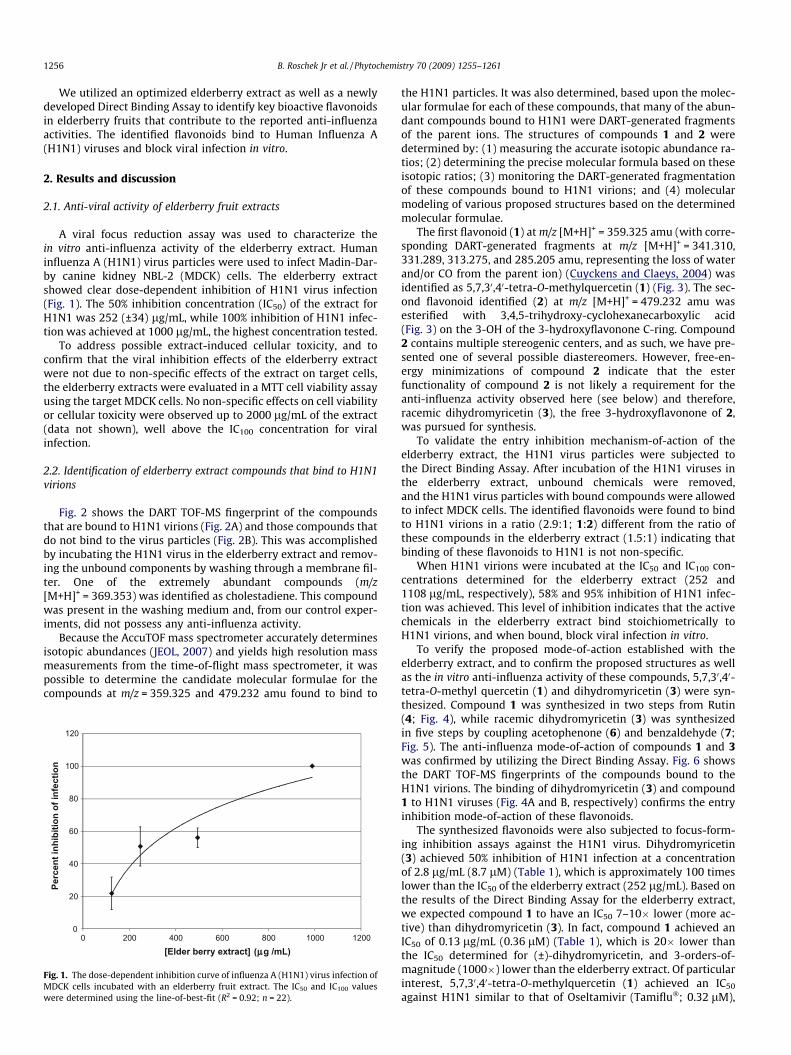
A viral focus reduction assay was used to characterize thein vitro anti-influenza activity of the elderberry extract. Humaninfluenza A (H1N1) virus particles were used to infect Madin-Dar-by canine kidney NBL-2 (MDCK) cells. The elderberry extractshowed clear dose-dependent inhibition of H1N1 virus infection(Fig. 1). The 50% inhibition concentration (IC50) of the extract forH1N1 was 252 (±34) lg/mL, while 100% inhibition of H1N1 infec-tion was achieved at 1000 lg/mL, the highest concentration tested.
To address possible extract-induced cellular toxicity, and toconfirm that the viral inhibition effects of the elderberry extractwere not due to non-specific effects of the extract on target cells,the elderberry extracts were evaluated in a MTT cell viability assayusing the target MDCK cells. No non-specific effects on cell viabilityor cellular toxicity were observed up to 2000 lg/mL of the extract(data not shown), well above the IC100 concentration for viralinfection.
2.2. Identification of elderberry extract compounds that bind to H1N1virions
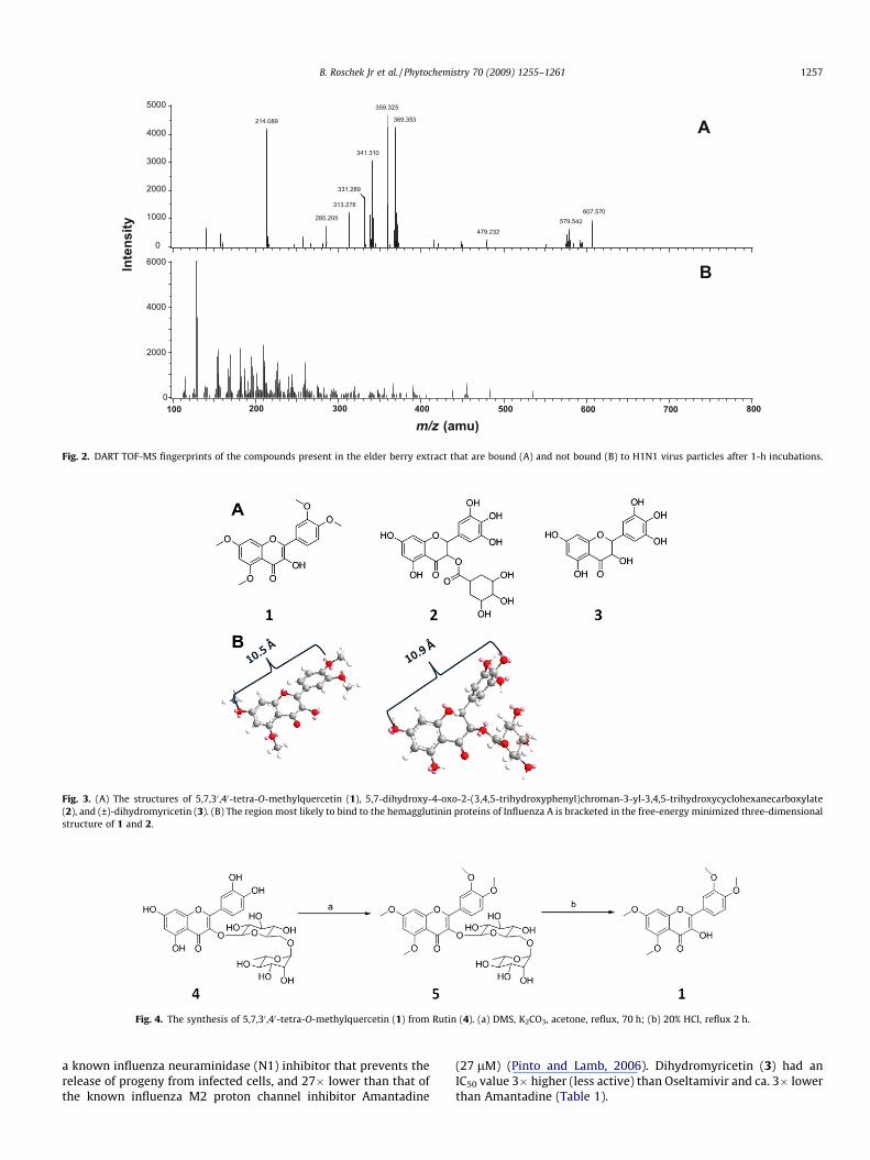
Fig. 2 shows the DART TOF-MS fingerprint of the compoundsthat are bound to H1N1 virions (Fig. 2A) and those compounds thatdo not bind to the virus particles (Fig. 2B). This was accomplishedby incubating the H1N1 virus in the elderberry extract and remov-ing the unbound components by washing through a membrane fil-ter. One of the extremely abundant compounds (m/z[M+H]+ = 369.353) was identified as cholestadiene. This compoundwas present in the washing medium and, from our control exper-iments, did not possess any anti-influenza activity.
Because the AccuTOF mass spectrometer accurately determinesisotopic abundances (JEOL, 2007) and yields high resolution massmeasurements from the time-of-flight mass spectrometer, it waspossible to determine the candidate molecular formulae for thecompounds at m/z = 359.325 and 479.232 amu found to bind to
0
20
40
60
80
100
120
0 200 400 600 800 1000 1200
Perc
ent i
nhib
ition
of i
nfec
tion
[Elder berry extract] (µµg /mL)
Fig. 1. The dose-dependent inhibition curve of influenza A (H1N1) virus infection ofMDCK cells incubated with an elderberry fruit extract. The IC50 and IC100 valueswere determined using the line-of-best-fit (R2 = 0.92; n = 22).
the H1N1 particles. It was also determined, based upon the molec-ular formulae for each of these compounds, that many of the abun-dant compounds bound to H1N1 were DART-generated fragmentsof the parent ions. The structures of compounds 1 and 2 weredetermined by: (1) measuring the accurate isotopic abundance ra-tios; (2) determining the precise molecular formula based on theseisotopic ratios; (3) monitoring the DART-generated fragmentationof these compounds bound to H1N1 virions; and (4) molecularmodeling of various proposed structures based on the determinedmolecular formulae.
The first flavonoid (1) at m/z [M+H]+ = 359.325 amu (with corre-sponding DART-generated fragments at m/z [M+H]+ = 341.310,331.289, 313.275, and 285.205 amu, representing the loss of waterand/or CO from the parent ion) (Cuyckens and Claeys, 2004) wasidentified as 5,7,30,40-tetra-O-methylquercetin (1) (Fig. 3). The sec-ond flavonoid identified (2) at m/z [M+H]+ = 479.232 amu wasesterified with 3,4,5-trihydroxy-cyclohexanecarboxylic acid(Fig. 3) on the 3-OH of the 3-hydroxyflavonone C-ring. Compound2 contains multiple stereogenic centers, and as such, we have pre-sented one of several possible diastereomers. However, free-en-ergy minimizations of compound 2 indicate that the esterfunctionality of compound 2 is not likely a requirement for theanti-influenza activity observed here (see below) and therefore,racemic dihydromyricetin (3), the free 3-hydroxyflavonone of 2,was pursued for synthesis.
To validate the entry inhibition mechanism-of-action of theelderberry extract, the H1N1 virus particles were subjected tothe Direct Binding Assay. After incubation of the H1N1 viruses inthe elderberry extract, unbound chemicals were removed,and the H1N1 virus particles with bound compounds were allowedto infect MDCK cells. The identified flavonoids were found to bindto H1N1 virions in a ratio (2.9:1; 1:2) different from the ratio ofthese compounds in the elderberry extract (1.5:1) indicating thatbinding of these flavonoids to H1N1 is not non-specific.
When H1N1 virions were incubated at the IC50 and IC100 con-centrations determined for the elderberry extract (252 and1108 lg/mL, respectively), 58% and 95% inhibition of H1N1 infec-tion was achieved. This level of inhibition indicates that the activechemicals in the elderberry extract bind stoichiometrically toH1N1 virions, and when bound, block viral infection in vitro.
To verify the proposed mode-of-action established with theelderberry extract, and to confirm the proposed structures as wellas the in vitro anti-influenza activity of these compounds, 5,7,30,40-tetra-O-methyl quercetin (1) and dihydromyricetin (3) were syn-thesized. Compound 1 was synthesized in two steps from Rutin(4; Fig. 4), while racemic dihydromyricetin (3) was synthesizedin five steps by coupling acetophenone (6) and benzaldehyde (7;Fig. 5). The anti-influenza mode-of-action of compounds 1 and 3was confirmed by utilizing the Direct Binding Assay. Fig. 6 showsthe DART TOF-MS fingerprints of the compounds bound to theH1N1 virions. The binding of dihydromyricetin (3) and compound1 to H1N1 viruses (Fig. 4A and B, respectively) confirms the entryinhibition mode-of-action of these flavonoids.
The synthesized flavonoids were also subjected to focus-form-ing inhibition assays against the H1N1 virus. Dihydromyricetin(3) achieved 50% inhibition of H1N1 infection at a concentrationof 2.8 lg/mL (8.7 lM) (Table 1), which is approximately 100 timeslower than the IC50 of the elderberry extract (252 lg/mL). Based onthe results of the Direct Binding Assay for the elderberry extract,we expected compound 1 to have an IC50 7–10� lower (more ac-tive) than dihydromyricetin (3). In fact, compound 1 achieved anIC50 of 0.13 lg/mL (0.36 lM) (Table 1), which is 20� lower thanthe IC50 determined for (±)-dihydromyricetin, and 3-orders-of-magnitude (1000�) lower than the elderberry extract. Of particularinterest, 5,7,30,40-tetra-O-methylquercetin (1) achieved an IC50
against H1N1 similar to that of Oseltamivir (Tamiflu�; 0.32 lM),
0
( )129.07
130.05
209.10181.13155.09
211.14 259.14
115.09260.14 366.19 455.28
356.22320.18 391.21 438.28 483.28 535.32
A( )
129.07
209.10155.09
211.14 259.14
260. 455.28356.22320.18 391.21 438.28 483.28 535.32
A
B0
359.33
369.36214.09
341.31
331.29
370.36313.28
371.33 607.57285.29140.11 579.54
158.03 215.09 257.26372.40160.03 479.51415.24 575.89449.09
216.08
B5000
4000
3000
2000
1000
6000
4000
2000
359.33
369.36214.09
341.31
331.29
370.36313.28
371.33 607.57285.29140.11 579.54
158.03 215.09257.26372.40160.03 479.51415.24 575.89449.09
216.08
BA
Inte
nsity
m/ z (a mu700600500400300200100 800
m/z (amu)
479.232579.542
607.570
369.353
359.325
341.310
331.289
313.276
285.205
214.089
Fig. 2. DART TOF-MS fingerprints of the compounds present in the elder berry extract that are bound (A) and not bound (B) to H1N1 virus particles after 1-h incubations.
Fig. 3. (A) The structures of 5,7,30 ,40-tetra-O-methylquercetin (1), 5,7-dihydroxy-4-oxo-2-(3,4,5-trihydroxyphenyl)chroman-3-yl-3,4,5-trihydroxycyclohexanecarboxylate(2), and (±)-dihydromyricetin (3). (B) The region most likely to bind to the hemagglutinin proteins of Influenza A is bracketed in the free-energy minimized three-dimensionalstructure of 1 and 2.
Fig. 4. The synthesis of 5,7,30 ,40-tetra-O-methylquercetin (1) from Rutin (4). (a) DMS, K2CO3, acetone, reflux, 70 h; (b) 20% HCl, reflux 2 h.
B. Roschek Jr et al. / Phytochemistry 70 (2009) 1255–1261 1257
a known influenza neuraminidase (N1) inhibitor that prevents therelease of progeny from infected cells, and 27� lower than that ofthe known influenza M2 proton channel inhibitor Amantadine
(27 lM) (Pinto and Lamb, 2006). Dihydromyricetin (3) had anIC50 value 3� higher (less active) than Oseltamivir and ca. 3� lowerthan Amantadine (Table 1).
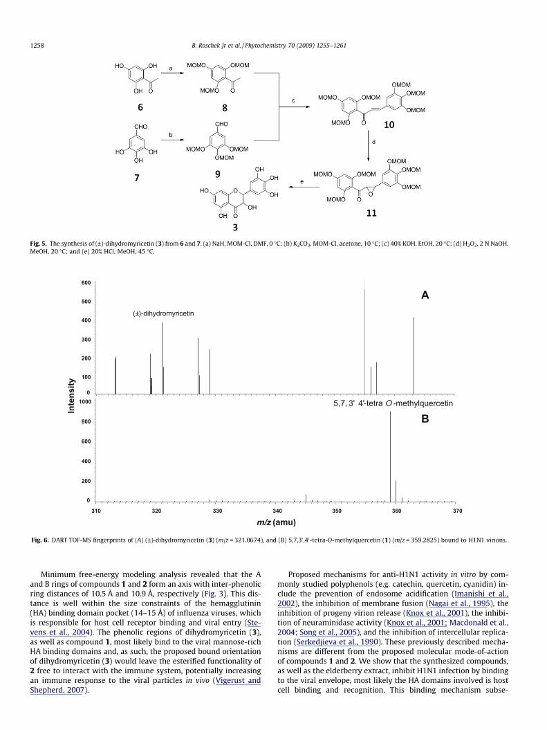
Fig. 5. The synthesis of (±)-dihydromyricetin (3) from 6 and 7. (a) NaH, MOM-Cl, DMF, 0 �C; (b) K2CO3, MOM-Cl, acetone, 10 �C; (c) 40% KOH, EtOH, 20 �C; (d) H2O2, 2 N NaOH,MeOH, 20 �C; and (e) 20% HCl, MeOH, 45 �C.
A
B
(±)-dihydromyricetin
310 320 330 340 350 360 370
600
500
400
300
200
100
01000
800
600
400
200
0
Inte
nsity
m/z (amu)
5,7, 3' 4'-tetra O -methylquercetin
Fig. 6. DART TOF-MS fingerprints of (A) (±)-dihydromyricetin (3) (m/z = 321.0674), and (B) 5,7,30 ,40-tetra-O-methylquercetin (1) (m/z = 359.2825) bound to H1N1 virions.
1258 B. Roschek Jr et al. / Phytochemistry 70 (2009) 1255–1261
Minimum free-energy modeling analysis revealed that the Aand B rings of compounds 1 and 2 form an axis with inter-phenolicring distances of 10.5 Å and 10.9 Å, respectively (Fig. 3). This dis-tance is well within the size constraints of the hemagglutinin(HA) binding domain pocket (14–15 Å) of influenza viruses, whichis responsible for host cell receptor binding and viral entry (Ste-vens et al., 2004). The phenolic regions of dihydromyricetin (3),as well as compound 1, most likely bind to the viral mannose-richHA binding domains and, as such, the proposed bound orientationof dihydromyricetin (3) would leave the esterified functionality of2 free to interact with the immune system, potentially increasingan immune response to the viral particles in vivo (Vigerust andShepherd, 2007).
Proposed mechanisms for anti-H1N1 activity in vitro by com-monly studied polyphenols (e.g. catechin, quercetin, cyanidin) in-clude the prevention of endosome acidification (Imanishi et al.,2002), the inhibition of membrane fusion (Nagai et al., 1995), theinhibition of progeny virion release (Knox et al., 2001), the inhibi-tion of neuraminidase activity (Knox et al., 2001; Macdonald et al.,2004; Song et al., 2005), and the inhibition of intercellular replica-tion (Serkedjieva et al., 1990). These previously described mecha-nisms are different from the proposed molecular mode-of-actionof compounds 1 and 2. We show that the synthesized compounds,as well as the elderberry extract, inhibit H1N1 infection by bindingto the viral envelope, most likely the HA domains involved is hostcell binding and recognition. This binding mechanism subse-
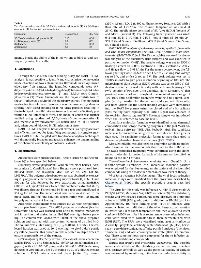
Table 1The IC50 values determined for 5,7,30 ,40-tetra-O-methylquercetin (1), the (±)-dihydr-omyricetin (3), Oseltamivir, and Amantadine.
Compound IC50 (lg/mL) IC50 (lM)
5,7,30 ,40-Tetra-O-methyl quercetin (1) 0.15 0.36(±)-Dihydromyricetin (3) 2.8 8.7Oseltamivir 0.1 0.32Amantadine 4.1 27
B. Roschek Jr et al. / Phytochemistry 70 (2009) 1255–1261 1259
quently blocks the ability of the H1N1 virions to bind to, and con-sequently enter, host cells.
3. Conclusions
Through the use of the Direct Binding Assay and DART TOF-MSanalysis, it was possible to identify and characterize the molecularmode-of-action of two anti-influenza flavonoids in an optimizedelderberry fruit extract. The identified compounds were 5,7-dihydroxy-4-oxo-2-(3,4,5-trihydroxyphenyl)chroman-3-yl-3,4,5-tri-hydroxycyclohexanecarboxylate (2) and 5,7,30,40-tetra-O-meth-ylquercetin (1). These flavonoids are the major contributors tothe anti-influenza activity of the elderberry extract. The molecularmode-of-action of these flavonoids was determined by demon-strating their direct binding to H1N1 virus particles resulting inthe inability of the H1N1 viruses to enter host cells, effectively pre-venting H1N1 infection in vitro. This mode-of-action was furtherverified using synthesized 5,7,30,40-tetra-O-methylquercetin (1)and racemic dihydromyricetin (3) which bind to H1N1 virionsand, when bound, blocked H1N1 infection in vitro.
DART TOF-MS analysis of botanical extracts is a highly accurateand efficient method for identifying compounds in complex mix-tures. DART TOF-MS coupled with traditional analytical techniqueswill dramatically broaden and greatly enhance the understandingof the chemical complexity of botanical extracts.
4. Experimental
All solvents were purchased from Thermo Fisher Scientific (Fair-lawn, NJ) unless specified below.
Elderberry extract preparation: Wild crafted elder berries (Sam-bucus nigra L., Caprifoliaceae) from Hungary were purchased fromBlessed Herbs, Inc. (Oakham, MA; Product No. 724, Lot No.L10379w). The polymer adsorbent extract was obtained by extract-ing 20 g of ground elderberries using supercritical CO2 at 60 �C and300 bar for 2 h, followed by two extractions using EtOH:H2O(100 mL, 4:1, v/v) EtOH for 2 h each. The combined extracted slurrywas filtered through Fisherbrand P4 filter paper and centrifuged at537�g for 20 min. The supernatant was vacuum distilled to re-move EtOH, and the final solution concentration was �35 mg/mLfor polymer adsorbent loading.
Adsorption experiments were carried out at room temperaturein an open batch system. The ADS5 polymer adsorbent (NankaiUniversity, China) was washed with EtOH to remove monomersand impurities and soaked in distilled H2O overnight before pack-ing. The column was loaded with 60 mL of the above preparedsolution and washed with two column volumes of distilled H2O.The column was eluted using EtOH/H2O (40 mL, 4:1, v/v). The col-lected fraction was dried at 50 �C overnight to yield a dark purplecrystalline powder. This procedure was repeated multiple times toensure reproducibility of the extract.
HPLC analysis of elderberry extracts: The extracts were character-ized by HPLC–UV on a Shimadzu LC-10AVP system (Shimadzu, Sin-gapore) with a LC10ADVP pump and a SPD-M 10AVP diode arraydetector at 280 and 350 nm by injecting 10 lL of a diluted extractsolution in EtOH onto a reversed phase Jupiter C18 column
(250 � 4.6 mm I.D., 5 l, 300 Å; Phenomenex, Torrance, CA) with aflow rate of 1 mL/min. The column temperature was held at25 �C. The mobile phase consisted of 5% (v/v) HCO2H (solvent A)and MeOH (solvent B). The following linear gradient was used:0–2 min, 5% B; 2–10 min, 5–24% B (hold 5 min); 15–30 min, 24–35% B (hold 5 min); 35–50 min, 45% B (hold 5 min); 55–65 min,5% B (hold 3 min).
DART TOF-MS analysis of elderberry extracts, synthetic flavonoidsand viral-bound compounds: The JEOL DARTTM AccuTOF mass spec-trometer (JMS-T100LC; Jeol USA, Peabody, MA) was used for chem-ical analysis of the elderberry fruit extracts and was executed inpositive ion mode [M+H]+. The needle voltage was set to 3500 V,heating element to 300 �C, electrode 1–150 V, electrode 2–250 V,and He gas flow to 3.98 L/min. For the mass spectrometer, the fol-lowing settings were loaded: orifice 1 set to 20 V, ring lens voltageset to 5 V, and orifice 2 set to 5 V. The peak voltage was set to1000 V in order to give peak resolution beginning at 100 m/z. Themicrochannel plate detector (MCP) voltage was set to 2550 V. Cal-ibrations were performed internally with each sample using a 10%(w/v) solution of PEG 600 (Ultra Chemical, North Kingston, RI) thatprovided mass markers throughout the required mass range of100–1000 amu. Calibration tolerances were held to 5 mmu. Sam-ples (as dry powders for the extracts and synthetic flavonoids,and fixed virions for the Direct Binding Assays) were introducedinto the DART He plasma using the closed end of a borosilicateglass melting point capillary tube until a signal was achieved inthe total-ion chromatogram (TIC). The next sample was introducedwhen the TIC returned to baseline levels.
Candidate molecular formulae were identified using elementalcomposition and isotope matching programs in the Jeol MassCen-terMain Suite software (JEOL USA, Peabody, MA). The candidatemolecular formulae were assigned with a confidence level greaterthan 90%. The candidate molecular formulae were then used todetermine plausible chemical structures (JEOL, 2007).
MassCenterMain was also used to determine candidate molec-ular formulae for the compounds that bind to the H1N1 virus.The DART-generated fragments were confirmed using the deter-mined molecular formulae for each of the masses identified asbound to the H1N1 virions.
Three-dimensional free-energy minimizations: Chem3D Ultra(Cambridgesoft, Cambridge, MA) molecular modeling packagewas employed for the free-energy minimizations of the identifiedcompounds using the molecular mechanics two level of theory.
Viral focus reduction infection assays: The viral focus reductioninfection assays were modified from the procedure described byOkuno et al. (1990). The specific procedure used is describedbelow.
The virus for this study was Influenza A (H1N1) virus strain A/PR/8/34 (ATCC, Manassas, VA; ATCC No. VR-1469). The elderberryextract and synthesized compounds were dissolved in a minimalvolume of EtOH (USP grade) prior to dilution in DMEM (pH 7.4).Approximately 100 focus-forming units (FFU) of influenza viruswere incubated with dilutions of the elder berry extract solutionin DMEM for 1 h at room temperature and then allowed to infectconfluent MDCK cells for 1 h at room temperature. After infection,cells were fixed with Formalde-fresh then permeabilized withEtOH (USP). The FFU’s were visualized using goat anti-influenzaA virus IgG polyclonal antibody, rabbit Anti-Goat IgG (H&L) horse-radish peroxidase conjugated affinity purified antibody (Chemicon,Temecula, CA) and AEC chromogen substrate (Dako, Carpinteria,CA). These same methods were employed for the re-infection as-says with viral-bound compounds.
Extract non-specific and cytotoxicity assessments: The possiblenon-specific effects of the elderberry extract on viral infection(e.g. positive control) as well as the potential toxicity of extractswas measured by monitoring mitochondrial reductase activity in
1260 B. Roschek Jr et al. / Phytochemistry 70 (2009) 1255–1261
MDCK cells using the TACSTM MTT cell proliferation assay (R&D Sys-tems, Inc., Minneapolis, MN) according to the manufacturer’sinstructions.
Direct Binding Assay: A Direct Binding Assay was developed todetermine which compounds in the elderberry extract bind toH1N1 virions. The H1N1 virus particles were incubated in theelderberry extract or the synthesized flavonoids, and were thenwashed 3 times on an Amicon 100 kDa filter (Ultracel PL-100; Mil-ipore Corp., Billerica, MA) with PBS to remove unbound com-pounds. The virus particles were then collected and a portionwas fixed in 100% (USP) EtOH for DART TOF-MS analysis. In addi-tion, the washed fractions containing the unbound chemicals werecollected and analyzed directly by DART TOF-MS for comparison.The closed end of a borosilicate glass capillary tube was immersedin the virion solution (in EtOH) and passed through the DART Heplasma to obtain mass spectra of virion surface-bound compoundsas described in Section 4.2.
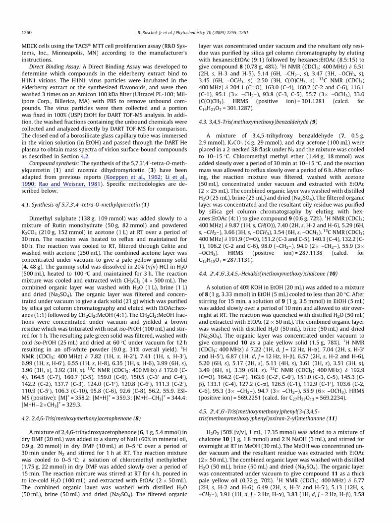
Compound synthesis: The synthesis of the 5,7,30,40-tetra-O-meth-ylquercetin (1) and racemic dihydromyricetin (3) have beenadapted from previous reports (Koeppen et al., 1962; Li et al.,1990; Rao and Weisner, 1981). Specific methodologies are de-scribed below.
4.1. Synthesis of 5,7,30,40-tetra-O-methylquercetin (1)
Dimethyl sulphate (138 g, 109 mmol) was added slowly to amixture of Rutin monohydrate (50 g, 82 mmol) and powderedK2CO3 (210 g, 152 mmol) in acetone (1 L) at RT over a period of30 min. The reaction was heated to reflux and maintained for80 h. The reaction was cooled to RT, filtered through Celite andwashed with acetone (250 mL). The combined acetone layer wasconcentrated under vacuum to give a pale yellow gummy solid(4, 48 g). The gummy solid was dissolved in 20% (v/v) HCl in H2O(500 mL), heated to 100 �C and maintained for 3 h. The reactionmixture was cooled and extracted with CH2Cl2 (4 � 500 mL). Thecombined organic layer was washed with H2O (1 L), brine (1 L)and dried (Na2SO4). The organic layer was filtered and concen-trated under vacuum to give a dark solid (21 g) which was purifiedby silica gel column chromatography and eluted with EtOAc:hex-anes (1:1) followed by CH2Cl2:MeOH (4:1). The CH2Cl2:MeOH frac-tions were concentrated under vacuum and yielded a brownresidue which was triturated with neat iso-PrOH (100 mL) and stir-red for 1 h. The resulting pale green solid was filtered, washed withcold iso-PrOH (25 mL) and dried at 60 �C under vacuum for 12 hresulting in an off-white powder (9.0 g, 31% overall yield). 1HNMR (CDCl3; 400 MHz) d 7.82 (1H, s, H-20), 7.41 (1H, s, H-30),6.99 (1H, s, H-60), 6.55 (1H, s, H-8), 6.35 (1H, s, H-6), 3.99 (6H, s),3.96 (3H, s), 3.92 (3H, s). 13C NMR (CDCl3; 400 MHz) d 172.0 (C-4), 164.5 (C-7), 160.7 (C-5), 159.0 (C-9), 150.5 (C-30 and C-40),142.2 (C-2), 137.7 (C-3), 124.0 (C-10), 120.8 (C-60), 111.3 (C-20),110.9 (C-50), 106.3 (C-10), 95.8 (C-6), 92.6 (C-8), 56.2, 55.9. ESI-MS (positive): [M]+ = 358.2; [M+H]+ = 359.3; [M+H�CH3]+ = 344.4;[M+H�2�CH3]+ = 329.3.
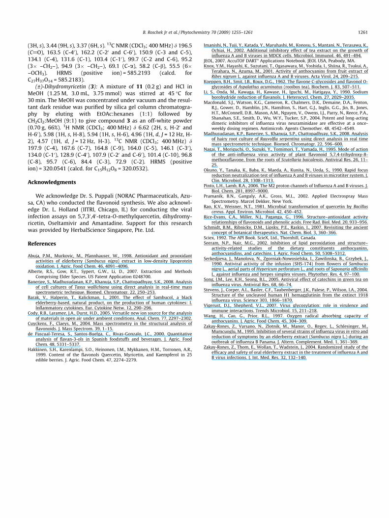
4.2. 2,4,6-Tris(methoxymethoxy)acetophenone (8)
A mixture of 2,4,6-trihydroxyacetophenone (6, 1 g, 5.4 mmol) indry DMF (20 mL) was added to a slurry of NaH (60% in mineral oil,0.9 g, 20 mmol) in dry DMF (10 mL) at 0–5 �C over a period of30 min under N2 and stirred for 1 h at RT. The reaction mixturewas cooled to 0–5 �C; a solution of chloromethyl methylether(1.75 g, 22 mmol) in dry DMF was added slowly over a period of15 min. The reaction mixture was stirred at RT for 4 h, poured into ice-cold H2O (100 mL), and extracted with EtOAc (2 � 50 mL).The combined organic layer was washed with distilled H2O(50 mL), brine (50 mL) and dried (Na2SO4). The filtered organic
layer was concentrated under vacuum and the resultant oily resi-due was purified by silica gel column chromatography by elutingwith hexanes:EtOAc (9:1) followed by hexanes:EtOAc (8.5:15) togive compound 8 (0.78 g, 48%). 1H NMR (CDCl3; 400 MHz) d 6.51(2H, s, H-3 and H-5), 5.14 (6H, –CH2–, s), 3.47 (3H, –OCH3, s),3.45 (6H, –OCH3, s), 2.50 (3H, C(O)CH3, s). 13C NMR (CDCl3;400 MHz) d 204.1 (C@O), 163.0 (C-4), 160.2 (C-2 and C-6), 116.1(C-1), 95.1 (3� –CH2–), 93.8 (C-3, C-5), 55.7 (3� –OCH3), 33.0(C(O)CH3). HRMS (positive ion) = 301.1281 (calcd. forC14H21O7 = 301.1287).
4.3. 3,4,5-Tris(methoxymethoxy)benzaldehyde (9)
A mixture of 3,4,5-trihydroxy benzaldehyde (7, 0.5 g,2.9 mmol), K2CO3 (4 g, 29 mmol), and dry acetone (100 mL) wereplaced in a 2-necked RB flask under N2 and the mixture was cooledto 10–15 �C. Chloromethyl methyl ether (1.44 g, 18 mmol) wasadded slowly over a period of 30 min at 10–15 �C, and the reactionmass was allowed to reflux slowly over a period of 6 h. After reflux-ing, the reaction mixture was filtered, washed with acetone(50 mL), concentrated under vacuum and extracted with EtOAc(2 � 25 mL). The combined organic layer was washed with distilledH2O (25 mL), brine (25 mL) and dried (Na2SO4). The filtered organiclayer was concentrated and the resultant oily residue was purifiedby silica gel column chromatography by eluting with hex-anes:EtOAc (4:1) to give compound 9 (0.6 g, 72%). 1H NMR (CDCl3;400 MHz) d 9.87 (1H, s, CH(O)), 7.40 (2H, s, H-2 and H-6), 5.29 (6H,s, –CH2–), 3.66 (3H, s, –OCH3), 3.54 (6H, s, –OCH3). 13C NMR (CDCl3;400 MHz) d 191.9 (C@O), 151.2 (C-3 and C-5), 140.3 (C-4), 132.2 (C-1), 106.2 (C-2 and C-6), 98.0 (–CH2–), 94.9 (2� –CH2–), 55.9 (3�–OCH3). HRMS (positive ion) = 287.1138 (calcd. forC13H19O7 = 287.1131).
4.4. 20,40,60,3,4,5,-Hexakis(methoxymethoxy)chalcone (10)
A solution of 40% KOH in EtOH (20 mL) was added to a mixtureof 8 (1 g, 3.33 mmol) in EtOH (5 mL) cooled to less than 20 �C. Afterstirring for 15 min, a solution of 9 (1 g, 3.5 mmol) in EtOH (5 mL)was added slowly over a period of 10 min and allowed to stir over-night at RT. The reaction was quenched with distilled H2O (50 mL)and extracted with EtOAc (2 � 50 mL). The combined organic layerwas washed with distilled H2O (50 mL), brine (50 mL) and dried(Na2SO4). The organic layer was concentrated under vacuum togive compound 10 as a pale yellow solid (1.5 g, 78%). 1H NMR(CDCl3; 400 MHz) d 7.22 (1H, d, J = 12 Hz, H-a), 7.04 (2H, s, H-30
and H-50), 6.87 (1H, d, J = 12 Hz, H-b), 6.57 (2H, s, H-2 and H-6),5.20 (6H, s), 5.17 (2H, s), 5.11 (4H, s), 3.61 (3H, s), 3.51 (3H, s),3.49 (6H, s), 3.39 (6H, s). 13C NMR (CDCl3; 400 MHz) d 192.9(C@O), 164.2 (C-40), 163.6 (C-20, C-60), 151.0 (C-3, C-5), 145.3 (C-b), 133.1 (C-4), 127.2 (C-a), 126.5 (C-1), 112.9 (C-10), 103.6 (C-2,C-6), 95.3 (3� –CH2–), 94.7 (3� –CH2–), 55.9 (6� –OCH3). HRMS(positive ion) = 569.2251 (calcd. for C27H37O13 = 569.2234).
4.5. 20,40,60-Tris(methoxymethoxy)phenyl(3-(3,4,5-tris(methoxymethoxy)phenyl)oxiran-2-yl)methanone (11)
H2O2 (50% [v/v], 1 mL, 17.35 mmol) was added to a mixture ofchalcone 10 (1 g, 1.8 mmol) and 2 N NaOH (3 mL), and stirred forovernight at RT in MeOH (30 mL). The MeOH was concentrated un-der vacuum and the resultant residue was extracted with EtOAc(2� 50 mL). The combined organic layer was washed with distilledH2O (50 mL), brine (50 mL) and dried (Na2SO4). The organic layerwas concentrated under vacuum to give compound 11 as a thickpale yellow oil (0.72 g, 70%). 1H NMR (CDCl3; 400 MHz) d 6.77(2H, s, H-2 and H-6), 6.49 (2H, s, H-30 and H-50), 5.13 (12H, s,–CH2–), 3.91 (1H, d, J = 2 Hz, H-a), 3.83 (1H, d, J = 2 Hz, H-b), 3.58
B. Roschek Jr et al. / Phytochemistry 70 (2009) 1255–1261 1261
(3H, s), 3.44 (9H, s), 3.37 (6H, s). 13C NMR (CDCl3; 400 MHz) d 196.5(C@O), 163.5 (C-40), 162.2 (C-20 and C-60), 150.9 (C-3 and C-5),134.1 (C-4), 131.6 (C-1), 103.4 (C-10), 99.7 (C-2 and C-6), 95.2(3� –CH2–), 94.9 (3� –CH2–), 69.1 (C-a), 58.2 (C-b), 55.5 (6�–OCH3). HRMS (positive ion) = 585.2193 (calcd. forC27H37O14 = 585.2183).
(±)-Dihydromyricetin (3): A mixture of 11 (0.2 g) and HCl inMeOH (1.25 M, 3.0 mL, 3.75 mmol) was stirred at 45 �C for30 min. The MeOH was concentrated under vacuum and the resul-tant dark residue was purified by silica gel column chromatogra-phy by eluting with EtOAc:hexanes (1:1) followed byCH2Cl2:MeOH (9:1) to give compound 3 as an off-white powder(0.70 g, 66%). 1H NMR (CDCl3; 400 MHz) d 6.62 (2H, s, H-20 andH-60), 5.98 (1H, s, H-8), 5.94 (1H, s, H-6), 4.96 (1H, d, J = 12 Hz, H-2), 4.57 (1H, d, J = 12 Hz, H-3). 13C NMR (CDCl3; 400 MHz) d197.9 (C-4), 167.6 (C-7), 164.8 (C-9), 164.0 (C-5), 146.1 (C-30),134.0 (C-10), 128.9 (C-40), 107.9 (C-20 and C-60), 101.4 (C-10), 96.8(C-8), 95.7 (C-6), 84.4 (C-3), 72.9 (C-2). HRMS (positiveion) = 320.0541 (calcd. for C15H12O8 = 320.0532).
Acknowledgments
We acknowledge Dr. S. Puppali (NORAC Pharmaceuticals, Azu-sa, CA) who conducted the flavonoid synthesis. We also acknowl-edge Dr. L. Holland (IITRI, Chicago, IL) for conducting the viralinfection assays on 5,7,30,40-tetra-O-methylquercetin, dihydromy-ricetin, Oseltamivir and Amantadine. Support for this researchwas provided by HerbalScience Singapore, Pte. Ltd.
References
Abuja, P.M., Murkovic, M., Pfannhauser, W., 1998. Antioxidant and prooxidantactivities of elderberry (Sambucus nigra) extract in low-density lipoproteinoxidation. J. Agric. Food Chem. 46, 4091–4096.
Alberte, R.S., Gow, R.T., Sypert, G.W., Li, D., 2007. Extraction and MethodsComprising Elder Species. US Patent Application 0248700.
Banerjee, S., Madhusudanan, K.P., Khanuja, S.P., Chattopadhyay, S.K., 2008. Analysisof cell cultures of Taxus wallichiana using direct analysis in real-time massspectrometric technique. Biomed. Chromatogr. 22, 250–253.
Barak, V., Halperin, T., Kalickman, I., 2001. The effect of Sambucol, a blackelderberry-based, natural product, on the production of human cytokines: I.Inflammatory cytokines. Eur. Cytokine Netw. 12, 290–296.
Cody, R.B., Laramee, J.A., Durst, H.D., 2005. Versatile new ion source for the analysisof materials in open air under ambient conditions. Anal. Chem. 77, 2297–2302.
Cuyckens, F., Claeys, M., 2004. Mass spectrometry in the structural analysis offlavonoids. J. Mass Spectrom. 39, 1–15.
de Pascual-Teresa, S., Santos-Buelga, C., Rivas-Gonzalo, J.C., 2000. Quantitativeanalysis of flavan-3-ols in Spanish foodstuffs and beverages. J. Agric. FoodChem. 48, 5331–5337.
Hakkinen, S.H., Karenlampi, S.O., Heinonen, I.M., Mykkanen, H.M., Torronen, A.R.,1999. Content of the flavonols Quercetin, Myricetin, and Kaempferol in 25edible berries. J. Agric. Food Chem. 47, 2274–2279.
Imanishi, N., Tuji, Y., Katada, Y., Maruhashi, M., Konosu, S., Mantani, N., Terasawa, K.,Ochiai, H., 2002. Additional inhibitory effect of tea extract on the growth ofinfluenza A and B viruses in MDCK cells. Microbiol. Immunol. 46, 491–494.
JEOL, 2007. AccuTOF DARTTM Applications Notebook. JEOL USA, Peabody, MA.Knox, Y.M., Hayashi, K., Suzutani, T., Ogasawara, M., Yoshida, I., Shiina, R., Tsukui, A.,
Terahara, N., Azuma, M., 2001. Activity of anthocyanins from fruit extract ofRibes nigrum L. against influenza A and B viruses. Acta Virol. 24, 209–215.
Koeppen, B.H., Smit, J.B., Roux, D.G., 1962. The flavone C-glycosides and flavonol O-glycosides of Aspalathus acuminatus (rooibos tea). Biochem. J. 83, 507–511.
Li, S., Onda, M., Kawaga, H., Kawase, H., Iguchi, M., Hatigaya, Y., 1990. Sodiumborohydride reduction of flavanols. J. Heterocycl. Chem. 27, 2029–2035.
Macdonald, S.J., Watson, K.G., Cameron, R., Chalmers, D.K., Demaine, D.A., Fenton,R.J., Gower, D., Hamblin, J.N., Hamilton, S., Hart, G.J., Inglis, G.G., Jin, B., Jones,H.T., McConnell, D.B., Mason, A.M., Nguyen, V., Owens, I.J., Parry, N., Reece, P.A.,Shanahan, S.E., Smith, D., Wu, W.Y., Tucker, S.P., 2004. Potent and long-actingdimeric inhibitors of influenza virus neuraminidase are effective at a once-weekly dosing regimen. Antimicrob. Agents Chemother. 48, 4542–4549.
Madhusudanan, K.P., Banerjee, S., Khanuja, S.P., Chattopadhyay, S.K., 2008. Analysisof hairy root culture of Rauvolfia serpentina using direct analysis in real timemass spectrometric technique. Biomed. Chromatogr. 22, 596–600.
Nagai, T., Moriguchi, O., Suzuki, Y., Tomimori, T., Yamada, H., 1995. Mode of actionof the anti-influenza virus activity of plant flavonoid 5,7,4-trihydroxy-8-methoxyflavone, from the roots of Scutellaria baicalensis. Antiviral Res. 26, 11–25.
Okuno, Y., Tanaka, K., Baba, K., Maeda, A., Kunita, N., Ueda, S., 1990. Rapid focusreduction neutralization test of influenza A and B viruses in microtiter system. J.Clin. Microbiol. 28, 1308–1313.
Pinto, L.H., Lamb, R.A., 2006. The M2 proton channels of Influenza A and B viruses. J.Biol. Chem. 281, 8997–9000.
Pramanik, B.N., Ganguly, A.K., Gross, M.L., 2002. Applied Electrospray MassSpectrometry. Marcel Dekker, New York.
Rao, K.V., Weisner, N.T., 1981. Microbial transformation of quercetin by Bacilluscereus. Appl. Environ. Microbiol. 42, 450–452.
Rice-Evans, C.A., Miller, N.J., Paganga, G., 1996. Structure–antioxidant activityrelationships of flavonoids and phenolic acids. Free Rad. Biol. Med. 20, 933–956.
Schmidt, B.M., Ribnicky, D.M., Lipsky, P.E., Raskin, I., 2007. Revisiting the ancientconcept of botanical therapeutics. Nat. Chem. Biol. 3, 360–366.
Sciex, 1992. The API Book. ScieX, Ltd., Thornhill, Canada.Seeram, N.P., Nair, M.G., 2002. Inhibition of lipid peroxidation and structure–
activity-related studies of the dietary constituents anthocyanins,anthocyanidins, and catechins. J. Agric. Food Chem. 50, 5308–5312.
Serkedjieva, J., Manolova, N., Zgorniak-Nowosielska, I., Zawilinska, B., Grzybek, J.,1990. Antiviral activity of the infusion (SHS-174) from flowers of Sambucusnigra L., aerial parts of Hypericum perforatum L., and roots of Saponaria officinilisL. against influenza and herpes simplex viruses. Phytother. Res. 4, 97–100.
Song, J.M., Lee, K.H., Seong, B.L., 2005. Antiviral effect of catechins in green tea oninfluenza virus. Antiviral Res. 68, 66–74.
Stevens, J., Corper, A.L., Basler, C.F., Taubenberger, J.K., Palese, P., Wilson, I.A., 2004.Structure of the uncleaved human H1 hemagglutinin from the extinct 1918influenza virus. Science 303, 1866–1870.
Vigerust, D.J., Shepherd, V.L., 2007. Virus glycosylation: role in virulence andimmune interactions. Trends Microbiol. 15, 211–218.
Wang, H., Cao, G., Prior, R.L., 1997. Oxygen radical absorbing capacity ofanthocyanins. J. Agric. Food Chem. 45, 304–309.
Zakay-Rones, Z., Varsano, N., Zlotnik, M., Manor, O., Regev, L., Schlesinger, M.,Mumcuoglu, M., 1995. Inhibition of several strains of influenza virus in vitro andreduction of symptoms by an elderberry extract (Sambucus nigra L.) during anoutbreak of influenza B Panama. J. Altern. Complement. Med. 1, 361–369.
Zakay-Rones, Z., Thom, E., Wollan, T., Wadstein, J., 2004. Randomized study of theefficacy and safety of oral elderberry extract in the treatment of influenza A andB virus infections. J. Int. Med. Res. 32, 132–140.
Related Documents