Research Article Efficacy of Platelet-Rich Plasma Containing Xenogenic Adipose Tissue-Derived Stromal Cells on Restoring Intervertebral Disc Degeneration: A Preclinical Study in a Rabbit Model Chao Ma, 1 Ran Wang , 1 Dingliang Zhao, 1 Naikun Wang, 1 Ying Han , 1 Shichong Wang , 1 Tianyun Gao, 2 Bin Wang , 2 and Lijuan Lu 1 1 Department of Pain, e Drum Tower Hospital Affiliated to Medical School of Nanjing University, No. 321 Zhongshan Road, Nanjing 210008, Jiangsu Province, China 2 Center for Clinic Stem Cell Research, e Drum Tower Hospital Affiliated to Medical School of Nanjing University, No. 321 Zhongshan Road, Nanjing 210008, Jiangsu Province, China Correspondence should be addressed to Bin Wang; [email protected] and Lijuan Lu; [email protected] Received 30 August 2018; Revised 8 January 2019; Accepted 5 March 2019; Published 16 April 2019 Academic Editor: Filippo Brighina Copyright©2019ChaoMaetal.isisanopenaccessarticledistributedundertheCreativeCommonsAttributionLicense, which permits unrestricted use, distribution, and reproduction in any medium, provided the original work is properly cited. Objective. Platelet-rich plasma (PRP) containing multiple growth factors is a promising strategy for disc degeneration. us, this study hypothesizes that the combination of PRP and adipose tissue-derived stromal cells (ADSCs) may repair de- generative disc more effectively than using each one of them alone. Methods. e model of early intervertebral disc de- generation was induced by annular puncture in the New Zealand rabbit. Autologous PRP was extracted from fresh arterial blood by using two centrifugation techniques. ADSC was offered by the Center for Clinic Stem Cell Research. Four weeks afterthefirstexperiment,PRPorADSCsoracombinationofPRPandADSCswasinjectedintothepuncturedintervertebral disc. Four weeks later, disc height and signal intensity on T2-weighted magnetic resonance imaging (MRI) were assessed. Results. One month after puncture, we detected relatively narrow discs and lower signal intensity in MRI T2-weighted images. At four weeks after injection, the PRP-ADSC group statistically significantly restored discs, compared with PRP, ADSCs, or negative control group. Conclusions. e combination of PRP and ADSCs shows an effective potential to restore degenerated intervertebral discs in the rabbit. 1. Introduction Intervertebral disc degeneration (IDD) is the leading cause of low back pain (LBP), leading to disability and placing a heavy burden on society [1]. Current treatment of IDD, such as conservative procedures (medication or physical therapy) and operative treatment, only partly alleviates symptoms, but does not reverse intervertebral disc cataplasia in etiology or pathology [2]. As a consequence, how to augment the number and enhance the function of nucleus pulposus cells has become a research hotspot [3]. Including PDGF, TGF-β, VEGF, and so on, PRP contains various growth factors that promote cell proliferation and tissue regeneration [4]. At the same time, PRP regulates the extracellular microenvironment by inducing extracellular matrix synthesis and secretion [5]. Furthermore, the role of PRP in immune inflammation is the production of in- terleukins and chemokines [6, 7]. PRP has been proven to be an effective therapy for IDD in rats, rabbits, and sheep treated in a large number of experimental animal models [2, 8]. Compared to other animals, rabbits are cheaper and their intervertebral discs are relatively large. Clinical research studies in the field of IDD have been indicative that treatment with PRP has a pronounced effect [9, 10]. Stem cell therapy is gradually becoming popular in multiple fields including IDD [11]. ADSCs have biological Hindawi Pain Research and Management Volume 2019, Article ID 6372356, 7 pages https://doi.org/10.1155/2019/6372356

Welcome message from author
This document is posted to help you gain knowledge. Please leave a comment to let me know what you think about it! Share it to your friends and learn new things together.
Transcript
Research ArticleEfficacy of Platelet-Rich Plasma Containing Xenogenic AdiposeTissue-Derived Stromal Cells on Restoring Intervertebral DiscDegeneration: A Preclinical Study in a Rabbit Model
Chao Ma,1 Ran Wang ,1 Dingliang Zhao,1 Naikun Wang,1 Ying Han ,1
Shichong Wang ,1 Tianyun Gao,2 Bin Wang ,2 and Lijuan Lu 1
1Department of Pain, �e Drum Tower Hospital Affiliated to Medical School of Nanjing University, No. 321 Zhongshan Road,Nanjing 210008, Jiangsu Province, China2Center for Clinic Stem Cell Research, �e Drum Tower Hospital Affiliated to Medical School of Nanjing University,No. 321 Zhongshan Road, Nanjing 210008, Jiangsu Province, China
Correspondence should be addressed to Bin Wang; [email protected] and Lijuan Lu; [email protected]
Received 30 August 2018; Revised 8 January 2019; Accepted 5 March 2019; Published 16 April 2019
Academic Editor: Filippo Brighina
Copyright © 2019 ChaoMa et al.-is is an open access article distributed under the Creative Commons Attribution License,which permits unrestricted use, distribution, and reproduction in any medium, provided the original work isproperly cited.
Objective. Platelet-rich plasma (PRP) containing multiple growth factors is a promising strategy for disc degeneration. -us,this study hypothesizes that the combination of PRP and adipose tissue-derived stromal cells (ADSCs) may repair de-generative disc more effectively than using each one of them alone. Methods. -e model of early intervertebral disc de-generation was induced by annular puncture in the New Zealand rabbit. Autologous PRP was extracted from fresh arterialblood by using two centrifugation techniques. ADSC was offered by the Center for Clinic Stem Cell Research. Four weeksafter the first experiment, PRP or ADSCs or a combination of PRP and ADSCs was injected into the punctured intervertebraldisc. Four weeks later, disc height and signal intensity on T2-weighted magnetic resonance imaging (MRI) were assessed.Results. One month after puncture, we detected relatively narrow discs and lower signal intensity in MRI T2-weightedimages. At four weeks after injection, the PRP-ADSC group statistically significantly restored discs, compared with PRP,ADSCs, or negative control group. Conclusions. -e combination of PRP and ADSCs shows an effective potential to restoredegenerated intervertebral discs in the rabbit.
1. Introduction
Intervertebral disc degeneration (IDD) is the leading causeof low back pain (LBP), leading to disability and placing aheavy burden on society [1]. Current treatment of IDD, suchas conservative procedures (medication or physical therapy)and operative treatment, only partly alleviates symptoms,but does not reverse intervertebral disc cataplasia in etiologyor pathology [2]. As a consequence, how to augment thenumber and enhance the function of nucleus pulposus cellshas become a research hotspot [3].
Including PDGF, TGF-β, VEGF, and so on, PRP containsvarious growth factors that promote cell proliferation and
tissue regeneration [4]. At the same time, PRP regulates theextracellular microenvironment by inducing extracellularmatrix synthesis and secretion [5]. Furthermore, the role ofPRP in immune inflammation is the production of in-terleukins and chemokines [6, 7]. PRP has been proven to bean effective therapy for IDD in rats, rabbits, and sheep treatedin a large number of experimental animal models [2, 8].Compared to other animals, rabbits are cheaper and theirintervertebral discs are relatively large. Clinical researchstudies in the field of IDD have been indicative that treatmentwith PRP has a pronounced effect [9, 10].
Stem cell therapy is gradually becoming popular inmultiple fields including IDD [11]. ADSCs have biological
HindawiPain Research and ManagementVolume 2019, Article ID 6372356, 7 pageshttps://doi.org/10.1155/2019/6372356
advantages in the proliferative capacity, secreted proteins,and immunomodulatory effects [12]. After passing throughthe needle at constant flow rates, ADSCs maintain pro-liferative capacity and metabolic function [13]. Scientistsverify favorable safety profile of ADSCs in a rabbit model forosteoarthritis [14]. In our current study, we hypothesizedthat the combination of PRP and ADSCs may possesssynergistic restorative function on the degenerative disc,considering ADSCs secreting protein to enhancing thefunction of PRP.-e purpose of this study was to investigatethe effects of ADSCs containing PRP on the regeneration ofearly degenerated intervertebral discs in vivo. To achieve thisaim, MRI evaluations were utilized to detect differences intreated discs.
2. Materials and Methods
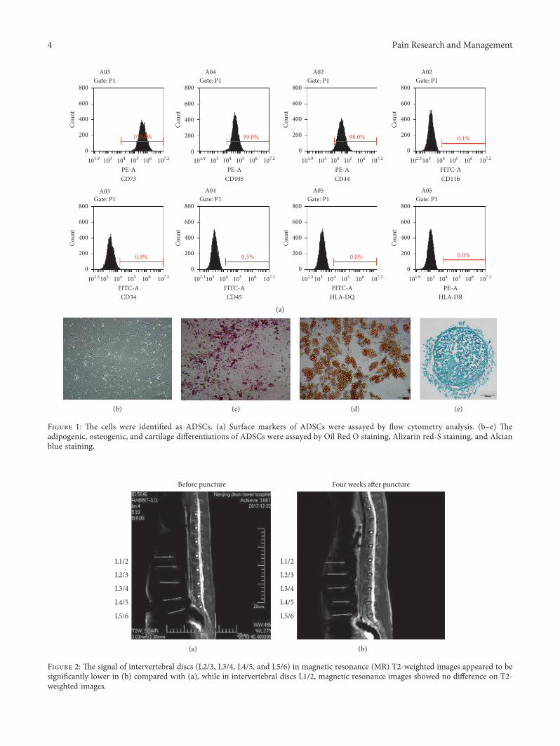
2.1. ADSC Isolation and Identification. -is study was ap-proved by the Research Ethics Board of Nanjing DrumTower Hospital. Written consent was obtained from thehealthy adipose tissue donors. ADSCs were prepared byfollowing the method in GMP (good manufacture practice)facility. After washing with phosphate-buffered saline (PBS),the adipose tissue was cut into small pieces of approximately1mm3 and was digested with 0.075% collagenase (type I;Sigma-Aldrich, St. Louis, MO, USA) at 37°C for 30min. -edigestion was stopped by adding α-MEM containing 10%FBS, and the solution was centrifuged at 1200× g for 10min.-e cell pellet was resuspended and filtered through a100 μmNylon mesh to remove tissue remains.-e cells wereincubated in the culture medium and were passaged threetimes to remove the contamination of other types of cells.ADSCs at the fourth passage were used for all of the ex-periments in this study.
Flow cytometry analysis was used for phenotypic anal-ysis in ADSCs. A total of 100,000 cells at the fourth passagewere incubated with fluorescein isothiocyanate (FITC) orphycoerythrin (PE) labeled monoclonal antibodies (CD11bFITC, CD34 FITC, CD44 PE, CD45 FITC, CD73 PE, CD105PE, HLA-DQ FITC, andHLA-DR FITC; BD, San Diego, CA,USA) for 30min in the dark at room temperature. ADSCswere centrifuged at 2000 rpm for 5min after washing threetimes with 1×PBS and resuspended in 1×PBS for flowcytometry analysis. Cells were analyzed using a FACScan(BD FACSAria™; BD, San Jose, CA, USA), and data wereanalyzed with the FACS software.
ADSCs were cultured in a 24-well tissue culture plate at adensity of 1× 10 exp 4 cells/well for adipogenic and oste-ogenic differentiation. ADSCs were cultured with adipo-genic differentiation medium, osteogenic differentiationmedium, and cartilage differentiation medium (Gibco,Grand Island, NY, USA) at 50–70% confluency. Every 3-4 days, the differentiation medium was changed. At day 21,Oil Red O staining (Sigma-Aldrich, St. Louis, MO, USA),Alizarin Red-S staining (Sigma-Aldrich, St. Louis, MO,USA), and Alcian blue staining (Sigma-Aldrich, St. Louis,MO, USA) were performed to check the adipogenesis,osteogenesis, and cartilage differentiation potentials of
ADSCs. -e cells used in this study come from a singledonor, and the quality was fully evaluated to meet the re-quirements and standards of MSC’s quality and safety forclinic settings.
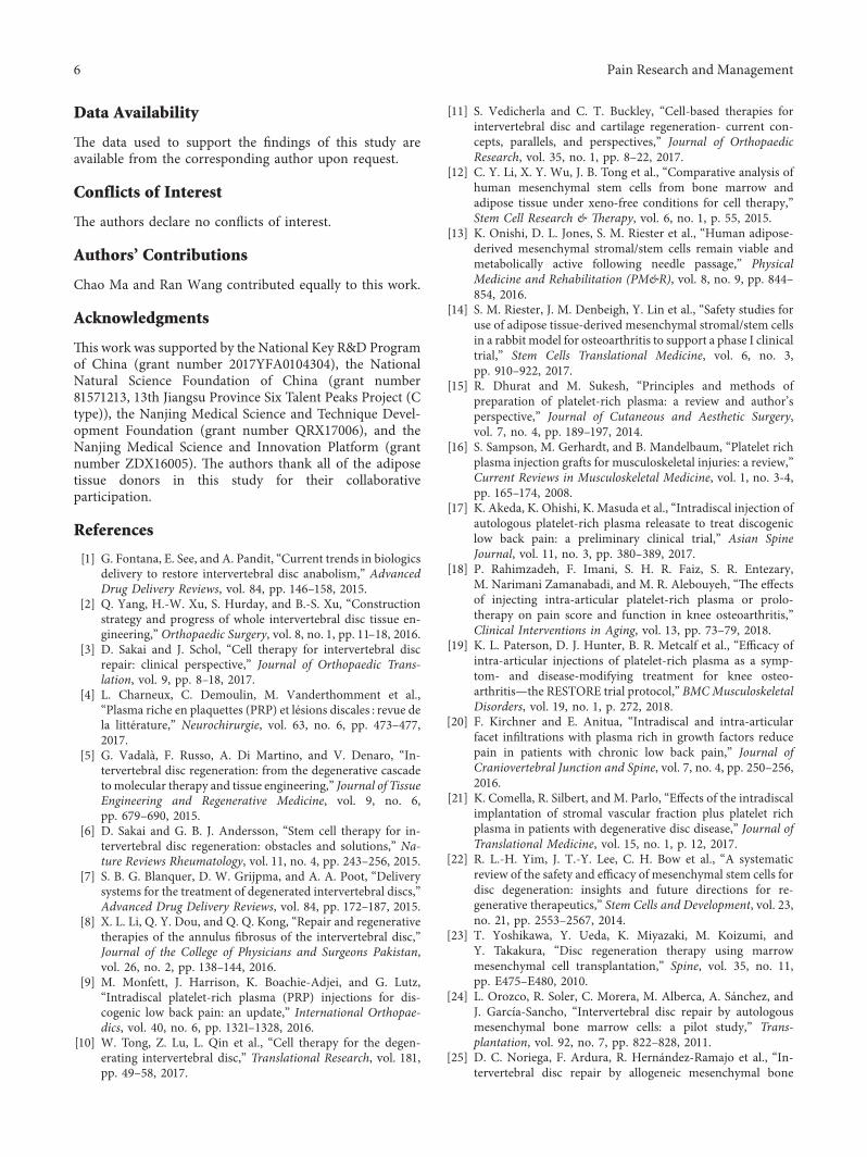
2.2. Experimental Animals. A total of five rabbits (female,2.5–3.5 kg, 4–6 weeks) were grown in the animal lab ofDrum Tower Hospital Affiliated to Medical School ofNanjing University. -e breed of rabbits was a NewZealand white rabbit. As the narrow area of our animallaboratory, our team had only five cages. To take fulladvantage of these rabbits, we established four de-generative discs (L2/3, L3/4, L4/5, and L5/6) in each rabbitunder the general anesthesia by intraperitoneal injection of10% chloral hydrate. -e discs were divided into fivegroups (Group A: L1/2 discs were set as normal controlgroup; Group B: L2/3 discs were set as degeneration andPBS injection; Group C: L3/4 discs were set as de-generation and PRP injection; Group D: L4/5 discs wereset as degeneration and ADSC injection; and Group E: L5/6 discs were set as degeneration and combined ADSC andPRP injection). Our study design was approved by theAnimal Ethics Committee of Drum Tower Hospital ofNanjing University.
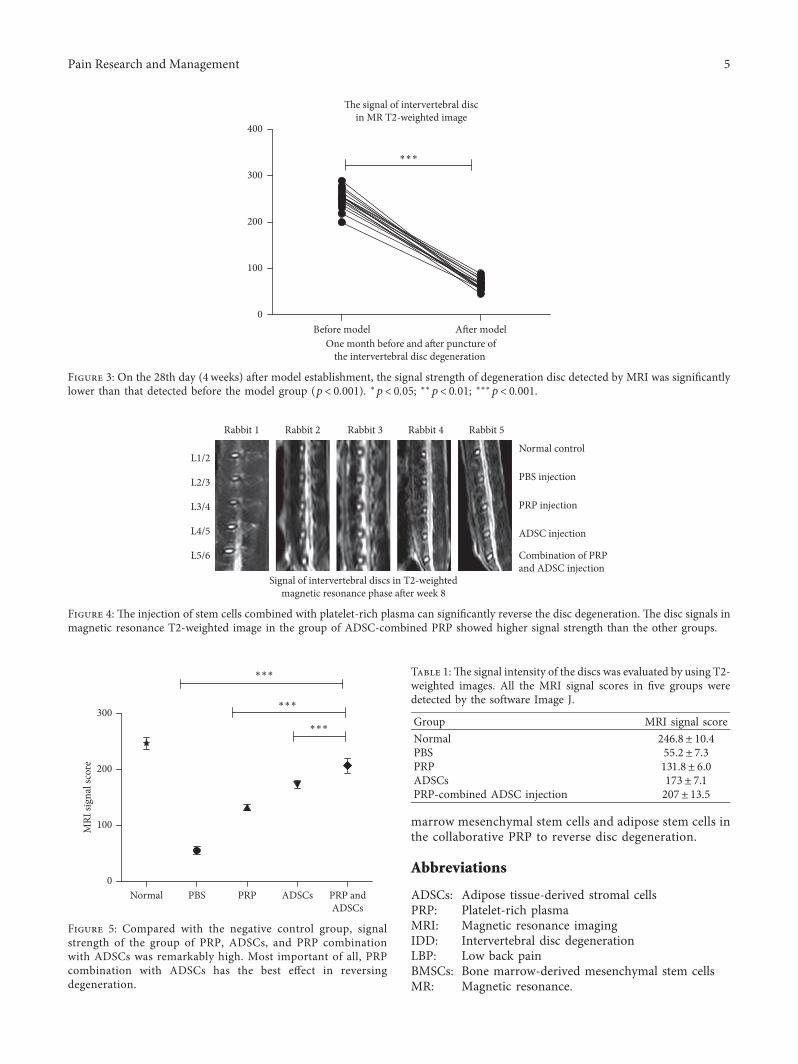
2.3. Establishment of IDD Model and Evaluation of MR.Preoperatively, rabbits were accepted intraperitoneal in-jection with 10% chloral hydrate. -e height of the in-tervertebral disc was measured by magnetic resonance(MR) as the base line prior to puncture. -en, we used asterile towel to remove the fur, disinfect the skin, andcover the rabbit. -e IDDmodel of rabbits was establishedby repeatedly puncture the annulus fibrosus of the in-tervertebral using the puncture needle (22G) under CT.Ct-guided disc puncture did not require an incision intothe rabbit’s posterior back, reducing bleeding and in-fection. -e nucleus pulposus and annulus fibrosus of theintervertebral disc were broken by puncture needle (22G)and negative pressure suction (the empty needle pistonscale ranged from 0ml to 2ml for 15 seconds) can destroythe nucleus pulposus and induce degeneration model. Toconfirm the IDD model and evaluate the effect of biologytherapy, we used MR to scan the target disc until 4 weeksafter the first experiment and injection of biologicalagents, respectively.
MRI was performed using a 1.5 T imager unit. Fol-lowing general anesthesia with chloral hydrate, the rabbitswere placed in a prone position for MRI scan. -e signalintensity of the discs was evaluated by using T2-weightedimages. IDD grade was evaluated using the modified-ompson classification of Grade I–IV. Two doctorsevaluated the images separately. -e parameters of the T2-weighted images wereas follows: time-to-repeat � 1,800ms;field of view � 140 ×140mm; time-to-echo� 70ms; slicethickness � 2mm; image matrix � 286× 385; averages� 9.All the MRI signal scores were detected by the software ofImage J.
2 Pain Research and Management

2.4. Preparation of PRP, ADSCs, and PRP-ADSC Mixture.PRP was prepared by the methods of Aghaloo [15]. -erabbits were fixed on the operating table and arterial bloodwas drawn from the auricular central artery. After mixingwith sodium citrate using as anticoagulant, the arterial bloodwas centrifuged in a two-step process. -e first centrifu-gation was carried out at a rate of 215 g for 10min, and theplasma above the white film was placed in another centrifugetube after centrifugation. -e second centrifugation wascarried out at a speed of 863 g for 6min. -e supernatant, orplatelet-poor plasma, was collected and transferred to an-other centrifuge tube. -e remaining platelet-rich plasmawas blown evenly. In our study, platelet-rich plasma con-tained small amounts of white blood cells. And wemade surethat the count of platelet was greater than 1000∗109/L (thecount of platelets was 1041∗ 109/L in our study) under themicroscope.
-e ADSCs were afforded by the Center for ClinicStem Cell Research, the Affiliated Nanjing Drum TowerHospital of Nanjing University Medical School (Fig-ure 1). -e number of stem cells in each group was tenmillion. A mixture of PRP, ADSCs, and PRP-ADSCs wasincluded, each having a volume of 200 μl. -e punctureneedle that injected the stem cells or PRP or PBS(phosphate-buffered solution) into the intervertebral discwas 22G.
2.5. StatisticalAnalysis. Statistical analysis was implementedby using the SPSS 18.0 software. Data of all groups arepresented as mean± standard deviation and were analyzedusing variance analysis. -e data of the MRI image signalwere analyzed using a one way ANOVA. All the data weremade by the software Graph Pad Prism. P< 0.05 wasregarded as statistically significant difference.
3. Results
3.1. IDD Model of Rabbits Was Confirmed Successfully.After 4 weeks of the first puncture, the signal images of discsincluded L2/3, L3/4, L4/5, and L5/6 were significantly lowerthan baseline as well as the control group (L1/2). -is meansthat our annulus fibrosus punctures lead to the degenerativedisc (Figures 2 and 3).
3.2. Either of PRP, ADSCs, or PRP-ADSC Mixture CouldRestore Degenerative Disc. Compared with disc L2/3, thesignal image of L3/4, L4/5, and L5/6 were significantlyhigher. -ese results suggest that biological therapy in-cluding but not limited to PRP and ADSCs could promotethe repair of disc regression. PRP-combined ADSCs had themost effect in restoring the intervertebral disc (Figures 4 and5 and Table 1).
4. Discussion
-is present study investigated the efficacy of PRP-containing ADSCs on degenerative disc regeneration in-duced by needle puncture. -e results of this study
demonstrated that the injection of PRP-containing ADSCswas effective in restoring the early degenerated discs. -eMRI examination revealed the repair effects of the PRP-ADSCs on the early degenerated discs.
In the United States and the European Union, there wasan increasing prevalence of the PRP utilization to facilitatehealing in a variety of diseases, including musculoskeletalinjuries, low back pain, and so on [16, 17]. Currently, PRPinjection is frequently used in the clinical treatment of kneeosteoarthritis [18, 19]. In the intervertebral disc de-generation, PRP treatment concentrates more on animalresearch or preliminary clinical trials, and more clinicalrandomized control trail (RCT) studies are still needed. InSpain, Japan, and American, retrospective single-centeranalysis has demonstrated that injection of autologousPRP was safe in patients with low back pain without adverseevents [17, 20, 21].
Based on the reported animal research, the use of stemcells for the treatment of intervertebral disc degeneration issafe and effective. -e incidence of complications in mes-enchymal stem cell therapy for disc degeneration is low [22].From 2010 to 2011, two literatures reported that in-tervertebral disc degeneration therapy using mesenchymalcell transplantation brought favorable results in patientsdiagnosed with lumber disc degeneration and have beenfollowed up for 1 or 2 years [23, 24]. A randomized con-trolled trial concerning allogeneic mesenchymal bonemarrow cells used in intervertebral disc repair displayedrapid and significant improvement [25].
-e joint use of PRP and stem cells in patients withpartial tear of the rotator cuff tendon alleviated pain andimproved shoulder function [26]. Scientists combined PRPand bone marrow-derived mesenchymal stem cells to treat amodel of disc degeneration caused by annular puncture inrabbit [27]. Literature studies manifested PRP can stimulateproliferation and differentiation of adipose tissue-derivedmesenchymal stem cells by releasing diverse growth factors[28, 29]. Although our team firstly applied ADSC and PRP todisc degeneration, there is still a long way to go in re-generative medicine. More in-depth foundational andclinical research is indispensable.
-ere are some drawbacks in our work. However, thereare still quite a few scientists using rabbits as models fordisc degeneration as in 2018. We have to admit that thepersistence of nucleus pulposus cells in rabbit models maybe affected by notochord cells. We all know that it is betterto do stem cell transplantation in immune-deficient mice,but the intervertebral discs of mice are too small and theintervertebral disc puncture is more difficult, which canlead to bleeding and death of animals. Due to the limi-tations of the animal room, the hospital only gave us a fewrabbit cages. We only had 5 rabbits in this trial, but we usedthe intervertebral discs of rabbits as far as possible. Afterall, the results suggest that the combination of PRP andADSC transplantation can be safe and effective in thetreatment of degenerative disc induced by annularpuncture in the New Zealand rabbit and encourage morestudies with a larger sample. Next, we will expand thesample size and compare the difference between bone
Pain Research and Management 3
Before puncture
L1/2
L2/3
L3/4
L4/5
L5/6
(a)
L1/2
L2/3
L3/4
L4/5
L5/6
Four weeks a�er puncture
(b)
Figure 2: �e signal of intervertebral discs (L2/3, L3/4, L4/5, and L5/6) in magnetic resonance (MR) T2-weighted images appeared to besigni�cantly lower in (b) compared with (a), while in intervertebral discs L1/2, magnetic resonance images showed no dierence on T2-weighted images.
PE-A101.9
800
600
400
200
0 0 0 0
0 0 0 0
800
600
400
200
800
600
400
200
800
600
400
200
103 104 105
100.0%
106 107.2 101.9 103 104 105
99.0% 98.0% 0.1%
0.0%0.0%0.5%0.9%
106 107.2 101.9 103 104 105 106 107.2 102.3103 104 105 106 107.2
Cou
nt
Cou
nt
Cou
nt
Cou
nt
800
600
400
200
102.3103 104 105 106 107.2
Cou
nt
800
600
400
200
102.3103 104 105 106 107.2
Cou
nt
800
600
400
200
102.3 103 104 105 106 107.2
Cou
nt
800
600
400
200
101.9 103 104 105 106 107.2
Cou
nt
CD73
PE-ACD34
CD105 CD44 CD11bPE-A PE-A FITC-A
FITC-ACD45
FITC-AHLA-DQ HLA-DRFITC-A
A03Gate: P1
A04Gate: P1
A02Gate: P1
A02Gate: P1
A03Gate: P1
A04Gate: P1
A05Gate: P1
A05Gate: P1
(a)
(b) (c) (d) (e)
Figure 1: �e cells were identi�ed as ADSCs. (a) Surface markers of ADSCs were assayed by �ow cytometry analysis. (b–e) �eadipogenic, osteogenic, and cartilage dierentiations of ADSCs were assayed by Oil Red O staining, Alizarin red-S staining, and Alcianblue staining.
4 Pain Research and Management
marrow mesenchymal stem cells and adipose stem cells inthe collaborative PRP to reverse disc degeneration.
Abbreviations
ADSCs: Adipose tissue-derived stromal cellsPRP: Platelet-rich plasmaMRI: Magnetic resonance imagingIDD: Intervertebral disc degenerationLBP: Low back painBMSCs: Bone marrow-derived mesenchymal stem cellsMR: Magnetic resonance.
Rabbit 1
L1/2
L2/3
L3/4
L4/5
L5/6
Rabbit 2 Rabbit 3
Signal of intervertebral discs in T2-weightedmagnetic resonance phase a�er week 8
Rabbit 4 Rabbit 5
Normal control
PBS injection
PRP injection
ADSC injection
Combination of PRPand ADSC injection
Figure 4: �e injection of stem cells combined with platelet-rich plasma can signi�cantly reverse the disc degeneration. �e disc signals inmagnetic resonance T2-weighted image in the group of ADSC-combined PRP showed higher signal strength than the other groups.
One month before and a�er puncture ofthe intervertebral disc degeneration
�e signal of intervertebral discin MR T2-weighted image
Before model
400
∗∗∗
300
200
100
0A�er model
Figure 3: On the 28th day (4weeks) after model establishment, the signal strength of degeneration disc detected by MRI was signi�cantlylower than that detected before the model group (p< 0.001). ∗p< 0.05; ∗∗p< 0.01; ∗∗∗p< 0.001.
300
∗∗∗
∗∗∗
∗∗∗
200
100
0Normal
MRI
sign
al sc
ore
PBS PRP ADSCs PRP andADSCs
Figure 5: Compared with the negative control group, signalstrength of the group of PRP, ADSCs, and PRP combinationwith ADSCs was remarkably high. Most important of all, PRPcombination with ADSCs has the best eect in reversingdegeneration.
Table 1:�e signal intensity of the discs was evaluated by using T2-weighted images. All the MRI signal scores in �ve groups weredetected by the software Image J.
Group MRI signal scoreNormal 246.8± 10.4PBS 55.2± 7.3PRP 131.8± 6.0ADSCs 173± 7.1PRP-combined ADSC injection 207± 13.5
Pain Research and Management 5
Data Availability
-e data used to support the findings of this study areavailable from the corresponding author upon request.
Conflicts of Interest
-e authors declare no conflicts of interest.
Authors’ Contributions
Chao Ma and Ran Wang contributed equally to this work.
Acknowledgments
-is work was supported by the National Key R&D Programof China (grant number 2017YFA0104304), the NationalNatural Science Foundation of China (grant number81571213, 13th Jiangsu Province Six Talent Peaks Project (Ctype)), the Nanjing Medical Science and Technique Devel-opment Foundation (grant number QRX17006), and theNanjing Medical Science and Innovation Platform (grantnumber ZDX16005). -e authors thank all of the adiposetissue donors in this study for their collaborativeparticipation.
References
[1] G. Fontana, E. See, and A. Pandit, “Current trends in biologicsdelivery to restore intervertebral disc anabolism,” AdvancedDrug Delivery Reviews, vol. 84, pp. 146–158, 2015.
[2] Q. Yang, H.-W. Xu, S. Hurday, and B.-S. Xu, “Constructionstrategy and progress of whole intervertebral disc tissue en-gineering,” Orthopaedic Surgery, vol. 8, no. 1, pp. 11–18, 2016.
[3] D. Sakai and J. Schol, “Cell therapy for intervertebral discrepair: clinical perspective,” Journal of Orthopaedic Trans-lation, vol. 9, pp. 8–18, 2017.
[4] L. Charneux, C. Demoulin, M. Vanderthomment et al.,“Plasma riche en plaquettes (PRP) et lesions discales : revue dela litterature,” Neurochirurgie, vol. 63, no. 6, pp. 473–477,2017.
[5] G. Vadala, F. Russo, A. Di Martino, and V. Denaro, “In-tervertebral disc regeneration: from the degenerative cascadetomolecular therapy and tissue engineering,” Journal of TissueEngineering and Regenerative Medicine, vol. 9, no. 6,pp. 679–690, 2015.
[6] D. Sakai and G. B. J. Andersson, “Stem cell therapy for in-tervertebral disc regeneration: obstacles and solutions,” Na-ture Reviews Rheumatology, vol. 11, no. 4, pp. 243–256, 2015.
[7] S. B. G. Blanquer, D. W. Grijpma, and A. A. Poot, “Deliverysystems for the treatment of degenerated intervertebral discs,”Advanced Drug Delivery Reviews, vol. 84, pp. 172–187, 2015.
[8] X. L. Li, Q. Y. Dou, and Q. Q. Kong, “Repair and regenerativetherapies of the annulus fibrosus of the intervertebral disc,”Journal of the College of Physicians and Surgeons Pakistan,vol. 26, no. 2, pp. 138–144, 2016.
[9] M. Monfett, J. Harrison, K. Boachie-Adjei, and G. Lutz,“Intradiscal platelet-rich plasma (PRP) injections for dis-cogenic low back pain: an update,” International Orthopae-dics, vol. 40, no. 6, pp. 1321–1328, 2016.
[10] W. Tong, Z. Lu, L. Qin et al., “Cell therapy for the degen-erating intervertebral disc,” Translational Research, vol. 181,pp. 49–58, 2017.
[11] S. Vedicherla and C. T. Buckley, “Cell-based therapies forintervertebral disc and cartilage regeneration- current con-cepts, parallels, and perspectives,” Journal of OrthopaedicResearch, vol. 35, no. 1, pp. 8–22, 2017.
[12] C. Y. Li, X. Y. Wu, J. B. Tong et al., “Comparative analysis ofhuman mesenchymal stem cells from bone marrow andadipose tissue under xeno-free conditions for cell therapy,”Stem Cell Research & �erapy, vol. 6, no. 1, p. 55, 2015.
[13] K. Onishi, D. L. Jones, S. M. Riester et al., “Human adipose-derived mesenchymal stromal/stem cells remain viable andmetabolically active following needle passage,” PhysicalMedicine and Rehabilitation (PM&R), vol. 8, no. 9, pp. 844–854, 2016.
[14] S. M. Riester, J. M. Denbeigh, Y. Lin et al., “Safety studies foruse of adipose tissue-derived mesenchymal stromal/stem cellsin a rabbit model for osteoarthritis to support a phase I clinicaltrial,” Stem Cells Translational Medicine, vol. 6, no. 3,pp. 910–922, 2017.
[15] R. Dhurat and M. Sukesh, “Principles and methods ofpreparation of platelet-rich plasma: a review and author’sperspective,” Journal of Cutaneous and Aesthetic Surgery,vol. 7, no. 4, pp. 189–197, 2014.
[16] S. Sampson, M. Gerhardt, and B. Mandelbaum, “Platelet richplasma injection grafts for musculoskeletal injuries: a review,”Current Reviews in Musculoskeletal Medicine, vol. 1, no. 3-4,pp. 165–174, 2008.
[17] K. Akeda, K. Ohishi, K. Masuda et al., “Intradiscal injection ofautologous platelet-rich plasma releasate to treat discogeniclow back pain: a preliminary clinical trial,” Asian SpineJournal, vol. 11, no. 3, pp. 380–389, 2017.
[18] P. Rahimzadeh, F. Imani, S. H. R. Faiz, S. R. Entezary,M. Narimani Zamanabadi, and M. R. Alebouyeh, “-e effectsof injecting intra-articular platelet-rich plasma or prolo-therapy on pain score and function in knee osteoarthritis,”Clinical Interventions in Aging, vol. 13, pp. 73–79, 2018.
[19] K. L. Paterson, D. J. Hunter, B. R. Metcalf et al., “Efficacy ofintra-articular injections of platelet-rich plasma as a symp-tom- and disease-modifying treatment for knee osteo-arthritis—the RESTORE trial protocol,” BMCMusculoskeletalDisorders, vol. 19, no. 1, p. 272, 2018.
[20] F. Kirchner and E. Anitua, “Intradiscal and intra-articularfacet infiltrations with plasma rich in growth factors reducepain in patients with chronic low back pain,” Journal ofCraniovertebral Junction and Spine, vol. 7, no. 4, pp. 250–256,2016.
[21] K. Comella, R. Silbert, and M. Parlo, “Effects of the intradiscalimplantation of stromal vascular fraction plus platelet richplasma in patients with degenerative disc disease,” Journal ofTranslational Medicine, vol. 15, no. 1, p. 12, 2017.
[22] R. L.-H. Yim, J. T.-Y. Lee, C. H. Bow et al., “A systematicreview of the safety and efficacy of mesenchymal stem cells fordisc degeneration: insights and future directions for re-generative therapeutics,” Stem Cells and Development, vol. 23,no. 21, pp. 2553–2567, 2014.
[23] T. Yoshikawa, Y. Ueda, K. Miyazaki, M. Koizumi, andY. Takakura, “Disc regeneration therapy using marrowmesenchymal cell transplantation,” Spine, vol. 35, no. 11,pp. E475–E480, 2010.
[24] L. Orozco, R. Soler, C. Morera, M. Alberca, A. Sanchez, andJ. Garcıa-Sancho, “Intervertebral disc repair by autologousmesenchymal bone marrow cells: a pilot study,” Trans-plantation, vol. 92, no. 7, pp. 822–828, 2011.
[25] D. C. Noriega, F. Ardura, R. Hernandez-Ramajo et al., “In-tervertebral disc repair by allogeneic mesenchymal bone
6 Pain Research and Management

marrow cells,” Transplantation, vol. 101, no. 8, pp. 1945–1951,2017.
[26] S. J. Kim, E. K. Kim, S. J. Kim, and D. H. Song, “Effects of bonemarrow aspirate concentrate and platelet-rich plasma onpatients with partial tear of the rotator cuff tendon,” Journal ofOrthopaedic Surgery and Research, vol. 13, no. 1, p. 1, 2018.
[27] S.-Z. Wang, J.-Y. Jin, Y.-D. Guo et al., “Intervertebral discregeneration using platelet-rich plasma-containing bonemarrow-derived mesenchymal stem cells: a preliminary in-vestigation,” Molecular Medicine Reports, vol. 13, no. 4,pp. 3475–3481, 2016.
[28] M. Tobita, S. Tajima, and H. Mizuno, “Adipose tissue-derivedmesenchymal stem cells and platelet-rich plasma: stem celltransplantation methods that enhance stemness,” Stem CellResearch & �erapy, vol. 6, no. 1, p. 215, 2015.
[29] P. Barba-Recreo, J. L. Del Castillo Pardo de Vera, T. Georgiev-Hristov et al., “Adipose-derived stem cells and platelet-richplasma for preventive treatment of bisphosphonate-relatedosteonecrosis of the jaw in a murine model,” Journal ofCranio-Maxillofacial Surgery, vol. 43, no. 7, pp. 1161–1168,2015.
Pain Research and Management 7

Stem Cells International
Hindawiwww.hindawi.com Volume 2018
Hindawiwww.hindawi.com Volume 2018
MEDIATORSINFLAMMATION
of
EndocrinologyInternational Journal of
Hindawiwww.hindawi.com Volume 2018
Hindawiwww.hindawi.com Volume 2018
Disease Markers
Hindawiwww.hindawi.com Volume 2018
BioMed Research International
OncologyJournal of
Hindawiwww.hindawi.com Volume 2013
Hindawiwww.hindawi.com Volume 2018
Oxidative Medicine and Cellular Longevity
Hindawiwww.hindawi.com Volume 2018
PPAR Research
Hindawi Publishing Corporation http://www.hindawi.com Volume 2013Hindawiwww.hindawi.com
The Scientific World Journal
Volume 2018
Immunology ResearchHindawiwww.hindawi.com Volume 2018
Journal of
ObesityJournal of
Hindawiwww.hindawi.com Volume 2018
Hindawiwww.hindawi.com Volume 2018
Computational and Mathematical Methods in Medicine
Hindawiwww.hindawi.com Volume 2018
Behavioural Neurology
OphthalmologyJournal of
Hindawiwww.hindawi.com Volume 2018
Diabetes ResearchJournal of
Hindawiwww.hindawi.com Volume 2018
Hindawiwww.hindawi.com Volume 2018
Research and TreatmentAIDS
Hindawiwww.hindawi.com Volume 2018
Gastroenterology Research and Practice
Hindawiwww.hindawi.com Volume 2018
Parkinson’s Disease
Evidence-Based Complementary andAlternative Medicine
Volume 2018Hindawiwww.hindawi.com
Submit your manuscripts atwww.hindawi.com
Related Documents