S1 Available online http://ccforum.com/supplements/12/S2 Critical Care Volume 12 Suppl 2, 2008 28th International Symposium on Intensive Care and Emergency Medicine Brussels, Belgium, 18–21 March 2008 Published online: 13 March 2008 These abstracts are available online at http://ccforum.com/supplements/12/S2 © 2008 BioMed Central Ltd P1 Analytical survey of human rabies and animal bite prevalence during one decade in the province of Kerman, Iran M Rezaeinasab 1 , M Rad 2 1 Rafsanjan University of Medical Sciences, Rafsanjan, Iran; 2 Faculty of Veterinary Medicine, University of Tehran, Iran Critical Care 2008, 12(Suppl 2):P1 (doi: 10.1186/cc6222) Introduction In order to find out the frequency rates of domestic and wild animal bites as well as the evaluation of the prevalence rates of rabies disease in the human population in the Province of Kerman, a retrospective study was designed to analyze statistically the collected recorded data related to this project. Methods This study was conducted within the framework of MPVM student research projects by means of collaboration between University of Tehran, Veterinary Organization of Kerman, Medical Science University of Kerman and Medical Science University of Rafsanjan and Networks of Health Centers of the 10 cities of Kerman Province. The required data such as the numbers of persons who were bitten by animals, the distribution of the studied variables such as geographical locations, age groups of people, jobs and professional relationships, pre-exposure prophylaxis treatment for rabies, and topographical conditions of the injured organs of bodies due to the animal bites, as well as the mortality rates of individuals resulting from rabies were collected during one decade from 21 March 1994 to 21 March 2003 in all 10 cities including the rural areas of the province of Kerman. All data were finally analyzed by SPSS software (version 11.5). Results On the basis of recorded statistical analysis, the mortality cases of human rabies in the province of Kerman during one decade was 10 persons (eight males and two females). One-half of them (50%) were bitten by dogs and the others (50%) by foxes. Among the reported deaths, 40% were from Kahnooj county (Jiroft region). The reported data indicated that 21,546 persons were bitten by animals during 10 years in the province of Kerman. The mean of age of the people who were bitten by dogs was 24.80 years (SD = ±14.6), while the mean age of the people who were bitten by foxes was 57.25 years (SD = ±1.50). There was a significant difference between the mean age of these two groups of the people (P < 0.05). The most frequent rate of injured people was reported in the age group 10–19 years old and the frequency rate of males (76.00%) was more than females (24.00%). Therefore, there was a statistically significant difference between males and females in this study (P < 0.01). About 60% of all persons that were bitten by animals were from rural areas and 40% of them were from urban areas (P < 0.05). Among the people who were bitten and injured by animals during one decade in the province of Kerman, 85.70% of them were not treated by the rabies prophylaxis treatment regimen. Among all of them who were bitten by animals, 50% were injured through hands and feet, 40% of them through heads and faces, and 10% of them through trunks, cervical regions and other organs of the bodies. In the persons who were bitten by animals in the head region, the mean latency period for rabies was 33 days (SD = ±12.2 days), while the mean latency period in the persons who were bitten through hands and feet was 77 days (SD = ±45.8 days). The P value was <0.1. The results of this study showed that there is a significant reciprocal correlation between annual raining level and the frequency rate of animal bites in the province of Kerman (r = 0.5, P < 0.01). Conclusions According to this study, the role of foxes in the epidemiology of human rabies in the province of Kerman, located in the southeast of Iran, seems very important. Since most of the animal bite individuals, during the one-decade survey in this region of Iran, did not seem aware of the risk of exposure to the viral infection of rabies through animal bites, the public education of preventive measurements of rabies seems imperative by the public health authorities as well as vaccination of animals against rabies, especially dogs and cats, as well as mass vaccination of wild animals by means of distribution of oral vaccines in the vast and scattered forests by helicopters belonging to Veterinary Organization Authorities being recommended. Collaboration of intersectional public health relationships of medical science universities of the province of Kerman as well as all related authorities to control rabies prevalence in the regional and inter- regional provinces of the southeast, the southwest and the neighbor provinces of Fars, Hormozgan, Sistan-Baluchestan and Yazd is very necessary. P2 What do people really know about MRSA? A survey of knowledge and attitudes in the general public and hospital visitors A Mclaughlin, J Canavan, E McAdam, R Mcdonagh, H Brar, J Hardt, K Sinead, G Fitzpatrick, M Donnelly Adelaide Meath Hospital, Dublin, Ireland Critical Care 2008, 12(Suppl 2):P2 (doi: 10.1186/cc6223) Introduction We set out to assess current understanding of MRSA among the lay public prior to writing an information booklet for relatives of patients in the ICU. Methods Trained researchers approached potential participants in the hospital entrance and public places to complete the questionnaire. Result Of 545 participants who completed the questionnaire, 24 had never heard of MRSA and 521 remained (176 visitors, 345 general public); 4.9% (n = 26) had previously contracted MRSA. The median age was 37 (21–49) years. The cohort first heard of MRSA 24 (±18) months previously. The most common sources of information were television and newspapers. Participants who had MRSA thought that the shortage of beds contributed to MRSA

Welcome message from author
This document is posted to help you gain knowledge. Please leave a comment to let me know what you think about it! Share it to your friends and learn new things together.
Transcript
S1
Available online http://ccforum.com/supplements/12/S2
Critical Care Volume 12 Suppl 2, 200828th International Symposium on Intensive Care and EmergencyMedicineBrussels, Belgium, 18–21 March 2008
Published online: 13 March 2008These abstracts are available online at http://ccforum.com/supplements/12/S2© 2008 BioMed Central Ltd
P1Analytical survey of human rabies and animal bite prevalenceduring one decade in the province of Kerman, Iran
M Rezaeinasab1, M Rad2
1Rafsanjan University of Medical Sciences, Rafsanjan, Iran; 2Facultyof Veterinary Medicine, University of Tehran, IranCritical Care 2008, 12(Suppl 2):P1 (doi: 10.1186/cc6222)
Introduction In order to find out the frequency rates of domesticand wild animal bites as well as the evaluation of the prevalencerates of rabies disease in the human population in the Province ofKerman, a retrospective study was designed to analyze statisticallythe collected recorded data related to this project.Methods This study was conducted within the framework ofMPVM student research projects by means of collaboration betweenUniversity of Tehran, Veterinary Organization of Kerman, MedicalScience University of Kerman and Medical Science University ofRafsanjan and Networks of Health Centers of the 10 cities ofKerman Province.The required data such as the numbers of persons who were bittenby animals, the distribution of the studied variables such asgeographical locations, age groups of people, jobs andprofessional relationships, pre-exposure prophylaxis treatment forrabies, and topographical conditions of the injured organs ofbodies due to the animal bites, as well as the mortality rates ofindividuals resulting from rabies were collected during one decadefrom 21 March 1994 to 21 March 2003 in all 10 cities includingthe rural areas of the province of Kerman. All data were finallyanalyzed by SPSS software (version 11.5).Results On the basis of recorded statistical analysis, the mortalitycases of human rabies in the province of Kerman during onedecade was 10 persons (eight males and two females). One-halfof them (50%) were bitten by dogs and the others (50%) by foxes.Among the reported deaths, 40% were from Kahnooj county (Jiroftregion). The reported data indicated that 21,546 persons werebitten by animals during 10 years in the province of Kerman. Themean of age of the people who were bitten by dogs was 24.80years (SD = ±14.6), while the mean age of the people who werebitten by foxes was 57.25 years (SD = ±1.50). There was asignificant difference between the mean age of these two groupsof the people (P < 0.05). The most frequent rate of injured peoplewas reported in the age group 10–19 years old and the frequencyrate of males (76.00%) was more than females (24.00%).Therefore, there was a statistically significant difference betweenmales and females in this study (P < 0.01). About 60% of allpersons that were bitten by animals were from rural areas and 40%of them were from urban areas (P < 0.05). Among the people whowere bitten and injured by animals during one decade in theprovince of Kerman, 85.70% of them were not treated by therabies prophylaxis treatment regimen. Among all of them who werebitten by animals, 50% were injured through hands and feet, 40%
of them through heads and faces, and 10% of them throughtrunks, cervical regions and other organs of the bodies. In thepersons who were bitten by animals in the head region, the meanlatency period for rabies was 33 days (SD = ±12.2 days), whilethe mean latency period in the persons who were bitten throughhands and feet was 77 days (SD = ±45.8 days). The P value was<0.1. The results of this study showed that there is a significantreciprocal correlation between annual raining level and thefrequency rate of animal bites in the province of Kerman (r = 0.5,P < 0.01).Conclusions According to this study, the role of foxes in theepidemiology of human rabies in the province of Kerman, located inthe southeast of Iran, seems very important. Since most of theanimal bite individuals, during the one-decade survey in this regionof Iran, did not seem aware of the risk of exposure to the viralinfection of rabies through animal bites, the public education ofpreventive measurements of rabies seems imperative by the publichealth authorities as well as vaccination of animals against rabies,especially dogs and cats, as well as mass vaccination of wildanimals by means of distribution of oral vaccines in the vast andscattered forests by helicopters belonging to VeterinaryOrganization Authorities being recommended. Collaboration ofintersectional public health relationships of medical scienceuniversities of the province of Kerman as well as all relatedauthorities to control rabies prevalence in the regional and inter-regional provinces of the southeast, the southwest and theneighbor provinces of Fars, Hormozgan, Sistan-Baluchestan andYazd is very necessary.
P2What do people really know about MRSA? A survey ofknowledge and attitudes in the general public and hospitalvisitors
A Mclaughlin, J Canavan, E McAdam, R Mcdonagh, H Brar, J Hardt, K Sinead, G Fitzpatrick, M DonnellyAdelaide Meath Hospital, Dublin, IrelandCritical Care 2008, 12(Suppl 2):P2 (doi: 10.1186/cc6223)
Introduction We set out to assess current understanding ofMRSA among the lay public prior to writing an information bookletfor relatives of patients in the ICU.Methods Trained researchers approached potential participants inthe hospital entrance and public places to complete thequestionnaire.Result Of 545 participants who completed the questionnaire, 24had never heard of MRSA and 521 remained (176 visitors, 345general public); 4.9% (n = 26) had previously contracted MRSA.The median age was 37 (21–49) years. The cohort first heard ofMRSA 24 (±18) months previously. The most common sources ofinformation were television and newspapers. Participants who hadMRSA thought that the shortage of beds contributed to MRSA

S2
Critical Care March 2008 Vol 12 Suppl 2 28th International Symposium on Intensive Care and Emergency Medicine
transmission (84% vs 69%). 46.3% of the public versus 16% ofthe MRSA group did not expect to acquire MRSA after routinesurgery (P = 0.0095). Most participants (65.3% of the public, 70%of visitors and 52% of the MRSA group) thought MRSA wasserious. Ninety-two percent of the MRSA group worried abouttransmission to family members. 3.6% of the cohort would notknow where to find more information.Conclusions MRSA is considered serious, information is obtainedthrough the media, and most participants can obtain furtherinformation.
P3Intensive care infections: risk factors and mortality
S Silvestri1, L Toma1, F Forfori2, C Mosca2, F Giunta2
1Scuola di specializzazione in Anestesia e Rianimazione, Universitàdegli Studi di Pisa, Pisa, Italy; 2Department of Surgery, AOUP,Pisa, ItalyCritical Care 2008, 12(Suppl 2):P3 (doi: 10.1186/cc6224)
Introduction The aim of this study was to elucidate the impact ofICU-acquired infection on ICU and hospital mortality. The maindeterminants of hospital infection onset were investigated and therole of the most used antibiotics in the ICU was considered a riskfactor for selection of peculiar bacterial species responsible forICU pneumonia.Methods Patients with a longer than 48 hour stay in a teachinghospital ICU were retrospectively enrolled between January 2005and December 2006. Risk factors for ICU and hospital mortalitywere analyzed with a logistic regression model adjusted for age,SAPS II, medical or surgical status of the patients. Univariate analysispermitted one to verify the relation between previous exposition to anantibiotic therapy and development of ICU pneumonia.Results Of 343 patients enrolled, 39 had a diagnosis for ICUinfection: 18 had an infection on admission developing a secondinfection during ICU stay, and 21 had a primary infection after ICUadmission. Among the patients with ICU-acquired infection, ICUmortality and hospital mortality were more than doubled (OR =2.51 (95% CI = 1.05–5.98) and OR = 2.32 (95% CI =1.10–4.86), respectively). Having more than one infectiondemonstrated an ICU mortality risk addiction more than tripled(OR = 3.36 (95% CI = 1.06–10.61)). Admission severity and aninfection before ICU admission emerged as important risk factorsfor ICU-acquired infections (OR = 5.71 (95% CI = 1.19–27.29)and OR = 3.14 (95% CI = 1.42–6.97), respectively). Previousfluoroquinolone use demonstrated a clear role in favouringPseudomonas aeruginosa pneumonia and linezolid inAcinetobacter baumannii pneumonia (Table 1).Conclusions ICU-acquired infections are an independent riskfactor for ICU and hospital mortality. Finally some antibioticcategories might show up as pneumonia inductors but furtherstudies are needed to confirm our hypothesis.Reference1. Aloush V, Navon-Venezia S: Antimicrob Agents Chemother
2006, 1:43–48.
Table 1 (abstract P3)
Pseudomonas Acinetobacter Stenotrophomonas aeruginosa baumannii maltophilia
Fluoroquinolones RR = 2.80 RR = 0.35 RR = 0.47 (1.03–7.62) (0.04–2.83) (0.05–4.06)
Linezolid RR = 0.38 RR = 6.21 RR = 1.38 (0.06–2.45) (1.27–30.40) (0.17–11.36)
RR, relative risk (95% confidence interval).
P4Gram-positive nosocomial infections in a general ICU:searching for a clue
G Georgiev, S Milanov, V Todorova, M MilanovPirogov Emergency Institute, Sofia, BulgariaCritical Care 2008, 12(Suppl 2):P4 (doi: 10.1186/cc6225)
Introduction The pattern of nosocomial pathogens has changedgradually since the mid 1980s and Gram(+) aerobes are theleading cause of infection in many ICUs today. Despite this trendthere are still no firm recommendations for empiric Gram(+) anti-microbial coverage in patients with severe nosocomial infections.Methods A historical cohort study was conducted and included allcases of documented nosocomial infections in our general ICU for a1-year period (November 2006–November 2007). Data on demo-graphic characteristics, primary diagnosis, comorbidity, number ofindwelling devices, previous microbial isolates and current antibioticswere cross-tabulated according to the presence and type ofGram(+) pathogens isolated. For the identified most likely riskfactors, separate contingency tables were constructed and analyzed.Results Sixty-six patients (39.05% of 169 with documentednosocomial infections) with Gram(+) isolates were identified.Methicillin-resistant Staphylococcus epidermidis (MRSE) (34.85%)and Enterococci (25.76%) were most commonly isolated, followedby methicillin-resistant Staphylococcus aureus (MRSA), methicillin-susceptible S. epidermidis (MSSE), Streptococci, and methicillin-susceptible S. aureus (MSSA). In eight (12.12%) of these 66patients the same pathogen was isolated more than once and in 14patients (21.21%) more than one Gram(+) pathogen was presentduring his/her ICU stay. There were no significant differencesbetween the groups according to demographic characteristics. Thefollowing independent risk factors for Gram(+) nosocomial infectionwere identified – for MRSE, gunshot wound, chronic obstructivepulmonary disease comorbidity, previous isolation of both Acineto-bacter spp. and Pseudomonas spp, previous/current treatment withcarbapenem; for Enterococcus spp., billiary peritonitis, previous/current treatment with the combination cefoperazone–sulbactam; forMRSA, clinical uroinfection; for MSSE, previous/current treatmentwith combination first/second-generation cephalosporin–metronida-zole; for MSSA, neurologic injury. Surprisingly the number ofindwelling devices was not linked with increased risk of coagulase-negative staphylococcal infections, nor there was found a long latentperiod for their clinical manifestation.Conclusions Exploratory hypotheses for further larger sampleconformations have been generated. Whether some of these arepertinent to a particular ICU or could be generalized remains to beelucidated. Identification of associated risk factors for Gram(+)nosocomial infections would aid initial antibiotic choice in suchpatients at risk.
P5Descriptive analysis of ICU patients with hospital-acquired,ventilator-associated, and healthcare-associatedpneumonia at four academic medical centersDH Kett1, JA Ramirez2, P Peyrani2, JE Mangino3, MJ Zervos4, E Cano1, KD Ford5, EG Scerpella5, IMPACT-HAP study group1
1University of Miami/Jackson Memorial Hospital, Miami, FL, USA;2University of Louisville, KY, USA; 3The Ohio State UniversityMedical Center, Columbus, OH, USA; 4Henry Ford Health System,Detroit, MI, USA; 5Pfizer, New York, USACritical Care 2008, 12(Suppl 2):P5 (doi: 10.1186/cc6226)
Introduction We developed an ICU performance improvementproject to evaluate patients with ventilator-associated pneumonia
S3
Available online http://ccforum.com/supplements/12/S2
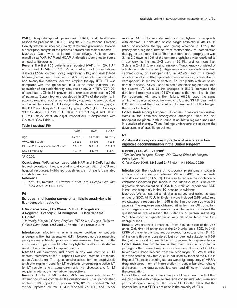
(VAP), hospital-acquired pneumonia (HAP), and healthcare-associated pneumonia (HCAP) using the 2005 American ThoracicSociety/Infectious Diseases Society of America guidelines. Below isa descriptive analysis of the patients enrolled and their outcomes.Methods Data were collected prospectively. Patients wereclassified as VAP, HAP and HCAP. Antibiotics were chosen basedon local antibiograms.Results The first 158 patients are reported (VAP n = 120, HAPn = 26 and HCAP n = 12). Patients often had comorbidities;diabetes (22%), cardiac (22%), respiratory (21%) and renal (16%).Microorganisms were identified in 78% of patients. One hundredand twenty-five patients received empiric therapy (ET). ET wascompliant with the guidelines in 31% of these patients. De-escalation of antibiotic therapy occurred on day 3 in 75% (77/103)of candidates. Clinical improvement and/or cure were seen in 70%of patients. Superinfections developed in 37% of the patients. Inpatients requiring mechanical ventilatory support, the average dayson the ventilator was 12 ± 17 days. Patients’ average stay (days) inthe ICU* and hospital* differed by group: VAP (17 ± 14 days,23 ± 19 days), HAP (9 ± 10 days, 13 ± 13 days) and HCAP(11 ± 19 days, 22 ± 36 days), respectively. *Comparisons withP < 0.05. See Table 1.
Table 1 (abstract P5)
VAP HAP HCAP
Age 57 ± 19 51 ± 18 64 ± 17
APACHE II score* 21 ± 6 18 ± 6 17 ± 8
Clinical Pulmonary Infection Score* 6.8 ± 2 5.7 ± 2 5.2 ± 2
Day 14 mortality* 19.7% 15.4% 8.3%
*P < 0.05.
Conclusions VAP, as compared with HAP and HCAP, had thehighest severity of illness, mortality, and consumption of ICU andhospital resources. Published guidelines are not easily translatedinto daily practice.Reference1. Kett DH, Ramirez JA, Peyrani P, et al.: Am J Respir Crit Care
Med 2005, 71:388-416.
P6European multicenter survey on antibiotic prophylaxis inliver transplant patients
E Vandecasteele1, J De Waele1, S Blot1, D Vogelaers1, X Rogiers1, D Vandijck1, M Bourgeois2, J Decruyenaere1, E Hoste1
1University Hospital, Ghent, Belgium; 2AZ St-Jan, Bruges, BelgiumCritical Care 2008, 12(Suppl 2):P6 (doi: 10.1186/cc6227)
Introduction Infection remains a major problem for patientsundergoing liver transplantation (LT). However, no data regardingperioperative antibiotic prophylaxis are available. The aim of thestudy was to gain insight into prophylactic antibiotic strategiesused in European liver transplant centers.Methods An electronic and postal survey was sent to all LTcenters, members of the European Liver and Intestine Transplan-tation Association. The questionnaire asked for the prophylacticantibiotic regimen used for LT recipients undergoing elective LT,for LT recipients with acute-on-chronic liver disease, and for LTrecipients with acute liver failure, respectively.Results A total of 59 centers (46% response rate) from 16different countries completed the questionnaire. Of all participatingcenters, 8.6% reported to perform <25, 37.9% reported 25–50,27.6% reported 50–75, 10.4% reported 75–100, and 15.5%
reported >100 LTs annually. Antibiotic prophylaxis for recipientswith elective LT consisted of one single antibiotic in 48.3%. In50%, combination therapy was given; whereas in 1.7%, theprophylactic regimen rotated from monotherapy to combinationtherapy on a 6-month basis. The mean duration of prophylaxis was3.1 ± 2.0 days. In 19% of the centers prophylaxis was restricted to1 day only, to the first 2–3 days in 55.2%, and for more than3 days in 24.1% (one missing answer). Monotherapy consisted ofa first-line antibiotic agent (first-generation and second-generationcephalosporin, or aminopenicillin) in 42.9%, and of a broad-spectrum antibiotic (third-generation cephalosporin, piperacillin, orcarbapenem) in 57.1% of centers. For recipients with acute-on-chronic disease, 73.7% used the same antibiotic regimen as usedfor elective LT, while 26.3% changed it (5.3% increased theduration of prophylaxis, and 21.0% changed the type of antibiotic).For recipients with acute liver failure, 66.7% used the sameantibiotic regimen as used for elective LT, while 33.3% changed it(10.5% changed the duration of prophylaxis, and 22.8% changedthe type of antibiotic).Conclusions Among European LT centers, considerable variationexists in the antibiotic prophylactic strategies used for livertransplant recipients, both in terms of antibiotic regimen used andin duration of therapy. These findings underscore the need for thedevelopment of specific guidelines.
P7A national survey on current practice of use of selectivedigestive decontamination in the United Kingdom
R Shah1, J Louw2, T Veenith2
1Frimley Park Hospital, Surrey, UK; 1Queen Elizabeth Hospital,Kings Lynn, UKCritical Care 2008, 12(Suppl 2):P7 (doi: 10.1186/cc6228)
Introduction The incidence of nosocomial pneumonia in patientsin intensive care ranges between 7% and 40%, with a crudemortality exceeding 50% [1]. One way to reduce the incidence ofventilator-associated pneumonia in the intensive care is selectivedigestive decontamination (SDD). In our clinical experience, SDDis not used frequently in the UK, despite its evidence.Methods We conducted a telephonic survey and collected dataon use of SDD. All ICUs in England were included (256 units) andwe obtained a response form 249 units. The average size was 5.8patients. The response was obtained either from an ICU consultantor a charge nurse in the intensive care. Before we discussed thequestionnaire, we assessed the suitability of person answering.We discussed our questionnaire with 73 consultants and 176charge nurses.Results We obtained a response from 249 units out of the 256units. Only 6% (15 units) out of the 249 units used SDD. In 94%(235) of the units this was not considered for use, and in 4% (12)of the units this was considered but not deemed suitable. In 0.8%(two) of the units it is currently being considered for implementation.Conclusions The oropharynx is the major source of potentialpathogens that cause lower airway infections. The role of SDD isto eradicate these bacteria from the oropharynx [1]. We found inour telephonic survey that SDD is not used by most of the ICUs inEngland. The main deterring factors were high frequency of MRSA,drug resistance, lack of incorporation in sepsis bundles, relativedisinterest in the drug companies, cost and difficulty in obtainingthe preparation.One of the drawbacks of our survey could have been the fact thatwe discussed with charge nurses and consultants who were notpart of decision-making for the use of SDD in the ICUs. But thebottom line is that SDD is not used in the majority of ICUs.
S4
Reference1. Baxby D, van Saene HKF, Stoutenbeek CP, Zandstra DF:
Selective decontamination of the digestive tract: 13 yearson, what it is and what it is not. Intensive Care Med 1996,22:699-706.
P8Community-acquired and healthcare-related urosepsis: a multicenter prospective study
T Cardoso1, O Ribeiro2, A Costa-Pereira2, A Carneiro1, A SACiUCI Study Group1
1Hospital Geral Sto António, Porto, Portugal; 2Faculty of Medicine,University of Oporto, Porto, PortugalCritical Care 2008, 12(Suppl 2):P8 (doi: 10.1186/cc6229)
Introduction Urinary infections are the third focus of infection insepsis. In this study we describe the epidemiology and microbiologyof community-acquired urosepsis, to determine the associatedcrude mortality and to identify independent predictors of mortality.Methods A prospective, multicentered, cohort study on community-acquired urosepsis cases admitted to Portuguese ICUs from1 December 2004 to 30 November 2005 with a follow-up untildischarge.Results Seventeen units entered the study from the north to southof Portugal, corresponding to 41% of all mixed national ICU beds.Over this period 4,142 patients were admitted to the study – 897(22%) had community-acquired sepsis, and of these 65 (7%) hadurosepsis.Compared with other focuses of infection, urosepsis was morefrequent in women (66% vs 33% in nonurosepsis, P < 0.001), andassociated with shorter ICU length of stay (7 days vs 9 days, P =0.002). No significant differences were observed regardingseverity of illness (SAPS II, sepsis severity) or crude mortality. Theisolation rate was 68% with 41% positive blood cultures. Allisolations, except one, were Gram-negative and no fungus wasisolated; Escherichia coli dominated the microbiological profile(63% of all isolations).Healthcare-related infection (HCRI) was found in 31% of thesepatients: E coli represents 58% of all isolations but the resistanceprofile was different, with resistance to ciprofloxacin and cotrimoxazolincreasing from 9% (in community-acquired sepsis) to 25% (inHCRI). The 28-day mortality was higher in the non-HCRI group (29%)than in the HCRI group (15%), although not statistically significant.Conclusions Although described as being the focus of infectionwith better prognosis we could not confirm this for community-acquired urosepsis in the present study. HCRI patients are aparticular group with a similar microbiological profile but differentresistance profile requiring a different empirical approach.Reference1. Friedman ND, Kaye KS, Stout JE, et al.: Health care-associ-
ated bloodstream infections in adults: a reason to changethe accepted definition of community-acquired infections.Ann Intern Med 2002, 137:791-797.
P9Bedside laparoscopy to diagnose intrabdominal pathologyin the ICU
S Matano, M Bonizzoli, A Di Filippo, G Manca, A PerisIntensive Care and Emergency Service, Florence, ItalyCritical Care 2008, 12(Suppl 2):P9 (doi: 10.1186/cc6230)
Introduction The aim of the study was to evaluate the accuracy ofbedside diagnostic laparoscopy (BDL) in critically ill patients (CIP)
suspected to suffer from intrabdominal pathology compared withoperative laparotomy or diagnostic imaging (CT scan) and to verifythe safety of the procedure. In fact, a delay in the diagnosis ofintrabdominal pathology could worsen the morbidity and mortalityin these patients. In ICU patients treated with prolonged parenteralnutrition, mechanical ventilation and high-dose opioid analgesics,acalculous cholecystitis (AC) is a severe complication [1]. Clinicalevaluation of the abdomen is difficult as deep sedation often maskssymptoms, and physical examination is inconclusive so they arepotentially eligible for exploratory laparoscopy after abdominal CT.Furthermore, performing CT is often impossible because of thedifficulty in safely transporting CIP.Methods From January 2006 to November 2007 a BDL wasperformed in 24 CIP to confirm the clinical diagnosis of AC. Everyday, liver function tests are collected and abdominal ultrasono-graphy is performed when the suspicion of AC is high. Elevatedliver function tests and ultrasonography signs such as gallbladderdistension or wall thickening (>3–4 mm) with or without perichole-cystic fluid were the more significant findings of suspected AC andwere considered admission criteria in the study. Twenty-fourpatients met the criteria. Ten were trauma victims, three were post-cardiac surgical patients, and 11 had sepsis of unknown origin.Fifteen were hypotensive and required haemodynamic support.BDL was performed with the Visiport. The pneumoperitoneum wascreated with a 10–15 mmHg CO2 pressure. The mean proceduretime was 40 minutes.Results The procedure was done a mean 8 days (range 5–15 days)after ICU admission. In two patients the BDL was positive forgangrenous colecystitis (both after cardiac surgery) requiringlaparoscopic cholecystectomies in the operating room. Purulentperitonitis was found in five patients with sepsis of unknown originbut microbiological tests on ascites resulted negative in all cases.The other BDLs resulted negative for intrabdominal pathology.Conclusions BDL seems to represent an alternative and effectivetechnique that might be more accurate than a CT scan and lessinvasive than laparotomy to obtain a diagnostic evaluation ofintrabdominal pathology in ICU patients.Reference1. Rehm CG: Crit Care Clin 2000, 16:101-112.
P10A potential role for the chest X-ray in the transmission ofresistant bacteria in the ICU
PD Levin, O Shatz, D Moriah, S Sviri, A Or-Barbash, CL Sprung,C BlockHadassah Hebrew University Hospital, Jerusalem, IsraelCritical Care 2008, 12(Suppl 2):P10 (doi: 10.1186/cc6231)
Introduction An investigation of infection control practices used byX-ray technicians during the performance of routine chest X-rayscans in the ICU, transmission of resistant bacteria to the X-raymachine, and the effect of an infection control intervention. Up to20% of patients acquire infections in the ICU, 44% of which maybe transferred on caregivers’ hands. Daily routine chest X-rayscans are performed sequentially, presenting the potential forbacterial spread. The degree to which X-ray technicians applyinfection control measures, and the extent to which bacteria aretransferred, is unknown.Methods Compliance with 14 infection control measures wasmeasured covertly during the performance of daily chest X-rayscans. Bacterial surface cultures were taken from the X-raymachines. An educational intervention (informing the techniciansabout resistant bacteria, machine culture results and correctalcohol and glove use) was instituted. Observations and machine
Critical Care March 2008 Vol 12 Suppl 2 28th International Symposium on Intensive Care and Emergency Medicine
S5
cultures were repeated. The appearance of resistant bacteria inpatient cultures was followed.Results Infection control practices were compared before andafter the intervention. Alcohol hand-rub use before patient contactincreased from 12% to 25% of occasions (P = 0.009), from 0% to62% prior to touching the X-ray machine (P < 0.001) and from 9%to 39% (P < 0.001) before touching the next patient. Glove usealso improved significantly.Resistant Gram-negative bacteria grew in 12/31 (39%) preinter-vention X-ray machine cultures and 0/29 (0%, P < 0.001) post-intervention cultures. Cultures with no bacterial growth increasedfrom 11/31 (33%) to 22/29 (67%, P = 0.002) pre to post inter-vention.New occurrences of resistant Gram-negative bacteria in clinicalcultures decreased from 19 in 68 patients (28%) pre interventionto 8/84 (10%, P = 0.003) post intervention.Conclusions Resistant Gram-negative bacteria are foundfrequently on the X-ray machine, probably being transferred ontechnicians’ hands. This represents the potential for patient-to-patient bacteria transfer. A simple infection control interventiondecreases X-ray machine contamination and is associated with adecrease in the appearance of resistant bacteria in patientcultures, although causality is not proven.References1. Grundmann H, et al.: Crit Care Med 33:946–951.2. Pittet D, et al.: Arch Intern Med 159:821–826.
P11Healthcare-related bacteraemia admitted to the ICU
G Castro1, T Cardoso1, R Carneiro1, O Ribeiro2, A Costa-Pereira2, A Carneiro1
1Hospital Geral de Santo António, Porto, Portugal; 2Faculty ofMedicine, University of Oporto, Porto, PortugalCritical Care 2008, 12(Suppl 2):P11 (doi: 10.1186/cc6232)
Introduction Bacteraemia developing in patients outside thehospital is categorized as community acquired. Accumulatingevidence suggests that healthcare-related bacteraemia (HCRB)are distinct from those that are community acquired.Methods A prospective, observational study of all the patients withcommunity-acquired bacteraemia sepsis (CABS) admitted to atertiary, mixed, 12-bed ICU, at a university hospital, between 1December 2004 and 30 November 2005. HCRB was definedaccording to criteria proposed by Friedman and colleagues [1].Results Throughout the study period, 160 patients were admittedwith CABS; 50 (31%) had HCRB. In the CABS group the mainfocus of infection was respiratory (41%), intra-abdominal (15%)and endovascular (15%); in the HCRB group respiratory infectionwas present in 14 (28%) patients, intra-abdominal in 13 (26%)patients and urological in 10 (20%) patients (P = 0.227). Themicrobiological profile was different between the two groups: inthe non-HCRB the main microbiological agents were Gram-positive 57 (63%), versus 34 (37%) Gram-negative. In the HCRBgroup the Gram-negative dominated the microbiological profile: 26(65%) versus 34 (37%) (P = 0.003). The ICU crude mortality wasdifferent in both groups (52% in HCRB versus 34% in CABS, P =0.028) and also hospital mortality (60% vs 39%, P = 0.013).Conclusions HCRB has a higher crude mortality and a differentmicrobiological profile was shown in the present study. Thisknowledge should prompt the necessity for early recognition ofpatients with HCRB that would need a different therapeuticapproach.Reference1. Friedman ND, Kaye KS, Stout JE, et al.: Health care-associ-
ated bloodstream infections in adults: a reason to changethe accepted definition of community-acquired infections.Ann Intern Med 2002, 137:791-797.
P12Incidence of nosocomial infection in patients withnontraumatic or traumatic coma
L Lorente Ramos, J Castedo, R Galván, C García, J Iribarren, J Jiménez, M Brouard, L Lorenzo, S Palmero, M Martín, M MoraHospital Universitario de Canarias, La Laguna, Tenerife, SpainCritical Care 2008, 12(Suppl 2):P12 (doi: 10.1186/cc6233)
Introduction To determine the rate of nosocomial infection innontraumatic or traumatic coma patients.Methods A prospective study for 24 months in a medical–surgicalICU. Infections were diagnosed according to CDC criteria.Infections were classified based on the diagnosis onset as: earlyonset (EO), developed during the first 4 days of ICU stay; and lateonset (LO), developed 5 days after ICU admission.Results We included 118 patients with nontraumatic coma (31intracerebral hemorrhage, 30 subarachnoid hemorrhage, 15 braininfarction, 12 intoxication, nine CNS infection, six status epilepticusand 15 others), 63 males. The mean age was 55.07 (±16.12years). The mean APACHE II score was 18.50 (±12.02). A total of47 patients (39.83%) developed 70 nosocomial infections (28 EOand 42 LO) and death in 32 patients (27.12%): 33 pneumonias(18 EO and 15 LO), 25 urinary tract infections (eight EO and 17LO), five primary bacteremias (two EO and three LO), threecatheter-related bacteremias (three LO), three ventriculitis (threeLO) and one wound surgical infection (one LO). Themicroorganisms responsible were: nine Pseudomonas, nine CNS,eight Escherichia coli, six MSSA, five MRSA, five Haemophillus,five Candida albicans, four Streptococcus faecalis, fourStreptococcus pneumoniae, four Proteus mirabilis and 11 others.Included were 67 patients with traumatic coma, 57 males. Themean age was 38.02 (±17.49 years). The mean APACHE II scorewas 18.32 (±12.21). A total of 27 patients (40.29%) developed38 nosocomial infections (18 EO and 20 LO) and death in 14patients (20.89%): 27 pneumonias (15 EO and 12 LO), six urinarytract infections (one EO and five LO), two primary bacteremias(one EO and one LO), one catheter-related bacteremia (one LO),one ventriculitis (one EO) and one wound surgical infection (oneLO). The microorganisms responsible were: eight MSSA, oneMRSA, seven Pseudomonas aeruginosa, five CNS, fiveHaemophillus influenzae and 12 others.Conclusions Forty percent of patients with nontraumatic andtraumatic coma developed infections – those with a respiratoryorigin being the most frequent.
P13Comparative study on infection of the central nervoussystem in patients with head trauma and spontaneouscerebral hemorrhage
P Vartzeli, A Yiambides, K Daskalakis, M Moukas, K Schulpis,K MandragosRed Cross Hospital, Ampelokipoi, GreeceCritical Care 2008, 12(Suppl 2):P13 (doi: 10.1186/cc6234)
Introduction The emergency neurosurgical procedure, the longduration of it (>4 hours) and the infected trauma are factors thathave, in studies, been connected with increased probability ofinfection of the central nervous system (CNS) during the post-operative period.
Available online http://ccforum.com/supplements/12/S2
S6
Objective To study the appearance of infection of the CNS inpatients who have been operated on after sustaining a head injuryor spontaneous cerebral hemorrhage that were hospitalized in theICU, over a period of 2 years.Materials Recordings of 118 patients who were hospitalized in theICU during the period 2005–2007. The selection of the patientswas based on the following criteria: the reason for admission to theICU was head injury (70 patients) or cerebral hemorrhage (48patients); all patients had undergone a neurosurgical procedure;and an infection occurred during hospitalization in the ICU.Methods All patients out of the 118 that presented fever orlaboratory findings of an infection which could not be attributed toan infection of any other reason except CNS underwent lumbarpuncture.Results Twenty-seven patients underwent lumbar puncture(22.88%). Findings from the lumbar puncture compatible with aninfection of the CNS occurred in six patients (five patients withcerebral injury and one patient with cerebral hemorrhage) out of118 patients, 5.08% of all patients (7.14% of head injury and2.08% of cerebral hemorrhages).The days that the lumbar puncture was performed were the4th–19th postoperative days. The mean GCS value during theadmittance to the hospital of the total patients was 8.88 (3–15),but the mean GCS value of those patients that developed CNSinfection was 7.86 (3–14).Conclusions The administration of antibiotics from the first day ofadmittance to the ICU probably is accountable for the very low rateof infection of the CNS in patients with head injury or cerebralhemorrhage. There is no important difference between thescheduled surgical procedure from the head injury and automaticcerebral hemorrhage. Further studies are needed for the reductionand control of the postoperative infections in these patients.References1. Korinek AM: Neurosurgery 1997, 41:1073-1079.2. Korinek AM, Golmard JL, Elcheick A, et al.: Br J Neurosurgery
2005, 19:155-162.3. Kourbeti IS, Jacobs AV, Koslow M, et al.: Neurosurgery 2007,
60:317-325.
P14Respiratory community-acquired and healthcare-relatedsepsis: are they different?
G Castro1, O Ribeiro2, A Costa Pereira2, A Carneiro1, T Cardoso1
1Hospital Geral de Santo António, Cuidados Intensivos, Porto,Portugal; 2Faculdade de Medicina do Porto, Serviço Biostastísticae Informática, Porto, PortugalCritical Care 2008, 12(Suppl 2):P14 (doi: 10.1186/cc6235)
Introduction Respiratory infection counts for more than one-half ofall admissions to the ICU with sepsis. In this study theepidemiology and microbiological profile of community-acquiredand healthcare-related (HCR) respiratory sepsis will be described.Methods A prospective, observational study of all the patients withcommunity-acquired sepsis (CAS) admitted to our ICU, over 1year. Respiratory CAS was defined by the presence of respiratoryinfection and at least two SIRS criteria at the time of hospitaladmission or within the first 48 hours. HCR infection was definedaccording to criteria proposed by Friedman and colleagues [1].Results In the study period, 347 patients were admitted – 149(43%) with CAS. Respiratory infection was present in 102 patients(68%). Comparing this group with nonrespiratory CAS, 73%versus 51% were male (P = 0.01), with a similar median age of 57years versus 62 years (P = 0.334), more severe sepsis (40% vs28%) and less septic shock (46% vs 68%) (P = 0.030). Blood
cultures were obtained in 96 (94%) patients, only 8% werepositive versus 39% in nonrespiratory CAS (P < 0.001). Gram-positive microorganisms represented 51% of all isolations, Gram-negative 26%, Mycobacterium tuberculosis 6%, atypical 5%, andfungus represented only 2% of all isolations. Polymicrobian infec-tions were documented in 5% of the patients. HCR respiratoryinfection was present in 17%. Gram-positive microorganismsrepresented 50% of all isolations, and Gram-negative 37%. ICUlength of stay (9 vs 8 days, P = 0.595), as well as ICU (35% vs32%, P = 0.686) and hospital (36% vs 41%, P = 0.559) mortalitywere similar between respiratory and non-respiratory CAS.Conclusions Respiratory CAS is a very important problem in theICU, representing 30% of all admissions. Although themicrobiological profile is similar to that described in the literature,in this population tuberculosis still plays a representative role andneeds to be considered. In this population, no significantdifferences in the microbiological profile were seen between CASand HCR infection.Reference1. Friedman ND, Kaye KS, Stout JE, et al.: Health care-associ-
ated bloodstream infections in adults: a reason to changethe accepted definition of community-acquired infections.Ann Intern Med 2002, 137:791-797.
P15Antibiotic costs in bacteremic and nonbacteremic patientstreated with the de-escalation approach
E Evodia1, P Myrianthefs1, P Prezerakos2, G Baltopoulos1
1KAT General Hospital, Athens, Greece; 2Municipality of Athens,Educational Centre, Athens, GreeceCritical Care 2008, 12(Suppl 2):P15 (doi: 10.1186/cc6236)
Introduction Antibiotic therapy significantly contributes to health-care costs and especially to those infections due to multidrugresistance pathogens. The purpose of the study was to investigateempiric antibiotic therapy costs compared with the consequentapplication of de-escalated therapy.Methods We prospectively collected data regarding demographicsand antibiotic costs in critically ill ICU patients experiencinginfection. We recorded daily costs of empiric antibiotic therapy onidentification–suspicion of infection as well as the costs after thepathogen identification and susceptibility.Results We included 27 critically ill patients (15 males) of meanage 49.9 ± 4.3 years and illness severity of APACHE II score 15.0± 1.7, SAPS II 32.4 ± 3.7, and SOFA score 6.0 ± 0.5. Daily costsof initial empiric antibiotic therapy were significantly highercompared with those of the therapy guided according tosusceptibility results in confirmed bacteremias. This was applicablefor Gram-positive (€61.0 ± 12.7 vs €130.4 ± 56.3, P = 0.009),Gram-negative (€181.0 ± 47.8 vs €142.7 ± 42.9, P = 0.0063)and mixed (€166.0 ± 21.1 vs €96.0 ± 34.0, P = 0.0016)bacteremias. In patients with other sites of infection the antibioticcosts did not differ (P = 0.112) between therapy guided accordingto susceptibility results compared with empiric therapy (€239.0 ±49.7 vs €242.0 ± 88.7).In patients with negative cultures the daily antibiotic cost was€110.7 ± 31.9. Therapy in those patients was discontinued earlierand they had a significantly lower length of ICU stay (P = 0.000,8.7 ± 0.9 days vs 24.6 ± 4.1 days).Conclusions According to our bacteriologic susceptibility results,the de-escalation therapy is applicable only in bacteremias whichmay lead to decreased antibiotic costs. Such an approach is notapplicable in infections of other sites possibly due to multidrugresistance pathogens.
Critical Care March 2008 Vol 12 Suppl 2 28th International Symposium on Intensive Care and Emergency Medicine
S7
P16When appropriate antibiotic therapy is relevant inbacteremic septic patients
H Bagnulo, M GodinoMaciel Hospital, Montevideo, UruguayCritical Care 2008, 12(Suppl 2):P16 (doi: 10.1186/cc6237)
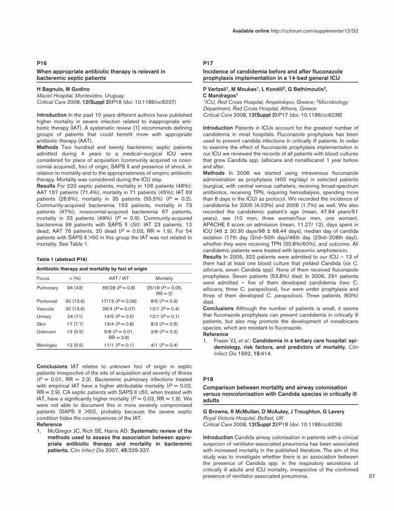
Introduction In the past 10 years different authors have publishedhigher mortality in severe infection related to inappropriate anti-biotic therapy (IAT). A systematic review [1] recommends defininggroups of patients that could benefit more with appropriateantibiotic therapy (AAT).Methods Two hundred and twenty bacteremic septic patientsadmitted during 4 years to a medical–surgical ICU wereconsidered for place of acquisition (community acquired vs noso-comial acquired), foci of origin, SAPS II and presence of shock, inrelation to mortality and to the appropriateness of empiric antibiotictherapy. Mortality was considered during the ICU stay.Results For 220 septic patients, mortality in 106 patients (48%):AAT 157 patients (71.4%), mortality in 71 patients (45%); IAT 63patients (28.6%), mortality in 35 patients (55.5%) (P = 0.2).Community-acquired bacteremia 153 patients, mortality in 73patients (47%); nosocomial-acquired bacteremia 67 patients,mortality in 33 patients (49%) (P = 0.9). Community-acquiredbacteremia 99 patients with SAPS II ≤50: IAT 23 patients, 12dead; AAT 76 patients, 20 dead (P = 0.03, RR = 1.9). For 54patients with SAPS II >50 in this group the IAT was not related tomortality. See Table 1.
Table 1 (abstract P16)
Antibiotic therapy and mortality by foci of origin
Focus n (%) AAT / IAT Mortality
Pulmonary 94 (43) 66/28 (P = 0.8) 25/18 (P = 0.05, RR = 2)
Peritoneal 30 (13.6) 17/13 (P = 0.08) 8/5 (P = 0.9)
Vascular 30 (13.6) 26/4 (P = 0.07) 12/1 (P = 0.4)
Urinary 24 (11) 19/5 (P = 0.5) 12/1 (P = 0.1)
Skin 17 (7.7) 13/4 (P = 0.8) 8/3 (P = 0.5)
Unknown 13 (5.5) 5/8 (P = 0.01, 2/6 (P = 0.2)RR = 3.8)
Meningeo 12 (5.5) 11/1 (P = 0.1) 4/1 (P = 0.4)
Conclusions IAT relates to unknown foci of origin in septicpatients irrespective of the site of acquisition and severity of illness(P = 0.01, RR = 2.3). Bacteremic pulmonary infections treatedwith empirical IAT have a higher attributable mortality (P = 0.02,RR = 2.9). CA septic patients with SAPS II ≤50, when treated withIAT, have a significantly higher mortality (P = 0.03, RR = 1.9). Wewere not able to document this in more severely compromisedpatients (SAPS II >50), probably because the severe septiccondition hides the consequences of the IAT.Reference1. McGregor JC, Rich SE, Harris AD: Systematic review of the
methods used to assess the association between appro-priate antibiotic therapy and mortality in bacteremicpatients. Clin Infect Dis 2007, 45:329-337.
P17Incidence of candidemia before and after fluconazoleprophylaxis implementation in a 14-bed general ICU
P Vartzeli1, M Moukas1, L Kondili2, G Bethimoutis2, C Mandragos1
1ICU, Red Cross Hospital, Ampelokipoi, Greece; 2MicrobiologyDepartment, Red Cross Hospital, Athens, GreeceCritical Care 2008, 12(Suppl 2):P17 (doi: 10.1186/cc6238)
Introduction Patients in ICUs account for the greatest number ofcandidemia in most hospitals. Fluconazole prophylaxis has beenused to prevent candida infections in critically ill patients. In orderto examine the effect of fluconazole prophylaxis implementation inour ICU we reviewed the records of all patients with blood culturesthat grew Candida spp. (albicans and nonalbicans) 1 year beforeand after.Methods In 2006 we started using intravenous fluconazoleadministration as prophylaxis (400 mg/day) in selected patients(surgical, with central venous catheters, receiving broad-spectrumantibiotics, receiving TPN, requiring hemodialysis, spending morethan 8 days in the ICU) as protocol. We recorded the incidence ofcandidemia for 2005 (4.03%) and 2006 (1.7%) as well. We alsorecorded the candidemic patient’s age (mean, 47.84 years/51years), sex (10 men, three women/four men, one woman),APACHE II score on admission (mean, 11.27/ 12), days spent inICU (46 ± 30.30 days/98 ± 68.44 days), median day of candidaisolation (17th day (2nd–50th day)/46th day (23rd–208th day)),whether they were receiving TPN (30.8%/60%), and outcome. Allcandidemic patients were treated with liposomic amphotericin.Results In 2005, 322 patients were admitted to our ICU – 13 ofthem had at least one blood culture that yielded Candida (six C.albicans, seven Candida spp). None of them received fluconazoleprophylaxis. Seven patients (53.8%) died. In 2006, 291 patientswere admitted – five of them developed candidemia (two C.albicans, three C. parapsilosis), four were under prophylaxis andthree of them developed C. parapsilosis. Three patients (60%)died.Conclusions Although the number of patients is small, it seemsthat fluconazole prophylaxis can prevent candidemia in critically illpatients, but also may promote the development of nonalbicansspecies, which are resistant to fluconazole.Reference1. Fraser VJ, et al.: Candidemia in a tertiary care hospital: epi-
demiology, risk factors, and predictors of mortality. ClinInfect Dis 1992, 15:414.
P18Comparison between mortality and airway colonisationversus noncolonisation with Candida species in critically illadults
G Browne, R McMullan, D McAuley, J Troughton, G LaveryRoyal Victoria Hospital, Belfast, UKCritical Care 2008, 12(Suppl 2):P18 (doi: 10.1186/cc6239)
Introduction Candida airway colonisation in patients with a clinicalsuspicion of ventilator-associated pneumonia has been associatedwith increased mortality in the published literature. The aim of thisstudy was to investigate whether there is an association betweenthe presence of Candida spp. in the respiratory secretions ofcritically ill adults and ICU mortality, irrespective of the confirmedpresence of ventilator-associated pneumonia.
Available online http://ccforum.com/supplements/12/S2
S8
Methods A retrospective analysis was performed on patientsadmitted to a large mixed ICU in Northern Ireland over a 1-yearperiod. Data were analysed to determine mortality in patientswhose respiratory secretions had cultured Candida spp. (both withand without coexisting bacteria), compared with those in whomcultures were negative for Candida spp. but positive for bacterialpathogens. Patients with persistently culture-negative respiratoryspecimens were excluded from analysis. Statistical significance ofobserved differences was evaluated by chi-square testing.Results In total, 287 patients were analysed. Of these, 202 (70%)were male. Bacteria only were cultured from respiratory secretions of208 (72%) patients (the ‘non-Candida’ group). The ‘Candida’ groupconsisted of 79 (28%) patients; of these, 39 had Candida spp. onlyand 40 had Candida spp. plus bacterial pathogens. Within the ‘non-Candida’ group, 39 patients died during the ICU episode; in the‘Candida’ group, 17 died (18.8% vs 21.5%, P = 0.597).Conclusions The presence of Candida spp. in the respiratorysecretions of this critically ill cohort was not associated with asignificant increase in ICU mortality. It appears, therefore, thatairway colonisation with Candida spp. in the absence of ventilator-associated pneumonia may not be regarded as a reliable predictorof ICU mortality.
P19Risk factors for lung colonization by Candida albicans in ageneral ICU
L Toma1, S Silvestri1, F Forfori2, G Licitra2, F Giunta2
1Scuola di specializzazione in Anestesia e Rianimazione, Pisa, Italy;2Azienda Ospedaliera Universitaria Pisana, Pisa, ItalyCritical Care 2008, 12(Suppl 2):P19 (doi: 10.1186/cc6240)
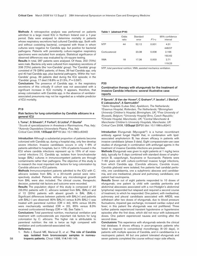
Introduction Although a substantial proportion of patients becomecolonized with Candida sp. during a hospital stay, only few developsevere infection. Invasive candidiasis occurs in only 1–8% ofpatients admitted to hospitals, but in 10% of patients housed in theICU where candida infections represent up to 15% of all noso-comial infections [1]. Candida sp. isolates from bronchoalveolarlavage (BAL) cultures in immunocompetent patients are throughcontaminants rather than pathogens. The objective of this study isto research the most important risk factors for lung colonization byCandida albicans in ICU patients.Methods Immunocompetent patients admitted to the ICU with C.albicans isolates from BAL in a 20-month period were retro-spectively studied. Patients without any microbiological growthfrom BAL were also included. The clinical course, therapeuticdecision, potential risk factors and outcome were recorded.Results The population object of this study is composed of 20(33.3%) patients with C. albicans isolated from BAL (BAL+) andof 12 (20%) patients with absent growth in BAL (BAL–).Significant differences between patients with BAL(+) and patientswith BAL(–) are observed: 80% BAL(+) versus 8.3% BAL(–) wastreated with parenteral nutrition (OR = 44), 90% versus 33.3%were mechanically ventilated (OR = 20), 65% versus 8.3%received corticosteroid therapy (OR = 18). See Table 1.Conclusions Total parenteral nutrition, mechanical ventilation andtreatment with corticosteroids are important risk factors for lungcolonization by C. albicans. The higher risk is attributable toparenteral nutrition: the risk is twice as high compared withventilation and corticosteroid-associated risk.Reference1. Rello J, Esandi ME, Mariscal D, et al.: The role of Candida
spp. isolated from broncoscopic samples in nonneu-tropenic patients. Chest 1998, 114:146-149.
P20Combination therapy with efungumab for the treatment ofinvasive Candida infections: several illustrative casereports
P Spronk1, B Van der Hoven2, C Graham3, F Jacobs4, J Sterba5,E Liakopoulou6, A Qamruddin7
1Gelre Hospitals (Lukas Site), Apeldoorn, The Netherlands;2Erasmus Hospital, Rotterdam, The Netherlands; 3BirminghamUniversity Children’s Hospital, Birmingham, UK; 4Free University ofBrussels, Belgium; 5University Hospital Brno, Czech Republic;6Christie Hospital, Manchester, UK; 7Central Manchester &Manchester Children’s University Hospitals, Manchester, UKCritical Care 2008, 12(Suppl 2):P20 (doi: 10.1186/cc6241)
Introduction Efungumab (Mycograb®) is a human recombinantantibody against fungal Hsp90 that, in combination with lipid-associated amphotericin B, has shown efficacy in patients withinvasive candidiasis (phase 3 data). Eight compassionate-use casestudies of efungumab in combination with antifungal agents in thetreatment of invasive Candida infections are presented.Methods Efungumab was given to eight patients at 1 mg/kg twicedaily, typically for 5 days combined with standard doses of ampho-tericin B, caspofungin, flucytosine or fluconazole. Patients were7–69 years old with culture-confirmed invasive fungal infections,from which Candida spp. (Candida albicans, Candida krusei,Candida glabrata) were isolated; five patients had candidal perito-nitis, one candidaemia, one a subphrenic abscess and candidae-mia, and one mediastinal, pleural and pulmonary candidiasis; onepatient had neutropenia.Results Seven out of eight patients responded to 10 doses ofefungumab; one patient (a child with candida peritonitis andabdominal abscesses associated with a non-Hodgkin’s abdominallymphoma) responded but relapsed and required a second courseof treatment, to which he responded. One patient, with mediastinal,pulmonary and pleural candidiasis associated with ARDS, waswithdrawn after two doses of efungumab, due to blood pressurefluctuations, impaired gas exchange, increased cardiac output andfever; in this patient the efungumab was not prefiltered. Threefurther patients experienced transient hypotensive or hypertensiveepisodes after the first dose, which did not recur with subsequentdoses. One patient experienced nausea and vomiting after thesecond dose.Conclusions This experience with efungumab extends the clinicaltrial database. It shows efficacy in poor-prognosis patients whofailed to respond to conventional monotherapy (6–20 days), inpatients with multiple species of Candida, and in candidaemia in aneutropenic patient. All but one patient tolerated efungumab andseven patients completed the course without major side effects.
Critical Care March 2008 Vol 12 Suppl 2 28th International Symposium on Intensive Care and Emergency Medicine
Table 1 (abstract P19)
Odds Standard 95% confidence ratio error P > z interval
NTP 44 52.12 0.001 4.31
448.57
Corticosteroid 23.38 0.008 2.166
192.62
VMA 18 17.36 0.003 2.71
119.23
NTP, total parenteral nutrition; VMA, assisted mechanics ventilation.
S9
P21Pooled analysis of safety for micafungin
OA Cornely1, P Maddison2, AJ Ullmann3
1Universität Klinikum Köln, Germany; 2Astellas Pharma Europe BV,Leiderdorp, The Netherlands; 3Klinikum derJohannes Gutenberg-Universität, Mainz, GermanyCritical Care 2008, 12(Suppl 2):P21 (doi: 10.1186/cc6242)
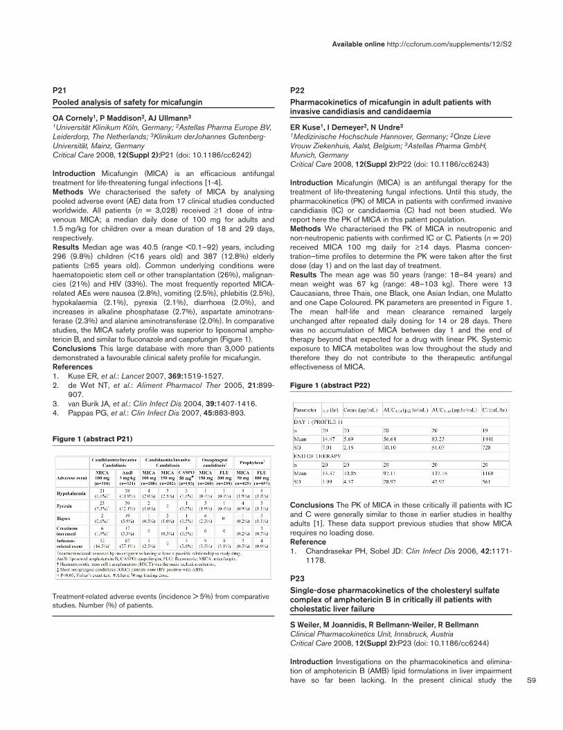
Introduction Micafungin (MICA) is an efficacious antifungaltreatment for life-threatening fungal infections [1-4].Methods We characterised the safety of MICA by analysingpooled adverse event (AE) data from 17 clinical studies conductedworldwide. All patients (n = 3,028) received ≥1 dose of intra-venous MICA; a median daily dose of 100 mg for adults and1.5 mg/kg for children over a mean duration of 18 and 29 days,respectively.Results Median age was 40.5 (range <0.1–92) years, including296 (9.8%) children (<16 years old) and 387 (12.8%) elderlypatients (≥65 years old). Common underlying conditions werehaematopoietic stem cell or other transplantation (26%), malignan-cies (21%) and HIV (33%). The most frequently reported MICA-related AEs were nausea (2.8%), vomiting (2.5%), phlebitis (2.5%),hypokalaemia (2.1%), pyrexia (2.1%), diarrhoea (2.0%), andincreases in alkaline phosphatase (2.7%), aspartate aminotrans-ferase (2.3%) and alanine aminotransferase (2.0%). In comparativestudies, the MICA safety profile was superior to liposomal ampho-tericin B, and similar to fluconazole and caspofungin (Figure 1).Conclusions This large database with more than 3,000 patientsdemonstrated a favourable clinical safety profile for micafungin.References1. Kuse ER, et al.: Lancet 2007, 369:1519-1527.2. de Wet NT, et al.: Aliment Pharmacol Ther 2005, 21:899-
907.3. van Burik JA, et al.: Clin Infect Dis 2004, 39:1407-1416.4. Pappas PG, et al.: Clin Infect Dis 2007, 45:883-893.
P22Pharmacokinetics of micafungin in adult patients withinvasive candidiasis and candidaemia
ER Kuse1, I Demeyer2, N Undre3
1Medizinische Hochschule Hannover, Germany; 2Onze LieveVrouw Ziekenhuis, Aalst, Belgium; 3Astellas Pharma GmbH,Munich, GermanyCritical Care 2008, 12(Suppl 2):P22 (doi: 10.1186/cc6243)
Introduction Micafungin (MICA) is an antifungal therapy for thetreatment of life-threatening fungal infections. Until this study, thepharmacokinetics (PK) of MICA in patients with confirmed invasivecandidiasis (IC) or candidaemia (C) had not been studied. Wereport here the PK of MICA in this patient population.Methods We characterised the PK of MICA in neutropenic andnon-neutropenic patients with confirmed IC or C. Patients (n = 20)received MICA 100 mg daily for ≥14 days. Plasma concen-tration–time profiles to determine the PK were taken after the firstdose (day 1) and on the last day of treatment.Results The mean age was 50 years (range: 18–84 years) andmean weight was 67 kg (range: 48–103 kg). There were 13Caucasians, three Thais, one Black, one Asian Indian, one Mulattoand one Cape Coloured. PK parameters are presented in Figure 1.The mean half-life and mean clearance remained largelyunchanged after repeated daily dosing for 14 or 28 days. Therewas no accumulation of MICA between day 1 and the end oftherapy beyond that expected for a drug with linear PK. Systemicexposure to MICA metabolites was low throughout the study andtherefore they do not contribute to the therapeutic antifungaleffectiveness of MICA.
Conclusions The PK of MICA in these critically ill patients with ICand C were generally similar to those in earlier studies in healthyadults [1]. These data support previous studies that show MICArequires no loading dose.Reference1. Chandrasekar PH, Sobel JD: Clin Infect Dis 2006, 42:1171-
1178.
P23Single-dose pharmacokinetics of the cholesteryl sulfatecomplex of amphotericin B in critically ill patients withcholestatic liver failure
S Weiler, M Joannidis, R Bellmann-Weiler, R BellmannClinical Pharmacokinetics Unit, Innsbruck, AustriaCritical Care 2008, 12(Suppl 2):P23 (doi: 10.1186/cc6244)
Introduction Investigations on the pharmacokinetics and elimina-tion of amphotericin B (AMB) lipid formulations in liver impairmenthave so far been lacking. In the present clinical study the
Available online http://ccforum.com/supplements/12/S2
Figure 1 (abstract P21)
Treatment-related adverse events (incidence > 5%) from comparativestudies. Number (%) of patients.
Figure 1 (abstract P22)
S10
pharmacokinetics of the cholesteryl sulfate complex of AMB wasassessed in critically ill patients with cholestatic liver failure.Methods Time–concentration profiles were determined in critically illpatients with cholestatic liver failure and in critically ill patients withnormal hepatic function requiring cholesteryl sulfate complex of AMBfor invasive fungal infections. The lipid-associated and liberatedfraction of AMB were separated by solid-phase extraction andsubsequently quantified by high-performance liquid chromatography.Results Three patients with impaired and three patients withnormal hepatic function on day 1 of ABCD therapy have so farbeen enrolled. After a single dose of ABCD (2.46 ± 0.54 mg vs2.94 ± 1.47 mg/kg in the impaired-liver group compared with thecontrol group), the maximum concentration in patients withimpaired liver function was fourfold increased compared with thecontrol group (1.98 ± 0.61 vs 0.52 ± 0.12 μg/ml for total AMB(P < 0.05), 1.25 ± 0.58 vs 0.46 ± 0.14 μg/ml for the liberatedfraction (P < 0.05), 0.74 ± 0.05 vs 0.06 ± 0.02 μg/ml for the lipid-associated fraction (P < 0.05)). The clearance was slower in theinvestigational group (0.15 ± 0.09 vs 0.38 ± 0.19 l/hour/kg fortotal AMB, 0.22 ± 0.10 vs 0.38 ± 0.19 l/hour/kg for the liberatedAMB fraction (P < 0.05) and 0.52 ± 0.45 vs 17.84 ± 15.45 l/hour/kgfor lipid-associated AMB (P < 0.05)). The volume of distribution atsteady state was significantly decreased (2.17 ± 0.58 vs 9.78 ±2.99 l/kg for total AMB (P < 0.05), 3.09 ± 0.88 vs 10.39 ± 2.70 l/kgfor liberated AMB (P < 0.05) and 8.18 ± 3.47 vs 83.27 ± 64.98 l/kgfor lipid-associated AMB (P < 0.05)).Conclusions The elimination of ABCD appears to be delayed incholestatic liver failure, particularly that of the lipid-associatedfraction. More pharmacokinetic data are required to establishreliable dose recommendations for ABCD in patients with liverfailure.
P24Serum tobramycin levels during selective decontaminationof the digestive tract in ICU patients on renal replacementtherapy
M Mol, H Van Kan, L Spanjaard, M Schultz, M Vroom, E De JongeAcademic Medical Center, Amsterdam, The NetherlandsCritical Care 2008, 12(Suppl 2):P24 (doi: 10.1186/cc6245)
Introduction Selective decontamination of the digestive tract (SDD)is an infection prophylaxis regimen that may improve survival in ICUpatients [1]. Antibiotics for SDD are nonabsorbable, are givenenterally and are therefore considered safe to use. The aim of ourstudy was to determine whether enteral administration of tobramycinas part of a SDD regimen may lead to detectable and potentiallytoxic serum tobramycin concentrations in patients with renal failure.Methods A prospective, observational study in ICU patients givenSDD treatment for at least 3 days. All patients were on continuousvenovenous hemofiltration with a filtration rate of 35 ml/kg/hour.Tobramycin serum concentrations were measured every 3 days.Results Serum samples were taken a median 6 days after the start ofSDD (IQR 3–9 days). Detectable tobramycin levels were found in 12of 19 patients (63%) and in 15 of 26 serum samples (58%). In fourpatients tobramycin concentrations were ≥1 mg/l, and in one of thesepatients a toxic concentration of 3 mg/l was found. All patients withtobramycin levels >1 mg/l had ischemic bowel disease. In contrast,no patients with lower concentrations had intestinal ischemia.Conclusions In patients with renal failure treated with continuousvenovenous hemofiltration, administration of SDD can lead todetectable and potentially toxic tobramycin serum concentrations.The risk of increased enteral absorption of tobramycin may beparticularly high in patients with intestinal ischemia. We advise
monitoring plasma tobramycin concentrations in patients with renalfailure on prolonged treatment with SDD.Reference1. de Jonge E, et al.: Effects of selective decontamination of
digestive tract on mortality and acquisition of resistantbacteria in intensive care: a randomised controlled trial.Lancet 2003, 362:1011-1016.
P25The pharmacokinetics of dalbavancin in subjects with mild,moderate, or severe hepatic impairment
J Dowell1, E Seltzer1, M Buckwalter1, T Marbury2, D Simoneau3,E Boudry3
1Vicuron Pharmaceuticals, Pfizer Inc., New York, USA; 2OrlandoClinical Research Center, Orlando, FL, USA; 3Pfizer InternationalOperations, Paris, FranceCritical Care 2008, 12(Suppl 2):P25 (doi: 10.1186/cc6246)
Introduction Dalbavancin (DAL) is a semisynthetic lipoglyco-peptide in phase 3 development with activity against Gram-positivebacteria. Weekly doses (1 g day 1/0.5 g day 8) are being investi-gated for the treatment of complicated skin and soft tissue infec-tions. DAL has both renal and nonrenal routes of elimination. Astudy was performed to assess the need for dosage adjustments inpatients with hepatic impairment.Methods Subjects received intravenously 1 g DAL on day 1 followedby 0.5 g on day 8. Subjects had mild, moderate, or severe hepaticimpairment as defined by Child–Pugh criteria A, B, or C. Age,gender, and weight-matched controls with normal hepatic functionwere also enrolled. DAL plasma concentrations were determinedand pharmacokinetic parameters were calculated. Drug exposurewas calculated as the cumulative area under the concentration–time curve through day 15; drug clearance and the elimination half-life were also determined. Safety was assessed by physicalexamination and adverse event and laboratory monitoring.Results Twenty-six subjects were enrolled, received DAL, and hadevaluable pharmacokinetics. The drug was well tolerated with noserious adverse events. DAL concentrations and exposures werenot increased due to hepatic impairment. The elimination half-lifewas not affected by hepatic impairment. Slightly lower exposuresand higher drug clearance were observed for subjects withmoderate and severe hepatic impairment, presumably due tovolume changes secondary to ascites and edema. The DALconcentrations observed for these subjects were comparable withthe ranges observed in other studies.Conclusions DAL concentrations are not increased due to hepaticimpairment and no dosage adjustment should be required forpatients with mild, moderate, or severe hepatic impairment.
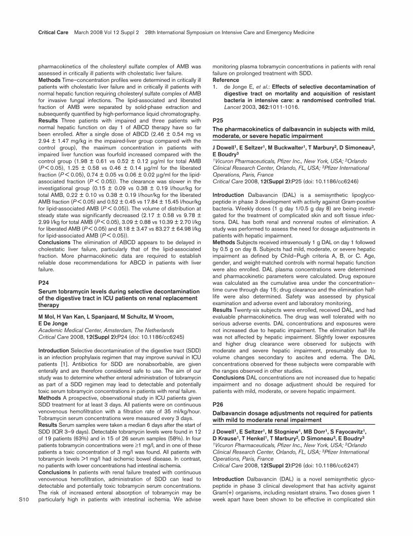
P26Dalbavancin dosage adjustments not required for patientswith mild to moderate renal impairment
J Dowell1, E Seltzer1, M Stogniew1, MB Dorr1, S Fayocavitz1, D Krause1, T Henkel1, T Marbury2, D Simoneau3, E Boudry3
1Vicuron Pharmaceuticals, Pfizer Inc., New York, USA; 2OrlandoClinical Research Center, Orlando, FL, USA; 3Pfizer InternationalOperations, Paris, FranceCritical Care 2008, 12(Suppl 2):P26 (doi: 10.1186/cc6247)
Introduction Dalbavancin (DAL) is a novel semisynthetic glyco-peptide in phase 3 clinical development that has activity againstGram(+) organisms, including resistant strains. Two doses given 1week apart have been shown to be effective in complicated skin
Critical Care March 2008 Vol 12 Suppl 2 28th International Symposium on Intensive Care and Emergency Medicine
S11
and soft tissue infections. A clinical study was performed todetermine the need for dosage adjustments in subjects with mild tomoderate renal impairment (RI).Methods Subjects with normal renal function (creatinine clearance(CrCL) > 80 ml/min), mild RI (CrCL of 50–79 ml/min), or moderateRI (CrCL of 30–49 ml/min) received DAL as a single intravenousinfusion (1,000 mg). Plasma samples were collected through atleast 14 days after the dose. DAL was assayed using validated LC-MS/MS methods. Pharmacokinetic (PK) data were analyzed usingnoncompartmental methods.Results Twenty-one subjects were enrolled, received one dose of1,000 mg dalbavancin, and were included in the PK analysis.Plasma concentration–time curves through 14 days (AUC0–14)were similar between subjects with normal renal function andsubjects with mild or moderate RI. An increased concentration wasobserved in subjects with moderate RI beyond day 14, at a point inthe profile when concentrations were below 40 m/l (Figure 1).Conclusions DAL does not require a dosage adjustment forpatients with mild or moderate RI. These results are consistent withprevious clinical and nonclinical PK studies showing that DAL hasdual (both renal and nonrenal) routes of elimination.
P27Dalbavancin safety in the phase 2/3 clinical developmentprogram
E Seltzer1, L Goldberg1, D Krause1, D Simoneau2, E Boudry2
1Vicuron Pharmaceuticals, Pfizer Inc., New York, USA; 2PfizerInternational Operations, Paris, FranceCritical Care 2008, 12(Suppl 2):P27 (doi: 10.1186/cc6248)
Introduction Dalbavancin (DAL) is a novel, next-generationlipoglycopeptide with a pharmacokinetic profile that allows weeklydosing. The safety of DAL in the treatment of complicated skin andsoft tissue infections was demonstrated versus comparators (COMP)in the phase 2/3 clinical development program.Methods Safety was assessed by analyzing adverse events (AEs),laboratory parameters, vital signs, and ECGs. Safety analyses wereconducted on the intent-to-treat (ITT) population, using descriptivestatistics only (consistent with ICH Guidance). COMP includedlinezolid, cefazolin, and vancomycin.
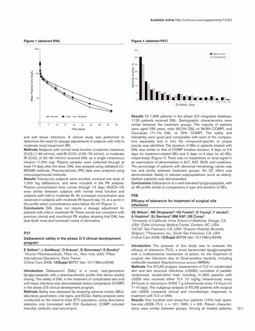
Results Of 1,699 patients in the phase 2/3 integrated database,1126 patients received DAL. Demographic characteristics weresimilar between the treatment groups. The majority of patientswere aged <65 years, male (60.2% DAL vs 58.8% COMP), andCaucasian (71.1% DAL vs 75% COMP). The safety andtolerability were good and comparable with each of the compara-tors separately and in toto. No compound-specific or uniquetoxicity was identified. The duration of AEs in patients treated withDAL was similar to that of COMP (median duration, 4 days vs 5.5days for treatment-related AEs and 3 days vs 4 days for all AEs,respectively) (Figure 1). There was no hepatotoxic or renal signal inan examination of abnormalities in ALT, AST, BUN, and creatinine.The percentage of patients with abnormal hematology values waslow and similar between treatment groups. No QT effect wasdemonstrated. Safety in relevant subpopulations (such as elderly,diabetic patients) was demonstrated.Conclusions Dalbavancin is a well-tolerated lipoglycopeptide, withan AE profile similar to comparators in type and duration of AEs.
P28Efficacy of telavancin for treatment of surgical siteinfections
SE Wilson1, ME Stryjewski2, VG Fowler2, D Young3, F Jacobs4,A Hopkins5, SL Barriere5, MM Kitt5, GR Corey2
1University of California, Irvine School of Medicine, Orange, CA,USA; 2Duke University Medical Center, Durham, NC, USA;3UCSF, San Francisco, CA, USA; 4Erasme Hospital, Brussels,Belgium; 5Theravance, Inc., South San Francisco, CA, USACritical Care 2008, 12(Suppl 2):P28 (doi: 10.1186/cc6249)
Introduction The purpose of this study was to evaluate theefficacy of telavancin (TLV), a novel bactericidal lipoglycopeptidewith a multifunctional mechanism of action, for the treatment ofsurgical site infections due to Gram-positive bacteria, includingmethicillin-resistant Staphylococcus aureus (MRSA).Methods The ATLAS program (assessment of TLV in complicatedskin and skin structure infections (cSSSI)) consisted of parallel,randomized, double-blind trials including >1,800 patients withcSSSI who received either TLV 10 mg/kg intravenously every24 hours or vancomycin (VAN) 1 g intravenously every 12 hours for7–14 days. This subgroup analysis of ATLAS patients with surgicalsite cSSSI compared clinical and microbiologic responses totreatment with TLV or VAN.Results One hundred and ninety-four patients (10%) had opera-tive-site cSSSI (TLV, n = 101; VAN, n = 93). Patient character-istics were similar between groups. Among all treated patients,
Available online http://ccforum.com/supplements/12/S2
Figure 1 (abstract P26) Figure 1 (abstract P27)
S12
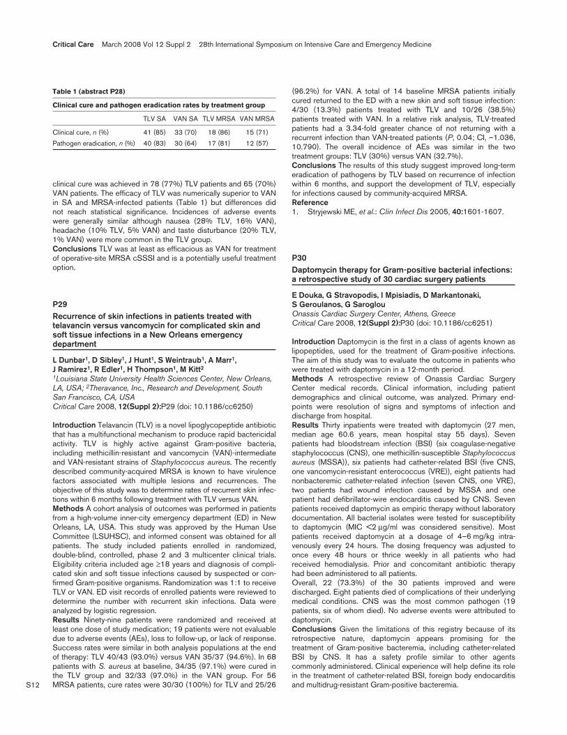
clinical cure was achieved in 78 (77%) TLV patients and 65 (70%)VAN patients. The efficacy of TLV was numerically superior to VANin SA and MRSA-infected patients (Table 1) but differences didnot reach statistical significance. Incidences of adverse eventswere generally similar although nausea (28% TLV, 16% VAN),headache (10% TLV, 5% VAN) and taste disturbance (20% TLV,1% VAN) were more common in the TLV group.Conclusions TLV was at least as efficacious as VAN for treatmentof operative-site MRSA cSSSI and is a potentially useful treatmentoption.
P29Recurrence of skin infections in patients treated withtelavancin versus vancomycin for complicated skin andsoft tissue infections in a New Orleans emergencydepartment
L Dunbar1, D Sibley1, J Hunt1, S Weintraub1, A Marr1, J Ramirez1, R Edler1, H Thompson1, M Kitt2
1Louisiana State University Health Sciences Center, New Orleans,LA, USA; 2Theravance, Inc., Research and Development, SouthSan Francisco, CA, USACritical Care 2008, 12(Suppl 2):P29 (doi: 10.1186/cc6250)
Introduction Telavancin (TLV) is a novel lipoglycopeptide antibioticthat has a multifunctional mechanism to produce rapid bactericidalactivity. TLV is highly active against Gram-positive bacteria,including methicillin-resistant and vancomycin (VAN)-intermediateand VAN-resistant strains of Staphylococcus aureus. The recentlydescribed community-acquired MRSA is known to have virulencefactors associated with multiple lesions and recurrences. Theobjective of this study was to determine rates of recurrent skin infec-tions within 6 months following treatment with TLV versus VAN.Methods A cohort analysis of outcomes was performed in patientsfrom a high-volume inner-city emergency department (ED) in NewOrleans, LA, USA. This study was approved by the Human UseCommittee (LSUHSC), and informed consent was obtained for allpatients. The study included patients enrolled in randomized,double-blind, controlled, phase 2 and 3 multicenter clinical trials.Eligibility criteria included age ≥18 years and diagnosis of compli-cated skin and soft tissue infections caused by suspected or con-firmed Gram-positive organisms. Randomization was 1:1 to receiveTLV or VAN. ED visit records of enrolled patients were reviewed todetermine the number with recurrent skin infections. Data wereanalyzed by logistic regression.Results Ninety-nine patients were randomized and received atleast one dose of study medication; 19 patients were not evaluabledue to adverse events (AEs), loss to follow-up, or lack of response.Success rates were similar in both analysis populations at the endof therapy: TLV 40/43 (93.0%) versus VAN 35/37 (94.6%). In 68patients with S. aureus at baseline, 34/35 (97.1%) were cured inthe TLV group and 32/33 (97.0%) in the VAN group. For 56MRSA patients, cure rates were 30/30 (100%) for TLV and 25/26
(96.2%) for VAN. A total of 14 baseline MRSA patients initiallycured returned to the ED with a new skin and soft tissue infection:4/30 (13.3%) patients treated with TLV and 10/26 (38.5%)patients treated with VAN. In a relative risk analysis, TLV-treatedpatients had a 3.34-fold greater chance of not returning with arecurrent infection than VAN-treated patients (P, 0.04; CI, –1.036,10.790). The overall incidence of AEs was similar in the twotreatment groups: TLV (30%) versus VAN (32.7%).Conclusions The results of this study suggest improved long-termeradication of pathogens by TLV based on recurrence of infectionwithin 6 months, and support the development of TLV, especiallyfor infections caused by community-acquired MRSA.Reference1. Stryjewski ME, et al.: Clin Infect Dis 2005, 40:1601-1607.
P30Daptomycin therapy for Gram-positive bacterial infections:a retrospective study of 30 cardiac surgery patients
E Douka, G Stravopodis, I Mpisiadis, D Markantonaki, S Geroulanos, G SaroglouOnassis Cardiac Surgery Center, Athens, GreeceCritical Care 2008, 12(Suppl 2):P30 (doi: 10.1186/cc6251)
Introduction Daptomycin is the first in a class of agents known aslipopeptides, used for the treatment of Gram-positive infections.The aim of this study was to evaluate the outcome in patients whowere treated with daptomycin in a 12-month period.Methods A retrospective review of Onassis Cardiac SurgeryCenter medical records. Clinical information, including patientdemographics and clinical outcome, was analyzed. Primary end-points were resolution of signs and symptoms of infection anddischarge from hospital.Results Thirty inpatients were treated with daptomycin (27 men,median age 60.6 years, mean hospital stay 55 days). Sevenpatients had bloodstream infection (BSI) (six coagulase-negativestaphylococcus (CNS), one methicillin-susceptible Staphylococcusaureus (MSSA)), six patients had catheter-related BSI (five CNS,one vancomycin-resistant enterococcus (VRE)), eight patients hadnonbacteremic catheter-related infection (seven CNS, one VRE),two patients had wound infection caused by MSSA and onepatient had defibrillator-wire endocarditis caused by CNS. Sevenpatients received daptomycin as empiric therapy without laboratorydocumentation. All bacterial isolates were tested for susceptibilityto daptomycin (MIC <2 μg/ml was considered sensitive). Mostpatients received daptomycin at a dosage of 4–6 mg/kg intra-venously every 24 hours. The dosing frequency was adjusted toonce every 48 hours or thrice weekly in all patients who hadreceived hemodialysis. Prior and concomitant antibiotic therapyhad been administered to all patients.Overall, 22 (73.3%) of the 30 patients improved and weredischarged. Eight patients died of complications of their underlyingmedical conditions. CNS was the most common pathogen (19patients, six of whom died). No adverse events were attributed todaptomycin.Conclusions Given the limitations of this registry because of itsretrospective nature, daptomycin appears promising for thetreatment of Gram-positive bacteremia, including catheter-relatedBSI by CNS. It has a safety profile similar to other agentscommonly administered. Clinical experience will help define its rolein the treatment of catheter-related BSI, foreign body endocarditisand multidrug-resistant Gram-positive bacteremia.
Critical Care March 2008 Vol 12 Suppl 2 28th International Symposium on Intensive Care and Emergency Medicine
Table 1 (abstract P28)
Clinical cure and pathogen eradication rates by treatment group
TLV SA VAN SA TLV MRSA VAN MRSA
Clinical cure, n (%) 41 (85) 33 (70) 18 (86) 15 (71)
Pathogen eradication, n (%) 40 (83) 30 (64) 17 (81) 12 (57)
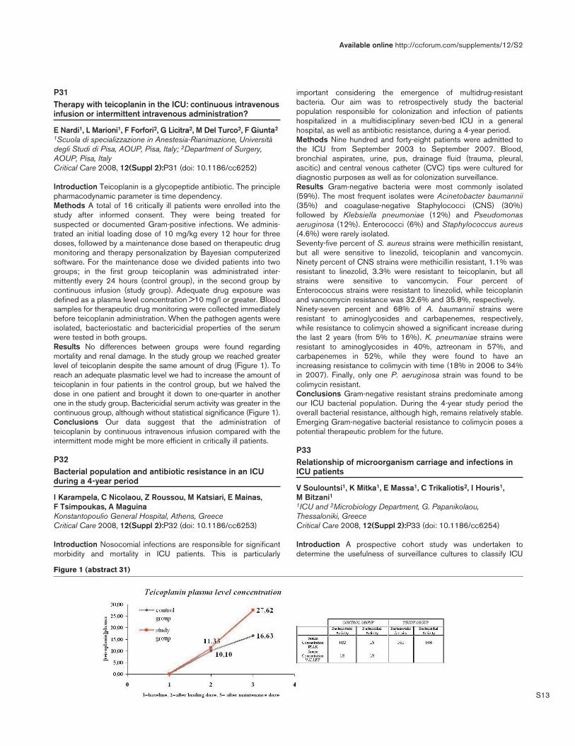
S13
P31Therapy with teicoplanin in the ICU: continuous intravenousinfusion or intermittent intravenous administration?
E Nardi1, L Marioni1, F Forfori2, G Licitra2, M Del Turco2, F Giunta2
1Scuola di specializzazione in Anestesia-Rianimazione, Universitàdegli Studi di Pisa, AOUP, Pisa, Italy; 2Department of Surgery,AOUP, Pisa, ItalyCritical Care 2008, 12(Suppl 2):P31 (doi: 10.1186/cc6252)
Introduction Teicoplanin is a glycopeptide antibiotic. The principlepharmacodynamic parameter is time dependency.Methods A total of 16 critically ill patients were enrolled into thestudy after informed consent. They were being treated forsuspected or documented Gram-positive infections. We adminis-trated an initial loading dose of 10 mg/kg every 12 hour for threedoses, followed by a maintenance dose based on therapeutic drugmonitoring and therapy personalization by Bayesian computerizedsoftware. For the maintenance dose we divided patients into twogroups; in the first group teicoplanin was administrated inter-mittently every 24 hours (control group), in the second group bycontinuous infusion (study group). Adequate drug exposure wasdefined as a plasma level concentration >10 mg/l or greater. Bloodsamples for therapeutic drug monitoring were collected immediatelybefore teicoplanin administration. When the pathogen agents wereisolated, bacteriostatic and bactericidial properties of the serumwere tested in both groups.Results No differences between groups were found regardingmortality and renal damage. In the study group we reached greaterlevel of teicoplanin despite the same amount of drug (Figure 1). Toreach an adequate plasmatic level we had to increase the amount ofteicoplanin in four patients in the control group, but we halved thedose in one patient and brought it down to one-quarter in anotherone in the study group. Bactericidial serum activity was greater in thecontinuous group, although without statistical significance (Figure 1).Conclusions Our data suggest that the administration ofteicoplanin by continuous intravenous infusion compared with theintermittent mode might be more efficient in critically ill patients.
P32Bacterial population and antibiotic resistance in an ICUduring a 4-year period
I Karampela, C Nicolaou, Z Roussou, M Katsiari, E Mainas, F Tsimpoukas, A MaguinaKonstantopoulio General Hospital, Athens, GreeceCritical Care 2008, 12(Suppl 2):P32 (doi: 10.1186/cc6253)
Introduction Nosocomial infections are responsible for significantmorbidity and mortality in ICU patients. This is particularly
important considering the emergence of multidrug-resistantbacteria. Our aim was to retrospectively study the bacterialpopulation responsible for colonization and infection of patientshospitalized in a multidisciplinary seven-bed ICU in a generalhospital, as well as antibiotic resistance, during a 4-year period.Methods Nine hundred and forty-eight patients were admitted tothe ICU from September 2003 to September 2007. Blood,bronchial aspirates, urine, pus, drainage fluid (trauma, pleural,ascitic) and central venous catheter (CVC) tips were cultured fordiagnostic purposes as well as for colonization surveillance.Results Gram-negative bacteria were most commonly isolated(59%). The most frequent isolates were Acinetobacter baumannii(35%) and coagulase-negative Staphylococci (CNS) (30%)followed by Klebsiella pneumoniae (12%) and Pseudomonasaeruginosa (12%). Enterococci (6%) and Staphylococcus aureus(4.6%) were rarely isolated.Seventy-five percent of S. aureus strains were methicillin resistant,but all were sensitive to linezolid, teicoplanin and vancomycin.Ninety percent of CNS strains were methicillin resistant, 1.1% wasresistant to linezolid, 3.3% were resistant to teicoplanin, but allstrains were sensitive to vancomycin. Four percent ofEnterococcus strains were resistant to linezolid, while teicoplaninand vancomycin resistance was 32.6% and 35.8%, respectively.Ninety-seven percent and 68% of A. baumannii strains wereresistant to aminoglycosides and carbapenemes, respectively,while resistance to colimycin showed a significant increase duringthe last 2 years (from 5% to 16%). K. pneumaniae strains wereresistant to aminoglycosides in 40%, aztreonam in 57%, andcarbapenemes in 52%, while they were found to have anincreasing resistance to colimycin with time (18% in 2006 to 34%in 2007). Finally, only one P. aeruginosa strain was found to becolimycin resistant.Conclusions Gram-negative resistant strains predominate amongour ICU bacterial population. During the 4-year study period theoverall bacterial resistance, although high, remains relatively stable.Emerging Gram-negative bacterial resistance to colimycin poses apotential therapeutic problem for the future.
P33Relationship of microorganism carriage and infections inICU patients
V Soulountsi1, K Mitka1, E Massa1, C Trikaliotis2, I Houris1, M Bitzani11ICU and 2Microbiology Department, G. Papanikolaou,Thessaloniki, GreeceCritical Care 2008, 12(Suppl 2):P33 (doi: 10.1186/cc6254)
Introduction A prospective cohort study was undertaken todetermine the usefulness of surveillance cultures to classify ICU
Available online http://ccforum.com/supplements/12/S2
Figure 1 (abstract 31)
S14
infections and to predict the causative agent of ICU-acquiredinfections.Methods A total of 48 community patients (36 men, 11 women,age 50.17 ± 17.974 years, APACHE II score 13.51 ± 6.153) whowere expected to stay in the ICU for >5 days were included in thisstudy. Surveillance cultures of the throat and rectum were obtainedon admission and thereafter at days 4, 8 and 12 to look forpotentially pathogenic microorganisms (PPMs) in order to distin-guish the community acquired from those acquired during the ICUstay. The epidemiological data and the alteration of carriage state ofthe patients during these days were recorded. Total infectionepisodes were classified into three categories according to thecarrier state: primary endogenous infection (PEI), caused by PPMscarried by the patient in surveillance cultures on admission;secondary endogenous infection (SEI),caused by PPMs of the ICUenvironment, yielded both in surveillance and diagnostic cultures;and exogenous infection (EXI) for those caused by PPMs that didnot yield in surveillance cultures. Statistical analysis was made usingPearson x2, paired t test, and ROC curve. P value ≤ a, a = 5%.Results On day 4, colonization was detected by throat and rectumsurveillance cultures in 81.1% and 75% of patients, respectively(P <<0.05). The most common microorganism isolated insurveillance cultures from the throat was Acinetobacter baumannii(22.9%) and that from the rectum was Escherichia coli (15.7%). Atotal of 100 infections were described during the patients’ ICUstay (length of stay: 26.44 ± 17.95) distinguished in 28 PEI, 44SEI and 25 EXI. ICU-acquired infections were 69% of cases. Themean day of PEI diagnosis was 6.2 ± 4.7, of SEI was 12.6 ± 8.2and of EXI was 12.6 ± 7.3. The causative agent could bepredicted in 72% of infections. The sixth day was the cutoff pointto predict the causative microbial agent from the surveillancecultures (sensitivity 80%, specificity 74.6%). Isolation of A.baumannii in surveillance cultures had a probability of 92.1% tocause infection.Conclusions Our data suggest that surveillance cultures may offeruseful information to improve hygiene in the ICU, to determine thecausative agent of infection and to follow better antimicrobialpolicy.
P34Rifampicin and colistin association in the treatment ofsevere multiresistant Acinetobacter baumannii infections
B Charra, A Hachimi, A Benslama, S MotaouakkilIbn Rochd University Hospital, Casablanca, MoroccoCritical Care 2008, 12(Suppl 2):P34 (doi: 10.1186/cc6255)
Introduction The increased incidence of nosocomial infections bymultidrug-resistant Acinetobacter baumannii creates demand onthe application of some combinations of older antimicrobials onthat species. We conducted the present observational study toevaluate the efficacy of intravenous and aerosolized colistincombined with rifampicin in the treatment of critically patients withnosocomial infections caused by multiresistant A. baumannii.Methods Critically ill patients with nosocomial infections causedby A. baumannii resistant to all antibiotics except colistin in amedical ICU were included. Diagnosis of infection was based onclinical data and isolation of bacteria. The bacterial susceptibilitiesto colistin were tested. Clinical response to colistin and rifampicinwas evaluated.Results There were 26 patients (age 43.58 ± 18.29 years,APACHE II score 6.35 ± 2.99), of whom were 16 cases of noso-comial pneumonia treated by aerosolized colistin (1 x 10⁶ IU threetimes/day) associated with intravenous rifampicin (10 mg/kg every
12 hours), nine cases of bacteraemia treated by intravenouscolistin (2 x 10⁶ IU three times/day) associated with intravenousrifampicin (10 mg/kg every 12 hours) in which three cases associa-ted with ventilator-associated pneumonia, and one case ofnosocomial meningitis treated by intrathecal use of colistinassociated with intravenous rifampicin. The clinical evolution wasfavourable for all ill patients. Concerning side effects, we noticed amoderate hepatic cytolysis in three patients.Conclusions This is a clinical report of colistin combined withrifampicin for treatment of A. baumannii infection. Despite the lackof a control group and the limited number of patients, the resultsseem to be encouraging.
P35Trends of resistance of Gram-negative bacteria in the ICUduring a 3-year period
A Skiada1, J Pavleas2, G Thomopoulos2, I Stefanou3, A Mega2,N Archodoulis2, P Vernikos2, K Rigas2, A Rigas2, A Kaskantamis2, A Zerva2, E Pagalou2, J Floros2
1Research Laboratory for Infectious Diseases and AntimicrobialChemotherapy, University of Athens, Greece; 2ICU, Laikon GeneralHospital, Athens, Greece; 3Microbiology Department, LaikonHospital, Athens, GreeceCritical Care 2008, 12(Suppl 2):P35 (doi: 10.1186/cc6256)
Introduction The aim of the study was to calculate the incidenceof bacteremias due to multidrug-resistant (MDR) Gram-negativebacteria (GNB) and to detect any emerging trends during a 3-yearperiod.Methods A prospective study of bloodstream infections in an ICUof a tertiary care hospital. The data collected prospectivelyincluded epidemiological and clinical characteristics of all patientsadmitted to the ICU from September 2004 to August 2007, thebacteria isolated from bloodstream infections and their patterns ofresistance. A bacterium was characterized as MDR when it wasresistant to three classes of antibiotics and as MDRc when it wasalso resistant to carbapenems. The study was divided into nine 4-month periods in order to calculate the incidence of MDRbacteremias in each such period and to evaluate each bacteriumseparately.Results During this study 390 patients were admitted to the ICU,of whom 60% were male. Their mean age was 65 years, the meanAPACHE score was 17.9 and the mean duration of stay in the ICUwas 18 days. One hundred bacteremias due to MDR GNBs wererecorded. Of the isolated MDR bacteria, 77 were MDRc and 95%of those 77 were Acinetobacter baumannii (25 isolates), Pseudo-monas aeruginosa (20 isolates) or Klebsiella pneumoniae (28isolates). A clear trend emerged for K. pneumoniae, whoseincidence increased exponentially during the study period. Of the28 isolates of MDRc K. pneumoniae, 7% were recorded during thefirst 12 months of the study, 33% during the next 12 months and60% during the last 12 months. The incidence of A. baumanniiremained relatively stable (36%, 32% and 32% of isolates wererecorded during each 12-month period) and the same was true forP. aeruginosa (25%, 40% and 35%, respectively).Conclusions The incidence of bacteremias due to MDR GNBsthat are also resistant to carbapenems is high in our ICU.Bacteremias due to MDRc K. pneumoniae have risen dramaticallyduring the past months. Further studies are needed to investigatethe risk factors and develop strategies to confront the problem.References1. Kollef M: Chest 2006, 130:1293-1296.2. Patel G: J Pharm Practice 2005, 18:91-99.
Critical Care March 2008 Vol 12 Suppl 2 28th International Symposium on Intensive Care and Emergency Medicine
S15
P36Colonization and infection by potential Gram-negativemultiresistant microorganism in a medical–surgical ICU
L Lorente Ramos, C García, J Iribarren, J Castedo, R Galván, J Jiménez, M Brouard, R Santacreu, L Lorenzo, M Martín, M MoraHospital Universitario de Canarias, La Laguna, Tenerife, SpainCritical Care 2008, 12(Suppl 2):P36 (doi: 10.1186/cc6257)
Introduction To determine the rate of colonization/infection bypotential Gram-negative multiresistant microorganisms (pseudo-monas, stenotrophomonas and acinetobacter) in critically ill patients.Methods A prospective study for 30 months in the ICU. Throatswabs, tracheal aspirates and urine were taken on admission andtwice weekly. The infections were classified based on thorat floraas: primary endogenous (PE), caused by germs that were alreadycolonizing the throat on ICU admission; secondary endogenous(SE), caused by germs that were not colonizing the throat on ICUadmission but were acquired during the stay in the ICU; andexogenous (EX), caused by germs that were not colonizing thethroat. The infections were classified based on the onset as: earlyonset (EO), when developed during the first 4 days of ICU stay;and late onset (LO), when developed 5 days after ICU admission.Results In total 1,582 patients were admitted. The mean APACHEII score was 13.95 (±8.93). Mortality was 14.79%. A total of 80patients had colonization by pseudomonas, 26 patients at ICUadmission and 54 patients during the ICU stay. We documented46 infections by pseudomonas (nine EO and 37 LO; four PE, 35SE and seven EX) with death in 13/46 patients (28.26%): 31pneumonias (six EO and 25 LO; two PE, 24 SE and five EX),seven urinary tract infections (one EO and six LO; two PE, threeSE and two EX), five primary bacteremias (two EO and three LO;five SE), two surgical wound infections (two LO and SE) and onepressure sore infection (one LO and SE). A total of 14 patients hadcolonization by stenotrophomonas, one patient at ICU admissionand 13 patients during the ICU stay. We documented eightinfections by stenotrophomonas (two EO and six LO; seven SEand one EX) with death in 3/8 patients (37.50%): all werepneumonias. A total of 12 patients had colonization byacinetobacter, one patient at ICU admission and 11 patientsduring the ICU stay. We documented eight infections by acineto-bacter (two EO and six LO; seven ES and one EX) with death in2/8 patients (25%): six pneumonias (two EO and four LO; five SEand one EX) and two bloodstream infection (two LO and two SE).Conclusions In our series, most of the infections caused bypseudomona, stenotrophomonas and acinetobacter were pneumo-nias, had a late onset and were secondary endogenous infections.
P37Changing resistance pattern for Acinetobacter baumanniithrough the years
F Alkaya Alver1, O Memikoglu1, E Özgencil1, E Eker2, M Oral1,N Unal1, M Tulunay1
1Ibni Sina Hospital, Ankara, Turkey; 2Bakent University, Adana,TurkeyCritical Care 2008, 12(Suppl 2):P37 (doi: 10.1186/cc6258)
Introduction Over the past two decades, Acinetobacter baumanniihas emerged as an important nosocomial pathogen, and hospitaloutbreaks caused by this organism have increased worldwide. Itsextraordinary ability to acquire resistance to almost all groups ofcommercially available antibiotics is a clinical problem of greatconcern. In fact, most A. baumannii strains isolated in the ICU arehighly resistant to carbapenems, aminogylcosides and β-lactam
antibiotics. The aim of this study is to determine the change in theresistance pattern of A. baumannii through the years.Methods Isolates from patients admitted to the ICU in 2003 and2007 were analyzed. Both colonization and infection isolates wereevaluated. In 2003, 118 isolates from 51 patients and in 2007,108 isolates from 21 patients were included in the study. Theclinical specimens were sampled from the tracheal aspirate,abscess, blood, wound, urine, pleura fluid, catheter and ascites.Susceptibilities to seven antibiotics were determined.Results Patterns of resistance in 2003 and 2007 were:54.7%/28.6% sulbactam–cefoperazone, 66.1%/85.7% imipenem,96.6%/95.2% piperacillin–tazobactam, 66.1%/61.9% cefepime,4.3%/0% colistin, 76.3%/85% amikacin, and 60.2%/66%tobramycin, respectively. Even though tobramycin is not on themarket in Turkey, drug resistance has been increased in 4 years.Conclusions Colistin seems to be the best alternative in ourhospital but, since it is not on the market in our country, the use ofit is limited. Carbepenems are therefore the first choice in thetreatment of multidrug-resistant A. baumannii in our country. Thischoice causes increased carbepenem resistance in A. baumanniiisolates. Our hospital’s antibiotic treatment policies and the cyclicusage of antibiotics must be reconsidered.Reference1. Del Mar Tomas M, Cartelle M, Pertega S, et al.: Hospital out-
break caused by a carbapenem-resistant strain of Acine-tobacter baumannii: patient prognosis and risk-factors forcolonisation and infection. Clin Microbiol Infect 2005, 11:540-546.
P38Catheter-related bloodstream infection according tocentral venous accesses
L Lorente Ramos, C García, J Iribarren, J Castedo, J Jiménez, M Brouard, C Henry, R Santacreu, M Martín, M MoraHospital Universitario de Canarias, La Laguna, Tenerife, SpainCritical Care 2008, 12(Suppl 2):P38 (doi: 10.1186/cc6259)
Introduction Which venous catheterization site is associated withthe higher risk of infection remains controversial. In the CDCguidelines of 1996 and in the latest guidelines of 2002, centralvenous catheter (CVC) insertion at the subclavian site is recom-mended rather than a femoral or a jugular access to minimizeinfection risk. The objective of this study was to analyze theincidence of catheter-related bloodstream infection (CRBSI) ofCVCs according to different accesses.Methods A prospective and observational study, conducted in apolyvalent medical–surgical ICU. We included all consecutivepatients admitted to the ICU during 4 years (1 May 2000–30 April2004). The comparison of CRBSI incidence per 1,000 catheter-days between the different central venous accesses wasperformed using Poisson regression. P < 0.05 was consideredstatistically significant.Results The number of CVCs, days of catheterization duration,number of bacteremias and the CRBSI incidence density per1,000 days were: global, 1,769, 15,683, 48 and 3.06; subclavian,877, 7,805, 8, 1.02; posterior jugular, 169, 1,647, 2 and 1.21;central jugular, 515, 4,552, 22 and 4.83; and femoral, 208, 1,679,16 and 9.52. The CRBSI incidence density was statistically higherfor femoral than for central jugular (OR = 1.40, 95% CI =1.04–infinite, P = 0.03), posterior jugular (OR = 1.99, 95% CI =1.30–infinite, P < 0.001) and subclavian accesses (OR = 9.30,95% CI = 4.27–infinite, P < 0.001); for central jugular than forposterior jugular (OR = 3.98, 95% CI = 1.15–infinite, P = 0.03)and subclavian accesses (OR = 4.72, 95% CI = 2.27–infinite, P <
Available online http://ccforum.com/supplements/12/S2
S16
0.001); and there were no significant differences betweenposterior jugular and subclavian access (OR = 1.09, 95% CI =0.43–infinite, P = 0.99).Conclusions Our results suggest that the order for venouspunction, to minimize the CVC-related infection risk, should besubclavian or posterior internal jugular as the first option,subsequently central internal jugular and finally the femoral vein.
P39Comparison of oligon central venous catheters withstandard multilumen central venous catheters in cardiacsurgery ICU patients
I Mpisiadis, E Douka, I Kriaras, D Markantonaki, S GeroulanosOnassis Cardiac Surgery Center, Athens, GreeceCritical Care 2008, 12(Suppl 2):P39 (doi: 10.1186/cc6260)
Introduction Catheter-related infections account for a large part ofall nosocomial infections, and clinical studies have suggested thatimpregnation of catheters with antiseptics or antibiotics coulddecrease the rates of colonization. The purpose of this study wasto assess the efficacy of oligon catheters to reduce bacterialcolonization.Methods A prospective, randomized clinical study was conductedamong patients admitted to our 16-bed cardiac surgery ICU from 1December 2006 to 1 December 2007 who required a centralvenous catheter after cardiac surgery. A total of 139 patients wereprospectively randomized to receive either an oligon (O group, n =69) or a standard catheter (S group, n = 70), expected to remain inplace for ≥3 days. Catheter colonization, catheter-related blood-stream infection and nonbacteremic catheter-related infection weredefined according to the Center for Disease Control andPrevention. Blood cultures were drawn at catheter removal, andthe removed catheters were analyzed with quantitative cultures.Catheters were removed aseptically if no longer necessary, thepatient died or there were signs of sepsis.Results A total of 69 catheters were studied in the oligon groupand 70 in the standard group. Characteristics of the patients, theinsertion site, the duration of catheterization, and other risk factorsfor infection were similar in the two groups. Catheter colonization,3 (4.35%) in O group versus 3 (4.28%) in S group, failed to reachsignificance despite the relative long median duration ofcatheterization of 9 days versus 8 days, respectively. Whencatheter colonization occurred, coagulase-negative staphylococcuswas found most frequently in both groups.Conclusions Oligon central venous catheters did not significantlyreduce bacterial catheter colonization or the catheter-relatedinfection rate compared with the standard catheters. This meansthat usual preventive measures are the cornerstone to controlcatheter-related infections.
P40A multicenter randomized controlled clinical trialcomparing central venous catheters impregnated witheither 5-fluorouracil or chlorhexidine/silver sulfadiazine inpreventing catheter colonization
JM Walz1, J Luber2, J Reyno3, G Stanford4, R Gitter5, KJ Longtine1, JK Longtine1, MA O’Neill1, SO Heard1
1University of Massachusetts Medical Center, Worcester, MA,USA; 2Franciscan Health System Research Center, Tacoma, WA,USA; 3Rapid City Regional Hospital, Rapid City, SD, USA;4Winchester Medical Center, Winchester, VA, USA; 5Mercy HeartCenter, Sioux City, IA, USACritical Care 2008, 12(Suppl 2):P40 (doi: 10.1186/cc6261)
Introduction We conducted a national multicenter randomizedclinical trial to compare the efficacy of a novel anti-infective centralvenous catheter (CVC) coated with 5-fluorouracil (5-FU)(Angiotech Pharmaceuticals, Vancouver, Canada) with a cathetercoated with chlorhexidine and silver sulfadiazine (CH-SS)(ARROWgard Blue; Arrow International, Inc., Reading, PA, USA) inpreventing catheter colonization, local site infection and catheter-related bloodstream infection (CRBSI).Methods Male and nonpregnant female subjects ≥18 years, whowere initially hospitalized in an ICU and required insertion of atriple-lumen CVC, were randomized (1:1) to receive either the 5-FUor CH-SS CVCs, implanted for a maximum of 28 days. Uponremoval, catheter tips were cultured using the roll-plate method.CRBSI was defined as isolation of the same species fromperipheral blood and the catheter tip. Incidence rates of bacterialcatheter colonization between the two treatments were comparedusing the Cochran–Mantel–Haenszel χ2 test. To evaluate bacterialisolates for evidence of acquired 5-FU resistance, isolates culturedfrom catheter tips were exposed to 5-FU in vitro for a second time.Results Of 960 subjects who were randomized, 817 completedthe study. Four hundred and nineteen were randomized to the 5-FU group and 398 to the CH-SS group. The rate of colonization of5-FU catheters was 2.9% (n = 12) compared with 5.3% (n = 21)in the CH-SS-coated catheters (relative reduction in colonizationwith 5-FU coating of 46%, P = 0.055). There were no statisticallysignificant differences (5-FU vs CH-SS) in local site infections(1.4% vs 0.9%), CRBSI (0% vs 2.8%), and the rate of adverseevents related to the study devices (3.4% vs 3.5%). There was noevidence for acquired resistance to 5-FU in clinical isolatesexposed to the drug for a second time.Conclusions The CVC coated with 5-FU is noninferior in its abilityto prevent bacterial colonization of the catheter tip when comparedwith catheters coated with CH-SS.
P41High epithelial lining fluid concentrations of NKTR-061(inhaled amikacin) twice daily achieved in pneumonicportions of lung
J Chastre1, K Corkery2, C Wood3, M Clavel4, K Guntupalli5, D Gribben2, C Luyt1
1Hopital Pitie-Salpetriere, Paris, France; 2Nektar Therapeutics, SanCarlos, CA, USA; 3University of Tennessee Health SciencesCenter, Memphis, TN, USA; 4Centre Hospitalier UniversitaireDupuytren, Limoges, France; 5Baylor College of Medicine,Houston, TX, USACritical Care 2008, 12(Suppl 2):P41 (doi: 10.1186/cc6262)
Introduction The use of systemic aminoglycosides to treatventilated patients with Gram-negative pneumonia (GNP) is limitedby their toxicity and poor penetration into the lung. Luyt andcolleagues demonstrated high amikacin epithelial lining fluid (ELF)concentrations after NKTR-061 twice daily, amikacin 400 mg(3.2 ml), administration to intubated patients with pneumonia (n =4) [1]. The present study was conducted to confirm high levels ofNKTR-061 in the ELF in the pneumonic portion of the lung after400 mg twice daily dosing.Methods NKTR-061 was delivered via the pulmonary drug deliverysystem (PDDS® Clinical; Nektar Therapeutics) in mechanicallyventilated patients with GNP for 7–14 days. The aerosol therapywas adjunctive to intravenous therapy per ATS guidelines. Twenty-eight evaluable patients received a daily dose of 800 mg in twodivided doses every 12 hours. On treatment day 3, all patientsunderwent bronchoalveolar lavages 30 minutes post aerosol. Thelung fluid was obtained from the infection-involved area of the lung.
Critical Care March 2008 Vol 12 Suppl 2 28th International Symposium on Intensive Care and Emergency Medicine
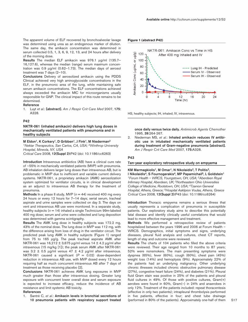
S17
The apparent volume of ELF recovered by bronchoalveolar lavagewas determined using urea as an endogenous marker of dilution.The same day, the amikacin concentration was determined inserum collected 0.5, 1, 3, 6, 9, 12, 13, and 24 hours after deliveryof the morning dose.Results The median ELF amikacin was 976.1 μg/ml (135.7–16,127.6), whereas the median (range) serum maximum concen-tration was 0.9 μg/ml (0.62–1.73). The median days of aerosoltreatment was 7 days (2–10).Conclusions Delivery of aerosolized amikacin using the PDDSClinical achieved very high aminoglycoside concentrations in theELF, in the pneumonic area of the lung, while maintaining safeserum amikacin concentrations. The ELF concentrations achievedalways exceeded the amikacin MIC for microorganisms usuallyresponsible for GNP. The clinical impact of this route remains to bedetermined.Reference1. Luyt et al.: [abstract]. Am J Respir Crit Care Med 2007, 175:
A328.
P42NKTR-061 (inhaled amikacin) delivers high lung doses inmechanically ventilated patients with pneumonia and inhealthy subjects
M Eldon1, K Corkery1, D Gribben1, J Fink1, M Niederman2
1Nektar Therapeutics, San Carlos, CA, USA; 2Winthrop UniversityHospital, Mineola, NY, USACritical Care 2008, 12(Suppl 2):P42 (doi: 10.1186/cc6263)
Introduction Intravenous antibiotics (AB) have a clinical cure rateof ~55% in mechanically ventilated patients (MVP) with pneumonia.AB inhalation delivers larger lung doses than intravenous AB, but isproblematic in MVP due to inefficient and variable current deliverysystems. NKTR-061, a proprietary amikacin (AMK) aerosolizationsystem optimized for ventilator circuits, is in clinical developmentas an adjunct to intravenous AB therapy for the treatment ofpneumonia.Methods In a phase II study, MVP (n = 44) received 400 mg every24 hours or every 12 hours for 7–14 days; serial serum, trachealaspirate and urine samples were collected on day 3. The days onvent and intravenous AB use were monitored. In a separate study,healthy subjects (n = 14) inhaled a single Technicium 99m-labeled400 mg dose; serum and urine were collected and lung depositionwas determined with gamma scintigraphy.Results The AMK lung dose in healthy subjects was 172.2 mg,43% of the nominal dose. The lung dose in MVP was 112 mg, withthe difference arising from loss of drug in the ventilator circuit. Thepredicted peak lung AMK in healthy subjects (Figure 1) rangedfrom 75 to 165 μg/g. The peak tracheal aspirate AMK afterNKTR-061 was 16,212 ± 3,675 μg/ml versus 14 ± 4.2 μg/ml afterintravenous (15 mg/kg [1]); the peak serum AMK after NKTR-061was 3.2 ± 0.5 μg/ml versus 47 ± 4.2 μg/ml after intravenous.NKTR-061 caused a significant (P = 0.02) dose-dependentreduction in intravenous AB use, with MVP dosed every 12 hoursrequiring half as much concurrent intravenous AB after 7 days oftreatment as those receiving placebo [2].Conclusions NKTR-061 achieves AMK lung exposures in MVPmuch greater than those after intravenous dosing. Greater lungexposure with concurrent lower overall dose and serum exposureis expected to increase efficacy, reduce the incidence of ABresistance and limit systemic AB toxicity.References1. Santré C, et al.: Amikacin levels in bronchial secretions of
10 pneumonia patients with respiratory support treated
once daily versus twice daily. Antimicrob Agents Chemother1995, 39:264-267.
2. Niederman MS, et al.: Inhaled amikacin reduces IV antibi-otic use in intubated mechanically ventilated patientsduring treatment of Gram-negative pneumonia [abstract].Am J Respir Crit Care Med 2007, 175:A328.
P43Ten-year exploratory retrospective study on empyema
KM Marmagkiolis1, M Omar1, N Nikolaidis2, T Politis3, I Nikolaidis4, S Fournogerakis4, MP Papamichail5, L Goldstein1
1Forum Health – WRCS, Youngstown, OH, USA; 2Aberdeen RoyalInfirmary Hospital, Aberdeen, UK; 3Northeastern Ohio UniversitiesCollege of Medicine, Rootstown, OH, USA; 4Tzanion GeneralHospital, Athens, Greece; 5Hospital Asklipion Voulas, Athens, GreeceCritical Care 2008, 12(Suppl 2):P43 (doi: 10.1186/cc6264)
Introduction Thoracic empyema remains a serious illness thatusually represents a complication of pneumonia in susceptiblepatients. Our exploratory study aims to describe this potentiallyfatal disease and identify clinically useful correlations that wouldlead to more effective management and treatment.Methods We performed a retrospective review of patientshospitalized between the years 1996 and 2006 at Forum Health –WRCS. Demographics, initial symptoms and signs, underlyingdiseases, pleural fluid analysis and cultures, chest CT reports,length of stay and outcome were reviewed.Results The charts of 104 patients who filled the above criteriawere reviewed. Their age ranged from 10 months to 87 years;52% were nonsmokers. The main presenting symptoms weredyspnea (65%), fever (60%), cough (60%), chest pain (45%)weight loss (14%) and hemoptysis (9%). Approximately 22% ofthe patients had an underlying malignancy. Other underlyingchronic illnesses included chronic obstructive pulmonary disease(27%), congestive heart failure (24%), and diabetes (21%). Pleuralfluid Gram stain was positive in 25% of the patients and pleuralfluid cultures in 49%. Of those with positive cultures, Gram(+)aerobes were found in 60%, Gram(–) in 24% and anaerobes inonly 12%. Treatment of the patients included: repeat thoracentesis(effective in only two patients); intrapleural thrombolysis performedin five patients, effective in four; and chest tube drainage(performed in 80% of the patients). Approximately one-half of them
Available online http://ccforum.com/supplements/12/S2
Figure 1 (abstract P42)
HS, healthy subjects; IH, inhaled; IV, intravenous.
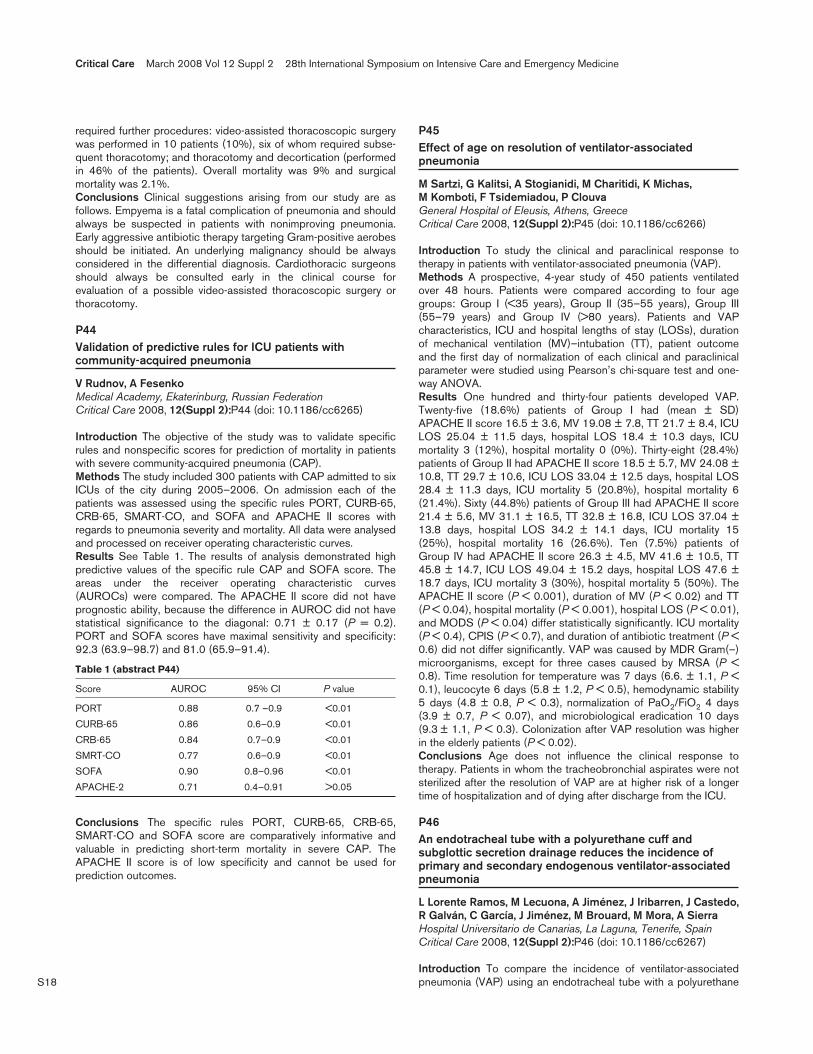
S18
required further procedures: video-assisted thoracoscopic surgerywas performed in 10 patients (10%), six of whom required subse-quent thoracotomy; and thoracotomy and decortication (performedin 46% of the patients). Overall mortality was 9% and surgicalmortality was 2.1%.Conclusions Clinical suggestions arising from our study are asfollows. Empyema is a fatal complication of pneumonia and shouldalways be suspected in patients with nonimproving pneumonia.Early aggressive antibiotic therapy targeting Gram-positive aerobesshould be initiated. An underlying malignancy should be alwaysconsidered in the differential diagnosis. Cardiothoracic surgeonsshould always be consulted early in the clinical course forevaluation of a possible video-assisted thoracoscopic surgery orthoracotomy.
P44Validation of predictive rules for ICU patients withcommunity-acquired pneumonia
V Rudnov, A FesenkoMedical Academy, Ekaterinburg, Russian FederationCritical Care 2008, 12(Suppl 2):P44 (doi: 10.1186/cc6265)
Introduction The objective of the study was to validate specificrules and nonspecific scores for prediction of mortality in patientswith severe community-acquired pneumonia (CAP).Methods The study included 300 patients with CAP admitted to sixICUs of the city during 2005–2006. On admission each of thepatients was assessed using the specific rules PORT, CURB-65,CRB-65, SMART-CO, and SOFA and APACHE II scores withregards to pneumonia severity and mortality. All data were analysedand processed on receiver operating characteristic curves.Results See Table 1. The results of analysis demonstrated highpredictive values of the specific rule CAP and SOFA score. Theareas under the receiver operating characteristic curves(AUROCs) were compared. The APACHE II score did not haveprognostic ability, because the difference in AUROC did not havestatistical significance to the diagonal: 0.71 ± 0.17 (P = 0.2).PORT and SOFA scores have maximal sensitivity and specificity:92.3 (63.9–98.7) and 81.0 (65.9–91.4).
Table 1 (abstract P44)
Score AUROC 95% CI P value
PORT 0.88 0.7 –0.9 <0.01
CURB-65 0.86 0.6–0.9 <0.01
CRB-65 0.84 0.7–0.9 <0.01
SMRT-CO 0.77 0.6–0.9 <0.01
SOFA 0.90 0.8–0.96 <0.01
APACHE-2 0.71 0.4–0.91 >0.05
Conclusions The specific rules PORT, CURB-65, CRB-65,SMART-CO and SOFA score are comparatively informative andvaluable in predicting short-term mortality in severe CAP. TheAPACHE II score is of low specificity and cannot be used forprediction outcomes.
P45Effect of age on resolution of ventilator-associatedpneumonia
M Sartzi, G Kalitsi, A Stogianidi, M Charitidi, K Michas, M Komboti, F Tsidemiadou, P ClouvaGeneral Hospital of Eleusis, Athens, GreeceCritical Care 2008, 12(Suppl 2):P45 (doi: 10.1186/cc6266)
Introduction To study the clinical and paraclinical response totherapy in patients with ventilator-associated pneumonia (VAP).Methods A prospective, 4-year study of 450 patients ventilatedover 48 hours. Patients were compared according to four agegroups: Group I (<35 years), Group II (35–55 years), Group III(55–79 years) and Group IV (>80 years). Patients and VAPcharacteristics, ICU and hospital lengths of stay (LOSs), durationof mechanical ventilation (MV)–intubation (TT), patient outcomeand the first day of normalization of each clinical and paraclinicalparameter were studied using Pearson’s chi-square test and one-way ANOVA.Results One hundred and thirty-four patients developed VAP.Twenty-five (18.6%) patients of Group I had (mean ± SD)APACHE II score 16.5 ± 3.6, MV 19.08 ± 7.8, TT 21.7 ± 8.4, ICULOS 25.04 ± 11.5 days, hospital LOS 18.4 ± 10.3 days, ICUmortality 3 (12%), hospital mortality 0 (0%). Thirty-eight (28.4%)patients of Group II had APACHE II score 18.5 ± 5.7, MV 24.08 ±10.8, TT 29.7 ± 10.6, ICU LOS 33.04 ± 12.5 days, hospital LOS28.4 ± 11.3 days, ICU mortality 5 (20.8%), hospital mortality 6(21.4%). Sixty (44.8%) patients of Group III had APACHE II score21.4 ± 5.6, MV 31.1 ± 16.5, TT 32.8 ± 16.8, ICU LOS 37.04 ±13.8 days, hospital LOS 34.2 ± 14.1 days, ICU mortality 15(25%), hospital mortality 16 (26.6%). Ten (7.5%) patients ofGroup IV had APACHE II score 26.3 ± 4.5, MV 41.6 ± 10.5, TT45.8 ± 14.7, ICU LOS 49.04 ± 15.2 days, hospital LOS 47.6 ±18.7 days, ICU mortality 3 (30%), hospital mortality 5 (50%). TheAPACHE II score (P < 0.001), duration of MV (P < 0.02) and TT(P < 0.04), hospital mortality (P < 0.001), hospital LOS (P < 0.01),and MODS (P < 0.04) differ statistically significantly. ICU mortality(P < 0.4), CPIS (P < 0.7), and duration of antibiotic treatment (P <0.6) did not differ significantly. VAP was caused by MDR Gram(–)microorganisms, except for three cases caused by MRSA (P <0.8). Time resolution for temperature was 7 days (6.6. ± 1.1, P <0.1), leucocyte 6 days (5.8 ± 1.2, P < 0.5), hemodynamic stability5 days (4.8 ± 0.8, P < 0.3), normalization of PaO2/FiO2 4 days(3.9 ± 0.7, P < 0.07), and microbiological eradication 10 days(9.3 ± 1.1, P < 0.3). Colonization after VAP resolution was higherin the elderly patients (P < 0.02).Conclusions Age does not influence the clinical response totherapy. Patients in whom the tracheobronchial aspirates were notsterilized after the resolution of VAP are at higher risk of a longertime of hospitalization and of dying after discharge from the ICU.
P46An endotracheal tube with a polyurethane cuff andsubglottic secretion drainage reduces the incidence ofprimary and secondary endogenous ventilator-associatedpneumonia
L Lorente Ramos, M Lecuona, A Jiménez, J Iribarren, J Castedo,R Galván, C García, J Jiménez, M Brouard, M Mora, A SierraHospital Universitario de Canarias, La Laguna, Tenerife, SpainCritical Care 2008, 12(Suppl 2):P46 (doi: 10.1186/cc6267)
Introduction To compare the incidence of ventilator-associatedpneumonia (VAP) using an endotracheal tube with a polyurethane
Critical Care March 2008 Vol 12 Suppl 2 28th International Symposium on Intensive Care and Emergency Medicine
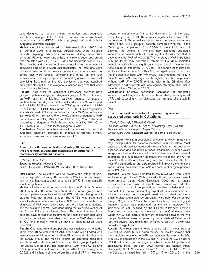
S19
cuff, designed to reduce channel formation, and subglotticsecretion drainage (ETT-PUC-SSD) versus an conventionalendotracheal tube (ETT-C) with a polyvinyl cuff and withoutsubglottic secretion drainage.Methods A clinical randomized trial, between 1 March 2006 and31 October 2006 in a medical–surgical ICU. Were includedpatients requiring mechanical ventilation during more than24 hours. Patients were randomized into two groups: one groupwas ventilated with ETT-PUC-SSD and another group with ETT-C.Throat swabs and tracheal aspirates were taken at the moment ofadmission and twice a week until discharge. The infections wereclassified based on thorat flora as: primary endogenous, caused bygerms that were already colonizing the throat on the ICUadmission; secondary endogenous, caused by germs that were notcolonizing the throat on the ICU admission but were acquiredduring the stay in ICU; and exogenous, caused by germs that werenot colonizing the throat.Results There were no significant differences between bothgroups of patients in age, sex, diagnosis groups, APACHE II score,pre-VAP use of antibiotics, paralytic agents, reintubation,tracheostomy, and days on mechanical ventilation. VAP was foundin 31 of 140 (22.1%) patients in the ETT-C group and in 11 of 140(7.9%) in the ETT-PUC-SSD group (P = 0.001). Cox regressionanalysis showed CET as a risk factor for global VAP (hazard rate =3.3, 95% CI = 1.66–6.67, P = 0.001), primary endogenous VAP(hazard rate = 5.12, 95% CI = 1.12–23.38, P = 0.04) andsecondary endogenous VAP (hazard rate = 2.87, 95% CI =1.20–6.84, P = 0.02); but not for exogenous VAP.Conclusions The endotracheal tube with a polyurethane cuff andsubglottic secretion drainage is effective to prevent primaryendogenous and secondary endogenous VAP.
P47Effect of continuous aspiration of subglottic secretions onthe prevention of ventilator-associated pneumonia inmechanically ventilated patients
C Yang, H Qiu, Y ZhuZhong-da Hospital, Nanjing, ChinaCritical Care 2008, 12(Suppl 2):P47 (doi: 10.1186/cc6268)
Introduction The objective was to evaluate the effect of con-tinuous aspiration of subglottic secretions (CASS) on the preven-tion of ventilator-associated pneumonia (VAP) in mechanicallyventilated patients.Methods Patients ventilated mechanically in the ICU from October2004 to April 2006 were randomly divided into two groups: onegroup of patients was treated with CASS and the other group ofpatients was not (NASS group). The CASS was performedimmediately after admission in the CASS group of patients. Thediagnosis of VAP was made based on the clinical presentations,and the evaluation of VAP was done using the simplified version ofthe Clinical Pulmonary Infection Score. The general status of thepatients, days of ventilated treatment, the volume of daily aspiratedsubglottic secretions, the morbidity and timing of VAP, days of stayin ICU and mortality within 28 days of hospitalization wererecorded.Results One hundred and one patients were included in the study.There were 48 patients in the CASS group who were treated withmechanical ventilation for longer than 48 hours, and 43 patients inthe NASS group. The median volume of aspirated subglotticsecretions within the first 24 hours in the CASS group of patients(48 cases) was 28.8 ml. The morbidity of VAP in the CASS andNASS groups of patients was 25.0% and 46.5%, respectively (P =0.032), and the length of time before the onset of VAP in these two
groups of patients was 7.3 ± 4.2 days and 5.1 ± 3.0 days,respectively (P = 0.088). There was a significant increase in thepercentage of Gram-positive cocci from the lower respiratorytracts in the NASS group of patients compared with that in theCASS group of patients (P = 0.004). In the CASS group ofpatients, the volume of the first daily aspirated subglotticsecretions in patients with VAP was significantly less than that inpatients without VAP (P = 0.006). The morbidity of VAP in patientswith the failed early aspiration (volume of first daily aspiratedsecretions ≤20 ml) was significantly higher than in patients whowere aspirated effectively (P < 0.01). The length of mechanicalventilation time in patients with VAP was significantly longer thanthat in patients without VAP (P = 0.000). The inhospital mortality inpatients with VAP was significantly higher than that in patientswithout VAP (P = 0.009), and mortality in the 28 days afteradmission in patients with VAP was significantly higher than that inpatients without VAP (P = 0.035).Conclusions Effective continuous aspiration of subglotticsecretions could significantly reduce the morbidity of early-onsetVAP, and, accordingly, may decrease the mortality of critically illpatients.
P48Effect of an oral care protocol in preventing ventilator-associated pneumonia in ICU patients
L Yao1, C Chang2, C Wang2, C Chen1
1National Taiwan University, School of Nursing, Taipei, Taiwan;2Mackay Memorial Hospital, Taipei, TaiwanCritical Care 2008, 12(Suppl 2):P48 (doi: 10.1186/cc6269)
Introduction Ventilator-associated pneumonia (VAP) remains amajor complication for patients incubated with ventilators. Mostcases are attributed to increased bacteria flora in the oropharyn-geal secretion and aspiration of those organisms. Evidence existssuggesting that oral care could reduce bacterial flora, preventaspiration, and subsequently decrease the incidence of VAP forpatients with ventilators. This study aims to evaluate the effective-ness of a standardized oral care protocol in improving oral hygieneand reducing the incidence of VAP in a sample of surgical patientsin the ICU (SICU).Methods Patients newly admitted to the SICU who were underventilator support for 48–72 hours and without pneumonia presentwere enrolled during March–November 2007 from a tertiarymedical center in Taiwan. Subjects were randomized into theexperimental or control groups and both received a 7-day oral careprotocol. For the experimental group (EG), a standardized 20-minute oral care protocol was performed using an electronic tooth-brush to clean and moisturize oral cavity twice daily. For the controlgroup (CG), a mimic 20-minute protocol involving moisturizing andattention control was performed for the same intervals. Theincidence of VAP defined by the Clinical Pulmonary InfectionScore and the oral hygiene measured by the Oral AssessmentGuide (OAG) and plaque index were compared between the twogroups. Variables were compared by the analysis of Fisher exacttest, chi-square test, and Mann–Whitney U test. P < 0.05 wasconsidered significant.Results Forty-four patients were studied with a mean age of60.6 ± 16.1 years, 63.6% being males. The results showed thatthe cumulative incidence of VAP was significantly lower in the EG,with 22.7% occurrence in the EG and 77.8% in the CG on day 9(P < 0.05). In terms of oral hygiene, subjects in the EG performedsignificantly better on both OAG scores and plaque index.Specifically, the OAG decreased from 16.3 ± 1.9 to 14.9 ± 2.6 inthe EG and remained high from 16.5 ± 1.6 to 16.6 ± 2.1 in the
Available online http://ccforum.com/supplements/12/S2
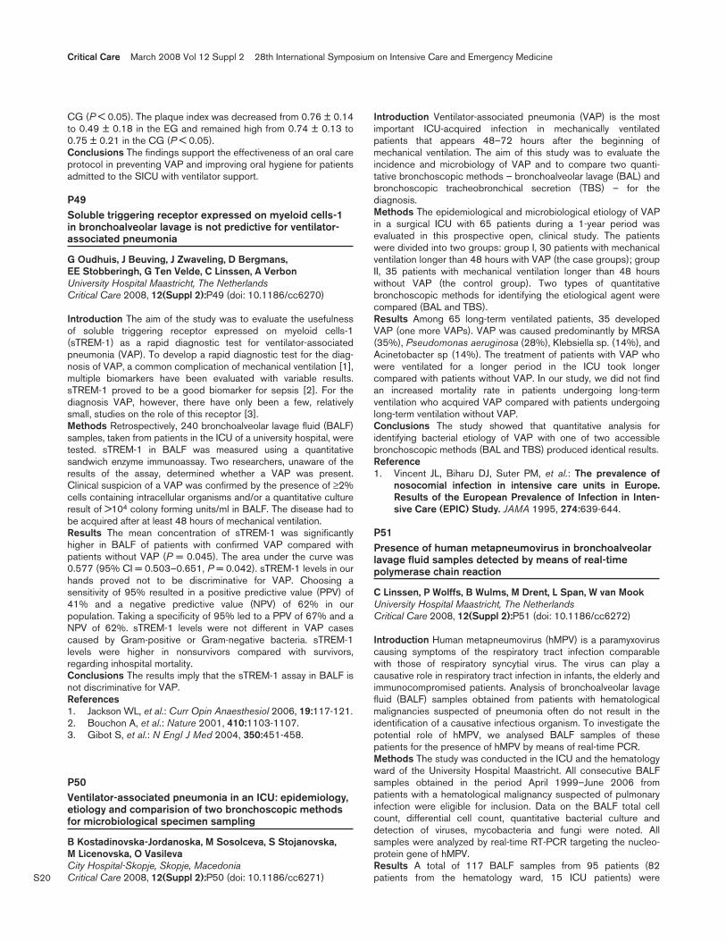
S20
CG (P < 0.05). The plaque index was decreased from 0.76 ± 0.14to 0.49 ± 0.18 in the EG and remained high from 0.74 ± 0.13 to0.75 ± 0.21 in the CG (P < 0.05).Conclusions The findings support the effectiveness of an oral careprotocol in preventing VAP and improving oral hygiene for patientsadmitted to the SICU with ventilator support.
P49Soluble triggering receptor expressed on myeloid cells-1in bronchoalveolar lavage is not predictive for ventilator-associated pneumonia
G Oudhuis, J Beuving, J Zwaveling, D Bergmans, EE Stobberingh, G Ten Velde, C Linssen, A VerbonUniversity Hospital Maastricht, The NetherlandsCritical Care 2008, 12(Suppl 2):P49 (doi: 10.1186/cc6270)
Introduction The aim of the study was to evaluate the usefulnessof soluble triggering receptor expressed on myeloid cells-1(sTREM-1) as a rapid diagnostic test for ventilator-associatedpneumonia (VAP). To develop a rapid diagnostic test for the diag-nosis of VAP, a common complication of mechanical ventilation [1],multiple biomarkers have been evaluated with variable results.sTREM-1 proved to be a good biomarker for sepsis [2]. For thediagnosis VAP, however, there have only been a few, relativelysmall, studies on the role of this receptor [3].Methods Retrospectively, 240 bronchoalveolar lavage fluid (BALF)samples, taken from patients in the ICU of a university hospital, weretested. sTREM-1 in BALF was measured using a quantitativesandwich enzyme immunoassay. Two researchers, unaware of theresults of the assay, determined whether a VAP was present.Clinical suspicion of a VAP was confirmed by the presence of ≥2%cells containing intracellular organisms and/or a quantitative cultureresult of >104 colony forming units/ml in BALF. The disease had tobe acquired after at least 48 hours of mechanical ventilation.Results The mean concentration of sTREM-1 was significantlyhigher in BALF of patients with confirmed VAP compared withpatients without VAP (P = 0.045). The area under the curve was0.577 (95% CI = 0.503–0.651, P = 0.042). sTREM-1 levels in ourhands proved not to be discriminative for VAP. Choosing asensitivity of 95% resulted in a positive predictive value (PPV) of41% and a negative predictive value (NPV) of 62% in ourpopulation. Taking a specificity of 95% led to a PPV of 67% and aNPV of 62%. sTREM-1 levels were not different in VAP casescaused by Gram-positive or Gram-negative bacteria. sTREM-1levels were higher in nonsurvivors compared with survivors,regarding inhospital mortality.Conclusions The results imply that the sTREM-1 assay in BALF isnot discriminative for VAP.References1. Jackson WL, et al.: Curr Opin Anaesthesiol 2006, 19:117-121.2. Bouchon A, et al.: Nature 2001, 410:1103-1107.3. Gibot S, et al.: N Engl J Med 2004, 350:451-458.
P50Ventilator-associated pneumonia in an ICU: epidemiology,etiology and comparision of two bronchoscopic methodsfor microbiological specimen sampling
B Kostadinovska-Jordanoska, M Sosolceva, S Stojanovska, M Licenovska, O VasilevaCity Hospital-Skopje, Skopje, MacedoniaCritical Care 2008, 12(Suppl 2):P50 (doi: 10.1186/cc6271)
Introduction Ventilator-associated pneumonia (VAP) is the mostimportant ICU-acquired infection in mechanically ventilatedpatients that appears 48–72 hours after the beginning ofmechanical ventilation. The aim of this study was to evaluate theincidence and microbiology of VAP and to compare two quanti-tative bronchoscopic methods – bronchoalveolar lavage (BAL) andbronchoscopic tracheobronchical secretion (TBS) – for thediagnosis.Methods The epidemiological and microbiological etiology of VAPin a surgical ICU with 65 patients during a 1-year period wasevaluated in this prospective open, clinical study. The patientswere divided into two groups: group I, 30 patients with mechanicalventilation longer than 48 hours with VAP (the case groups); groupII, 35 patients with mechanical ventilation longer than 48 hourswithout VAP (the control group). Two types of quantitativebronchoscopic methods for identifying the etiological agent werecompared (BAL and TBS).Results Among 65 long-term ventilated patients, 35 developedVAP (one more VAPs). VAP was caused predominantly by MRSA(35%), Pseudomonas aeruginosa (28%), Klebsiella sp. (14%), andAcinetobacter sp (14%). The treatment of patients with VAP whowere ventilated for a longer period in the ICU took longercompared with patients without VAP. In our study, we did not findan increased mortality rate in patients undergoing long-termventilation who acquired VAP compared with patients undergoinglong-term ventilation without VAP.Conclusions The study showed that quantitative analysis foridentifying bacterial etiology of VAP with one of two accessiblebronchoscopic methods (BAL and TBS) produced identical results.Reference1. Vincent JL, Biharu DJ, Suter PM, et al.: The prevalence of
nosocomial infection in intensive care units in Europe.Results of the European Prevalence of Infection in Inten-sive Care (EPIC) Study. JAMA 1995, 274:639-644.
P51Presence of human metapneumovirus in bronchoalveolarlavage fluid samples detected by means of real-timepolymerase chain reaction
C Linssen, P Wolffs, B Wulms, M Drent, L Span, W van MookUniversity Hospital Maastricht, The NetherlandsCritical Care 2008, 12(Suppl 2):P51 (doi: 10.1186/cc6272)
Introduction Human metapneumovirus (hMPV) is a paramyxoviruscausing symptoms of the respiratory tract infection comparablewith those of respiratory syncytial virus. The virus can play acausative role in respiratory tract infection in infants, the elderly andimmunocompromised patients. Analysis of bronchoalveolar lavagefluid (BALF) samples obtained from patients with hematologicalmalignancies suspected of pneumonia often do not result in theidentification of a causative infectious organism. To investigate thepotential role of hMPV, we analysed BALF samples of thesepatients for the presence of hMPV by means of real-time PCR.Methods The study was conducted in the ICU and the hematologyward of the University Hospital Maastricht. All consecutive BALFsamples obtained in the period April 1999–June 2006 frompatients with a hematological malignancy suspected of pulmonaryinfection were eligible for inclusion. Data on the BALF total cellcount, differential cell count, quantitative bacterial culture anddetection of viruses, mycobacteria and fungi were noted. Allsamples were analyzed by real-time RT-PCR targeting the nucleo-protein gene of hMPV.Results A total of 117 BALF samples from 95 patients (82patients from the hematology ward, 15 ICU patients) were
Critical Care March 2008 Vol 12 Suppl 2 28th International Symposium on Intensive Care and Emergency Medicine
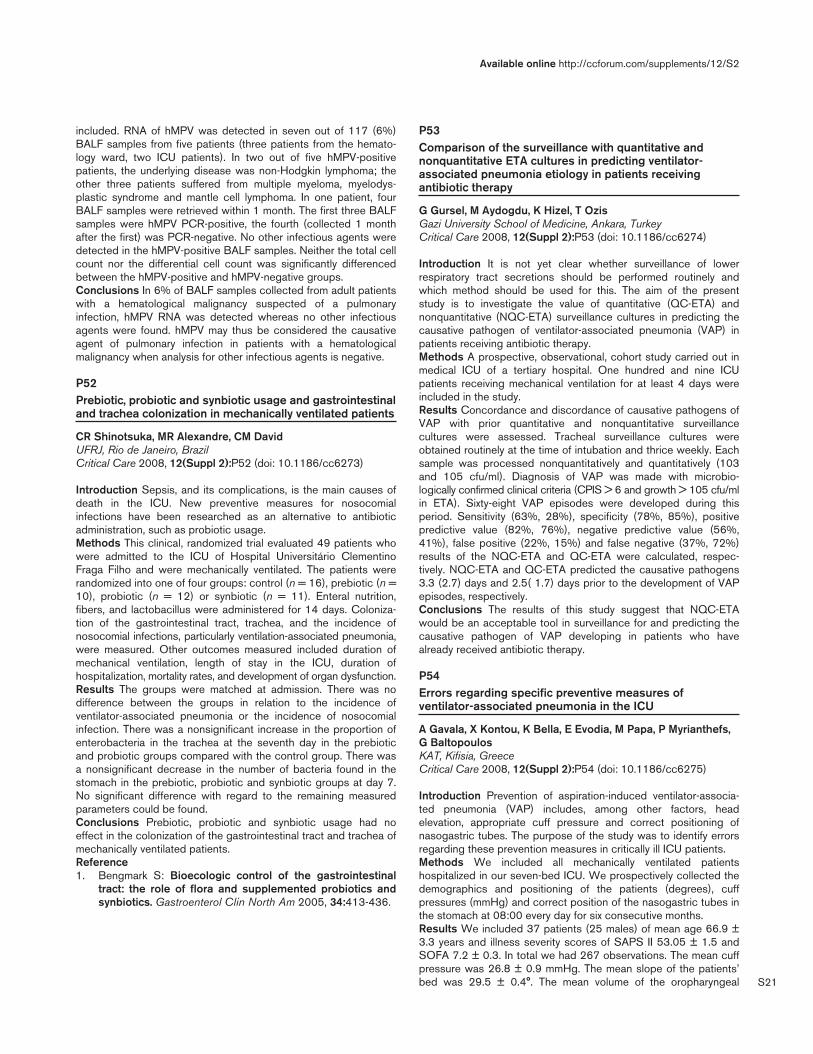
S21
included. RNA of hMPV was detected in seven out of 117 (6%)BALF samples from five patients (three patients from the hemato-logy ward, two ICU patients). In two out of five hMPV-positivepatients, the underlying disease was non-Hodgkin lymphoma; theother three patients suffered from multiple myeloma, myelodys-plastic syndrome and mantle cell lymphoma. In one patient, fourBALF samples were retrieved within 1 month. The first three BALFsamples were hMPV PCR-positive, the fourth (collected 1 monthafter the first) was PCR-negative. No other infectious agents weredetected in the hMPV-positive BALF samples. Neither the total cellcount nor the differential cell count was significantly differencedbetween the hMPV-positive and hMPV-negative groups.Conclusions In 6% of BALF samples collected from adult patientswith a hematological malignancy suspected of a pulmonaryinfection, hMPV RNA was detected whereas no other infectiousagents were found. hMPV may thus be considered the causativeagent of pulmonary infection in patients with a hematologicalmalignancy when analysis for other infectious agents is negative.
P52Prebiotic, probiotic and synbiotic usage and gastrointestinaland trachea colonization in mechanically ventilated patients
CR Shinotsuka, MR Alexandre, CM DavidUFRJ, Rio de Janeiro, BrazilCritical Care 2008, 12(Suppl 2):P52 (doi: 10.1186/cc6273)
Introduction Sepsis, and its complications, is the main causes ofdeath in the ICU. New preventive measures for nosocomialinfections have been researched as an alternative to antibioticadministration, such as probiotic usage.Methods This clinical, randomized trial evaluated 49 patients whowere admitted to the ICU of Hospital Universitário ClementinoFraga Filho and were mechanically ventilated. The patients wererandomized into one of four groups: control (n = 16), prebiotic (n =10), probiotic (n = 12) or synbiotic (n = 11). Enteral nutrition,fibers, and lactobacillus were administered for 14 days. Coloniza-tion of the gastrointestinal tract, trachea, and the incidence ofnosocomial infections, particularly ventilation-associated pneumonia,were measured. Other outcomes measured included duration ofmechanical ventilation, length of stay in the ICU, duration ofhospitalization, mortality rates, and development of organ dysfunction.Results The groups were matched at admission. There was nodifference between the groups in relation to the incidence ofventilator-associated pneumonia or the incidence of nosocomialinfection. There was a nonsignificant increase in the proportion ofenterobacteria in the trachea at the seventh day in the prebioticand probiotic groups compared with the control group. There wasa nonsignificant decrease in the number of bacteria found in thestomach in the prebiotic, probiotic and synbiotic groups at day 7.No significant difference with regard to the remaining measuredparameters could be found.Conclusions Prebiotic, probiotic and synbiotic usage had noeffect in the colonization of the gastrointestinal tract and trachea ofmechanically ventilated patients.Reference1. Bengmark S: Bioecologic control of the gastrointestinal
tract: the role of flora and supplemented probiotics andsynbiotics. Gastroenterol Clin North Am 2005, 34:413-436.
P53Comparison of the surveillance with quantitative andnonquantitative ETA cultures in predicting ventilator-associated pneumonia etiology in patients receivingantibiotic therapy
G Gursel, M Aydogdu, K Hizel, T OzisGazi University School of Medicine, Ankara, TurkeyCritical Care 2008, 12(Suppl 2):P53 (doi: 10.1186/cc6274)
Introduction It is not yet clear whether surveillance of lowerrespiratory tract secretions should be performed routinely andwhich method should be used for this. The aim of the presentstudy is to investigate the value of quantitative (QC-ETA) andnonquantitative (NQC-ETA) surveillance cultures in predicting thecausative pathogen of ventilator-associated pneumonia (VAP) inpatients receiving antibiotic therapy.Methods A prospective, observational, cohort study carried out inmedical ICU of a tertiary hospital. One hundred and nine ICUpatients receiving mechanical ventilation for at least 4 days wereincluded in the study.Results Concordance and discordance of causative pathogens ofVAP with prior quantitative and nonquantitative surveillancecultures were assessed. Tracheal surveillance cultures wereobtained routinely at the time of intubation and thrice weekly. Eachsample was processed nonquantitatively and quantitatively (103and 105 cfu/ml). Diagnosis of VAP was made with microbio-logically confirmed clinical criteria (CPIS > 6 and growth > 105 cfu/mlin ETA). Sixty-eight VAP episodes were developed during thisperiod. Sensitivity (63%, 28%), specificity (78%, 85%), positivepredictive value (82%, 76%), negative predictive value (56%,41%), false positive (22%, 15%) and false negative (37%, 72%)results of the NQC-ETA and QC-ETA were calculated, respec-tively. NQC-ETA and QC-ETA predicted the causative pathogens3.3 (2.7) days and 2.5( 1.7) days prior to the development of VAPepisodes, respectively.Conclusions The results of this study suggest that NQC-ETAwould be an acceptable tool in surveillance for and predicting thecausative pathogen of VAP developing in patients who havealready received antibiotic therapy.
P54Errors regarding specific preventive measures ofventilator-associated pneumonia in the ICU
A Gavala, X Kontou, K Bella, E Evodia, M Papa, P Myrianthefs,G BaltopoulosKAT, Kifisia, GreeceCritical Care 2008, 12(Suppl 2):P54 (doi: 10.1186/cc6275)
Introduction Prevention of aspiration-induced ventilator-associa-ted pneumonia (VAP) includes, among other factors, headelevation‚ appropriate cuff pressure and correct positioning ofnasogastric tubes. The purpose of the study was to identify errorsregarding these prevention measures in critically ill ICU patients.Methods We included all mechanically ventilated patientshospitalized in our seven-bed ICU. We prospectively collected thedemographics and positioning of the patients (degrees)‚ cuffpressures (mmHg) and correct position of the nasogastric tubes inthe stomach at 08:00 every day for six consecutive months.Results We included 37 patients (25 males) of mean age 66.9 ±3.3 years and illness severity scores of SAPS II 53.05 ± 1.5 andSOFA 7.2 ± 0.3. In total we had 267 observations. The mean cuffpressure was 26.8 ± 0.9 mmHg. The mean slope of the patients’bed was 29.5 ± 0.4°. The mean volume of the oropharyngeal
Available online http://ccforum.com/supplements/12/S2
S22
aspirates was 9.9 ± 0.4 ml and of tracheal aspirates was 7.1 ± 0.2 ml.In 24 and 69 observations‚ tracheal and oropharyngeal aspirates,respectively, were >10 ml. In 109/267 (40.1%) observations. theslope of the patients was <30°. All patients had at least onepositioning with a slope <30°. In 64/267 (23.9%) observations,the cuff pressures were <20.0 mmHg. One-half of the patients hadat least one measurement <20.0 mmHg. In 10 cases, the end ofthe nasogastric tube was in the esophagus and in five cases it wasobstructed. Twenty patients developed VAP (20/37‚ 54.1%).Patients with a large amount of oropharyngeal aspirates (>10 ml)and low cuff pressures (<20 mmHg) had a significantly higherincidence of subsequent VAP (72.2% vs 36.8%) compared withthose with a low amount of oropharyngeal aspirates (<5 ml) andnormal cuff pressures (chi-square test‚ P = 0.049; RR = 1.960‚CI = 1.018–3.774; OD = 4.457‚ CI = 1.110–17.90).Conclusions Errors regarding specific prevention measures ofVAP are frequently observed. Our data also show the significanceof the amount of oropharyngeal aspirates and cuff pressures forthe subsequent development of VAP. The tightness of thestomach–oropharyngeal–tracheal axis seems to be a significantfactor influencing the subsequent development of VAP.Reference1. Chastre J: Respir Care 2005, 50:975-983.
P55Clinical predictors for septic shock in patients withventilator-associated pneumonia
G Gursel, M AydogduGazi University School of Medicine, Ankara, TurkeyCritical Care 2008, 12(Suppl 2):P55 (doi: 10.1186/cc6276)
Introduction Ventilator-associated pneumonia (VAP) is one of themost frequent infections of ICUs, and nearly 50% of patientsdevelop septic shock during VAP. Septic shock is an independentpredictor for mortality in these patients. The aim of this study is toinvestigate the predictors for septic shock in patients with VAPreceiving appropriate antibiotic therapy.Methods Eighty-nine patients with microbiologically confirmedVAP and receiving appropriate antibiotic therapy were included inthe study. The patients were divided into two groups according tothe existence of septic shock. Clinical, hematological, biochemicaland microbiological characteristics of the patients were compared.Results Thirty-one percent of the patients developed septic shock,and advanced age (OR = 1.07, 95% CI = 1.02–1.13, P = 0.009),lymphocytopenia <1,000 mm3 (OR = 7.48, 95% CI = 1.91–29,P = 0.004), high blood glucose levels >120 mg/dl (OR = 4.75,95% CI = 1.38–16, P = 0.014), and higher Clinical PulmonaryInfection Scores (OR = 1.64, 95% CI = 1.16–2.33, P = 0.006)were independent predictors for the development of septic shock.Conclusions Some clinical parameters such as lymphocytopenia,advanced age, higher blood glucose levels and Clinical PulmonaryInfection Scores can predict septic shock during VAP but largerandomized controlled studies are needed to confirm these results.
P56Antibiotic-related acute effects within the intestinalmicrocirculation in experimental sepsis
CH Lehmann1, V Bac1, J Richter2, M Gründling2, M Wendt2, S Whynot1, O Hung1, M Murphy1, T Issekutz1, D Pavlovic2
1Dalhousie University, Halifax, NS, Canada; 2Ernst-Moritz-Arndt-Universität Greifswald, GermanyCritical Care 2008, 12(Suppl 2):P56 (doi: 10.1186/cc6277)
Introduction Although the benefit of antibiotic therapy in infectiousinflammatory diseases remains unquestioned [1], little is knownabout the effects of these antibiotics on the inflamedmicrocirculation independent of their antimicrobial property. Theaim of this study was to evaluate acute effects related to antibioticsadministration upon the intestinal microcirculation, which plays acrucial role in the pathogenesis of sepsis and subsequent multi-organ failure [2,3].Methods Experimental sepsis was induced in 50 Lewis rats usingthe colon ascendens stent peritonitis model [4]. Four frequentlyused antibiotics were included in the study (20 mg/kg imipenem/cilastatin (IMI), 25 mg/kg tobramycin (TOB), 70 mg/kg vancomycin(VAN), 5 mg/kg erythromycin (ERY)). The antibiotics wereadministered as a single intravenous bolus following 16 hours ofobservation time. The intestinal functional capillary density andleukocyte–endothelial interactions were evaluated using intravitalmicroscopy 1 hour following antibiotic treatment. Additional experi-ments were performed in an abacterial setting with comparablemicrocirculatory disturbances (2 hours endotoxemia; n = 50).Results Acute IMI or TOB administration, respectively, did notaffect the intestinal microcirculation. VAN treatment aggravated theleukocyte rolling behavior in this acute setting. In contrast, ERYadministration significantly reduced leukocyte activation andimproved the functional capillary density within the intestinalmicrocirculation during experimental sepsis. These effects couldbe confirmed during endotoxemia, suggesting that ERY exerts anti-inflammatory effects in addition to its antibacterial action.Conclusions When choosing antimicrobial agents in septicconditions, possible effects of the antibiotics within the pathogene-tically important intestinal microcirculation should be considered. Inconjunction with microbial sensitivity tests, the results of suchstudies assist in selecting the appropriate antibiotic therapy.References1. Nobre V, et al.: Curr Opin Crit Care 2007, 13:586-591.2. Suliburk J, et al.: Eur Surg Res 2007, 40:184-118.3. Clark JA, et al.: Shock 2007, 28:384-393.4. Lustig MK, et al.: Shock 2007, 28:59-64.
P57Intraperitoneal lipopolysaccharide-induced neutrophilsequestration in the lung microvasculature is due to afactor produced in the peritoneal cavity
S Mullaly, S Johnson, P KubesUniversity of Calgary, Calgary, AB, CanadaCritical Care 2008, 12(Suppl 2):P57 (doi: 10.1186/cc6278)
Introduction Intraperitoneal injection of Gram-negative lipopoly-saccharide (LPS) results in a profound decrease in circulatingleukocyte counts with a significant increase in neutrophil seques-tration in the lung microvasculature as measured by lung myelo-peroxidase levels. Mice made deficient in toll-like receptor 4 (TLR4)are resistant to LPS challenges. Furthermore, it has been demon-strated that the systemic effects of LPS are due to parenchymal,possibly endothelial, cells and not due to bone marrow-derivedcells, such as leukocytes.Methods C57B/6 mice were anesthetized and sacrificed.Peritoneal lavage using 3 ml PBS followed by a gentle abdominalmassage for 1 minute was performed. Lavage fluid was aspiratedand centrifuged to concentrate the peritoneal cells. Cells werethen treated with normal saline or 10 μg LPS. The treatedperitoneal cells were then injected into the peritoneal cavities ofTLR4-deficient mice (C57B/10ScNJ) for a duration of 4 hours.Measured outcomes included circulating leukocyte counts andlung myeloperoxidase (MPO) levels.
Critical Care March 2008 Vol 12 Suppl 2 28th International Symposium on Intensive Care and Emergency Medicine
S23
Results Normal saline-treated control C57B/6 mice had circulatingcounts of 6.38 ± 0.56 million cells/ml, and the MPO levels in normalsaline-treated peritoneal cells transferred into C57B/6 mice were5.80 ± 0.73 units. Normal saline-treated peritoneal cells of C57B/6mice injected into TLR4-deficient mice resulted in circulating countsof 5.18 ± 0.22 million cells/ml and MPO levels of 4.23 ± 0.73 units.LPS-treated control C57B/6 mice had circulating counts of 1.94 ±0.22 million cells/ml, and the MPO levels in LPS-treated peritonealcells transferred into C57B/6 mice were 16.52 ± 4.3 units. LPS-treated peritoneal cells of C57B/6 mice injected into TLR4-deficientmice resulted in circulating counts of 2.48 ± 0.44 million cells/mland MPO levels of 12.06 ± 1.74 units. The liver endothelium wasthe only organ activated in this model. Furthermore, this process ofLPS-treated, peritoneal cell-induced neutrophil sequestration in thelung microvasculature was found to be independent of mast cellsand NKT cells.Conclusions An as yet to be identified factor, or factors, producedfrom the peritoneal lavage cells, can produce neutrophilsequestration in the lung microvasculature.Reference1. Andonegui G, et al.: J Clin Invest 2003, 111:1011-1020.
P58Correlation between microcirculatory flow, density andheterogeneity scores in septic shock patients
C Ruiz, G Izquierdo, R Lopez, M Andresen, G Hernandez, A BruhnPontificia Universidad Católica de Chile, Santiago, ChileCritical Care 2008, 12(Suppl 2):P58 (doi: 10.1186/cc6279)
Introduction A recent conference recommended that micro-circulatory images should be analyzed with several scores thatevaluate density, flow and flow heterogeneity [1]. Following theserecommendations, we analyzed sublingual microcirculatory imagesfrom septic shock patients to determine how these different scoreswere correlated with each other and with clinical hemodynamicand perfusion parameters.Methods Using side dark-field videomicroscopy (Microscan®;Microvision Medical) we repeatedly evaluated sublingual micro-circulation at different time points in septic shock patients. In total,we performed 17 microcirculatory single-time-point assessments(3–6 site images/time point), in parallel with hemodynamic andperfusion measurements (mean arterial pressure (MAP), nor-adrenaline dose (NA), cardiac index (CI), mixed venous O2 satura-tion (SmvO2), arterial lactate). Images were analyzed by semi-quantitative scores of flow (mean flow index (MFI) and proportionof perfused vessels (PPV)) and density (perfused vascular density(PVD)) of small vessels (<20 μm). Heterogeneity indexes (HetIndex = maximum – minimum / mean) were calculated for the MFIand PPV. Correlations between parameters were determined bythe Pearson coefficient and considered significant if P < 0.05.Results We found that PVD was correlated to PPV (r = 0.55), andnegatively to Het Index PVD (r = –0.54) and Het index PPV (r =–0.43), but we found no correlation of PVD with any homodynamicor perfusion parameter. Flow indexes (PPV and MFI) were stronglycorrelated with each other (r = 0.81) and inversely with theirrespective heterogeneity indexes (PPV and Het Index PPV, r =–0.88; MFI and Het Index MFI, r = –0.83). In addition, PPV andMFI were correlated to SmvO2 (r = 0.44 and 0.52), and CI (r =0.49 and 0.47), and inversely to lactate levels (r = –0.46 and–0.4). Only the MFI was correlated to MAP (r = 0.5).Heterogeneity indexes were correlated to lactate (r = 0.40 withPPV and r = 0.44 with MFI), and inversely to MAP (r = –0.40 withHet Index PPV and r = –0.64 with Het index MFI). The Het IndexMFI was also correlated to NA (r = 0.5).
Conclusions Higher microcirculatory flow scores, but not density,are associated with higher CI and better systemic perfusionparameters. Both MFI and PPV seem equally effective to assessmicrocirculatory flow.Reference1. De Backer D, et al.: Crit Care 2007, 11:R101.
P59Effect of iloprost on the microcirculation and liver functionafter orthotopic liver transplantation
S Klinzing, C Stumme, K Albin, O Habrecht, U Settmacher, G MarxFriedrich Schiller University, Jena, GermanyCritical Care 2008, 12(Suppl 2):P59 (doi: 10.1186/cc6280)
Introduction Iloprost, a prostacyclin analogon, can probablyimprove the microcirculation in various tissues. It may have apositive effect on perfusion and function of the liver, especially inpatients after liver transplantation. We therefore estimated liverfunction with the indocyanine green (ICG) plasma disappearancerate (PDR). We also evaluated iloprost effects on microcirculationin the oral mucosal tissue.Methods Sixteen patients after orthotopic liver transplantationwere randomly included in the study. They received either iloprost(n = 9) or placebo (n = 7). Iloprost was given in a dose of 1 ng/kg/min.The oral mucosal tissue oxygen saturation, microcirculatory bloodflow and blood flow velocity were measured in a depth of one witha laser Doppler flowmetry and remission spectroscopy system(O2C; LEA, Gießen, Germany). The ICG-PDR was determinedwith the LIMON (Pulsion, München, Germany). All measurementswere performed 6 hours, 12 hours, 24 hours, 48 hours, 72 hoursand 96 hours postoperatively.Results We saw a significant increase in the microcirculatoryblood flow and blood flow velocity 6 and 12 hours postoperativelyin the iloprost group in comparison with the placebo group. After6 hours the blood flow was 156 (minimum, 54; maximum, 460;without units) in the placebo group and 213 (minimum, 57;maximum, 456; P < 0.05) in the iloprost group. After 12 hours itwas 175 (minimum, 53; maximum, 483) and 318 (minimum, 86;maximum, 569; P < 0.05), respectively. The blood flow velocitywas 18 (minimum, 14; maximum, 50) in the placebo group and 29(minimum, 17; maximum, 51; P < 0.05) in the iloprost group6 hours postoperatively. Twelve hours postoperatively the velocitywas 21 (minimum, 16; maximum, 52) and 37.5 (minimum, 19;maximum, 64; P < 0.05). The oral mucosal tissue oxygen saturationdid not change. We could not find any difference in the ICG-PDRbetween the two groups.Conclusions Iloprost improved the microcirculatory blood flow andblood flow velocity of the oral mucosa in the early postoperativephase. This effect did not lead to an improved ICG clearance inpatients after liver transplantation.
P60Noninvasive monitoring of peripheral perfusion withphysical examination and the peripheral flow indexcorrelates with dynamic near-infrared spectroscopymeasurements in patients with septic shock
A Lima, T Jansen, C Ince, J BakkerErasmus MC University Medical Centre Rotterdam, The NetherlandsCritical Care 2008, 12(Suppl 2):P60 (doi: 10.1186/cc6281)
Introduction Peripheral blood flow can be markedly impaired inseptic shock. Bedside assessment of this derangement has not yet
Available online http://ccforum.com/supplements/12/S2
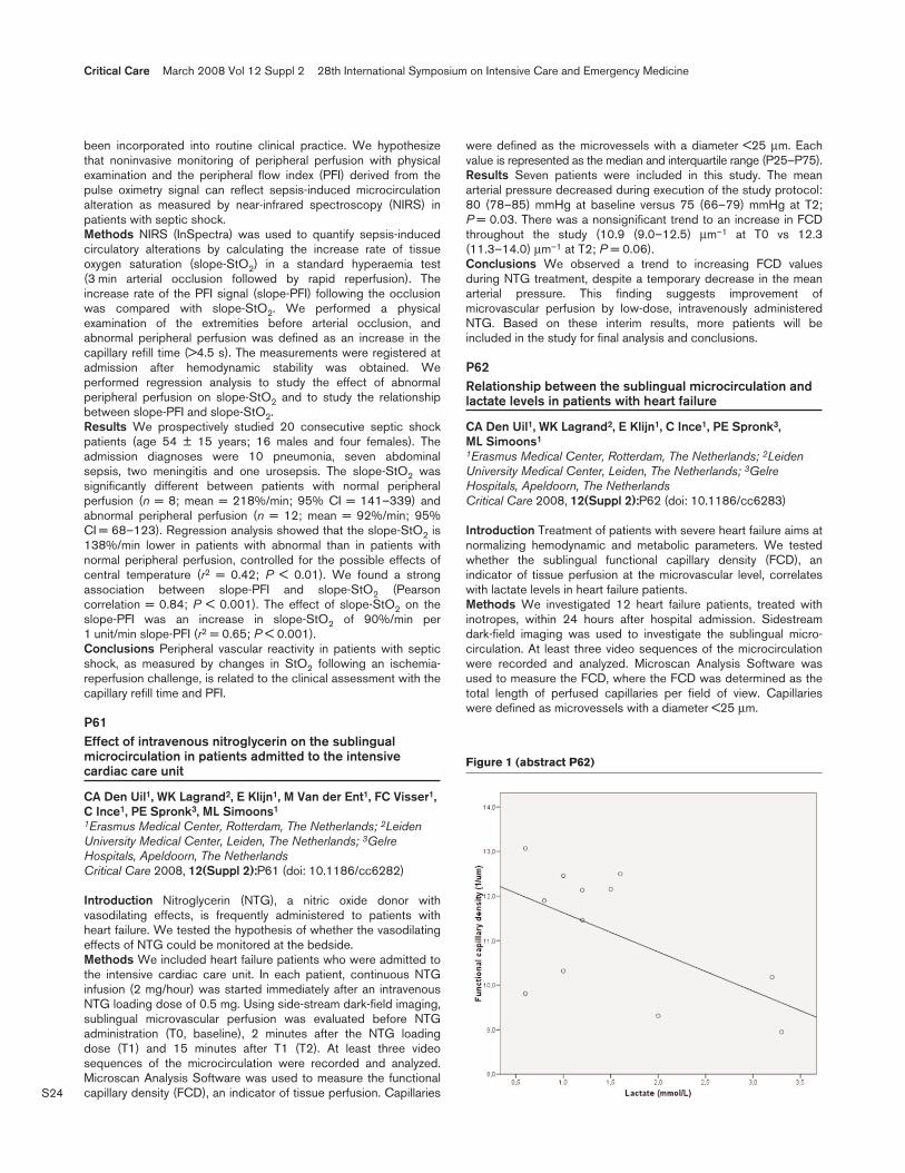
S24
been incorporated into routine clinical practice. We hypothesizethat noninvasive monitoring of peripheral perfusion with physicalexamination and the peripheral flow index (PFI) derived from thepulse oximetry signal can reflect sepsis-induced microcirculationalteration as measured by near-infrared spectroscopy (NIRS) inpatients with septic shock.Methods NIRS (InSpectra) was used to quantify sepsis-inducedcirculatory alterations by calculating the increase rate of tissueoxygen saturation (slope-StO2) in a standard hyperaemia test(3 min arterial occlusion followed by rapid reperfusion). Theincrease rate of the PFI signal (slope-PFI) following the occlusionwas compared with slope-StO2. We performed a physicalexamination of the extremities before arterial occlusion, andabnormal peripheral perfusion was defined as an increase in thecapillary refill time (>4.5 s). The measurements were registered atadmission after hemodynamic stability was obtained. Weperformed regression analysis to study the effect of abnormalperipheral perfusion on slope-StO2 and to study the relationshipbetween slope-PFI and slope-StO2.Results We prospectively studied 20 consecutive septic shockpatients (age 54 ± 15 years; 16 males and four females). Theadmission diagnoses were 10 pneumonia, seven abdominalsepsis, two meningitis and one urosepsis. The slope-StO2 wassignificantly different between patients with normal peripheralperfusion (n = 8; mean = 218%/min; 95% CI = 141–339) andabnormal peripheral perfusion (n = 12; mean = 92%/min; 95%CI = 68–123). Regression analysis showed that the slope-StO2 is138%/min lower in patients with abnormal than in patients withnormal peripheral perfusion, controlled for the possible effects ofcentral temperature (r2 = 0.42; P < 0.01). We found a strongassociation between slope-PFI and slope-StO2 (Pearsoncorrelation = 0.84; P < 0.001). The effect of slope-StO2 on theslope-PFI was an increase in slope-StO2 of 90%/min per1 unit/min slope-PFI (r2 = 0.65; P < 0.001).Conclusions Peripheral vascular reactivity in patients with septicshock, as measured by changes in StO2 following an ischemia-reperfusion challenge, is related to the clinical assessment with thecapillary refill time and PFI.
P61Effect of intravenous nitroglycerin on the sublingualmicrocirculation in patients admitted to the intensivecardiac care unit
CA Den Uil1, WK Lagrand2, E Klijn1, M Van der Ent1, FC Visser1,C Ince1, PE Spronk3, ML Simoons1
1Erasmus Medical Center, Rotterdam, The Netherlands; 2LeidenUniversity Medical Center, Leiden, The Netherlands; 3GelreHospitals, Apeldoorn, The NetherlandsCritical Care 2008, 12(Suppl 2):P61 (doi: 10.1186/cc6282)
Introduction Nitroglycerin (NTG), a nitric oxide donor withvasodilating effects, is frequently administered to patients withheart failure. We tested the hypothesis of whether the vasodilatingeffects of NTG could be monitored at the bedside.Methods We included heart failure patients who were admitted tothe intensive cardiac care unit. In each patient, continuous NTGinfusion (2 mg/hour) was started immediately after an intravenousNTG loading dose of 0.5 mg. Using side-stream dark-field imaging,sublingual microvascular perfusion was evaluated before NTGadministration (T0, baseline), 2 minutes after the NTG loadingdose (T1) and 15 minutes after T1 (T2). At least three videosequences of the microcirculation were recorded and analyzed.Microscan Analysis Software was used to measure the functionalcapillary density (FCD), an indicator of tissue perfusion. Capillaries
were defined as the microvessels with a diameter <25 μm. Eachvalue is represented as the median and interquartile range (P25–P75).Results Seven patients were included in this study. The meanarterial pressure decreased during execution of the study protocol:80 (78–85) mmHg at baseline versus 75 (66–79) mmHg at T2;P = 0.03. There was a nonsignificant trend to an increase in FCDthroughout the study (10.9 (9.0–12.5) μm–1 at T0 vs 12.3(11.3–14.0) μm–1 at T2; P = 0.06).Conclusions We observed a trend to increasing FCD valuesduring NTG treatment, despite a temporary decrease in the meanarterial pressure. This finding suggests improvement ofmicrovascular perfusion by low-dose, intravenously administeredNTG. Based on these interim results, more patients will beincluded in the study for final analysis and conclusions.
P62Relationship between the sublingual microcirculation andlactate levels in patients with heart failure
CA Den Uil1, WK Lagrand2, E Klijn1, C Ince1, PE Spronk3, ML Simoons1
1Erasmus Medical Center, Rotterdam, The Netherlands; 2LeidenUniversity Medical Center, Leiden, The Netherlands; 3GelreHospitals, Apeldoorn, The NetherlandsCritical Care 2008, 12(Suppl 2):P62 (doi: 10.1186/cc6283)
Introduction Treatment of patients with severe heart failure aims atnormalizing hemodynamic and metabolic parameters. We testedwhether the sublingual functional capillary density (FCD), anindicator of tissue perfusion at the microvascular level, correlateswith lactate levels in heart failure patients.Methods We investigated 12 heart failure patients, treated withinotropes, within 24 hours after hospital admission. Sidestreamdark-field imaging was used to investigate the sublingual micro-circulation. At least three video sequences of the microcirculationwere recorded and analyzed. Microscan Analysis Software wasused to measure the FCD, where the FCD was determined as thetotal length of perfused capillaries per field of view. Capillarieswere defined as microvessels with a diameter <25 μm.
Critical Care March 2008 Vol 12 Suppl 2 28th International Symposium on Intensive Care and Emergency Medicine
Figure 1 (abstract P62)

S25
Results The mean arterial pressure was 75 (68–78) mmHg,lactate levels were 1.2 (1.0–1.7) mmol/l and SvO2 was 0.78(0.65–0.84) mol/mol. The FCD was 11.7 (10.1–12.2) μm–1. Thecorrelation between lactate and FCD is shown in Figure 1(regression line: β1 = –0.88, P = 0.05).Conclusions In this study, FCD correlated with lactate levels. Thisfinding is indicative for a relationship between globalhemodynamics and the sublingual microcirculation in patientstreated for heart failure. Based on the interim results, more patientswill be included in this study for final analysis and conclusions.
P63Sidestream dark-field imaging versus orthogonalpolarization spectroscopic imaging: a comparative study
R Bezemer, P Goedhart, M Khalilzada, C InceAcademic Medical Center, Amsterdam, The NetherlandsCritical Care 2008, 12(Suppl 2):P63 (doi: 10.1186/cc6284)
Introduction Sidestream dark-field (SDF) imaging, a stroboscopicLED ring-based imaging modality for clinical observation of themicrocirculation, was validated by quantitative and qualitativecomparison with orthogonal polarization spectral (OPS) imaging.Methods For OPS imaging a Cytocan-II backfocus type device(Cytometrics, Philadelphia, PA, USA) was used, and for SDFimaging a MicroScan Video Microscope (MicroVision Medical Inc.,Amsterdam, The Netherlands) was employed. To validate SDFimaging, nailfold capillary diameters and red blood cell velocitieswere measured in the exact same capillaries using OPS and SDFimaging. For quantitative comparison of the quality of sublinguallyacquired microcirculatory images, an image quality quantificationsystem was developed to score venular and capillary contrast andsharpness on scales from 0 to 1.Results After introduction of a scaling factor to correct for theslightly higher magnification of the SDF device with respect to theOPS device, equal quantitative results for capillary diameters andred blood cell velocities were obtained. Venular contrast andsharpness were shown to be comparable for OPS and SDFimaging. Capillary sharpness and contrast, however, were shownto be significantly higher using SDF imaging (Figure 1). Venulargranularity, in addition, was more clearly observable employing theSDF device.
Conclusions SDF imaging provided significantly higher imagequality by the use of stroboscopic LED ring-based SDF illumi-nation. It is anticipated that SDF imaging will serve as a reliableimaging modality for the clinical assessment of the microcirculationand will enhance computer-aided image analysis.Reference1. Goedhart PT, et al.: Opt Express 2007, 15:15101-15114.
P64Finger reactive hyperaemia to measure endothelialfunction in sepsis and health (the FRESH study)
JS Davis1, J Thomas2, M McMillan1, T Yeo1, D Celermajer3, D Stephens2, NM Anstey1
1Menzies School of Health Research, Darwin, Australia; 2RoyalDarwin Hospital, Darwin, Australia; 3Royal Prince Alfred Hospital,Sydney, AustraliaCritical Care 2008, 12(Suppl 2):P64 (doi: 10.1186/cc6285)
Introduction Endothelial dysfunction is thought to be an importantmechanism of organ failure in sepsis. We hypothesised thatendothelial function (EF) would be impaired in adult patients withsepsis; that it would improve with treatment; and that the degree ofits impairment would correlate with disease severity and outcome.Methods EF was measured using a novel, noninvasive techniqueat the bedside (reactive hyperaemia peripheral arterial tonometry(RH-PAT)) in three groups: patients with sepsis requiringadmission to the ICU (ICU sepsis); patients with sepsis requiringhospital but not ICU admission (ward sepsis); and control patientswithout sepsis. Measurements were taken on days 0, 2 and 7 inthe sepsis patients and at baseline in the control patients.Results Planned interim analysis was performed on 38 ICU sepsispatients, 19 ward sepsis patients and 28 control patients. Themean (95% CI) baseline RH-PAT index was significantly lower inICU sepsis (1.56 (1.41–1.71)) than in control patients (2.03(1.87–2.19)), P = 0.0001. It was intermediate in the ward sepsisgroup: baseline RH-PAT index = 1.72 (1.52–1.92) (P = 0.02 cfcontrols, not significant cf ICU sepsis). See Figure 1. The RH-PATindex improved markedly in the ward sepsis patients over the first 2days (1.72 (1.52–1.92) to 2.29 (2.08–2.57); P = 0.0004); how-ever, it did not change significantly in the ICU sepsis patients (1.56(1.41–1.71) to 1.77 (1.56–1.98)).Conclusions Noninvasive measurement of EF is feasible in sepsis.EF in sepsis is initially markedly impaired. It improves over the first2 days in those patients with moderate sepsis but not in those with
Available online http://ccforum.com/supplements/12/S2
Figure 1 (abstract P63)
Sidestream dark-field (SDF) imaging versus orthogonal polarizationspectral (OPS) imaging: capillary contrast and sharpness.
Figure 1 (abstract P64)
Baseline endothelial function.
S26
sepsis requiring ICU admission. These data will be further analysedto explore correlations, and blood samples have been stored forthe measurement of serum arginine and markers of endothelialactivation.
P65Microcirculatory investigation in severe trauma injury
E GrigoryevMedical University, Kemerovo, Russian FederationCritical Care 2008, 12(Suppl 2):P65 (doi: 10.1186/cc6286)
Introduction Disturbances of regional microcirculation do notcorrelate with the global indices of perfusion such as the cardiacindex and cardiac output. Research of local regional micro-circulation must therefore be useful for diagnostic and prognosticvalue. The aim of study was to research the diagnostic and prog-nostic consequence of the microcirculatory indices in severetrauma injury.Methods Thirty-four patients with severe trauma injury wereentered into a prospective trial (scale TRISS, excluding patientswith penetrating trauma and severe head injury). The patients weredivided into two groups according to the TRISS scale: group 1(TRISS 10–15 points, lethal probability 30%, favorable outcome)and group 2 (TRISS 16–25 points, lethal probability >30%,unfavorable outcome). The standard of the intensive therapyincluded respiratory support, infusion therapy and sedation/musclerelaxation, and analgesia. We investigated the red blood cell flux bylaser Doppler flowmetry (perfusion unit) – the device was placedon skin and antral gastric mucosa (LAKK 01, Russia); noninvasivehemodynamic monitoring with thoracic bioimpedansometry (DiamantM, Russia) and invasive by PiCCO+ in unstable patients (Pulsion,Germany). The characteristics of blood gas and oxygenconsumption were evaluated by Bayer RapidLab (Bayer, Germany)and lactate/pyruvat concentration (Boeringer Mannheim, Germany).The dates were analyzed by t test, Fisher criteria. P < 0.05 wasconsidered statistically significant.Results The serum lactate/pyruvate level was increased in bothgroups of patients on day 1 (m ± SD, 3.5 ± 1.2 vs 3.8 ± 1.0 mmol/l;nonsignificant). According to laser Doppler flowmetry the skin andthe mucosa perfusion were decreased versus the control group(red blood cell flux 0.45 ± 0.12 in control vs 0.12 ± 0.03 inperfusion unit; significant). Group 1 was defined as a normalizationof perfusion to day 2, whereas the disturbances of microcirculationremained in group 2 up to day 2. The microcirculatory index wasnot correlated with cardiac index (r = 0.26, nonsignificant). Theextra lung water index by PiCCO+ was correlated with themicrocirculatory index (r = 0.56, P < 0.05): group 1 associatedwith a normal index versus group 2 with increased extra lung water(13 ± 4 vs 23 ± 12; significant).Conclusions The recovery of the microcirculatory index (red blood cellflux) is associated with favorable outcome in severe trauma patients.
P66Assessment of tissue hypoperfusion by subcutaneousmicrodialysis during septic shock: cases with bacteremiaversus nonbacteremia
K Morisawa, S Fujitani, H Takahashi, H Oohashi, S Ishigouoka,M Yanai, Y Masui, Y TairaSt Marianna University, Kawasaki-shi, JapanCritical Care 2008, 12(Suppl 2):P66 (doi: 10.1186/cc6287)
Introduction Plasma lactate has been used as a better marker oftissue hypoperfusion in patients with sepsis. Plasma lactate
elevation can be delayed compared with tissue hypoperfusion.Microdialysis has been used for an assessment of tissuehypoperfusion in the area of neurosurgery; however, limited studieshave been published in the area of septic shock. We hypothesizedthat septic patients with bacteremia (BA) suffered from moresevere hypoperfusion than those with nonbacteremia (Non-BA).We therefore investigated subcutaneous lactate and lactate/pyruvate ratio in cases with BA versus Non-BA for an assessmentof tissue hypoperfusion in both groups.Methods Cases with septic shock were enrolled between April2006 and November 2007 in a mixed ICU of a tertiary carehospital in Japan. Microdialysis (CMA/Microdialysis, Sweden) wasused as in a previous study [1]. Lactate, pyruvate and glucose insubcutaneous tissue of cases with BA and Non-BA weremeasured three times with 8-hour intervals after ICU admission.Two groups were then compared in terms of above measurements.All data were reported as medians and interquartile ranges (IQR).The Mann–Whitney U test was used for statistical analysis and P <0.05 was considered statistically significant.Results Fourteen cases were evaluated; the male/female ratio ofBA was 2/5 (age 62–86 years) and Non-BA was 4/3 (age 57–88years). No difference of APACHE II score was observed (mean:BA 30 vs Non-BA 29). The lactate level (mmol/l) in BA (median3.8, IQR 1.9–5.4) was significantly higher than in Non-BA (median1.9, IQR 1.6–2.6) (P = 0.012). The glucose level (mmol/l) in BA(median 3.9, IQR 2.6–7.1) was significantly less than that in Non-BA (median 6.3, IQR 4.9–10.1) (P = 0.004). The lactate/pyruvateratio in BA (median 1.8%, IQR 1.4–2.5%) was significantly higherthan those in Non-BA (median 1.4%, IQR 1.2–1.6%) (P = 0.023).Conclusions Our data suggest that tissue ischemia was moreprominent in septic patients with BA than those with Non-BA.Microdialysis can be a promising method to differentiate betweenseptic shock with BA and Non-BA.Reference1. Ungerstedt U, et al.: Microdialysis – principles and applica-
tions for studies in animals and man. J Intern Med 1991,230:365-373.
P67Tissue perfusion evaluation with near-infraredspectroscopy during treatment with activated protein C
A Donati, L Botticelli, C Anastasi, M Romanelli, L Romagnoli, V Beato, P PelaiaUniversità Politecnica delle Marche, Torrette di Ancona, ItalyCritical Care 2008, 12(Suppl 2):P67 (doi: 10.1186/cc6288)
Introduction In sepsis the link between the systemic inflammatoryresponse and the development of multiorgan failure is representedby microcirculatory and mitochondrial distress syndrome, whichcauses an important cellular deoxygenation not corrigible exclusivelywith a restoration of a normal hemodynamic state and a satisfactorysystemic transport of oxygen. We determine the changes caused bya stagnant ischemia in the tissue oxygenation with near-infraredspectroscopy (NIRS) in patients with severe sepsis, during therapywith activated protein C (APC), evaluating whether APC influencestissue saturation (index of O2ER) and whether alterations of thehemodynamic state are connected with these changes.Methods A prospective observational study. We evaluated 10septic patients (treated with APC) from December 2005 toSeptember 2007. We carried out evaluation with NIRS of thetissue oxygen saturation (StO2) with the InSpectra spectrometer(Hutchinson Technology Inc., MN, USA), putting a probe of 15 mminto the brachioradialis muscle of the patients. The measurementswere made in five steps: pre-APC, at 24 hours, at 48 hours, at
Critical Care March 2008 Vol 12 Suppl 2 28th International Symposium on Intensive Care and Emergency Medicine
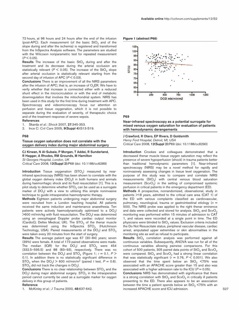
S27
72 hours, at 96 hours and 24 hours after the end of the infusion(post-APC). Each measurement (of the basic StO2 and of theslope during and after the ischemia) is registered and transformedfrom the InSpectra Analysis software. The parameters are studiedwith the Wilcoxon nonparametric test for repeated measurement(P < 0.05).Results The increase of the basic StO2 during and after thetreatment and its decrease during the arterial occlusion arestatistically relevant (P < 0.05). The increase of the StO2 slopeafter arterial occlusion is statistically relevant starting from thesecond day of infusion of APC (P < 0.03).Conclusions There is an improvement of all the NIRS parametersafter the infusion of APC; that is, an increase of O2ER. We have toverify whether that increase is connected either with a reducedshunt effect in the microcirculation or with the end of metabolicdownregulation that involves the mitochondrial system. NIRS hasbeen used in this study for the first time during treatment with APC.Spectroscopy and videomicroscopy focus our attention onperfusion and tissue oxygenation, which it is not possible toseparate during the evaluation of severity, of therapeutic choiceand of the treatment response of severe sepsis.References1. Skarda et al.: Shock 2007, 27:345-353.2. Ince C: Crit Care 2005, 9(Suppl 4):S13-S19.
P68Tissue oxygen saturation does not correlate with theoxygen delivery index during major abdominal surgery
CJ Kirwan, N Al-Subaie, P Morgan, T Addei, R Sunderland, R Hagger, A Rhodes, RM Grounds, M HamiltonSt Georges Hospital, London, UKCritical Care 2008, 12(Suppl 2):P68 (doi: 10.1186/cc6289)
Introduction Tissue oxygenation (STO2) measured by near-infrared spectroscopy (NIRS) has been shown to correlate with theglobal oxygen delivery index (DO2I) in both humans and animalsduring haemorrhagic shock and its fluid resuscitation [1]. This is apilot study to determine whether STO2 can be used as a surrogatemarker of DO2I with a view to utilising this simple noninvasivetechnique to guide intraoperative haemodynamic therapy.Methods Eighteen patients undergoing major abdominal surgerywere recruited from a London teaching hospital. All patientsreceived the same induction and maintenance anaesthesia. Tenpatients were actively haemodynamically optimised to a DO2I>600 ml/min/kg with fluid resuscitation. The DO2I was determinedusing an oesophageal Doppler probe cardiac output monitor(CardioQ; Deltex Medical, UK). The STO2 of the thenar musclewas determined using the InSpectra STO2 (HutchinsonTechnology, USA). Paired measurements of the DO2I and STO2were taken every 20 minutes from the start of surgery.Results The average patient age was 67 (30–84) years; seven(39%) were female. A total of 173 paired observations were made.The median (IQR) for the DO2I and STO2 were 454(332.5–595.5) and 88 (83–93), respectively. There was nocorrelation between the DO2I and STO2 (Figure 1; r = 0.1, P >0.1). In addition there is no statistically significant difference inSTO2 when the DO2I > 600 ml/min/m2 (paired t test, P = 0.6).STO2 did not track the changes in DO2I.Conclusions There is no clear relationship between STO2 and theDO2I during major abdominal surgery. STO2 in the intraoperativeperiod cannot currently be used as a surrogate marker for oxygendelivery in this group of patients.Reference1. McKinley et al.: J Trauma 2000, 48:637-642.
P69Near-infrared spectroscopy as a potential surrogate formixed venous oxygen saturation for evaluation of patientswith hemodynamic derangements
J Crawford, R Otero, EP Rivers, D GoldsmithHenry Ford Hospital, Detroit, MI, USACritical Care 2008, 12(Suppl 2):P69 (doi: 10.1186/cc6290)
Introduction Crookes and colleagues demonstrated that adecreased thenar muscle tissue oxygen saturation may reflect thepresence of severe hypoperfusion (shock) in trauma patients betterthan traditional hemodynamic parameters [1]. Near-infraredspectroscopy (NIRS) may be a novel method for rapidly andnoninvasively assessing changes in tissue level oxygenation. Thepurpose of this study was to compare and correlate NIRSmeasurements (StO2) with central venous blood saturationmeasurement (ScvO2) in the setting of compromised systemicperfusion in critical patients in the emergency department (ED).Methods A prospective, nonrandomized, observational, study inpatients >18 years, admitted to the critical care area (CAT 1) ofthe ED with various complaints classified as cardiovascular,pulmonary, neurological, trauma or gastrointestinal etiology (n =500). The NIRS probe was applied to the right thenar eminenceand data were collected and stored for analysis. StO2 and ScvO2monitoring was performed within 15 minutes of admission to CAT1, and values were recorded at a single point in time. The EDphysicians were blinded to StO2 values. Exclusion criteria includedthe Do Not Resuscitate status, peripheral vascular disease, cardiacarrest, amputated upper extremities or skin abnormalities in themonitoring site as well as refusal to participate.Results StO2 correlation analysis was performed against allcontinuous variables. Subsequently, ANOVA was run for all of thecontinuous variables allowing pairwise comparisons. For thiscohort of 500 patients, 305 paired data points of StO2 and ScvO2were compared. StO2 and ScvO2 had a strong linear correlationthat was statistically significant (r = 0.76, P < 0.001). We alsoobserved that the time spent below an StO2 <75% wasassociated with an APACHE score greater than 15 and also wasassociated with a higher admission rate to the ICU (P = 0.05).Conclusions NIRS has demonstrated with significance that thereis a strong correlation with StO2 and ScvO2 in critically ill patientspresenting to the ED. There also appears to be an associationbetween the time a patient spends below an StO2 <75% with anincreased APACHE score and ICU admission.
Available online http://ccforum.com/supplements/12/S2
Figure 1 (abstract P68)
S28
Reference1. Crookes BA, Cohn SM, Bloch S, et al.: Can near-infrared
spectroscopy identify the severity of shock in traumapatients? J Trauma 2005, 58:806-813.
P70Changes in thenar eminence tissue oxygen saturationmeasured using near-infrared spectroscopy suggestischaemic preconditioning in a repeated arterial occlusionforearm ischaemia model
A Sen, D Martin, M Grocott, H MontgomeryUniversity College London, Institute of Human Health andPerformance, London, UKCritical Care 2008, 12(Suppl 2):P70 (doi: 10.1186/cc6291)
Introduction Ischaemic preconditioning (IP) describes the processwhereby a tissue exposed to brief sublethal periods of ischaemiabecomes protected from longer lethal episodes of ischaemia. Onemechanism by which skeletal muscle may effect protection fromischaemic insult is to reduce the resting rate of oxygenconsumption (VO2) following a preconditioning stimulus. Tissueoxygen saturation (StO2) reflects the dynamic balance betweenoxygen supply and utilisation. We hypothesised that using near-infrared spectroscopy to measure thenar eminence StO2 repeatedarterial occlusion of the upper arm would induce an IP effect.Methods The study was approved by the UCL Research EthicsCommittee and written consent was obtained from 20 healthyvolunteers. StO2 was measured using the InSpectra TissueSpectrometer (Model 325; Hutchinson Technology Inc., USA). Thetissue spectrometer probe was attached to the left thenareminence and a blood pressure cuff was placed around the leftupper arm. The repeated arterial occlusion forearm ischaemiamodel (RAOFIM) consisted of resting measurements and then acycle of four cuff inflations (200 mmHg, 3 min) and four deflations(5 min). Finally the cuff was inflated for 3 minutes on the rightupper arm while the StO2 was measured from the right thenareminence. Paired t tests were used to compare rates of oxygendesaturation; P < 0.05 was considered statistically significant.Results There was a fall in thenar eminence StO2 during all arterialocclusions. The rate of decline of StO2 was significantly reducedduring the fourth inflation (0.160%/s) as compared with the first inthe left arm (0.213%/s), P < 0.001. There was an increase in therate of StO2 decline in the right arm (0.268%/s) when comparedwith the first left occlusion (P < 0.001).Conclusions The data from this pilot study demonstrate that,following preconditioning using a RAOFIM, the rate of oxygendesaturation in resting skeletal muscle during subsequent arterialocclusion manoeuvres is reduced. This could be explained by a fallin resting muscle VO2 as a result of the preceding short ischaemicstimuli and therefore represents evidence of IP in skeletal muscle.These data do not provide evidence to support a remote IP effectin the contralateral arm.
P71Tissue oxygen saturation during anaesthesia,cardiopulmonary bypass and intensive care stay forcardiac surgery
J Sanders1, D Martin1, A Smith2, B Keogh2, M Mutch2, H Montgomery1, M Hamilton3, M Mythen4
1University College London, Institute of Human Health andPerformance, London, UK; 2The Heart Hospital, University CollegeLondon Hospitals NHS Trust, London, UK; 3St Georges HealthcareNHS Trust, London, UK; 4Great Ormond Street Hospital, UCLInstitute of Child Health, London, UKCritical Care 2008, 12(Suppl 2):P71 (doi: 10.1186/cc6292)
Introduction Near-infrared spectroscopy is a novel method for rapidand noninvasive assessment of tissue oxygen saturation (StO2). Anassociation between StO2 and oxygen delivery has beendemonstrated during shock, trauma and resuscitation. Weconducted a pilot observational study in which the aims were tomeasure changes in StO2 during the perioperative period forscheduled cardiac surgery and to explore correlations betweenStO2 and routine haemodynamic measures.Methods The study was approved by the UCLH Joint ResearchEthics Committee. Written informed consent was gained from 74patients undergoing scheduled coronary artery bypass grafting(CABG) and valvular surgery requiring cardiopulmonary bypass(CPB). The thenar eminence StO2 was measured continuouslyduring the perioperative period for a maximum of 24 hours usingthe InSpectra Tissue Spectrometer (Model 325; HutchinsonTechnology Inc., USA). Haemodynamic variables were collectedfrom patient records. The mean StO2 was calculated for varioustime points within the study.Results The tissue spectrometer performed well throughout thestudy. From a baseline of 81.7% the StO2 rose significantly duringinduction of anaesthesia to 88.5% (P < 0.001). Prior to and duringCPB the StO2 fell to a minimum of 77.6%, and rose significantly to83.1% after CPB (P < 0.001). The mean StO2 decreased duringthe ICU stay to a minimum of 70.0% at 2 hours post operation.There was marginal association between StO2 measures andhaemodynamic changes although all analyses resulted in areasunder ROC curves <0.70.Conclusions The present study demonstrates interesting changesin tissue StO2 during the perioperative period surroundingscheduled cardiac surgery. The trends suggest a fall in StO2throughout CPB and during early recovery in the ICU. Changes inStO2 may reflect underlying tissue perfusion; therefore theutilisation of StO2 as both an index for tissue hypoperfusion and asa therapeutic goal needs further exploration.
P72Near-infrared spectroscopy during stagnant ischemia: amarker of ScvO2–SvO2 mismatch in septic patients withlow cardiac output
H Mozina, M PodbregarUniversity Medical Center Ljubljana, SloveniaCritical Care 2008, 12(Suppl 2):P72 (doi: 10.1186/cc6293)
Introduction Monitoring of oxygen saturation in the superior venacava (ScvO2) was suggested as a simpler and cheaper assess-ment of the global DO2:VO2 ratio and was used successfully as agoal in treatment of patients with septic shock and severe sepsis[1]. In patients with low cardiac output (CO) the differencebetween SvO2 and ScvO2 is more expressed and problematicallylarge confidence limits and poor correlation were found between
Critical Care March 2008 Vol 12 Suppl 2 28th International Symposium on Intensive Care and Emergency Medicine
S29
the two values [2]. The thenar muscle oxygen saturation (StO2)measured with near-infrared spectroscopy (NIRS) during stagnantischemia (cuff inflation-induced vascular occlusion) decreasesslower in septic shock patients [3]. This may be due to slowermuscle tissue oxygen consumption in sepsis. This phenomenonpossibly contributes to the ScvO2–SvO2 mismatch in patients withlow CO by adding more oxygenated venous blood to flow throughthe superior vena cava. The aim of present study was to determinethe relationship between the StO2 deceleration rate and theScvO2–SvO2 difference in septic patients with low CO.Methods In septic patients with low CO and no signs ofhypovolaemia, catheterization with a pulmonary artery floatingcatheter was performed. Blood was drawn from the superior venacava and pulmonary artery at the time of each StO2 measurementin order to determine ScvO2 and SvO2. The thenar muscle StO2during stagnant ischemia was measured using NIRS (InSpectra™)and the StO2 deceleration rate (StO2%/min) was obtained usingthe Inspectra Analysis Program V2.0.Results Fifty-four patients (47 male, seven female), age 68 ± 13years, SOFA score 12.2 ± 2.5 points. CI 2.5 ± 0.7 l/min/m2, SvO267 ± 10%, ScvO2 77 ± 8%. Lactate 3.5 ± 3.0 mmol/l, CRP 127 ±78 mg/l. NIRS data: basal StO2 89 ± 8%, deceleration rate–12.6 ± 4.9%/min, StO2 deceleration rate versus ScvO2–SvO20.651, P = 0.001(Pearson correlation, P value).Conclusions The StO2 deceleration rate during cuffing is inverselyproportional to the difference between ScvO2 and SvO2 in septicpatients with low CO. When using ScvO2 as a treatment goal, thissimple noninvasive NIRS measurement might be useful to discoverthose patients with normal ScvO2 but probably abnormally lowSvO2.References1. Rivers E, et al.: N Engl J Med 2001, 345:1368-1377.2. Martin C, et al.: Intensive Care Med 1992, 18:101-104.3. Pareznik R, et al.: Intensive Care Med 2006, 32:87-92.
P73Relationship between central venous oxygen saturationmeasured in the inferior and superior vena cava
T Leiner, A Mikor, Z Heil, Z MolnarUniversity of Pecs, HungaryCritical Care 2008, 12(Suppl 2):P73 (doi: 10.1186/cc6294)
Introduction Central venous saturation ScvO2 proved a usefulalternative to mixed venous saturation, and also a reliableparameter to evaluate the oxygen debt in emergency and intensivecare [1-3]. Under special circumstances of anatomical orcoagulation disorders, however, canulation of the jugular orsubclavian veins are not recommended. In these cases the femoralvein has to be catheterised. The aim of our prospective study wasto investigate the relationship between the ScvO2 measured in thesuperior vena cava (ScvsO2) and in the inferior vena cava (ScviO2).Methods After local ethics committee approval every ICU patientwith two central venous catheters (one subclavian/internal jugularand one femoral) entered the study. Parallel blood gas analyseswere performed in random, whenever ScvO2 was requested by theattending physician. Vital parameters (heart rate, mean arterialpressure, Glasgow Coma Scale, respiratory parameters) and thedose of vasopressor and sedative drugs were also recorded.Results were compared using Wilcoxon test, Pearson correlationand Bland–Altman plots.Results In 13 patients 47 matched pairs were compared. AlthoughScvsO2 = median 79% (range: 50–85) was significantly higherthan ScviO2 = 71% (45–87), P < 0.001, there was significant
correlation between the two variables (r = 0.690, P < 0.001).Bland–Altman plots showed a mean bias of 7.6% with lower andupper levels of agreement of –5.6 and 20.7, respectively. The doseof vasopressor (norepinephrine) and dose of sedative (propofol)had a significant influence on the measured difference between theinvestigated variables (r = 0.562, P < 0.001; r = 0.538, P < 0.001,respectively).Conclusions The preliminary results of this study show thatScviO2 underestimates ScvsO2 with a low level of agreement andthat this difference is affected by vasopressor support andsedation. ScviO2 may be useful, however, for monitoring the trendof ScvsO2.References1. Reinhart K, et al.: Intensive Care Med 2004, 30:1572-1578.2. Rivers E, et al.: N Engl J Med 2001, 345:1368-1377.3. Molnar Z, et al.: Intensive Care Med 2007, 33:1767-1770.
P74Early worst central venous oxygen saturation is predictiveof mortality in severe head trauma but not in moderatehead trauma
A Di Filippo, S Matano, R Spina, L Perretta, C Gonnelli, A PerisTeaching Hospital Careggi, Florence, ItalyCritical Care 2008, 12(Suppl 2):P74 (doi: 10.1186/cc6295)
Introduction The aim of the present study was to evaluate themortality prediction power of central venous oxygen saturation(ScVO2) in critically ill patients suffering from major trauma andhead trauma.Methods In an ED, eight-bed, ICU of a teaching hospital fromJanuary 2004 to November 2007, all patients with major trauma(RTS < 10) and head trauma were included in the study. On thebasis of the severity of head trauma the patients were divided intotwo groups: severe (GCS ≤ 8; n = 91) and moderate head trauma(GCS > 8 ≤ 12; n = 116). Each group was in turn divided into twoother groups: patient survivors and dead patients. In eachsubgroup, the age, sex, ISS, SAPS II, worst ScVO2 on the first dayfrom trauma (emogasanalysis of venous blood sampled by acatheter inserted in the superior vena cava 2 hours from trauma),and worst lactate level in circulating blood on the first day fromtrauma were compared. Statistics were performed with theStudent t test and the χ2 test.Results The results showing a significant difference aresummarized in Table 1.
Conclusions ScVO2 seems to be predictive of major outcome insevere head trauma but not in moderate head trauma. Venousmixing of the superior vena cava could play a role in this difference.Reference1. Reinhart K, et al.: Intensive Care Med 2004, 30:1572.
Available online http://ccforum.com/supplements/12/S2
Table 1 (abstract P74)
Severe head trauma Moderate head trauma
Survivors Dead Survivors Dead (n = 76) (n = 15) (n = 99) (n = 17)
ISS (pt) 30.3 ± 10.7 43.4 ± 18° 27.9 ± 16 41.1 ± 16.3°
ScVO2 (%) 71 ± 7 62 ± 9* 73 ± 7 75 ± 7
Lactate (mmol/l) 3.5 ± 1.9 9 ± 5.4° 2.9 ± 1.6 6.1 ± 4°
Data presented as the mean ± SD. *P < 0.05, °P < 0.01.
S30
P75Comparison between continuous and discontinuouscentral venous oxygen saturation in the ICU: a prospectivestudy and preliminary results
H Gharsallah, I Labbene, W Koubaa, F Gargouri, K Lamine, M FerjaniMilitary Hospital of Tunis, TunisiaCritical Care 2008, 12(Suppl 2):P75 (doi: 10.1186/cc6296)
Introduction Benefits of central venous oxygen saturation (ScvO2)optimization, at early stages of severe sepsis and septic shock,have been shown [1]. Discontinuous ScvO2 measurement has notbeen studied earlier. The purpose of our study was to comparecontinuous and discontinuous ScvO2 in terms of the number oftherapeutic interventions in septic patients.Methods After approval by our institutional ethics committee, 16patients with severe sepsis or septic shock were included in thisprospective study. Patients were randomly assigned to thecontinuous ScvO2 group (central venous catheter; EdwardsLifescience X3820HS ) or to the discontinuous ScvO2 group.Blood pressure, heart rates and pulse oxymetry were continuouslymonitored and the lactate concentration measured in all patients. Inboth groups we noted that the number of therapeutic interventionsdue to the ScvO2 value is <70%. Statistical analysis used theFisher test exact for qualitative variables and the Student t test(Mann–Whitney) for quantitative variables. P < 0.05 was consideredsignificant.Results There were no significant differences between the groupswith respect to baseline characteristics. The median number oftherapeutic interventions was significantly higher in the continuousScvO2 group (13 vs 7, P = 0.016). No significant differencesoccurred between the length of stay in the two groups. See Table 1.
Table 1 (abstract P75)
Comparison between continuous and discontinuous centralvenous oxygen saturation
Continuous Discontinuous P value
Age 63 ± 17 66.8 ± 5.6 0.64
Weight 63 ± 17 73 ± 8.9 0.44
MODS 6.62 ± 3.9 6.12 ± 1.8 0.87
APACHE I 18.8 ± 7.4 18.1 ± 6.8 0.87
Length of stay 6.1 ± 2.51 10.2 ± 5 0.95
Therapeutic interventions 13 (6–19) 7 (4–12) 0.01*
*Significant at P < 0.05.
Conclusions These preliminary results showed that continuousScvO2 measurements increase the number of therapeuticinterventions. We conclude that continuous measurement ofScvO2 is helpful for early therapeutic interventions. Controlled trialsof sufficient size, however, are needed to confirm these results.Reference1. Rivers E, et al.: N Engl J Med 2001, 345:1368-1377.
P76Influence of tissue perfusion on the outcome of high-risksurgical patients needing blood transfusion
JM Silva Jr, DO Toledo, A Gulinelli, FG Correa, KR Genga, T Cezario, M Pinto, A Isola, E RezendeHospital do Servidor Público Estadual, São Paulo, BrazilCritical Care 2008, 12(Suppl 2):P76 (doi: 10.1186/cc6297)
Introduction The objective of this study was to evaluate the clinicaloutcomes of patients who required intraoperative bloodtransfusion, aiming to compare the pretransfusion hematimetricvalues with tissue perfusion markers.Methods A prospective single-center cohort study. Patients wereselected in the operative room of a tertiary hospital. Adult patientswho required blood transfusion during the intraoperative periodwere included in the study. Arterial and central venous bloodsamples were collected at the moment in which the bloodtransfusion decision was made.Results Sixty-one patients were included, with a mean age of 68years. The POSSUM score was 36.2 ± 10.3 and the MODS scorewas 2.4 ± 1.9. At the time of the blood transfusion the meanhemoglobin level was 8.4 ± 1.8 g/dl. The overall inhospitalmortality rate was 24.6%. The ScvO2 cutoff point for the ROCcurve was equal to 80% (AUC = 0.75; sensitivity = 80%;specificity = 65.2%). Patients who received a blood transfusionand had ScvO2 ≤ 80% (n = 29), in comparison with those withScvO2 > 80% (n = 32), had lower mortality rates (12.5% vs47.1%; P = 0.008) and lower incidence of postoperative compli-cations (58.9% vs 72.9%; P = 0.06). Blood transfusion with aScvO2 ≤ 80% was also associated with reduced use of vaso-pressors (5.9% vs 36.8%; P = 0.009), lower incidence ofhypoperfusion (17.6% vs 52.6%; P = 0.009) and lower incidenceof infection (23.5% vs 52.6%; P = 0.038) in the postoperativeperiod.Conclusions In patients submitted to major surgery, the ScvO2appears an important variable to be taken into consideration todecide for or against blood transfusion, since transfusions withadequate perfusion, reflected by ScvO2 > 80%, are associatedwith higher mortality rates and worse clinical outcomes.References1. Hebert PC, Wells G, Blajchman MA, et al.: A multicenter,
randomized, controlled clinical trial of transfusion require-ments in critical care. N Engl J Med 1999; 340:409-417.
2. Vincent JL, Baron JF, Reinhart K, et al.: Anemia and bloodtransfusion in critically ill patients. JAMA 2002, 288:1499-1507.
3. Rivers E, Nguyen B, Havstad S, et al.: Early goal-directedtherapy in the treatment of severe sepsis and septicshock. N Engl J Med 2001, 345:1368-1377.
P77Hyperlactatemia and low central venous saturation canpredict prognosis after cardiac surgery
L Hajjar, M Rego, F Maeda, M Sundin, L Sampaio, F Galas, J Auler JrHeart Institute, Faculdade de Medicina da Universidade de São Paulo, BrazilCritical Care 2008, 12(Suppl 2):P77 (doi: 10.1186/cc6298)
Introduction Hyperlactatemia and low central venous saturation arecommonly encountered in critically ill patients and carry a prognosticsignificance in those with sepsis. Few reports, however, haveexamined the relationship between these parameters immediatelyafter cardiac surgery and clinical outcome. We examined the abilityof the arterial plasma lactate concentration and central venoussaturation to predict patient outcome after cardiac surgery.Methods We performed a consecutive observational study in auniversity hospital. A total of 125 patients undergoing cardiacsurgery were studied. Demographic data were analyzed. Samplesof arterial lactate, arterial gases, and central venous saturationwere collected at the time of admission in the ICU, 12 and 24hours later. Univariate and multivariate analyses were performed.
Critical Care March 2008 Vol 12 Suppl 2 28th International Symposium on Intensive Care and Emergency Medicine
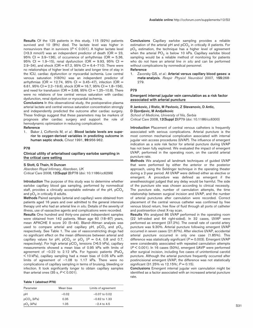
S31
Results Of the 125 patients in this study, 115 (92%) patientssurvived and 10 (8%) died. The lactate level was higher innonsurvivors than in survivors (P < 0.001). A higher lactate level(>3.3 mmol/l) was an independent predictor of death (OR = 23,95% CI = 3.9–136), of occurrence of arrhythmias (OR = 5.36,95% CI = 1.9–15), renal dysfunction (OR = 9.93, 95% CI =2.9–34), and shock (OR = 67.2, 95% CI = 6.4–710). There wereno relationships of higher level of lactate and longer time of stay inthe ICU, cardiac dysfunction or myocardial ischemia. Low centralvenous saturation (<60%) was an independent predictor ofarrhythmias (OR = 12.74, 95% CI = 3.45–47), infection (OR =6.61, 95% CI = 2.2–19.6), shock (OR = 16.7, 95% CI = 1.8–156),and need for transfusion (OR = 3.68, 95% CI = 1.25–10.8). Therewere no relations of low central venous saturation with cardiacdysfunction, renal dysfunction or myocardial ischemia.Conclusions In this observational study, the postoperative plasmaarterial lactate and central venous saturation concentration stronglyand independently predicted the outcome after cardiac surgery.These findings suggest that these parameters may be markers ofprognosis after cardiac surgery and support the role ofhemodynamic optimization in reducing complications.Reference1. Baker J, Coffernils M, et al.: Blood lactate levels are supe-
rior to oxygen-derived variables in predicting outcome inhuman septic shock. Chest 1991, 99:956-962.
P78Clinical utility of arterialised capillary earlobe sampling inthe critical care setting
S Stott, G Thain, N DuncanAberdeen Royal Infirmary, Aberdeen, UKCritical Care 2008, 12(Suppl 2):P78 (doi: 10.1186/cc6299)
Introduction The purpose of this study was to determine whetherearlobe capillary blood gas sampling, performed by nonmedicalstaff, provides a clinically acceptable estimate of the pH, pCO2and pO2 in critically ill adults.Methods Paired samples (arterial and capillary) were obtained frompatients aged 16 years and over admitted to the general intensivetherapy unit who had an arterial line in situ. Details of the severity ofillness, use of vasoactive agents and complications were recorded.Results One hundred and thirty-one paired independent sampleswere obtained from 142 patients. Mean age 60 (18–87) years,mean APACHE II score 20 (5–44). Bland–Altman analysis wasused to compare arterial and capillary pH, pCO2 and pO2,respectively. See Table 1. The use of vasoconstricting drugs hadno significant effect on the mean differences between arterial andcapillary values for pH, pCO2 or pO2 (P = 0.4, 0.8 and 0.7,respectively). For high arterial pCO2 tensions (>6.5 kPa), capillarymeasurements showed a mean bias of 0.95 kPa with limits ofagreement of –0.22 to 2.12 kPa. For hypoxic patients (PaO2< 10 kPa), capillary sampling had a mean bias of 0.05 kPa withlimits of agreement of –1.08 to 1.17 kPa. There were nocomplications of capillary sampling in terms of bruising, bleeding orinfection. It took significantly longer to obtain capillary samplesthan arterial ones (35 s, P < 0.001).
Conclusions Capillary earlobe sampling provides a reliableestimation of the arterial pH and pCO2 in critically ill patients. ForpO2 estimation, the technique has a higher level of agreementwhen the arterial PO2 is below 10 kPa. Capillary earlobe bloodsampling would be a reliable method of monitoring for patientswho do not have an arterial line in situ and can be performedwithout complications by nonmedical personnel.Reference1. Zavorsky GS, et al.: Arterial versus capillary blood gases: a
meta-analysis. Respir Physiol Neurobiol 2007, 155:268-279.
P79Emergent internal jugular vein cannulation as a risk factorassociated with arterial puncture
R Jankovic, J Ristic, M Pavlovic, Z Stevanovic, D Antic, D Djordjevic, M AndjelkovicSchool of Medicine, University of Nis, SerbiaCritical Care 2008, 12(Suppl 2):P79 (doi: 10.1186/cc6300)
Introduction Placement of central venous catheters is frequentlyassociated with serious complications. Arterial puncture is themost common mechanical complication associated with internaljugular vein access procedures (IJVAP). The influence of emergentindication as a sole risk factor for arterial puncture during IJVAPhas not been fully explored. We evaluated the impact of emergentIJVAP, performed in the operating room, on the carotid arterialpuncture rate.Methods We analyzed all landmark techniques of guided IJVAPthat were performed by either the anterior or the posteriorapproach, using the Seldinger technique in the operating theaterduring a 2-year period. All IJVAP were defined either as elective oremergent. A procedure was defined as emergent if theanesthesiologist judged that any delay would be harmful. The sideof the puncture site was chosen according to clinical necessity.The puncture side, number of cannulation attempts, the timerelationship between surgical incision and IJVAP, and the numberof arterial punctures after cannulation were recorded. Correctplacement of the central venous catheter was confirmed by freevenous blood return, free flow of fluid through all ports of catheterand postinsertion chest X-ray scan.Results We analyzed 86 IJVAP performed in the operating room(22 left-sided and 64 right-sided). In 32 cases, IJVAP wereperformed as emergent (37.2%). The overall rate of carotid arterypuncture was 9.30%. Arterial puncture following emergent IJVAPoccurred in seven cases (21.87%). After elective IJVAP, accidentalarterial puncture occurred in only one case (1.85%). Thisdifference was statistically significant (P = 0.003). Emergent IJVAPwere considerably associated with repeated cannulation attempts(P < 0.001). In 16 cases (50%), emergent IJAVP were performedafter surgical incision, including five cases of unintentional carotidpuncture. Although the arterial puncture frequently occurred afterpostincisional emergent IJVAP, the difference was not statisticallysignificant (31.25% vs 12.15%; P = 0.15).Conclusions Emergent internal jugular vein cannulation might beidentified as a factor associated with an increased arterial puncturerate.
Available online http://ccforum.com/supplements/12/S2
Table 1 (abstract P78)
Parameter Mean bias Limits of agreement
pH –0.02 –0.07 to 0.02
pCO2 (kPa) 0.35 –0.62 to 1.33
pO2 (kPa) 1.05 –2.4 to 4.5
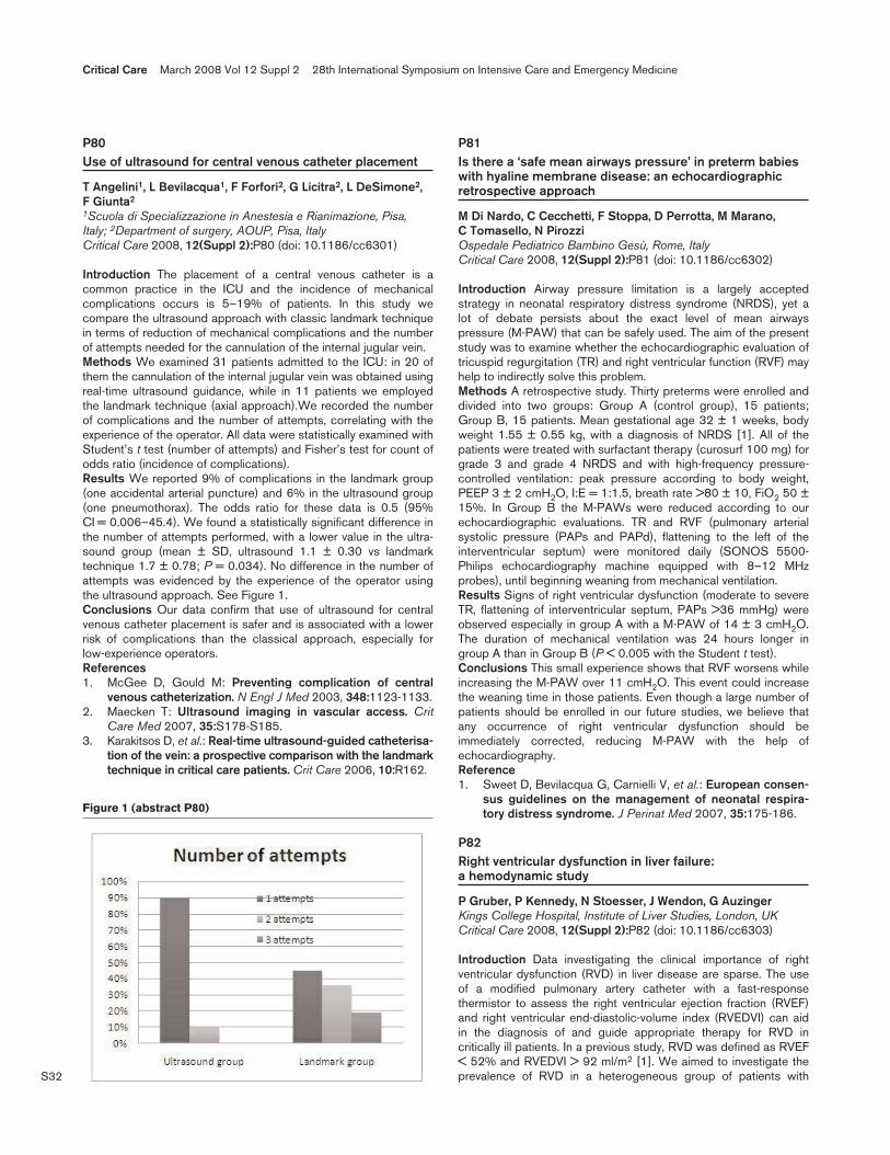
S32
P80Use of ultrasound for central venous catheter placement
T Angelini1, L Bevilacqua1, F Forfori2, G Licitra2, L DeSimone2, F Giunta2
1Scuola di Specializzazione in Anestesia e Rianimazione, Pisa,Italy; 2Department of surgery, AOUP, Pisa, ItalyCritical Care 2008, 12(Suppl 2):P80 (doi: 10.1186/cc6301)
Introduction The placement of a central venous catheter is acommon practice in the ICU and the incidence of mechanicalcomplications occurs is 5–19% of patients. In this study wecompare the ultrasound approach with classic landmark techniquein terms of reduction of mechanical complications and the numberof attempts needed for the cannulation of the internal jugular vein.Methods We examined 31 patients admitted to the ICU: in 20 ofthem the cannulation of the internal jugular vein was obtained usingreal-time ultrasound guidance, while in 11 patients we employedthe landmark technique (axial approach).We recorded the numberof complications and the number of attempts, correlating with theexperience of the operator. All data were statistically examined withStudent’s t test (number of attempts) and Fisher’s test for count ofodds ratio (incidence of complications).Results We reported 9% of complications in the landmark group(one accidental arterial puncture) and 6% in the ultrasound group(one pneumothorax). The odds ratio for these data is 0.5 (95%CI = 0.006–45.4). We found a statistically significant difference inthe number of attempts performed, with a lower value in the ultra-sound group (mean ± SD, ultrasound 1.1 ± 0.30 vs landmarktechnique 1.7 ± 0.78; P = 0.034). No difference in the number ofattempts was evidenced by the experience of the operator usingthe ultrasound approach. See Figure 1.Conclusions Our data confirm that use of ultrasound for centralvenous catheter placement is safer and is associated with a lowerrisk of complications than the classical approach, especially forlow-experience operators.References1. McGee D, Gould M: Preventing complication of central
venous catheterization. N Engl J Med 2003, 348:1123-1133.2. Maecken T: Ultrasound imaging in vascular access. Crit
Care Med 2007, 35:S178-S185.3. Karakitsos D, et al.: Real-time ultrasound-guided catheterisa-
tion of the vein: a prospective comparison with the landmarktechnique in critical care patients. Crit Care 2006, 10:R162.
P81Is there a ‘safe mean airways pressure’ in preterm babieswith hyaline membrane disease: an echocardiographicretrospective approach
M Di Nardo, C Cecchetti, F Stoppa, D Perrotta, M Marano, C Tomasello, N PirozziOspedale Pediatrico Bambino Gesù, Rome, ItalyCritical Care 2008, 12(Suppl 2):P81 (doi: 10.1186/cc6302)
Introduction Airway pressure limitation is a largely acceptedstrategy in neonatal respiratory distress syndrome (NRDS), yet alot of debate persists about the exact level of mean airwayspressure (M-PAW) that can be safely used. The aim of the presentstudy was to examine whether the echocardiographic evaluation oftricuspid regurgitation (TR) and right ventricular function (RVF) mayhelp to indirectly solve this problem.Methods A retrospective study. Thirty preterms were enrolled anddivided into two groups: Group A (control group), 15 patients;Group B, 15 patients. Mean gestational age 32 ± 1 weeks, bodyweight 1.55 ± 0.55 kg, with a diagnosis of NRDS [1]. All of thepatients were treated with surfactant therapy (curosurf 100 mg) forgrade 3 and grade 4 NRDS and with high-frequency pressure-controlled ventilation: peak pressure according to body weight,PEEP 3 ± 2 cmH2O, I:E = 1:1.5, breath rate >80 ± 10, FiO2 50 ±15%. In Group B the M-PAWs were reduced according to ourechocardiographic evaluations. TR and RVF (pulmonary arterialsystolic pressure (PAPs and PAPd), flattening to the left of theinterventricular septum) were monitored daily (SONOS 5500-Philips echocardiography machine equipped with 8–12 MHzprobes), until beginning weaning from mechanical ventilation.Results Signs of right ventricular dysfunction (moderate to severeTR, flattening of interventricular septum, PAPs >36 mmHg) wereobserved especially in group A with a M-PAW of 14 ± 3 cmH2O.The duration of mechanical ventilation was 24 hours longer ingroup A than in Group B (P < 0.005 with the Student t test).Conclusions This small experience shows that RVF worsens whileincreasing the M-PAW over 11 cmH2O. This event could increasethe weaning time in those patients. Even though a large number ofpatients should be enrolled in our future studies, we believe thatany occurrence of right ventricular dysfunction should beimmediately corrected, reducing M-PAW with the help ofechocardiography.Reference1. Sweet D, Bevilacqua G, Carnielli V, et al.: European consen-
sus guidelines on the management of neonatal respira-tory distress syndrome. J Perinat Med 2007, 35:175-186.
P82Right ventricular dysfunction in liver failure: a hemodynamic study
P Gruber, P Kennedy, N Stoesser, J Wendon, G AuzingerKings College Hospital, Institute of Liver Studies, London, UKCritical Care 2008, 12(Suppl 2):P82 (doi: 10.1186/cc6303)
Introduction Data investigating the clinical importance of rightventricular dysfunction (RVD) in liver disease are sparse. The useof a modified pulmonary artery catheter with a fast-responsethermistor to assess the right ventricular ejection fraction (RVEF)and right ventricular end-diastolic-volume index (RVEDVI) can aidin the diagnosis of and guide appropriate therapy for RVD incritically ill patients. In a previous study, RVD was defined as RVEF< 52% and RVEDVI > 92 ml/m2 [1]. We aimed to investigate theprevalence of RVD in a heterogeneous group of patients with
Critical Care March 2008 Vol 12 Suppl 2 28th International Symposium on Intensive Care and Emergency Medicine
Figure 1 (abstract P80)
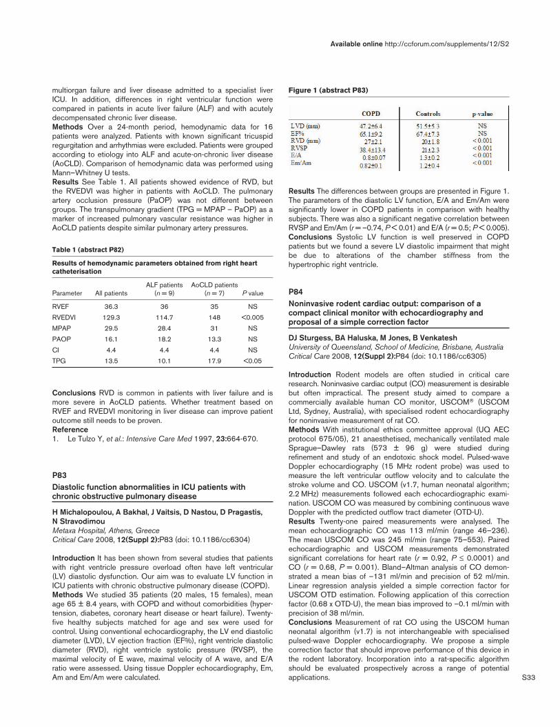
S33
multiorgan failure and liver disease admitted to a specialist liverICU. In addition, differences in right ventricular function werecompared in patients in acute liver failure (ALF) and with acutelydecompensated chronic liver disease.Methods Over a 24-month period, hemodynamic data for 16patients were analyzed. Patients with known significant tricuspidregurgitation and arrhythmias were excluded. Patients were groupedaccording to etiology into ALF and acute-on-chronic liver disease(AoCLD). Comparison of hemodynamic data was performed usingMann–Whitney U tests.Results See Table 1. All patients showed evidence of RVD, butthe RVEDVI was higher in patients with AoCLD. The pulmonaryartery occlusion pressure (PaOP) was not different betweengroups. The transpulmonary gradient (TPG = MPAP – PaOP) as amarker of increased pulmonary vascular resistance was higher inAoCLD patients despite similar pulmonary artery pressures.
Table 1 (abstract P82)
Results of hemodynamic parameters obtained from right heartcatheterisation
ALF patients AoCLD patients Parameter All patients (n = 9) (n = 7) P value
RVEF 36.3 36 35 NS
RVEDVI 129.3 114.7 148 <0.005
MPAP 29.5 28.4 31 NS
PAOP 16.1 18.2 13.3 NS
CI 4.4 4.4 4.4 NS
TPG 13.5 10.1 17.9 <0.05
Conclusions RVD is common in patients with liver failure and ismore severe in AoCLD patients. Whether treatment based onRVEF and RVEDVI monitoring in liver disease can improve patientoutcome still needs to be proven.Reference1. Le Tulzo Y, et al.: Intensive Care Med 1997, 23:664-670.
P83Diastolic function abnormalities in ICU patients withchronic obstructive pulmonary disease
H Michalopoulou, A Bakhal, J Vaitsis, D Nastou, D Pragastis, N StravodimouMetaxa Hospital, Athens, GreeceCritical Care 2008, 12(Suppl 2):P83 (doi: 10.1186/cc6304)
Introduction It has been shown from several studies that patientswith right ventricle pressure overload often have left ventricular(LV) diastolic dysfunction. Our aim was to evaluate LV function inICU patients with chronic obstructive pulmonary disease (COPD).Methods We studied 35 patients (20 males, 15 females), meanage 65 ± 8.4 years, with COPD and without comorbidities (hyper-tension, diabetes, coronary heart disease or heart failure). Twenty-five healthy subjects matched for age and sex were used forcontrol. Using conventional echocardiography, the LV end diastolicdiameter (LVD), LV ejection fraction (EF%), right ventricle diastolicdiameter (RVD), right ventricle systolic pressure (RVSP), themaximal velocity of E wave, maximal velocity of A wave, and E/Aratio were assessed. Using tissue Doppler echocardiography, Em,Am and Em/Am were calculated.
Results The differences between groups are presented in Figure 1.The parameters of the diastolic LV function, E/A and Em/Am weresignificantly lower in COPD patients in comparison with healthysubjects. There was also a significant negative correlation betweenRVSP and Em/Am (r = –0.74, P < 0.01) and E/A (r = 0.5; P < 0.005).Conclusions Systolic LV function is well preserved in COPDpatients but we found a severe LV diastolic impairment that mightbe due to alterations of the chamber stiffness from thehypertrophic right ventricle.
P84Noninvasive rodent cardiac output: comparison of acompact clinical monitor with echocardiography andproposal of a simple correction factor
DJ Sturgess, BA Haluska, M Jones, B VenkateshUniversity of Queensland, School of Medicine, Brisbane, AustraliaCritical Care 2008, 12(Suppl 2):P84 (doi: 10.1186/cc6305)
Introduction Rodent models are often studied in critical careresearch. Noninvasive cardiac output (CO) measurement is desirablebut often impractical. The present study aimed to compare acommercially available human CO monitor, USCOM® (USCOMLtd, Sydney, Australia), with specialised rodent echocardiographyfor noninvasive measurement of rat CO.Methods With institutional ethics committee approval (UQ AECprotocol 675/05), 21 anaesthetised, mechanically ventilated maleSprague–Dawley rats (573 ± 96 g) were studied duringrefinement and study of an endotoxic shock model. Pulsed-waveDoppler echocardiography (15 MHz rodent probe) was used tomeasure the left ventricular outflow velocity and to calculate thestroke volume and CO. USCOM (v1.7, human neonatal algorithm;2.2 MHz) measurements followed each echocardiographic exami-nation. USCOM CO was measured by combining continuous waveDoppler with the predicted outflow tract diameter (OTD-U).Results Twenty-one paired measurements were analysed. Themean echocardiographic CO was 113 ml/min (range 46–236).The mean USCOM CO was 245 ml/min (range 75–553). Pairedechocardiographic and USCOM measurements demonstratedsignificant correlations for heart rate (r = 0.92, P ≤ 0.0001) andCO (r = 0.68, P = 0.001). Bland–Altman analysis of CO demon-strated a mean bias of –131 ml/min and precision of 52 ml/min.Linear regression analysis yielded a simple correction factor forUSCOM OTD estimation. Following application of this correctionfactor (0.68 x OTD-U), the mean bias improved to –0.1 ml/min withprecision of 38 ml/min.Conclusions Measurement of rat CO using the USCOM humanneonatal algorithm (v1.7) is not interchangeable with specialisedpulsed-wave Doppler echocardiography. We propose a simplecorrection factor that should improve performance of this device inthe rodent laboratory. Incorporation into a rat-specific algorithmshould be evaluated prospectively across a range of potentialapplications.
Available online http://ccforum.com/supplements/12/S2
Figure 1 (abstract P83)
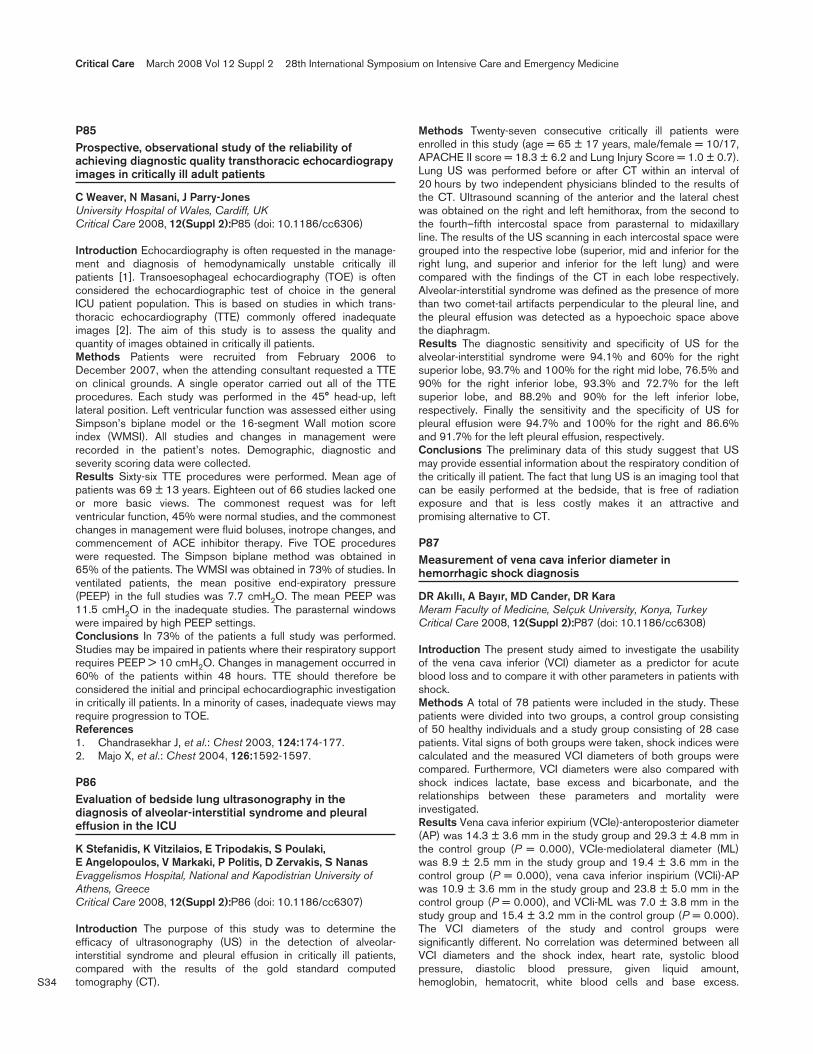
S34
P85Prospective, observational study of the reliability ofachieving diagnostic quality transthoracic echocardiograpyimages in critically ill adult patients
C Weaver, N Masani, J Parry-JonesUniversity Hospital of Wales, Cardiff, UKCritical Care 2008, 12(Suppl 2):P85 (doi: 10.1186/cc6306)
Introduction Echocardiography is often requested in the manage-ment and diagnosis of hemodynamically unstable critically illpatients [1]. Transoesophageal echocardiography (TOE) is oftenconsidered the echocardiographic test of choice in the generalICU patient population. This is based on studies in which trans-thoracic echocardiography (TTE) commonly offered inadequateimages [2]. The aim of this study is to assess the quality andquantity of images obtained in critically ill patients.Methods Patients were recruited from February 2006 toDecember 2007, when the attending consultant requested a TTEon clinical grounds. A single operator carried out all of the TTEprocedures. Each study was performed in the 45° head-up, leftlateral position. Left ventricular function was assessed either usingSimpson’s biplane model or the 16-segment Wall motion scoreindex (WMSI). All studies and changes in management wererecorded in the patient’s notes. Demographic, diagnostic andseverity scoring data were collected.Results Sixty-six TTE procedures were performed. Mean age ofpatients was 69 ± 13 years. Eighteen out of 66 studies lacked oneor more basic views. The commonest request was for leftventricular function, 45% were normal studies, and the commonestchanges in management were fluid boluses, inotrope changes, andcommencement of ACE inhibitor therapy. Five TOE procedureswere requested. The Simpson biplane method was obtained in65% of the patients. The WMSI was obtained in 73% of studies. Inventilated patients, the mean positive end-expiratory pressure(PEEP) in the full studies was 7.7 cmH2O. The mean PEEP was11.5 cmH2O in the inadequate studies. The parasternal windowswere impaired by high PEEP settings.Conclusions In 73% of the patients a full study was performed.Studies may be impaired in patients where their respiratory supportrequires PEEP > 10 cmH2O. Changes in management occurred in60% of the patients within 48 hours. TTE should therefore beconsidered the initial and principal echocardiographic investigationin critically ill patients. In a minority of cases, inadequate views mayrequire progression to TOE.References1. Chandrasekhar J, et al.: Chest 2003, 124:174-177.2. Majo X, et al.: Chest 2004, 126:1592-1597.
P86Evaluation of bedside lung ultrasonography in thediagnosis of alveolar-interstitial syndrome and pleuraleffusion in the ICU
K Stefanidis, K Vitzilaios, E Tripodakis, S Poulaki, E Angelopoulos, V Markaki, P Politis, D Zervakis, S NanasEvaggelismos Hospital, National and Kapodistrian University ofAthens, GreeceCritical Care 2008, 12(Suppl 2):P86 (doi: 10.1186/cc6307)
Introduction The purpose of this study was to determine theefficacy of ultrasonography (US) in the detection of alveolar-interstitial syndrome and pleural effusion in critically ill patients,compared with the results of the gold standard computedtomography (CT).
Methods Twenty-seven consecutive critically ill patients wereenrolled in this study (age = 65 ± 17 years, male/female = 10/17,APACHE II score = 18.3 ± 6.2 and Lung Injury Score = 1.0 ± 0.7).Lung US was performed before or after CT within an interval of20 hours by two independent physicians blinded to the results ofthe CT. Ultrasound scanning of the anterior and the lateral chestwas obtained on the right and left hemithorax, from the second tothe fourth–fifth intercostal space from parasternal to midaxillaryline. The results of the US scanning in each intercostal space weregrouped into the respective lobe (superior, mid and inferior for theright lung, and superior and inferior for the left lung) and werecompared with the findings of the CT in each lobe respectively.Alveolar-interstitial syndrome was defined as the presence of morethan two comet-tail artifacts perpendicular to the pleural line, andthe pleural effusion was detected as a hypoechoic space abovethe diaphragm.Results The diagnostic sensitivity and specificity of US for thealveolar-interstitial syndrome were 94.1% and 60% for the rightsuperior lobe, 93.7% and 100% for the right mid lobe, 76.5% and90% for the right inferior lobe, 93.3% and 72.7% for the leftsuperior lobe, and 88.2% and 90% for the left inferior lobe,respectively. Finally the sensitivity and the specificity of US forpleural effusion were 94.7% and 100% for the right and 86.6%and 91.7% for the left pleural effusion, respectively.Conclusions The preliminary data of this study suggest that USmay provide essential information about the respiratory condition ofthe critically ill patient. The fact that lung US is an imaging tool thatcan be easily performed at the bedside, that is free of radiationexposure and that is less costly makes it an attractive andpromising alternative to CT.
P87Measurement of vena cava inferior diameter inhemorrhagic shock diagnosis
DR AkIllI, A BayIr, MD Cander, DR KaraMeram Faculty of Medicine, Selçuk University, Konya, TurkeyCritical Care 2008, 12(Suppl 2):P87 (doi: 10.1186/cc6308)
Introduction The present study aimed to investigate the usabilityof the vena cava inferior (VCI) diameter as a predictor for acuteblood loss and to compare it with other parameters in patients withshock.Methods A total of 78 patients were included in the study. Thesepatients were divided into two groups, a control group consistingof 50 healthy individuals and a study group consisting of 28 casepatients. Vital signs of both groups were taken, shock indices werecalculated and the measured VCI diameters of both groups werecompared. Furthermore, VCI diameters were also compared withshock indices lactate, base excess and bicarbonate, and therelationships between these parameters and mortality wereinvestigated.Results Vena cava inferior expirium (VCIe)-anteroposterior diameter(AP) was 14.3 ± 3.6 mm in the study group and 29.3 ± 4.8 mm inthe control group (P = 0.000), VCIe-mediolateral diameter (ML)was 8.9 ± 2.5 mm in the study group and 19.4 ± 3.6 mm in thecontrol group (P = 0.000), vena cava inferior inspirium (VCIi)-APwas 10.9 ± 3.6 mm in the study group and 23.8 ± 5.0 mm in thecontrol group (P = 0.000), and VCIi-ML was 7.0 ± 3.8 mm in thestudy group and 15.4 ± 3.2 mm in the control group (P = 0.000).The VCI diameters of the study and control groups weresignificantly different. No correlation was determined between allVCI diameters and the shock index, heart rate, systolic bloodpressure, diastolic blood pressure, given liquid amount,hemoglobin, hematocrit, white blood cells and base excess.
Critical Care March 2008 Vol 12 Suppl 2 28th International Symposium on Intensive Care and Emergency Medicine
S35
Lactate (r = 55) was correlated with all VCI diameters; however,this correlation was better for VCIe-ML. The shock index was lesscorrelated with base excess and lactate (r = 37 and 43, respec-tively). A significant decrease was found in diastolic blood pressure,VCIe-ML and VCIi-ML in addition to lactate, bicarbonate and baseexcess when dead and alive patients were compared (P < 0.05).Conclusions The VCI diameter can give more reliable informationcompared with the shock index and other parameters, especially intrauma patients, to determine acute blood loss, and it can be usedas a follow-up parameter of hemorrhagic shock. A decreased VCIdiameter measured on admission in patients with hemorrhagicshock might be a predictor of high mortality.References1. Tetsuka T, et al.: ASAIO J 1995, 41:105-110.2. Morishita Y, et al.: Clin Exp Nephrol 2005, 9:233-237.3. Marcelino P, et al.: Rev Port Pneumol 2006, 12:637-658.4. Barbier C, et al.: Intensive Care Med 2004, 30:1740-1746.5. Feissel M, et al.: Intensive Care Med 2004, 30:1834-1837.
P88Arterial pressure changes during the Valsalva maneuver topredict fluid responsiveness in spontaneously breathingpatients
MI Monge García, A Gil Cano, JC Díaz MonroveHospital de Jerez, Jerez de la Frontera, SpainCritical Care 2008, 12(Suppl 2):P88 (doi: 10.1186/cc6309)
Introduction Although superiority of dynamic parameters comparedwith static values of preload is widely accepted, predicting fluidresponsiveness in spontaneously breathing patients is still achallenging problem. A sudden increase in intrathoracic pressureduring the Valsalva maneuver transiently impairs venous return and,only in preload-dependent patients, decreases the stroke volumeand arterial pulse pressure (PP). We designed this study to assessthe predictive value of arterial pressure response to a Valsalva maneu-ver for fluid responsiveness in spontaneously breathing patients.Methods In 27 spontaneously breathing patients, the Valsalvamaneuver, consisting of a forced expiration through a closedmouthpiece, was performed. Patients were encouraged to maintaina constant pressure of 30 cmH2O for 10 seconds. The Valsalvapulse pressure variation (ΔVPP) was defined using the maximumPP at the beginning of the strain (PPmax) and the minimum PP(PPmin) according to the known formula: ΔVPP (%) = (PPmax –PPmin) / [(PPmax + PPmin) / 2]. A first set of measurements wasobtained at baseline and immediately after the Valsalva maneuverwas performed. Cardiac output (FloTrac/Vigileo™), central venouspressure, invasive arterial pressure and respiratory pressure werecontinuously recorded during the whole strain time. After volumeexpansion, new measurements were obtained and the Valsalvamaneuver was performed again post infusion.Results The volume expansion-induced increase in the strokevolume index was ≥15% in 10 patients (responders) and <15% in17 patients (nonresponders). The baseline ΔVPP was higher anddecreased more after volume expansion in responders (P < 0.0001).The baseline ΔVPP and ΔVPP increase after volume expansionwere statistically correlated with changes in the stroke volumeindex (r = 0.83 and r = 0.74, P < 0.0001, respectively). Thebaseline ΔVPP accurately predicted changes induced by volumeexpansion with a sensibility of 90% and a specificity of 94%. Thearea under the ROC curve for ΔVPP was 0.96 (95% CI =0.81–0.99, P = 0.0001), with a best cutoff value of 50%.Conclusions Arterial pressure variations induced by a Valsalvamaneuver reliably predict fluid responsiveness in spontaneouslybreathing patients.
P89Passive leg raising predicts fluid responsiveness aftercardiac surgery
F Galas, L Hajjar, T Polastri, T Faustino, W Leao, L Sampaio, J Auler JrHeart Institute, Faculdade de Medicina da Universidade de SãoPaulo, BrazilCritical Care 2008, 12(Suppl 2):P89 (doi: 10.1186/cc6310)
Introduction Passive leg raising (PLR) represents a self-volumechallenge that could predict fluid response with less limitationsthan other methods as variation of stroke volume and pulsepressure variation. We hypothesized that the hemodynamic responseto PLR is able to predict fluid responsiveness in mechanicallyventilated patients after cardiac surgery.Methods A prospective study in a surgical ICU of a universityhospital was performed. We investigated 44 patients in theimmediate postoperative period after cardiac surgery while inmechanical ventilation with no spontaneous breathing activity.Fourteen patients had arrhythmias. The hemodynamic status wasevaluated at baseline, after PLR and after volume expansion(500 ml HAES 130/04 infusion over 10 min). In patients withoutarrhythmias, the pulse pressure variation was calculated.Results In 22 patients (responders), the cardiac index increasedby >15% after fluid infusion. A PLR increase of cardiac index>15% predicted fluid responsiveness with a sensitivity of 95% anda specificity of 94%. In patients without arrhythmias, a respiratoryvariation in pulse pressure >13% predicted fluid responsivenesswith a sensitivity of 92% and a specificity of 88%.Conclusions In a group of patients submitted to cardiac surgery,the changes in cardiac index induced by PLR predict fluidresponsiveness in ventilated patients with higher sensitivity andspecificity than respiratory variation in pulse pressure.References1. Monnet X, Rienzo M, Osman D, et al.: Passive leg raising
predicts fluid responsiveness in the critically ill. Crit CareMed 2006, 34:1402-1407.
2. Michard F, Teboul J-L: Predicting fluid responsiveness inICU patients: a critical analysis of the literature. Chest2002, 121:2000-2008.
P90Variation of hemodynamic parameters after fluid challenge
A Donati, L Botticelli, C Anastasi, M Romanelli, L Romagnoli, R Nardella, P PelaiaUniversità Politecnica delle Marche, Torrette di Ancona, ItalyCritical Care 2008, 12(Suppl 2):P90 (doi: 10.1186/cc6311)
Introduction We compared thoracic electrical bioimpedance(TEB) with transpulmonary thermodilution (TT) to evaluate theaccuracy of cardiac index with the ICG (CI-I) and cardiac indexwith the PiCCO (CI-P) before and after fluid challenge (FC), todetermine the correlation between intrathoracic blood volume(ITBV) and corrected flow time (FTc) before and after FC, to verifythe parameters’ response after FC, and to establish the credibilityof total fluid content (TFC) as a pulmonary fluid index incomparison with extravascular lung water (EVLW).Methods We recruited 33 patients from May 2006 to July 2007.Inclusion criteria: instable hemodynamic conditions, mechanicventilation. Exclusion criteria: intraaortic balloon pump, aorticfailure. We used 7 ml/kg hydroxyethyl starch 6% in 30 minutes forthe FC. We used the PiCCO (Pulsion Medical Systems AG) andthe ICG (Solar ICG module; GE Medical Systems Technology,
Available online http://ccforum.com/supplements/12/S2
S36
Milwaukee, USA) to monitor hemodynamic parameters. Westudied the strength of the association between all of thehemodynamic parameters of the ICG and the PiCCO with thecorrelation coefficient (P < 0.005).Results The correlation coefficient between the differences of CI-Iand CI-P before and after FC is 0.6090 (P = 0.0002). Thecorrelation coefficient between the differences of EVLW and TFCbefore and after FC is 0.1192 (P = 0.51). The correlationcoefficient between the differences of FTc and ITBV before andafter FC is 0.3443 (P = 0.04).Conclusions The study demonstrates that the ICG can individuatean increase of cardiac output after FC, but less than the PiCCO.The correlation coefficient between CI-P and CI-I results is inferiorafter FC, so CI-I seems less accurate in identifying the fillingresponse. There is an agreement between TFC and EVLW beforethe FC. The ITBV from the PiCCO demonstrates more clinicalutility in identifying a response to FC. Even if TEB is clinicallyuseful, it does not represent an available option instead of the TT.The parameters we studied have less clinical efficacy than theclassic methods of TT, as recent studies of the literaturedemonstrate. TEB should be used when catheterization of a centralartery is contraindicated, when there is no other method to monitorand especially when there is a need for rapid monitoring.References1. Engoren et al.: Am J Crit Care 2005, 14:40-45.2. Heringlake et al.: Intensive Care Med 2007, 33:2168-2172.
P91Comparison of pulse pressure variation and end-diastolicvolume index in an experimental model of hemorrhagicshock in the pig
MA Oliveira, DA Otsuki, JO Auler JrFaculdade de Medicina da Universidade de São Paulo, BrazilCritical Care 2008, 12(Suppl 2):P91 (doi: 10.1186/cc6312)
Introduction Different hemodynamic parameters, including staticindicators of cardiac preload such as the end-diastolic volumeindex (EDVI) and dynamic parameters such as the pulse pressurevariation (PPV) have been used in the decision-making processregarding volume expansion in critically ill patients. The objective ofthis study was to compare fluid resuscitation guided by either PPVor EDVI, after hemorrhagic shock.Methods Twenty anesthetized and mechanically ventilated pigswere randomly allocated into two groups: PPV and EDVI. Hemor-rhagic shock was induced by removing blood to a target pressureof 40 mmHg and was maintained for 60 minutes. Parameters weremeasured at baseline, at the time of the shock (Shock0),60 minutes after the shock (Shock60), immediately after resusci-tation with hydroxyethyl starch 6% (130/0.4) (R0), and 1 hour(R60) and 2 hours (R120) after resuscitation. The endpoint of fluidreplacement was to re-establish the baseline values of PPV orEDVI. Data were submitted to ANOVA for repeated measuresfollowed by the Bonferroni test.Results The resuscitation solution volume was higher in the EDVIgroup when compared with PPV (EDVI = 1,305 ± 331 ml and PPV =965 ± 245 ml; P < 0.05). The time required to reach the endpointwas also different between groups (PPV = 8.8 ± 1.3 min and EDV =24.8 ± 4.7 min). The cardiac index decreased after shock (Shock0and Shock60, P < 0.01) and increased after resuscitation (R0, P <0.01) in the PPV group. In the EDVI group, the cardiac indexdecreased at Shock0 (P < 0.05) and increased during R0 and R60
(P < 0.05). The right atrial pressure and pulmonary artery wedgepressure decreased after shock in both groups (Shock0 andShock60, P < 0.05), reaching baseline values after resuscitation.Oxygen delivery decreased after shock in both groups (Shock0and Shock60, P < 0.001), recovered the baseline value at R0 inboth groups, but decreased at R60 and R120 in group PPV and atR120 in the EDVI group. Lactate increased at Shock60 in bothgroups and remained high at R0 in the PPV group and at R0 andR60 in the EDVI group.Conclusions After hemorrhagic shock, the resuscitation to anestablished endpoint was quicker and required less fluid with PPVwhen compared with EDVI.Acknowledgements Supported by grants from FAPESP(05/59470-0). Performed at LIM08.Reference1. Frédéric M, et al.: Crit Care 2007, 11:131.
P92Evaluation of systolic pressure variation and pulsepressure variation in an experimental model of acutenormovolemic hemodilution
AJ Sant´Ana1, DA Otsuki1, DT Fantoni2, JO Auler Jr1
1Faculdade de Medicina da Universidade de São Paulo, Brazil;2Faculdade de Medicina Veterinaria da Universidade de São Paulo,BrazilCritical Care 2008, 12(Suppl 2):P92 (doi: 10.1186/cc6313)
Introduction Dynamic indicators derived from the arterial pressurewaveform analysis as systolic pressure variation (SPV) and pulsepressure variation (PPV) have been shown to be a useful tool tooptimize tissue perfusion in clinical and experimental studies.These indicators, however, have not been adequately explored insituations of acute variation of blood viscosity as occurs duringacute normovolemic hemodilution (ANH). The purpose of thisresearch was to compare the behavior of these dynamic indicatorsin a porcine model of ANH with two different solutions.Methods Fourteen pigs were anesthetized and randomly allocatedinto two groups: GI (ANH with 6% hydroxyethyl starch, 1:1) andGII (ANH with 0.9% saline solution, 1:3). Static and dynamichemodynamic parameters were evaluated at baseline (T1),immediately after ANH (T2), and 1 hour and 2 hours after ANH (T3and T4). Data were submitted to ANOVA for repeated measuresfollowed by the Bonferroni test.Results The cardiac index increased in GI after ANH (T2 and T3,P < 0.001). The mean arterial pressure was sustained in GI, butdecreased after ANH in GII (T2, T3 and T4, P < 0.05). The rightatrial pressure and pulmonary artery wedge pressure decreased inGI (T3 and T4, P < 0.05). The PPV increased in GII (T3 and T4,P < 0.001). The SPV increased in GI (T4, P < 0.05) and GII (T3and T4, P < 0.001).Conclusions The fluid responsiveness evaluated by PPV and SPV,as well as the cardiac index and mean arterial pressure, in animalssubmitted to ANH with hydroxyethyl starch showed that thehemodynamics were better preserved. We can suppose thatstarch remains longer in the vessels when compared with normalsaline solution.Acknowledgements Supported by grants from FAPESP(06/55221-8). Performed at LIM08.References1. Grocott MPW, et al.: Anesth Analg 2005, 100:1093-106.2. Michard F: Anesthesiology 2005, 103:419-428.3. Lopes et al.: Clinics 2006, 61:345-350.
Critical Care March 2008 Vol 12 Suppl 2 28th International Symposium on Intensive Care and Emergency Medicine
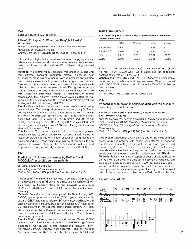
S37
P93Venous return in ICU patients
J Maas1, WK Lagrand1, PC Van den Berg1, MR Pinsky2, JR Jansen1
1Leiden University Medical Center, Leiden, The Netherlands;2University of Pittsburgh, PA, USACritical Care 2008, 12(Suppl 2):P93 (doi: 10.1186/cc6314)
Introduction Guyton’s theory on venous return, implying a linearrelationship between blood flow and central venous pressure, wastested in 12 mechanically ventilated ICU patients during standardcare.Methods The central venous pressure was changed by applyingfour different constant inspiratory plateau pressures over12 seconds. Mean values of central venous pressure and cardiacoutput were measured with pulse contour analysis over the last3 seconds of this plateau period and were plotted against eachother to construct a venous return curve. During the inspiratoryplateau periods, hemodynamic steady-state circumstances weremet without an observable change in cardiovascular controlmechanisms. Two different volemic states were created: normo-volemia in the supine position (SUP) and hypervolemia by volumeloading with 0.5 l intravenously (SUP-V).Results Guyton’s linear venous return pressure–flow relationshipwas confirmed. The average slope of the relation during SUP wasnot significantly different from the slope during SUP-V. The meansystemic filling pressures derived from these venous return curvesduring SUP and SUP-V were 18.8 ± 4.5 mmHg and 29.1 ± 5.2mmHg, respectively (P < 0.001). During SUP the calculated totalcirculatory mean compliance was 0.98 ml/mmHg/kg and the meanstressed volume was 1,677 ml.Conclusions The mean systemic filling pressure, systemiccompliance and stressed volume can be determined in mecha-nically ventilated patients with intact circulation using inspiratorypause procedures. These results may imply a physiological tool toassess the volume state of the circulation as well as fluidresponsiveness of mechanically ventilated patients in the ICU.
P94Prediction of fluid responsiveness by FloTrac™ andPiCCOplus™ in cardiac surgery patients
C Hofer, A Senn, A ZollingerTriemli City Hospital, Zurich, SwitzerlandCritical Care 2008, 12(Suppl 2):P94 (doi: 10.1186/cc6315)
Introduction The aim of this study was to compare the predictionof fluid responsiveness [1] using the stroke volume variation (SVV)determined by FloTrac™ (SVV-FloTrac; Edwards Lifesciences,USA) and PiCCOplus™ (SVV-PiCCO; Pulsion Medical Systems,Germany).Methods With ethics committee approval, the SVV-FloTrac, SVV-PiCCO, pulse pressure variation (PPV), global end-diastolicvolume (GEDV) and stroke volume (SV) were measured before andafter a volume shift induced by body positioning (30° head-up to30° head-down) in 40 patients after cardiac surgery. A t test,Bland–Altman analysis, Pearson correlation and area under thereceiver operating curves (AUC) were calculated. P < 0.05 wasconsidered significant.Results Body positioning resulted in a significant SV and GEDVincrease, while SVV-FloTrac, SVV-PiCCO and PPV significantlydecreased. Comparably strong correlations between SVV-FloTrac/SVV-PiCCO and ΔSV were observed (Table 1). The bestAUC was found for SVV-FloTrac (threshold value: 12.1%) and
SVV-PiCCO (threshold value: 9.6%). Mean bias ± 2SD (SVV-FloTrac – SVV-PiCCO) was –2.5 ± 6.2%, and the correlationcoefficient (r2) was 0.72 (P < 0.01).Conclusions SVV-FloTrac and SVV-PiCCO showed a comparableperformance in predicting fluid responsiveness. When comparedwith SVV-PiCCO, a lower threshold value for SVV-FloTrac has tobe considered.Reference1. Hofer CK, et al.: Chest 2005, 128:848-854.
P95Myocardial dysfunction in sepsis studied with the pressurerecording analytical method
F Pratesi1, T Balderi1, C Sommario1, P Bertini1, F Forfori2, SM Romano3, F Giunta2
1Scuola di specializzazione in Anestesia e Rianimazione, Universitàdegli studi di Pisa, AOUP, Pisa, Italy; 2Department of Surgery,AOUP, Pisa, Italy; 3University of Florence, School of Medicine,Florence, ItalyCritical Care 2008, 12(Suppl 2):P95 (doi: 10.1186/cc6316)
Introduction Myocardial dysfunction is one of the most commonorgan failures in patients with sepsis characterized by transientbiventricular contractility impairment, as well as systolic anddiastolic dysfunction. The aim of this study is to value earlyhemodynamic alterations and myocardial dysfunction in severesepsis using the pressure recording analytical method (PRAM).Methods Patients with severe sepsis or septic shock admitted tothe ICU were enrolled. We studied hemodynamic variations andcardiac performance measured with PRAM (cardiac output, strokevolume, systemic vascular resistance, stroke volume variation,pulse pressure variation, cardiac cycle efficiency (CCE), maximalrate of rise in the arterial pulse curve (dP/dt max)) for the first
Available online http://ccforum.com/supplements/12/S2
Table 1 (abstract P94)
AUC predicting ΔSV > 25% and Pearson correlation of baselineindices versus ΔSV
AUC P value r2 value P value
SVV-FloTrac 0.824 0.001 0.426 <0.001
SVV-PiCCO 0.858 <0.001 0.492 <0.001
PPV 0.718 0.011 0.334 <0.001
GEDV 0.509 0.924 0.091 0.580
Figure 1 (abstract P95)
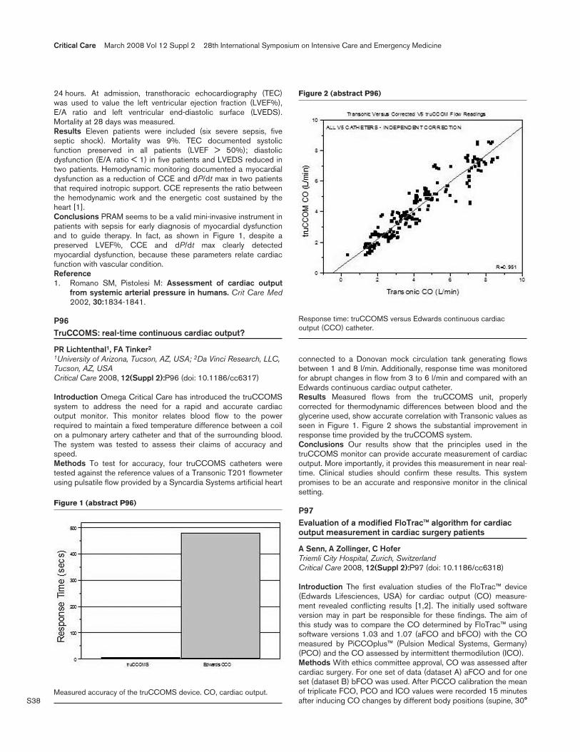
S38
24 hours. At admission, transthoracic echocardiography (TEC)was used to value the left ventricular ejection fraction (LVEF%),E/A ratio and left ventricular end-diastolic surface (LVEDS).Mortality at 28 days was measured.Results Eleven patients were included (six severe sepsis, fiveseptic shock). Mortality was 9%. TEC documented systolicfunction preserved in all patients (LVEF > 50%); diastolicdysfunction (E/A ratio < 1) in five patients and LVEDS reduced intwo patients. Hemodynamic monitoring documented a myocardialdysfunction as a reduction of CCE and dP/dt max in two patientsthat required inotropic support. CCE represents the ratio betweenthe hemodynamic work and the energetic cost sustained by theheart [1].Conclusions PRAM seems to be a valid mini-invasive instrument inpatients with sepsis for early diagnosis of myocardial dysfunctionand to guide therapy. In fact, as shown in Figure 1, despite apreserved LVEF%, CCE and dP/dt max clearly detectedmyocardial dysfunction, because these parameters relate cardiacfunction with vascular condition.Reference1. Romano SM, Pistolesi M: Assessment of cardiac output
from systemic arterial pressure in humans. Crit Care Med2002, 30:1834-1841.
P96TruCCOMS: real-time continuous cardiac output?
PR Lichtenthal1, FA Tinker2
1University of Arizona, Tucson, AZ, USA; 2Da Vinci Research, LLC,Tucson, AZ, USACritical Care 2008, 12(Suppl 2):P96 (doi: 10.1186/cc6317)
Introduction Omega Critical Care has introduced the truCCOMSsystem to address the need for a rapid and accurate cardiacoutput monitor. This monitor relates blood flow to the powerrequired to maintain a fixed temperature difference between a coilon a pulmonary artery catheter and that of the surrounding blood.The system was tested to assess their claims of accuracy andspeed.Methods To test for accuracy, four truCCOMS catheters weretested against the reference values of a Transonic T201 flowmeterusing pulsatile flow provided by a Syncardia Systems artificial heart
connected to a Donovan mock circulation tank generating flowsbetween 1 and 8 l/min. Additionally, response time was monitoredfor abrupt changes in flow from 3 to 6 l/min and compared with anEdwards continuous cardiac output catheter.Results Measured flows from the truCCOMS unit, properlycorrected for thermodynamic differences between blood and theglycerine used, show accurate correlation with Transonic values asseen in Figure 1. Figure 2 shows the substantial improvement inresponse time provided by the truCCOMS system.Conclusions Our results show that the principles used in thetruCCOMS monitor can provide accurate measurement of cardiacoutput. More importantly, it provides this measurement in near real-time. Clinical studies should confirm these results. This systempromises to be an accurate and responsive monitor in the clinicalsetting.
P97Evaluation of a modified FloTrac™ algorithm for cardiacoutput measurement in cardiac surgery patients
A Senn, A Zollinger, C HoferTriemli City Hospital, Zurich, SwitzerlandCritical Care 2008, 12(Suppl 2):P97 (doi: 10.1186/cc6318)
Introduction The first evaluation studies of the FloTrac™ device(Edwards Lifesciences, USA) for cardiac output (CO) measure-ment revealed conflicting results [1,2]. The initially used softwareversion may in part be responsible for these findings. The aim ofthis study was to compare the CO determined by FloTrac™ usingsoftware versions 1.03 and 1.07 (aFCO and bFCO) with the COmeasured by PiCCOplus™ (Pulsion Medical Systems, Germany)(PCO) and the CO assessed by intermittent thermodilution (ICO).Methods With ethics committee approval, CO was assessed aftercardiac surgery. For one set of data (dataset A) aFCO and for oneset (dataset B) bFCO was used. After PiCCO calibration the meanof triplicate FCO, PCO and ICO values were recorded 15 minutesafter inducing CO changes by different body positions (supine, 30°
Critical Care March 2008 Vol 12 Suppl 2 28th International Symposium on Intensive Care and Emergency Medicine
Figure 1 (abstract P96)
Measured accuracy of the truCCOMS device. CO, cardiac output.
Figure 2 (abstract P96)
Response time: truCCOMS versus Edwards continuous cardiacoutput (CCO) catheter.
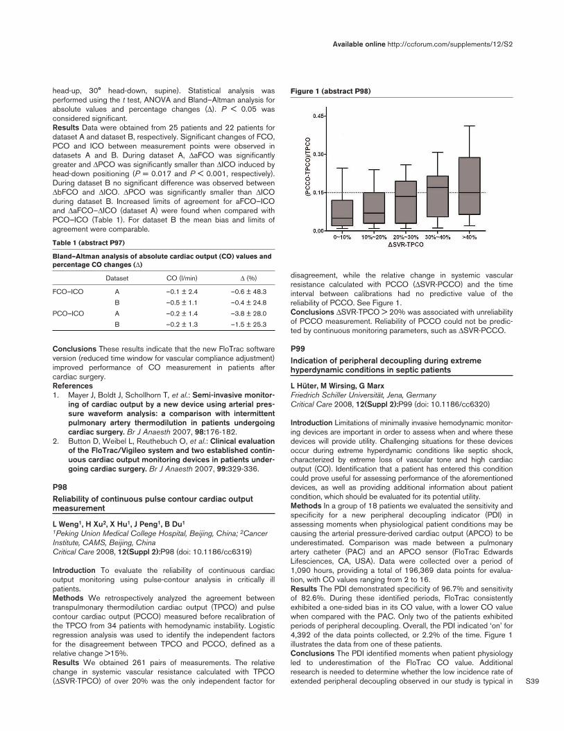
S39
head-up, 30° head-down, supine). Statistical analysis wasperformed using the t test, ANOVA and Bland–Altman analysis forabsolute values and percentage changes (Δ). P < 0.05 wasconsidered significant.Results Data were obtained from 25 patients and 22 patients fordataset A and dataset B, respectively. Significant changes of FCO,PCO and ICO between measurement points were observed indatasets A and B. During dataset A, ΔaFCO was significantlygreater and ΔPCO was significantly smaller than ΔICO induced byhead-down positioning (P = 0.017 and P < 0.001, respectively).During dataset B no significant difference was observed betweenΔbFCO and ΔICO. ΔPCO was significantly smaller than ΔICOduring dataset B. Increased limits of agreement for aFCO–ICOand ΔaFCO–ΔICO (dataset A) were found when compared withPCO–ICO (Table 1). For dataset B the mean bias and limits ofagreement were comparable.
Table 1 (abstract P97)
Bland–Altman analysis of absolute cardiac output (CO) values andpercentage CO changes (Δ)
Dataset CO (l/min) Δ (%)
FCO–ICO A –0.1 ± 2.4 –0.6 ± 48.3
B –0.5 ± 1.1 –0.4 ± 24.8
PCO–ICO A –0.2 ± 1.4 –3.8 ± 28.0
B –0.2 ± 1.3 –1.5 ± 25.3
Conclusions These results indicate that the new FloTrac softwareversion (reduced time window for vascular compliance adjustment)improved performance of CO measurement in patients aftercardiac surgery.References1. Mayer J, Boldt J, Schollhorn T, et al.: Semi-invasive monitor-
ing of cardiac output by a new device using arterial pres-sure waveform analysis: a comparison with intermittentpulmonary artery thermodilution in patients undergoingcardiac surgery. Br J Anaesth 2007, 98:176-182.
2. Button D, Weibel L, Reuthebuch O, et al.: Clinical evaluationof the FloTrac/Vigileo system and two established contin-uous cardiac output monitoring devices in patients under-going cardiac surgery. Br J Anaesth 2007, 99:329-336.
P98Reliability of continuous pulse contour cardiac outputmeasurement
L Weng1, H Xu2, X Hu1, J Peng1, B Du1
1Peking Union Medical College Hospital, Beijing, China; 2CancerInstitute, CAMS, Beijing, ChinaCritical Care 2008, 12(Suppl 2):P98 (doi: 10.1186/cc6319)
Introduction To evaluate the reliability of continuous cardiacoutput monitoring using pulse-contour analysis in critically illpatients.Methods We retrospectively analyzed the agreement betweentranspulmonary thermodilution cardiac output (TPCO) and pulsecontour cardiac output (PCCO) measured before recalibration ofthe TPCO from 34 patients with hemodynamic instability. Logisticregression analysis was used to identify the independent factorsfor the disagreement between TPCO and PCCO, defined as arelative change >15%.Results We obtained 261 pairs of measurements. The relativechange in systemic vascular resistance calculated with TPCO(ΔSVR-TPCO) of over 20% was the only independent factor for
disagreement, while the relative change in systemic vascularresistance calculated with PCCO (ΔSVR-PCCO) and the timeinterval between calibrations had no predictive value of thereliability of PCCO. See Figure 1.Conclusions ΔSVR-TPCO > 20% was associated with unreliabilityof PCCO measurement. Reliability of PCCO could not be predic-ted by continuous monitoring parameters, such as ΔSVR-PCCO.
P99Indication of peripheral decoupling during extremehyperdynamic conditions in septic patients
L Hüter, M Wirsing, G MarxFriedrich Schiller Universität, Jena, GermanyCritical Care 2008, 12(Suppl 2):P99 (doi: 10.1186/cc6320)
Introduction Limitations of minimally invasive hemodynamic monitor-ing devices are important in order to assess when and where thesedevices will provide utility. Challenging situations for these devicesoccur during extreme hyperdynamic conditions like septic shock,characterized by extreme loss of vascular tone and high cardiacoutput (CO). Identification that a patient has entered this conditioncould prove useful for assessing performance of the aforementioneddevices, as well as providing additional information about patientcondition, which should be evaluated for its potential utility.Methods In a group of 18 patients we evaluated the sensitivity andspecificity for a new peripheral decoupling indicator (PDI) inassessing moments when physiological patient conditions may becausing the arterial pressure-derived cardiac output (APCO) to beunderestimated. Comparison was made between a pulmonaryartery catheter (PAC) and an APCO sensor (FloTrac EdwardsLifesciences, CA, USA). Data were collected over a period of1,090 hours, providing a total of 196,369 data points for evalua-tion, with CO values ranging from 2 to 16.Results The PDI demonstrated specificity of 96.7% and sensitivityof 82.6%. During these identified periods, FloTrac consistentlyexhibited a one-sided bias in its CO value, with a lower CO valuewhen compared with the PAC. Only two of the patients exhibitedperiods of peripheral decoupling. Overall, the PDI indicated ‘on’ for4,392 of the data points collected, or 2.2% of the time. Figure 1illustrates the data from one of these patients.Conclusions The PDI identified moments when patient physiologyled to underestimation of the FloTrac CO value. Additionalresearch is needed to determine whether the low incidence rate ofextended peripheral decoupling observed in our study is typical in
Available online http://ccforum.com/supplements/12/S2
Figure 1 (abstract P98)
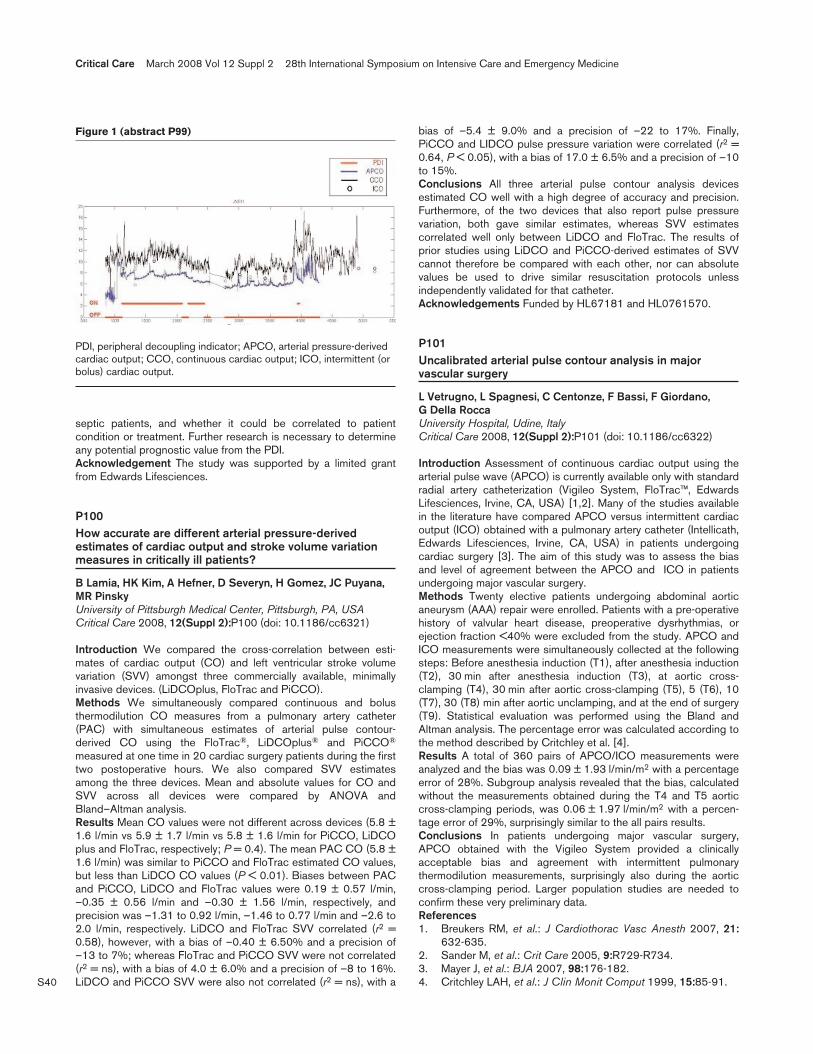
S40
septic patients, and whether it could be correlated to patientcondition or treatment. Further research is necessary to determineany potential prognostic value from the PDI.Acknowledgement The study was supported by a limited grantfrom Edwards Lifesciences.
P100How accurate are different arterial pressure-derivedestimates of cardiac output and stroke volume variationmeasures in critically ill patients?
B Lamia, HK Kim, A Hefner, D Severyn, H Gomez, JC Puyana,MR PinskyUniversity of Pittsburgh Medical Center, Pittsburgh, PA, USACritical Care 2008, 12(Suppl 2):P100 (doi: 10.1186/cc6321)
Introduction We compared the cross-correlation between esti-mates of cardiac output (CO) and left ventricular stroke volumevariation (SVV) amongst three commercially available, minimallyinvasive devices. (LiDCOplus, FloTrac and PiCCO).Methods We simultaneously compared continuous and bolusthermodilution CO measures from a pulmonary artery catheter(PAC) with simultaneous estimates of arterial pulse contour-derived CO using the FloTrac®, LiDCOplus® and PiCCO®
measured at one time in 20 cardiac surgery patients during the firsttwo postoperative hours. We also compared SVV estimatesamong the three devices. Mean and absolute values for CO andSVV across all devices were compared by ANOVA andBland–Altman analysis.Results Mean CO values were not different across devices (5.8 ±1.6 l/min vs 5.9 ± 1.7 l/min vs 5.8 ± 1.6 l/min for PiCCO, LiDCOplus and FloTrac, respectively; P = 0.4). The mean PAC CO (5.8 ±1.6 l/min) was similar to PiCCO and FloTrac estimated CO values,but less than LiDCO CO values (P < 0.01). Biases between PACand PiCCO, LiDCO and FloTrac values were 0.19 ± 0.57 l/min,–0.35 ± 0.56 l/min and –0.30 ± 1.56 l/min, respectively, andprecision was –1.31 to 0.92 l/min, –1.46 to 0.77 l/min and –2.6 to2.0 l/min, respectively. LiDCO and FloTrac SVV correlated (r2 =0.58), however, with a bias of –0.40 ± 6.50% and a precision of–13 to 7%; whereas FloTrac and PiCCO SVV were not correlated(r2 = ns), with a bias of 4.0 ± 6.0% and a precision of –8 to 16%.LiDCO and PiCCO SVV were also not correlated (r2 = ns), with a
bias of –5.4 ± 9.0% and a precision of –22 to 17%. Finally,PiCCO and LIDCO pulse pressure variation were correlated (r2 =0.64, P < 0.05), with a bias of 17.0 ± 6.5% and a precision of –10to 15%.Conclusions All three arterial pulse contour analysis devicesestimated CO well with a high degree of accuracy and precision.Furthermore, of the two devices that also report pulse pressurevariation, both gave similar estimates, whereas SVV estimatescorrelated well only between LiDCO and FloTrac. The results ofprior studies using LiDCO and PiCCO-derived estimates of SVVcannot therefore be compared with each other, nor can absolutevalues be used to drive similar resuscitation protocols unlessindependently validated for that catheter.Acknowledgements Funded by HL67181 and HL0761570.
P101Uncalibrated arterial pulse contour analysis in majorvascular surgery
L Vetrugno, L Spagnesi, C Centonze, F Bassi, F Giordano, G Della RoccaUniversity Hospital, Udine, ItalyCritical Care 2008, 12(Suppl 2):P101 (doi: 10.1186/cc6322)
Introduction Assessment of continuous cardiac output using thearterial pulse wave (APCO) is currently available only with standardradial artery catheterization (Vigileo System, FloTrac™, EdwardsLifesciences, Irvine, CA, USA) [1,2]. Many of the studies availablein the literature have compared APCO versus intermittent cardiacoutput (ICO) obtained with a pulmonary artery catheter (Intellicath,Edwards Lifesciences, Irvine, CA, USA) in patients undergoingcardiac surgery [3]. The aim of this study was to assess the biasand level of agreement between the APCO and ICO in patientsundergoing major vascular surgery.Methods Twenty elective patients undergoing abdominal aorticaneurysm (AAA) repair were enrolled. Patients with a pre-operativehistory of valvular heart disease, preoperative dysrhythmias, orejection fraction <40% were excluded from the study. APCO andICO measurements were simultaneously collected at the followingsteps: Before anesthesia induction (T1), after anesthesia induction(T2), 30 min after anesthesia induction (T3), at aortic cross-clamping (T4), 30 min after aortic cross-clamping (T5), 5 (T6), 10(T7), 30 (T8) min after aortic unclamping, and at the end of surgery(T9). Statistical evaluation was performed using the Bland andAltman analysis. The percentage error was calculated according tothe method described by Critchley et al. [4].Results A total of 360 pairs of APCO/ICO measurements wereanalyzed and the bias was 0.09 ± 1.93 l/min/m2 with a percentageerror of 28%. Subgroup analysis revealed that the bias, calculatedwithout the measurements obtained during the T4 and T5 aorticcross-clamping periods, was 0.06 ± 1.97 l/min/m2 with a percen-tage error of 29%, surprisingly similar to the all pairs results.Conclusions In patients undergoing major vascular surgery,APCO obtained with the Vigileo System provided a clinicallyacceptable bias and agreement with intermittent pulmonarythermodilution measurements, surprisingly also during the aorticcross-clamping period. Larger population studies are needed toconfirm these very preliminary data.References1. Breukers RM, et al.: J Cardiothorac Vasc Anesth 2007, 21:
632-635.2. Sander M, et al.: Crit Care 2005, 9:R729-R734.3. Mayer J, et al.: BJA 2007, 98:176-182.4. Critchley LAH, et al.: J Clin Monit Comput 1999, 15:85-91.
Critical Care March 2008 Vol 12 Suppl 2 28th International Symposium on Intensive Care and Emergency Medicine
Figure 1 (abstract P99)
PDI, peripheral decoupling indicator; APCO, arterial pressure-derivedcardiac output; CCO, continuous cardiac output; ICO, intermittent (orbolus) cardiac output.
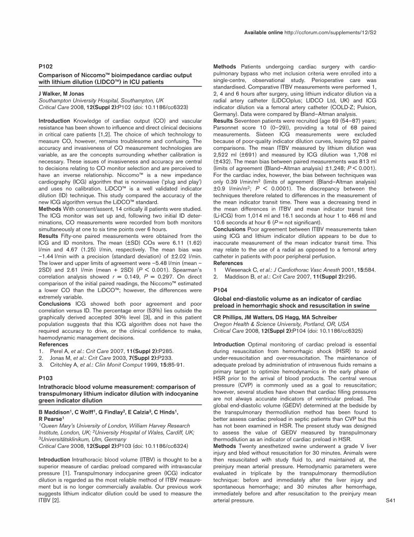
S41
P102Comparison of Niccomo™ bioimpedance cardiac outputwith lithium dilution (LIDCO™) in ICU patients
J Walker, M JonasSouthampton University Hospital, Southampton, UKCritical Care 2008, 12(Suppl 2):P102 (doi: 10.1186/cc6323)
Introduction Knowledge of cardiac output (CO) and vascularresistance has been shown to influence and direct clinical decisionsin critical care patients [1,2]. The choice of which technology tomeasure CO, however, remains troublesome and confusing. Theaccuracy and invasiveness of CO measurement technologies arevariable, as are the concepts surrounding whether calibration isnecessary. These issues of invasiveness and accuracy are centralto decisions relating to CO monitor selection and are perceived tohave an inverse relationship. Niccomo™ is a new impedancecardiography (ICG) algorithm that is noninvasive (‘plug and play’)and uses no calibration. LiDCO™ is a well validated indicatordilution (ID) technique. This study compared the accuracy of thenew ICG algorithm versus the LiDCO™ standard.Methods With consent/assent, 14 critically ill patients were studied.The ICG monitor was set up and, following two initial ID deter-minations, CO measurements were recorded from both monitorssimultaneously at one to six time points over 6 hours.Results Fifty-one paired measurements were obtained from theICG and ID monitors. The mean (±SD) COs were 6.11 (1.62)l/min and 4.67 (1.25) l/min, respectively. The mean bias was–1.44 l/min with a precision (standard deviation) of ±2.02 l/min.The lower and upper limits of agreement were –5.48 l/min (mean –2SD) and 2.61 l/min (mean + 2SD) (P < 0.001). Spearman’scorrelation analysis showed r = 0.149, P = 0.297. On directcomparison of the initial paired readings, the Niccomo™ estimateda lower CO than the LiDCO™; however, the differences wereextremely variable.Conclusions ICG showed both poor agreement and poorcorrelation versus ID. The percentage error (53%) lies outside thegraphically derived accepted 30% level [3], and in this patientpopulation suggests that this ICG algorithm does not have therequired accuracy to drive, or the clinical confidence to make,haemodynamic management decisions.References1. Perel A, et al.: Crit Care 2007, 11(Suppl 2):P285.2. Jonas M, et al.: Crit Care 2003, 7(Suppl 2):P233.3. Critchley A, et al.: Clin Monit Comput 1999, 15:85-91.
P103Intrathoracic blood volume measurement: comparison oftranspulmonary lithium indicator dilution with indocyaninegreen indicator dilution
B Maddison1, C Wolff1, G Findlay2, E Calzia3, C Hinds1, R Pearse1
1Queen Mary’s University of London, William Harvey ResearchInstitute, London, UK; 2University Hospital of Wales, Cardiff, UK;3Universitätsklinikum, Ulm, GermanyCritical Care 2008, 12(Suppl 2):P103 (doi: 10.1186/cc6324)
Introduction Intrathoracic blood volume (ITBV) is thought to be asuperior measure of cardiac preload compared with intravascularpressure [1]. Transpulmonary indocyanine green (ICG) indicatordilution is regarded as the most reliable method of ITBV measure-ment but is no longer commercially available. Our previous worksuggests lithium indicator dilution could be used to measure theITBV [2].
Methods Patients undergoing cardiac surgery with cardio-pulmonary bypass who met inclusion criteria were enrolled into asingle-centre, observational study. Perioperative care wasstandardised. Comparative ITBV measurements were performed 1,2, 4 and 6 hours after surgery, using lithium indicator dilution via aradial artery catheter (LiDCOplus; LIDCO Ltd, UK) and ICGindicator dilution via a femoral artery catheter (COLD-Z; Pulsion,Germany). Data were compared by Bland–Altman analysis.Results Seventeen patients were recruited (age 69 (54–87) years;Parsonnet score 10 (0–29)), providing a total of 68 pairedmeasurements. Sixteen ICG measurements were excludedbecause of poor-quality indicator dilution curves, leaving 52 pairedcomparisons. The mean ITBV measured by lithium dilution was2,522 ml (±691) and measured by ICG dilution was 1,708 ml(±432). The mean bias between paired measurements was 813 ml(limits of agreement (Bland–Altman analysis) ±1,248; P < 0.001).For the cardiac index, however, the bias between techniques wasonly 0.39 l/min/m2 (limits of agreement (Bland–Altman analysis)±0.9 l/min/m2; P < 0.0001). The discrepancy between thetechniques therefore related to differences in the measurement ofthe mean indicator transit time. There was a decreasing trend inthe mean differences in ITBV and mean indicator transit time(Li-ICG) from 1,014 ml and 16.1 seconds at hour 1 to 466 ml and10.6 seconds at hour 6 (P = not significant).Conclusions Poor agreement between ITBV measurements takenusing ICG and lithium indicator dilution appears to be due toinaccurate measurement of the mean indicator transit time. Thismay relate to the use of a radial as opposed to a femoral arterycatheter in patients with poor peripheral perfusion.References1 Wiesenack C, et al.: J Cardiothorac Vasc Anesth 2001, 15:584.2. Maddison B, et al.: Crit Care 2007, 11(Suppl 2):295.
P104Global end-diastolic volume as an indicator of cardiacpreload in hemorrhagic shock and resuscitation in swine
CR Phillips, JM Watters, DS Hagg, MA SchreiberOregon Health & Science University, Portland, OR, USACritical Care 2008, 12(Suppl 2):P104 (doi: 10.1186/cc6325)
Introduction Optimal monitoring of cardiac preload is essentialduring resuscitation from hemorrhagic shock (HSR) to avoidunder-resuscitation and over-resuscitation. The maintenance ofadequate preload by administration of intravenous fluids remains aprimary target to optimize hemodynamics in the early phase ofHSR prior to the arrival of blood products. The central venouspressure (CVP) is commonly used as a goal to resuscitation;however, several studies have shown that cardiac filling pressuresare not always accurate indicators of ventricular preload. Theglobal end-diastolic volume (GEDV) determined at the bedside bythe transpulmonary thermodilution method has been found tobetter assess cardiac preload in septic patients than CVP but thishas not been examined in HSR. The present study was designedto assess the value of GEDV measured by transpulmonarythermodilution as an indicator of cardiac preload in HSR.Methods Twenty anesthetized swine underwent a grade V liverinjury and bled without resuscitation for 30 minutes. Animals werethen resuscitated with study fluid to, and maintained at, thepreinjury mean arterial pressure. Hemodynamic parameters wereevaluated in triplicate by the transpulmonary thermodilutiontechnique: before and immediately after the liver injury andspontaneous hemorrhage; and 30 minutes after hemorrhage,immediately before and after resuscitation to the preinjury meanarterial pressure.
Available online http://ccforum.com/supplements/12/S2
S42
Results Changes in the GEDV index were more highly correlatedwith changes in stroke volume (SV) as compared with changes inCVP versus changes in SV (Figure 1).Conclusions In this porcine model of traumatic hemorrhagic shockand resuscitation, the GEDV in contrast to the CVP behaved as anindicator of cardiac preload.
P105Prognostic value of the extravascular lung water index incritically ill septic shock patients
J Mallat, P Salaun, D Thevenin, L Tronchon, C Patoir, G GazanHospital of Lens, FranceCritical Care 2008, 12(Suppl 2):P105 (doi: 10.1186/cc6326)
Introduction The study investigated the prognostic value of theextravascular lung water index (EVLWI) determined by the singletranspulmonary thermodilution technique and its relationship withphysiologic indexes of lung injury in critically ill patients with septicshock in the ICU.Methods The EVLWI was determined using a PiCCO monitor, andthe daily fluid balance, oxygenation ratio (PaO2/FiO2), pulmonaryvascular index (PVI), lung compliance and lung injury score (LIS)were recorded. The final outcome was assessed at day 28. Data(mean ± SD) were compared using Student’s t test for continuousvariables and by the chi-squared test for discrete variables. Thecorrelations were estimated using Pearson’s coefficient. P < 0.05was regarded as statistically significant.Results Thirty patients with septic shock were admittedprospectively. Fourteen (47%) patients died before day 28. At day1 and day 3 the EVLWI was correlated to PaO2/FiO2 (r = –0.4 andr = –0.47, respectively; P < 0.05) and to LIS (r = 0.47 and r =0.43, respectively; P < 0.05). No correlation was found, however,between the EVLWI and lung compliance and fluid balance. Theaverage EVLWI at baseline was 12 ± 5 ml/kg, and the differencewas not different between survivors and nonsurvivors; P = 0.14.The EVLWI and PVI for day 3 in nonsurvivors were significantlyhigher than in the survivors (13.7 ± 4.5 vs 8.6 ± 2.6 ml/kg; P =0.001 and 2.69 ± 0.98 vs 1.93 ± 0.65; P = 0.01, respectively).ROC statistics using the highest EVLWI value at day 3 in eachindividual revealed an area under the curve of 0.868 ± 0.128; P =0.001 with a cutoff point >11.5 ml/kg. At day 3, the hospitalmortality of patients with EVLWI >11.5 ml/kg was significantlyhigher than those with EVLWI <11.5 ml/kg (77% vs 19%; P =0.02) with sensitivity of 77% and specificity of 80%. During thecourse of illness, the EWLI, PVI and fluid balance decreased fromdays 1 to 3 only in the survivors (P < 0.05).Conclusions In human septic shock, the EVLWI demonstratedmoderate correlation with markers of the severity of pulmonaryaggression. Dynamic observation of the EVLWI can be one of the
factors for predicting the prognosis of patients with septic shock. Areduction of the EVLWI at early treatment was associated with abetter prognosis.
P106Conjunctival microcirculation in patients with traumaticbrain injury
E Klijn1, R Van Zijderveld2, C Den Uil1, C Ince1, J Bakker1
1Erasmus Medical Center, Rotterdam, The Netherlands; 2AcademicMedical Centre, Amsterdam, The NetherlandsCritical Care 2008, 12(Suppl 2):P106 (doi: 10.1186/cc6327)
Introduction Traumatic brain injury (TBI) is one of the most importantcauses of death in young adults. Treatment aims at controlling theintracranial pressure (ICP) in order to maintain an adequate cerebralblood flow, to reduce the risk of secondary ischemic damage.Abnormal blood flow in the middle cerebral artery in patients with TBIwas previously associated with poor outcome. Because perfusion ofthe brain shares a common origin with blood flow in the conjunctiva,we hypothesized that conjunctival microcirculation is altered afterTBI in comparison with healthy subjects.Methods We used sidestream dark-field (SDF) imaging forevaluation of the readily accessible microcirculation of the bulbarconjunctiva as a noninvasive research site. Conjunctival micro-circulation was studied in eight patients with TBI requiring sedationand continuous ICP monitoring. In addition, we investigated eightage-matched healthy control individuals. Using MAS software wedetermined the functional vascular density (FVD) as the total lengthof perfused vessels per field of view as well as the microvascularflow index (MFI).Results Data are presented as the median (interquartile range).The TBI patients had an ICP of 20 (15–25) mmHg and a cerebralperfusion pressure of 61 (53–77) mmHg. The conjunctival MFI inTBI patients was 2.94 (2.88–3.00) in comparison with 2.93(2.79–3.00) in healthy controls. The FVD was 7.78 (7.54–8.14)and 8.53 (7.60–9.97) in TBI patients and healthy controls, respec-tively. There was no significant difference in microcirculatoryparameters found between the groups.Conclusions We found that the FVD and MFI did not differbetween healthy subjects and patients with TBI. Based on theseinterim results, further research will focus on the effect of anelevated ICP on conjunctival microvascular blood flow.
P107Novel models for the prediction of mortality after traumaticbrain injury requiring intensive care
JA Hyam1, CA Welch2, DA Harrison2, DK Menon3
1Charing Cross Hospital, London, UK; 2ICNARC, Case MixProgramme, London, UK; 3University of Cambridge, UKCritical Care 2008, 12(Suppl 2):P107 (doi: 10.1186/cc6328)
Introduction Major head injury is a common reason for admissionto the ICU. Knowledge of factors that predict mortality providesclues to the pathophysiology of head injury, how clinicians’interventions can be most effective, allows audit between differentunits or time points and provides objective data with which tocommunicate with patients’ relatives. Several established riskprediction models exist in the ICU; however, they have been shownto have suboptimal discrimination and calibration in this patientgroup [1]. Our aim was therefore to develop a novel model topredict mortality specifically for head injury.Methods A literature review was undertaken to identify variablespredictive for mortality after severe head injury. The ICNARC Case
Critical Care March 2008 Vol 12 Suppl 2 28th International Symposium on Intensive Care and Emergency Medicine
Figure 1 (abstract P104)
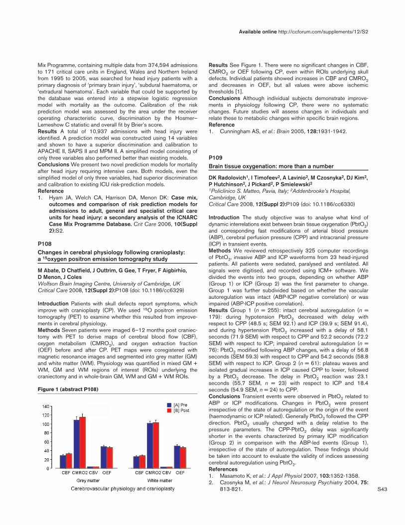
S43
Mix Programme, containing multiple data from 374,594 admissionsto 171 critical care units in England, Wales and Northern Irelandfrom 1995 to 2005, was searched for head injury patients with aprimary diagnosis of ‘primary brain injury’, ‘subdural haematoma, or‘extradural haematoma’. Each variable that could be supported bythe database was entered into a stepwise logistic regressionmodel with mortality as the outcome. Calibration of the riskprediction model was assessed by the area under the receiveroperating characteristic curve, discrimination by the Hosmer–Lemeshow C statistic and overall fit by Brier’s score.Results A total of 10,937 admissions with head injury wereidentified. A prediction model was constructed using 14 variablesand shown to have a superior discrimination and calibration toAPACHE II, SAPS II and MPM II. A simplified model consisting ofonly three variables also performed better than existing models.Conclusions We present two novel prediction models for mortalityafter head injury requiring intensive care. Both models, even thesimplified model of only three variables, had superior discriminationand calibration to existing ICU risk-prediction models.Reference1. Hyam JA, Welch CA, Harrison DA, Menon DK: Case mix,
outcomes and comparison of risk prediction models foradmissions to adult, general and specialist critical careunits for head injury: a secondary analysis of the ICNARCCase Mix Programme Database. Crit Care 2006, 10(Suppl2):S2.
P108Changes in cerebral physiology following cranioplasty: a 15oxygen positron emission tomography study
M Abate, D Chatfield, J Outtrim, G Gee, T Fryer, F Aigbirhio, D Menon, J ColesWolfson Brain Imaging Centre, University of Cambridge, UKCritical Care 2008, 12(Suppl 2):P108 (doi: 10.1186/cc6329)
Introduction Patients with skull defects report symptoms, whichimprove with cranioplasty (CP). We used 15O positron emissiontomography (PET) to examine whether this resulted from improve-ments in cerebral physiology.Methods Seven patients were imaged 6–12 months post craniec-tomy with PET to derive maps of cerebral blood flow (CBF),oxygen metabolism (CMRO2), and oxygen extraction fraction(OEF) before and after CP. PET maps were coregistered withmagnetic resonance images and segmented into grey matter (GM)and white matter (WM). Physiology was quantified in mixed GM +WM, GM and WM regions of interest (ROIs) underlying thecraniectomy and in whole-brain GM, WM and GM + WM ROIs.
Results See Figure 1. There were no significant changes in CBF,CMRO2 or OEF following CP, even within ROIs underlying skulldefects. Individual patients showed increases in CBF and CMRO2and decreases in OEF, but all values were above ischemicthresholds [1].Conclusions Although individual subjects demonstrate improve-ments in physiology following CP, there were no systematicchanges. Future studies will assess changes in individuals andrelate these to metabolic changes within specific brain regions.Reference1. Cunningham AS, et al.: Brain 2005, 128:1931-1942.
P109Brain tissue oxygenation: more than a number
DK Radolovich1, I Timofeev2, A Lavinio2, M Czosnyka2, DJ Kim2,P Hutchinson2, J Pickard2, P Smielewski21Policlinico S. Matteo, Pavia, Italy; 2Addenbrooke’s Hospital,Cambridge, UKCritical Care 2008, 12(Suppl 2):P109 (doi: 10.1186/cc6330)
Introduction The study objective was to analyse what kind ofdynamic interrelations exist between brain tissue oxygenation (PbtO2)and corresponding fast modifications of arterial blood pressure(ABP), cerebral perfusion pressure (CPP) and intracranial pressure(ICP) in transient events.Methods We reviewed retrospectively 325 computer recordingsof PbtO2, invasive ABP and ICP waveforms from 23 head-injuredpatients. All patients were sedated, paralysed and ventilated. Allsignals were digitised, and recorded using ICM+ software. Wedivided the events into two groups, depending on whether ABP(Group 1) or ICP (Group 2) was the first parameter to change.Group 1 was further subdivided based on whether the vascularautoregulation was intact (ABP-ICP negative correlation) or wasimpaired (ABP-ICP positive correlation).Results Group 1 (n = 255): intact cerebral autoregulation (n =179): during hypotension PbtO2 decreased with delay withrespect to CPP (48.5 s; SEM 92.1) and ICP (39.9 s; SEM 91.4),and during hypertension PbtO2 increased with a delay of 58.1seconds (71.9 SEM) with respect to CPP and 52.2 seconds (72.2SEM) with respect to ICP; impaired cerebral autoregulation (n =76): PbtO2 modified following ABP changes, with a delay of 56.8seconds (SEM 59.3) with respect to CPP and 54.2 seconds (58.8SEM) with respect to ICP. Group 2 (n = 61): plateau waves andisolated gradual increases in ICP caused CPP to lower, followedby a PbtO2 decrease. The delay in PbtO2 reaction was 23.1seconds (55.7 SEM, n = 23) with respect to ICP and 18.4seconds (54.9 SEM, n = 24) to CPP.Conclusions Transient events were observed in PbtO2 related toABP or ICP modifications. Changes in PbtO2 were presentirrespective of the state of autoregulation or the origin of the event(haemodynamic or ICP related). Generally PbtO2 followed the CPPdirection. PbtO2 usually changed with a delay relative to thepressure parameters. The CPP-PbtO2 delay was significantlyshorter in the events characterized by primary ICP modification(Group 2) in comparison with the ABP-led events (Group 1),irrespective of the state of autoregulation. These findings shouldbe taken into account to evaluate the validity of indices assessingcerebral autoregulation using PbtO2.References1. Masamoto K, et al.: J Appl Physiol 2007, 103:1352-1358.2. Czosnyka M, et al.: J Neurol Neurosurg Psychiatry 2004, 75:
813-821.
Available online http://ccforum.com/supplements/12/S2
Figure 1 (abstract P108)
S44
P110Fatty acid binding protein and tau levels are related tobrain damage and outcome after subarachnoidhemorrhage
T Zoerle1, M Fiorini2, L Longhi1, ER Zanier1, A Bersano1, L Cracco2, S Monaco2, N Stocchetti11University of Milano, Fondazione IRCCS Ospedale MaggiorePoliclinico, Milan, Italy; 2Ospedale Borgo Roma, Verona, ItalyCritical Care 2008, 12(Suppl 2):P110 (doi: 10.1186/cc6331)
Introduction We measured the fatty acid binding protein (H-FABP) and tau levels in the cerebrospinal fluid (CSF) of patientsafter subarachnoid hemorrhage (SAH): to evaluate the relationshipbetween SAH severity and H-FABP/tau values; to test thehypothesis that H-FABP/tau might help in the diagnosis ofvasospasm; and to evaluate their association with outcome.Methods We studied 38 SAH patients, whose severity wasassessed by the Glasgow Coma Scale (GCS). Serial CSFsamples were obtained in every patient starting on the day of SAHand up 2 weeks post-SAH. H-FABP/tau levels were measured byELISA. Vasospasm was defined as neuro-worsening (loss of atleast one point of the motor component of GCS and/or appear-ance of a new focal deficit) + angiographic confirmation. The 6-month outcome was assessed by the dichotomized GlasgowOutcome Score (GOS): good (GOS 4–5) and bad (GOS 1–3).Multiple logistic regression analyses were performed to assess theassociation between H-FABP/tau values and GOS.Results H-FABP and tau increased after SAH. We observed asignificant association between the peak H-FABP/tau values andadmission mGCS (Spearman r = –0.581, P = 0.0001 and r =–0.582, P = 0.0001, respectively). Eight patients underwent braindeath. Within the survivors we observed vasospasm in 11 patients.Both proteins were significantly higher in this group compared withthose without ischemia (H-FABP = 15,958 ± 21,736 pg/ml vs2,527 ± 2,427 pg/ml, P < 0.05; tau = 5,821 ± 3,774 pg/ml vs1,118 ± 1,547 pg/ml, P < 0.05). The H-FABP rise precededclinical recognition of vasospasm in seven patients and wassimultaneous in four patients. Tau increased before clinicalrecognition of vasospasm in five patients. Patients with bad out-come showed higher peak levels of both proteins than patientswith good outcome: respectively, H-FABP = 23,977 ± 25,593 pg/mland 3,374 ± 2,549 pg/ml, P < 0.001; tau = 6,756 ± 4,544 pg/mland 1,591 ± 1,639 pg/ml, P < 0.001. Logistic regression showedthat, after correction for age, sex and SAH severity, the peak valueof tau protein was an independent predictor of outcome.Conclusions The H-FABP and tau increase following SAH andmight add complementary information for the diagnosis ofvasospasm. There is an association between their CSF values andoutcome following SAH.
P111Transdermal nicotine replacement is associated with lowermortality among active smokers admitted withspontaneous subarachnoid hemorrhage
D Seder, M Schmidt, N Badjatia, F Rincon, J Claassen, E Gordon, E Carrera, M Oddo, L Fernandez, C Lesch, K Lee, E Connolly, S MayerColumbia University, New York, USACritical Care 2008, 12(Suppl 2):P111 (doi: 10.1186/cc6332)
Introduction Active smokers comprise 35–55% of patientsadmitted with acute spontaneous subarachnoid hemorrhage (SAH).Transdermal nicotine replacement is sometimes prescribed to these
patients to prevent a withdrawal syndrome, but the safety ofexogenous nicotine during the acute period after SAH is unknown.Methods We conducted a prospective, observational study from2001 to 2007 in the neurological ICU of a major academic medicalcenter. All active smokers admitted with SAH were included in theanalysis, but we excluded patients who died within 7 days ofadmission to remove those whose death was due to discon-tinuation of life support. The primary endpoint was 3-monthmortality. Secondary endpoints were delayed cerebral ischemia(DCI) and clinical vasospasm.Results One hundred and ninety-two active smokers, including104 (54%) who received transdermal nicotine, were well matchedon demographics, gender, age, Hunt and Hess grade, SAH sumscore, aneurism size, and smoking pack-year history, but a higherpercentage of current heavy smokers (>10 cigarettes daily)received nicotine (67%, P < 0.001). There was no association ofnicotine replacement and clinical vasospasm or DCI. Aftercontrolling for disease severity and cerebral edema on head CT(OR = 13.9, CI = 1.5–125.3), multivariable logistic regressionrevealed that heavy smokers were more likely than light smokers todie (OR = 6.0, CI = 1.11–32.7). Smokers who received nicotinehad lower mortality (OR = 0.26, CI = 0.68–0.98), an effect thatseemed on secondary analysis to be driven by high mortalityamong heavy smokers who did not receive nicotine.Conclusions Transdermal nicotine replacement is not associatedwith clinical vasospasm or DCI in smokers admitted with SAH, andis associated with lower mortality, particularly among smokers ofmore than 10 cigarettes daily. This may be due to prevention of thephysiological derangements associated with nicotine withdrawal.Nicotine replacement after acute SAH is probably safe, and shouldbe given to active heavy smokers at the time of admission. Moreresearch is needed to verify these findings and define thetherapeutic role of nicotine in the ICU.Reference1. Lee AH, et al.: The association of nicotine replacement
therapy with mortality in a medical intensive care unit. CritCare Med 2007, 35:1517.
P112Impact of treatment with pravastatin on delayed ischemicdisease and mortality after aneurysmal subarachnoidhemorrhage
U Jaschinski, K Scherer, M Lichtwarck, H ForstKlinikum Augsburg, GermanyCritical Care 2008, 12(Suppl 2):P112 (doi: 10.1186/cc6333)
Introduction Statins have neuroprotective properties includingimproved vasomotor reactivity, reduced platelet activation and anti-inflammatory effects [1]. A prospective observational controlledstudy was conducted to evaluate the impact of pravastatin on thedevelopment of delayed ischemic disease (DID) and ICU mortalityafter aneurysmal subarachnoid hemorrhage (aSAH).Methods A total of 98 patients (20–80 years old) with aSAH wererandomized to receive either pravastatin 40 mg (n = 40) ornonstatin treatment (n = 58) within 24 hours after the ictus. Primaryendpoints, incidence of DID and extent of disability measured by theGlasgow Outcome Scale; secondary endpoint, ICU mortality.Results Groups were comparable with respect to age (54.2(50.3–58.3) vs 53.2 (49.8–56.7) 95% CI), grade of aSAH(Hess/Hunt) (2.6 (2.17–3.03) vs 3.06 (3.00–3.80) 95% CI) andstroke severity (Glasgow Coma Scale 10.9 (9.4–12.4) vs 10.5(9.3–11.8) 95% CI). There was a trend towards less DID in thestatin group (37.5% vs 60.3% nonstatin; standard error of thedifference of the means 9.8 (3.64–28.00) 95% CI). The extent of
Critical Care March 2008 Vol 12 Suppl 2 28th International Symposium on Intensive Care and Emergency Medicine
S45
disability between the groups, however, was not different(Glasgow Outcome Scale 3.65 (3.16–4.14) statin vs 3.39(3.00–3.80) nonstatin 95% CI). Mortality was unchanged as well(22.5% statin vs 22.4% nonstatin).Conclusions These results are in line with a recently publishedstudy demonstrating reduced vasospasm-related DID in patientstreated with pravastatin after aSAH [2]. We could not confirm thebenefit of statin treatment regarding mortality as mentioned in thecited trial since our study was not powered to detect a differencein mortality. So it is to be hoped that the Statins for AneurysmalHemorrhage STASH trial will clarify this topic.References1. Topcuoglu MA: Expert Opin Drug Saf 2006, 5:57.2. Tseng MY: Stroke 2007, 38:1545.
P113Pentraxin 3 as a marker of vasospasm followingsubarachnoid hemorrhage
ER Zanier1, G Peri2, G Brandi1, L Longhi1, M Tettamanti3, C Garlanda2, A Mantovani2, MG De Simoni3, N Stocchetti11University of Milano, Fondazione IRCCS Ospedale MaggiorePoliclinico, Milan, Italy; 2Clinical Institute Humanitas, Milan, Italy;3Mario Negri Institute, Milan, ItalyCritical Care 2008, 12(Suppl 2):P113 (doi: 10.1186/cc6334)
Introduction We studied the induction of Pentraxin 3 (PTX3), aprototypic long pentraxin protein induced by proinflammatorysignals in subarachnoid hemorrhage (SAH) patients, to investigatea possible relation with SAH-associated ischemic brain damage.Methods PTX3 was measured in the plasma and cerebrospinalfluid (CSF) of 38 SAH patients admitted to the neuroscience ICU,who were divided into three groups: occurrence of vasospasm,defined as neuro-worsening (loss of at least one point of theGlasgow Coma Scale motor component and/or appearance of anew focal deficit) and angiographic confirmation of vasospasm;presence of an early hypodense lesion, defined as the appearanceof a new hypodense lesion at CT scan following endovascular orsurgical treatment, or around the initial intracerebral hematoma;and absence of a hypodense lesion. Arterial and CSF sampleswere obtained every 12 hours starting on the day of SAH and up 2weeks post SAH.Results PTX3 was induced in the plasma and CSF of SAHpatients. CSF peak concentrations were significantly higher inpatients with vasospasm (21.5 ± 5.1 ng/ml) compared with thosewith no CT hypodense lesion (5.8 ± 4.5 ng/ml, P < 0.05). Patientswith an early hypodense lesion showed a peak concentration thatwas intermediate between the other two groups (12.4 ± 5.2 ng/ml).No difference was observed in plasma levels among the threegroups. The temporal pattern of CSF PTX3 in patients withvasospasm was triphasic: there was an initial increase of PTX3during the first 48 hours following SAH (acute phase, up to 17.2 ±5.2 ng/ml), followed by a subsequent decrease in the next 48–96hours (subacute phase, up to 1.2 ± 0.3 ng/ml, P < 0.01 comparedwith the acute phase). With the appearance of vasospasm, asecondary peak of PTX3 was detected (up to 7.1 ± 1.4 ng/ml, P <0.01 compared with the subacute phase). No changes weredetectable in plasma.Conclusions PTX3 is induced in the CSF and in plasma followingSAH; however, the CSF but not plasma levels are directly relatedto the degree of brain injury. In addition the data show that PTX3measured in the CSF might be a reliable marker of vasospasmfollowing SAH, and suggest that measurements of PTX3-CSFlevels associated with clinical evaluation could improve earlydiagnosis of vasospasm in these patients.
P114Transcranial sonography investigations of the cerebralblood flow disturbances after hypothalamic pituitary andbrain stem surgery
S Madorskiy, O Shovikova, A SafinNeurosurgery Institute, Moscow, Russian FederationCritical Care 2008, 12(Suppl 2):P114 (doi: 10.1186/cc6335)
Introduction Cerebral blood flow (CBF) disturbances at focallesions of the hypothalamopituitary system and brain stem structuresare one of the unexplored and pressing questions of modernneurosurgery [1]. Transcranial duplex sonography (TCDS) is awidely used method for determination of the cerebral blood-flowvelocity (FV) in neurosurgical patients.Methods We studied characteristics of cerebral hemodynamics byTCDS after hypothalamic and brain stem tumor excision in 186patients. The data obtained were compared with CT-MRI data,clinical parameters and factors of neurohumoral regulation.Results Our study showed that FV disturbances were observed in82% of patients after surgery for hypothalamic and brain stemlesion. The revealed FV disturbances were evaluated as vasomotorspasm of different degrees of manifestation; distress of FV wascaused by a thrombosis of branches of cerebral vessels, hyper-perfusion, hypoperfusion and infringements of venous outflow andCBF autoregulation. Stable neurological disorders were observedin 100% of patients at FV <40 cm/s and >200 cm/s. At FV>120 cm/s we observed 61% of patients with transientneurological disorders and 24% with stable, and at FV >150 cm/s22% with transient and 78% with stable neurological disorders.Middle cerebral artery (MCA)/internal carotid artery ratio >3.0 andFV >120 cm/s and basilar artery (BA)/external vertebral artery ratio>2.0 with BA velocities >85 cm/s was associated with 92%sensitivity and 97% specificity for vasospasm in the MCA and BAaccordingly. In 87% of patients with FV in MCA >185 cm/s weobserved focal ischemic brain lesions verified on CT scan. Fixedpathologic interactions between degrees of FV and factors ofneurohumoral regulations suggest existing pathogenetic mechanismsof CBF disturbances in focal lesions of the hypothalamic pituitarysystem and brain stem. The dependence of FV disturbances andvasopressin plasma level was established. The revealed FVdisorders allowed us to develop algorithms for therapy andpreventing secondary ischemic brain lesions.Conclusions The investigation of FV by the TCDS method at focalbrain lesions, together with MRI and neurological research, hasallowed us to specify pathogenic mechanisms of CBFdisturbances and algorithms for their therapy.Reference1. Macdonald RL, et al.: J Clin Neurosci 1997, 4:348-352.
P115Flow velocity in head injury of different severity: findings oftranscranial duplex sonography
A Safin, A Parfenov, S Madorsky, E GrinenkoNeurosurgical Institute, Moscow, Russian FederationCritical Care 2008, 12(Suppl 2):P115 (doi: 10.1186/cc6336)
Introduction Most commonly, transcranial duplex sonography(TCDS) is used to evaluate the flow velocity (FV). The main signsof cerebral blood flow disturbances in head injury are olygemia,hyperemia and vasospasm [1], which are closely connected withtraumatic brain injury and dynamics of the disease. The influence ofFV values measured by TCDS on the course and outcome of headinjury of different severity is of special importance.
Available online http://ccforum.com/supplements/12/S2
S46
Methods FV was measured using an ultrasound triplex system in83 patients with head injuries. The traumatic brain injury substratewas verified by CT and nuclear MRI. Mean values for FV wereregistered in the MCA every 48 hours. The hemispheric index (HI)was measured to differentiate a vasospasm (HI = mean MCA /mean ICA).Results Depending on values of CFV, all patients were divided intothree groups: Group I, 22 patients with FV < 70 cm/s; Group II, 23patients with FV 70–120 cm/s and HI < 3; Group III, 38 patientswith FV > 120 cm/s and HI > 3.0. Severity of cerebral lesions inGroup I was caused by unilateral intracranial haematomas in sixcases, contusion of type 1–2 in nine cases and diffuse axonalinjury (DAI) in 12 cases. Patients in Group II and Group III revealedbilateral intracranial haematomas combined with type 2 and 3contusions and DAI; patients in Group II showed contusionpredominance, and patients in Group III had concomitant braindamage predominance (that is, intracranial haematomas combinedwith type 2–3 contusions and post-traumatic SAH). Outcomeanalysis in Group I revealed a GOS score of 1–2 in 13 patients, of3 in seven patients and of 4 in two patients. In Group II the GOSscore was 1 or 2 in 17 patients, 3 in four patients and 4 in twopatients. In Group III the GOS score was 1 or 2 in 12 patients, 3 in14 patients and 4 in six patients, and mortality was marked in fivepatients.Conclusions The performed analysis allowed us to conclude thatthere existed a close relationship between the severity of traumaticbrain damage and the character of FV disturbances. Markedtraumatic brain injuries presented by multiple contusions andintracranial haematomas, DAI of type 2–3 and combined with SAHresulted in development of vasospasm in the MCA. Low values ofFV as well as development of vasospasm in the cerebral middleartery are regarded as unfavourable prognosis for patients in theacute period of severe head injury.Reference1. Oertel M, et al.: J Neurosurg 2005, 103:812-824.
P116Transcranial Doppler in serious malaria
V Mardelle, A Nau, E PeytelHIA Laveran, Marseille, FranceCritical Care 2008, 12(Suppl 2):P116 (doi: 10.1186/cc6337)
Introduction Many assumptions have been proposed to explainthe confinement of red blood cells infested by Plasmodiumfalciparum in the cerebral capillaries (cell adherence, rosetting),involving an increase in blood viscosity and a deceleration in bloodflow inside capillaries.Methods In this study, 10 nonimmune adults were included withserious malaria according to the WHO classification. All of themhad one or more criteria of gravity. A Quantitative Buffy Coatmalaria test, a microscopic examination of thick and thin bloodsmear and transcranial Doppler were carried out from entry. Wecompared the transcranial Doppler findings, the pulsatility index(PI), with the degree of parasitemia. Data are expressed as themean, standard deviation, extremes and percentage.Results The age of the patients was 40 ± 13 (SD) years (19–62).The sex ratio was 0.9. SAPS II was 34.3 ± 10 (SD) (20–53). TheGlasgow Coma Scale score was 10 ± 4 (SD) (14–3). Theparasitemia was 12.2 ± 16.9% (SD) (0.01–50). The PI (by averagingthe two middle cerebral arteries’ PI) was 1.9 ± 2.5 (0.8–9). Thecorrelation coefficient between parasitemia and the PI was 0.86.Conclusions Some studies, carried out in children, demonstratedthe interest in monitoring cerebral perfusion pressure andtranscranial Doppler in prognostic evaluation of cerebral malaria
[1]. In the adult, the interest in monitoring cerebral perfusionpressure was also demonstrated [2]. Nevertheless, measurementof intracranial pressure is related with hemorrhagic risk because ofhomeostasis disorder usually observed during serious malaria. Weknow there is no exact correlation between the degree ofparasitemia and the quantity of red blood cells confined in cerebralcapillaries. Nevertheless, in our preliminary study, there is acorrelation between the degree of parasitemia and disturbance ofthe cerebral flow. Indeed, the PI rises when parasitemia increases.References1. Newton CR, et al.: Pediatr Neurol 1996, 15:41.2. Angel G, et al.: Med Trop 1997, 57(3S):76.
P117Noninvasive assessment of intracranial pressure usingocular sonography in neurocritical care patients
T Geeraerts, S Merceron, D Benhamou, B Vigue, J DuranteauCHU de Bicetre, Le Kremlin Bicetre, FranceCritical Care 2008, 12(Suppl 2):P117 (doi: 10.1186/cc6338)
Introduction Invasive devices are the ‘gold standard’ for measure-ment of intracranial pressure (ICP). Their placement, however, canbe challenging (coagulation disorders, lack of surgical availability).Noninvasive sonography of the optic nerve sheath diameter(ONSD) has been proposed to detect elevated ICP [1,2]. How-ever, this method needs further validation. This study was per-formed to assess the relationship between the ONSD and ICP inneurocritical care patients.Methods After approval from the local ethics committee, 37 adultpatients with severe traumatic brain injury (n = 22), subarachnoidalhemorrhage (n = 6), intracranial hematoma (n = 8) and stroke (n =1) requiring sedation and ICP monitoring (intraparenchymal probein the frontal lobe; Codman, Johnson & Johnson) were included.For each optic nerve, two measurements of ONSD were madeusing a 7.5 MHz linear probe (HP Sonos 5500®; Hewlett Packard)(2D mode, 3 mm behind the globe, one measure in the sagittal andone in the transverse plane). The mean value for both eyes wasretained. The ONSD and ICP were measured simultaneously oncea day during the first 2 days after ICP probe placement and incases of important changes in ICP.Results There was a significant linear relationship between theONSD and ICP (Spearman correlation ρ = 0.75, P < 0.0001;Figure 1a). Changes in ICP (delta) were also significantlycorrelated with ONSD variations (ρ = 0.78, P < 0.001; Figure 1b).The ONSD cutoff for detecting ICP > 20 mmHg was 5.8 mm (areaunder ROC curve = 0.91). The negative likelihood ratio of thiscutoff was 0.07.
Critical Care March 2008 Vol 12 Suppl 2 28th International Symposium on Intensive Care and Emergency Medicine
Figure 1 (abstract P117)
Relationship between intracranial pressure (ICP) and the optic nervesheath diameter (ONSD).
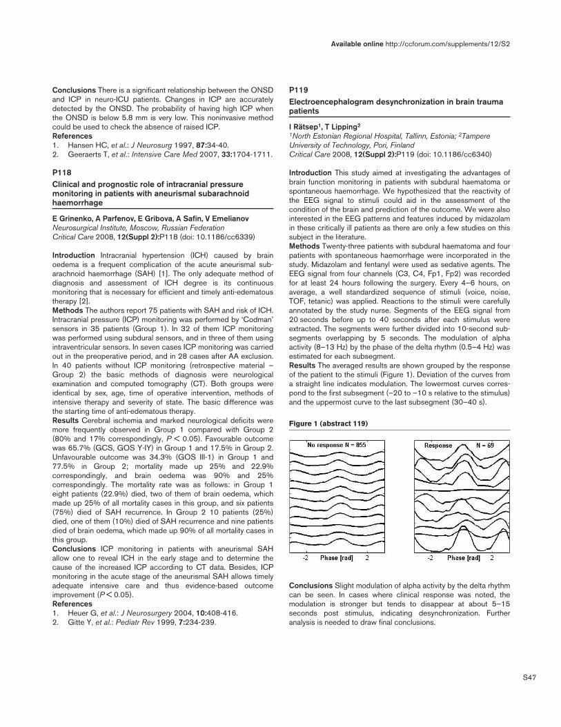
S47
Conclusions There is a significant relationship between the ONSDand ICP in neuro-ICU patients. Changes in ICP are accuratelydetected by the ONSD. The probability of having high ICP whenthe ONSD is below 5.8 mm is very low. This noninvasive methodcould be used to check the absence of raised ICP.References1. Hansen HC, et al.: J Neurosurg 1997, 87:34-40.2. Geeraerts T, et al.: Intensive Care Med 2007, 33:1704-1711.
P118Clinical and prognostic role of intracranial pressuremonitoring in patients with aneurismal subarachnoidhaemorrhage
E Grinenko, A Parfenov, E Gribova, A Safin, V EmelianovNeurosurgical Institute, Moscow, Russian FederationCritical Care 2008, 12(Suppl 2):P118 (doi: 10.1186/cc6339)
Introduction Intracranial hypertension (ICH) caused by brainoedema is a frequent complication of the acute aneurismal sub-arachnoid haemorrhage (SAH) [1]. The only adequate method ofdiagnosis and assessment of ICH degree is its continuousmonitoring that is necessary for efficient and timely anti-edematoustherapy [2].Methods The authors report 75 patients with SAH and risk of ICH.Intracranial pressure (ICP) monitoring was performed by ‘Codman’sensors in 35 patients (Group 1). In 32 of them ICP monitoringwas performed using subdural sensors, and in three of them usingintraventricular sensors. In seven cases ICP monitoring was carriedout in the preoperative period, and in 28 cases after AA exclusion.In 40 patients without ICP monitoring (retrospective material –Group 2) the basic methods of diagnosis were neurologicalexamination and computed tomography (CT). Both groups wereidentical by sex, age, time of operative intervention, methods ofintensive therapy and severity of state. The basic difference wasthe starting time of anti-edematous therapy.Results Cerebral ischemia and marked neurological deficits weremore frequently observed in Group 1 compared with Group 2(80% and 17% correspondingly, P < 0.05). Favourable outcomewas 65.7% (GCS, GOS Y-IY) in Group 1 and 17.5% in Group 2.Unfavourable outcome was 34.3% (GOS III-1) in Group 1 and77.5% in Group 2; mortality made up 25% and 22.9%correspondingly, and brain oedema was 90% and 25%correspondingly. The mortality rate was as follows: in Group 1eight patients (22.9%) died, two of them of brain oedema, whichmade up 25% of all mortality cases in this group, and six patients(75%) died of SAH recurrence. In Group 2 10 patients (25%)died, one of them (10%) died of SAH recurrence and nine patientsdied of brain oedema, which made up 90% of all mortality cases inthis group.Conclusions ICP monitoring in patients with aneurismal SAHallow one to reveal ICH in the early stage and to determine thecause of the increased ICP according to CT data. Besides, ICPmonitoring in the acute stage of the aneurismal SAH allows timelyadequate intensive care and thus evidence-based outcomeimprovement (P < 0.05).References1. Heuer G, et al.: J Neurosurgery 2004, 10:408-416.2. Gitte Y, et al.: Pediatr Rev 1999, 7:234-239.
P119Electroencephalogram desynchronization in brain traumapatients
I Rätsep1, T Lipping2
1North Estonian Regional Hospital, Tallinn, Estonia; 2TampereUniversity of Technology, Pori, FinlandCritical Care 2008, 12(Suppl 2):P119 (doi: 10.1186/cc6340)
Introduction This study aimed at investigating the advantages ofbrain function monitoring in patients with subdural haematoma orspontaneous haemorrhage. We hypothesized that the reactivity ofthe EEG signal to stimuli could aid in the assessment of thecondition of the brain and prediction of the outcome. We were alsointerested in the EEG patterns and features induced by midazolamin these critically ill patients as there are only a few studies on thissubject in the literature.Methods Twenty-three patients with subdural haematoma and fourpatients with spontaneous haemorrhage were incorporated in thestudy. Midazolam and fentanyl were used as sedative agents. TheEEG signal from four channels (C3, C4, Fp1, Fp2) was recordedfor at least 24 hours following the surgery. Every 4–6 hours, onaverage, a well standardized sequence of stimuli (voice, noise,TOF, tetanic) was applied. Reactions to the stimuli were carefullyannotated by the study nurse. Segments of the EEG signal from20 seconds before up to 40 seconds after each stimulus wereextracted. The segments were further divided into 10-second sub-segments overlapping by 5 seconds. The modulation of alphaactivity (8–13 Hz) by the phase of the delta rhythm (0.5–4 Hz) wasestimated for each subsegment.Results The averaged results are shown grouped by the responseof the patient to the stimuli (Figure 1). Deviation of the curves froma straight line indicates modulation. The lowermost curves corres-pond to the first subsegment (–20 to –10 s relative to the stimulus)and the uppermost curve to the last subsegment (30–40 s).
Conclusions Slight modulation of alpha activity by the delta rhythmcan be seen. In cases where clinical response was noted, themodulation is stronger but tends to disappear at about 5–15seconds post stimulus, indicating desynchronization. Furtheranalysis is needed to draw final conclusions.
Available online http://ccforum.com/supplements/12/S2
Figure 1 (abstract 119)
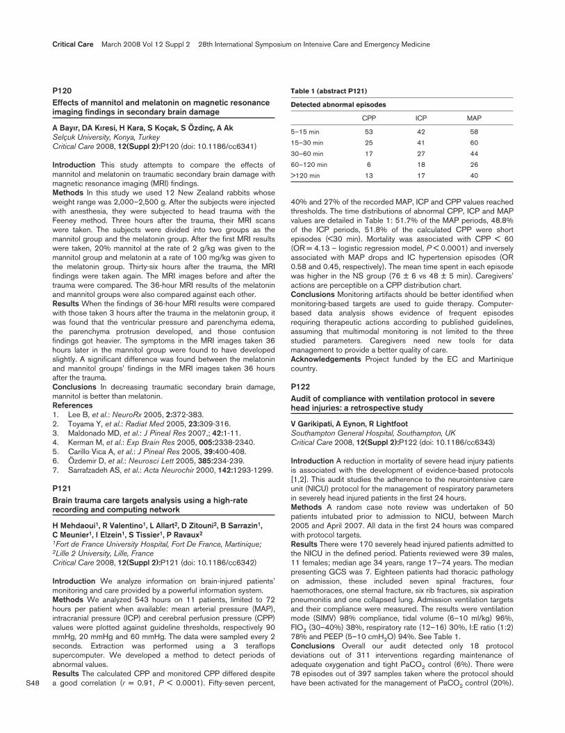
S48
P120Effects of mannitol and melatonin on magnetic resonanceimaging findings in secondary brain damage
A BayIr, DA KIresi, H Kara, S Koçak, S Özdinç, A AkSelçuk University, Konya, TurkeyCritical Care 2008, 12(Suppl 2):P120 (doi: 10.1186/cc6341)
Introduction This study attempts to compare the effects ofmannitol and melatonin on traumatic secondary brain damage withmagnetic resonance imaging (MRI) findings.Methods In this study we used 12 New Zealand rabbits whoseweight range was 2,000–2,500 g. After the subjects were injectedwith anesthesia, they were subjected to head trauma with theFeeney method. Three hours after the trauma, their MRI scanswere taken. The subjects were divided into two groups as themannitol group and the melatonin group. After the first MRI resultswere taken, 20% mannitol at the rate of 2 g/kg was given to themannitol group and melatonin at a rate of 100 mg/kg was given tothe melatonin group. Thirty-six hours after the trauma, the MRIfindings were taken again. The MRI images before and after thetrauma were compared. The 36-hour MRI results of the melatoninand mannitol groups were also compared against each other.Results When the findings of 36-hour MRI results were comparedwith those taken 3 hours after the trauma in the melatonin group, itwas found that the ventricular pressure and parenchyma edema,the parenchyma protrusion developed, and those contusionfindings got heavier. The symptoms in the MRI images taken 36hours later in the mannitol group were found to have developedslightly. A significant difference was found between the melatoninand mannitol groups’ findings in the MRI images taken 36 hoursafter the trauma.Conclusions In decreasing traumatic secondary brain damage,mannitol is better than melatonin.References1. Lee B, et al.: NeuroRx 2005, 2:372-383.2. Toyama Y, et al.: Radiat Med 2005, 23:309-316.3. Maldonado MD, et al.: J Pineal Res 2007,; 42:1-11.4. Kerman M, et al.: Exp Brain Res 2005, 005:2338-2340.5. Carillo Vica A, et al.: J Pineal Res 2005, 39:400-408.6. Özdemir D, et al.: Neurosci Lett 2005, 385:234-239.7. Sarrafzadeh AS, et al.: Acta Neurochir 2000, 142:1293-1299.
P121Brain trauma care targets analysis using a high-raterecording and computing network
H Mehdaoui1, R Valentino1, L Allart2, D Zitouni2, B Sarrazin1, C Meunier1, I Elzein1, S Tissier1, P Ravaux2
1Fort de France University Hospital, Fort De France, Martinique;2Lille 2 University, Lille, FranceCritical Care 2008, 12(Suppl 2):P121 (doi: 10.1186/cc6342)
Introduction We analyze information on brain-injured patients’monitoring and care provided by a powerful information system.Methods We analyzed 543 hours on 11 patients, limited to 72hours per patient when available: mean arterial pressure (MAP),intracranial pressure (ICP) and cerebral perfusion pressure (CPP)values were plotted against guideline thresholds, respectively 90mmHg, 20 mmHg and 60 mmHg. The data were sampled every 2seconds. Extraction was performed using a 3 teraflopssupercomputer. We developed a method to detect periods ofabnormal values.Results The calculated CPP and monitored CPP differed despitea good correlation (r = 0.91, P < 0.0001). Fifty-seven percent,
40% and 27% of the recorded MAP, ICP and CPP values reachedthresholds. The time distributions of abnormal CPP, ICP and MAPvalues are detailed in Table 1: 51.7% of the MAP periods, 48.8%of the ICP periods, 51.8% of the calculated CPP were shortepisodes (<30 min). Mortality was associated with CPP < 60(OR = 4.13 – logistic regression model, P < 0.0001) and inverselyassociated with MAP drops and IC hypertension episodes (OR0.58 and 0.45, respectively). The mean time spent in each episodewas higher in the NS group (76 ± 6 vs 48 ± 5 min). Caregivers’actions are perceptible on a CPP distribution chart.Conclusions Monitoring artifacts should be better identified whenmonitoring-based targets are used to guide therapy. Computer-based data analysis shows evidence of frequent episodesrequiring therapeutic actions according to published guidelines,assuming that multimodal monitoring is not limited to the threestudied parameters. Caregivers need new tools for datamanagement to provide a better quality of care.Acknowledgements Project funded by the EC and Martiniquecountry.
P122Audit of compliance with ventilation protocol in severehead injuries: a retrospective study
V Garikipati, A Eynon, R LightfootSouthampton General Hospital, Southampton, UKCritical Care 2008, 12(Suppl 2):P122 (doi: 10.1186/cc6343)
Introduction A reduction in mortality of severe head injury patientsis associated with the development of evidence-based protocols[1,2]. This audit studies the adherence to the neurointensive careunit (NICU) protocol for the management of respiratory parametersin severely head injured patients in the first 24 hours.Methods A random case note review was undertaken of 50patients intubated prior to admission to NICU, between March2005 and April 2007. All data in the first 24 hours was comparedwith protocol targets.Results There were 170 severely head injured patients admitted tothe NICU in the defined period. Patients reviewed were 39 males,11 females; median age 34 years, range 17–74 years. The medianpresenting GCS was 7. Eighteen patients had thoracic pathologyon admission, these included seven spinal fractures, fourhaemothoraces, one sternal fracture, six rib fractures, six aspirationpneumonitis and one collapsed lung. Admission ventilation targetsand their compliance were measured. The results were ventilationmode (SIMV) 98% compliance, tidal volume (6–10 ml/kg) 96%,FIO2 (30–40%) 38%, respiratory rate (12–16) 30%, I:E ratio (1:2)78% and PEEP (5–10 cmH2O) 94%. See Table 1.Conclusions Overall our audit detected only 18 protocoldeviations out of 311 interventions regarding maintenance ofadequate oxygenation and tight PaCO2 control (6%). There were78 episodes out of 397 samples taken where the protocol shouldhave been activated for the management of PaCO2 control (20%).
Critical Care March 2008 Vol 12 Suppl 2 28th International Symposium on Intensive Care and Emergency Medicine
Table 1 (abstract P121)
Detected abnormal episodes
CPP ICP MAP
5–15 min 53 42 58
15–30 min 25 41 60
30–60 min 17 27 44
60–120 min 6 18 26
>120 min 13 17 40
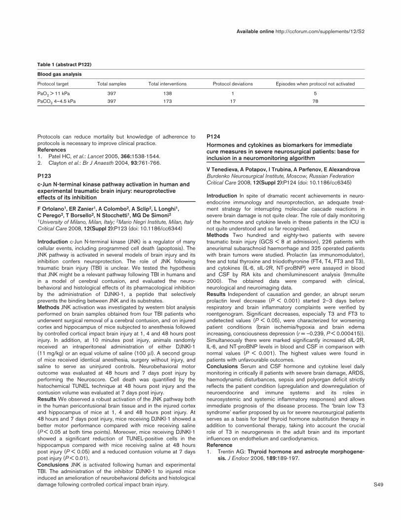
S49
Protocols can reduce mortality but knowledge of adherence toprotocols is necessary to improve clinical practice.References1. Patel HC, et al.: Lancet 2005, 366:1538-1544.2. Clayton et al.: Br J Anaesth 2004, 93:761-766.
P123c-Jun N-terminal kinase pathway activation in human andexperimental traumatic brain injury: neuroprotectiveeffects of its inhibition
F Ortolano1, ER Zanier1, A Colombo2, A Sclip2, L Longhi1, C Perego2, T Borsello2, N Stocchetti1, MG De Simoni21University of Milano, Milan, Italy; 2Mario Negri Institute, Milan, ItalyCritical Care 2008, 12(Suppl 2):P123 (doi: 10.1186/cc6344)
Introduction c-Jun N-terminal kinase (JNK) is a regulator of manycellular events, including programmed cell death (apoptosis). TheJNK pathway is activated in several models of brain injury and itsinhibition confers neuroprotection. The role of JNK followingtraumatic brain injury (TBI) is unclear. We tested the hypothesisthat JNK might be a relevant pathway following TBI in humans andin a model of cerebral contusion, and evaluated the neuro-behavioral and histological effects of its pharmacological inhibitionby the administration of DJNKI-1, a peptide that selectivelyprevents the binding between JNK and its substrates.Methods JNK activation was investigated by western blot analysisperformed on brain samples obtained from four TBI patients whounderwent surgical removal of a cerebral contusion, and on injuredcortex and hippocampus of mice subjected to anesthesia followedby controlled cortical impact brain injury at 1, 4 and 48 hours postinjury. In addition, at 10 minutes post injury, animals randomlyreceived an intraperitoneal administration of either DJNKI-1(11 mg/kg) or an equal volume of saline (100 μl). A second groupof mice received identical anesthesia, surgery without injury, andsaline to serve as uninjured controls. Neurobehavioral motoroutcome was evaluated at 48 hours and 7 days post injury byperforming the Neuroscore. Cell death was quantified by thehistochemical TUNEL technique at 48 hours post injury and thecontusion volume was evaluated at 7 days post injury.Results We observed a robust activation of the JNK pathway bothin the human pericontusional brain tissue and in the injured cortexand hippocampus of mice at 1, 4 and 48 hours post injury. At48 hours and 7 days post injury, mice receiving DJNKI-1 showed abetter motor performance compared with mice receiving saline(P < 0.05 at both time points). Moreover, mice receiving DJNKI-1showed a significant reduction of TUNEL-positive cells in thehippocampus compared with mice receiving saline at 48 hourspost injury (P < 0.05) and a reduced contusion volume at 7 dayspost injury (P < 0.01).Conclusions JNK is activated following human and experimentalTBI. The administration of the inhibitor DJNKI-1 to injured miceinduced an amelioration of neurobehavioral deficits and histologicaldamage following controlled cortical impact brain injury.
P124Hormones and cytokines as biomarkers for immediatecure measures in severe neurosurgical patients: base forinclusion in a neuromonitoring algorithm
V Tenedieva, A Potapov, I Trubina, A Parfenov, E AlexandrovaBurdenko Neurosurgical Institute, Moscow, Russian FederationCritical Care 2008, 12(Suppl 2):P124 (doi: 10.1186/cc6345)
Introduction In spite of dramatic recent achievements in neuro-endocrine immunology and neuroprotection, an adequate treat-ment strategy for interrupting molecular cascade reactions insevere brain damage is not quite clear. The role of daily monitoringof the hormone and cytokine levels in these patients in the ICU isnot quite understood and so far recognized.Methods Two hundred and eighty-two patients with severetraumatic brain injury (GCS < 8 at admission), 226 patients withaneurismal subarachnoid haemorrhage and 325 operated patientswith brain tumors were studied. Prolactin (as immunomodulator),free and total thyroxine and triiodothyronine (FT4, T4, FT3 and T3),and cytokines (IL-6, sIL-2R, NT-proBNP) were assayed in bloodand CSF by RIA kits and chemiluminescent analysis (Immulite2000). The obtained data were compared with clinical,neurological and neuroimaging data.Results Independent of causation and gender, an abrupt serumprolactin level decrease (P < 0.001) started 2–3 days beforerespiratory and brain inflammatory complaints were verified byroentgenogram. Significant decreases, especially T3 and FT3 toundetected values (P < 0.05), were characterized for worseningpatient conditions (brain ischemia/hypoxia and brain edemaincreasing, consciousness depression (r = –0.239, P < 0.000415)).Simultaneously there were marked significantly increased sIL-2R,IL-6, and NT-proBNP levels in blood and CSF in comparison withnormal values (P < 0.001). The highest values were found inpatients with unfavourable outcomes.Conclusions Serum and CSF hormone and cytokine level dailymonitoring in critically ill patients with severe brain damage, ARDS,haemodynamic disturbances, sepsis and polyorgan deficit strictlyreflects the patient condition (upregulation and downregulation ofneuroendocrine and immune systems and its roles inneurosystemic and systemic inflammatory responses) and allowsimmediate prognosis of the disease process. The ‘brain low T3syndrome’ earlier proposed by us for severe neurosurgical patientsserves as a basis for brief thyroid hormone substitution therapy inaddition to conventional therapy, taking into account the crucialrole of T3 in neurogenesis in the adult brain and its importantinfluences on endothelium and cardiodynamics.Reference1. Trentin AG: Thyroid hormone and astrocyte morphogene-
sis. J Endocr 2006, 189:189-197.
Available online http://ccforum.com/supplements/12/S2
Table 1 (abstract P122)
Blood gas analysis
Protocol target Total samples Total interventions Protocol deviations Episodes when protocol not activated
PaO2 > 11 kPa 397 138 1 5
PaCO2 4–4.5 kPa 397 173 17 78
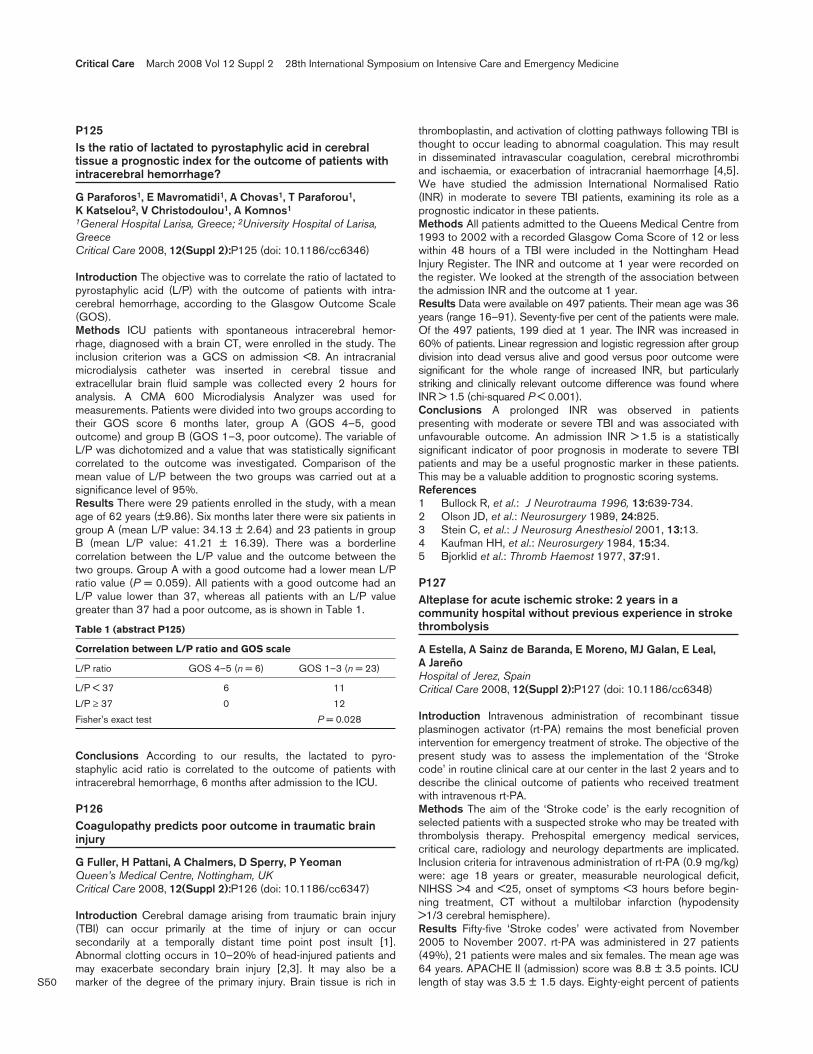
S50
P125Is the ratio of lactated to pyrostaphylic acid in cerebraltissue a prognostic index for the outcome of patients withintracerebral hemorrhage?
G Paraforos1, E Mavromatidi1, A Chovas1, T Paraforou1, K Katselou2, V Christodoulou1, A Komnos1
1General Hospital Larisa, Greece; 2University Hospital of Larisa,GreeceCritical Care 2008, 12(Suppl 2):P125 (doi: 10.1186/cc6346)
Introduction The objective was to correlate the ratio of lactated topyrostaphylic acid (L/P) with the outcome of patients with intra-cerebral hemorrhage, according to the Glasgow Outcome Scale(GOS).Methods ICU patients with spontaneous intracerebral hemor-rhage, diagnosed with a brain CT, were enrolled in the study. Theinclusion criterion was a GCS on admission <8. An intracranialmicrodialysis catheter was inserted in cerebral tissue andextracellular brain fluid sample was collected every 2 hours foranalysis. A CMA 600 Microdialysis Analyzer was used formeasurements. Patients were divided into two groups according totheir GOS score 6 months later, group A (GOS 4–5, goodoutcome) and group B (GOS 1–3, poor outcome). The variable ofL/P was dichotomized and a value that was statistically significantcorrelated to the outcome was investigated. Comparison of themean value of L/P between the two groups was carried out at asignificance level of 95%.Results There were 29 patients enrolled in the study, with a meanage of 62 years (±9.86). Six months later there were six patients ingroup A (mean L/P value: 34.13 ± 2.64) and 23 patients in groupB (mean L/P value: 41.21 ± 16.39). There was a borderlinecorrelation between the L/P value and the outcome between thetwo groups. Group A with a good outcome had a lower mean L/Pratio value (P = 0.059). All patients with a good outcome had anL/P value lower than 37, whereas all patients with an L/P valuegreater than 37 had a poor outcome, as is shown in Table 1.
Table 1 (abstract P125)
Correlation between L/P ratio and GOS scale
L/P ratio GOS 4–5 (n = 6) GOS 1–3 (n = 23)
L/P < 37 6 11
L/P ≥ 37 0 12
Fisher’s exact test P = 0.028
Conclusions According to our results, the lactated to pyro-staphylic acid ratio is correlated to the outcome of patients withintracerebral hemorrhage, 6 months after admission to the ICU.
P126Coagulopathy predicts poor outcome in traumatic braininjury
G Fuller, H Pattani, A Chalmers, D Sperry, P YeomanQueen’s Medical Centre, Nottingham, UKCritical Care 2008, 12(Suppl 2):P126 (doi: 10.1186/cc6347)
Introduction Cerebral damage arising from traumatic brain injury(TBI) can occur primarily at the time of injury or can occursecondarily at a temporally distant time point post insult [1].Abnormal clotting occurs in 10–20% of head-injured patients andmay exacerbate secondary brain injury [2,3]. It may also be amarker of the degree of the primary injury. Brain tissue is rich in
thromboplastin, and activation of clotting pathways following TBI isthought to occur leading to abnormal coagulation. This may resultin disseminated intravascular coagulation, cerebral microthrombiand ischaemia, or exacerbation of intracranial haemorrhage [4,5].We have studied the admission International Normalised Ratio(INR) in moderate to severe TBI patients, examining its role as aprognostic indicator in these patients.Methods All patients admitted to the Queens Medical Centre from1993 to 2002 with a recorded Glasgow Coma Score of 12 or lesswithin 48 hours of a TBI were included in the Nottingham HeadInjury Register. The INR and outcome at 1 year were recorded onthe register. We looked at the strength of the association betweenthe admission INR and the outcome at 1 year.Results Data were available on 497 patients. Their mean age was 36years (range 16–91). Seventy-five per cent of the patients were male.Of the 497 patients, 199 died at 1 year. The INR was increased in60% of patients. Linear regression and logistic regression after groupdivision into dead versus alive and good versus poor outcome weresignificant for the whole range of increased INR, but particularlystriking and clinically relevant outcome difference was found whereINR > 1.5 (chi-squared P < 0.001).Conclusions A prolonged INR was observed in patientspresenting with moderate or severe TBI and was associated withunfavourable outcome. An admission INR > 1.5 is a statisticallysignificant indicator of poor prognosis in moderate to severe TBIpatients and may be a useful prognostic marker in these patients.This may be a valuable addition to prognostic scoring systems.References1 Bullock R, et al.: J Neurotrauma 1996, 13:639-734.2 Olson JD, et al.: Neurosurgery 1989, 24:825.3 Stein C, et al.: J Neurosurg Anesthesiol 2001, 13:13.4 Kaufman HH, et al.: Neurosurgery 1984, 15:34.5 Bjorklid et al.: Thromb Haemost 1977, 37:91.
P127Alteplase for acute ischemic stroke: 2 years in acommunity hospital without previous experience in strokethrombolysis
A Estella, A Sainz de Baranda, E Moreno, MJ Galan, E Leal, A JareñoHospital of Jerez, SpainCritical Care 2008, 12(Suppl 2):P127 (doi: 10.1186/cc6348)
Introduction Intravenous administration of recombinant tissueplasminogen activator (rt-PA) remains the most beneficial provenintervention for emergency treatment of stroke. The objective of thepresent study was to assess the implementation of the ‘Strokecode’ in routine clinical care at our center in the last 2 years and todescribe the clinical outcome of patients who received treatmentwith intravenous rt-PA.Methods The aim of the ‘Stroke code’ is the early recognition ofselected patients with a suspected stroke who may be treated withthrombolysis therapy. Prehospital emergency medical services,critical care, radiology and neurology departments are implicated.Inclusion criteria for intravenous administration of rt-PA (0.9 mg/kg)were: age 18 years or greater, measurable neurological deficit,NIHSS >4 and <25, onset of symptoms <3 hours before begin-ning treatment, CT without a multilobar infarction (hypodensity>1/3 cerebral hemisphere).Results Fifty-five ‘Stroke codes’ were activated from November2005 to November 2007. rt-PA was administered in 27 patients(49%), 21 patients were males and six females. The mean age was64 years. APACHE II (admission) score was 8.8 ± 3.5 points. ICUlength of stay was 3.5 ± 1.5 days. Eighty-eight percent of patients
Critical Care March 2008 Vol 12 Suppl 2 28th International Symposium on Intensive Care and Emergency Medicine
S51
had vascular risk factor, 33.3% were receiving aspirin at strokeonset.Post-treatment study imaging was performed 48 hours afterthrombolysis: three patients developed CT haemorrhagic infarcttype 1 (asymptomatic small petechiae along the margins of theinfarct). Two patients died, because of cerebral infarction withcerebral edema. The median NIHSS score was 12.8 points atadmission and 10.2, 8 and 7.2 at 2 hours, 24 hours and 48 hoursafter treatment, respectively.Conclusions In selected patients rt-PA is effective when usedwithin 3 hours of stroke onset [1]. rt-PA is safe in routine clinicaluse despite limited prior experience of thrombolysis for acutestroke [2].References1. Broderick J, et al.: Stroke 2007, 38:2001-2023.2. Wahlgren N, et al.: Lancet 2007, 369:275-282.
P128Acute lung injury in a neurosciences critical care unit
RD Stevens, E Lin, RE HoeschJohns Hopkins University, Baltimore, MD, USACritical Care 2008, 12(Suppl 2):P128 (doi: 10.1186/cc6349)
Introduction Acute lung injury (ALI) may complicate neurologicalillness, but the mechanisms and outcomes of ALI in this setting arepoorly understood. We hypothesized that ALI is linked to severityof neurological illness, mechanical ventilation (MV) parameters, andoutcomes in brain-injured patients.Methods We identified consecutive patients admitted over a 2-year period to a tertiary hospital neurosciences critical care unitand requiring MV for >48 hours. ALI was determined using AECCcriteria. Univariable and multivariable predictors of ALI and ofmortality were assessed.Results We evaluated 124 patients with head trauma (34patients), intracerebral hemorrhage (29 patients), subarachnoidhemorrhage (25 patients), ischemic stroke (12 patients), and otherbrain disorders (24 patients). The primary indication for MV wasneurological (impaired consciousness, seizures, intracranial hyper-tension) in 89 patients, respiratory failure in 22 patients, surgery in10 patients, and other in three patients. ALI developed in 36patients (29%) a mean (SD) of 2.7 (1.8) days after initiation of MV.Neither ALI risk factors (pneumonia, aspiration, sepsis, trauma,transfusion, pancreatitis) or neurological insult severity (GlasgowComa Scale on admission, absence of brainstem reflexes) weresignificantly associated with ALI. Tidal volumes and positive end-expiratory pressures on days 1 and 2 of MV were not significantlydifferent in patients with and without ALI. Fifty-two patients (42%)died during hospitalization, and independent predictors of deathwere admission with intracerebral hemorrhage (OR = 4.3, 95% CI =1.5–12.2), absence of corneal reflex (OR = 5.0, 95% CI =1.2–20.0), and circulatory shock (OR = 6.2, 95% CI = 1.9–20.9).There was no independent association between ALI and mortality.Conclusions ALI developed in nearly one-third of patientsundergoing MV following either traumatic or nontraumatic braininjury. The postulated relationships between ALI and MVparameters, neurological severity of illness, and short-term mortalitywere not confirmed in this population.
P129Hemodynamic changes after hypothalamic and brain stemsurgery: interdisciplinary approach to studying
S Madorskiy, A Parfenov, A ZakharovaNeurosurgey Institute, Moscow, Russian FederationCritical Care 2008, 12(Suppl 2):P129 (doi: 10.1186/cc6350)
Introduction We discuss prognostic criteria of hemodynamicchanges in patients after hypothalamic and brain stem surgery. Wehope that better understanding of mechanisms of adaptivedisorders in local brain lesions will help to optimize postoperativemanagement of these patients. This study was based on theinterdisciplinary neurocardiologic approach [1].Methods Cardiac output measured by the echocardiographicmethod as well as hemodynamic and humoral parameters wereinvestigated in 139 patients with pituitary adenomas and cranio-pharyngiomas and in 148 patients with brain stem tumors.Results We consider that unfavorable hemodynamic changes maybe used as prognostic criteria of severe damage of regulatorycenters in the hypothalamus or brain stem. It is clear that afavorable type of hemodynamic change is a kind of postoperativestress reaction. The reaction after pituitary tumor surgery wasreduced or delayed and grew to its peak by the third day after brainstem surgery. The main unfavorable type of hemodynamics wasdecreased cardiac output (CO). However, the causes of thisdecrease are quite different. In damage of the hypothalamus,decrease of CO was connected with decreased blood volume andthe latter was connected with a decrease of vasopressin secretion.Our research has shown that patients with lesions of differentstructures of the hypothalamus and brain stem revealed specificchanges of various neurohumoral systems. In damage of thedorsomedial part of the medulla oblongata, the decease of COwas caused by primary neurogenic cardiac insufficiency. Indamage of hypothalamic structures, we see increased amplitudepower spectral density of the respiratory period of heart ratevariability (HRV), a decrease of the amplitude of the low-frequencypeak and a very high degree of coherence between HRV andrespiratory variability. In brain stem structure damage, we can seelow-frequency components only on the power spectral density ofHRV. We postulate that the revealed distinctions of power spectraldensity of HRV showed that with hemodynamic disturbance inhypothalamic and brain stem lesions a different pathological typeof cerebral regulation of hemodynamics forms.Conclusions Disorders of a humoral regulation at focal lesions ofthe hypothalamus and brain stem are specific. Intensive careshould therefore be carried out taking into account that thesechanges should be directed to regeneration of a normal humoralpattern.Reference1. Goldstein D: The Autonomic Nervous System in Health and
Disease. New York: Marcel Dekker; 2001.
P130Evaluation of development of diabetes insipidus in theearly phase following traumatic brain injury in critically illpatients
V Karali, E Massa, G Vassiliadou, I Chouris, I Rodin, M BitzaniG. Papanikolaou, Thessaloniki, GreeceCritical Care 2008, 12(Suppl 2):P130 (doi: 10.1186/cc6351)
Introduction The purpose of this study was to define theprevalence and outcome of diabetes insipidus (DI) in the earlypost-traumatic brain injury (TBI) period in ICU patients. Inadequate
Available online http://ccforum.com/supplements/12/S2
S52
antidiuretic hormone secretion, which results in DI, is a wellrecognized complication of TBI, owing to post-traumatic posteriorpituitary dysfunction.Methods This prospective study was performed in 73 ICU-TBIpatients (with or without multisystem trauma) admitted to a generalICU at a tertiary center between December 2005 and November2007. Patients had suffered severe TBI, according to the initialGCS score (≤8). DI was diagnosed if plasma sodium exceeded145 mmol/l in the presence of inappropriate dilute urine with 24-hour urine volume >30 ml/kg body weight, urine specific gravity<1,005 or urine osmolality <300 mOsm/kg with a simultaneousplasma osmolality ≥300 mOsm/kg. The age, gender, GCS, InjurySeverity Score (ISS), onset of DI, peak recorded plasma sodiumand outcome were noted. Statistical analysis was computed by ttest and Fischer exact test. P < 0.05 was considered statisticallysignificant.Results Twenty-one ICU-TBI patients (28.7%) developed acuteDI. Comparison was made between two groups of these patients:Group A, nine survivors and Group B, 12 nonsurvivors of TBI.There was no statistical significance between them with respect toage, gender (P > 0.05). Group B had a lower GCS (4.5 ± 1.5) ascompared with Group A (7.8 ± 3, P = 0.003). The ISS wassignificant greater in Group B: 38 ± 8 versus 17 ± 7 in Group A,P < 0.001. Peak plasma sodium was significantly greater in GroupB: 167 ± 4 mmol/l versus 156 ± 3 mmol/l in Group A, P < 0.05.The mean onset time of DI in Group B (1.7 ± 0.9 days) was shorterthan in Group A (7.4 ± 3.3 days), P = 0.004. Overall mortality was57.1%. The mortality rate for the development of DI within the first3 days after TBI was 90% versus 27.2% if DI occurred later.Nonsurvivors died from brain death and not as a result of theirassociated injuries.Conclusions Our results demonstrate that DI is common,following severe TBI. ICU-TBI patients presenting with features ofDI have an overall high mortality. This study shows that thedevelopment of DI within the first 3 days of TBI is associated withhigh mortality rate and impending brain death. On the contrary,ICU-TBI patients who develop DI later have a better prognosis.
P131Hypernatremia and mortality in patients with severetraumatic brain injury
E Picetti, E Antonucci, M Mergoni, A Vezzani, E Parenti, G Regolisti, U Maggiore, A Cabassi, E FiaccadoriParma University Medical School, Parma, ItalyCritical Care 2008, 12(Suppl 2):P131 (doi: 10.1186/cc6352)
Introduction Hypernatremia (HyperNa) carries on an increasedrisk of death in critically ill patients [1]. It is not known, however,whether this is true also in patients with severe traumatic braininjury (TBI).Methods We analyzed prospective data from all patients admittedfor severe TBI (GCS < 8) to a trauma ICU over a 3-year timeperiod. We collected demographics, clinical variables, complica-tions, and the available laboratory data for each day of ICU stay.Major outcomes were ICU and hospital mortality, and ICU length ofstay (LOS). We used Cox proportional-hazards regression modelswith time-dependent variates designed to reflect the exposure tothe varying sodium (Na) levels over time during the ICU stay. Thesame models were adjusted for age, gender, and Na levels atadmission as baseline covariates.Results We included in the study 130 TBI patients (mean age52 years, SD 23, range 18–96; males 74%; median GCS 3, range3–8; mean SAPS II 50, SD 14, range 9–84; all mechanicallyventilated; tracheostomy in 64/130, 49%). ICU mortality was
36/130 (27.7%), hospital mortality was 42/130 (32.3%). Follow-up included a total of 1,334 patient-days (average of 2.9 measure-ments of serum Na/day). Serum Na values were computed as thedaily average, which was 140 mmol/l (range 133–153); the patientaverage of the daily maximum Na levels was 143 mmol/l (range131–164). Twenty-six percent of the days in the ICU were compli-cated by HyperNa (that is, at least one value of Na > 145 mmol/l),with 70% of the patients showing this abnormality. The averagetime of first occurrence of HyperNa was 5 days from ICUadmission, while only five patients had HyperNa at ICU admission.A daily increase from the cumulative patient-average by 1 SD unit(about 2.4 mmol/l Na) was associated with a 2.15 times increasehazard of death (95% CI = 1.28–3.59; P = 0.004). Adjustment forthe daily use of hypertonic solutions did not change our findings.HyperNa was slightly associated with increased ICU LOS.Conclusions Our study suggests a strong relation betweenincreased Na levels and mortality in patients with severe TBI.Although these results do not prove a causal relation betweenincreased Na levels and death, we urge for interventional studies toascertain the safety of treatment strategies that might increaseserum Na levels in patients with severe TBI.Reference1. Lindner G, et al.: Am J Kidney Dis 2007, 50:952-957.
P132Gastric tubes in patients with severe brain injury
R Lyon1, GR Nimmo2
1Royal Infirmary of Edinburgh, UK; 2Western General Hospital,Edinburgh, UKCritical Care 2008, 12(Suppl 2):P132 (doi: 10.1186/cc6353)
Introduction Following severe brain injury most patients requireintubation and ventilation. Gastric tubes, whether nasogastric ororogastric, allow the stomach to be decompressed, which can aidmechanical ventilation, reduce the risk of aspiration and provide aroute for drug administration and subsequently nutrition.Methods A 4-month prospective audit was carried out on patientsadmitted to the ICU of a regional neurosurgical centre followingsevere brain injury. Patients were included following primaryintracerebral haemorrhage or traumatic brain injury.Results All patients (n = 25) were admitted to the ICU from anEmergency Department. All had a Glasgow Coma Score of 3 onadmission to the ICU and were intubated and ventilated prior toarrival. The mean time from accident to arrival in the ICU was15 hours. Only 32% of patients had a gastric tube in situ on arrivalin the ICU; 16% had a nasogastric tube and 16% had an oro-gastric tube in situ. Only 16% of patients had the gastric tubeinserted at the time of rapid sequence intubation. Thirty-fivepercent of patients who required gastric tube insertion afteradmission to the ICU had documented changes in management orcomplications as a consequence of the procedure. These includedthe need for bolus sedation and muscle relaxant use, with ensuinghypotension requiring inotrope support; delay in commencement ofenteral feeding and the need for extra chest radiographs to confirmthe tube position.Conclusions Instrumentation to pass a gastric tube may cause arise in intracranial pressure or induce hypertension, which mayprecipitate rebleeding in patients with intracerebral haemorrhage.Transfer times to regional neurosurgical units can be long. Optimalmanagement of the brain-injured patient should include insertion ofgastric tubes at the time of initial rapid sequence intubation. This isnot current practice in the emergency department and improvedawareness of the need to place gastric tubes early in brain-injuredpatients may avoid unnecessary complications.
Critical Care March 2008 Vol 12 Suppl 2 28th International Symposium on Intensive Care and Emergency Medicine
S53
P133Alcohol: a risk factor for head injury
JE Johnston, SJ McGovernNorth Tyneside General Hospital, Newcastle Upon Tyne, UKCritical Care 2008, 12(Suppl 2):P133 (doi: 10.1186/cc6354)
Introduction The study objective was to determine whether thereis a significant difference in the pattern and severity of injurysustained during falls in patients who have consumed alcohol andthose who have not. To determine how the pattern and severity ofinjury correlates with the blood alcohol level (BAL).Methods A prospective quasi-randomised controlled studybetween November 2001 and July 2002. All healthy adultsbetween 16 and 60 years old who had fallen from standing heightwere included. A systematic history and examination allowedcalculation of injury severity scores as per the abbreviated injuryscale update 1998. BALs were obtained from intoxicated patientswith consent.Results Three hundred and fifty-one healthy adult patients wereincluded in the study, there were 238 in the no alcohol group, 113had consumed alcohol, and blood alcohol levels were obtained for47 patients. The alcohol group had a higher incidence of headinjuries (46 (48%) vs 22 (9%)) with a lower incidence of limbinjuries (39 (39%) vs 183 (76%)) than the no alcohol group. Therewas a significant difference in the pattern of injury between thealcohol and no alcohol groups (χ2, P < 0.001) and there was asignificant difference in the injury severity scores (P < 0.001,Z –2.5). In the alcohol group, the severity and pattern correlatedwith the alcohol level at the time of injury. Patients with an alcohollevel <200 mg/dl had mostly soft-tissue limb injuries (58%),200–250 mg/dl mostly significant limb fractures (55%) and>250 mg/dl mostly significant head injuries (90%).Conclusions Alcohol-related falls are more often associated withsevere craniofacial injury. The severity of both limb and head injuryis greater and correlates directly with the BAL.
P134Existence of microalbuminuria during evolution of acutecoronary syndrome is a powerful short-term and long-termprognostic factor
J Garcia Acuna, E Gonzalez Babarro, A Lopez Lago, J Fernandez Villanueva, S De Lange, M Gutierrez Feijoo, J Gonzalez JuanateyHospital Clinico Universitario, Santiago de Compostela, SpainCritical Care 2008, 12(Suppl 2):P134 (doi: 10.1186/cc6355)
Introduction Microalbuminuria (MA) is considered a risk factor inthe hypertensive and diabetic population. The presence of MAduring the evolution of acute coronary syndrome (ACS) is a badprognosis criterion.Methods We studied the presence of MA by 24-hour urine test in396 hospitalized patients with ACS consecutively. During theirhospitalization period blood samples were taken in the first 24hours for all of them (leukocyte recount, hemoglobin and hemato-crit levels, troponin I, total cholesterol, LDL-cholesterol, fibrinogen,ultrasensible C-reactive protein (US-CRP), glucose andglycosylated hemoglobin (HbA1) serum levels). The left ventricularfunction was determined in all cases through echocardiography.We made a follow-up of 2.5 years.Results One hundred and forty-seven patients presented MA(37%). We found this group was also the one with older age (P =0.001), higher hypertension level (P = 0.001) and more diabetes
(P = 0.0001), strokes (P = 0.04), periferal arteriopathy (P =0.0001) and chronic renal failure (P = 0.0001) cases. Thirty-sevenpercent of patients were hospitalized in Killip >I stage (P =0.0001). This group was characterized to have poor left ventricularejection function (51% vs 46%, P = 0.001), worse renal function(P = 0.001) and higher glycemic levels (P = 0.0001). Patients withMA presented a high intrahospital mortality ratio (9% vs 4%; P =0.004), more heart failure development (45% vs 21%; P = 0.0001),atrial fibrillation (25% vs 12%; P = 0.004), abnormalities ofconduction syndromes (15% vs 7%; P = 0.02), and strokes (4%vs 1%; P = 0.02). In the follow-up, the mortality rate in the MAgroup rose to 15% (P = 0.0001). In the multivariant analysis due toage, gender, left ventricular ejection function, troponin-I serumlevels, existence of anemia and creatinine clearance, MA wasfound to be an independent risk factor of heart failure (OR = 1.75;95% CI = 1.02–3.01; P = 0.04) and of mortality (OR = 2.6; 95%CI = 1.05–6.41).Conclusions The presence of MA during evolution of ACS isassociated with high-profile vascular risk and is a powerful short-term and long-term prognostic factor.
P135Nonoperative management of blunt trauma in abdominalsolid organ: a prospective study to evaluate the successrate and predictive factors of failure
S Hashemzadeh, KH Hashemzadeh, S Resaeii, MJ Dehdilani,MZ DehdilaniTabriz University of Medical Sciences, Tabriz, IranCritical Care 2008, 12(Suppl 2):P135 (doi: 10.1186/cc6356)
Introduction Over the past several years, nonoperative manage-ment (NOM) has increasingly been recommended for the care ofselected blunt abdominal solid organ injuries. No prospective studyhas evaluated the rate of NOM of blunt abdominal trauma in thenorthwest of Iran. The objective of our study was to evaluate thesuccess rate of this kind of management in patients who do notrequire emergency surgery.Methods This prospective study was performed in Imam KhomeiniHospital (as a referral center of trauma) at Tabriz University ofMedical Sciences, Iran, between 20 March 2004 and 20 March2007. All trauma patients who had sustained injury to a solidabdominal organ (kidney, liver, or spleen) were selected for initialanalysis, using the student’s t test or chi-square test.Results During the 3 years of the study, 98 patients (83 male and15 female) with blunt trauma were selected for NOM for renal,hepatic and splenic injuries. Mean age was 26.1 ± 17.7 years(range, 2–89) and the mean injury severity score (ISS) was 14.5 ±7.4. The success rate of NOM was 93.8%. Fifty-one patients (43men, eight women; mean ISS, 14.2 ± 5.8) underwent NOM ofsplenic trauma, 38 patients (33 men, five women; mean ISS,12.9 ± 8.2) hepatic trauma, and nine patients (seven men, twowomen; mean ISS, 22.2 ± 7.6) renal trauma. Six patientsunderwent laparotomy due to the failure of NOM. The successrates of this treatment were 94.1%, 94.7% and 88.8% for thespleen, liver and kidney injuries, respectively. Female gender andISS were significant predictors of the failure of NOM (P = 0.005and P = 0.039, respectively).Conclusions We suggest that NOM can be undertakensuccessfully for the hemodynamically stable patients with solidorgan blunt trauma. The study indicates that the rates of NOM varyin relation to the severity of the organ injury. These suggest thatthis approach to the care of blunt injury in abdominal solid organsis being led by trauma centers.
Available online http://ccforum.com/supplements/12/S2
S54
P136Evidence for early presence of intestinal epithelial celldamage in multitrauma patients
J De Haan1, J Derikx1, B Relja2, T Lubbers1, MD Luyer3, WA Buurman1, JW Greve1, I Marzi21NUTRIM, Maastricht University Medical Center, Maastricht, TheNetherlands; 2University Hospital, JW Goethe University, Frankfurtam Main, Germany; 3Maasland Ziekenhuis, Sittard, The NetherlandsCritical Care 2008, 12(Suppl 2):P136 (doi: 10.1186/cc6357)
Introduction The present study investigates the presence ofintestinal epithelial cell damage in multitrauma patients on admis-sion. In trauma patients, the development of SIRS and sepsis areimportant determinants of clinical outcome. Intestinal damage isconsidered to play an important role in development of theseinflammatory syndromes. However, clinical evidence remainsscarce. Previously, in a rat model of hemorrhagic shock, wedemonstrated that interventions reducing intestinal damagestrongly attenuated the inflammatory response. In order to explorepotential applicability of such therapies in trauma patients, thepresence of early intestinal damage is assessed after trauma.Methods Trauma patients (n = 95) admitted to the emergencyroom (ER) were divided into four groups regarding the InjurySeverity Score (ISS) and the presence of abdominal injury (+AI or–AI): ISS < 25 +AI (n = 27); ISS > 25 +AI (n = 26); ISS < 25 –AI(n = 24) and ISS > 25 –AI (n = 18). Plasma was obtained directlyafter admittance to the ER. Intestinal fatty acid binding protein(I-FABP), a cytosolic protein constitutively present in matureenterocytes and released after cellular damage, was measured byELISA. Circulating procalcitonin (PCT), representing inflammation,was assessed by Kryptor assay.Results On admission, concentrations of I-FABP (1,395 ± 438 pg/ml)were significantly (P < 0.05) elevated in patients with ISS > 25+AI compared with all other groups on admission (ISS > 25 –AI:309 ± 67 pg/ml; ISS < 25 +AI: 531 ± 202 pg/ml; ISS < 25 –AI:221 ± 46 pg/ml) (MWU). Noteworthy, I-FABP was significantlyincreased in patients without AI (ISS > 25) in comparison with 76healthy volunteers (102 ± 12 pg/ml). On admission, I-FABP levelswere correlated positive with ISS (Pearson r2 = 0.28; P < 0.0001).Furthermore, I-FABP concentrations at ER correlated to PCT levelson day 1 (Pearson r2 = 0.50; P < 0.0001).Conclusions This is the first study to provide evidence for rapiddevelopment of intestinal epithelial cell damage in severe multi-trauma patients with and without abdominal trauma. The extent ofearly intestinal damage is associated with the inflammatoryresponse present at 24 hours. Further studies are needed todetermine whether therapies aimed at reduction of intestinaldamage improve clinical outcome of patients with severe trauma.
P137ICU predictors of morbidity after major trauma
F Franchi, S Scolletta, P Mongelli, E Casadei, M Cozzolino, P GiomarelliUniversity of Siena, ItalyCritical Care 2008, 12(Suppl 2):P137 (doi: 10.1186/cc6358)
Introduction ICU injured patients often experience a condition oftissue hypoperfusion due to low cardiac output and oxygendelivery (DO2). The imbalance between oxygen demand and DO2could be responsible for an anaerobic metabolism that iscorrelated with poor outcome. Several authors demonstrated thattraditional (that is, serum lactate, base deficit) and oxygen-derivedand carbon dioxide-derived parameters of anaerobiosis are helpful
indicators of bad outcome in trauma patients. We aimed to identifypredictors of morbidity in our ICU trauma patients.Methods Data for 175 adult trauma patients (age mean 50 ± 18.5years) admitted to our ICU were prospectively collected from May2006 and April 2007. Seventy hemodynamic, ventilatory, andmetabolic parameters were evaluated within 3 hours after ICUadmission. Accordingly to the GIVITI (Italian Group for theEvaluation of Interventions in ICU) database definitions, complica-tions were defined as one or more organ dysfunctions or failuresoccurring during the ICU stay. Multivariate and receiver operatingcharacteristic (ROC) curve analyses were applied.Results Morbidity was 40.5%. The Simplified Acute PhysiologyScore II, a high CO2 production (VCO2), and a low DO2/VCO2ratio were significant in the multivariate analysis (Table 1). TheDO2/VCO2 ratio was the best predictor of morbidity. Its cutoffvalue for morbidity was 3, and its area under the ROC curve was0.87 (sensitivity 82%, specificity 75%). The ICU stay was longerfor complicated patients (4.4 vs 14.5 days, P < 0.001), andmortality was higher (9% vs 22%, P < 0.001).
Table 1 (abstract P137)
Multivariate analysis results
OR 95% CI P value
DO2/VCO2 1.9 1.35–2.9 0.012
VCO2 1.7 1.2–2.3 0.03
SAPSII 1.2 1.01–2.1 0.04
Conclusions This study demonstrated that the DO2/VCO2 ratiocorrelated well with morbidity. This ratio represents the imbalancebetween oxygen demand and delivery. The ratio might becontinuously monitored in critically ill patients to assess ananaerobiosis state. This ratio together with the SAPS II ratio couldpredict complications in trauma patients.Reference1. Husain FA, et al.: Serum lactate and base deficit as predic-
tors of mortality and morbidity. Am J Surg 2003, 185:485-491.
P138Hospital mortality and length of ICU stay in severelyburned patients
S Meier1, G Kleger1, W Künzi2, R Stocker2
1Kantonsspital St Gallen, Switzerland; 2Universitätsspital Zürich,SwitzerlandCritical Care 2008, 12(Suppl 2):P138 (doi: 10.1186/cc6359)
Introduction Survival and the length of ICU stay (LOS) of severelyill or injured patients are dependent on demographic (for example,age, gender) and organizational factors as well as pre-existingdiseases and the degree of physiological abnormalities. Differentscores allow one to predict hospital mortality of general ICUpatients. Such scores (for example, APACHE II or SAPS II) aredeveloped by multivariate statistical methods. Burned patients,however, have been excluded in the development of most scoringsystems. We are interested in finding relevant risk factorsconcerning hospital mortality and LOS.Methods Patients with >10% burned surface area (BSA) admittedto the burn unit of the University Hospital Zurich between 1997and 2006 were retrospectively analysed. Relevant epidemiologicand clinical parameters were included in a univariate analysis andsubsequently in a multivariate analysis with either hospital mortalityor LOS as endpoints.
Critical Care March 2008 Vol 12 Suppl 2 28th International Symposium on Intensive Care and Emergency Medicine
S55
Results Six hundred and sixty-two burned patients were treatedbetween 1997 and 2006. Four hundred and eighty-nine patientshaving a BSA > 10% were included. One hundred and forty-one(28.8%) died and the median LOS was 19 days in survivors. Therewere no changes in overall mortality, gender distribution, surgicaltreatment ore intensive care throughout the whole study period.Conclusions We could confirm age, burned surface area, malesex, inhalation injury, diabetes mellitus and psychiatric illness of anykind as important risk factors for mortality. Additionally, suicideattempts were included in the model but did not reach statisticalsignificance. LOS in survivors was correlated with burned surfacearea, inhalation injury and the presence of suicide attempt.References1. Knaus WA, et al.: Crit Care Med 1985, 13:818-829.2, Le Gall JR, et al.: JAMA 1993, 270:2957-2963.
P139Sympathetic responses during hemorrhagic shock
A Terada, A Caricati, J Mitsunaga, L Poli-de-FigueiredoFederal University of São Paulo, BrazilCritical Care 2008, 12(Suppl 2):P139 (doi: 10.1186/cc6360)
Introduction Hemorrhagic shock is associated with adrenergicdischarge that has been linked to neurohumoral and immuneinflammatory responses, vasoconstriction and end-organ perfusiondeficits. Our goal is to characterize the sympathetic activity tohemorrhage in tissues with a rich supply of sympathetic nerves(deferens duct).Methods Seventy anesthetized male Wistar rats were submitted tofemoral artery and vein catheterization for mean arterial pressure(MAP) measurement and blood withdrawal to reach a MAP of40 mmHg. Deferens ducts were removed from rats after 10 minutes,30 minutes and 60 minutes, and were placed in isolated organbaths between two platinum electrodes for transmural electricalstimulation (TES) (0.1–20 Hz, 1 ms, 60 V). This technique allowsthe evaluation of neurotransmissors released by sympatheticnerves (noradrenaline and ATP).Results Controls maintained a MAP of 105 ± 3 mmHg in allexperimental groups. Hemorrhaged rats presented a MAP of 39 ±3 mmHg after 10, 30 or 60 minutes. The contraction profile afterATP and noradrenaline after TES were similar between controlsand hemorrhaged rats. The amplitude was greater, however, for thethree hemorrhaged groups. The addition of tetrodotoxin abolishedcontractions induced by TES, confirming the neurogenic nature ofthose contractions. The ATP-mediated contraction was blocked bythe selective P2 purinoreceptor antagonist suramin. Noradrenalin-mediated contraction was blocked by the prazosin, a selective α-adrenoreceptor.Conclusions We conclude that, based on the increased amplitudecontraction induced by both noradrenaline and ATP, sympatheticnerve activity is increased in hemorrhagic shock animals.
P140Metabolic evaluation during weaning from mechanicalventilation using indirect calorimetry
LJ Santos, SR VieiraHospital de Clínicas de Porto Alegre, BrazilCritical Care 2008, 12(Suppl 2):P140 (doi: 10.1186/cc6361)
Introduction Indirect calorimetry (IC) can be useful in theevaluation of metabolic status from critical care patients, especiallyduring weaning from mechanical ventilation (MV) when energyexpenditure can increase. The goals of this study were to compare
the energy expenditure (EE) from patients during weaning fromMV, comparing pressure support ventilation (PSV) and T tube (TT)using IC, as well as to compare these findings with resultscalculated with Harris–Benedict equation.Methods Patients clinically ready to discontinue MV support wereevaluated from August 2006 to January 2007. They were studied,in a random order, during PSV and TT. Measurements from EEwere registered during 20 minutes in both methods. Indirect calori-metry was registered using a specific metabolic monitor (Datex-Ohmeda/M-COVX). EE was also estimated using the Harris–Benedict equation with and without an activity factor. Results areshown as the mean ± standard deviation. Statistical analysis wasperformed with the paired t test, Pearson’s correlation coefficientand the Bland–Altman test. The significance level was P < 0.05.Results Forty patients were enrolled. The mean age was 56 ± 16years, APACHE II score was 23 ± 8 and the majority of patientswere male (70%). The mean EE during TT was 14.43% greaterthan during PSV (P < 0.001). The mean EE estimated by theHarris–Benedict equation was 1,455.05 ± 210.4 kcal/24 hours,and considering the activity factor 1,608 ± 236.14 kcal/24 hours.Both calculated values showed correlation with that measured byindirect calorimetry during PSV (r = 0.647) and TT (r = 0.539). Theagreement limits comparing measured and estimated EE with theBland–Altman analysis suggest that the Harris–Benedict equationunderestimates EE during TT.Conclusions Comparing EE during PSV and TT, using IC, weobserved that during TT there was, as expected, an increase in EE(14.43%). The results also suggest that the Harris–Benedictequation underestimates energy expenditure during TT.References1. Haugen HA, et al.: Indirect calorimetry: a practical guide for
clinicians. Nutr Clin Practice 2007, 22:377-388.2. Cheng CH, et al.: Measured versus estimated energy
expenditure in mechanically ventilated critically illpatients. Clin Nutr 2002, 21:165-172.
P141Hypocaloric nutrition and outcome in critically ill patientswith prolonged ICU stay
S Iff, M Leuenberger, Z Stanga, SM JakobUniversity Hospital, Bern, SwitzerlandCritical Care 2008, 12(Suppl 2):P141 (doi: 10.1186/cc6362)
Introduction While implementation of protocols for nutritionalsupport is associated with less energy deficit [1,2], the impact ofhypocaloric feeding on clinical relevant outcomes is morecontroversial: recent studies suggested both positive [3,4] andnegative effects [5] in patients receiving the recommended intakes.The aim of this study was to assess the incidence and magnitudeof hypocaloric feeding in an ICU without explicit nutrition protocolstogether with standardized mortality ratios.Methods A retrospective analysis of data from all patients staying>72 hours in a mixed medical–surgical 30-bed university hospitalICU in 2006.Results Data from 562 patients (medical 270 patients, surgical292 patients) were analyzed. The lengths of ICU and hospital staywere 9 ± 9 days and 27 ± 25 days. The age was 61 ± 16 years,weight 77 ± 17 kg, BMI 26 ± 5 kg/m2, and APACHE II and SAPSscores 24 ± 8 and 50 ± 17. Daily energy and protein intake were302 ± 33 kcal and 12 ± 1 g (recommended amount of energy andprotein intake according to the European Society of Parenteral andEnteral Nutrition: 1,549 ± 34 kcal and 114 ± 2 g). Patients weremechanically ventilated during 7 ± 8 days. ICU mortality was 14%(expected by APACHE II and SAPS II: 50% and 46%), and
Available online http://ccforum.com/supplements/12/S2
S56
hospital mortality was 22%. The total caloric deficit per patient was9,820 ± 1,126 kcal. The distribution of the acquired energy deficitwas: 0–5,000 kcal (20%), 5,000–7,500 kcal (31%), 7,500–10,000 kcal (20%), 10,000–20,000 kcal (22%), 20,000–30,000kcal (4%), >30,000 kcal (3%).Conclusions Most patients with an ICU stay >72 hours acquired asubstantial caloric deficit during the study period when comparedwith recommendations. Despite this, mortality was relatively low forthe measured APACHE II and SAPS II scores. Nutrition protocolsshould be used and their impact on both the delivered calories andclinically relevant outcome parameters be monitored.References1. Berger et al.: Nutrition 2006, 22:221-229.2. Heyland et al.: Crit Care Med 2004, 32:2260-2266.3. Villet et al.: Clin Nutr 2005, 24:502-509.4. Artinian et al.: Chest 2006, 129:960-967.5. Krishnan et al.: Chest 2003, 124:297-305.
P142Early introduction of enteral feeding for patients withpercutaneous cardiopulmonary support
H Hayami, O Yamaguchi, H Yamada, S Nagai, S Oohama, Y Sugawara, A SakuraiYokohama City University Medical Center, Yokohama, JapanCritical Care 2008, 12(Suppl 2):P142 (doi: 10.1186/cc6363)
Introduction Early enteral nutrition has been shown to have abeneficial effect on intestinal integrity and motility, immuno-competence, and patient outcome. Generally, circulatory stability isneeded for the introduction but, because there is no precisedefinition, we might miss the right time to begin. In the presentstudy we attempted to establish early enteral nutrition for patientswith cardiogenic shock with a ventricular assist device.Methods Ten postoperative patients with cardiogenic shock underpercutaneous cardiopulmonary support were included. An enteralfeeding tube was placed beyond the pylorus within 36 hours ofoperation under the observation of an upper gastrointestinalfiberscope. We estimated the mobility rate of the stomach bycounting the number of vermiculation for 3 minutes at the pylorus. Weassessed the movement of the intestine by observing the X-ray film tosee whether the contrast medium we injected 3 hours before hadmoved or not. If the medium had moved rapidly to the colon, enteralformula was started at the rate of 20 ml/hour. The serum prealbuminconcentration was measured every 7 days. Other laboratory data wascompared with five control TPN patients retrospectively.Results The mobility rate of the stomach was decreased to 4.6 ±3.2 times/3 minutes, but contrast media moved rapidly to theascending colon in two patients, to the transverse colon in threepatients, to the sigmoid colon in one patient, and to the rectum inthree patients. One patient needed to stop enteral nutritiontransiently because of reflux, but for the other nine patients enteralnutrition was well established. The prealbumin level also rose to 13± 3.5, 14.1 ± 4.9, 22 ± 2.8 weekly, but it was difficult to comparewith control TPN patients because many of them died early. SerumALP, total bilirubin, and direct bilirubin concentration 1 week afterin survivors was lower in ED patients (ALP 437 ± 248 vs 566 ±300, P = 0.57; total bilirubin 2.5 ± 2.5 vs 3.1 ± 1.0, P = 0.09; anddirect bilirubin 1.5 ± 1.8 vs 2.1 ± 0.8, P = 0.09). Seven (70%) ofthe ED patients survived over 90 days (all five patients died in theTPN group).Discussion If mesenteric circulation were stable, enteral nutritioncould not be contraindication. Even an improvement in patientoutcome can be expected in the view of avoiding complicationssuch as bacterial translocation.
Conclusions Intestinal mobility is fairly maintained in patients withcardiopulmonary support, and early enteral nutrition can beestablished under close observation.
P143Nutritional activation of the cholinergic pathway afterhemorrhagic shock reduces inflammation and preservesintestinal integrity
J De Haan1, T Lubbers1, M Hadfoune1, MD Luyer2, CH Dejong1,WA Buurman1, JW Greve1
1NUTRIM, Maastricht University Medical Center, Maastricht, TheNetherlands; 2Maasland Ziekenhuis, Sittard, The NetherlandsCritical Care 2008, 12(Suppl 2):P143 (doi: 10.1186/cc6364)
Introduction This study investigates the effects of lipid-enrichednutrition administered early after hemorrhagic shock. Previously wehave shown that high-lipid feeding effectively inhibits systemicinflammation and preserves intestinal integrity when given beforehemorrhagic shock by stimulation of the cholinergic anti-inflam-matory pathway via activation of CCK receptors. Control of theinflammatory status of trauma patients forms a major clinicalproblem since the inflammatory cascade is already ongoing uponpresentation. The anti-inflammatory effects of high-lipid interventionafter shock are therefore examined.Methods Hemorrhagic shock in rats was induced by extracting30–40% of the circulating volume. Animals were subsequentlyfasted or given enteral feedings containing high or low concen-trations of lipids at 30 and 180 minutes after shock (n = 8). CCK-receptor antagonists were administered 10 minutes before feed-ing. Tissue and plasma were collected 4 hours after shock toassess inflammation and intestinal integrity.Results Administration of lipid-enriched nutrition early after shocksignificantly reduced plasma levels of IFNγ at 4 hours (0.39 ± 0.06ng/ml) compared with low-lipid treated (0.77 ± 0.09; P < 0.01)and fasted animals (1.38 ± 0.11; P < 0.001). Enterocyte damage,expressed as circulating levels of ileal lipid binding protein, wasprevented by high-lipid feeding compared with animals thatreceived a low-lipid composition or were fasted (3.7 ± 0.3 vs 4.9 ±0.5 vs 8.0 ± 1.1 pg/ml; P < 0.05 respective P < 0.0001). Further-more, early post-shock intervention with lipid-enriched feedingsignificantly reduced translocation of bacteria to distant organs(69.7 ± 6.4 vs low lipid: 100.9 ± 9.2 CFU/g tissue; P < 0.05).Blockage of CCK receptors abrogated the anti-inflammatoryeffects of high-lipid nutrition (IFNγ 1.18 ± 0.15 vs vehicle 0.58 ±0.14 ng/ml; P < 0.05).Conclusions Administration of lipid-enriched nutrition afterhemorrhagic shock reduces inflammation and preserves intestinalintegrity. This study implicates lipid-enriched nutrition as a potentialtherapeutic option in settings in which inflammation and tissuedamage are already present, such as in trauma patients.
P144Inflammatory response in patients requiring parenteralnutrition: comparison of a new fish-oil-containing emulsion(SMOF®) versus an olive/soybean oil-based formula
I Schade, KD Röhm, A Schellhaass, A Mengistu, J Boldt, SN PiperKlinikum Ludwigshafen, GermanyCritical Care 2008, 12(Suppl 2):P144 (doi: 10.1186/cc6365)
Introduction Lipid emulsions are an essential part of parenteralnutrition (PN), such as energy supply and source of essential fattyacids. It has been shown that the composition of cell membranes is
Critical Care March 2008 Vol 12 Suppl 2 28th International Symposium on Intensive Care and Emergency Medicine
S57
influenced by the fatty acid profile of dietary lipids, and may there-fore be responsible for modulations in immune response. The aimof this study was to assess the effects of a new lipid emulsionbased on soybean oil, medium-chain triglycerides, olive oil and fishoil (SMOF®) compared with a lipid emulsion based on olive andsoybean oil (ClinOleic®) on the inflammatory response in post-operative ICU patients.Methods A prospective randomised study. After approval from theethical committee, 44 postoperative surgical patients with anindication for PN were included in this study. Nonprotein calorieswere given as 60% glucose and 40% lipid emulsion. The totalenergy intake per day was calculated as 25 kcal/kg body weight.The sedation regimen was standardized, excluding propofoladministration. Patients were thus allocated to one of two nutritionregimens: group A (n = 22) received SMOFlipid® 20%, and groupB (n = 22) a lipid emulsion based on olive and soybean oil(ClinOleic® 20%). Lipid emulsions were administered during 5 dayspostoperatively, corresponding to the observation time. IL-6, TNFα,and soluble E-selectin levels (sE-selectin) were measured before thestart of infusion (d0), at day 2 (d2) and at day 5 (d5) after the start ofadministration. The significance level was defined at P < 0.05.Results There were no significant differences between the twogroups in the inflammatory response at d0 and d2. But at d5,significantly lower IL-6 (group A: 73 ± 58 vs group B: 123 ± 107pg/ml), TNFα (group A: 15.2 ± 7.9 vs group B: 22.6 ± 12.9 pg/nl),and soluble E-selectin concentrations (group A: 21.5 ± 13.7 vsgroup B: 32.6 ± 21.2 ng/ml) were seen in patients receivingSMOF® compared with patients administered ClinOleic®.Conclusions The administration of SMOFlipid® within a PNregimen led to a significantly reduced inflammatory response atday 5 of the nutrition regimen compared with a lipid emulsionbased on olive and soybean oil, including measurements of IL-6,TNFα, and soluble E-selectin values.
P145Lipid-enriched nutrition reduces inflammation via localactivation of the autonomic nervous system bycholecystokinin
T Lubbers1, J De Haan1, M Luyer2, M Hadfoune1, C Dejong1, W Buurman1, J Greve1
1Maastricht University Medical Center, Nutrition and ToxicologyInstitute (NUTRIM), Maastricht, The Netherlands; 2MaaslandHospital, Sittard, The NetherlandsCritical Care 2008, 12(Suppl 2):P145 (doi: 10.1186/cc6366)
Introduction The present study investigates the nutritional activationof the cholinergic anti-inflammatory pathway. Lipid-enriched nutritioneffectively attenuates systemic inflammation and prevents gut barrierfailure by stimulation of the cholinergic pathway via cholecystokinin(CCK) receptors. This study investigates whether enteral lipidsactivate the autonomic nervous system via local stimulation of CCKreceptors on the afferent vagus or by activation of receptors withinthe central nervous system via circulating CCK.Methods Sprague–Dawley rats were subjected to hemorrhagicshock. Before shock, animals were fasted or fed a lipid-enrichedoral nutrition at 18 hours, 2 hours and 45 minutes. Peripheralactivation of the autonomic nervous system was determined byperforming deafferentations with perivagal application of capsaicinprior to shock. Central activation of the autonomic nervous systemby circulating levels of CCK was studied by infusion of high levelsof sulfated CCK8 starting 30 minutes prior to shock until sacrificein fasted animals. Plasma and tissue samples were collected90 minutes after shock to assess the inflammatory status and gutbarrier function.
Results Deafferentation significantly abrogated the inhibitory effect ofdietary fat on TNFα (133.7 ± 31.6 pg/ml vs 45.3 ± 12.9 pg/ml(sham); P < 0.001) and IL-6 (168 ± 14 pg/ml vs 69 ± 9 pg/ml(sham); P < 0.001). Preservation of gut barrier function was hinderedby vagal deafferentation, expressed as increased leakage of HRP inileal segments (6.1 ± 0.3 μg/ml vs 2.7 ± 0.3 μg/ml (sham); P <0.001) and bacterial translocation (113 ± 20 CFU/g tissue vs 33 ± 4CFU/g tissue (sham); P < 0.001). Infusion of sulfated CCK8 (arteriallevels: 13 ± 2 pM at shock and 19 ± 4 pM at sacrifice) failed toattenuate inflammation and improve gut barrier function.Conclusions Our study shows for the first time that lipid-enrichednutrition attenuates systemic inflammation and improves intestinalintegrity via local activation of the afferent vagus nerve. Thepresence of enteral lipids is essential to exert these protectiveeffects. Clinically, nutritional activation of this potent anti-inflam-matory pathway could provide a novel therapeutic treatment forpatients prone to develop excessive inflammation.
P146Efficacy of glutamine dipeptide-supplemented totalparenteral nutrition in critically ill patients: a prospective,double-blind randomized trial
TG Grau1, A Bonet2, E Miñambres3, L Piñeiro2, A Robles4, JA Irles5, J Acosta6, J Lopez1
1Hospital Severo Ochoa, Madrid, Spain; 2Hospital Josep Trueta,Girona, Spain; 3Hospital Marques de Valdecilla, Santander, Spain;4Hospital Vall d’Hebron, Barcelona, Spain; 5Hospital de Valme,Seville, Spain; 6Hospital General de Alicante, SpainCritical Care 2008, 12(Suppl 2):P146 (doi: 10.1186/cc6367)
Introduction The aim of this study was to assess the clinicalefficacy of glutamine dipeptide-supplemented total parenteralnutrition (TPN), defined by the occurrence of nosocomial infectionsor new organ failure as clinical endpoints.Methods Patients received a glutamine dipeptide-supplementedTPN (Glu-TPN) or a standard TPN (S-TPN). Entry criteria: adultpatients in the ICU requiring TPN for 3 days or more and APACHEII score >12. Exclusion criteria: malnutrition or obesity, chronicrenal or hepatic failure, immunocompromised patients and poor lifeexpectancy. Both groups received isonitrogenous and isocaloricTPN. Nutritional needs were calculated: 0.25 g N/kg/day and25 kcal/kg/day. The Glu-TPN group received 0.5 g/kg/day glutaminedipeptide and the S-TPN group a similar amount of amino acids.Vitals, sepsis and septic shock on admission, type of patient, dailySOFA score, daily calories administered, nosocomial infectionsbased on CDC criteria, ICU and hospital lengths of stay and ICUmortality were recorded. Intent-to-treat and per-protocol analyseswere done. Infections rates were compared using density ratesand the ΔSOFA score was analyzed using ANOVA.Results One hundred and seventeen patients received anyintervention, 53 assigned to Glu-TPN and 64 to S-TPN. Baselinecharacteristics were similar in both groups. Less new infectionsoccurred in Glu-TPN patients: nosocomial pneumonia 8.04 versus29.25 episodes-‰ days of mechanical ventilation (RR = 1.4; 95%CI = 1.2–1.7; P = 0.02), and urinary tract infections 2.5 versus16.7 episodes-‰ days of urinary catheter (RR = 1.6; 95% CI =1.3–2.1; P = 0.04). There were no differences in the incidence ofcatheter-related sepsis, primary bacteremias and intra-abdominalinfections. There was a trend to improved ΔSOFA score in patientsreceiving Glu-TPN: ΔSOFA 72 hours (1.9 ± 2.4 vs 2.6 ± 2.7, P =0.07). There were no differences in ICU and hospital lengths ofstay or ICU mortality (15% vs 18%).Conclusions Glu-TPN used in critically ill patients for longer than 3days significantly reduces the incidence of nosocomial pneu-
Available online http://ccforum.com/supplements/12/S2
S58
monias and urinary tract infections, and decreases the severity oforgan failures.
P147Arginine reduces leukocyte/endothelial cell interaction in amodel of normotensive endotoxemia without attenuatingcapillary perfusion failure
J Fertmann1, M Laschke2, B Vollmar3, MD Menger2, JN Hoffmann1
1LMU Munich, Germany; 2University of Saarland, Homburg-Saar,Germany; 3University of Rostock, GermanyCritical Care 2008, 12(Suppl 2):P147 (doi: 10.1186/cc6368)
Introduction Sepsis and septic multiorgan failure are stillassociated with a high mortality. Recent pathophysiological studiescould show that a substantial depletion of the semi-essential aminoacid arginine occurs during sepsis. However, the effects of a high-dosed supplementation of L-arginine on the microcirculation havenot been well characterised. This study addresses the effect of anintravenous L-arginine application on the microcirculation in a well-established model of normotensive endotoxemia.Methods In a dorsal skinfold chamber preparation in male Syriangolden hamsters, normotensive endotoxemia was induced byintravenous lipopolysaccharide (LPS) administration (Escherichiacoli, 2 mg/kg BW). Before and 30 minutes, 3 hours, 8 hours and24 hours after LPS application, arteriolar and venular leukocyterolling and adhesion as well as functional capillary density as aparameter of microvascular perfusion injury were quantified byintravital microscopy. In the treatment group, animals receivedintravenous L-arginine (50 mg/kg BW, n = 5) 15 minutes beforeLPS administration. Animals infused with the stereo isomer D-arginine (n = 4, 50 mg/kg BW) or sodium chloride (NaCl 0.9%,vehicle) served as controls.Results Administration of LPS markedly increased leukocyte rollingand adherence in control animals (P < 0.01 vs baseline). L-Arginineinduced a significant reduction of leukocyte rolling (P < 0.05) andadherence (P < 0.01) in postcapillary venules, whereas D-argininedid not lead to significant differences when compared with vehiclecontrols. Interestingly, despite its effect on leukocyte/endothelial cellinteraction, L-arginine did not attenuate capillary perfusion failure.Conclusions L-Arginine supplementation results in a significantreduction of LPS-induced leukocyte/endothelial cell interaction inthis in-vivo microcirculation model (dorsal skinfold chamber). Thelack of improvement in capillary perfusion has to be furthercharacterised in additional studies.
P148Antioxidant intake by intensive care patients
S Friar, S StottAberdeen Royal Infirmary, Aberdeen, UKCritical Care 2008, 12(Suppl 2):P148 (doi: 10.1186/cc6369)
Introduction Evidence shows that in critical illness antioxidantdefences are overwhelmed by a massive increase in reactiveoxygen species [1]. Antioxidant supplementation may be beneficialin these patients. We quantified antioxidant intake from enteral
nutrition by our patients and compared this with the dietaryreference value (DRV) for the healthy population [2].Methods Data were collected from a retrospective case notereview during January 2007. Patients’ volume and type of feeddelivered was recorded each day. Antioxidant intake wascalculated from the volume of feed and the feed nutritional data.Results Antioxidant intake of vitamins and traces elements wasassessed for the enterally fed patients over the first 7 days in theICU or part thereof. This amounted to 117 days of feeding. Themean intake per day and the intake as a percentage of DRV arepresented in Table 1.Conclusions There is no evidence to recommend an optimalintake of antioxidants, but doses of antioxidants used in clinicaltrials with beneficial outcomes have been up to 10–20 times theDRV [3]. Antioxidant intakes in our patients were much lower thanthis. The present audit shows that beneficial antioxidantsupplementation is unlikely to be met by standardised feeddelivery, and additional supplementation will be required.References1. Goodyear-Bruch C, et al.: Am J Crit Care 2002, 11:543-551.2. Dietary Reference Values for Food Energy and Nutrients for
the United Kingdom. Report of the Panel on Dietary Refer-ence Values of the Committee on Medical Aspects of FoodPolicy. 11th impression. London: HMSO; 2001.
3. Berger MM: Clin Nutr 2005, 24:172-183.
P149Influence of first glycemia determination in acute coronarysyndrome: long-term prognosis
E Gonzalez Babarro, J Garcia Acuna, A Lopez Lago, J Fernanadez Villanueva, S De Lange, M Gutierrez Feijoo, J Gonzalez JuanateyHospital Clinico Universitario, Santiago de Compostela, SpainCritical Care 2008, 12(Suppl 2):P149 (doi: 10.1186/cc6370)
Introduction Hyperglycemia at the moment of hospitalization isassociated with a worse prognosis in patients with acute coronarysyndrome (ACS). We introduce a study about the influence ofglycemic levels at hospital admission in patients with ACS.Methods We studied glycemic levels of 611 patients with ACS athospitalized admission consecutively. We established three groupsbased on the glycemic levels results: Group 1, <114 mg/dl; Group2, 114–163 mg/dl; Group 3, >163 mg/dl. The clinical andevolution characteristics were evaluated and we made a median of3 years follow-up.Results Group 3 presented significantly older age, higher hyper-tension levels and more diabetes, peripheral arteriopathy andchronic renal failure cases. During the follow-up we found in Group3 the worst Killip stage at the moment of hospitalization, a higherrate of heart failure (44%) and atrial fibrillation and a minor survivalrate at the end of the pursuit (Group 1, 92%; Group 2, 89% andGroup 3, 82%, P = 0.03).Conclusions High levels of glycemia at the first determination inACS patients are a long-term prognostic factor. It is necessary toknow the influence of glycemic levels for ACS prognosis when acorrect control of the level is obtained during the ACS acute phase.
Critical Care March 2008 Vol 12 Suppl 2 28th International Symposium on Intensive Care and Emergency Medicine
Table 1 (abstract P148)
Intake of antioxidants
Vitamin A Vitamin C Vitamin E Selenium Copper Zinc
Mean intake 806.9 μg 98.6 mg 13.6 mg 56.9 μg 1.7 mg 12.2 mg
Percentage of DRV 119% 248% 158% 79% 150% 137%
S59
P150Tight blood glucose control decreases surgical woundinfection in the cardiac surgical patient population in theICU
E Saad, N Shwaihet, AM Mousa, AK Kalloghlian, BA Afrane,MG Guy, CC CanverKing Faisal Specialist Hospital, Riyadh, Saudi ArabiaCritical Care 2008, 12(Suppl 2):P150 (doi: 10.1186/cc6371)
Introduction Tight blood glucose control (TBGC) results in adecrease in the infection rate in critically ill patients. In 2002 aretrospective analysis of 38 postoperative patients in our cardiacsurgical ICU revealed that most of the patients had a high serumglucose level upon arrival and remained so throughout their stayirrespective of their diabetes status. Additionally it was noted thatthe number of infections exceeded the international accepted rate.Methods Based on those findings, we initiated a prospectiveobservational study implementing a continuous insulin intravenousinfusion protocol as recommended internationally in our patients,both diabetic and nondiabetic, to achieve a blood glucose level(BGL) between 4 and 8 mmol/l. Our sample study populationincluded 116 patients, mean age 54 (±17.9) years, 65 (56%) weremales, 62 (53%) received coronary artery bypass grafting and 46(40%) were diabetic. Initially there was resistance to implementthis protocol and compliance was poor. We therefore embarked ona nursing and physician education program for more than 1 year.We initiated a new prospective study in 2006–2007. The studyincluded 270 patients, mean age 52 years (±15.8), 155 (57%)were males, 136 (50%) received coronary artery bypass graftingand 97 (36%) were diabetic.Results The demographics of the study patients were similar. Themean admission BGL, highest BGL, lowest BGL and dischargeBGL for 2003 and 2006–2007 were 8.1/13/7.9/11 mmol/l and7.8/12.8/4.6/8.3 mmol/l, respectively. A comparison of woundinfection rates before and after full implementation of TBGCshowed a decrease in the rate from 7.25% in 2002 to 3.3% in2007 (P = 0.02). The blood stream infection rate, however, did notshowed any statistical significant change, 2% in 2003 versus1.9% in 2007 (P = 0.4).Conclusions Our study showed that implementing TBGC incardiac surgical patients decreases surgical wound infection butdoes not change significantly the bloodstream infection.References1. Van den Berghe G, Wouters P, Weekers F, et al.: Intensive
insulin therapy in critically ill patients. N Engl J Med 2001,345:1359-1367.
2. Butler SO, Btaiche IF, Alaniz C: Relationship betweenhyperglycemia and infection in critically ill patients.Pharmacotherapy 2005, 25:963-976.
P151Mechanisms of kidney protection by intensive insulintherapy during critical illness
I Vanhorebeek1, B Ellger1, J Gunst1, M Boussemaere1, Y Debaveye1,N Rabbani2, P Thornalley2, M Schetz1, G Van den Berghe1
1Katholieke Universiteit Leuven, Belgium; 2University of Warwick, UKCritical Care 2008, 12(Suppl 2):P151 (doi: 10.1186/cc6372)
Introduction Strict blood glucose control with intensive insulintherapy reduces mortality and morbidity of critical illness, includingnewly acquired kidney injury [1-3].Methods To study the underlying mechanisms, we independentlymanipulated blood glucose (G) and insulin (I) to normal (N) or high
(H) levels in our rabbit model of prolonged critical illness [4],resulting in four experimental groups: NI/NG, HI/NG, NI/HG andHI/HG.Results Plasma creatinine levels were elevated in the two HGcompared with the two NG groups. Light microscopy showedsevere renal structural abnormalities in HG rabbits, with formationof tubular casts. These effects of blood glucose control on kidneyfunction and structure were not explained by an effect on bloodflow or oxygen delivery to the kidney. In contrast, in the renal cortexof HG rabbits, the activities of the mitochondrial respiratory chainenzymes were reduced to below 50% to 30% of the valuesobserved in controls and NG rabbits, a finding that was indepen-dent of insulin. No significant correlations were found betweenrespiratory chain complex activities and blood flow or oxygendelivery to the cortex. Strongly significant inverse correlations werefound between the enzyme activities and plasma levels ofcreatinine, suggesting that mitochondrial protection by intensiveinsulin therapy mediated at least part of the prevention of kidneyinjury. The glucose content in the renal cortex was more than four-fold higher in the HG than the NG groups and correlated directlywith creatinine levels and inversely with enzyme activities, suppor-ting glucose toxicity as the mediator of renal mitochondrial damage.The dicarbonyls glyoxal, methylglyoxal and 3-deoxyglucosone wereelevated in plasma of the HG groups and strongly correlated withglucose in the cortex and plasma creatinine, suggesting a possiblecontribution of these toxic metabolites of glucose.Conclusions Intensive insulin therapy during critical illness confersrenal protection by prevention of hyperglycemia-inducedmitochondrial damage rather than by improving perfusion andoxygen delivery.References1. Van den Berghe et al.: N Engl J Med 2001, 345:1359.2. Van den Berghe et al.: N Engl J Med 2006, 354:449.3. Schetz M, et al.: J Am Soc Nephrol, in press.4. Ellger et al.: Diabetes 2006, 55:1096.
P152Complement activation after uncomplicated coronaryartery bypass grafting: role of strict glucose control
C Hoedemaekers1, M Van Deuren1, T Sprong1, P Pickkers1, TE Mollnes2, I Klasen1, J Van derHoeven1
1Radboud University Nijmegen Medical Centre, Nijmegen, TheNetherlands; 2Rikshospitalet, Oslo, NorwayCritical Care 2008, 12(Suppl 2):P152 (doi: 10.1186/cc6373)
Introduction The complement system is a key component in theSIRS response after cardiac surgery. The aim of this study was toinvestigate whether strict glucose control modifies complementactivation in this setting and to analyze the route of complementactivation.Methods We performed a randomized trial in 20 adult patientsafter coronary artery bypass grafting (CABG). Patients wereassigned to receive intensive or conventional insulin treatmentimmediately after admission to the ICU. Components of thecomplement system were determined by ELISA. Changes incomplement levels over time were analyzed with one-way ANOVArepeated measures.Results Blood glucose levels were significantly lower in theintensive treatment group (P < 0.003). Serum concentrations ofterminal complement complex were increased on admission to theICU in both groups (2.80 ± 1.45 AU/ml vs 3.21 ± 2.17 AU/ml, P =0.817) and declined significantly thereafter. All complementactivation pathways converge at the point of C3 activation. TheC3bc concentration was strongly increased on admission in both
Available online http://ccforum.com/supplements/12/S2
S60
groups (78.9 ± 36.7 AU/ml vs 103.4 ± 68.0 AU/ml, P = 0.355)and declined in the following hours with a second peak at 8 hoursafter admission (P = 0.005). C3bBbP (alternative pathwayactivation) was increased on admission in both groups (106.44 ±42.72 AU/ml vs 144.44 ± 73.51 AU/ml respectively, P = 0.199),followed by a significant decline in the following hours (P < 0.001).C1rs–C1inh complexes (classical pathway) were increased onadmission in both groups (38.00 ± 12.27 AU/ml vs 40.78 ± 16.41AU/ml, P = 0.690), followed in time by a gradual decrease andlater by an increase (P < 0.001). No differences in C4bc(combined classical and lectin pathway) concentrations weremeasured between the treatment groups, and the concentrationsremained constant during ICU stay. MBL (lectin pathway)concentrations were comparable in both treatment groups and didnot change significantly during the 24-hour follow-up.Conclusions Strict glucose regulation does not alter theconcentration of complement components or route of activation.Complement activation after CABG shows a biphasic pattern.Initially complement is activated trough the classical/lectin pathwayand augmented by the alternative pathway. In a second phase,complement is activated by the classical/lectin pathway to thepoint of C3b formation without production of terminal complementcomplexes, indicating inhibition beyond C3b.
P153Relationship between admission blood glucose level andprognosis in acute ischemic and hemorrhagic strokepatients
A BayIr, S Özdinç, A Ak, B Cander, F KaraSelçuk University, Konya, TurkeyCritical Care 2008, 12(Suppl 2):P153 (doi: 10.1186/cc6374)
Introduction The aim of this study was to investigate therelationship between blood glucose level measured on admissionand hospital mortality with the Glasgow Coma Score (GCS) inischemic and hemorrhagic stroke patients.Methods Those patients who experienced ischemic andhemorrhagic stroke and who arrived at the hospital within the first3 hours after the beginning of the symptoms were included in thestudy. On arrival the GCS was detected. Blood glucose levelswere determined for each patient. The patients were allocated asischemic and hemorrhagic stroke groups on admission. In addition,ischemic and hemorrhagic stroke groups were allocated as GCS≤ 8 and GCS ≥ 9 groups. The patients were observed in terms ofmortality during their stay in the hospital. The data were comparedusing Kruskal–Wallis variance analysis and the Mann–Whitney Utest with Bonferroni correction. P ≤ 0.05 was considered significant.Results We enrolled 113 patients (26 hemorrhagic, 87 ischemicstroke) in the study. The mean blood glucose level in the ischemicstroke and GCS ≤ 8 group (25 patients) was 189 ± 69.23 mg/dl onadmission. The mean blood glucose level for the ischemic stroke andGCS ≥ 9 group (62 patients) was 165 ± 79.8 mg/dl. The meanblood glucose level of the hemorrhagic stroke and GCS ≤ 8 group(16 patients) was 291.7 ± 162.63 mg/dl. On admission, the meanblood glucose level of the hemorrhagic stroke and GCS ≥ 9 group(10 patients) was 141.8 ± 35.46 mg/dl. The mean blood glucoselevel of dead patients (n = 35) was 236.25 ± 128.88 mg/dl. Asignificant reverse relationship was found between GCS and bloodglucose level (P = 0.00). A significant reverse relationship was foundbetween GCS and blood glucose level for dead patients (P = 0.00).Conclusions In patients with ischemic and hemorrhagic strokewho referred to the emergency clinic within the first 3 hours afterthe stroke developed, a measured high glucose level on admissioncould be an indicator of bad prognosis and high hospital mortality.
References1. Perttu J, et al.: Stroke 2004, 35:363-364.2. Martini SR, et al.: J Cerebral Blood Flow Metab 2007, 27:
435-451.
P154Significance of the suppression of blood glucose variabilityin acutely ill severe patients with glucose intoleranceevaluated by means of bedside-type artificial pancreas
M Hoshino1, Y Haraguchi2, I Mizushima3, S Kajiwara1, M Takagi11Shisei Hospital, Saitama, Japan; 2National Hospital OrganizationDisaster Medical Center, Tokyo, Japan; 3Tokyo Police Hospital,Tokyo, JapanCritical Care 2008, 12(Suppl 2):P154 (doi: 10.1186/cc6375)
Introduction We hereby report the usefulness of continuous useof an artificial pancreas (AP) to control blood glucose (BG)clinically. In this report we analyzed the significance of BG stability,or variability under strict control of BG, by the use of an AP.Methods BG control was performed by an AP, STG22. Patientswere evaluated at early (E) phase and late (L) phase (1 week afterthe E phase). Based on the daily mean BG (BGm), basicallycalculated by 24 data obtained hourly, patients were classified intotwo groups. Patients with BGm <200 mg/dl and those with BGm>200 mg/dl were denoted as group B and group A, respectively.Each group was classified into two subgroups based on the dailyBG difference (BGd), or 100 mg/dl, high and low variabilitysubgroups. Group B patients with BGd <100 mg/dl were denotedBL, and group B patients with BGd >100 mg/d as BH. SubgroupsAL and AH were classified similarly. The parameters studied wereBGm, BGd, SOFA score and mortality.Results (1) Group A had BGm in the E phase and L phase of 231± 24 (n = 11) and 220 ± 19 (n = 7), respectively. Group B hadBGm in the E phase and L phase of 175 ± 19 (n = 35) and 166 ±21 (n = 42), respectively. (2) Relationship between BGm andBGd: (E phase) group A had the tendency of higher BGd ascompared with group B (101 ± 60 vs 68 ± 46, P < 0.10); (Lphase) group A had significantly higher BGd as compared withgroup B (109 ± 43 vs 66 ± 46, P < 0.025). (3) Relationshipsbetween BGd and SOFA score, mortality: (E phase) group AH hadthe tendency of higher mortality as compared with group AL(100%, n = 3 vs 50%, n = 8); (L phase) group BH had thetendency of higher SOFA score and mortality as compared withgroup BL (8.0 ± 6.7, 80%, n = 5 vs 6.7 ± 5.7, 38%, n = 37).Conclusions Although this is a preliminary study, based on theprecise data measured by the AP, the following conclusions weresuggested. High BG variability or unstability supported highmorbidity and mortality. BG control aimed at the suppression ofBG variability, or BGd lower than 100 mg/dl, may thereforeimprove the outcome as well as the improvement of BGm.
P155Implementing intensive insulin therapy in daily practicereduces the incidence of critical illness polyneuropathyand/or myopathy
N Berends, G Hermans, B Bouckaert, P Van Damme, M Schrooten, W De Vooght, P Wouters, G Van den BergheKU Leuven, BelgiumCritical Care 2008, 12(Suppl 2):P155 (doi: 10.1186/cc6376)
Introduction In two randomised controlled trials (RCTs) on theeffect of intensive insulin therapy (IIT) in a surgical ICU (SICU) and
Critical Care March 2008 Vol 12 Suppl 2 28th International Symposium on Intensive Care and Emergency Medicine
S61
a medical ICU (MICU), IIT reduced the incidence of critical illnesspolyneuropathy and/or myopathy (CIP/CIM) and the need forprolonged mechanical ventilation (MV ≥ 14 days). Here we investi-gated whether these effects are present in daily practice when IITis implemented outside a study protocol.Methods We retrospectively studied all electronically availableelectrophysiological data (electroneuromyography (ENMG)) frompatients in the SICU and MICU before and after implementation ofIIT in routine practice (omitting data obtained during the twoRCTs). All ENMGs were performed because of clinical weaknessand/or weaning failure. As in the RCTs, CIP/CIM was diagnosedby the presence of abundant spontaneous electrical activity(fibrillation potentials or positive sharp waves). Baseline and out-come variables were compared using Student’s t test, chi-squaretest or Mann–Whitney U test when appropriate. The effect ofimplementing IIT on CIP/CIM and prolonged MV were assessedusing univariate analysis and multivariate logistic regression analysis(MVLR) correcting for baseline and ICU risk factors.Results ENMGs were performed in 193 long-stay ICU patientsbefore and 494 after implementing IIT. This population comprised4.6% of all patients before and 5.6% after IIT implementation in theMICU and 4.0% before and 3.9% of all patients after IITimplementation in the SICU. With IIT, mean glycemia wassignificantly lowered (median 142 (130–153) to 106 mg/dl(100–113)). IIT implementation significantly reduced ENMGdiagnosis of CIP/CIM in this population (71.6% to 48.7% (P <0.0001)). MVLR identified implementing IIT as an independentprotective factor (P < 0.0001, OR = 0.24 (95% CI = 0.14–0.43)).MVLR confirmed the independent protective effect of IIT onprolonged MV (P = 0.03, OR = 0.55 (95% CI = 0.31–0.95)). Thiseffect was explained by the reduction in CIP/CIM (P = 0.009,OR = 1.13 (95% CI = 1.65–2.42)).Conclusions Implementing IIT in daily practice evokes a similarbeneficial effect on neuromuscular function, as observed in twoRCTs. IIT significantly improves glycemic control and significantlyand independently reduces the electrophysiological incidence ofCIP/CIM. This reduction explains the beneficial effect of IIT-prolonged MV.References1. Van den Berghe G, et al.: Neurology 2005, 64:1348-1353.2. Hermans G, et al.: Am J Respir Crit Care Med 2007, 175:
480-489.3. Van den Berghe G, et al.: N Engl J Med 2001, 345:1359-
1367.4. Van den Berghe G, et al.: N Engl J Med 2005, 354:449-461.
P156Evaluation of the implementation of a fully automatedalgorithm (eMPC) in an interacting infusion pump systemfor the establishment of tight glycaemic control in medicalICU patients
J Plank1, R Kulnik1, C Pachler1, R Hovorka2, D Röthlein3, N Kachel3, M Wufka3, K Smolle1, S Perl1, R Zweiker1, T Pieber1,M Ellmerer1
1Medical University Graz, Austria; 2Addenbrooke´s Hospital,Cambridge, UK; 3B. Braun Melsungen AG, Melsungen, GermanyCritical Care 2008, 12(Suppl 2):P156 (doi: 10.1186/cc6377)
Introduction The purpose of this study was to investigate theperformance of a newly developed prototype decision supportsystem for the establishment of tight glycaemic control in patientsin the medical ICU for a period of 72 hours.Methods The study was conducted as a single-center, open,noncontrolled clinical investigation in 10 mechanically ventilated
patients at the Medical University Graz. After admittance to theICU, arterial blood glucose values were monitored and the CS-1Decision Support System (interacting infusion pumps withintegrated algorithm eMPC and user interface) was used to adjustthe infusion rate of intravenously administered human solubleinsulin to normalize arterial blood glucose. The efficacy and safetywere assessed by calculating the percentage within the targetrange (4.4–6.1 mM), the hyperglycaemic index (HGI), mean glucoseand the number of hypoglycaemic episodes (<2.2 mM).Results The percentage of readings within the target range was47.0% (±13.0). The average blood glucose concentration and HGIwere 6.08 mM (±0.73) and 0.54 mM (±0.52), respectively. Nohypoglycaemic episode (<2.2 mM) was detected. Severaltechnical malfunctions of the device, such as repetitive errormessages and missing data in the data log owing to com-munication problems between the new hardware components, areshortcomings of the present version of the device. Owing to thesetechnical failures of system integration, treatment had to bestopped ahead of schedule in three patients.Conclusions For the first time a decision support system fullyintegrated into an infusion pump system was tested clinically.Despite technical malfunctions, the performance of this prototypesystem of the CS-1 Decision Support System device was, from aclinical point of view, already effective in maintaining tightglycaemic control. Accordingly, and with technical improvementrequired, the CS1 system has the capacity to be further improvedin the next phase of the development process and to serve as areliable tool for routine establishment of glycaemic control forcritically ill patients.
P157Tight glucose control by intensive insulin therapy inBelgian ICU: an evaluation of practice
JC Preiser1, T Sottiaux2
1University Hospital Centre of Liege, Belgium; 2Notre-Dame deGrâce Hospital, Gosselies, BelgiumCritical Care 2008, 12(Suppl 2):P157 (doi: 10.1186/cc6378)
Introduction Recent data suggest that a tight glucose control byintensive intensive therapy (TGCIIT) may improve survival ofcritically ill patients. The optimal target for blood glucose (BG),however, is a matter of debate and controversy, and the constraintsassociated with the implementation of TGCIIT are considerable.Methods The present study surveyed the current practice ofglucose management. We sent a multiple-choice questionnaire to120 Belgian ICUs, on behalf of the Belgian Federal Board forIntensive Care.Results Fifty-two ICUs (43%) answered. A total of 489 patientswere staying in the ICUs when the questionnaire was filled. Thenumber of glucometers per ICU bed averaged 0.6 ± 0.4 and thenurse/patient ratio averaged 0.6 ± 0.3. Glucose control is felt to bean important issue by all participants, while 96% are aware of theresults of the landmark 2001 study of Van den Berghe andcolleagues. Ninety percent changed their practice following thisstudy. Fifty percent of the responders use TGCIIT for every patient,while others restricted TGCIIT to long-stayers, septic or diabeticpatients. An algorithm is used for glucose control by 98% of theparticipants. The BG target is 80–110 mg/dl and 110–140 mg/dlfor 27% and for 56% of the responders, respectively. BG ischecked systematically two to eight times per day (five to eighttimes for 54%), on blood and capillary samples. Prior to theachievement of the target BG, checks are performed hourly (60%)or every 2 hours (28%). Once the target BG is reached, checksare performed six times (45%) to 12 times (36%) a day. The
Available online http://ccforum.com/supplements/12/S2
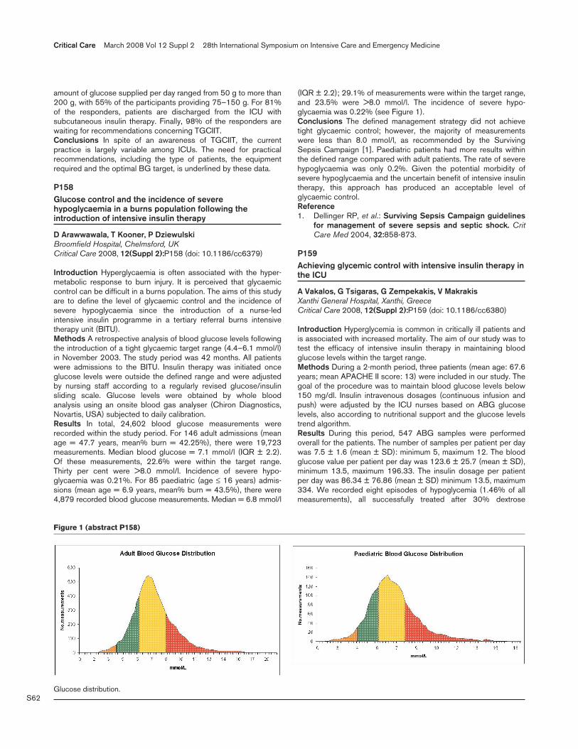
S62
amount of glucose supplied per day ranged from 50 g to more than200 g, with 55% of the participants providing 75–150 g. For 81%of the responders, patients are discharged from the ICU withsubcutaneous insulin therapy. Finally, 98% of the responders arewaiting for recommendations concerning TGCIIT.Conclusions In spite of an awareness of TGCIIT, the currentpractice is largely variable among ICUs. The need for practicalrecommendations, including the type of patients, the equipmentrequired and the optimal BG target, is underlined by these data.
P158Glucose control and the incidence of severehypoglycaemia in a burns population following theintroduction of intensive insulin therapy
D Arawwawala, T Kooner, P DziewulskiBroomfield Hospital, Chelmsford, UKCritical Care 2008, 12(Suppl 2):P158 (doi: 10.1186/cc6379)
Introduction Hyperglycaemia is often associated with the hyper-metabolic response to burn injury. It is perceived that glycaemiccontrol can be difficult in a burns population. The aims of this studyare to define the level of glycaemic control and the incidence ofsevere hypoglycaemia since the introduction of a nurse-ledintensive insulin programme in a tertiary referral burns intensivetherapy unit (BITU).Methods A retrospective analysis of blood glucose levels followingthe introduction of a tight glycaemic target range (4.4–6.1 mmol/l)in November 2003. The study period was 42 months. All patientswere admissions to the BITU. Insulin therapy was initiated onceglucose levels were outside the defined range and were adjustedby nursing staff according to a regularly revised glucose/insulinsliding scale. Glucose levels were obtained by whole bloodanalysis using an onsite blood gas analyser (Chiron Diagnostics,Novartis, USA) subjected to daily calibration.Results In total, 24,602 blood glucose measurements wererecorded within the study period. For 146 adult admissions (meanage = 47.7 years, mean% burn = 42.25%), there were 19,723measurements. Median blood glucose = 7.1 mmol/l (IQR ± 2.2).Of these measurements, 22.6% were within the target range.Thirty per cent were >8.0 mmol/l. Incidence of severe hypo-glycaemia was 0.21%. For 85 paediatric (age ≤ 16 years) admis-sions (mean age = 6.9 years, mean% burn = 43.5%), there were4,879 recorded blood glucose measurements. Median = 6.8 mmol/l
(IQR ± 2.2); 29.1% of measurements were within the target range,and 23.5% were >8.0 mmol/l. The incidence of severe hypo-glycaemia was 0.22% (see Figure 1).Conclusions The defined management strategy did not achievetight glycaemic control; however, the majority of measurementswere less than 8.0 mmol/l, as recommended by the SurvivingSepsis Campaign [1]. Paediatric patients had more results withinthe defined range compared with adult patients. The rate of severehypoglycaemia was only 0.2%. Given the potential morbidity ofsevere hypoglycaemia and the uncertain benefit of intensive insulintherapy, this approach has produced an acceptable level ofglycaemic control.Reference1. Dellinger RP, et al.: Surviving Sepsis Campaign guidelines
for management of severe sepsis and septic shock. CritCare Med 2004, 32:858-873.
P159Achieving glycemic control with intensive insulin therapy inthe ICU
A Vakalos, G Tsigaras, G Zempekakis, V MakrakisXanthi General Hospital, Xanthi, GreeceCritical Care 2008, 12(Suppl 2):P159 (doi: 10.1186/cc6380)
Introduction Hyperglycemia is common in critically ill patients andis associated with increased mortality. The aim of our study was totest the efficacy of intensive insulin therapy in maintaining bloodglucose levels within the target range.Methods During a 2-month period, three patients (mean age: 67.6years; mean APACHE II score: 13) were included in our study. Thegoal of the procedure was to maintain blood glucose levels below150 mg/dl. Insulin intravenous dosages (continuous infusion andpush) were adjusted by the ICU nurses based on ABG glucoselevels, also according to nutritional support and the glucose levelstrend algorithm.Results During this period, 547 ABG samples were performedoverall for the patients. The number of samples per patient per daywas 7.5 ± 1.6 (mean ± SD): minimum 5, maximum 12. The bloodglucose value per patient per day was 123.6 ± 25.7 (mean ± SD),minimum 13.5, maximum 196.33. The insulin dosage per patientper day was 86.34 ± 76.86 (mean ± SD) minimum 13.5, maximum334. We recorded eight episodes of hypoglycemia (1.46% of allmeasurements), all successfully treated after 30% dextrose
Critical Care March 2008 Vol 12 Suppl 2 28th International Symposium on Intensive Care and Emergency Medicine
Figure 1 (abstract P158)
Glucose distribution.
S63
infusion. Within the target range were 427 blood glucose levels(78.06%), while the higher glucose values were associated withthe initial hyperglycemia correction. The regration between glucosevalues and insulin dosage was not linear but rather polynomial,while the higher values of insulin dosage correlated with both thehigher and the lower glucose levels.Conclusions The blood glucose level target is difficult to achievewith intensive insulin therapy in a population of ICU patients withhigh severity score on admission. In our study, the glycemic controltarget below 150 mg/dl was achieved in more than two-thirds ofmeasurements using a high insulin dosage. On the other hand, therate of hypoglycemia was high in our study (1.46%), probablybecause of application failure of the insulin dosage algorithmduring nutritional interruption. We suggest that application of anintensive insulin therapy protocol adjusted to nutritional supportand the glucose levels trend will achieve glycemic control in clinicalpractice, while minimizing the risk of hypoglycemia.
P160Intensive insulin therapy: protocols in use in The Netherlands
MJ De Graaff1, H Kieft2, JP Van der Sluijs3, AA Royakkers4, JC Korevaar1, PE Spronk5, MJ Schultz1
1Academic Medical Centre, Amsterdam, The Netherlands; 2IsalaHospitals, Zwolle, The Netherlands; 3Medical Centre Haaglanden,The Hague, The Netherlands; 4Tergooi Hospitals, Blaricum, TheNetherlands; 5Gelre Hospitals, Apeldoorn, The NetherlandsCritical Care 2008, 12(Suppl 2):P160 (doi: 10.1186/cc6381)
Introduction Intensive insulin therapy (IIT) reduces mortality andmorbidity of critically ill patients [1]. The ‘original IIT protocol’ asused by the group of van den Berghe (Leuven, Belgium) is a simpletext-based protocol aiming for blood glucose values between 4.4and 6.1 mmol/l. We conducted a postal survey amongst intensivecare physicians and nurses in February 2007. As part of thissurvey, respondents were asked to send in a copy of their protocolon glycemic control (GC).Methods All Dutch ICUs with ≥5 beds available for mechanicalventilation received a questionnaire on GC policies, in particularthresholds for blood glucose values to start insulin, and the targetsof GC. Respondents were explicitly asked to send in their GCprotocol too, when available.Results Of 71 ICUs responding to the questionnaire, 46 (65%)sent in their GC protocol. Formats of the glucose control protocolvaried widely, four different types of protocol formats could berecognized: ‘flow chart’ based (n = 17), ‘sliding scales’ based (n =16), ‘text’ based (n = 7), and ‘others’ (n = 5). In three ICUs the GCprotocol was computer based. In only 11 GC protocols (24%)were blood glucose targets between 4.4 and 6.1 mmol/l. In themajority of GC protocols (87%), the lower target for blood glucosewas <4.5 mmol/l; in only one-half of GC protocols (43%), theupper target for blood glucose was <6.1 mmol/l. In four GCprotocols, the thresholds for starting insulin were unclear.Conclusions There is large variability in the presently used GSprotocols in The Netherlands. In only 24% did the GC-protocoltargets reflect those of the original IIT protocol as used by van denBerghe.Reference1. van den Berghe G, et al.: N Engl J Med 2001, 345:1359-
1367.
P161Serum insulin-like growth factor binding protein 1 andC-peptide to assess insulin resistance in septic patients
C Chelazzi, AR De GaudioUniversity of Florence, ItalyCritical Care 2008, 12(Suppl 2):P161 (doi: 10.1186/cc6382)
Introduction Insulin resistance and hyperglycemia are importantfeatures of the care of the critically ill. They are part of themetabolic pathophysiology of acute conditions [1,2]. In septicpatients, insulin resistance is linked to the synergic effect ofcytokines, bacterial products and catecholamines [3]. Laboratoryfindings include elevated C-peptide and serum insulin-like growthfactor binding protein 1 (IGFBP-1) [4]. This is secreted by the liverand its secretion is inhibited by insulin. In the insulin-resistantpatient, a rise in insulin fails to reduce its plasma levels. We dosedserum IGFBP-1 in samples of patients with sepsis and compared itwith glycemia and serum C-peptide.Methods Only patients with a definite diagnosis of sepsis wereincluded. Five blood samples were taken from each patient at thetime of their entrance and every 24 hours for the next 96 hours. Foreach sample, glycemia, C-peptide and IGFBP-1 were dosed andtheir values compared.Results C-peptide levels constantly remained high, even innormoglycemic patients. Higher glycemias were associated withraised serum IGFBP-1 levels. Insulin increases could not inhibitIGFBP-1 and, hence, the worse the insulin resistance, the higherthe glycemia, the higher the IGFBP-1. The relation between C-peptide and IGFBP-1 showed that higher levels of circulatinginsulin were associated with higher levels of IGFBP-1. This wasthought as a direct sign of the existing insulin-resistant state.Conclusions IGFBP-1 can be used to asses insulin resistance inseptic critical patients, particularly compared with glycemia. Itscorrelation with C-peptide might better define the severity of insulinresistance and, thus, of the underlying sepsis.References1. Vincent JL: Metabolic support in sepsis and multiple organ
failure: more questions than answers. Crit Care Med 2007,35(Suppl):S436-S440.
2. Vanhorebeek I, et al.: Tight blood glucose control withinsulin in the ICU: facts and controversies. Chest 2007,132:268-278.
3. Turina M, et al.: Acute hyperglicemia and the innateimmune system: clinical, cellular and molecular aspects.Crit Care Med 2005, 33:1624-1633.
4. Langouche L, et al.: Effect of intensive insulin therapy oninsulin sensitivity in the critically ill. J Clin Endocrinol Metab2007, 92:3890-3897.
P162Insulin increases deformation-induced lung cell death
B Wilcox, N Vlahakis, G LiuMayo Clinic Rochester, MN, USACritical Care 2008, 12(Suppl 2):P162 (doi: 10.1186/cc6383)
Introduction With insulin use increasing to treat ICU hyper-glycemia, we wished to determine whether insulin is cytoprotectivein the presence of deformation-induced lung cell injury.Methods A549 epithelial cells were grown in full growth media for 24hours on deformable six-well culture plates coated with type 1collagen. Cells were then grown in glucose concentrations of1.26 g/l, 4.26 g/l and 7.26 g/l for an additional 24 hours. After2 hours of serum starvation, cells were exposed to 100 nM insulin for
Available online http://ccforum.com/supplements/12/S2
S64
30 minutes, followed by stretching for 2 minutes at 8 Hz, 30% strainamplitude and 140%/s strain velocity in the presence of 1% FITCsolution, a marker of plasma membrane injury and reseal. Followingstretching, the cells were incubated with 1% PI, a marker of celldeath, with quantification of death performed by confocal microscopy.Results In undeformed cells the rate of cell death was 0.8%, 0.5%and 1.2% at 1.26 g/l, 4.26 g/l and 7.26 g/l glucose, respectively.In undeformed cells treated with 100 nM insulin, cell death was0.2%, 1.3% and 2.1%. Deformation increased the percentage ofcellular death to 3%, 5% and 2% compared with undeformedcells. Deformation increased the percentage of death in cellstreated with 100 nM insulin to 15%, 15%, and 8% as comparedwith undeformed cells treated with insulin and deformed cellsgrown in glucose alone. Cell death did not differ from control atinsulin concentrations ranging up to 100 nM but doubled andplateaued, with concentrations of 100–300 nM.Conclusions Insulin increases deformation-induced death in lungcells. This is consistent with a previously published multivariateanalysis of patients with respiratory failure showing that the amountof infused insulin and the mean glucose level were independentrisk factors for increased mortality. Our findings may haveimportant implications for the use of insulin therapy in critically illand ventilated patients. The mechanism by which insulin influenceslung cell death after deformation is not yet known.
P163Factors influencing accuracy of blood glucosemeasurements in critically ill patients
J Peng, Y Liu, Y Meng, X Song, L Weng, B DuPeking Union Medical College Hospital, Beijing, ChinaCritical Care 2008, 12(Suppl 2):P163 (doi: 10.1186/cc6384)
Introduction Rapid and accurate blood glucose measurement isessential for treatment of critically ill patients. This prospectiveobservational study, performed in a nine-bed medical ICU, wasdesigned to evaluate factors affecting the accuracy of differentmethods of blood glucose measurement.Methods A total of 29 consecutively admitted patients wereincluded. Blood glucose was measured with a glucometer(Lifescan surestep™, capillary and arterial), a blood gas analyzer(Radiometer ABL-735, arterial), and a central laboratory (OlympusAU5400, arterial). Each value was compared with the referencelaboratory result. Discrepancy was defined as the percentage ofpaired values not in accordance (>0.83 mmol/l difference forlaboratory values <4.12 mmol/l, and >20% difference for values≥4.12 mmol/l). Patient demographics, and clinical data includingthe presence of peripheral edema, vasopressor dependence,hematocrit, arterial pH and PaO2 were also recorded. Binarylogistic regression analysis was used to determine the inde-pendent factor of discrepancy of blood glucose measurements.Results Discrepancy occurred in 46% (152/332) of bloodglucose measurements. Independent factors of discrepancy arepresented in Table 1.
Conclusions Decreased hematocrit, poor oxygenation, and use ofglucometer significantly increased the risk of discrepancy ofglucose measurements in critically ill patients.
P164Evaluation of a noninvasive blood glucose monitoringdevice for critically ill patients
O Amir1, D Dvir2, B Grunberg2, J Cohen2, E Gabis1, P Singer2
1OrSense Ltd., Nes Ziona, Israel; 2Rabin Medical Center, PetachTikva, IsraelCritical Care 2008, 12(Suppl 2):P164 (doi: 10.1186/cc6385)
Introduction The purpose of this study was to evaluate the feasibilityof the NBM device (OrSense Ltd) for noninvasive continuous glucosemonitoring in critically ill patients. Critically ill patients frequentlyexperience abnormalities in carbohydrate metabolism and a severeinsulin resistance state. Hyperglycemia is a negative predictor ofoutcome in these patients, as high blood glucose (BG) values areassociated with an increased risk of morbidity and mortality. CurrentBG monitoring methods do not provide the continuous glucosemonitoring needed to implement tight glucose control protocols.Methods The NBM uses a sensor shaped like a ring, placed on thebase of thumb. Red/near-infrared occlusion spectroscopy detectsand analyzes BG and hemoglobin concentrations. A study wasconducted on 14 patients (seven females, seven males, ages34–92 years) in the ICU of the Rabin Medical Center. The NBMprobe performed noninvasive continuous glucose monitoring for upto 24 hours, with readings every 10 minutes. There were a total of22 sessions, with two excluded due to insufficient calibration. NBMresults were compared with arterial blood samples taken through anarterial line every 30–60 minutes and were analyzed with a bloodgas machine (ABL 700; Radiometer). In all sessions there wasgood patient compliance and no adverse effects were identified.Results A prospective analysis based on a uniform model withpersonal calibration was performed on the NBM readings, for atotal of 195 paired data points. The calibration phase lasted 3 hoursutilizing reference BG values taken at t0+0:30, t0+1:30, t0+2:30,and t0+3:30. The reference BG range was 62–369 mg/dl. Themedian relative absolute error was 7.6%. A Clarke error gridanalysis showed that 95.9% of the measurements fell within zonesA (66.7%) and B (29.2%). Furthermore, the NBM and ABL 700showed comparable estimates for the average percentage of time inhypoglycemia (8% ABL 700, 11% NBM), euglycemia (25% ABL700, 26% NBM), and hyperglycemia (49% ABL 700, 57% NBM).Conclusions This study indicates the potential use of thenoninvasive NBM as a device for continual, accurate, safe, andeasy-to-use BG evaluation for the ICU. Consequently, it will improvepatient care and survival, as well as reducing staff workload. Thedevice has the promise for trend analysis, hypoglycemia detectionand closed-loop systems enabling automatic glycemic control.
P165Evaluation of a near-infrared automated blood glucosemonitor for use in critical care settings
S Hendee, S Vanslyke, F Stout, M Borrello, D Welsh, A Ross, A Fettig, S Martha, A Truong, R Robinson, R ThompsonLuminous Medical, Inc., Carlsbad, CA, USACritical Care 2008, 12(Suppl 2):P165 (doi: 10.1186/cc6386)
Introduction Luminous Medical (Carlsbad, CA, USA) is develop-ing an automated, patient-attached system that uses near-infraredspectroscopy to measure glucose in whole blood. The systemunder development will aid caregivers in achieving tight glycemic
Critical Care March 2008 Vol 12 Suppl 2 28th International Symposium on Intensive Care and Emergency Medicine
Table 1 (abstract P163)
Independent factors of discrepancy of blood glucose measurements
Factor OR 95% CI P value
Hematocrit 0.93 0.89–0.98 0.006PaO2 0.98 0.98–0.99 0.003Assay
Laboratory 1.0ABG 2.4 1.1–4.9 0.021Glucometer 7.5 3.6–15.6 0.000

S65
control in critical care patients. Luminous Medical conducted apilot study to characterize system performance in terms of auto-mated blood access and glucose measurement accuracy.Methods Four volunteers with type 2 diabetes (mean BMI = 32)participated in an IRB-approved study of the Luminous MedicalAutomated Glucose Monitor (AGM). Two subjects were enrolledfor 24-hour sessions, and two for 48-hour sessions. Two AGMsystems were used in the study. A standard peripheral intravenouscatheter was placed in the subject’s arm to provide venous access.The AGM was attached to the catheter via a sterile, patient-dedicated, disposable tubing set. The system was configured toautomatically draw a blood sample through a flow cell integratedinto the disposable set at 30-minute intervals. Near-infraredtransmission spectra were collected as blood was drawn throughthe flow cell. After measurement, the system reversed flow toreturn the blood to the subject and to flush the circuit with saline.Glucose measurements were determined from collected spectrausing partial least-squares regression applied in subject-out cross-validation. Simultaneous blood samples collected and analyzedwith a YSI 2700 Select provided reference glucose values.Results The Luminous Medical AGM systems collected 283 bloodglucose measurements during 144 hours of operation. The systemoperated with a single disposable set without interruption duringeach of the four sessions, infrequently requiring only minoroperator interventions (such as slight adjustment of the armposition). Glucose values ranged from 75 to 340 mg/dl. Bland–Altman analysis showed good agreement between LuminousMedical AGM glucose measurements and paired reference values,with a mean difference of 4.15 mg/dl, 95% confidence limits of–18.8 to 10.5 mg/dl, and R2 = 0.97.Conclusions Luminous Medical’s AGM provides reliable access toperipheral venous blood samples in volunteers with type 2diabetes, and accurately measures glucose in these samples.Luminous technology holds considerable promise for providing animproved critical care glucose monitoring solution over currentlyavailable methods.
P166Comparison of accuracy of three point-of-careglucometers in an adult ICU
A Roman, C Hanicq, P Flament, T El Mahi, F Vertongen, E StevensCHU Saint-Pierre, Brussels, BelgiumCritical Care 2008, 12(Suppl 2):P166 (doi: 10.1186/cc6387)
Introduction Obtaining accurate blood glucose levels at thebedside is mandatory to titrate insulin infusions in ICU patientsunder tight glycemic control. We evaluated concurrently theperformance of three point-of-care devices – one blood gasanalyzer and two glucometers – in an adult ICU.Methods Simultaneously, arterial blood glucose was measuredwith RapidLab 1265, the Accu-chek Aviva, the Nova StatStrip and
in the central laboratory as reference using the hexokinase method.The Bland–Altman approach and a modified Kanji approach [1]were used.Results A total of 330 matched analysis were randomly performedin 275 patients. The mean SOFA score was 4.5 (minimum 0;maximum 21). The range of laboratory glucose was 34–526 mg/dl.One patient showed 1,025 mg/dl and was not included instatistical analysis as glucometers all indicated a high out-of-rangevalue. No patient was receiving peritoneal dialysis with icodextrin,and none had a paracetamol overdose. Biases are defined aspoint-of-care minus laboratory glucose values. These mean biaseswere –2.9 mg/dl for the RapidLab 1265 blood gas analyzer,–1.2 mg/dl for the Accu-Chek Aviva and –0.3 mg/dl for the NovaStatStrip. The analysis of the 20% discrepancy showed, respec-tively, zero cases, five cases and one case in the study, whileanother 22 cases, 40 cases and 19 cases revealed more than10% discrepancy. See Table 1.Conclusions The very low biases and the low rate of significant(>20%) discrepancy appear sufficient for safe tight glucosecontrol monitoring in the adult ICU.Reference1. Kanji S, et al.: Crit Care Med 2005, 33:2778-2785.
P167Accuracy of point-of-care blood glucose measurements inthe medical ICU
Y Liu, D Wu, X Song, Y Meng, L Weng, B DuPeking Union Medical College Hospital, Beijing, ChinaCritical Care 2008, 12(Suppl 2):P167 (doi: 10.1186/cc6388)
Introduction Accurate blood glucose measurement is essential forstrict glycemic control. We evaluated four methods of point-of-careblood glucose measurement in critically ill patients.Methods In this prospective observational study, blood glucosewas measured with the Roche Accu-Chek (capillary and arterial),the Radiometer ABL-700 analyzer (arterial), and in the centrallaboratory (arterial). Each value was compared with the referencelaboratory result. Discrepancy was defined as the percentage ofpaired values not in accordance (>0.83 mmol/l difference forlaboratory values <4.12 mmol/l, and >20% difference for values≥4.12 mmol/l).
Available online http://ccforum.com/supplements/12/S2
Table 1 (abstract P166)
Mean bias n >10% n >20% n (mg/dl) SD discrepancy discrepancy
RapidLab 1265 329 –2.9 5.6 22 (6.6%) 0
Accu-Chek Aviva 329 –1.2 7.7 45 (13.6%) 5 (1.5%)
Nova StatStrip 329 –0.4 5.6 20 (6.0%) 1 (0.3%)
Table 1 (abstract P167)
Mean ± SD (mmol/l) Bias (mmol/l) r2 (95% CI) Cb Discrepancy (%)
Reference 7.7 ± 2.8 NA NA NA NA
Accu-Chek, capillary 9.3 ± 2.7 1.6 ± 1.4 0.874 (0.818–0.914) 0.848 65.3 (62/95)
Accu-Chek, arterial 9.1 ± 2.6 1.4 ± 1.5 0.835 (0.758–0.889) 0.867 59.8 (52/87)
ABL-700, arterial 8.7 ± 3.0 1.1 ± 1.1 0.925 (0.890–0.950) 0.930 35.8 (34/95)
Laboratory, arterial 8.3 ± 2.8 0.6 ± 1.2 0.908 (0.864–0.938) 0.978 21.5 (20/93)
r2, Pearson correlation coefficient; Cb, bias correction factor
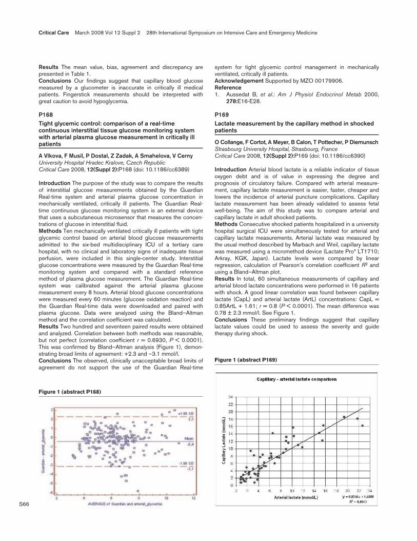
S66
Results The mean value, bias, agreement and discrepancy arepresented in Table 1.Conclusions Our findings suggest that capillary blood glucosemeasured by a glucometer is inaccurate in critically ill medicalpatients. Fingerstick measurements should be interpreted withgreat caution to avoid hypoglycemia.
P168Tight glycemic control: comparison of a real-timecontinuous interstitial tissue glucose monitoring systemwith arterial plasma glucose measurement in critically illpatients
A Vlkova, F Musil, P Dostal, Z Zadak, A Smahelova, V CernyUniversity Hospital Hradec Kralove, Czech RepublicCritical Care 2008, 12(Suppl 2):P168 (doi: 10.1186/cc6389)
Introduction The purpose of the study was to compare the resultsof interstitial glucose measurements obtained by the GuardianReal-time system and arterial plasma glucose concentration inmechanically ventilated, critically ill patients. The Guardian Real-time continuous glucose monitoring system is an external devicethat uses a subcutaneous microsensor that measures the concen-trations of glucose in interstitial fluid.Methods Ten mechanically ventilated critically ill patients with tightglycemic control based on arterial blood glucose measurementsadmitted to the six-bed multidisciplinary ICU of a tertiary carehospital, with no clinical and laboratory signs of inadequate tissueperfusion, were included in this single-center study. Interstitialglucose concentrations were measured by the Guardian Real-timemonitoring system and compared with a standard referencemethod of plasma glucose measurement. The Guardian Real-timesystem was calibrated against the arterial plasma glucosemeasurement every 8 hours. Arterial blood glucose concentrationswere measured every 60 minutes (glucose oxidation reaction) andthe Guardian Real-time data were downloaded and paired withplasma glucose. Data were analyzed using the Bland–Altmanmethod and the correlation coefficient was calculated.Results Two hundred and seventeen paired results were obtainedand analyzed. Correlation between both methods was reasonable,but not perfect (correlation coefficient r = 0.6930, P < 0.0001).This was confirmed by Bland–Altman analysis (Figure 1), demon-strating broad limits of agreement: +2.3 and –3.1 mmol/l.Conclusions The observed, clinically unacceptable broad limits ofagreement do not support the use of the Guardian Real-time
system for tight glycemic control management in mechanicallyventilated, critically ill patients.Acknowledgement Supported by MZO 00179906.Reference1. Aussedat B, et al.: Am J Physiol Endocrinol Metab 2000,
278:E16-E28.
P169Lactate measurement by the capillary method in shockedpatients
O Collange, F Cortot, A Meyer, B Calon, T Pottecher, P DiemunschStrasbourg University Hospital, Strasbourg, FranceCritical Care 2008, 12(Suppl 2):P169 (doi: 10.1186/cc6390)
Introduction Arterial blood lactate is a reliable indicator of tissueoxygen debt and is of value in expressing the degree andprognosis of circulatory failure. Compared with arterial measure-ment, capillary lactate measurement is easier, faster, cheaper andlowers the incidence of arterial puncture complications. Capillarylactate measurement has been already validated to assess fetalwell-being. The aim of this study was to compare arterial andcapillary lactate in adult shocked patients.Methods Consecutive shocked patients hospitalized in a universityhospital surgical ICU were simultaneously tested for arterial andcapillary lactate measurements. Arterial lactate was measured bythe usual method described by Marbach and Weil, capillary lactatewas measured using a micromethod device (Lactate Pro* LT1710;Arkray, KGK, Japan). Lactate levels were compared by linearregression, calculation of Pearson’s correlation coefficient R2 andusing a Bland–Altman plot.Results In total, 60 simultaneous measurements of capillary andarterial blood lactate concentrations were performed in 16 patientswith shock. A good linear correlation was found between capillarylactate (CapL) and arterial lactate (ArtL) concentrations: CapL =0.85ArtL + 1.61; r = 0.8 (P < 0.0001). The mean difference was0.78 ± 2.3 mmol/l. See Figure 1.Conclusions These preliminary findings suggest that capillarylactate values could be used to assess the severity and guidetherapy during shock.
Critical Care March 2008 Vol 12 Suppl 2 28th International Symposium on Intensive Care and Emergency Medicine
Figure 1 (abstract P168)
Figure 1 (abstract P169)

S67
P170Influence of Stewart acid–base variables on plasma pH incritically ill patients
A Marcelli, L Bucci, A Pochini, L Errico, RA De Blasi, M MercieriUniversità La Sapienza di Roma, Rome, ItalyCritical Care 2008, 12(Suppl 2):P170 (doi: 10.1186/cc6391)
Introduction The study objective was to determine and quantifythe influence of strong-ion approach variables on the plasma pH incritically ill patients.Methods A retrospective analysis of clinical records for 284consecutive patients admitted to the medical adult ICU of auniversity hospital. Analysis was made of plasma acid–base datafor 5,172 blood samples collected at admission and throughouthospitalization (one sample per patient). By substituting bicar-bonate with the apparent strong-ion difference (SID), the weakacid anionic component (A–) and unmeasured ions (UI) in theHenderson–Hasselbalch equation, and after selecting samples
having at least one variable within the normal range, we determinedpossible linear relationships between the study variables. We thencompared our results with those calculated using derivatives of thesimplified strong-ion equation.Results In samples with normal UI and PaCO2, the SID had astrong correlation with plasma pH (r2 = 0.84), yielding a δpH/δSIDratio of +0.013 (strong-ion acidosis/alkalosis) (Figure 1). Insamples with normal SID and PaCO2, UI also correlated stronglywith pH (r2 = 0.69), yielding a δpH/δUI ratio of –0.014 (uncompen-sated metabolic acidosis) (Figure 2). Hypoalbuminemia caused acompensatory reduction in SID of about 3 mEq/l per g/dl, therebyalso influencing the pH (δpH/δalbumin ratio of –0.040). In sampleswith normal SID and UI, PaCO2 correlated strongly with pH (r2 =0.85), yielding a δpH/δPaCO2 ratio of –0.01 (pure acute respira-tory acidosis/alkalosis) (Figure 3).Conclusions The SID correlates strongly with changes in pH, thusidentifying a strong-ion acidosis or alkalosis. The changes in pHrelated to the SID, total nonvolatile weak anions and PaCO2 incritically ill patients almost match those calculated using theStewart’s simplified strong-ion equation.Reference1. Constable PD: J Appl Physiol 1997, 83:297-311.
P171Value of postoperative C-reactive protein and leukocytecount after elective thoracoabdominal aneurysm surgery
GB Brunnekreef1, E Scholten2, LJ Bras2, MA Schepens2, LP Aarts3, EP Van Dongen2
1University Medical Centre, Utrecht, The Netherlands; 2St AntoniusHospital, Nieuwegein, The Netherlands; 3University MedicalCentre, Leiden, The NetherlandsCritical Care 2008, 12(Suppl 2):P171 (doi: 10.1186/cc6392)
Introduction There are many causes for a systemic inflammatoryresponse after thoracoabdominal aortic aneurysm (TAAA) repair.The aneurysm itself, the surgical trauma, ischemia-reperfusioninjury and reactions to graft material can all cause an inflammatoryresponse. This makes it difficult to identify postoperative infection.A PubMed search revealed no study on how to discern betweennormal postoperative levels of inflammation and postoperativeinfection after TAAA repair.
Available online http://ccforum.com/supplements/12/S2
Figure 2 (abstract P170)
Correlation between UI and pH (patients with normal SID and PaCO2).
Figure 3 (abstract P170)
Correlation between the PaCO2 and pH (patients with normal UI and SID).Figure 1 (abstract P170)
Correlation between the SID and pH (patients with normal UI and PaCO2).
S68
Methods In this prospective single-centre study we included 34patients. They underwent elective surgical TAAA repair. Immuno-compromised patients and patients using immunosuppressiveagents were excluded. C-reactive protein (CRP) levels andleukocyte count were measured in the operating room and onevery postoperative day until discharge from the ICU, with amaximum of 14 days. We also determined the occurrence of fever.Results Five patients (15%) suffered a postoperative infection:three pulmonary infections, one bacteraemia of unknown origin andone patient suffered a septic period without positive cultures. In allpatients there was a postoperative rise in CRP with a maximum onthe second and third postoperative day. The median CRP was229 mg/l on the second day and 221 mg/l on the thirdpostoperative day. CRP declined towards preoperative levelsduring the first 2 weeks after surgery. The leukocyte countcontinued to rise postoperatively to 13 x 109/l on day 14. Therewas no correlation between fever or leukocyte count and infection.In only three of five patients with postoperative infection was asecond rise in CRP noted.Conclusions This study shows the CRP levels and leukocytecount that can be expected in the ICU after TAAA surgery.Surprisingly the leukocyte count continued to rise. This may becaused by the fact that patients with infection tend to stay longer inthe ICU. The median CRP level on day 14, however, was only22 mg/l. Not all postoperative infections caused a rise in thealready high CRP levels. So in some cases CRP, leukocyte andtemperature may be of no clinical value. Clinical evaluationcombined with positive cultures may be the only method todiagnose postoperative infection in this group of patients.
P172Role of inflammation in nonhemorrhagic strokes
H Michalopoulou, A Bakhal, J Vaitsis, D Nastou, N Stravodimou, D PragastisMetaxa Hospital, Athens, GreeceCritical Care 2008, 12(Suppl 2):P172 (doi: 10.1186/cc6393)
Introduction In recent years considerable interest has beenfocused on the role of inflammation in the pathophysiology of acutecoronary syndromes. There are limited data, however, about itsparticipation in the pathogenesis of strokes. We investigatedwhether inflammation markers are increased in the acute phase ofstrokes.Methods We studied consecutively 54 patients aged 55 ± 8 yearsold (32 males) that were hospitalized in the ICU from June 2005 toDecember 2007 with the diagnosis of nonhemorrhagic strokeproven by computed (CT) or magnetic (MRI) tomography. Within24 hours of their admission, C-reactive protein (CRP), IL-6 andfibrinogen values were determined in all patients. Seventy patients,who were comparable as regards their age and sex, were used asa control group.Results See Figure 1.Conclusions Inflammation markers are increased in the acutephase of ischemic strokes. Further studies are needed to showwhether this increase is secondary to or contributes itself in thepathogenesis of ischemic strokes.
P173Biomarkers that might improve prognostic accuracy in ICUpatients
M Karvouniaris, M Stougianni, A Tefas, D LagonidisGeneral Hospital of Giannitsa, Thessaloniki, GreeceCritical Care 2008, 12(Suppl 2):P173 (doi: 10.1186/cc6394)
Introduction There is often a discrepancy between physicianprediction of mortality and clinical prediction scores, and theaccuracy of the latter is rather moderate. Nevertheless, it istempting to just measure a biochemical parameter – a biomarker –to find prognostic information. Serum lipoproteins, for example, canmirror inflammatory activity and prognosis as well.Methods A prospective study of 29 patients that stayed in the ICUfor at least 4 days and had the following characteristics: age 62.28± 16.92 years, length of stay in the ICU 15.55 ± 10.51 days andAPACHE II score 21.28 ± 7.83. C-reactive protein (CRP), totalcholesterol and high-density lipoprotein (HDL) were measured onadmission and on day 4 in the ICU. First, we correlated theseparameters with the length of stay in the ICU using Pearson’scorrelation method. Secondly, we compared the means betweensurvivors and nonsurvivors after 6 months with an independent-samples’ t test. We finally performed receiver operating curves ofthe above parameters according to mortality.Results CRP both on admission and on day 4 was positivelycorrelated with length of stay in the ICU (P < 0.05). Mortality in6 months was 18/29 (62%). According to the independent-samples’ t test, statistical significance (P < 0.05) was only foundfor CRP on admission. On admission, the values of areas under thecurve (AUC) were: CRP = 0.793, HDL = 0.667, total cholesterol =0.604. Furthermore, on day 4 the values of AUC were: CRP =0.629, HDL = 0.712 and cholesterol = 0.629. Using a cutoff CRPvalue on admission of ≤0.87 mg/dl, there was a better chance ofsurvival with a sensitivity of 63.6% and a specificity of 94.44%(95% CI = 72.6–99.1). In addition to that, a cutoff HDL value onday 4 of ≥28.4 mg/dl predicts survival with a sensitivity of 63.6%and a specificity of 83.3% (95% CI = 46.5–90.2).Conclusions Serial measurements of CRP and HDL are both easyto perform and can add prognostic information. On the other hand,total cholesterol seems not to have any prognostic significance.Reference1. Schuetz et al.: Curr Opin Crit Care 2007, 13:578-585.
P174Plasma C-reactive protein and albumin as predictors ofreadmission to intensive care
N Al-Subaie, T Reynolds, R Sunderland, A Myers, P Morgan,RM Grounds, A RhodesSt Georges Hospital, London, UKCritical Care 2008, 12(Suppl 2):P174 (doi: 10.1186/cc6395)
Introduction Readmission to intensive care is associated withpoor outcome. C-reactive protein (CRP) and low albumin are asso-ciated with systemic inflammation, and this study aims to assesstheir usefulness in predicting readiness for discharge from the ICU.Methods An observational study based in a London teachinghospital mixed medical/surgical ICU. Plasma CRP and albumin onthe day of ICU discharge and patients’ demographic and outcomedata were collected.Results Seven hundred consecutive patients were identified, ofwhich 125 were excluded as they were not suitable for readmission.Eleven patients did not have plasma CRP and albumin data on theday of discharge or the outcome was unknown. Of the 564 patients
Critical Care March 2008 Vol 12 Suppl 2 28th International Symposium on Intensive Care and Emergency Medicine
Figure 1 (abstract P172)
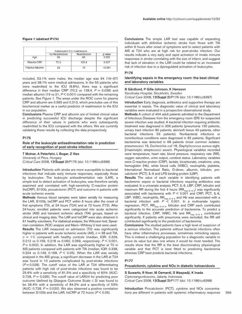
S69
included, 53.1% were males, the median age was 64 (16–97)years and 38.1% were medical admissions. In the 55 patients whowere readmitted to the ICU (9.8%), there was a significantdifference in their median CRP (70.2 vs 128.4, P = 0.036) andmedian albumin (19 vs 21, P < 0.001) compared with the remainingpatients. See Figure 1. The areas under the ROC curve for plasmaCRP and albumin are 0.583 and 0.313, which precludes use of thisbiochemical marker as a useful predictor of readmission to the ICUin our population.Conclusions Plasma CRP and albumin are of limited clinical valuein predicting successful ICU discharge despite the significantdifference of their values in patients who were subsequentlyreadmitted to the ICU compared with the others. We are currentlyvalidating these results by collecting the data prospectively.
P175Role of the leukocyte antisedimentation rate in predictionof early recognition of post-stroke infection
T Molnar, A Peterfalvi, L Bogar, Z IllesUniversity of Pecs, HungaryCritical Care 2008, 12(Suppl 2):P175 (doi: 10.1186/cc6396)
Introduction Patients with stroke are more susceptible to bacterialinfections that indicate early immune responses, especially thoseby leukocytes. The leukocyte antisedimentation rate (LAR), asimple test to detect activation of leukocytes, was therefore seriallyexamined and correlated with high-sensitivity C-reactive protein(hsCRP), S100b, procalcitonin (PCT) and outcome in patients withacute ischemic events.Methods Venous blood samples were taken serially for measuringthe LAR, S100b, hsCRP and PCT within 6 hours after the onset offirst symptoms (T0), at 24 hours (T24) and at 72 hours (T72). After24 hours, enrolled patients were categorized into acute ischemicstroke (AIS) and transient ischemic attack (TIA) groups, based onclinical and imaging data. The LAR and hsCRP were also obtained in61 healthy volunteers. For statistical analysis, the Wilcoxon test, Spear-man correlation, ROC analysis and Mann–Whitney U test were used.Results The LAR measured on admission (T0) was significantlyhigher in patients with acute ischemic events (AIS, n = 38 and TIA,n = 11) compared with healthy controls (median, IQR: 0.329,0.212 vs 0.159, 0.218 vs 0.060, 0.069, respectively; P < 0.001,P = 0.002). In addition, the LAR was significantly higher at T0 inAIS patients compared with patients with TIA (median, IQR: 0.338,0.204 vs 0.149, 0.168, P < 0.05). When the LAR was seriallyanalyzed in the AIS group, a significant decrease in the LAR at T24was found in 10 patients complicated by post-stroke infections(P = 0.028). The cutoff value of the LAR at T24 differentiatingpatients with high risk of post-stroke infections was found to be25.6% with a sensitivity of 81.5% and a specificity of 60% (AUC:0.728, P = 0.035). The cutoff value of LAR24 for predicting pooroutcome (defined by Glasgow Outcome Scale ≤ 3) was found tobe 26.4% with a sensitivity of 84.2% and a specificity of 53%(AUC: 0.728, P = 0.020). We also observed a positive correlationbetween S100b and the LAR, hsCRP at 72 hours (P < 0.05).
Conclusions The simple LAR test was capable of separatingindividuals with definitive ischemic stroke from those with TIAwithin 6 hours after onset of symptoms and to select patients withAIS at T24 who are at high risk for post-stroke infection. Ourresults indicate a very early and rapid activation of innate immuneresponses in stroke correlating with the size of infarct, and suggestthat lack of elevation in the LAR could be related to an increasedrisk of infection due to a dysregulated activation of leukocytes.
P176Identifying sepsis in the emergency room: the best clinicaland laboratory variables
B Gårdlund, P Gille-Johnson, K HanssonKarolinska Hospital, Stockholm, SwedenCritical Care 2008, 12(Suppl 2):P176 (doi: 10.1186/cc6397)
Introduction Early diagnosis, antibiotics and supportive therapy areessential in sepsis. The diagnostic value of clinical and laboratoryvariables were evaluated in a prospective observational study.Methods A cohort of 404 adult patients admitted to the Departmentof Infectious Diseases from the emergency room (ER) for suspectedsevere infection was studied. A bacterial infection requiring antibiotictreatment was diagnosed in 306 patients (pneumonia 130 patients,urinary tract infection 80 patients, skin/soft tissue 43 patients, otherbacterial infections 53 patients). Nonbacterial infections ornoninfectious conditions were diagnosed in 82 patients. Significantbacteremia was detected in 68 patients (most common isolates:pneumococci 19, Escherichia coli 18, Staphylococcus aureus eight,β-haemolytic streptococci seven). Physiological variables recordedwere temperature, heart rate, blood pressure, respiratory rate (RR),oxygen saturation, urine output, cerebral status. Laboratory variableswere C-reactive protein (CRP), lactate, bicarbonate, creatinine, urea,hemoglobin (Hb), white blood cells (WBC), neutrophils, platelets,International Normalized Ratio, D-dimer, albumin, bilirubin, pro-calcitonin (PCT), IL-6 and LPS binding protein (LBP).Results The value of each variable in identifying patients withbacteremic sepsis or bacterial infections requiring antibiotics wasevaluated. In a univariate analysis, PCT, IL-6, LBP, CRP, bilirubin andmaximum RR during the first 4 hours (RRmax 0–4 h) was significantlyassociated with bacteremia with P < 0.001 and CRP, PCT, IL-6,LBP, WBC, neutrophils, RRmax 0–4 h and Hb was associated with abacterial infection with P < 0.001. In a multivariate logisticregression, PCT, RRmax 0–4 h, bilirubin and CRP each contributedsignificantly to the accurate prediction of bacteremia. To predict abacterial infection, CRP, WBC, Hb and RRmax 0–4 h contributedsignificantly. If patients with pneumonia were excluded, the RR stillcontributed significantly to the prediction of bacteremia.Conclusions The studied patients have a high level of suspicion ofa serious infection. The patients without bacterial infections oftenhave other inflammatory processes, sometimes mimicking sepsis.This is indeed a challenging population for a diagnostic variable toprove its value but also one where it would be most needed. Theresults show that the RR is the best discriminatory physiologicalvariable and that PCT is best fitted to predicting bacteremiawhereas CRP best predicts bacterial infections.
P177Procalcitonin, cytokine and NOx in diabetic ketoacidosis
S Suwarto, R Noer, M Oemardi, S Waspadji, K InadaCiptomangunkusumo, Jakarta, IndonesiaCritical Care 2008, 12(Suppl 2):P177 (doi: 10.1186/cc6398)
Introduction Procalcitonin (PCT), cytokine and NOx concentra-tions are different in patients with systemic inflammatory response
Available online http://ccforum.com/supplements/12/S2
Figure 1 (abstract P174)
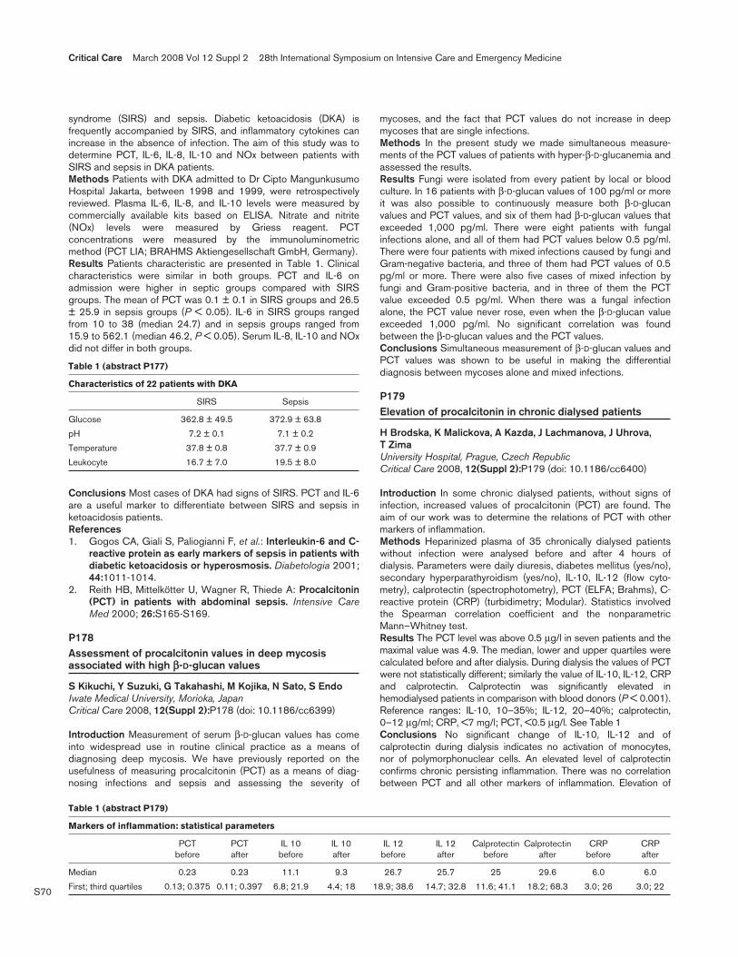
S70
syndrome (SIRS) and sepsis. Diabetic ketoacidosis (DKA) isfrequently accompanied by SIRS, and inflammatory cytokines canincrease in the absence of infection. The aim of this study was todetermine PCT, IL-6, IL-8, IL-10 and NOx between patients withSIRS and sepsis in DKA patients.Methods Patients with DKA admitted to Dr Cipto MangunkusumoHospital Jakarta, between 1998 and 1999, were retrospectivelyreviewed. Plasma IL-6, IL-8, and IL-10 levels were measured bycommercially available kits based on ELISA. Nitrate and nitrite(NOx) levels were measured by Griess reagent. PCTconcentrations were measured by the immunoluminometricmethod (PCT LIA; BRAHMS Aktiengesellschaft GmbH, Germany).Results Patients characteristic are presented in Table 1. Clinicalcharacteristics were similar in both groups. PCT and IL-6 onadmission were higher in septic groups compared with SIRSgroups. The mean of PCT was 0.1 ± 0.1 in SIRS groups and 26.5± 25.9 in sepsis groups (P < 0.05). IL-6 in SIRS groups rangedfrom 10 to 38 (median 24.7) and in sepsis groups ranged from15.9 to 562.1 (median 46.2, P < 0.05). Serum IL-8, IL-10 and NOxdid not differ in both groups.
Table 1 (abstract P177)
Characteristics of 22 patients with DKA
SIRS Sepsis
Glucose 362.8 ± 49.5 372.9 ± 63.8
pH 7.2 ± 0.1 7.1 ± 0.2
Temperature 37.8 ± 0.8 37.7 ± 0.9
Leukocyte 16.7 ± 7.0 19.5 ± 8.0
Conclusions Most cases of DKA had signs of SIRS. PCT and IL-6are a useful marker to differentiate between SIRS and sepsis inketoacidosis patients.References1. Gogos CA, Giali S, Paliogianni F, et al.: Interleukin-6 and C-
reactive protein as early markers of sepsis in patients withdiabetic ketoacidosis or hyperosmosis. Diabetologia 2001;44:1011-1014.
2. Reith HB, Mittelkötter U, Wagner R, Thiede A: Procalcitonin(PCT) in patients with abdominal sepsis. Intensive CareMed 2000; 26:S165-S169.
P178Assessment of procalcitonin values in deep mycosisassociated with high ββ-D-glucan values
S Kikuchi, Y Suzuki, G Takahashi, M Kojika, N Sato, S EndoIwate Medical University, Morioka, JapanCritical Care 2008, 12(Suppl 2):P178 (doi: 10.1186/cc6399)
Introduction Measurement of serum β-D-glucan values has comeinto widespread use in routine clinical practice as a means ofdiagnosing deep mycosis. We have previously reported on theusefulness of measuring procalcitonin (PCT) as a means of diag-nosing infections and sepsis and assessing the severity of
mycoses, and the fact that PCT values do not increase in deepmycoses that are single infections.Methods In the present study we made simultaneous measure-ments of the PCT values of patients with hyper-β-D-glucanemia andassessed the results.Results Fungi were isolated from every patient by local or bloodculture. In 16 patients with β-D-glucan values of 100 pg/ml or moreit was also possible to continuously measure both β-D-glucanvalues and PCT values, and six of them had β-D-glucan values thatexceeded 1,000 pg/ml. There were eight patients with fungalinfections alone, and all of them had PCT values below 0.5 pg/ml.There were four patients with mixed infections caused by fungi andGram-negative bacteria, and three of them had PCT values of 0.5pg/ml or more. There were also five cases of mixed infection byfungi and Gram-positive bacteria, and in three of them the PCTvalue exceeded 0.5 pg/ml. When there was a fungal infectionalone, the PCT value never rose, even when the β-D-glucan valueexceeded 1,000 pg/ml. No significant correlation was foundbetween the β-D-glucan values and the PCT values.Conclusions Simultaneous measurement of β-D-glucan values andPCT values was shown to be useful in making the differentialdiagnosis between mycoses alone and mixed infections.
P179Elevation of procalcitonin in chronic dialysed patients
H Brodska, K Malickova, A Kazda, J Lachmanova, J Uhrova, T ZimaUniversity Hospital, Prague, Czech RepublicCritical Care 2008, 12(Suppl 2):P179 (doi: 10.1186/cc6400)
Introduction In some chronic dialysed patients, without signs ofinfection, increased values of procalcitonin (PCT) are found. Theaim of our work was to determine the relations of PCT with othermarkers of inflammation.Methods Heparinized plasma of 35 chronically dialysed patientswithout infection were analysed before and after 4 hours ofdialysis. Parameters were daily diuresis, diabetes mellitus (yes/no),secondary hyperparathyroidism (yes/no), IL-10, IL-12 (flow cyto-metry), calprotectin (spectrophotometry), PCT (ELFA; Brahms), C-reactive protein (CRP) (turbidimetry; Modular). Statistics involvedthe Spearman correlation coefficient and the nonparametricMann–Whitney test.Results The PCT level was above 0.5 μg/l in seven patients and themaximal value was 4.9. The median, lower and upper quartiles werecalculated before and after dialysis. During dialysis the values of PCTwere not statistically different; similarly the value of IL-10, IL-12, CRPand calprotectin. Calprotectin was significantly elevated inhemodialysed patients in comparison with blood donors (P < 0.001).Reference ranges: IL-10, 10–35%; IL-12, 20–40%; calprotectin,0–12 μg/ml; CRP, <7 mg/l; PCT, <0.5 μg/l. See Table 1Conclusions No significant change of IL-10, IL-12 and ofcalprotectin during dialysis indicates no activation of monocytes,nor of polymorphonuclear cells. An elevated level of calprotectinconfirms chronic persisting inflammation. There was no correlationbetween PCT and all other markers of inflammation. Elevation of
Critical Care March 2008 Vol 12 Suppl 2 28th International Symposium on Intensive Care and Emergency Medicine
Table 1 (abstract P179)
Markers of inflammation: statistical parameters
PCT PCT IL 10 IL 10 IL 12 IL 12 Calprotectin Calprotectin CRP CRP before after before after before after before after before after
Median 0.23 0.23 11.1 9.3 26.7 25.7 25 29.6 6.0 6.0
First; third quartiles 0.13; 0.375 0.11; 0.397 6.8; 21.9 4.4; 18 18.9; 38.6 14.7; 32.8 11.6; 41.1 18.2; 68.3 3.0; 26 3.0; 22
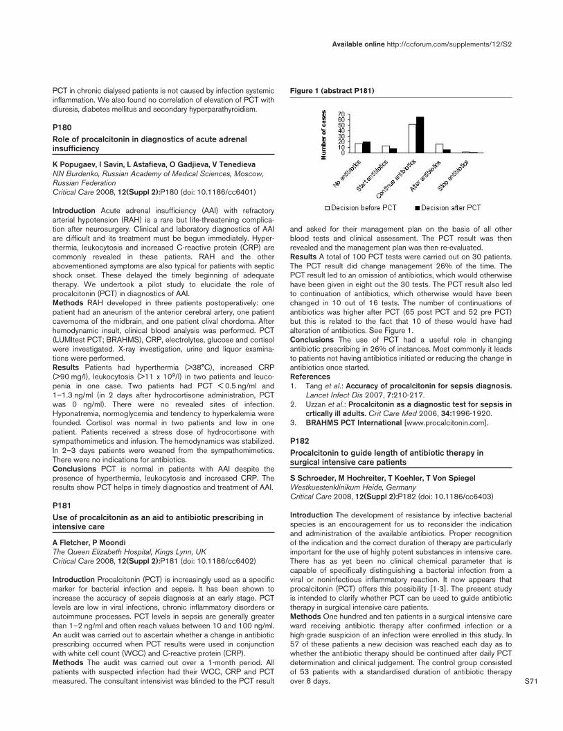
S71
PCT in chronic dialysed patients is not caused by infection systemicinflammation. We also found no correlation of elevation of PCT withdiuresis, diabetes mellitus and secondary hyperparathyroidism.
P180Role of procalcitonin in diagnostics of acute adrenalinsufficiency
K Popugaev, I Savin, L Astafieva, O Gadjieva, V TenedievaNN Burdenko, Russian Academy of Medical Sciences, Moscow,Russian FederationCritical Care 2008, 12(Suppl 2):P180 (doi: 10.1186/cc6401)
Introduction Acute adrenal insufficiency (AAI) with refractoryarterial hypotension (RAH) is a rare but life-threatening complica-tion after neurosurgery. Clinical and laboratory diagnostics of AAIare difficult and its treatment must be begun immediately. Hyper-thermia, leukocytosis and increased C-reactive protein (CRP) arecommonly revealed in these patients. RAH and the otherabovementioned symptoms are also typical for patients with septicshock onset. These delayed the timely beginning of adequatetherapy. We undertook a pilot study to elucidate the role ofprocalcitonin (PCT) in diagnostics of AAI.Methods RAH developed in three patients postoperatively: onepatient had an aneurism of the anterior cerebral artery, one patientcavernoma of the midbrain, and one patient clival chordoma. Afterhemodynamic insult, clinical blood analysis was performed. PCT(LUMItest PCT; BRAHMS), CRP, electrolytes, glucose and cortisolwere investigated. X-ray investigation, urine and liquor examina-tions were performed.Results Patients had hyperthermia (>38°C), increased CRP(>90 mg/l), leukocytosis (>11 x 109/l) in two patients and leuco-penia in one case. Two patients had PCT < 0.5 ng/ml and1–1.3 ng/ml (in 2 days after hydrocortisone administration, PCTwas 0 ng/ml). There were no revealed sites of infection.Hyponatremia, normoglycemia and tendency to hyperkalemia werefounded. Cortisol was normal in two patients and low in onepatient. Patients received a stress dose of hydrocortisone withsympathomimetics and infusion. The hemodynamics was stabilized.In 2–3 days patients were weaned from the sympathomimetics.There were no indications for antibiotics.Conclusions PCT is normal in patients with AAI despite thepresence of hyperthermia, leukocytosis and increased CRP. Theresults show PCT helps in timely diagnostics and treatment of AAI.
P181Use of procalcitonin as an aid to antibiotic prescribing inintensive care
A Fletcher, P MoondiThe Queen Elizabeth Hospital, Kings Lynn, UKCritical Care 2008, 12(Suppl 2):P181 (doi: 10.1186/cc6402)
Introduction Procalcitonin (PCT) is increasingly used as a specificmarker for bacterial infection and sepsis. It has been shown toincrease the accuracy of sepsis diagnosis at an early stage. PCTlevels are low in viral infections, chronic inflammatory disorders orautoimmune processes. PCT levels in sepsis are generally greaterthan 1–2 ng/ml and often reach values between 10 and 100 ng/ml.An audit was carried out to ascertain whether a change in antibioticprescribing occurred when PCT results were used in conjunctionwith white cell count (WCC) and C-reactive protein (CRP).Methods The audit was carried out over a 1-month period. Allpatients with suspected infection had their WCC, CRP and PCTmeasured. The consultant intensivist was blinded to the PCT result
and asked for their management plan on the basis of all otherblood tests and clinical assessment. The PCT result was thenrevealed and the management plan was then re-evaluated.Results A total of 100 PCT tests were carried out on 30 patients.The PCT result did change management 26% of the time. ThePCT result led to an omission of antibiotics, which would otherwisehave been given in eight out the 30 tests. The PCT result also ledto continuation of antibiotics, which otherwise would have beenchanged in 10 out of 16 tests. The number of continuations ofantibiotics was higher after PCT (65 post PCT and 52 pre PCT)but this is related to the fact that 10 of these would have hadalteration of antibiotics. See Figure 1.Conclusions The use of PCT had a useful role in changingantibiotic prescribing in 26% of instances. Most commonly it leadsto patients not having antibiotics initiated or reducing the change inantibiotics once started.References1. Tang et al.: Accuracy of procalcitonin for sepsis diagnosis.
Lancet Infect Dis 2007, 7:210-217.2. Uzzan et al.: Procalcitonin as a diagnostic test for sepsis in
crtically ill adults. Crit Care Med 2006, 34:1996-1920.3. BRAHMS PCT International [www.procalcitonin.com].
P182Procalcitonin to guide length of antibiotic therapy insurgical intensive care patients
S Schroeder, M Hochreiter, T Koehler, T Von SpiegelWestkuestenklinikum Heide, GermanyCritical Care 2008, 12(Suppl 2):P182 (doi: 10.1186/cc6403)
Introduction The development of resistance by infective bacterialspecies is an encouragement for us to reconsider the indicationand administration of the available antibiotics. Proper recognitionof the indication and the correct duration of therapy are particularlyimportant for the use of highly potent substances in intensive care.There has as yet been no clinical chemical parameter that iscapable of specifically distinguishing a bacterial infection from aviral or noninfectious inflammatory reaction. It now appears thatprocalcitonin (PCT) offers this possibility [1-3]. The present studyis intended to clarify whether PCT can be used to guide antibiotictherapy in surgical intensive care patients.Methods One hundred and ten patients in a surgical intensive careward receiving antibiotic therapy after confirmed infection or ahigh-grade suspicion of an infection were enrolled in this study. In57 of these patients a new decision was reached each day as towhether the antibiotic therapy should be continued after daily PCTdetermination and clinical judgement. The control group consistedof 53 patients with a standardised duration of antibiotic therapyover 8 days.
Available online http://ccforum.com/supplements/12/S2
Figure 1 (abstract P181)
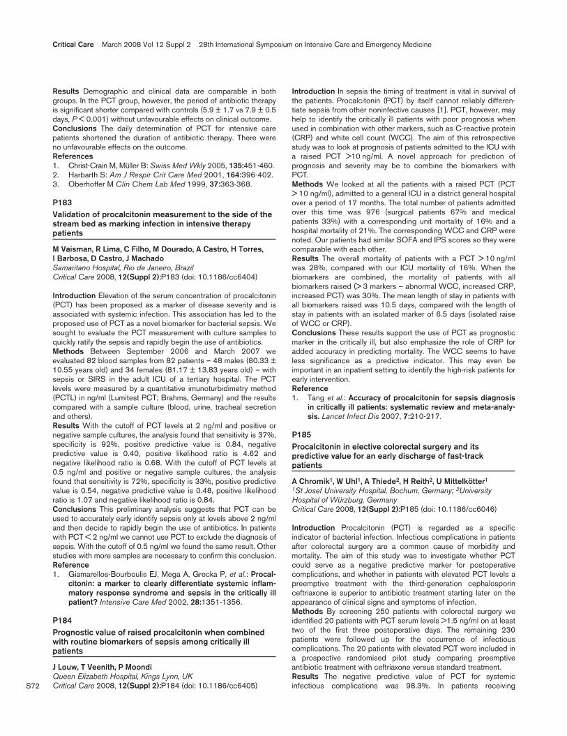
S72
Results Demographic and clinical data are comparable in bothgroups. In the PCT group, however, the period of antibiotic therapyis significant shorter compared with controls (5.9 ± 1.7 vs 7.9 ± 0.5days, P < 0.001) without unfavourable effects on clinical outcome.Conclusions The daily determination of PCT for intensive carepatients shortened the duration of antibiotic therapy. There wereno unfavourable effects on the outcome.References1. Christ-Crain M, Müller B: Swiss Med Wkly 2005, 135:451-460.2. Harbarth S: Am J Respir Crit Care Med 2001, 164:396-402.3. Oberhoffer M Clin Chem Lab Med 1999, 37:363-368.
P183Validation of procalcitonin measurement to the side of thestream bed as marking infection in intensive therapypatients
M Vaisman, R Lima, C Filho, M Dourado, A Castro, H Torres, I Barbosa, D Castro, J MachadoSamaritano Hospital, Rio de Janeiro, BrazilCritical Care 2008, 12(Suppl 2):P183 (doi: 10.1186/cc6404)
Introduction Elevation of the serum concentration of procalcitonin(PCT) has been proposed as a marker of disease severity and isassociated with systemic infection. This association has led to theproposed use of PCT as a novel biomarker for bacterial sepsis. Wesought to evaluate the PCT measurement with culture samples toquickly ratify the sepsis and rapidly begin the use of antibiotics.Methods Between September 2006 and March 2007 weevaluated 82 blood samples from 82 patients – 48 males (80.33 ±10.55 years old) and 34 females (81.17 ± 13.83 years old) – withsepsis or SIRS in the adult ICU of a tertiary hospital. The PCTlevels were measured by a quantitative imunoturbidimetry method(PCTL) in ng/ml (Lumitest PCT; Brahms, Germany) and the resultscompared with a sample culture (blood, urine, tracheal secretionand others).Results With the cutoff of PCT levels at 2 ng/ml and positive ornegative sample cultures, the analysis found that sensitivity is 37%,specificity is 92%, positive predictive value is 0.84, negativepredictive value is 0.40, positive likelihood ratio is 4.62 andnegative likelihood ratio is 0.68. With the cutoff of PCT levels at0.5 ng/ml and positive or negative sample cultures, the analysisfound that sensitivity is 72%, specificity is 33%, positive predictivevalue is 0.54, negative predictive value is 0.48, positive likelihoodratio is 1.07 and negative likelihood ratio is 0.84.Conclusions This preliminary analysis suggests that PCT can beused to accurately early identify sepsis only at levels above 2 ng/mland then decide to rapidly begin the use of antibiotics. In patientswith PCT < 2 ng/ml we cannot use PCT to exclude the diagnosis ofsepsis. With the cutoff of 0.5 ng/ml we found the same result. Otherstudies with more samples are necessary to confirm this conclusion.Reference1. Giamarellos-Bourboulis EJ, Mega A, Grecka P, et al.: Procal-
citonin: a marker to clearly differentiate systemic inflam-matory response syndrome and sepsis in the critically illpatient? Intensive Care Med 2002, 28:1351-1356.
P184Prognostic value of raised procalcitonin when combinedwith routine biomarkers of sepsis among critically illpatients
J Louw, T Veenith, P MoondiQueen Elizabeth Hospital, Kings Lynn, UKCritical Care 2008, 12(Suppl 2):P184 (doi: 10.1186/cc6405)
Introduction In sepsis the timing of treatment is vital in survival ofthe patients. Procalcitonin (PCT) by itself cannot reliably differen-tiate sepsis from other noninfective causes [1]. PCT, however, mayhelp to identify the critically ill patients with poor prognosis whenused in combination with other markers, such as C-reactive protein(CRP) and white cell count (WCC). The aim of this retrospectivestudy was to look at prognosis of patients admitted to the ICU witha raised PCT >10 ng/ml. A novel approach for prediction ofprognosis and severity may be to combine the biomarkers withPCT.Methods We looked at all the patients with a raised PCT (PCT> 10 ng/ml), admitted to a general ICU in a district general hospitalover a period of 17 months. The total number of patients admittedover this time was 976 (surgical patients 67% and medicalpatients 33%) with a corresponding unit mortality of 16% and ahospital mortality of 21%. The corresponding WCC and CRP werenoted. Our patients had similar SOFA and IPS scores so they werecomparable with each other.Results The overall mortality of patients with a PCT > 10 ng/mlwas 28%, compared with our ICU mortality of 16%. When thebiomarkers are combined, the mortality of patients with allbiomarkers raised (> 3 markers – abnormal WCC, increased CRP,increased PCT) was 30%. The mean length of stay in patients withall biomarkers raised was 10.5 days, compared with the length ofstay in patients with an isolated marker of 6.5 days (isolated raiseof WCC or CRP).Conclusions These results support the use of PCT as prognosticmarker in the critically ill, but also emphasize the role of CRP foradded accuracy in predicting mortality. The WCC seems to haveless significance as a predictive indicator. This may even beimportant in an inpatient setting to identify the high-risk patients forearly intervention.Reference1. Tang et al.: Accuracy of procalcitonin for sepsis diagnosis
in critically ill patients: systematic review and meta-analy-sis. Lancet Infect Dis 2007, 7:210-217.
P185Procalcitonin in elective colorectal surgery and itspredictive value for an early discharge of fast-trackpatients
A Chromik1, W Uhl1, A Thiede2, H Reith2, U Mittelkötter1
1St Josef University Hospital, Bochum, Germany; 2UniversityHospital of Würzburg, GermanyCritical Care 2008, 12(Suppl 2):P185 (doi: 10.1186/cc6046)
Introduction Procalcitonin (PCT) is regarded as a specificindicator of bacterial infection. Infectious complications in patientsafter colorectal surgery are a common cause of morbidity andmortality. The aim of this study was to investigate whether PCTcould serve as a negative predictive marker for postoperativecomplications, and whether in patients with elevated PCT levels apreemptive treatment with the third-generation cephalosporinceftriaxone is superior to antibiotic treatment starting later on theappearance of clinical signs and symptoms of infection.Methods By screening 250 patients with colorectal surgery weidentified 20 patients with PCT serum levels >1.5 ng/ml on at leasttwo of the first three postoperative days. The remaining 230patients were followed up for the occurrence of infectiouscomplications. The 20 patients with elevated PCT were included ina prospective randomised pilot study comparing preemptiveantibiotic treatment with ceftriaxone versus standard treatment.Results The negative predictive value of PCT for systemicinfectious complications was 98.3%. In patients receiving
Critical Care March 2008 Vol 12 Suppl 2 28th International Symposium on Intensive Care and Emergency Medicine
S73
preemptive antibiotic treatment (ceftriaxone), both the incidenceand the severity of postoperative systemic infections weresignificantly lower compared with those in a control group(Pearson’s chi-squared test P = 0.001 and P = 0.007,respectively). Major differences were also observed with respect tothe duration of antibiotic treatment and the length of hospital stay.Conclusions PCT is an early marker for systemic infectiouscomplications after colorectal surgery with a high negativepredictive value. A significant reduction in the rate of postoperativeinfections in patients with elevated PCT serum concentrations wasachieved by means of preemptive antibiotic treatment.
P186Endotoxin adsorption method may affect serumprocalcitonin
T Ikeda, K Ikeda, T Ueno, S SudaTokyo Medical University, Hachioji Medical Center, Tokyo, JapanCritical Care 2008, 12(Suppl 2):P186 (doi: 10.1186/cc6407)
Introduction When septic patients progress to endotoxin shock,they present a high mortality rate. The mortality rate of septic patientswith multiple organ failure has been reported to be 30–80%. Theendotoxin adsorption method (PMX-DHP: Toray Industries, Inc.,Tokyo, Japan) has been used for treatment of patients with severesepsis and septic shock primarily caused by Gram-negativeinfections in Japan. We also reported that PMX-DHP removedplasma endotoxin and improved hemodynamic parameters in clinicaltrials [1]. The purpose of this study was to assess the changes ofprocalcitonin (PCT) values during PMX-DHP.Methods The retrospective study was carried out in our ICU. Inthis study, 68 septic patients who had multiple organ failure due tointra-abdominal infection were treated with PMX-DHP. Sepsis wasdiagnosed according to the criteria of the ACCP/SCCM Consen-sus Conference Committee. These patients were separated intotwo groups: those who survived for at least 28 days after the startof PMX-DHP therapy (S group; 49 patients), and those who didnot (nonsurvival group; 19 patients). Background factors andinflammatory mediators were examined in each group. PCT wasmeasured by immunoluminometric assay before and after PMX-DHP and 24 hours later. The luminometer used was an AutolumatLB953 (Berthord, Bad Wildbad, Germany). Endotoxin (kineticturbidimetric method) was also measured just before andimmediately after PMX-DHP.Results The 28-day survival rate was 72.1% (49 survivors, 19nonsurvivors). The APACHE II scores were 22.0 ± 8.3 and 28.3 ±7.0 and the Sequential Organ Failure Assessment scores were9.1 ± 3.9 and 11.1 ± 2.8 in the survival and nonsurvival groups,respectively, showing significantly higher scores in the nonsurvivalgroup. PCT before PMX-DHP in all patients was 59.1 ± 97.2ng/ml and tended to decrease 54.7 ± 81.7 ng/ml after PMX-DHP.PCT was 59.4 ± 109.4 ng/ml before PMX-DHP and significantlydecreased to 42.2 ± 67.4 ng/ml at 24 hours after PMX-DHP in thesurvival group, but it did not change significantly in the nonsurvivalgroup. There was a significant correlation between endotoxin andPCT (r = 0.527, P < 0.001).Conclusions Our results may suggest that PMX-DHP can reducesystemic inflammatory cytokines and serum PCT in the survivalgroup.Reference1. Ikeda T, et al.: Clinical evaluation of PMX-DHP for hypercy-
tokinemia caused by septic multiple organ failure. TherApher Dial 2004, 8:293-298.
P187Discriminative procalcitonin values for diagnosis ofsystemic inflammatory response syndrome and sepsis inthe ICU
H Gharsallah, I Labbene, B Fatthallah, N Hichri, K Lamine, M FerjaniMilitary Hospital of Tunis, TunisiaCritical Care 2008, 12(Suppl 2):P187 (doi: 10.1186/cc6408)
Introduction No study has found a procalcitonin level thatdistinguishes between systemic inflammatory response syndrome(SIRS) and sepsis [1]. The goal of our study was to determine acutoff value of serum procalcitonin concentration for diagnosis ofSIRS and sepsis.Methods In this prospective and observational study, we includedall patients admitted to the ICU. The procalcitonin level wasdetermined on day 0, day 2, day 4 and day 7 of hospitalization usingthe immunoluminometric method (PCT-lumin; Brahms Diagnostica,Berlin, Germany). Normal values are < 0.1 ng/ml. The detection limitwas 0.3 ng/ml. P < 0.05 was considered significant. Time pointswere defined as the procalcitonin concentrations measured atdifferent times in all patients. Time points were associated withSIRS, sepsis and septic shock (SS) according to the establishedACCP/SCCM consensus definition. Statistical analysis wasperformed using SPSS software for windows version 10.Results A total of 70 patients were included in our study. Twohundred and sixty-five time points were categorized into threegroups (SIRS, sepsis and SS). The mean IGS II score was 32 ±14; the mean APACHE II score 15 ± 7. The median procalcitoninlevels in the SIRS group and the sepsis + SS group were,respectively, 0.325 and 1.115 ng/ml (P < 0.001). The area underthe ROC curve to distinguish the presence or absence of sepsiswas 0.745 (0.685–0.805). A cutoff value of 1.3 has a specificityand a sensitivity of 89% and 45%, respectively. If we excludepatient patients with SS, a cutoff value of 1.3 has the samespecificity (89%) and was less sensitive (58%). A cutoff value of0.265 had a specificity of 58% and a sensitivity of 80%.Conclusions According to these preliminary results a procalcitoninlevel between 0.265 and 1.3 ng/ml cannot distinguish betweenSIRS and sepsis. This can be explained by the fact that weconsidered different time points in the same patients and by thefact that we did not separate medical and surgical patients.Reference1. Simon L, Gauvin F, Amre DK, et al.: Clin Infect Dis 2004,
39:206-217.
P188Measurement of procalcitonin in bronchoalveolar lavageand serum as early predictors in acute respiratory distresssyndrome
A Abdel Razek, E Abdel Moneim Arida, A DeghadyAlexandria Faculty of Medicine, Alexandria, EgyptCritical Care 2008, 12(Suppl 2):P188 (doi: 10.1186/cc6409)
Introduction Procalcitonin (PCT) is a diagnostic for identifyingsevere bacterial infections and reliably indicating complicationssecondary to systemic inflammation. PCT levels increase in casesof sepsis, septic shock and in severe systemic inflammatoryreactions. The early pathological features of acute respiratorydistress syndrome (ARDS) are generally described as diffusealveolar damage, which can be diagnosed by cytologicalexamination of bronchoalveolar lavage fluid (BAL) [1]. The aim ofthis study was to evaluate the value of PCT measurement in BAL
Available online http://ccforum.com/supplements/12/S2
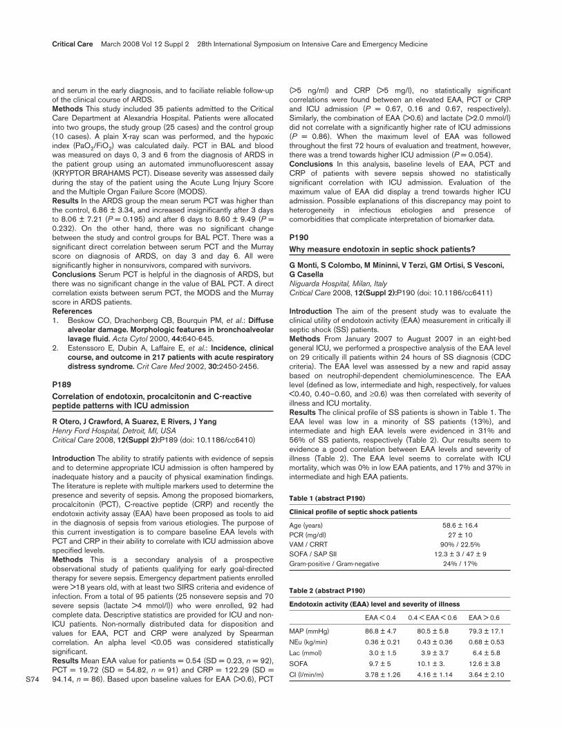
S74
and serum in the early diagnosis, and to faciliate reliable follow-upof the clinical course of ARDS.Methods This study included 35 patients admitted to the CriticalCare Department at Alexandria Hospital. Patients were allocatedinto two groups, the study group (25 cases) and the control group(10 cases). A plain X-ray scan was performed, and the hypoxicindex (PaO2/FiO2) was calculated daily. PCT in BAL and bloodwas measured on days 0, 3 and 6 from the diagnosis of ARDS inthe patient group using an automated immunofluorescent assay(KRYPTOR BRAHAMS PCT). Disease severity was assessed dailyduring the stay of the patient using the Acute Lung Injury Scoreand the Multiple Organ Failure Score (MODS).Results In the ARDS group the mean serum PCT was higher thanthe control, 6.86 ± 3.34, and increased insignificantly after 3 daysto 8.06 ± 7.21 (P = 0.195) and after 6 days to 8.60 ± 9.49 (P =0.232). On the other hand, there was no significant changebetween the study and control groups for BAL PCT. There was asignificant direct correlation between serum PCT and the Murrayscore on diagnosis of ARDS, on day 3 and day 6. All weresignificantly higher in nonsurvivors, compared with survivors.Conclusions Serum PCT is helpful in the diagnosis of ARDS, butthere was no significant change in the value of BAL PCT. A directcorrelation exists between serum PCT, the MODS and the Murrayscore in ARDS patients.References1. Beskow CO, Drachenberg CB, Bourquin PM, et al.: Diffuse
alveolar damage. Morphologic features in bronchoalveolarlavage fluid. Acta Cytol 2000, 44:640-645.
2. Estenssoro E, Dubin A, Laffaire E, et al.: Incidence, clinicalcourse, and outcome in 217 patients with acute respiratorydistress syndrome. Crit Care Med 2002, 30:2450-2456.
P189Correlation of endotoxin, procalcitonin and C-reactivepeptide patterns with ICU admission
R Otero, J Crawford, A Suarez, E Rivers, J YangHenry Ford Hospital, Detroit, MI, USACritical Care 2008, 12(Suppl 2):P189 (doi: 10.1186/cc6410)
Introduction The ability to stratify patients with evidence of sepsisand to determine appropriate ICU admission is often hampered byinadequate history and a paucity of physical examination findings.The literature is replete with multiple markers used to determine thepresence and severity of sepsis. Among the proposed biomarkers,procalcitonin (PCT), C-reactive peptide (CRP) and recently theendotoxin activity assay (EAA) have been proposed as tools to aidin the diagnosis of sepsis from various etiologies. The purpose ofthis current investigation is to compare baseline EAA levels withPCT and CRP in their ability to correlate with ICU admission abovespecified levels.Methods This is a secondary analysis of a prospectiveobservational study of patients qualifying for early goal-directedtherapy for severe sepsis. Emergency department patients enrolledwere >18 years old, with at least two SIRS criteria and evidence ofinfection. From a total of 95 patients (25 nonsevere sepsis and 70severe sepsis (lactate >4 mmol/l)) who were enrolled, 92 hadcomplete data. Descriptive statistics are provided for ICU and non-ICU patients. Non-normally distributed data for disposition andvalues for EAA, PCT and CRP were analyzed by Spearmancorrelation. An alpha level <0.05 was considered statisticallysignificant.Results Mean EAA value for patients = 0.54 (SD = 0.23, n = 92),PCT = 19.72 (SD = 54.82, n = 91) and CRP = 122.29 (SD =94.14, n = 86). Based upon baseline values for EAA (>0.6), PCT
(>5 ng/ml) and CRP (>5 mg/l), no statistically significantcorrelations were found between an elevated EAA, PCT or CRPand ICU admission (P = 0.67, 0.16 and 0.67, respectively).Similarly, the combination of EAA (>0.6) and lactate (>2.0 mmol/l)did not correlate with a significantly higher rate of ICU admissions(P = 0.86). When the maximum level of EAA was followedthroughout the first 72 hours of evaluation and treatment, however,there was a trend towards higher ICU admission (P = 0.054).Conclusions In this analysis, baseline levels of EAA, PCT andCRP of patients with severe sepsis showed no statisticallysignificant correlation with ICU admission. Evaluation of themaximum value of EAA did display a trend towards higher ICUadmission. Possible explanations of this discrepancy may point toheterogeneity in infectious etiologies and presence ofcomorbidities that complicate interpretation of biomarker data.
P190Why measure endotoxin in septic shock patients?
G Monti, S Colombo, M Mininni, V Terzi, GM Ortisi, S Vesconi,G CasellaNiguarda Hospital, Milan, ItalyCritical Care 2008, 12(Suppl 2):P190 (doi: 10.1186/cc6411)
Introduction The aim of the present study was to evaluate theclinical utility of endotoxin activity (EAA) measurement in critically illseptic shock (SS) patients.Methods From January 2007 to August 2007 in an eight-bedgeneral ICU, we performed a prospective analysis of the EAA levelon 29 critically ill patients within 24 hours of SS diagnosis (CDCcriteria). The EAA level was assessed by a new and rapid assaybased on neutrophil-dependent chemioluminescence. The EAAlevel (defined as low, intermediate and high, respectively, for values<0.40, 0.40–0.60, and ≥0.6) was then correlated with severity ofillness and ICU mortality.Results The clinical profile of SS patients is shown in Table 1. TheEAA level was low in a minority of SS patients (13%), andintermediate and high EAA levels were evidenced in 31% and56% of SS patients, respectively (Table 2). Our results seem toevidence a good correlation between EAA levels and severity ofillness (Table 2). The EAA level seems to correlate with ICUmortality, which was 0% in low EAA patients, and 17% and 37% inintermediate and high EAA patients.
Critical Care March 2008 Vol 12 Suppl 2 28th International Symposium on Intensive Care and Emergency Medicine
Table 1 (abstract P190)
Clinical profile of septic shock patients
Age (years) 58.6 ± 16.4PCR (mg/dl) 27 ± 10VAM / CRRT 90% / 22.5%SOFA / SAP SII 12.3 ± 3 / 47 ± 9Gram-positive / Gram-negative 24% / 17%
Table 2 (abstract P190)
Endotoxin activity (EAA) level and severity of illness
EAA < 0.4 0.4 < EAA < 0.6 EAA > 0.6
MAP (mmHg) 86.8 ± 4.7 80.5 ± 5.8 79.3 ± 17.1
NEu (kg/min) 0.36 ± 0.21 0.43 ± 0.36 0.68 ± 0.53
Lac (mmol) 3.0 ± 1.5 3.9 ± 3.7 6.4 ± 5.8
SOFA 9.7 ± 5 10.1 ± 3. 12.6 ± 3.8
CI (l/min/m) 3.78 ± 1.26 4.16 ± 1.14 3.64 ± 2.10
S75
Conclusions Although our sample is too small to reach statisticalsignificance, the EAA level could be a good marker of severity inSS patients. A high level of EAA seems to correlate with worseprognosis in SS patients.
P191Measuring endotoxin with newly developed endotoxinscattering photometry
Y Kase, A Endo, T ObataJikei University School of Medicine, Tokyo, JapanCritical Care 2008, 12(Suppl 2):P191 (doi: 10.1186/cc6412)
Introduction Endotoxin scattering photometry (ESP) is a newlydeveloped endotoxin assay. The mechanism of ESP is the same asfor the turbidimetric method, which is a conventional endotoxinassay in Japan; however, ESP enables one to detect a very smallamount of endotoxin within 1 hour. This is because ESP can detectthe clotting enzyme product coagulin, which is the first appearanceof limulus amebocyte lysate cascade evoked by endotoxin [1].Methods For measurement of clinical samples of endotoxin withESP, three groups of patients were examined. The three groupswere normal healthy volunteers (n = 14), patients with electivesurgery (n = 10) and patients with sepsis (n = 19) who wereadmitted to the ICU between February and September 2007.Sepsis is defined by the American College of Chest Physicians/Society of Critical Care Medicine as systemic inflammatoryresponse syndrome resulting from infection.Results Using endotoxin measurement with ESP, the value washigher in patients with sepsis (median, 20.7 pg/ml (interquartilerange, 5.1–64.1 pg/ml)) than in patients with elective surgery(0.259 pg/ml (0.050–0.875 pg/ml)) and in normal healthyvolunteers (0.073 pg/ml (0.031–0.345 pg/ml)).Conclusions Endotoxin could be detectable in every clinicalsample by ESP, even though the turbidimetric method could detectpositive for only 15% of patents with sepsis. These data suggestthe potential value of measuring endotoxin with ESP for huntingdown a hidden infection or an early manifestation for Gram-negative infection.Reference1. Obata T, Nomura M, Kase Y, Sasaki H, Shirasawa Y: Early
detection of the limulus amebocyte lysate reaction evokedby endotoxins. Anal Biochem 2008, 373:281-286.
P192Biochemical markers of the iron metabolism and theirrelationship with the inflammatory status in multipletrauma patients
E Pavlou, K Makris, A Palaiologou, O Tsimpoukidou, P Sarafidou, A Kotouzas, M Stavropoulou, E Katsioula, I Drakopoulos, E IoannidouKAT General Hospital, Athens, GreeceCritical Care 2008, 12(Suppl 2):P192 (doi: 10.1186/cc6413)
Introduction Anaemia is a common problem in critically ill patients.The pathophysiology of anaemia involves altered iron metabolismand impaired erythropoiesis. In this study we compared the ironmetabolism in septic and nonseptic patients on admission andduring their stay in the ICU.Methods Sixty polytrauma patients under mechanical ventilationwere studied, 34 septic patients (Group I) and 26 nonsepticpatients (Group II). The mean age of all patients was 51 ± 19years, APACHE II score 13 ± 6, ISS 24 ± 11, and the mean ICUstay 25 ± 8 days. Blood samples were collected on admission, on
the 7th day or the day of the onset of sepsis, and on the 15th day,and were tested for serum iron (Fe), ferritin (Ft), transferrin (Tf), andsoluble transferrin receptor (sTfR). The measured inflammatoryparameters were white blood cell count, C-reactive protein andprocalcitonin. Statistical analysis involved Student’s t test andlinear regression analysis.Results In Group I the mean values were Fe 22 μg/dl (12–78), Ft877 ng/dl (85–4,797), and Tf 128 mg/dl (61–212). In group II themean values were Fe 43 μg/dl (23–97), Ft 377 ng/dl (36–1,127)and Tf 151 mg/dl (69–216). There was a statistical differencebetween the two groups (P < 0.05). No difference was observed forsTfR: 1.08 mg/dl (0.44–2.16) vs 0.94 mg/dl (0.6–1.49) between thetwo groups. No correlation could be established between any of themarkers of the iron metabolism and the patient outcome. For allpatients C-reactive protein was weakly correlated with Ft (r = 0.43, P< 0.001) and inversely with Tf (r = –0.39, P < 0.001).Conclusions The iron metabolism is altered in patients whodevelop sepsis in the ICU but this does not seem to outline patientoutcome.
P193Compartmentalization of the inflammatory response inabdominal sepsis
E GrigoryevMedical University, Kemerovo, Russian FederationCritical Care 2008, 12(Suppl 2):P193 (doi: 10.1186/cc6414)
Introduction There are three forms of translocation phenomena insepsis: translocation in a proximal way to the small intestine, in thelymphatic way to peritoneal exudates and lymphatic collectors, andto the portal vein and hepatic circulation. Such factors of sepsisevolution are called decompartmentalization. The aim of our studywas to investigate the prognostic value of several biochemicalmarkers in peritoneal exudates in abdominal sepsis.Methods One hundred and four patients with general peritonitisand abdominal sepsis were examined. According to the ConsensusConference ACCP/SCCM (1992), the patients were divided intothree groups: the sepsis group (n = 34, the focus of infection andtwo SIRS symptoms; APACHE II 5 ± 1; SOFA 1.0 ± 0.5); severesepsis (n = 50, sepsis + multiorgan dysfunction; APACHE II 15 ±2; SOFA 4.5 ± 2.5); and septic shock (n = 20, severe sepsis +vasopressor agents; APACHE II 25 ± 6; SOFA 7.6 ± 3.5). Weresearched the markers of SIRS in blood serum and in peritonealexudates: TNFα (ELISA; DPC Biermann, Bad Nauheim, Germany),IL-1 (ELISA, LIA; Sangtec Medical, Bromma, Sweden), lactoferrine(Vector Best, Russia). The data were analyzed by t test, Fishercriteria. P < 0.05 was considered statistically significant.Results The sepsis group was characterized by a brief increase ofTNF and IL-1 levels in blood serum on the first day (mean ± SD:TNF, 0.24 ± 0.1 pg/ml vs 0.1 ± 0.06 pg/ml; IL-1, 0.34 ± 0.12pg/ml vs 0.1 pg/ml, significant). The severe sepsis group wascharacterized by an increase of TNF and IL-1 levels in blood serum,the considerable increase of TNF level in peritoneal exudates(severe sepsis 0.56 ± 0.21 pg/ml vs sepsis 0.12 ± 0.08 pg/ml,significance), and a significant increase of lactoferrine level inperitoneal exudates. The septic shock group was characterized bythe low level of proinflammatory cytokines in blood serum, theincrease of the IL-1 level in peritoneal exudates (septic shock0.78 ± 0.24 pg/ml vs severe shock 0.54 ± 0.25 vs sepsis 0.18 ±0.09 pg/ml, significant), and the low concentration of lactoferrine inperitoneal exudates.Conclusions The nonfavourable outcome in abdominal sepsis wasassociated with the increase of TNFα and IL-1 levels, and thedecrease of the lactoferrine level in peritoneal exudates.
Available online http://ccforum.com/supplements/12/S2
S76
Reference1. Dugernier TL, Laterre PF, Wittebole X, et al.: Compartmen-
talization of the inflammatory response during acute pan-creatitis. Correlation with local and systemic complications.Am J Respir Crit Care Med 2003, 168:148-157.
P194Early elevation of plasma soluble CD14 subtype, a novelbiomarker for sepsis, in a rabbit cecal ligation andpuncture model
M Nakamura, T Takeuchi, K Naito, K Shirakawa, Y Hosaka, F Yamasaki, S FurusakoMochida Pharmaceutical Co., Ltd, Gotemba, JapanCritical Care 2008, 12(Suppl 2):P194 (doi: 10.1186/cc6415)
Introduction To reduce the mortality rates of patients with sepsis,rapid diagnosis and therapeutic decision are required. We havetherefore discovered the soluble CD14 subtype (sCD14-ST),which is specific for sepsis and is elevated at an early stage duringthe disease progression [1]. Additionally, we have beenresearching a novel fusion protein, MR1007, which consists of themodified light chain of interalpha inhibitor and the anti-CD14antibody as an anti-sepsis agent.Methods We developed an ELISA using two rat monoclonalantibodies against N-terminal and C-terminal peptide sequences ofrabbit sCD14-ST, respectively, to determine sCD14-ST concen-trations in rabbit plasma. Survival rates and the time course ofplasma levels of sCD14-ST, IL-6, and D-dimer were examined in arabbit cecal ligation and puncture (CLP) model. Blood bacterialcounts were also determined as colony-forming units.Results The plasma sCD14-ST levels in seven dead animalsclearly increased at 2 hours or later together with blood bacterialcounts, reached the peak at 3 hours, and then gradually decreasedat 4–8 hours, whereas those in one surviving animal did not. Theinduction phase was about 24 minutes and the half-life rangedfrom 4 to 5 hours. Additionally, the plasma IL-6 and D-dimer levelsin dead animals clearly increased at 3 hours or later, whereasthose in one surviving animal did not. Intravenous administration ofMR1007 with an antibiotic, latamoxef sodium, following theobservation of increases in sCD14-ST levels and blood bacterialcounts, improved the survival and the plasma D-dimer levels in arabbit CLP model (n = 9, P < 0.05).Conclusions Plasma sCD14-ST levels were elevated earlier thanIL-6 and D-dimer along with occurrence of blood bacteria in arabbit CLP model. Therapy with an anti-sepsis agent such asMR1007 following the elevation of sCD14-ST improved theoutcome in the CLP model. These results suggest that sCD14-STis useful to determine the earlier initiation of anti-sepsis therapy.Reference1. Yaegashi Y, et al.: J Infect Chemother 2005, 11:234-238.
P195Proinflammatory versus anti-inflammatory cytokine profilesas an early predictor of outcome in severe multiple trauma
E Apostolidou1, V Grosomanidis2, A Mouzaki3, E Xenou3, M Rodi3, C Gogos4, C Skourtis2, D Vassilakos2, M Giala2
1Mpodosakeio Hospital, Ptolemaida, Greece; 2Ahepa UniversityHospital, Thessaloniki, Greece; 3School of Medicine, Patras,Greece; 4Patras University Hospital, Rion-Patras, GreeceCritical Care 2008, 12(Suppl 2):P195 (doi: 10.1186/cc6416)
Introduction In the present study we investigated the early prog-nostic value of the serum levels of the main proinflammatory and anti-
inflammatory cytokines and soluble cytokine inhibitors for mortalityand late complications such as sepsis and multiorgan failure (MOF)in a well-defined population of patients with severe trauma.Methods A total of 62 previously healthy immunocompetentpatients with severe multiple trauma (ISS > 16) admitted to theEmergency Room and aged less than 65 years were includedduring a period of 18 months. Sixty-four healthy individuals servedas controls. Sera for sequential cytokine determination frompatients were obtained on admission, 12 hours and 24 hours aftertrauma. We used an ELISA kit for quantitative determination of awide spectrum of proinflammatory and anti-inflammatory cytokinessimultaneously (TNFα, IL-1β, IL-6, IL-10, sTNFR type I and type II,IL-1ra and TGFβ). All patients were evaluated clinically andmicrobiologically and were followed up for clinical outcome untildischarge from the hospital.Results The patient characteristics (57 men and five women) wereage 34.51 ± 11.65 years and ISS 22.16 ± 12.43. They had amortality rate of 11.29%, MOF 22.58%, ARDS 8.06% and sepsis33.87%. On admission, trauma patients had significantly higherlevels of IL-6, IL-10, sTNFRII, IL-1ra and TGFβ than did controls.Among the various cytokines, IL-6 (admission, 12 hours, 24 hours)and IL-10 (24 hours) were more closely related to the severity oftrauma and the ISS (P < 0.001). Elevated serum IL-6 (24 hours),TGFβ (admission) and IL-1ra (24 hours) were associated withintrahospital death, whereas higher levels of IL-6 (24 hours), IL-10(24 hours), sTNFRI (24 hours), sTNFRII (12 hours and 24 hours)and IL-1ra (24 hours) were detected in patients who developedlater sepsis and higher levels of IL-6 (admission, 12 hours and24 hours), IL-10 (12 hours and 24 hours) and IL-1ra (12 and24 hours) were detected in patients who developed later MOF. Inthe multivariate analysis, higher values of IL-6 (12 hours and24 hours) were detected in sepsis and MOF (P = 0.006 and P =0.029, respectively). In addition a significant decline in IL-10 at12 hours and 24 hours was observed in patients without sepsisand MOF, as well as a decline in IL-1ra at 24 hours in survivors.Conclusions The levels of IL-6 as well as a sustained IL-10 andIL-1ra production may predict death and late complications asearly as into the first 24 hours following severe trauma.
P196Immunoparalysis in patients with acute respiratorydistress syndrome
A Lahana1, E Galiatsou2, G Nakos2
1NIMTS Hospital, Athens, Greece; 2University Hospital, Ioannina, GreeceCritical Care 2008, 12(Suppl 2):P196 (doi: 10.1186/cc6417)
Introduction Dysregulation of innate immunity may contribute toboth the initiation and progression of acute respiratory distresssyndrome (ARDS) [1]. The deactivation of alveolar macrophages(AMs), which is expressed by reduced HLA-DR surface molecules,was associated with higher mortality rate in patients with acutelung injury [2]. Our aim was to investigate the immune status in thelungs and systemically in early ARDS, by evaluating the AM andperipheral blood monocyte (PBM) HLA-DR expression.Methods Forty-one mechanically ventilated patients, 34 with earlyARDS and seven without lung disease (control), were studied. Onthe third day after the onset of ARDS, all patients underwentfiberoptic bronchoscopy. Bronchoalveolar lavage fluid wasobtained, and, besides the cell differential analysis, evaluation ofAM HLA-DR expression was performed. At the same time,peripheral blood samples were obtained for evaluation of HLA-DRexpression on PBMs. The three-step immunoperoxidase methodwas applied using the streptavidin–biotin complex Kit and themonoclonal mouse anti-human HLA-DR antigen. Levels of HLA-DR
Critical Care March 2008 Vol 12 Suppl 2 28th International Symposium on Intensive Care and Emergency Medicine
S77
expression were determined from the percentages of cells withpositive cytoplasmic staining to the total number of cells.Results Patients were characterized as having direct ARDS (groupA, 17 patients) and indirect ARDS (group B, 17 patients), respec-tively. In both groups, percentages of polymorphonuclear cells andlymphocytes were higher, while AM percentages were lower incomparison with the control group. HLA-DR expressions on AMs inboth ARDS groups were lower than in controls (19.9 ± 11.4%(group A), 32.1 ± 10.4% (group B) vs 56.4 ± 10.5% (control),respectively; P < 0.05). AM HLA-DR expression in group A waslower than in group B (P < 0.05). PBM HLA-DR expressions inboth ARDS groups were lower than in controls (38.06 ± 15.7%(group A), 27.5 ± 12.6% (group B) vs 54.1 ± 15.4% (control),respectively; P < 0.05). PBM HLA-DR expression in group B waslower than in group A (P = 0.01).Conclusions In early ARDS, HLA-DR expressions on AMs as wellas on PBMs were low. In direct ARDS, however, local immuno-paralysis was more profound, while more intense peripheralmonocyte deactivation was observed in the indirect syndrome. Theunderstanding of the immune dysfunction in ARDS may allow theassessment of novel treatments in an attempt to modify lung injury.References1. Gunther A, et al.: Am J Respir Crit Care Med 1996, 53:176-184.2. Pugin J, et al.: Crit Care Med 1999, 27:304-312.
P197Transforming growth factor beta 1 gene transcription ininfection and severe sepsis displays distinguishingcharacteristics
M White1, MJ O’Dwyer1, R Grealy1, P Stordeur2, B O’Connell1,DK Kelleher1, R McManus3, T Ryan1
1St James Hospital, Dublin, Ireland; 2Hôpital Erasme, UniversitéLibre de Bruxelles, Brussels, Belgium; 3Trinity Centre for HealthSciences, Dublin, IrelandCritical Care 2008, 12(Suppl 2):P197 (doi: 10.1186/cc6418)
Introduction Transforming growth factor beta (TGFβ) is a pleo-trophic cytokine that promotes a CD4 Th1 response to infection.We examined the gene expression of TGFβ by quantitative RT-PCR in three study groups: 10 healthy controls, 15 patients withGram-negative bacteraemia but without severe sepsis, and 58patients with severe sepsis.Methods Blood samples were collected from healthy controls atone time point. In bacteraemic patients, blood sampling was carriedout within 24 hours of the positive blood culture being reported. In58 patients presenting with severe sepsis, blood sampling wascarried on day 1 of intensive care admission and on day 7 insurvivors. Mononuclear cells were isolated and TGFβ mRNA wasquantified using the technique of quantitative QRT-PCR. All valuesare stated as the median and interquartile range. Between-groupcomparisons were performed by Wilcoxon rank sum test.Results TGFβ mRNA copy numbers were significantly reduced inthe bacteraemic group (1.99 x 10⁶; 2.22 x 10⁶–1.92 x 10⁶)compared with controls (3.8 x 10⁶; 4.1 x 10⁶–2.9 x 10⁶), P = 0.01,and was significantly reduced in the sepsis group (1.97 x 10⁶;2.8 x 10⁶–0.76 x 10⁶) compared with the control group, P = 0.009.While median TGFβ copy numbers were similar in sepsis andbacteraemia groups, 18 of 58 (30%) patients with sepsis hadTGFβ copy numbers less than the lowest of the bacteraemic group(P = 0.02). In the sepsis group, 19 patients died. There was noassociation between TGFβ mRNA copy numbers and outcomemeasures such as mortality, the presence of shock after prolongedsepsis, duration of vasopressor support, duration of mechanicalventilation and duration of intensive care stay.
Conclusions The human host response to infection is related to adistinct pattern of TGFβ gene transcription, with deficient TGFβgene transcription related to the occurrence of infection and onsetof septic shock rather than recovery from a shocked state orsurvival. This information could be used to structure genomicstudies in sepsis and infection.
P198Accumulation of advanced glycation end products inintensive care patients
WL Greven, J Smit, JH Rommes, PE SpronkGelre Hospitals, Apeldoorn, The NetherlandsCritical Care 2008, 12(Suppl 2):P198 (doi: 10.1186/cc6419)
Introduction Oxidative stress plays an important role in the courseand eventual outcome of a majority of patients admitted to the ICU.Markers to estimate oxidative stress are not readily available in aclinical setting. Recently, advanced glycation endproducts (AGEs),compounds that accumulate with age and play an important role inthe development of end organ damage in several conditions, haveemerged as one of the very few stable end products of oxidativestress. Skin autofluorescence (AF) is a validated marker of tissuecontent of AGEs, and can be rapidly and noninvasively measured.We hypothesized that AGEs, measured by AF accumulate in ICUpatients, are a prognostic factor for outcome.Methods Skin AF was measured using an AGE reader in 40consecutive ICU patients (with a small subgroup of five diabeticpatients), age >18 years. As a comparison, historical data of a non-diabetic control group (n = 231) and a diabetic control group (n =973) were also used to calculate age-adjusted AF levels (AF-adj).Values are expressed as the median and interquartile range(P25–P75). Differences between groups were tested by the Mann–Whitney U test. P < 0.05 was considered statistically significant.Results AF-adj values were higher in nondiabetic ICU patients(0.333 (0.002–0.676)) than in nondiabetic controls (–0.070(–0.290 to 0.240); P < 0.001). AF-adj values were also higher indiabetic ICU patients (0.770 (0.566–0.892)), compared withdiabetic controls (0.000 (0.000–0.000); P < 0.001). No differencesin skin AF were observed between acute or planned admissions,nor was skin AF related to severity of disease as estimated by theAPACHE II score, length of ICU and hospital stays or mortality.Conclusions Acute AGE accumulation occurs in ICU patients,probably reflecting oxidative stress. The group was too small to allowany conclusions on the possible predictive value of skin AF forprognosis for patients on the ICU. Further studies should revealwhether AGE accumulation will be a useful parameter in ICU patients.
P199Total antioxidant status and lipid metabolism in patientswith severe multiple trauma
M Muravyeva1, A Zhanataev2, V Moroz1, A Durnev2, V Reshetnyak1
1Science Research Institute of General Reanimatology and2Zakusov’s State Research Intstitute of Pharmacology, RussianAcademy of Medical Sciences, Moscow, Russia FederationCritical Care 2008, 12(Suppl 2):P199 (doi: 10.1186/cc6420)
Introduction The objective was to study the parameters of freeradical processes and cholesterol metabolism in sufferers withsevere multiple trauma (SMT).
Available online http://ccforum.com/supplements/12/S2
S78
Methods The investigation included 77 persons. The patientswere divided into two groups in relation to the outcome of disease:group I, nonsurvivors; group II, survivors. The concentrations oflipid metabolic parameters, total antioxidant status (TAOS) and anumber of biochemical plasma parameters were determined on thebiochemical analyzer on days 1, 3, 5, 7 and 15. Very low-densitylipoprotein and low-density lipoprotein (LDL) cholesterols werecalculated. 8-Hydroxy-2-desoxyguanosine was determined usingthe method of gel electrophoresis of isolated blood cells.Results The study indicated normal levels of 8-hydroxy-2-desoxyguanosine in group II in the early period after SMT. Therewas a rise of this parameter on days 5 and 7 in group I. The TAOSwas decreased in comparison with the normal range in bothgroups and has a tendency to decrease later. The level of totalcholesterol was decreased in both groups during the first weekafter the SMT. A rise of total cholesterol occurred in group II onday 15 (4.48 ± 1.81 mmol/l). At the same time, this parameterremained decreased in group I. The content of LDL cholesterol inthe first week after trauma tended to increase in group II and todecrease in group I. The study findings suggest that a level of LDLcholesterol lower than 2.0 mmol/l during the first week after SMTwith a decreased (<3.2 mmol/l) level of total cholesterol areunfavourable prognostic factors of disease. There was a reductionof high-density lipoprotein cholesterol in the early period aftertrauma. This parameter, however, tended to increase in group IIand to decrease in group I. There was a rise of GGT in group I,although the total protein tended to decrease. Enhanced alkalinephosphatase activity was observed in both groups, and on day 15was in 1.5–2 times higher than the normal range.Conclusions The dynamics of changes of total cholesterol, LDLcholesterol, total protein, GGT and 8-hydroxy-2-desoxyguanosinecan be used as a prognostic factor in sufferers in the early periodafter SMT.
P200Evaluation of plasma thiolic groups and reactive oxygenmetabolites in critically ill patients
L Montini, M Antonelli, M Calabrese, C Rossi, A Minucci, S Persichilli, P De SoleUniversità Cattolica del Sacro Cuore, Roma, ItalyCritical Care 2008, 12(Suppl 2):P200 (doi: 10.1186/cc6421)
Introduction Free thiolic group (SH) and reactive oxygenmetabolite (ROM) determination could provide helpful informationon the balance between oxidative damage and antioxidant capacity[1]. In previous work we reported the change of the relationshipbetween ROMs and SHs in a group of patients with severe sepsis[2]. In this work we show the values of ROMs and SHs of patientsin the ICU divided into three groups according to the gravity ofsepsis to investigate a possible relationship between theseparameters and the clinical state.Methods Sixty patients admitted to the ICU were divided into threegroups (sepsis, severe sepsis, septic shock). At least threedeterminations of ROMs and SHs for patient were assayed in 2–3weeks. Control cases: 20 surgical patients without complications.The blood for ROM and SH determinations was drawn during24 hours after surgery. SH groups were assayed in plasma byEllman’s reaction with spectrophotometric methods applied to anautomatic instrument (OLYMPUS AU 460) [3]. The plasmaticROM values were assayed by a DIACRON-Italia kit, applied to anautomatic instrument (OLYMPUS AU 640).Results The results obtained show a significant reduction of bothplasma SHs and ROMs in the three groups according to their level
of sepsis. The analysis of variability (CV) of ROMs shows a clearCV increase in the three groups of patients (CV 40–60%) incomparison with the relatively low values in the control group (CV20%). If the septic shock patients are divided in two groupsaccording to their ROM levels (lower and higher than 150 Ucarr),the frequency of deaths in the group of low ROM values (12/20) isdecidedly higher than that observed in survivors patients during theobservation time (3/17).Conclusions This last result suggests that plasma ROM levelsdecrease significantly when the clinical situation gets worse, andallows one to hypothesize a possible diagnostic use of thisparameter as a prognostic index.References1. Bergamini CM, et al.: Current Pharmac Design 2004, 10:
1611-1626.2. Montini L, et al.: 20th ESICM Annual Congress, Berlin, 7–10
October 2007 [abstract 0843].3. Ellman G, Lysko H: A precise method for the determination
of whole blood and plasma sulphydryl groups. AnalBiochem 1979, 93:98-102.
P201Decreased apolipoprotein A1 levels correlate with sepsisand adverse outcome among ICU patients
E Pavlou1, K Makris1, A Palaiologou1, B Kaldis1, G Vrioni2, E Economou1, M Eforakopoulou1, L Zerva1, I Drakopoulos1, E Ioannidou1
1KAT General Hospital, Athens, Greece; 2Atticon, Athens, GreeceCritical Care 2008, 12(Suppl 2):P201 (doi: 10.1186/cc6422)
Introduction Although changes in lipoprotein levels occur in avariety of inflammatory disorders, little is known about lipoproteinmetabolism among septic patients. This study investigated thedynamics of plasma apolipoprotein A1 (apoA1), as well as otherinflammatory markers, in ICU patients with and without sepsis.Methods Sixty patients (34 with sepsis and 26 without) onmechanical ventilation, mean age 51 ± 19.6 years, mean ICU stay24 ± 18.8 days, APACHE II score 13 ± 6.8, admitted directly toour ICU were enrolled in our study. Three blood samples werecollected on day 0, on day 7 or the day of sepsis onset and on day15 for the determination of plasma apoA1, C-reactive protein andserum amyloid A levels by the nephelometric technique(BNProSpec; Dade-Behring).Results Among septic patients apoA1 levels decreased from81.9 ± 28.3 on day 0 to 56.2 ± 16.0 mg/dl the on day of sepsisonset (63.2 ± 14.4 and 50.6 ± 15.4 mg/dl for survivors and non-survivors, respectively). On day 15, surviving patients demon-strated increasing values (79.3 ± 16.4 mg/dl); the opposite wastrue for nonsurvivors (30.3 ± 15.4 mg/dl on the third sample).Among nonseptic patients, the apoA1 values corresponded to92.8 ± 26.2 on day 0, 85.2± 19.3 on day 7, and 87.2 ± 20.9mg/dl on day 15. Significantly different levels (paired Student’s ttest, P < 0.05) were detected between septic and nonsepticpatients on day 7 or on the day of sepsis onset, between survivingsepsis and nonsurviving sepsis patients on the same day, andbetween surviving sepsis and nonsurviving sepsis patients on day15. C-reactive protein and serum amyloid A concentrationsshowed no difference between patients who survived and thosewho passed away (second or third sample).Conclusions Among ICU patients with sepsis, the apoA1concentrations decrease rapidly, but not in nonseptic patients. LowapoA1 levels on the day of onset of sepsis appear to be apredictive factor for adverse outcome.
Critical Care March 2008 Vol 12 Suppl 2 28th International Symposium on Intensive Care and Emergency Medicine
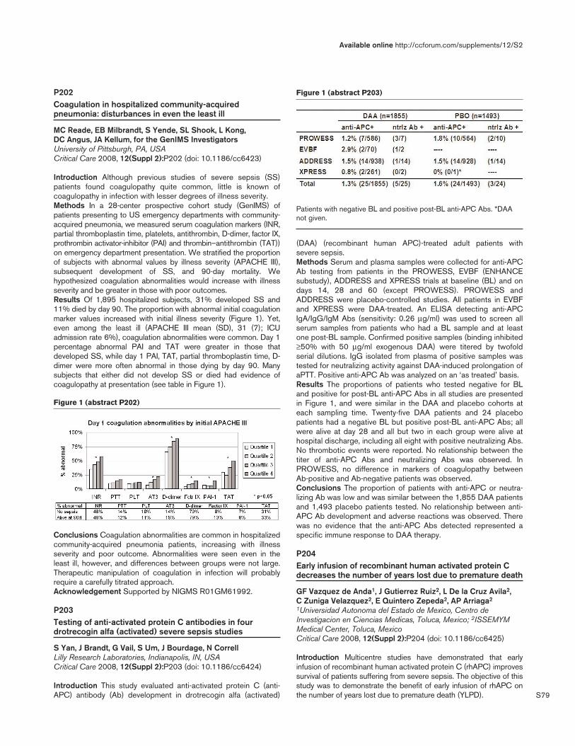
S79
P202Coagulation in hospitalized community-acquiredpneumonia: disturbances in even the least ill
MC Reade, EB Milbrandt, S Yende, SL Shook, L Kong, DC Angus, JA Kellum, for the GenIMS InvestigatorsUniversity of Pittsburgh, PA, USACritical Care 2008, 12(Suppl 2):P202 (doi: 10.1186/cc6423)
Introduction Although previous studies of severe sepsis (SS)patients found coagulopathy quite common, little is known ofcoagulopathy in infection with lesser degrees of illness severity.Methods In a 28-center prospective cohort study (GenIMS) ofpatients presenting to US emergency departments with community-acquired pneumonia, we measured serum coagulation markers (INR,partial thromboplastin time, platelets, antithrombin, D-dimer, factor IX,prothrombin activator-inhibitor (PAI) and thrombin–antithrombin (TAT))on emergency department presentation. We stratified the proportionof subjects with abnormal values by illness severity (APACHE III),subsequent development of SS, and 90-day mortality. Wehypothesized coagulation abnormalities would increase with illnessseverity and be greater in those with poor outcomes.Results Of 1,895 hospitalized subjects, 31% developed SS and11% died by day 90. The proportion with abnormal initial coagulationmarker values increased with initial illness severity (Figure 1). Yet,even among the least ill (APACHE III mean (SD), 31 (7); ICUadmission rate 6%), coagulation abnormalities were common. Day 1percentage abnormal PAI and TAT were greater in those thatdeveloped SS, while day 1 PAI, TAT, partial thromboplastin time, D-dimer were more often abnormal in those dying by day 90. Manysubjects that either did not develop SS or died had evidence ofcoagulopathy at presentation (see table in Figure 1).
Conclusions Coagulation abnormalities are common in hospitalizedcommunity-acquired pneumonia patients, increasing with illnessseverity and poor outcome. Abnormalities were seen even in theleast ill, however, and differences between groups were not large.Therapeutic manipulation of coagulation in infection will probablyrequire a carefully titrated approach.Acknowledgement Supported by NIGMS R01GM61992.
P203Testing of anti-activated protein C antibodies in fourdrotrecogin alfa (activated) severe sepsis studies
S Yan, J Brandt, G Vail, S Um, J Bourdage, N CorrellLilly Research Laboratories, Indianapolis, IN, USACritical Care 2008, 12(Suppl 2):P203 (doi: 10.1186/cc6424)
Introduction This study evaluated anti-activated protein C (anti-APC) antibody (Ab) development in drotrecogin alfa (activated)
(DAA) (recombinant human APC)-treated adult patients withsevere sepsis.Methods Serum and plasma samples were collected for anti-APCAb testing from patients in the PROWESS, EVBF (ENHANCEsubstudy), ADDRESS and XPRESS trials at baseline (BL) and ondays 14, 28 and 60 (except PROWESS). PROWESS andADDRESS were placebo-controlled studies. All patients in EVBFand XPRESS were DAA-treated. An ELISA detecting anti-APCIgA/IgG/IgM Abs (sensitivity: 0.26 μg/ml) was used to screen allserum samples from patients who had a BL sample and at leastone post-BL sample. Confirmed positive samples (binding inhibited≥50% with 50 μg/ml exogenous DAA) were titered by twofoldserial dilutions. IgG isolated from plasma of positive samples wastested for neutralizing activity against DAA-induced prolongation ofaPTT. Positive anti-APC Ab was analyzed on an ‘as treated’ basis.Results The proportions of patients who tested negative for BLand positive for post-BL anti-APC Abs in all studies are presentedin Figure 1, and were similar in the DAA and placebo cohorts ateach sampling time. Twenty-five DAA patients and 24 placebopatients had a negative BL but positive post-BL anti-APC Abs; allwere alive at day 28 and all but two in each group were alive athospital discharge, including all eight with positive neutralizing Abs.No thrombotic events were reported. No relationship between thetiter of anti-APC Abs and neutralizing Abs was observed. InPROWESS, no difference in markers of coagulopathy betweenAb-positive and Ab-negative patients was observed.Conclusions The proportion of patients with anti-APC or neutra-lizing Ab was low and was similar between the 1,855 DAA patientsand 1,493 placebo patients tested. No relationship between anti-APC Ab development and adverse reactions was observed. Therewas no evidence that the anti-APC Abs detected represented aspecific immune response to DAA therapy.
P204Early infusion of recombinant human activated protein Cdecreases the number of years lost due to premature death
GF Vazquez de Anda1, J Gutierrez Ruiz2, L De la Cruz Avila2, C Zuniga Velazquez2, E Quintero Zepeda2, AP Arriaga2
1Universidad Autonoma del Estado de Mexico, Centro deInvestigacion en Ciencias Medicas, Toluca, Mexico; 2ISSEMYMMedical Center, Toluca, MexicoCritical Care 2008, 12(Suppl 2):P204 (doi: 10.1186/cc6425)
Introduction Multicentre studies have demonstrated that earlyinfusion of recombinant human activated protein C (rhAPC) improvessurvival of patients suffering from severe sepsis. The objective of thisstudy was to demonstrate the benefit of early infusion of rhAPC onthe number of years lost due to premature death (YLPD).
Available online http://ccforum.com/supplements/12/S2
Figure 1 (abstract P202)
Figure 1 (abstract P203)
Patients with negative BL and positive post-BL anti-APC Abs. *DAAnot given.
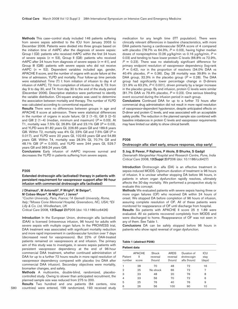
S80
Methods This case–control study included 146 patients sufferingfrom severe sepsis admitted to the ICU from January 2003 toDecember 2006. Patients were divided into three groups based onthe initiation time of rhAPC after the diagnosis of severe sepsis:Group I (GI): patients who received rhAPC within the first 24 hoursof severe sepsis (n = 53), Group II (GII): patients who receivedrhAPC after 24 hours from diagnosis of severe sepsis (n = 41), andGroup III (GIII): patients with severe sepsis who did not receiverhAPC (n = 52). Dependent variables included age, gender,APACHE II score, and the number of organs with acute failure at thetime of admission, YLPD and mortality. Four follow-up time periodswere established: Time (T) I: from initiation of infusion to day 4 ofinfusion of rhAPC, T2: from completion of infusion to day 8, T3: fromday 9 to day 30, and T4: from day 30 to the end of the study period(December 2006). Descriptive statistics were performed to identifythe variable distribution. Chi-square analysis was used to determinethe association between mortality and therapy. The number of YLPDwas calculated according to conventional equations.Results There were no differences between groups in age andAPACHE II score at admission. There were statistical differencesin the number of organs in acute failure; GI 2 (1–5), GII 3 (2–5)and GIII 2 (1–4) (median, minimum and maximum) (P = 0.03). AtT1, mortality was 7.5% GI, 26.8% GII and 23.1% GIII (P = 0.03),and YLPD were 81.82 years GI, 226.65 years GII and 189.6 yearsGIII. Within T2, mortality was 4% GI, 23% GII and 7.5% GIII (P =0.017), and YLPD were 20 years GI, 102.63 years GII and 54.89years GIII. Within T4, mortality was 28.3% GI, 70.7% GII and48.1% GIII (P = 0.000), and YLPD were 244 years GI, 529.7years GII and 369.24 years GIII.Conclusions Early infusion of rhAPC improves survival anddecreases the YLPD in patients suffering from severe sepsis.
P205Extended drotrecogin alfa (activated) therapy in patients withpersistent requirement for vasopressor support after 96-hourinfusion with commercial drotrecogin alfa (activated)
J Dhainaut1, M Antonelli2, P Wright3, M Belger4, M Cobas-Meyer4, M Mignini4, J Janes4
1Cochin University, Paris, France; 2A Gemelli University, Rome,Italy; 3Moses Cone Memorial Hospital, Greensboro, NC, USA; 4EliLilly & Co. Ltd, Windlesham, UKCritical Care 2008, 12(Suppl 2):P205 (doi: 10.1186/cc6426)
Introduction In the European Union, drotrecogin alfa (activated)(DAA) is licensed (intravenous infusion, 96 hours) for adults withsevere sepsis with multiple organ failure. In the PROWESS trial,DAA treatment was associated with significant mortality reductionand more rapid improvement in cardiovascular function over 7 days(decreased need for vasopressors). But 22% of DAA-treatedpatients remained on vasopressors at end infusion. The primaryaim of this study was to investigate, in severe sepsis patients withpersistent vasopressor dependency at the end of 96-hourcommercial DAA treatment, whether continued administration ofDAA for up to a further 72 hours results in more rapid resolution ofvasopressor dependency compared with placebo (no DAA aftercommercial DAA infusion). Secondary objectives were mortality,biomarker changes, and safety.Methods A multicentre, double-blind, randomized, placebo-controlled study. Owing to slower than anticipated recruitment, theplanned sample size was reduced from 275 to 200.Results Two hundred and one patients (64 centers, ninecountries) were entered, 199 randomized, 193 received study
medication for any length time (ITT population). There wereclinically relevant differences in baseline characteristics, with moreDAA patients having a cardiovascular SOFA score of 4 comparedwith placebo (78.7% vs 64.3%, P = 0.03), having higher mediandoses of norepinephrine (0.26 μg/kg/min vs 0.16 μg/kg/min, P =0.03) and tending to have lower protein C levels (66.8% vs 72.9%,P = 0.23). There was no statistically significant difference forprimary endpoint resolution of vasopressor dependency (log-rankP = 0.42), nor in the proportion of resolvers (34.0% DAA vs40.4% placebo, P = 0.36). Day 28 mortality was 39.8% in theDAA group, 32.3% in the placebo group (P = 0.28). The DAAgroup had significantly lower percentage change in D-dimers(21.9% vs 63.2%, P < 0.001), driven primarily by a larger increasein the placebo group. By end infusion, protein C levels were similar(81.7% DAA vs 79.4% placebo, P = 0.23). One serious bleedingevent occurred during the infusion period in each group.Conclusions Continued DAA for up to a further 72 hours aftercommercial drug administration did not result in more rapid resolutionof vasopressor-dependent hypotension, despite anticipated effects onD-dimer and protein C levels, and was associated with an acceptablesafety profile. The reduction in the planned sample size combined withbaseline imbalances in protein C levels and vasopressor requirementsmay have limited our ability to show clinical benefit.
P206Drotrecogin alfa: start early, ensure response, stop early!
S Jog, B Pawar, P Rajhans, P Akole, B Bhurke, S GadgilDeenanath Mangeshkar Hospital and Research Centre, Pune, IndiaCritical Care 2008, 12(Suppl 2):P206 (doi: 10.1186/cc6427)
Introduction Drotrecogin alfa (DA) is an effective treatment insepsis-induced MODS. Optimum duration of treatment is 96 hoursof infusion. It is unclear whether stopping DA before 96 hours, inpatients in whom organ dysfunction rapidly resolves, ultimatelyaffects the 30-day mortality. We performed a prospective study toevaluate this concept.Methods We evaluated patients with severe sepsis having three ormore organ failures (OF) who received DA within 24 hours ofonset. We stopped DA before completion of 96 hours of infusion,assuring complete resolution of OF. All of these patients weremonitored for reappearance of OF until discharge from hospital.Results Six patients with APACHE II score 25 ± 1.89 wereevaluated. All six patients recovered completely from MODS andwere discharged to home. Reappearance of OF was not seen inany of them. See Table 1.Conclusions DA can be safely stopped before 96 hours inpatients who show rapid reversal of organ dysfunction.
Critical Care March 2008 Vol 12 Suppl 2 28th International Symposium on Intensive Care and Emergency Medicine
Table 1 (abstract P206)
Patient data
APACHE Shock ARDS Duration of ICU Patient II reversal reversal drotrecogin stay number score (hours) (hours) alfa (hours) (days)
1 28 70 48 72 162 25 No shock 66 72 73 23 48 20 76 84 23 56 70 72 65 25 76 40 76 96 26 56 100 90 10
S81
P207Association of mortality in the surgical ICU with plasmaconcentrations of plasminogen activator inhibitor-1 andsoluble E-selectin
T Yasuda, M Nakahara, Y Kakihana, Y KanmuraKagoshima University Hospital, Kagoshima, JapanCritical Care 2008, 12(Suppl 2):P207 (doi: 10.1186/cc6428)
Introduction Both plasminogen activator inhibitor-1 (PAI-1) andsoluble E-selectin (sES) are substances activated by cytokines understrong inflammation. PAI-1 is a rapid inhibitor of tissue plasminogenin vivo. PAI-1 is known as one of the markers of systemicinflammatory response syndrome, which is followed by multipleorgan dysfunctions. sES is an adhesion molecule that is expressedfrom endothelial cells activated by TNF. It is reported that elevation ofsES is followed by respiratory failure, which causes acute respiratorydistress syndrome. But it is not clear whether their plasma levelsaffect the mortality and morbidity of critically ill patients. We thereforedivide patients into two groups by the plasma levels of PAI-1 and s-ES and evaluate the mortality respectively.Methods We compared the levels of PAI-1 and sES in survivors withthose in nonsurvivors. We examined 29 patients admitted to oursurgical ICU in the hospital of Kagoshima University. High levels ofPAI-1 are known to be accompanied by hemorrhage after surgery.To evaluate, we therefore use the values of PAI-1 and sES on theadmission day (day 1), day 2 and the day when hemorrhage iscontrolled. The plasma levels of PAI-1 and sES are measured by thelatex agglutination assay with an automatic analyzer (LPIA-NV7;Mitsubishi Kagaku Iatron Co., Tokyo, Japan). For statistical analysis, atwo-sided Fisher exact probability test was used to analyze thedifference in the mortality. P < 0.05 indicated statistical significance.Results Among the patients examined, 11 patients showedelevated tPAI levels (>50 ng/ml) (PE group) and 18 patientsshowed normal tPAI levels (≤50 ng/ml) (PN group). Fourteenpatients showed elevated sES levels (>30 ng/ml) (EE group) and15 patients showed normal sES levels (<30 ng/ml) (EN group).Mortality is significantly higher in the PE group (9/11, 81.8%) andEE group (8/14, 57.1%) than in the PN group (1/18, 5.5%) (P <0.0001) and EN group (2/15, 13.3%) (P < 0.0209), respectively.Conclusions Both the levels of PAI-1 and sES are useful forevaluating prognosis of critically ill patients in the surgical ICU.
P208Efficacy of antithrombin administration in the acute phaseof burn injury
A Lavrentieva, I Houris, S Aspragathou, K Mitka, K Kazanas, M BitzaniG. Papanikolaou Hospital, Thessaloniki, GreeceCritical Care 2008, 12(Suppl 2):P208 (doi: 10.1186/cc6429)
Introduction Severe burn injury is characterized by the activationof coagulation, decreased fibrinolytic activity and decreasednatural anticoagulant activity. The aim of our study was to investi-gate the effect of antithrombin administration on the coagulationstatus and on organ function in the early postburn period.Methods Thirty-one patients admitted to the burn ICU wererandomized into two groups, antithrombin-treated (n = 15) andcontrol (n = 16), for four consecutive days after thermal injury. Theclinical data, coagulation parameters and fibrinolysis parameterswere compared and the adverse effects were monitored.Results Significant differences in the time trend of D-dimers andthrombin–antithrombin complexes were observed between anti-thrombin-treated and control groups (decrease in the antithrombin-
treated group and increase in the control group). According to theInternational Society on Thrombosis and Hemostasis criteria,disseminated intravascular coagulation (DIC) diagnosis was set for28 from 31 patients. The presence of overt DIC was associated withmortality (P = 0.002). The Sequential Organ Failure Assessmentscore time trend differed significantly between the two investigationgroups (decreased in the treated group and did not change in thecontrol group). Antithrombin-treated patients had an absolutereduction in 28-day mortality of 25% compared with the controlgroup (P = 0.004). No treatment-related side effects were observed.Conclusions Treatment with antithrombin seems to affect thecoagulation status and to reduce multiple organ failure incidenceand mortality in the early postburn period.
P209Whole blood coagulation and platelet activation in theathlete: a comparison of marathon, triathlon and long-distance running
A Hanke1, A Staib1, K Görlinger2, M Perrey1, D Dirkmann2, P Kienbaum1
1Heinrich-Heine-Universität Düsseldorf, Germany; 2UniklinikumEssen, GermanyCritical Care 2008, 12(Suppl 2):P209 (doi: 10.1186/cc6430)
Introduction Thromboembolic events have been reported inmarathon athletes during competition. We tested the hypothesisthat activation of coagulation and platelets depends on the type ofendurance sport and running fraction.Methods After ethic committee approval, 68 healthy athletesparticipating in a marathon (MAR, running 42 km, n = 24), atriathlon (TRI, swimming 2.5 km + cycling 90 km + running 21 km,n = 22), and long-distance cycling (CYC, 151 km, n = 22) wereincluded in the study. Blood samples were taken before andimmediately after competition. Rotational thrombelastometry wasperformed (ROTEM; Pentapharm, Germany). The coagulation time(CT) and maximum clot firmness (MCF) after intrinsic activationwas assessed. Platelet aggregation was tested using a multipleplatelet function analyzer (Multiplate; Dynabyte, Germany) byactivation with ADP as well as thrombin-activating peptide 6 andexpressed as the area under the curve (AUC). Statistics used theWilcoxon signed rank test, P < 0.05.Results Complete datasets were obtained in 59 athletes (MAR: n =21, TRI: n = 19, CYC: n = 19). The CT significantly decreased inMAR (from 172 ± 15.3 s to 155 ± 18.3 s), TRI (from 168.1 ± 12.9s to 154.2 ± 11.3 s), and CYC (from 164.7 ± 17.7 s to 152.5 ±13.0 s) without differences between groups. In parallel, the MCFincreased in all groups (MAR: from 58.1 ± 3.9 mm to 62.4 ± 3.8mm, TRI: from 56.1 ± 3.2 mm to 59.5 ± 3.1 mm, CYC: from 59.3 ±5.0 mm to 64.2 ± 4.2 mm). Platelets were only activated during theMAR and TRI, however, as indicated by an increased AUC duringTRAP activation in the MAR (from 919 ± 149 to 1,074 ± 290) andan increased AUC during ADP activation in the MAR (from 532 ±184 to 827 ± 262) and TRI (from 505 ± 205 to 799 ± 329).Conclusions As shown before, coagulation is activated duringphysical activity. We observed significant platelet activation duringa marathon and to a lesser extent during a triathlon. We concludethat prolonged running may increase platelet activity. Moreover, wespeculate that direct mechanical stress during running contributesto the observed effect. Running therefore activates both coagula-tion and platelet activity, resulting in an increased risk of thrombo-embolic incidents in running athletes.Reference1. Sumann G, Fries D, Griesmacher A, et al.: Blood Coagul Fib-
rinolysis 2007, 18:435-440.
Available online http://ccforum.com/supplements/12/S2
S82
P210Admission platelet count as a prognostic indicator inintensive care
V Hariharan, J PaddleRoyal Cornwall Hospital, Truro, UKCritical Care 2008, 12(Suppl 2):P210 (doi: 10.1186/cc6431)
Introduction Abnormal platelet counts are common findings in ICUpatients. Thrombocytopenia is associated with a poor outcome [1].Conversely, thrombocytosis may be associated with an improvedoutcome [2]. We therefore conducted a retrospective observa-tional study in our own unit to investigate this further.Methods All patients admitted to the ICU of a large district generalhospital (Royal Cornwall Hospital) from January 2002 to April2005 were included in this retrospective study. We collected dataon age, sex, admission category, platelet count, APACHE II score,APACHE II predicted mortality, and hospital mortality. The plateletvalue was taken as the lowest platelet count obtained within thefirst 24 hours of ICU admission. The primary outcome was hospitalmortality. Statistical analysis was conducted with SPSS version15.0 using logistical regression models.Results A total of 1,767 patients were admitted during the studyperiod. We excluded 119 patients with no recorded platelet data.We found a strong negative correlation between the admissionplatelet count and mortality, which was significant (P = 0.001,logistic regression). To test this relationship with actual hospitalmortality we divided the cohort into deciles of platelet count andplotted the data against mortality. Those with platelet counts below67 had a mortality rate of 57.2%. This was substantially higher thenthe remaining deciles (P = 0.0001, Fisher’s exact test). We did notdemonstrate any significant reduction in mortality in patients withthrombocytosis (P = 0.523, Fisher’s exact test). We comparedmedical versus surgical patients and found that, for any givenplatelet value, the predicted outcome for surgical patients wasbetter (P = 0.008, t test). We analysed a model that includedplatelets as an additional indicator for outcome. In binary logisticregression analysis there was a significant association betweenplatelet count and mortality (coefficient = 0.998, CI = 0.996–0.999). This association remained significant in a multiple logisticregression model, which included APACHE II (P < 0.001). Amodel including both APACHE II and platelet count improved theproportion of deaths correctly predicted from 69.5% withAPACHE II alone to 71.3% with platelets included.Conclusions We confirmed previous findings that there is acorrelation between low platelet counts and adverse outcome, andwe have further demonstrated that the correlation between plateletcount and predicted mortality exists across the spectrum of plateletvalues. In addition we have demonstrated a difference in mortalitybetween medical and surgical patients for any given admissionplatelet values. Finally, we have demonstrated that platelet valuesprovide additional prognostic information above the APACHE II score.References1. Vanderschueren S, et al.: Crit Care Med 2000, 28:1871-1876.2. Gurung AM, et al.: Br J Anaesth 2001, 87:926-968.
P211A phase 1 trial of nebulized heparin in acute lung injury
B Dixon, D Santamaria, J Campbell, A TobinSt.Vincent’s Health, Melbourne, AustraliaCritical Care 2008, 12(Suppl 2):P211 (doi: 10.1186/cc6432)
Introduction Animal studies of acute lung injury (ALI) suggestnebulized heparin may limit damage from fibrin deposition in the
alveolar space and microcirculation. We therefore undertook a trialto assess the safety and tolerability of nebulized heparin in patientswith ALI.Methods An open-label phase 1 trial of four escalating doses ofnebulized heparin was administered over 2 days. A total of 16ventilated patients with ALI were studied. Each dose was assessedin four patients. The first group was administered 50,000 U/day,the second 100,000 U/day, the third 200,000 U/day and thefourth 400,000 U/day. We measured the arterial to inspired oxygenratio (PaO2/FiO2), lung compliance, the alveolar dead spacefraction, the blood thrombin clotting time and the activated partialthromboplastin time (APTT). Bronchoalveolar lavage (BAL) fluidwas collected and the prothrombin fragment and tissue plasmino-gen activator levels assessed.Results There was no difference between groups in thePaO2/FiO2, lung compliance or the alveolar dead space fractionover the study period. A trend to reduced prothrombin fragmentlevels in BAL fluid was present with higher doses of nebulizedheparin (P = 0.1). Nebulized heparin did not increase tissueplasminogen activator levels in BAL fluid. A trend to increasedblood thrombin clotting time and APTT levels was present withhigher doses of nebulized heparin (P = 0.1 and P = 0.09,respectively). For the highest dose, the APTT reached 64 seconds.Conclusions Nebulized heparin can be administered safely toventilated patients with ALI. At higher doses, nebulized heparinmay limit coagulation activation in the lungs and increase systemicAPTT levels.
P212Thromboelastography in clinical decision-making in thecritically ill patient in a district general hospital ICU
J Louw, T Veenith, P MoondiQueen Elizabeth Hospital, Kings Lynn, UKCritical Care 2008, 12(Suppl 2):P212 (doi: 10.1186/cc6433)
Introduction Thromboelastography (TEG) is a point-of-caremonitoring tool that could help in managing coagulopathy in thecritically ill. This may be beneficial in reducing the length of stay in theICU, guide blood product transfusion and improve patient outcome.Methods We conducted a retrospective analysis of the use of TEGin a busy district general hospital ICU. We included all 100 patientsin whom TEG was performed over 1 year. They required >4 unitsblood intraoperatively or >2 units blood on the ICU, abdominalaortic aneurysm repair or had sepsis. TEG was performed on 212occasions, in parallel with routine coagulation studies.Results We transfused 656 units of packed RBCs, 27 units ofcryoprecipitate, 180 units of FFP and 130 units of platelets,incurring an expenditure of £722,682. The cost of running TEG forthat year was £1,845. Two hundred and twelve clinical decisionswere made following TEG along with clotting results. We identified174 (82.08%) abnormal TEG results, of which 88 (50.57%) wereaccompanied by abnormal clotting. One hundred and eighty-seven(88.21%) clinical decisions were influenced by the TEG result. Inthis group, 171 (91.44%) were related to guiding transfusion ofblood products. Fifteen (8.02%) resulted in a change of medicalmanagement, guiding activated protein C administration, renalreplacement therapy, invasive procedures and starting secondaryanticoagulation prophylaxis.Conclusions Standard coagulation assays do not provide anyinformation on platelet function or fibrinolysis [1]. TEG can replaceclotting studies and the assessment of platelet function [2]. TEGcan guide blood product transfusion in cardiac surgery [1]. TEGcan be done with a fraction of the total costs of transfusion andprovides confidence during the management of coagulopathy.
Critical Care March 2008 Vol 12 Suppl 2 28th International Symposium on Intensive Care and Emergency Medicine
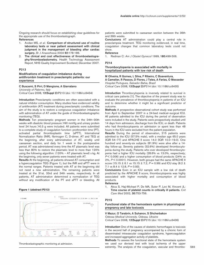
S83
Ongoing research should focus on establishing clear guidelines forthe appropriate use of the thromboelastograph.References1. Avidan MS, et al.: Comparison of structured use of routine
laboratory tests or near patient assessment with clinicaljudgment in the management of bleeding after cardiacsurgery. Br J Anaesthesia 2004 92:178-186.
2. The clinical and cost effectiveness of thromboelastogra-phy/thromboelastometry. Health Technology AssessmentReport, NHS Quality Improvement Scotland; December 2007.
P213Modifications of coagulation imbalance duringantithrombin treatment in preeclamptic patients: ourexperience
C Buscemi, S Pirri, D Mangione, A GiarratanoUniversity of Palermo, ItalyCritical Care 2008, 12(Suppl 2):P213 (doi: 10.1186/cc6434)
Introduction Preeclamptic conditions are often associated with anatural inhibitor consumption. Many studies have evidenced validityof antithrombin (AT) treatment during preeclamptic conditions. Theaim of the study is to restore a congruous coagulation imbalancewith administration of AT under the guide of thromboelastographicmonitoring (TEG).Methods Ten preeclamptic pregnant women in the 24th–30thweeks with diastolic blood pressure >90 mmHg and urinary proteinlevel 24 hours >0.3 g were included. All patients were submittedto a complete study of coagulation function: prothrombin time (PT),activated partial thromboplastin time (aPTT), InternationalNormalization Ratio (INR), fibrinogen C, D-dimer, AT and TEG atthe beginning, after every administration of AT, weekly untilcaesarean section, and daily for 1 week in the postoperativeperiod. AT was administered every time the AT plasmatic level wasless than 80% to restore the plasmatic level to more than 120%using the following algorithm: (120% – AT plasmatic level) x kg. Atthe beginning, only seven patients were treated with AT.Results At the beginning, all patients showed AT consumption anda hypercoagulation TEG (Figure 1), but the INR and aPTT were inthe normal ranges. Patients treated with AT at the beginning didnot need a new administration. The remaining patients weretreated at the 31st, 33rd and 34th weeks, respectively. In allpatients, AT administration determined a normalization of TEGwithout any modification of the PT and aPTT or bleeding. All
patients were submitted to caesarean section between the 36thand 39th weeks.Conclusions AT administration could play a central role inpreeclampsia treatment. TEG monitoring evidenced, in real time,coagulation changes that common laboratory tests could notshow.Reference1. Redman C: Am J Obstet Gynecol 1999, 180:499-506.
P214Thrombocytopenia is associated with mortality inhospitalized patients with low risk of death
M Oliveira, R Gomes, L Silva, F Ribeiro, C Boaventura, A Camelier, R Passos, D Flores, J Teles, A Farias, O MessederHospital Portugues, Salvador Bahia, BrazilCritical Care 2008, 12(Suppl 2):P214 (doi: 10.1186/cc6435)
Introduction Thrombocytopenia is inversely related to survival incritical care patients [1]. The objective of the present study was toevaluate the prevalence of thrombocytopenia in patients of an ICUand to determine whether it might be a significant predictor ofoutcome.Methods A prospective observational cohort study was performedfrom April to September 2007 in a 24-bed medical–surgical ICU.All patients admitted to the ICU during the period of observationwere included in the study. Patients were prospectively studied until14 days from admission, discharge from the ICU, or death. Patientswho had thrombocytopenia on admission or spent less than 48hours in the ICU were excluded from the patient population.Results During the period of observation, 215 patients wereadmitted to the ICU (57.5% male), with a median age 65.0 years(IQR 54–77) and APACHE II score 14.0 (IQR 10.0–19.0). Onehundred and seventy-six subjects (81.9%) were alive after a 14-day follow-up. Seventy patients (32.6%) developed thrombocyto-penia during the study. Patients who ever developed thrombocyto-penia had a higher ICU mortality (28.6% vs 13.0%, respectively;P < 0.006) and a higher consumption of blood products (24% vs2%, P < 0.0001). However, both groups had the same APACHE IIscore (15.15 ± 6.1 vs 15.15 ± 7.2, P = 0.99) and ICU stay (8.2 ±7.1 vs 8.4 ± 12.8, P = 0.93).Conclusions Even in an ICU sample with a low risk of deathpredicted by the APACHE II score, thrombocytopenia was highlyassociated with higher mortality and consumption of bloodproducts.Reference1. Akca S, Haji-Michael P, De MA, Suter P, Levi M, Vincent JL:
Time course of platelet counts in critically ill patients. CritCare Med 2002, 30:753-756.
P215Functional state of the hemostasis system in physiologicalpregnancy and late toxicosis
V Mazur, O Tarabrin, A Suhanov, S ShcherbakovOdessa Medical University, Odessa, UkraineCritical Care 2008, 12(Suppl 2):P215 (doi: 10.1186/cc6436)
Introduction One of the causes of obstetric hemorrhages is toxicosisin the second half of pregnancy accompanied by a chronic form ofdisseminated intravascular coagulation syndrome, hypercoagulationand increased aggregation activity of platelets.Methods To assess the functional state of the hemostasis systemwe used our devised test with local ischemia of the upperextremity. The analysis of the coagulation, vascular and thrombo-
Available online http://ccforum.com/supplements/12/S2
Figure 1 (abstract P213)
Thromboelastographic monitoring at admission.
S84
cytic components of hemostasis and fibrinolysis was made on thebasis of parameters of the blood aggregate state obtained usingthe method of haemoviscoelastography.Results We examined 30 healthy pregnant women in the agerange 20–31 years (control group), and 30 pregnant women withrevealed late toxicosis of different severity degree (nephropathy ofII degree, 10 women; nephropathy of III degree, 20 women). Whileanalyzing the functional state of the hemostasis system in healthypregnant women we distinguished two types of response to thetest: compensated type (1) in 30% and subcompensated type (2)in 70%. The pregnant women suffering from late toxicosis wereregistered to have a subcompensated type of the hemostasissystem response (3) in 20% of cases and a decompensated type(4) in 80% of cases. The functional test in group 1 resulted indecreased aggregation activity of platelets, reduced activity of the Iand II phases of blood coagulation (elevation of r and k) andactivation of the fibrinolytic system. Group 2 is noted to haveenhanced aggregation activity of platelets, enhanced thrombinactivity and acceleration of the thrombin formation and activation ofII and III coagulation phases. The total fibrinolytic blood activity wasreduced by 42%.Conclusions Late toxicosis is therefore accompanied by changesin the hemostasis system, causing exhaustion of compensatorypotentials of the regulation system of the blood aggregate stateand promoting a high risk of thrombohemorrhagic complicationsduring delivery and in the postpartum period.
P216Plasma fibrinolysis is related to the SOFA score but not tothe von Willebrand factor on ICU admission
K Zouaoui Boudjeltia1, S Ollieuz2, M Piagnerelli3, P Biston2, P Cauchie1, M Vanhaeverbeek1
1ISPPC CHU Charleroi, Vesale Hospital, Montigny-le-Tilleul,Belgium; 2ISPPC CHU Charleroi, Belgium; 3Erasme University,Brussels, BelgiumCritical Care 2008, 12(Suppl 2):P216 (doi: 10.1186/cc6437)
Introduction Endothelial cell activation and injuries are importantcauses of multiorgan failure. Altered fibrinolysis promotes fibrindeposition and may create microvascular alterations duringinflammation. C-reactive protein (CRP) is correlated with anincreased risk of MOF [1] and CRP may inhibit fibrinolysis [2]. Weaimed to determine whether plasma fibrinolysis is related to theSOFA score and von Willebrand factor (vWF antigen), as a markerof endothelium dysfunction, in critically ill patients at ICU admission.Methods A cross-sectional study in an adult medicosurgical ICU.Patients were 49 consecutive patients (31 nonseptic and 18septic). Plasma fibrinolysis was assessed by the euglobulin clotlysis time (ECLT) at ICU admission [3].Results The ECLT was significantly longer in septic than in nonsepticpatients (1,219 ± 574 min versus 701 ± 224 min, P = 0.001).Significant correlation between the ECLT and CRP (R = 0.67, P <0.001) and between the ECLT and SOFA score (R = 0.36, P =0.009) were observed. CRP was weakly correlated with vWF (R =0.29; P = 0.04). The vWF was not correlated either with the ECLT(R = –0.06, P = 0.65) or the SOFA score (R = –0.02, P = 0.88).Conclusions The ECLT measurement could be a marker of organdysfunction and a prognosis factor in critically ill patients. Furtherstudies with measurement of plasma fibrinolysis by the ECLTshould be investigated in ICU patients.References1. Lobo SM, et al.: Chest 2003, 123:2043-2049.2. Devaraj S, et al.: Circulation 2003, 107:398-404.3. Boudjeltia Z, et al.: BMC Biotechnol 2002, 2:8.
P217Perioperative monitoring of coagulation in patients afterabdominal surgery
O Tarabrin, V Mazur, A Suhanov, S ShcherbakovOdessa Medical University, Odessa, UkraineCritical Care 2008, 12(Suppl 2):P217 (doi: 10.1186/cc6438)
Introduction Despite the evidence of perioperative hypercoagula-bility in cancer patients, there are no consistent data evaluating theextent, duration, and specific contribution of platelets andprocoagulatory proteins by in vitro testing. This study comparedthe efficacy of haemoviscoelastography (HVG) versus thrombo-elastography (TEG) for monitoring coagulation imbalance.Methods In 108 patients undergoing surgery for abdominal cancerwe examined the efficacy of a variety of coagulation tests. Acomplete coagulation screening, TEG and HVG were performedbefore and at the end of surgery.Results We calculated the elastic shear modulus of standardmaximum amplitude (MA) (Gt) and HVG MA (Gh), which reflect thetotal clot strength and procoagulatory protein component, respec-tively. The difference was an estimate of the platelet component(Gp). There was a 14% perioperative increase of standard MA,corresponding to a 48% increase of Gt (<0.05/J) and an 80–86%contribution of the calculated Gp to Gt. We conclude that serialstandard TEG and HVG viscoelastic tests may reveal theindependent contribution of platelets and procoagulatory proteins toclot strength. The results showed that some components of the TEGfailed to identify hypercoagulation (r < 0.2, P > 0.75). All compo-nents of the HVG test reflected postoperative coagulopathies.Conclusions Hypercoagulability is not reflected completely bystandard coagulation monitoring and TEG, and seems to bepredominantly caused by increased platelet reactivity. HVGprovides a fast and easy-to-perform bedside test to quantify in vitrocoagulation, and may be useful in determining the coagulationstatus of cancer patients perioperatively.Reference1. Samama CM, et al.: Anesthesiology 2001, 94:74-78.
P218Neutrophil oxidative burst evaluation during acutenormovolemic hemodilution
MA Kahvegian1, DT Fantoni2, DA Otsuki1, CA Holms1, CO Massoco2, JO Auler Jr1
1Faculdade de Medicina da Universidade de São Paulo, Brazil;2Faculdade de Medicina Veterinaria da Universidade de São Paulo,BrazilCritical Care 2008, 12(Suppl 2):P218 (doi: 10.1186/cc6439)
Introduction In recent years there has been increasing evidencethat resuscitation strategies with different fluids can have widelydivergent impacts on the immune response and neutrophilactivation. This study was undertaken to determine the neutrophil’soxidative burst in a swine model during the acute normovolemichemodilution (ANH) procedure with hydroxyethyl starch (HES),normal saline solution (NSS) or gelatin (GEL).Methods Twenty-four pigs were anesthetized, instrumented andrandomized into four groups: Control, ANH + HES, ANH + NSSand ANH + GEL. Animals in the ANH group were submitted toacute normovolemic hemodilution to a target hematocrit of 15%with volume replacement performed with HES 130/0.4 and GEL ata 1:1 ratio and NSS at a 3:1 ratio. The withdrawn blood wasreturned to the animals 120 minutes after the end of hemodilution.Neutrophil oxidative burst was performed with blood samples
Critical Care March 2008 Vol 12 Suppl 2 28th International Symposium on Intensive Care and Emergency Medicine
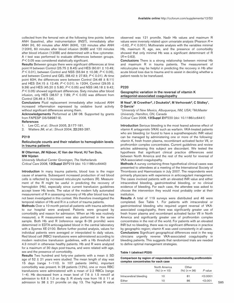
S85
collected from the femoral vein at the following time points: beforeANH (baseline), after instrumentation (INST), immediately afterANH (H), 60 minutes after ANH (60H), 120 minutes after ANH(120H), 60 minutes after blood infusion (60BI) and 120 minutesafter blood infusion (120BI) and determined with a flow cytometer.A t test was performed to evaluate differences between groups.P < 0.05 was considered statistically significant.Results Between groups there were significant differences at timepoint H between Control (25.75 ± 8.45) and HES (60.61 ± 10.49;P < 0.01), between Control and NSS (55.94 ± 10.38; P < 0.05),and between Control and GEL (68.42 ± 27.83; P < 0.01). At timepoint 60H, the differences were between Control (34.48 ± 8.11)and HES (54.15 ± 12.49; P < 0.01). In 120H, Control (29.05 ±9.39) and HES (45.20 ± 5.80; P < 0.05) and NSS (46.18 ± 9.42;P < 0.05) showed significant differences. Sixty minutes after bloodinfusion, only HES (38.57 ± 7.89; P < 0.05) was different fromControl (26.46 ± 7.54).Conclusions Fluid replacement immediately after induced ANHincreased inflammation expressed by oxidative burst activitywithout significant differences among them.Acknowledgements Performed at LIM 08. Supported by grantsfrom FAPESP (05/58987-9).References1. Lee CC, et al.: Shock 2005, 2:177-181.2. Watters JM, et al.: Shock 2004, 22:283-287.
P219Reticulocyte counts and their relation to hemoglobin levelsin trauma patients
M Otterman, JM Nijboer, IC Van der Horst, HJ Ten Duis, MW NijstenUniversity Medical Center Groningen, The NetherlandsCritical Care 2008, 12(Suppl 2):P219 (doi: 10.1186/cc6440)
Introduction In many trauma patients, blood loss is the majorcause of anaemia. Subsequent increased production of red bloodcells is reflected by increased reticulocyte numbers (R). Measure-ment of the R might be useful in predicting the recovery ofhemoglobin (Hb), especially since current transfusion guidelinesaccept lower Hb levels. The value of the modern fully automatedmeasurement of R in assessing recovery of Hb after blood loss hasnot been investigated in this context. We therefore investigated thetemporal relation of Hb and R in a cohort of trauma patients.Methods Over a 10-month period all patients with trauma admittedto our hospital were analysed. Patients were grouped bycomorbidity and reason for admission. When an Hb was routinelymeasured, a R measurement was also performed in the samesample. Both Hb and R (reference range 8–26 promille) weredetermined in EDTA-anticoagulated blood in the central laboratorywith a Sysmex XE-2100. Before further pooled analysis, values forindividual patients were averaged or interpolated to daily values.Red blood cell (RBC) transfusions were administered according tomodern restrictive transfusion guidelines, with a Hb threshold of4.3 mmol/l in otherwise healthy patients. Hb and R were analyzedfor a maximum of 30 days post-trauma, and were related with age,sex and the presence of comorbidity.Results Two hundred and forty-one patients with a mean ± SDage of 52 ± 21 years were studied. The mean length of stay was15 days (range 1–110). In 107 patients (44%), importantcomorbidity was present. In 28 patients (12%), one or more RBCtransfusions were administered with a mean of 2.2 RBCs (range1–4). Hb decreased from a mean level of 7.6 ± 1.5 mmol/l atadmission to 6.8 ± 1.3 on day 3. R slowly rose from 16 ± 11 atadmission to 38 ± 21 promille on day 13. The highest R value
observed was 121 promille. Nadir Hb values and maximum Rvalues were inversely related upon univariate analysis (Pearson R =–0.62, P < 0.001). Multivariate analysis with the variables minimalHb, maximum R, age, sex, and the presence of comorbidityshowed that only minimal Hb was a significant determinant of R(R = 0.63).Conclusions There is a strong relationship between minimal Hband maximum R in trauma patients. The measurement ofreticulocytes may be helpful in predicting the recovery in Hb afteracute blood loss due to trauma and to assist in deciding whether apatient needs to be transfused.
P220Geographic variation in the reversal of vitamin Kantagonist-associated coagulopathy
M Neal1, M Crowther2, J Douketis2, M Verhovsek2, C Stidley1, D Garcia1
1University of New Mexico, Albuquerque, NM, USA; 2McMasterUniversity, Hamilton, ON, CanadaCritical Care 2008, 12(Suppl 2):P220 (doi: 10.1186/cc6441)
Introduction Serious bleeding is the most feared adverse effect ofvitamin K antagonists (VKA) such as warfarin. VKA-treated patientswho are bleeding (or found to have a supratherapeutic INR value)can be managed by administering one or more of the following:vitamin K, fresh frozen plasma, recombinant activated factor VII, orprothrombin complex concentrates. Current guidelines and reviewarticles addressing this subject are discordant. We tested thehypothesis that significant clinical practice differences existbetween North America and the rest of the world for reversal ofVKA-associated coagulopathy.Methods A survey containing three hypothetical clinical cases waspresented to attendees at a meeting of the International Society ofThrombosis and Haemostasis in July 2007. The respondents wereprimarily physicians with experience in anticoagulant management.The cases involved patients with an elevated INR value and eitherintracerebral bleeding, gastrointestinal bleeding, or no clinicalevidence of bleeding. For each case, the attendee was asked tochoose the intervention they would most probably order at theirinstitution.Results A total of 119 surveys were distributed and 46 werecompleted. See Table 1. For patients with intracerebral orgastrointestinal bleeding who required urgent reversal of VKA-associated coagulopathy, there was significantly greater use offresh frozen plasma and recombinant activated factor VII in NorthAmerica and significantly greater use of prothrombin complexconcentrates in the rest of the world. For patients with an elevatedINR but no bleeding, there was no significant difference in practiceby geographic region; vitamin K was used consistently in all cases.Conclusions Significant geographical differences exist in the wayclinicians urgently reverse VKA-associated coagulopathy inbleeding patients. This suggests that randomized trials are neededto define optimal management strategies.
Available online http://ccforum.com/supplements/12/S2
Table 1 (abstract P220)
Comparison by region of respondents recommending prothrombincomplex concentrates for each case
North America Other (%) (n = 10) (%) (n = 36) P value
Intracerebral bleeding 10 81 <0.0001
Either 10 86 <0.001
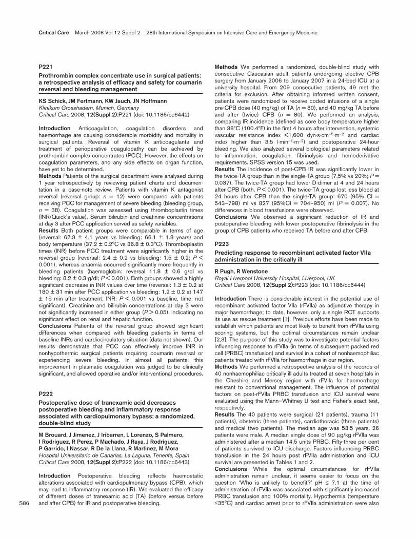
S86
P221Prothrombin complex concentrate use in surgical patients:a retrospective analysis of efficacy and safety for coumarinreversal and bleeding management
KS Schick, JM Fertmann, KW Jauch, JN HoffmannKlinikum Grosshadern, Munich, GermanyCritical Care 2008, 12(Suppl 2):P221 (doi: 10.1186/cc6442)
Introduction Anticoagulation, coagulation disorders andhaemorrhage are causing considerable morbidity and mortality insurgical patients. Reversal of vitamin K anticoagulants andtreatment of perioperative coagulopathy can be achieved byprothrombin complex concentrates (PCC). However, the effects oncoagulation parameters, and any side effects on organ function,have yet to be determined.Methods Patients of the surgical department were analysed during1 year retrospectively by reviewing patient charts and documen-tation in a case-note review. Patients with vitamin K antagonistreversal (reversal group: n = 12) were compared with patientsreceiving PCC for management of severe bleeding (bleeding group,n = 38). Coagulation was assessed using thromboplastin times(INR/Quick’s value). Serum bilirubin and creatinine concentrationsat day 3 after PCC application served as safety variables.Results Both patient groups were comparable in terms of age(reversal: 67.3 ± 4.1 years vs bleeding: 66.1 ± 1.8 years) andbody temperature (37.2 ± 0.2°C vs 36.8 ± 0.3°C). Thromboplastintimes (INR) before PCC treatment were significantly higher in thereversal group (reversal: 2.4 ± 0.2 vs bleeding: 1.5 ± 0.2; P <0.001), whereas anaemia occurred significantly more frequently inbleeding patients (haemoglobin: reversal 11.8 ± 0.6 g/dl vsbleeding: 8.2 ± 0.3 g/dl; P < 0.001). Both groups showed a highlysignificant decrease in INR values over time (reversal: 1.3 ± 0.2 at180 ± 31 min after PCC application vs bleeding: 1.2 ± 0.2 at 147± 15 min after treatment; INR: P < 0.001 vs baseline, time: notsignificant). Creatinine and bilirubin concentrations at day 3 werenot significantly increased in either group (P > 0.05), indicating nosignificant effect on renal and hepatic function.Conclusions Patients of the reversal group showed significantdifferences when compared with bleeding patients in terms ofbaseline INRs and cardiocirculatory situation (data not shown). Ourresults demonstrate that PCC can effectively improve INR innonhypothermic surgical patients requiring coumarin reversal orexperiencing severe bleeding. In almost all patients, thisimprovement in plasmatic coagulation was judged to be clinicallysignificant, and allowed operative and/or interventional procedures.
P222Postoperative dose of tranexamic acid decreasespostoperative bleeding and inflammatory responseassociated with cardiopulmonary bypass: a randomized,double-blind study
M Brouard, J Jimenez, J Iribarren, L Lorenzo, S Palmero, I Rodriguez, R Perez, P Machado, J Raya, J Rodriguez, P Garrido, I Nassar, R De la Llana, R Martinez, M MoraHospital Universitario de Canarias, La Laguna, Tenerife, SpainCritical Care 2008, 12(Suppl 2):P222 (doi: 10.1186/cc6443)
Introduction Postoperative bleeding reflects haemostaticalterations associated with cardiopulmonary bypass (CPB), whichmay lead to inflammatory response (IR). We evaluated the efficacyof different doses of tranexamic acid (TA) (before versus beforeand after CPB) for IR and postoperative bleeding.
Methods We performed a randomized, double-blind study withconsecutive Caucasian adult patients undergoing elective CPBsurgery from January 2006 to January 2007 in a 24-bed ICU at auniversity hospital. From 209 consecutive patients, 49 met thecriteria for exclusion. After obtaining informed written consent,patients were randomized to receive coded infusions of a singlepre-CPB dose (40 mg/kg) of TA (n = 80), and 40 mg/kg TA beforeand after (twice) CPB (n = 80). We performed an analysis,comparing IR incidence (defined as core body temperature higherthan 38ºC (100.4ºF) in the first 4 hours after intervention, systemicvascular resistance index <1,600 dyn·s·cm–5·m–2 and cardiacindex higher than 3.5 l·min–1·m–2) and postoperative 24-hourbleeding. We also analyzed several biological parameters relatedto inflammation, coagulation, fibrinolysis and hemoderivativerequirements. SPSS version 15 was used.Results The incidence of post-CPB IR was significantly lower inthe twice-TA group than in the single-TA group (7.5% vs 20%; P =0.037). The twice-TA group had lower D-dimer at 4 and 24 hoursafter CPB (both, P < 0.001). The twice-TA group lost less blood at24 hours after CPB than the single-TA group: 670 (95% CI =543–798) ml vs 827 (95%CI = 704–950) ml (P = 0.007). Nodifferences in blood transfusions were observed.Conclusions We observed a significant reduction of IR andpostoperative bleeding with lower postoperative fibrinolysis in thegroup of CPB patients who received TA before and after CPB.
P223Predicting response to recombinant activated factor VIIaadministration in the critically ill
R Pugh, R WenstoneRoyal Liverpool University Hospital, Liverpool, UKCritical Care 2008, 12(Suppl 2):P223 (doi: 10.1186/cc6444)
Introduction There is considerable interest in the potential use ofrecombinant activated factor VIIa (rFVIIa) as adjunctive therapy inmajor haemorrhage; to date, however, only a single RCT supportsits use as rescue treatment [1]. Previous efforts have been made toestablish which patients are most likely to benefit from rFVIIa usingscoring systems, but the optimal circumstances remain unclear[2,3]. The purpose of this study was to investigate potential factorsinfluencing response to rFVIIa (in terms of subsequent packed redcell (PRBC) transfusion) and survival in a cohort of nonhaemophiliacpatients treated with rFVIIa for haemorrhage in our region.Methods We performed a retrospective analysis of the records of40 nonhaemophiliac critically ill adults treated at seven hospitals inthe Cheshire and Mersey region with rFVIIa for haemorrhageresistant to conventional management. The influence of potentialfactors on post-rFVIIa PRBC transfusion and ICU survival wereevaluated using the Mann–Whitney U test and Fisher’s exact test,respectively.Results The 40 patients were surgical (21 patients), trauma (11patients), obstetric (three patients), cardiothoracic (three patients)and medical (two patients). The median age was 53.5 years, 26patients were male. A median single dose of 90 μg/kg rFVIIa wasadministered after a median 14.5 units PRBC. Fifty-three per centof patients survived to ICU discharge. Factors influencing PRBCtransfusion in the 24 hours post rFVIIa administration and ICUsurvival are presented in Tables 1 and 2.Conclusions While the optimal circumstances for rFVIIaadministration remain unclear, it seems easier to focus on thequestion ‘Who is unlikely to benefit?’ pH ≤ 7.1 at the time ofadministration of rFVIIa was associated with significantly increasedPRBC transfusion and 100% mortality. Hypothermia (temperature≤35°C) and cardiac arrest prior to rFVIIa administration were also
Critical Care March 2008 Vol 12 Suppl 2 28th International Symposium on Intensive Care and Emergency Medicine
S87
associated with significantly increased PRBC transfusion.Although there have been previous reports of survival followingrFVIIa administration for haemorrhage in the presence of pH < 7.1,such profound acidaemia provides a strong indication that rFVIIa islikely to be futile.References1. Boffard et al.: J Trauma 2005, 59:8-18.2. Biss et al.: Vox Sang 2006, 90:45-52.3. Bowles et al.: Br J Anaesth 2006, 97:476-481.
P224A multicentre prospective open-label study assessingefficacy and safety of a triple-secured fibrinogenconcentrate in the treatment of postpartum haemorrhage
AS Ducloy-Bouthors1, F Broisin2, C Teboul3, A Jaffry3, T Waegemans3, B Padrazzi31Hôpital Jeanne de Flandre, Lille, France; 2Hôpital de la CroixRousse, Lyon, France; 3LFB, Les Ulis, FranceCritical Care 2008, 12(Suppl 2):P224 (doi: 10.1186/cc6445)
Introduction Postpartum haemorrhage (PPH) is a major cause ofglobal maternal morbidity and mortality. The use of haemostaticdrugs in the therapeutic management of patients is mainly empirical.A rationale for an early treatment with fibrinogen, however, has beenrecently suggested [1].Methods A multicentre, noncontrolled, phase 2 study wasperformed to assess the efficacy and the safety of a new triple-secured fibrinogen concentrate (FGT1; LFB, Les Ulis, France) inthe treatment of PPH. A single median dose of 30 mg/kg wasadministered in addition to standard care. Patients were followeduntil 6 weeks after the inclusion. Failure of treatment was definedwhen ultimate resources (that is, invasive hæmostatic interventionor treatment with activated recombinant factor VII) were required to
stop the hæmorrhage, or in case of massive transfusion or death.Other clinical criteria included the course of haemorrhage andinvestigator’s assessment using a four-point scale. Laboratoryassessments were changes in fibrinogen plasma levels and in theFibTEM A15 parameter (RoTEM®). Safety included adverse eventsand vital signs.Results Sixteen patients were included with a median (range)volume of haemorrhage of 1,667 (800; 3,160) ml at baseline.FGT1 succeeded in controlling the haemorrhage in 75% of the 12patients who were clinically assessable. A convergence wasobserved for all efficacy criteria used. The median fibrinogen incre-mental recovery was 10.0 (g/l)/(g/kg) with a 7% concomitantmedian increase of A15 FibTEM. Biological ranges were also verylarge. Higher incremental recovery and relative increases ofFibTEM A15 were associated with clinical success. FGT1 was welltolerated in all patients. Among the 14 adverse events reported,only one was serious, but all were reported as not related to FGT1.No thrombosis or allergic reaction to the study drug occurred.Conclusions This exploratory study suggests efficacy of FGT1 tocontrol PPH in cases of failure of first-line treatments. Most severePPH may require higher doses than used in this study. Theseresults are to be confirmed by larger controlled trials.Reference1. Charbit B, et al.: J Thromb Haemost 2007, 5:266-273.
P225Comparison of prothrombin complex concentrate andfresh frozen plasma on thrombin generation andhemorrhage in experimental dilutional coagulopathy
G Dickneite, I PragstCSL Behring, Marburg, GermanyCritical Care 2008, 12(Suppl 2):P225 (doi: 10.1186/cc6446)
Introduction Thrombin, the key enzyme of blood clotting, isresponsible for the conversion of fibrinogen to fibrin, and for theactivation coagulation factors and platelets. Sufficient thrombingeneration (TG) is therefore crucial for hemostasis. In an experi-mental dilutional coagulapathy in pigs with subsequent bone orspleen injury, the effect of a substitution therapy with prothrombincomplex concentrate (PCC) (Beriplex P/N; CSL Behring) orautologous porcine fresh frozen plasma (pFFP) on TG andhemorrhage was evaluated.Methods A dilutional coagulopathy was induced in 44 anaes-thetized pigs. Erythrocytes were retransfused, the lost volume wasreplaced by hydroxyethyl starch. Animals were randomized to thefollowing two study groups – A: bone injury, (1) placebo (n = 7),(2) PCC 25 U/kg (n = 7), (3) 15 ml/kg pFFP (pFFP15, n = 7), or(4) 40 ml/kg pFFP (pFFP40, n = 4); B: spleen injury, (1) placebo(n = 7), (2) PCC 25 U/kg (n = 6), or (3) 15 ml/kg pFFP (n = 6). A3 mm bone injury was performed by drilling a hole into the femurneck. A spleen incision was created by a scalpel blade. Blood loss(BL) and the time to hemostasis (TH) were determined. TG, theprothrombin time (PT) and coagulation factor levels were measured.Results The dilutional coagulopathy led to a decrease incirculating coagulation factors, a prolonged PT and a decreasedTG. The substitution with PCC and pFFP normalized the impairedPT, but only PCC could normalize the TG. PCC but not pFFPcould restore the decreased plasma levels of coagulation factors ofthe prothrombin complex to sufficiently high levels. In the placebogroup, TH and BL after bone injury were 90.0 ± 27.4 minutes and625 ± 330 ml, and were 82.8 ± 24.5 minutes and 757 ± 251 mlafter spleen injury. After substitution therapy with PCC, asignificantly faster TH of 39.6 ± 9.8 minutes (P = 0.0004) and adecreased BL of 191 ± 119 ml (P = 0.0127) was observed after
Available online http://ccforum.com/supplements/12/S2
Table 1 (abstract P223)
Median post-rFVIIa PRBC transfusion in the presence or absenceof factor
PRBC transfusion (units)
Factor Present Absent P value
pH ≤ 7.1 6 3 0.050*
Temperature ≤ 35°C 8 3 0.001*
Prior cardiac arrest 10 3 0.034*
Prior PRBC transfusion ≥ 20 units 4.5 3 0.062
INR ≥ 1.5 3 3 0.621
Platelet count ≤ 50 x 109/l 4.5 3 0.609
*Significant at P < 0.05.
Table 2 (abstract P223)
Survival in the presence or absence of factor
Survival (%)
Factor Present Absent P value
pH ≤ 7.1 0 63 0.003*
Temperature ≤ 35°C 44 65 0.711
Prior cardiac arrest 33 56 0.398
Prior PRBC transfusion ≥ 20 units 47 56 0.745
INR ≥ 1.5 42 61 0.342
Platelet count ≤ 50 x 109/l 25 56 0.331
*Significant at P < 0.05.
S88
bone injury. After spleen injury, TH was 45.3 ± 9.1 minutes (P =0.0129) and BL was 356 ± 175 ml (P = 0.0152). Neither dose ofpFFP could correct hemorrhage.Conclusions Substitution with PCC but not FFP could normalizeTG and provide sufficient coagulation factors to reduce hemor-rhage. In both models of either venous or arterial injury, TH and BLwere significantly decreased by PCC. It was concluded that thecorrection of TG correlates with hemostasis from a trauma injury.
P226Potential effects of infused particles in paediatric intensivecare patients
T Jack1, B Brent1, M Mueller2, M Boehne1, A Wessel1, M Sasse1
1Medical School Hanover, Germany; 2Fraunhofer ITEM, Hanover,GermanyCritical Care 2008, 12(Suppl 2):P226 (doi: 10.1186/cc6447)
Introduction As a part of a clinical trial to evaluate the potentialbenefits of inline filtration on reducing major complications of paediatricICU (PICU) patients (Clinical Trials.gov ID NCT 00209768), weexamined the physical aspects and chemical composition of par-ticles captured by inline microfilters. Additionally we investigatedthe inflammatory and cytotoxic effects of particles on human endo-thelial cells and macrophages in vitro.Methods We analysed 22 filters used by critically ill children withelectron microscopy and energy dispersion spectroscopy. Theaverage number of particles on the surface as well as theircomposition was examined. In the in vitro model, humanendothelial cells and murine macrophages were exposed todifferent solutions of glass particles and the cytokine levelsassayed to assess their immune response. Levels of IL-1β, IL-6,IL-8, and TNFα were measured.Results The average number of particles found on the surface of afilter membrane was 542/cm2 and energy dispersion spectroscopyanalysis confirmed silicon as one of the major particle constituents.When human endothelial cells and murine macrophages wereexposed to different solutions of glass particles (according to theparticles found on the filter membranes), levels of IL-1β, IL-6, IL-8,and TNFα were found to be significantly suppressed.Conclusions Inline filtration prevents the infusion of potentiallyharmful particles. The suppression of macrophage and endothelialcell cytokine secretion by particles in vitro suggests that theinfusion of microparticles may also contribute to immunecompromisation, which is often seen in vivo in the clinical courseof PICU patients. These findings and their effect on the clinicaloutcome of our PICU patients may be further elucidated.
P227What is the efficacy of a filter needle in the retention oftypical bacterial pathogens?
W Elbaz, J Moore, C Goldsmith, G McCarthy, T MawhinneyBelfast Health and Social Services Trust, Belfast, UKCritical Care 2008, 12(Suppl 2):P227 (doi: 10.1186/cc6448)
Introduction The use of filter needles to prepare medication issuggested as a way of preventing particulate contamination ofinfusions, which is regarded as a source of infection or inflammationin critical care patients [1]. To assess the impact of such needlespurely on bacterial contamination we carried out a comparison ofthe retention of four bacterial pathogens through a standard 25-swgneedle (BD), a standard fill needle with 5 μm filter (BD) and the filterof an Epidural minipack system (Portex). Comparisons were madeat high and then low (‘real world’) bacterial contamination levels.
Methods We prepared four bacterial suspensions mixed togetherin peptone saline to produce Staphylococcus aureus (2.35 x 10⁶
colony-forming units (cfu)/ml, Bacillus cereus (5.77 x 10⁵ cfu/ml),Escherichia coli (4.38 x 10⁶ cfu/ml) and Pseudomonas aeruginosa(3.86 x 10⁶ cfu/ml) per 10 ml test fluid. This volume was theninjected through each type of needle and the epidural filter, afterwhich the filtrate was taken for culture. Small standard amountswere plated on Columbia blood agar while the remainder of thesample (9.9 ml) was mixed with double-strength nutrient broth to beincubated for 24 hours at 37°C. This was repeated with three setsof needles and filters. We then prepared a low-density inoculate of1 ml equating to a total density of 2.63 x 10² cfu of an equal mix ofthe above bacteria. This was injected through six sets of the devicesin question, and both quantitative counts and cultures wereperformed. Student’s t test was used to compare counts.Results No bacteria could be cultured following the use of the 0.2μm epidural filter either from Columbia blood agar or from broth athigh or low contamination levels. In contrast, it was easy to isolateall four pathogens from both needles. In quantitative counts therewas no difference in the mean counts (175 cfu/ml vs 190 cfu/ml)between filtered and unfiltered needles.Conclusions In terms of preventing bacterial transmission wherean infusion is contaminated (even at low levels), the use of a 5 μmfilter needle is no better than a normal needle. In contrast a 0.2 μmfilter is highly efficient at preventing transmission, at least whenresisting a single challenge.Reference1. Stein HG: Medsurg Nurs 2006, 15:290-294.
P228Administration of a standardized plasma-protein solution(Biseko®) in high-risk patients with systemic inflammatoryresponse syndrome: influence on cytokine levels and survival
B Meyer1, A Bojic1, GJ Locker1, T Staudinger1, B Wulkersdorfer1, M Müllner1, A Spittler1, E Schuster1, B Reith2, A Kaye3, M Frass1
1Medical University of Vienna, Austria; 2Clinic of Konstanz,Germany; 3LSU School of Medicine, New Orleans, LA, USACritical Care 2008, 12(Suppl 2):P228 (doi: 10.1186/cc6449)
Introduction Morbidity and mortality in patients suffering fromsystemic inflammatory response syndrome (SIRS) remain high. Thepresent study was carried out because proteins might serve aspromising agents to reduce mortality in SIRS patients. Wetherefore investigated the effects of a plasma-protein solutioncontaining immunoglobulins on serum cytokine levels and survival.Methods This prospective, double-blind, randomized, controlledtrial was performed in the medical ICU of a university hospital.Forty consecutive patients with SIRS were randomized to receiveeither a commercially available standardized plasma-protein solution(Biseko®; Biotest, Dreieich, Germany) [1] consisting of all impor-tant transport and inhibitor proteins as well as immunoglobulins ora 5% albumin solution. Plasma/albumin was given intravenously ata volume of 1,000 ml on the first day and 500 ml/day during thefollowing 4 days. Serum cytokine levels of IL-1β and IL-6 weremeasured on days 1–6, TNFα and TNF-R levels were determinedon days 1 and 14 and at day 28. Survival was assessed on day 28and on day 180.Results Eighteen patients received Biseko®, 20 patients receivedalbumin. Two patients died before receiving the complete studymedication. During days 1–6 of the study period, serum levels ofIL-1β were significantly lower in patients with Biseko® therapycompared with patients receiving albumin (IL-1β AUC 65 ± 71days.pg/ml vs 111 ± 157 days.pg/ml, P = 0.03). No statistically
Critical Care March 2008 Vol 12 Suppl 2 28th International Symposium on Intensive Care and Emergency Medicine
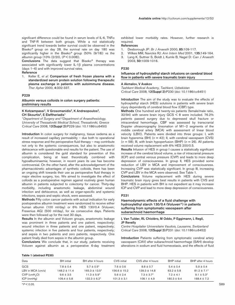
S89
significant difference could be found in serum levels of IL-6, TNFαand TNF-R between both groups. While a not statisticallysignificant trend towards better survival could be observed in theBiseko® group on day 28, the survival rate on day 180 wassignificantly higher in the Biseko® group (50% (9/18)) vs thealbumin group (10% (2/20), (P < 0.008)).Conclusions The data suggest that Biseko® therapy wasassociated with significantly lower IL-1β plasma concentrations(days 1–6) and with improved survival rates.Reference1. Keller E, et al: Comparison of fresh frozen plasma with a
standardized serum protein solution following therapeuticplasma exchange in patients with autoimmune disease.Ther Apher 2000, 4:332-337.
P229Albumin versus colloids in colon surgery patients:preliminary results
K Kotzampassi1, V Grosomanidis2, K Andreopoulos2, CH Skourtis2, E Eleftheriadis1
1Department of Surgery and 2Department of Anaesthesiology,University of Thessaloniki Medical School, Thessaloniki, GreeceCritical Care 2008, 12(Suppl 2):P229 (doi: 10.1186/cc6450)
Introduction In colon surgery for malignancy, tissue oedema as aresult of increased capillary permeability – due both to operationalstress response and the perioperative fluid therapy – may contributenot only to the systemic consequences, but also to anastomoticdehiscence with questionable end results for the patient. The use ofalbumin is considered the gold standard for prevention of thiscomplication, being at least theoretically combined withhypoalbuminaemia; however, in recent years its use has becomecontroversial. On the other hand, after the acknowledgement of thepharmacokinetic advantages of synthetic colloids, there has beenan ongoing shift towards their use as perioperative fluid therapy inmajor elective surgery, too. We aimed to investigate the effect ofcolloids as a postoperative regimen against routinely given humanalbumin in patients subjected to colectomy for cancer. Thirty-daymorbidity, including anastomotic leakage, abdominal woundinfection and dehiscence, as well as organ-specific and systemicinfections, sepsis and septic shock, were assessed.Methods Fifty colon cancer patients with actual indication for earlypostoperative albumin treatment were randomized to receive eitherhuman albumin (100 ml/day) or 6% HES 130/0.4 (Voluven;Fresenius AG) (500 ml/day), for six consecutive days. Patientswere then followed up for the next 30 days.Results In the albumin and Voluven groups, anastomotic leakagewas prominent in three patients and one patient, respectively;wound infection in three patients and one patient, respectively;systemic infection in five patients and four patients, respectively;and sepsis in two patients and zero patients, respectively. Onepatient finally died from sepsis in the albumin group.Conclusions We conclude that, in our study, patients receivingVoluven against albumin as a perioperative 6-day treatment
exhibited lower morbidity rates. However, further research isrequired.References1. Desborough JP: Br J Anaesth 2000, 85:109-117.2. Wilkes MM, Navickis RJ: Ann Intern Med 2001, 135:149-164.3. Lang K, Suttner S, Boldt J, Kumle B, Nagel D: Can J Anaesth
2003, 50:1009-1016.
P230Influence of hydroxylethyl starch infusions on cerebral bloodflow in patients with severe traumatic brain injury
A Akmalov, V AvakovTashkent Medical Academy, Tashkent, UzbekistanCritical Care 2008, 12(Suppl 2):P230 (doi: 10.1186/cc6451)
Introduction The aim of the study was to evaluate the effects ofhydroxylethyl starch (HES) solutions in patients with severe braininjury dependently of cerebral blood flow (CBF) type.Methods One hundred and twenty-six patients (female/male ratio,32/94) with severe brain injury GCS < 8 were included. 76.2%patients passed surgery due to depressed skull fracture orintracerebral hemorrhage. CBF was assessed by transcranialDoppler ultrasonography (insonation of M1–2 segments of themiddle cerebral artery (MCA) with assessment of linear bloodvelocity (LBV)). Patients were divided into three groups: I, withbrain hyperemia (BH) (n = 42); II, with cerebral vasospasm (CVS)(n = 58); III, with brain hypoperfusion (BHP) (n = 26). All patientsreceived volume replacement with 6% HES 200/0.5.Results Infusion of HES in group I causes a statistically significantincrease of the cerebral blood volume, a rise of intracranial pressure(ICP) and central venous pressure (CVP) and leads to more deepdepression of consciousness. In group II, HES provided somereduction of LBV in MCA and improvement of consciousness.Increasing CVP was statistically significant. In group III, increasingCVP and LBV in the MCA were observed. See Table 1.Conclusions Volume replacement with HES during severetraumatic brain injury gives best results in patients with CVS andBHP. HES in patients with BH is not expedient as it may increaseICP and CVP and lead to more deep depression of consciousness.
P231Haemodynamic effects of a fluid challenge withhydroxylethyl starch 130/0.4 (Voluven®) in patientssuffering from symptomatic vasospasm aftersubarachnoid haemorrhage
L Van Tulder, RL Chioléro, M Oddo, P Eggimann, L Regli, JP RevellyCentre Hospitalier Universitaire Vaudois, Lausanne, SwitzerlandCritical Care 2008, 12(Suppl 2):P231 (doi: 10.1186/cc6452)
Introduction Patients suffering from symptomatic cerebral arteryvasospasm (CAV) after subarachnoid haemorrhage (SAH) developalterations in sodium and fluid homeostasis, and the effects of fluid
Available online http://ccforum.com/supplements/12/S2
Table 1 (abstract P230)
Data BH initial BH after 4 hours CVS initial CVS after 4 hours BHP initial BHP after 4 hours
GCS 7.8 ± 0.4 5.7 ± 0.5* 7.6 ± 0.6 8.8 ± 0.7 5.4 ± 0.4 5.9 ± 0.4LBV in MCA (cm/s) 148.2 ± 11.4 165.3 ± 13.5* 156.6 ± 15.2 139.3 ± 14.8 63.2 ± 5.8 81.2 ± 7.1*CVP (cmH2O) 9.6 ± 3.5 11.3 ± 5.9* 5.6 ± 2.4 7.3 ± 3.7* 7.2 ± 4.1 9.1 ± 5.3*ICP (mmHg) 109.4 ± 5.8 132.3 ± 6.2* 101.3 ± 3.1 108.1 ± 4.9 180.3 ± 9.4 168.4 ± 7.2
*P < 0.05.
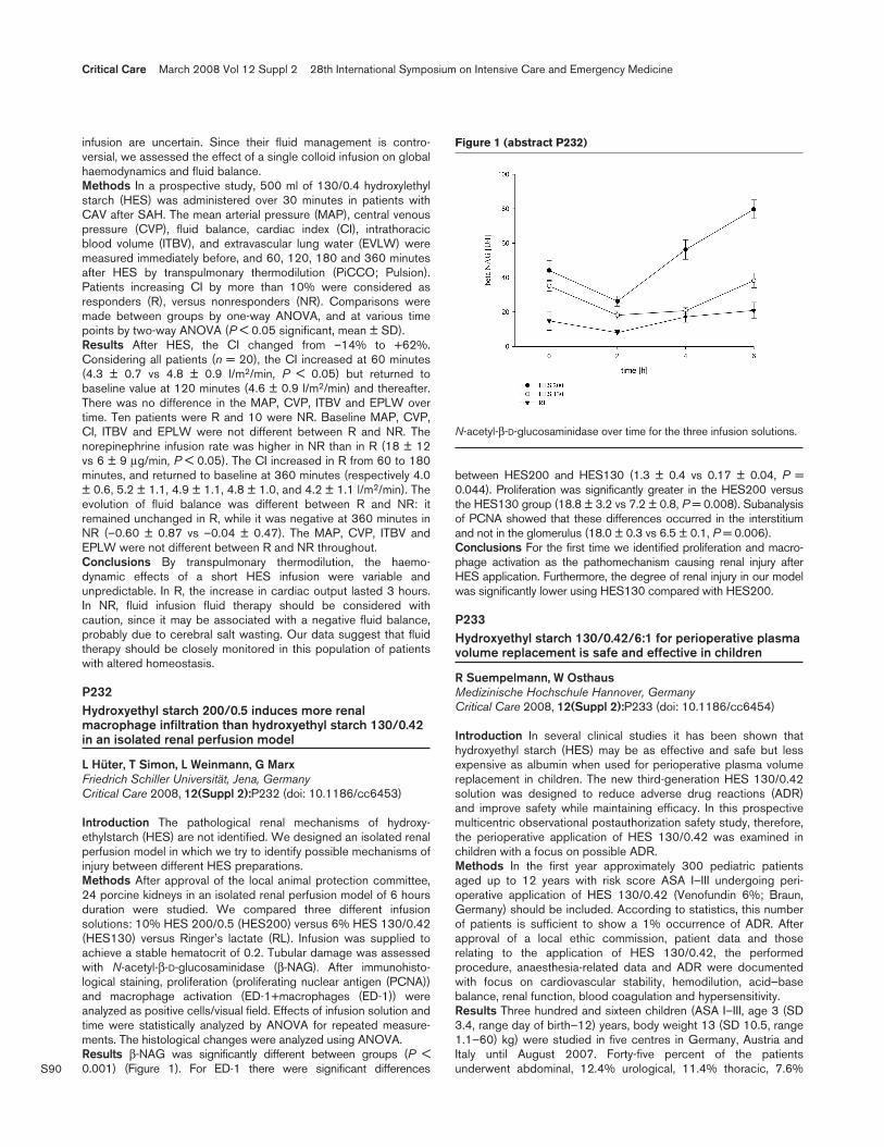
S90
infusion are uncertain. Since their fluid management is contro-versial, we assessed the effect of a single colloid infusion on globalhaemodynamics and fluid balance.Methods In a prospective study, 500 ml of 130/0.4 hydroxylethylstarch (HES) was administered over 30 minutes in patients withCAV after SAH. The mean arterial pressure (MAP), central venouspressure (CVP), fluid balance, cardiac index (CI), intrathoracicblood volume (ITBV), and extravascular lung water (EVLW) weremeasured immediately before, and 60, 120, 180 and 360 minutesafter HES by transpulmonary thermodilution (PiCCO; Pulsion).Patients increasing CI by more than 10% were considered asresponders (R), versus nonresponders (NR). Comparisons weremade between groups by one-way ANOVA, and at various timepoints by two-way ANOVA (P < 0.05 significant, mean ± SD).Results After HES, the CI changed from –14% to +62%.Considering all patients (n = 20), the CI increased at 60 minutes(4.3 ± 0.7 vs 4.8 ± 0.9 l/m2/min, P < 0.05) but returned tobaseline value at 120 minutes (4.6 ± 0.9 l/m2/min) and thereafter.There was no difference in the MAP, CVP, ITBV and EPLW overtime. Ten patients were R and 10 were NR. Baseline MAP, CVP,CI, ITBV and EPLW were not different between R and NR. Thenorepinephrine infusion rate was higher in NR than in R (18 ± 12vs 6 ± 9 μg/min, P < 0.05). The CI increased in R from 60 to 180minutes, and returned to baseline at 360 minutes (respectively 4.0± 0.6, 5.2 ± 1.1, 4.9 ± 1.1, 4.8 ± 1.0, and 4.2 ± 1.1 l/m2/min). Theevolution of fluid balance was different between R and NR: itremained unchanged in R, while it was negative at 360 minutes inNR (–0.60 ± 0.87 vs –0.04 ± 0.47). The MAP, CVP, ITBV andEPLW were not different between R and NR throughout.Conclusions By transpulmonary thermodilution, the haemo-dynamic effects of a short HES infusion were variable andunpredictable. In R, the increase in cardiac output lasted 3 hours.In NR, fluid infusion fluid therapy should be considered withcaution, since it may be associated with a negative fluid balance,probably due to cerebral salt wasting. Our data suggest that fluidtherapy should be closely monitored in this population of patientswith altered homeostasis.
P232Hydroxyethyl starch 200/0.5 induces more renalmacrophage infiltration than hydroxyethyl starch 130/0.42in an isolated renal perfusion model
L Hüter, T Simon, L Weinmann, G MarxFriedrich Schiller Universität, Jena, GermanyCritical Care 2008, 12(Suppl 2):P232 (doi: 10.1186/cc6453)
Introduction The pathological renal mechanisms of hydroxy-ethylstarch (HES) are not identified. We designed an isolated renalperfusion model in which we try to identify possible mechanisms ofinjury between different HES preparations.Methods After approval of the local animal protection committee,24 porcine kidneys in an isolated renal perfusion model of 6 hoursduration were studied. We compared three different infusionsolutions: 10% HES 200/0.5 (HES200) versus 6% HES 130/0.42(HES130) versus Ringer’s lactate (RL). Infusion was supplied toachieve a stable hematocrit of 0.2. Tubular damage was assessedwith N-acetyl-β-D-glucosaminidase (β-NAG). After immunohisto-logical staining, proliferation (proliferating nuclear antigen (PCNA))and macrophage activation (ED-1+macrophages (ED-1)) wereanalyzed as positive cells/visual field. Effects of infusion solution andtime were statistically analyzed by ANOVA for repeated measure-ments. The histological changes were analyzed using ANOVA.Results β-NAG was significantly different between groups (P <0.001) (Figure 1). For ED-1 there were significant differences
between HES200 and HES130 (1.3 ± 0.4 vs 0.17 ± 0.04, P =0.044). Proliferation was significantly greater in the HES200 versusthe HES130 group (18.8 ± 3.2 vs 7.2 ± 0.8, P = 0.008). Subanalysisof PCNA showed that these differences occurred in the interstitiumand not in the glomerulus (18.0 ± 0.3 vs 6.5 ± 0.1, P = 0.006).Conclusions For the first time we identified proliferation and macro-phage activation as the pathomechanism causing renal injury afterHES application. Furthermore, the degree of renal injury in our modelwas significantly lower using HES130 compared with HES200.
P233Hydroxyethyl starch 130/0.42/6:1 for perioperative plasmavolume replacement is safe and effective in children
R Suempelmann, W OsthausMedizinische Hochschule Hannover, GermanyCritical Care 2008, 12(Suppl 2):P233 (doi: 10.1186/cc6454)
Introduction In several clinical studies it has been shown thathydroxyethyl starch (HES) may be as effective and safe but lessexpensive as albumin when used for perioperative plasma volumereplacement in children. The new third-generation HES 130/0.42solution was designed to reduce adverse drug reactions (ADR)and improve safety while maintaining efficacy. In this prospectivemulticentric observational postauthorization safety study, therefore,the perioperative application of HES 130/0.42 was examined inchildren with a focus on possible ADR.Methods In the first year approximately 300 pediatric patientsaged up to 12 years with risk score ASA I–III undergoing peri-operative application of HES 130/0.42 (Venofundin 6%; Braun,Germany) should be included. According to statistics, this numberof patients is sufficient to show a 1% occurrence of ADR. Afterapproval of a local ethic commission, patient data and thoserelating to the application of HES 130/0.42, the performedprocedure, anaesthesia-related data and ADR were documentedwith focus on cardiovascular stability, hemodilution, acid–basebalance, renal function, blood coagulation and hypersensitivity.Results Three hundred and sixteen children (ASA I–III, age 3 (SD3.4, range day of birth–12) years, body weight 13 (SD 10.5, range1.1–60) kg) were studied in five centres in Germany, Austria andItaly until August 2007. Forty-five percent of the patientsunderwent abdominal, 12.4% urological, 11.4% thoracic, 7.6%
Critical Care March 2008 Vol 12 Suppl 2 28th International Symposium on Intensive Care and Emergency Medicine
Figure 1 (abstract P232)
N-acetyl-β-D-glucosaminidase over time for the three infusion solutions.
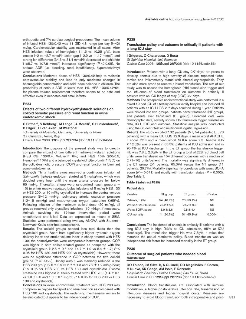
S91
orthopedic and 7% cardiac surgical procedures. The mean volumeof infused HES 130/0.42 was 11 (SD 4.8, range per day 5–42)ml/kg. Cardiovascular stability was maintained in all cases. AfterHES infusion, values of hemoglobin (11.5 vs 10.25 g/dl), baseexcess (–2 vs –2.7 mmol/l), anion gap (12.9 vs 11.17 mmol/l) andstrong ion difference (34.3 vs 31.4 mmol/l) decreased and chloride(105.7 vs 107.8 mmol/l) increased significantly (P < 0.05). Noserious ADR (i.e. bleeding, renal insufficiency, hypersensitivity)were observed.Conclusions Moderate doses of HES 130/0.42 help to maintaincardiovascular stability and lead to only moderate changes inhaemoglobin concentration and acid–base balance in children. Theprobability of serious ADR is lower than 1%. HES 130/0.42/6:1for plasma volume replacement therefore seems to be safe andeffective even in neonates and small infants.
P234Effects of two different hydroxyethylstarch solutions oncolloid osmotic pressure and renal function in ovineendotoxemic shock
C Ertmer1, S Rehberg1, M Lange1, A Morelli2, C Hucklenbruch1,B Ellger1, H Van Aken1, M Westphal11University of Muenster, Germany; 2University of Rome ‘La Sapienza’, Rome, ItalyCritical Care 2008, 12(Suppl 2):P234 (doi: 10.1186/cc6455)
Introduction The purpose of the present study was to directlycompare the impact of two different hydroxyethylstarch solutions(HES 6% 130/0.4, Voluven® 6%; and HES 10% 200/0.5,Hemohes® 10%) and a balanced crystalloid (Sterofundin® ISO) onthe colloid-osmotic pressure (COP) and renal function in fulminantovine endotoxemia.Methods Thirty healthy ewes received a continuous infusion ofSalmonella typhosa endotoxin started at 5 ng/kg/min, which wasdoubled every hour until the mean arterial pressure fell below65 mmHg. Thereafter, sheep were randomized (each group n =10) to either receive repeated bolus infusions of 5 ml/kg HES 130or HES 200, or 10 ml/kg crystalloid to increase the central venouspressure (8–12 mmHg), pulmonary arterial occlusion pressure(12–15 mmHg) and mixed-venous oxygen saturation (≥65%).Following infusion of the maximum colloid dose (20 ml/kg), allgroups received only crystalloid infusions (10 ml/kg), if necessary.Animals surviving the 12-hour intervention period wereanesthetized and killed. Data are expressed as means ± SEM.Statistics were performed using two-way ANOVA with Student–Newman–Keuls post-hoc comparisons.Results The colloid groups needed less total fluids than thecrystalloid group. Apart from significantly higher systemic oxygendelivery index and stroke volume index in sheep treated with HES130, the hemodynamics were comparable between groups. COPwas higher in both colloid-treated groups as compared with thecrystalloid group (12.5 ± 0.6 and 14.7 ± 1.0 vs 8.4 ± 1.7; P <0.05 for HES 130 and HES 200 vs crystalloids). However, therewas no significant difference in COP between the two colloidgroups (P = 0.429). Urinary output was markedly reduced in theHES 200 group (2.5 ± 0.9 vs 5.7 ± 1.3 and 7.2 ± 1.2 ml/kg/hour;P < 0.05 for HES 200 vs HES 130 and crystalloids). Plasmacreatinine was highest in sheep treated with HES 200 (1.4 ± 0.1vs 1.0 ± 0.0 and 1.0 ± 0.1 mg/dl; P < 0.05 for HES 200 vs HES130 and crystalloids).Conclusions In ovine endotoxemia, treatment with HES 200 maycompromise oxygen transport and renal function as compared withHES 130 and crystalloids. The underlying mechanisms remain tobe elucidated but appear to be independent of COP.
P235Transfusion policy and outcome in critically ill patients witha long ICU stay
I Grigoras, O Chelarescu, D RusuSf Spiridon Hospital, Iasi, RomaniaCritical Care 2008, 12(Suppl 2):P235 (doi: 10.1186/cc6456)
Introduction Patients with a long ICU stay (>7 days) are prone todevelop anemia due to high severity of disease, repeated flebo-tomies and inflammatory status with altered erythropoiesis. Theyare also more prone to receive a blood transfusion. The aim of ourstudy was to assess the hemoglobin (Hb) transfusion trigger andthe influence of blood transfusion on outcome in critically illpatients with an ICU length of stay (LOS) >7 days.Methods The prospective noninterventional study was performed in amixed 19-bed ICU of a tertiary care university hospital and included allpatients with an ICU LOS > 7 days admitted during 1 year. Patientswere divided into two groups: patients never transfused (NT group),and patients ever transfused (ET group). Collected data weredemographic data, severity scores, Hb transfusion trigger, transfusiondata, ICU LOS and outcome. Statistical analysis was conductedusing the Student t test and multinomial logistic regression.Results The study enrolled 132 patients (NT, 54 patients; ET, 78patients) with a mean ICU LOS 12.9 days, a mean worst APACHEII score 22.8 and a mean worst SOFA score 9.3. Anemia (Hb< 12 g%) was present in 83.3% patients at ICU admission and in95.4% at ICU discharge. In the ET group the transfusion triggerHb was 7.8 ± 2.3g%. In the ET group a total of 228 red blood cellunits were transfused on 154 different occasions with a median of2 (1–16) units/patient. The mortality was significantly different inthe ET group (51 patients, 65.3%) versus the NT group (11patients, 20.7%). Mortality significantly correlates with worst SOFAscore (P = 0.041) and mostly with transfusion status (P = 0.002).See Table 1.
Table 1 (abstract P235)
Patient data
Variable NT group ET group P value
Patients, n (%) 54 (40.9%) 78 (59.1%) NS
Worst APACHE score 23.2 ± 9.5 22.2 ± 9.8 NS
Worst SOFA score 8.9 ± 3.6 9.8 ± 4.4 NS
ICU mortality 11 (20.7%) 51 (65.3%) 0.0004
Conclusions The incidence of anemia in critically ill patients with along ICU stay is high (83% at ICU admission, 95% at ICUdischarge). The transfusion trigger Hb was 7.8g%, a value thatmatches the actual restrictive policy. Blood transfusion was anindependent risk factor for increased mortality in the ET group.
P236Outcome of surgical patients who needed bloodtransfusion
DO Toledo, JM Silva Jr, A Gulinelli, DD Magalhães, F Correa, H Nuevo, KR Genga, AM Isola, E RezendeHospital do Servidor Público Estadual, São Paulo, BrazilCritical Care 2008, 12(Suppl 2):P236 (doi: 10.1186/cc6457)
Introduction Blood transfusions are associated with immunemodulation, a higher postoperative infection rate, transmission ofinfectious diseases and higher hospital costs. Measures arenecessary to avoid blood transfusion both intraoperative and post-
Available online http://ccforum.com/supplements/12/S2
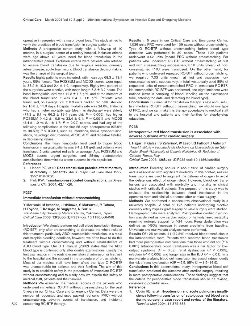
S92
operative in surgeries with a major blood loss. This study aimed toverify the practices of blood transfusion in surgical patients.Methods A prospective cohort study, with a follow-up of 10months, in a surgical center of a tertiary hospital. Inclusion criteriawere age above 18 years, need for blood transfusion in theintraoperative period. Exclusion criteria were patients who refusedto receive blood transfusion due to religious reasons, coronaryartery disease, acute brain injury. Blood transfusion decision-takingwas the charge of the surgical team.Results Eighty patients were included, with mean age 68.3 ± 13.1years, 55% female. The POSSUM and MODS scores were equalto 36.2 ± 10.3 and 2.4 ± 1.9, respectively. Eighty-two percent ofthe surgeries were elective, with mean length 6.3 ± 3.2 hours. Thebasal hemoglobin level was 12.3 ± 1.6 g/dl, and at the moment ofthe blood transfusion it was 8.4 + 1.8 g/dl. Patients weretransfused, on average, 2.3 ± 0.9 units packed red cells, stockedfor 16.8 ± 11.8 days. Hospital mortality rate was 24.6%. Patientswho had a higher mortality rate (death vs discharge) were elderly(77.3 ± 8.1 vs 66.2 ± 13.4 years old; P = 0.005), had higherPOSSUM (44.2 ± 10.6 vs 33.4 ± 9.1; P = 0.001) and MODS(3.4 ± 1.9 vs 2.1 ± 1.7; P = 0.02) scores, and had any of thefollowing complications in the first 28 days postoperative (92.9%vs 39.5%; P < 0.001), such as infections, tissue hypoperfusion,shock, neurologic disturbances, ARDS, ARF, and digestive fistulae,in decreasing values.Conclusions The mean hemoglobin level used to trigger bloodtransfusion in surgical patients was 8.4 ± 1.8 g/dl, and patients weretransfused 2 units packed red cells on average. Age, POSSUM andMODS scores, urgent surgeries, and 28-day postoperativecomplications determined a worse outcome in this population.References1. Hèbert PC, et al.: Does transfusion practice affect mortality
in critically ill patients? Am J Respir Crit Care Med 1997,155:1618-1623.
2. Park KW: Transfusion-associated complications. Int Anes-thesiol Clin 2004, 42:11-26.
P237Immediate transfusion without crossmatching
Y Moriwaki, M Iwashita, J Ishikawa, S Matsuzaki, Y Tahara, H Toyoda, T Kosuge, M Sugiyama, N SuzukiYokohama City University Medical Center, Yokohama, JapanCritical Care 2008, 12(Suppl 2):P237 (doi: 10.1186/cc6458)
Introduction We should perform red cell blood transfusion therapy(RC-BTF) only after crossmatching to decrease the whole risks ofthis treatment, particularly ABO-incompatible transfusion. In a rapidcatastrophic bleeding condition, however, we often have to do thistreatment without crossmatching and without establishment ofABO blood type. Our BTF manual (2002) states that the ABOblood type is confirmed only after double examinations, usually thefirst examination in the routine examination at admission or first visitto the hospital and the second in the procedure of crossmatching.Most of our medical staff have been afraid that type O RC-BTFwas unacceptable for patients and their families. The aim of thisstudy is to establish safety in the procedure of immediate RC-BTFwithout crossmatching and to clarify how we explain this safety tomedical staff, patients and their families.Methods We examined the medical records of the patients whounderwent immediate RC-BTF without crossmatching for the past5 years in our Critical Care and Emergency Center. Data were thenumber of requested and used packed red cells (PRC) withoutcrossmatching, adverse events of transfusion, and incidentsconcerning RC-BTF therapy.
Results In 5 years in our Critical Care and Emergency Center,1,036 units PRC were used for 109 cases without crossmatching.Type O RC-BTF without crossmatching before blood typedetection was performed in 30 cases. These 109 patientsunderwent 9.42 units (mean) PRC without crossmatching. Forpatients who underwent RC-BTF without crossmatching at firstand with crossmatching successively, 6.10 units (mean) of non-crossmatched PRC were transfused. On the other hand, forpatients who underwent repeated RC-BTF without crossmatching,we required 7.03 units (mean) at first and excessive non-crossmatched units successively. In total, we actually used 85% ofrequested units of noncrossmatched PRC in immediate RC-BTF.No incompatible RC-BTF was performed, and eight incidents werenoticed (error in sampling of blood, labeling on the examinationtube, entering the data, and detecting the blood type).Conclusions Our manual for transfusion therapy is safe and useful.In immediate RC-BTF without crossmatching, we should use typeO PRC, and we can make this therapy acceptable to medical staffin the hospital and patients and their families for step-by-stepeducation.
P238Intraoperative red blood transfusion is associated withadverse outcome after cardiac surgery
L Hajjar1, F Galas1, S Zeferino1, W Leao1, G Fallico2, J Auler Jr1
1Heart Institute – Faculdade de Medicina da Universidade de SãoPaulo, Brazil; 2University of Catania Policlinico G. Rodolico ofCatania, Trieste, ItalyCritical Care 2008, 12(Suppl 2):P238 (doi: 10.1186/cc6459)
Introduction Bleeding occurs in about 20% of cardiac surgeryand is associated with significant morbidity. In this context, red celltransfusions are used to augment the delivery of oxygen to avoidthe deleterious effect of oxygen debt. However, blood cell trans-fusions are associated with morbidity and mortality in clinicalstudies with critically ill patients. The purpose of this study was toevaluate the relationship between blood transfusion in theoperative room and clinical outcomes after cardiac surgery.Methods We performed a consecutive observational study in auniversity hospital. A total of 125 patients undergoing electivecoronary artery bypass graft surgery or valve surgery were studied.Demographic data were analyzed. Postoperative cardiac dysfunc-tion was defined as low cardiac output or hemodynamic instabilityrequiring inotropic support for >24 hours. Renal dysfunction wasdefined as >50% increase in serum creatinine from baseline.Univariate and multivariate analyses were performed.Results Of 125 patients, 41 (32.8%) received blood transfusion inthe intraoperative room. Patients who received blood transfusionhad more postoperative complications than those who did not (P <0.001). Intraoperative blood transfusion was a risk factor for low-output syndrome (P = 0.02), renal dysfunction (P < 0.003),infection (P < 0.008) and longer stay in the ICU (P = 0.01). In amultivariate analysis, blood cell transfusion increased independentlythe risk of renal dysfunction (OR = 5.5, 95% CI = 1.5–16.9).Conclusions In this observational study, intraoperative blood celltransfusion predicted the outcome after cardiac surgery, resultingin more postoperative complications. These findings suggest thatthe criteria for perioperative blood transfusion should be revised,considering potential risks.Reference1. Covin RB, et al.: Hypotension and acute pulmonary insuffi-
ciency following transfusion of autologous red blood cellsduring surgery: a case report and review of the literature.Transfus Med 2004, 14:375-383.
Critical Care March 2008 Vol 12 Suppl 2 28th International Symposium on Intensive Care and Emergency Medicine
S93
P239Acute transfusion reactions in critically ill pediatric patients
N Ozata, D Demirkol, M Karabocuoglu, A Citak, R Ucsel, N UzelIstanbul Medical Faculty, Istanbul, TurkeyCritical Care 2008, 12(Suppl 2):P239 (doi: 10.1186/cc6460)
Introduction This study was undertaken to determine the inci-dence, type, and severity of acute transfusion reactions observed ina tertiary care pediatric ICU.Methods All transfusions of blood product administered toconsecutive patients admitted to our pediatric ICU, betweenFebruary 2006 and February 2007, were prospectively recorded.For each transfusion, the bedside nurse recorded the patient’sstatus before, during, and up to 4 hours after the transfusion, aswell as the presence of any new sign or symptom suggesting anacute transfusion reaction.Results A total of 651 transfusions were administered during thestudy period. Sixty-one febrile nonhemolytic transfusion reactions(9.4%) were recorded. No allergic and hypotansive reactions andtransfusion-related acute lung injury were seen. Seventy-sevenpercent (n = 47) of the febrile reactions were recorded during thered blood cell transfusions.Conclusions The incidence of febrile nonhemolytic reactions washigher when compared with similar studies. Possible cause may benot using leuko-reduced components. Transfusion-related acutelung injury is not common in critically ill pediatric patients. Theseestimates are useful for decisions concerning transfusion therapy,and for evaluating efficacy of interventions to reduce risk incritically ill pediatric patients.References1. Gauvin F, Lacroix J, Robillard P, Lapointe H, Hume H: Acute
transfusion reactions in the pediatric intensive care unit.Transfusion 2006. 46:1899-1908.
2. Sanchez R, Toy P: Transfusion related acute lung injury: apediatric perspective. Pediatr Blood Cancer 2005, 45:248-255.
P240Utility of an artificial oxygen carrier in a rat haemorrhagicshock model
M Yanai, K Morisawa, H Takahashi, S Fujitani, K Ishigooka, Y TairaSt Marianna University School of Medicine, Kawasaki, JapanCritical Care 2008, 12(Suppl 2):P240 (doi: 10.1186/cc6461)
Introduction Hemorrhagic shock is a frequent encountered entityin a critical situation. The initial treatment for hemorrhagic shock israpid replacement of massive extracellular fluid (ECF); however,sufficient oxygen transportation to peripheral tissue cannot beobtained only by ECF replacement. Lately, using state-of-the-artnanotechnology, an artificial oxygen transporter with the ribosomeinclusion body (hemoglobin endoplasmic reticulum (HbV)) hasbeen developed. In this study, measuring the serum lactate leveland tissue lactate level, we investigated the effect of HbV on theperipheral oxygen metabolism by the microdialysis method in afatal rat (Wister rat) model with hemorrhagic shock.Methods Cannulation was placed in the femoral vein of a rat andprobes for measurement of the tissue oxygen partial pressure andmicrodialysis method were inserted into subcutaneous tissue ofthe abdominal wall. A shock model was made by exsanguinationsfrom the femoral vein by 60% of the total body blood volume untilthe mean arterial pressure decreased to 25 ± 5 mmHg. Weclassified this shock model into two arms at random; ECF
replacement arm (n = 10) and HbV arm (n = 8). The mean arterialpressure, plasma oxygen pressure, plasma lactate (p-lac), tissuepartial oxygen pressure and tissue lactate (t-lac) of this shockmodel were measured every 50 minutes after exsanguinations until250 minutes elapsed.Results Elevation of p-lac and t-lac was observed in each arm afterexsanguinations. In both arms, p-lac elevated rapidly and thendecreased after exsanguinations. While p-lac in the ECF arm re-elevated, that in the HbV arm decreased steadily. The p-lac in theHbV arm showed a significantly lower value than that in the ECFarm at each measured point (P < 0.05). After exsanguinations, t-lacelevated rapidly and then steadily elevated thereafter in the ECFarm, while t-lac in the HbV arm decreased. Similarly, t-lac in theHbV arm showed a significantly lower value than that in the ECFarm at each measured point (P < 0.05). In conclusion, the HbVarm significantly decreased p-lac and t-lac compared with the ECFarm in a fatal rat model with hemorrhagic shock.Conclusions Our study showed that HbV had better outcomesthan ECF in terms of oxygen metabolism in peripheral tissue.References1. Klaus S, et al.: Intensive Care Med 2003, 29:634-641.2. Sakai H, et al.: Biomaterials 2004, 25:4317-4325.
P241Clinical outcome and mortality associated withpostoperative low cardiac output after cardiopulmonarybypass: a cohort study
J Jimenez, J Iribarren, M Brouard, L Lorente, R Perez, S Palmero, C Henry, J Malaga, J Lorenzo, N Serrano, R Martinez, M MoraHospital Universitario de Canarias, La Laguna, Tenerife, SpainCritical Care 2008, 12(Suppl 2):P241 (doi: 10.1186/cc6462)
Introduction Postoperative low cardiac output (PLCO) remains aserious complication after cardiopulmonary bypass (CPB). Our aimwas to determine the incidence and clinical outcome of PLCO.Methods We performed a cohort study in consecutive patientswho underwent CPB surgery in a period of 8 months. PLCO wasdefined as dobutamine requirements >5 μg/kg/min at least longerthan 4 hours after optimized pulmonary capillary wedge pressure =18 mmHg, to achieve a cardiac index higher than 2.2 l/min/m2. Werecorded the preoperative left ventricular function, postoperativehaemodynamic parameters, and clinical outcomes (postoperativearrhythmias, length of mechanical ventilation, ICU and hospitalstays, and mortality). SPSS version 15 was used.Results We studied 166 patients, 50 (30.1%) women and 116(69.9%) men, mean age 67 ± 1 years. Surgical procedures were92 (55.4%) coronary artery bypass grafting, 55 (33.1%) valvular,16 (9.5%) combined surgery and three (1.8%) other procedures.The preoperative left ventricular function was 65 ± 10%, and therewas no difference between patients regarding PLCO. Thirty-nine(23.5%) patients developed PLCO. Aortic clamping and CPB timeshowed no differences. According to the type of surgery, valvularprocedures had 19 (48.7%), coronary artery bypass grafting 14(35.9%) and combined surgery six (15.4%) PLCO (P = 0.037).According to the type of valvulopathy, PLCO was associated with16 (59.3%) mitral valvulopathy versus 11 (40.7%) othervalvulopathies (P = 0.011). Patients with PLCO needed longermechanical ventilation (15 (7–37) hours versus 7 (5–10) hours(P < 0.001)), ICU stay (5 (3.5–12.5) days versus 3 (2–4) days(P < 0.001)) and hospital stay (20 (15–28) days versus 24(18–37) (P = 0.022)). We observed 50 postoperative arrhythmias,and in 22 patients were associated with PLCO (P < 0.001). Therewere nine deaths, seven of them had PLCO (P < 0.001).
Available online http://ccforum.com/supplements/12/S2
S94
Conclusions PLCO was associated with valvular procedures,particularly mitral valvulopathy. PLCO had a higher incidence ofarrhythmias, longer ICU and hospital stays, longer mechanicalventilation and higher mortality.
P242Renal insufficiency is a powerful predictor of worseoutcome in patients with acute heart failure
H Michalopoulou, P Stamatis, A Bakhal, E Ktorou, J Vaitsis, E Drabalou, D Nastou, D Stamatis, D Pragastis‘Metaxa’ Hospital, Athens, GreeceCritical Care 2008, 12(Suppl 2):P242 (doi: 10.1186/cc6463)
Introduction Large clinical trials have revealed that renal dys-function (RD) is a common problem and one of the major negativepredictors of survival in patients with acute heart failure (AHF);however, the influence of different degrees of RD on prognosis hasbeen less well defined.Methods We studied 82 patients (mean age 69 ± 11 years, 64male patients) admitted to our unit for AHF from July 2005 toNovember 2006. Fifty-eight percent of them (48 patients) met thecriteria for RD. The aim of our study was to evaluate whethercreatinine clearance (Cr-C) values calculated by Cockroft’s formula[(140 – age (years)) x weight (kg)] / [72 x plasma creatinine level(mg/dl)] adjusted by sex correlated with inhospital mortality in thisICU population with AHF. We analyzed four subgroups of patientsaccording their Cr-C: ≥90 ml/min, 89–60 ml/min, 59–30 ml/minand <30 ml/min. Kidney failure was defined as Cr-C < 60 ml/min.The etiology of AHF was mainly ischemic heart disease (68%) andmean the left ventricular ejection fraction was 31.6 ± 12.7%. Wecompared baseline characteristics and used a multivariable modelto adjust and compare inhospital all-cause mortality across the Cr-C groups.Results Lower Cr-C was significantly related to older age, femalegender, lower blood pressure and ischemic etiology. Cardiogenicshock was more frequent in patients with reduced Cr-C. Inhospitaltotal mortality was significantly higher in RD patients than in thosewithout RD (10% vs 3.3%), P < 0.0001. Mortality was 3.3% inpatients with Cr-C ≥ 90 ml/min, 13.4% in patients with Cr-Cbetween 89 and 60 ml/min, 24.2% in patients with Cr-C between59 and 30 ml/min, and 62.5% in patients with Cr-C < 30 ml/min.OR = 3.2 (2.23–4.58), P < 0.001.Conclusions Among patients with AHF, RD is a frequent findingand a major risk factor for inhospital mortality. Even mild degrees ofCr-C impairment showed higher mortality rates than normal values.
P243Study of risk factors and prognoses in female patientsyounger than 60 years old with acute myocardial infarction
H Fu1, Q Hua1, Y Zhao2
1Xuanwu Hospital of Capital Medical University, Beijing, China;2Gerontic Cardiovascular Disease Institution in Chinese PeopleLiberation Army Hospital, Beijing, ChinaCritical Care 2008, 12(Suppl 2):P243 (doi: 10.1186/cc6464)
Introduction Acute myocardial infarction (AMI) is one of the mostcommon cardiovascular emergencies. Studies about old femalepatients with AMI are much more frequent than those aboutyounger patients. The objective of our study is to analyze the riskfactors and prognoses of female patients younger than 60 yearsold with AMI.Methods Seventy-five female patients younger than 60 years oldwith AMI were compared with 440 male patients regarding
hypertension, hyperlipemia, diabetes, smoking, occupation, bodymass index, complications and hospital mortality.Results The morbidity of hyperlipemia and the ratios of mentallabors and smoking in female patients were significantly lower thanthose in male patients (P < 0.001, P < 0.05, P < 0.001, respec-tively); the morbidity of hypertension and the ratios of physicallabors in female patients were significantly higher than those inmale patients (P < 0.001, all); the morbidity of diabetes and bodymass index were similar in both sexes. The incidence ofcomplications in female patients was significantly higher than thatin male patients (P < 0.05), and the hospital mortality was similar inboth sexes.Conclusions The incidence of AMI in female patients younger than60 years old was much less than that in male patients, whichprobably related to lower blood fat and more physical labors infemale patients. Hypertension played a more important role infemale patients younger than 60 years old with AMI as comparedwith male patients. The prognoses in female patients were worsethan those in male patients, probably owing to the higher morbidityof hypertension in female patients.
P244Impact factors of multiple organ dysfunction syndromescomplicating acute myocardial infarction in the elderly:multivariate logistic regression analysis
Y Zhao, X Li, Q Xue, W Gao, S WangInstitute of Geriatric Cardiology, Chinese PLA General Hospital,Beijing, ChinaCritical Care 2008, 12(Suppl 2):P244 (doi: 10.1186/cc6465)
Introduction Multiple organ dysfunction syndrome (MODS) is oneof the leading causes of inhospital mortality after acute myocardialinfarction (AMI) in the elderly. The identification of patients atincreased risk of MODS in the immediate postmyocardial infarctionperiod could therefore aid in targeting more aggressive treatment,thereby leading to improved outcomes in these patients.Methods Eight hundred patients 60 years of age and older, whopresented after onset of symptoms and had either ST elevation inany two contiguous leads or new left bundle branch block, wereadmitted to the Chinese PLA General Hospital from 1 January1993 to 30 June 2006. Patients were divided into two groups,based on the patients with or without MODS in the immediatepostmyocardial infarction period. Data were obtained from case-record forms. The clinical characteristics, risk factors, clinicalpresentation, and complications were analyzed. All statistical testswere two-sided and nominal P values with a threshold of 0.05were used in these exploratory analyses. All baseline variables onunivariate analyses with P < 0.05 were included as candidatevariables in the multivariable models.Results Of the 800 patients enrolled, 27 patients (3.4%)developed MODS within 30 days after AMI. Patients with MODShad higher mortality rates (55.6% vs 11.6%, P < 0.001) and morecomplications of cardiogenic shock (25.9% vs 6.2%, P < 0.001),heart failure (59.3% vs 18.2%, P < 0.001), arrhythmia (44.4% vs26.4%, P < 0.05) and pneumonia (55.6% vs 16.3%, P < 0.001) at30 days, compared with patients without MODS. From multivariatelogistic regression analysis using MODS as the dependent variableand the major acute symptoms during AMI, risk history, inhospitalcomplication and so on as the independent variables, the majordeterminants of the MODS secondary to AMI inhospital wereshortness of breath (OR = 2.64, 95% CI = 1.13–6.16), heart rateon the first day of admission (OR = 1.74, 95% CI = 1.14–2.64),inhospital complication of heart failure (OR = 3.03, 95% CI =1.26–7.26) and pneumonia (OR = 2.82, 95% CI = 1.18–6.77).
Critical Care March 2008 Vol 12 Suppl 2 28th International Symposium on Intensive Care and Emergency Medicine
S95
Conclusions These findings demonstrate that the heart rate on thefirst day of admission and inhospital complication of heart failureand pneumonia were the independent impact factors of MODScomplicating AMI in the elderly.
P245Impact factors of pneumonia in hospitalized patients withacute myocardial infarction
X Li, Y Zhao, D Wang, X WuInstitute of Geriatric Cardiology, Chinese PLA General Hospital,Beijing, ChinaCritical Care 2008, 12(Suppl 2):P245 (doi: 10.1186/cc6466)
Introduction Acute myocardial infarction (AMI) may be accom-panied by acute, severe, concomitant, noncardiac conditions. Themost common concomitant condition was pneumonia. The studyobjective was to investigate the impact factors of pneumonia inhospitalized patients with AMI.Methods A total of 1,443 patients admitted with an AMI wereprospectively enrolled in Chinese PLA General Hospital betweenJanuary 1993 and June 2006. Patients were divided into twogroups, based on whether or not the patient was ill with pneumoniawithin 30 days in hospital. The clinical characteristics, risk factors,clinical treatment and complications were analyzed.Results From multivariate logistic regression analysis usingpneumonia as a dependent variable and the history, inhospitalcomplications and so on as independent variables, the majordeterminants of pneumonia were age (OR = 1.983, 95% CI =1.499–2.623), history of coronary heart disease (OR = 1.566,95% CI = 1.034–2.371), heart rate (OR = 1.823, 95% CI =1.452–2.287) and white blood cell count (OR = 1.409, 95% CI =1.071–1.853) at admission, and complications of heart failure(OR = 3.264, 95% CI = 2.130–5.002), ventricular tachycardia orfibrillation (OR = 2.347, 95% CI = 1.231–4.476), anemia (OR =2.292, 95% CI = 1.482–3.543) and percutaneous coronaryinterventions (OR = 0.519, 95% CI = 0.327–0.824).Conclusions These findings demonstrate that aging, history ofcoronary heart disease, heart rate and white blood cell count atadmission, and complications of heart failure, ventricular tachycardiaor fibrillation, anemia and percutaneous coronary interventions areindependent impact factors of pneumonia complicating AMI.Reference1. Lichtman JH, Spertus JA, Reid KJ, et al.: Acute noncardiac
conditions and in-hospital mortality in patients with acutemyocardial infarction. Circulation 2007, 116:1925-1930.
P246Left ventricular TEI index: comparison between flow andtissue Doppler analyses and its association with post-operative atrial fibrillation in cardiopulmonary bypass surgery
J Iribarren, C Naranjo, M Mora, R Galvan, R Perez, L Lorenzo, M Brouard, A Barragan, I Laynez, J JimenezHospital Universitario de Canarias, La Laguna, Tenerife, SpainCritical Care 2008, 12(Suppl 2):P246 (doi: 10.1186/cc6467)
Introduction The TEI index (myocardial performance index), anindicator of combined ventricular systolic and diastolic function, isdefined as the ratio of the sum of the isovolumic relaxation time andthe isovolumic contraction time. The TEI index is independent ofventricular geometry, and is not significantly affected by heart rateor blood pressure. We sought to determine whether there wasassociation with postoperative atrial fibrillation (AF) aftercardiopulmonary bypass surgery.
Methods We performed a preoperative and the postoperative firsthour comparison between flow and tissue Doppler imaginganalyses in patients who underwent cardiopulmonary bypasssurgery. The Doppler sample volume was placed at the tips of themitral leaflets to obtain the left ventricular inflow waveforms fromthe apical four-chamber view and just below the aortic valve toobtain the left ventricular outflow waveforms from the apical long-axis view, sequentially. All sample volumes were positioned withultrasonic beam alignment to flow. Tissue Doppler imaging wasobtained with the sample volume placed at the lateral and septalcorner of the mitral annulus from the apical four-chamber view. Weanalyzed the mean of five consecutive measures. We comparedthe result according to the presence of postoperative atrialfibrillation. SPSS version 15 was used.Results We studied 166 patients, 50 (30.1%) women and 116(69.9%) men, mean age 67 ± 1 years. Surgical procedures were92 (55.4%) coronary artery bypass grafting, 55 (33.1%) valvular,16 (9.5%) combined surgery and three (1.8%) other procedures.The onset of postoperative AF was 38 ± 5 hours. We observed ahigher preoperative lateral-mitral tissue Doppler TEI index (0.87 ±0.43 versus 0.68 ± 0.32, P = 0.017) and preoperative septal-mitral tissue Doppler (0.96 ± 0.45 versus 0.67 ± 0.31, P = 0.004)in patients who developed postoperative AF. The flow Doppler TEIindex showed no differences between both groups. Thepostoperative tissue Doppler TEI index showed no differences:lateral-mitral, 0.76 ± 0.42 versus 0.71 ± 0.43 and septal-mitral,0.76 ± 0.45 versus 0.78 ± 0.44, and the postoperative flow TEIindex showed similar values, 0.66 ± 0.30 versus 0.64 ± 0.27.Conclusions Higher values of the preoperative tissue Doppler TEIindex, which reflects a worse global ventricular function, wereassociated with postoperative AF.
P247Color-coded speckle tracking radial strain dyssynchronyanalysis in a canine model of left bundle branch block andcardiac resynchronization therapy
B Lamia, M Tanabe, H Kim, J Gorcsan III, M PinskyUniversity of Pittsburgh Medical Center, Pittsburgh, PA, USACritical Care 2008, 12(Suppl 2):P247 (doi: 10.1186/cc6468)
Introduction Quantification of left ventricular (LV) dyssynchrony isimportant for heart failure patients with left bundle branch block toassess the effectiveness of cardiac resynchronization therapy (CRT).We tested the hypothesis that LV contraction dyssynchrony and theimpact of CRT on restoration of efficient synchronous contractioncould be quantified from discordant regional radial strain.Methods Seven open-chest dogs had grayscale mid-LV short axisecho images. Right atrial (RA) and right ventricular (RV) to simulateleft bundle branch block, and LV free wall (LVf) and apical LV (LVa)pacing leads were placed to create CRT. Regional radial strain wasanalyzed by custom software (Toshiba Corp.) for color-codedspeckle tracking in six radial sites during four different pacing modes:RA, RA–RV, RA–RV–LVf (CRTf) and RA–RV–LVa (CRTa).Dyssynchrony was assessed as the maximum time differencebetween the earliest and latest time to peak segmental strain. RApacing was used as minimal dyssynchrony and RA–RV pacing asmaximal dyssynchrony. For each pacing mode, the global efficientstrain was calculated as the area under the curve (AUC) of the globalpositive strain. During RA–RV we calculated the global negativestrain as the sum of AUCs of negative individual segment strains.Results Baseline dyssynchrony during RA pacing control wasminimal (58 ± 40 ms). RV pacing increased dyssynchrony (213 ±67 ms, P < 0.05 vs control) and reduced LV stroke work (89 ± 46mJ, P < 0.05 vs RA). Radial dyssynchrony was improved by both
Available online http://ccforum.com/supplements/12/S2
S96
CRTf and CRTa (116 ± 47 ms, 50 ± 34 ms, respectively, P < 0.05vs RV). RV pacing displayed early septal wall thickening andopposing wall thinning with a lower efficient strain compared withRA (257 ± 124%/ms vs 129 ± 80%/ms, P < 0.05), whereas bothCRTf and CRTa restored efficient strain to RA pacing levels(205 ± 78%/ms and 223 ± 76%/ms). During RA–RV, the globalefficient strain and negative global strain were similar (230 ±88%/ms vs 257 ± 123%/ms, P < 0.05) and correlated (r2 = 0.96)with RA global efficient strain.Conclusions LV contraction efficiency and cardiac performancecan be quantified by speckle tracking radial strain analysis.RA–RV-induced decreased efficiency and improved efficiency withboth CRTa and CRTf can be characterized by summed regionalstrain changes.
P248Endotoxin impairs the human pacemaker current If
K Zorn-Pauly1, B Pelzmann1, P Lang1, H Mächler2, H Schmidt3,H Ebelt3, K Werdan3, K Koidl1, U Mueller-Werdan3
1Institut für Biophysik, Graz, Austria; 2Klinik für Chirurgie, Graz,Austria; 3Martin-Luther-University Halle, GermanyCritical Care 2008, 12(Suppl 2):P248 (doi: 10.1186/cc6469)
Introduction Lipopolysaccharides (LPSs) trigger the developmentof sepsis by Gram-negative bacteria and cause a variety ofbiological effects on host cells including alterations in ionicchannels. As heart rate variability is reduced in human sepsis andendotoxemia, we hypothesized that LPS affects the pacemakercurrent If in the human heart, which might – at least in part –explain this phenomenon.Methods Isolated human myocytes from right atrial appendageswere incubated for 6–10 hours with LPS (1 μg/ml and 10 μg/ml),and afterwards used to investigate the pacemaker current If. The Ifwas measured with the whole-cell patch-clamp technique (at 37°C).Results Incubation of atrial myocytes with 10 μg/ml LPS wasfound to significantly impair If by suppressing the current atmembrane potentials positive to –80 mV and slowing down currentactivation, but without effecting maximal current conductance.Furthermore, in incubated cells (10 μg/ml) the response of If to β-adrenergic stimulation (1 μM isoproterenol) was significantly largercompared with control cells (the shift of half-maximal activationvoltage to more positive potentials amounted to 10 mV and 14 mVin untreated and treated cells, respectively). Simulations using aspontaneously active sinoatrial cell model indicated that LPS-induced If impairment reduced the responsiveness of the modelcell to fluctuations of autonomic input.Conclusions This study showed a direct impact of LPS on thecardiac pacemaker current If. The LPS-induced If impairment maycontribute to the clinically observed reduction in heart ratevariability under septic conditions and in cardiac diseases like heartfailure where endotoxin could be of pathophysiological relevance.
P249Cardiac cycle efficiency correlates with pro-B-typenatriuretic peptide in cardiac surgery patients
S Scolletta, F Carlucci, A Tabucchi, F Franchi, S Romano, P Giomarelli, B BiagioliUniversity of Siena, ItalyCritical Care 2008, 12(Suppl 2):P249 (doi: 10.1186/cc6470)
Introduction Cardiac cycle efficiency (CCE) can be calculated bythe pressure recording analytical method (PRAM), a less invasivepulse-contour system that can provide beat-to-beat monitoring of
cardiac output (CO). CCE is an innovative parameter that rangesfrom –1 to +1, with –1 being the worse and +1 the best possibleefficiency of the cardiac cycle (that is, better ventricular–arterialcoupling). Pro-BNP-type natriuretic peptide (pro-BNP) ispredominantly secreted from the cardiac ventricles in response toincreases in ventricular wall stress (VWS). Pro-BNP has beenshown to correlate with myocardial hypertrophy and dysfunction[1]. We studied the feasibility of the CCE by PRAM whencompared with pro-BNP to monitor the VWS and myocardialimpairment and recovery in cardiac surgery.Methods Ten patients with myocardial hypertrophy undergoingaortic valve replacement were studied. Plasma pro-BNP concen-trations were obtained 15 minutes after the induction of anesthesia(t0), 15 minutes after myocardial reperfusion (t1), and 24 hoursafter surgery (t2). CCE measurements were acquired at the sametimes and correlations with pro-BNP levels were assessed.Results CCE values ranged from –0.38 to +0.44. CCE decreasedfrom 18% to 42% at t1 with respect to t0 (P < 0.05). Also, at t1 adecrease of CO from 10% to 25% with respect to t0 wasobserved (P < 0.05). The t2 and t0 intervals showed similar valuesfor CCE (+0.37 ± 0.08 vs +0.35 ± 0.11) and CO (5.0 ± 0.9 vs4.8 ± 1.1 l/min). Pro-BNP was 1,270 ± 1,560 pg/ml at t0,increased moderately at t1, and peaked significantly at t2 (2,839 ±873 pg/ml; P < 0.001). Overall, a negative correlation betweenCCE and pro-BNP values was found (r = –0.89, P < 0.01). Ateach time of the study, correlations between CCE and pro-BNPwere –0.91, –0.83, and –0.88 (t0, t1, and t2, respectively; P < 0.01).Conclusions This study demonstrated an inverse correlationbetween CCE and pro-BNP values. The feasibility of PRAM toassess VWS, myocardial impairment and recovery during variousphases of surgery sounds good. This new pulse-contour systemseems a valuable tool that, together with pro-BNP measurements,may provide new insights into cardiac and hemodynamicassessment of patients scheduled for cardiac surgery.Reference1. Ikeda T, et al.: Pro-BNP and ANP elevate in proportion to
left ventricular end-systolic wall stress in patients withaortic stenosis. Am Heart J 1997, 133:307–314.
P250Bradford Acute Coronary Syndrome study – the impact ofprimary percutaneous coronary intervention in a tertiarycentre: a review of the process
A Alfian Yusof, MA Khan, N Denbow, A KhanBradford Teaching Hospital, Bradford, UKCritical Care 2008, 12(Suppl 2):P250 (doi: 10.1186/cc6471)
Introduction The Bradford Acute Coronary Syndrome (BACS) studywas set up after the introduction of 24-hour primary percutaneouscoronary intervention (pPCI) service in a tertiary centre, the LeedsGeneral Infirmary (LGI), approximately 15 miles away from theinitial site of presentation. This paper reviews the process, thedemographics and the challenges faced with the introduction ofpPCI in a tertiary centre remote from our hospital, the BradfordRoyal Infirmary (BRI). The BRI is an urban hospital serving apopulation of about half a million people, and our busy emergencydepartment (ED) sees in excess of 110,000 new patients per year.Methods Data from all acute coronary syndromes presenting tothe ED at the BRI are stored prospectively on a database. TheBACS study reviewed patients presenting between 22 May 2005and 21 May 2007, 1 year before and 1 year after the introductionof 24-hour pPCI, which commenced on 22 May 2006. Astructured analysis of the database was performed for the purposeof this paper. Data concerning treatment modalities, times to
Critical Care March 2008 Vol 12 Suppl 2 28th International Symposium on Intensive Care and Emergency Medicine
S97
achieve treatments and all the complications were tabulated andpresented graphically.Results The study looked at 161 patients who had presented in theyear prior to the introduction of pPCI, and 156 patients who hadattended the ED at the BRI in the year after pPCI was introduced.After the introduction of 24-hour pPCI, 87 (56%) patients had primaryangioplasty at the LGI, 24 (15%) had angiogram only at the LGI, two(1.3%) had primary angioplasty at the BRI, eight (5%) werethrombolysed at the BRI, three (2%) were thrombolysed at the LGI,one (0.7%) was thrombolysed prehospitally, 26 (17%) had medicalmanagement at the BRI and five (3%) patients had medicalmanagement at the LGI. Of 119 patients transferred to the LGI, 87(73%) had primary angioplasty, 24 (20%) had only angiogram, three(2.5%) were thrombolysed, and five (4.5%) were managed medically.Conclusions pPCI is the gold standard for the management ofacute myocardial infarction. This paper by the BACS study groupreviews the processes, the demographics of patients, and thecomplications that can occur in patients who present with acutemyocardial infarctions and need to be transferred to a tertiarycentre where onsite 24-hour pPCI service is available.
P251Preoperative tissue Doppler imaging and diastolic fillingpatterns on postoperative new-onset atrial fibrillation incardiopulmonary bypass surgery
J Iribarren, J Jimenez, I Laynez, A Barragan, J Lacalzada, L Lorente, R Perez, L Lorenzo, R Galvan, M MoraHospital Universitario de Canarias, La Laguna, Tenerife, SpainCritical Care 2008, 12(Suppl 2):P251 (doi: 10.1186/cc6472)
Introduction Postoperative atrial fibrillation is one of the mostfrequent complications after cardiopulmonary bypass (CPB)surgery. We evaluated the value of preoperative transthoracicechocardiography and tissue Doppler imaging (TDI) analysis of themitral annulus and the incidence of postoperative new-onset atrialfibrillation (NOAF) in CPB surgery.Methods A cohort of CPB surgery patients underwent apreoperative transthoracic echocardiography. The annular TDIwaveforms were obtained from the apical four-chamber view. Thesample volume was located at the septal and lateral side of themitral annulus. Early (E´) and late (A´) diastolic mitral annulusvelocities and the ratio of early to late peak velocities (E´/A´) wereobtained. SPSS version 15 was used.Results We studied 166 patients, 50 (30.1%) women and 116(69.9%) men, mean age 67 ± 1 years. Surgical procedures were 92(55.4%) coronary artery bypass grafting, 55 (33.1%) valvular, 16(9.5%) combined surgery and three (1.8%) other procedures.Postoperative NOAF developed in 37 (74%) patients out of 50patients with postoperative atrial fibrillation. There were no differencesin preoperative left ventricular function between groups. We found ahigher distance of the left atrium in systole and diastole in patientswith NOAF (P = 0.005 and P = 0.038, respectively) and a higher Dpulmonary vein peak velocity (52 ± 18 cm/s versus 41 ± 14 cm/s,P = 0.02). Patients with NOAF had a higher TDI E/A septal ratio(0.90 ± 0.33 versus 0.74 ± 0.29, P = 0.22). Attending topreoperative diastolic filling patterns, patients with NOAF had nine(24.3%) normal pattern, 19 (51.3%) abnormal relaxation, eight(21.6%) pseudonormal pattern and one (2.7%) restrictive pattern(P = 0.11), but NOAF patients were prompted to have an alteration inthe diastolic filling pattern (28 (75.7%) versus 9 (24.3%), P = 0.028).Conclusions Higher size of the left atrium, preoperative Dpulmonary vein peak velocity and TDI E/A ratio together with anydegree of alteration of preoperative diastolic filling pattern wereassociated with postoperative NOAF in CPB surgery.
P252Risk factors of hospitalisation in general surgery units:new application of International Classification of Diseases
M Piechota1, M Banach2, M Marczak3, A Jacoñ3
1Boleslaw Szarecki University Hospital No. 5 in Lodz, Poland;2University Hospital No. 3 in Lodz, Poland; 3Medical University ofLodz, PolandCritical Care 2008, 12(Suppl 2):P252 (doi: 10.1186/cc6473)
Introduction The authors decided to estimate the risk of death forpatients admitted to general surgery units dependent on thesuggested risk factor comprised in the description of basicdiagnosis (according to International Classification of Diseases(ICD-10)).Methods The study was a retrospective analysis of mortality ingeneral surgery units located at three university hospitals: N.Barlicki University Hospital No. 1 in Lodz, WAM University HospitalNo. 2 in Lodz and B. Szarecki University Hospital No. 5. The studycomprised 26,020 patients treated in these units from 1 January2003 to 31 December 2006. One of the distinguished death riskfactors – malignant neoplasm, suspicion of malignant neoplasm,acute diffuse peritonitis, paralytic ileus, acute pancreatitis, otherinflammatory conditions, bleeding from digestive tract, acutevascular disorders of intestines (included in basic diagnosis),states with peritoneal obliteration, perforation or peritonitis(included in basic diagnosis), states with acute hepatic failure orcirrhosis (included into basic diagnosis) or lack of death risk factor– is ascribed to each basic diagnosis of patients hospitalised inone of the selected units (after modification of the structure). Thedeath risk groups formed in this way were subjected to furtherstatistical analysis in order to estimate the occurrence of significantdifferences in mortality between the group without the risk factorand the groups containing determined risk factors.Results Among the risk factors subjected to analysis, only one(malignant neoplasm) demonstrated a significant difference inmortality in relation to the group of diagnoses without a risk factorin every general surgery unit subjected to analysis. Three riskfactors (paralytic ileus, acute vascular disorders of intestines,states with peritoneal obliteration, perforation or peritonitis)manifested a significant difference in mortality in relation to thegroup of diagnoses without a risk factor in one of the three surgicalunits subjected to analysis.Conclusions 1. A patient hospitalised in a general surgery unitwith basic diagnosis (according to ICD-10) comprising malignantneoplasm is a patient at increased risk of death (high-risk factor). 2.A patient hospitalised with basic diagnosis comprising paralyticileus, acute vascular disorders of intestines or states withperitoneal obliteration, perforation or peritonitis is a patient withmoderately increased risk of death (low-risk factor). 3. A patienthospitalised with basic diagnosis comprising acute diffuseperitonitis and the states with acute hepatic failure or cirrhosisrequires further studies (necessity for increase of the sample size).
P253Prophylactic modalities against venous thromboembolismin complicated surgery for cancer patients
W Salem, R Abulmagd, A ShakerNational Cancer Institute, Cairo, EgyptCritical Care 2008, 12(Suppl 2):P253 (doi: 10.1186/cc6474)
Introduction Venous thromboembolism (VTE) is the most frequentcomplication following surgery in cancer patients. This complica-tion becomes more serious in complicated surgery. The morbidity
Available online http://ccforum.com/supplements/12/S2
S98
and mortality associated with VTE remains unacceptably high. Thesurgeon may not perceive VTE as a significant problem and wouldnot be aware of the effects of prophylaxis. The aim of the work is toevaluate a different modality for prophylaxis against VTE amongpatients with complicated, major surgery, for cancer treatment.Methods One hundred and seventy-four patients admitted to thesurgical ICU with complicated (unexpected long duration (morethan 6 hours) or vascular injury) major surgery for cancer treatment,in the period from January 2006 to June 2007, were included. Thepatients were randomized to receive enoxaparin, 40 mg/12 hours(group (E)), intermittent pneumatic compression (group (PC)) orenoxaparin 40 mg/24 hours + intermittent pneumatic compres-sions (group (E + PC)). All patients underwent duplex venousultrasonography examination on day 0; at discharge and at clinicalsuspicion of deep vein thrombosis (DVT) or pulmonary embolism(PE) (complaint of chest discomfort or shortness of breath, changeon ECG), a same-day chest X-ray scan and ventilation-perfusionscan was obtained, to confirm PE. The incidence of DVT, PE andbleeding was recorded.Results Calf DVT was only recorded in one patient in group (E).The incidence of proximal DVT was significantly higher in group(E), 3.6%, compared with group (PC), 1.7%, and group (E + PC),1.6%. No significant difference occurred in the incidence of clinicalPE between the three groups, but the incidences of total and fatalPE were higher in group (PC), 3.4% and 1.7%, respectively. Thebleeding complication was recorded in three patients in group (E),5.5%, one patient in group (PC), 1.7%, and one patient in group (E+ PC), 1.6%. The total incidence of mortality in the 174 patientsadmitted to the surgical ICU was 5.75%, 30% of deaths wereascribed to PE, 20% were sudden cardiac deaths (whichundoubtedly included some undiagnosed PE). Fifty percent weredue to surgical complication and cancer, of which 60% wereconsidered due to respiratory failure, which may also have includedsome deaths due to PE.Conclusions In high-risk patients with complicated surgery the useof multimodality (intermittent pneumatic compression plus LMWH)provided excellent and safe prophylaxis against VTE.Reference1. Patiar S, et al.: Br J Surg 2007, 94:412-420.
P254Cardiac output and oxygen delivery are affected byintraoperative hyperthermic intrathoracic chemotherapy
S Scolletta, F Franchi, M Garosi, L Voltolini, M Caciorgna, S Romano, P Giomarelli, B BiagioliUniversity of Siena, ItalyCritical Care 2008, 12(Suppl 2):P254 (doi: 10.1186/cc6475)
Introduction Pleural space hyperthermic perfusion with cisplatin(hyperthermic intrathoracic chemotherapy (HIC)) in themultimodality treatment of malignant mesothelioma is a relativemodern procedure [1]. Published data are related to postoperativelung function and medium–long-term outcome. To our knowledge,no study describes the effects of HIC on cardiovascular andmetabolic parameters. We aimed to evaluate the influence of theHIC on cardiac output (CO) and oxygen delivery (DO2) in thoracicsurgery patients.Methods Ten patients (mean age 67 years) undergoing thoracicsurgery for malignant mesothelioma were studied. HIC was appliedwith 3 l of 0.9% saline solution warmed at 42.5°C, containingcisplatin (100 mg/m2), and infused in 60 minutes. CO, DO2 andsystemic vascular resistance (SVR) were calculated with a pulsecontour system called the pressure recording analytical method
(PRAM) [2]. PRAM parameters were blinded to the anaesthesiolo-gists who based their management (for example, fluids and/orvasoactive drugs) on standard protocols. Data were retrievedbefore, during and after the HIC.Results When the HIC started, the mean arterial pressure (MAP)and SVR decreased from 81 to 51 mmHg, and from 1,500 to1,050 dyne*s/cm5, respectively (P < 0.05). The MAP quickly wentup to pre-HIC values before the end of HIC (within 10 min).Conversely, SVR achieved pre-HIC values after 3 hours. CO andDO2 decreased from 4.6 to 2.6 l/min, and from 610 to 370 ml/min,respectively (P < 0.05). They increased after the end of HIC andreached the pre-HIC values after 2 hours. Serum lactates peakedduring the HIC from 0.9 to 2.8 mmol/l (basal vs on-HIC values, P <0.01) and slowly decreased to reach pre-HIC values after 3 hours.Conclusions The hemodynamic and metabolic state of patientsundergoing thoracic surgery is severely affected by HIC. Standardmonitoring may not disclose the intraoperative hemodynamicchanges of patients undergoing HIC. Furthermore, it does notprovide key information about oxygen delivery with the hazard of animbalance between tissue oxygen demand and consumption. Webelieve that a beat-to-beat hemodynamic monitoring should beused whenever a HIC is scheduled for thoracic surgery patients toavoid the risk of a low output state, tissue hypoperfusion, and badoutcome.References1. Ratto GB, et al.: Pleural perfusion with cisplatin in the
treatment of malignant mesothelioma. J Thorac CardiovascSurg 1999, 117:759-765.
2. Scolletta S, et al.: PRAM for measurement of CO duringvarious hemodynamic states. Br J Anesth 2005, 95:159-165.
P255Influence of donor gender in early outcome after lungtransplantation
E Miñambres1, J Llorca2, B Suberviola1, A Vallejo1, F Ortíz-Melón1, A González-Castro1
1Hospital Universitario Marques de Valdecilla, Santander, Spain;2University of Cantabria School of Medicine, Santander, SpainCritical Care 2008, 12(Suppl 2):P255 (doi: 10.1186/cc6476)
Introduction In the current practice of lung transplantation (LT),donor and recipient genders are neither directly considered normatched. However, donor female gender has been suggested as asignificant risk factor for mortality in recipients after solid organtransplantation. The purpose of this study was to evaluate the earlymortality (30 days) in LT recipients according to the donor gender(male or female).Methods We analysed the potential effect of donor gender onearly survival in all lung transplant recipients performed in ourinstitution between January 1999 and December 2006. The curvesof survival were calculated by the Kaplan–Meier method and thecomparison among curves was made by the log-rank method.Results During the study period 153 LT procedures wereperformed in 150 patients. There was a total of 99 male donorsand 54 female donors. The study groups were found to behomogeneous with regard to the major preoperative risk factors(etiology, status at transplantation, donor and recipient age, totalischemic time). The mean age of recipients was 54 ± 10 years(range 14–70). Indications included chronic obstructive pulmonarydisease in 49%, idiopathic pulmonary fibrosis in 40%, and other in11%. The 30-day survival was 86% (95% CI, 77–91%) forrecipients who received male donor lungs and 80% (95% CI,
Critical Care March 2008 Vol 12 Suppl 2 28th International Symposium on Intensive Care and Emergency Medicine
S99
66–88%) for recipients who received female donor lungs. Nodifferences were observed between both curves of survivalaccording to the log-rank test (P = 0.983). A Cox proportionalhazards analysis for overall survival at 30 days showed a hazardratio of 0.99 (95% CI, 0.63–1.58; P = 0.98) in recipients whoreceived male donor lungs.Conclusions Even though previous reports suggest that gendernegatively affects survival, this factor proved to have no influenceon the early outcome of the present series.Reference1. International Society of Heart and Lung Transplantation Reg-
istry, Sato M, Gutierrez C, Kaneda H, et al.: The effect ofgender combinations on outcome in human lung trans-plantation: the International Society of Heart and LungTransplantation Registry experience. J Heart Lung Trans-plant 2006, 25:634-637.
P256Early outcome following single versus bilateral lungtransplantation in recipients 60 years of age and older
A González-Castro1, J Llorca2, B Suberviola1, A Vallejo1, F Ortíz-Melón1, E Miñambres1
1Hospital Universitario Marqués de Valdecilla, Santander, Spain;2University of Cantabria School of Medicine, Santander, SpainCritical Care 2008, 12(Suppl 2):P256 (doi: 10.1186/cc6477)
Introduction Lung transplantation (LT) has been increasinglyapplied to patients over 60 years. The outcome of LT recipients inthis age group has not been analyzed systematically. The purposeof this study was to evaluate the early mortality (30 days) in LTrecipients older than 60 years according to the type of procedure(single vs bilateral LT).Methods We retrospectively reviewed our experience with olderrecipients between January 1999 and August 2007. The curves ofsurvival were calculated by the Kaplan–Meier method and thecomparison among curves was made by the log-rank method.Results During the study period 167 LT procedures wereperformed in 164 patients, of which 51 (30.5%) were aged 60years and older (range 60–70, mean 63.3 ± 2.4 years). Thirty-seven of the recipients 60 years and older received a single LT and14 a bilateral LT. Indications included chronic obstructive pulmonarydisease in 51% (26/51), idiopathic pulmonary fibrosis in 43%(22/51), and other in 6% (3/51). The 30-day survival was 84%(95% CI, 67–92%) for patients who underwent a single LT and93% (95% CI, 59–99%) for patients who underwent a bilateral LT.No differences were observed between both curves of survivalaccording to the log-rank test (P = 0.896). A Cox proportionalhazards analysis for overall survival at 30 days showed a hazardratio of 1.05 (95% CI, 0.46–2.38; P = 0.897) in the unilateral LTgroup.Conclusions The early survival of lung transplant recipients 60years of age or older who underwent bilateral versus single LT iscomparable. The type of procedure is not a predictor of mortality inthis age group. In carefully selected recipients ≥60 years of age, LToffers acceptable early survival.Reference1. Nwakanma LU, Simpkins CE, Williams JA, et al.: Impact of
bilateral versus single lung transplantation on survival inrecipients 60 years of age and older: analysis of UnitedNetwork for Organ Sharing database. J Thorac CardiovascSurg 2007, 133:541-547.
P257Acute mesenteric ischemia: a comparative study of causesand mortality rates in Shiraz, Southern Iran
P Hasanpour1, K Bagheri Lankarani21Shiraz University of Medical Education, Shiraz, Iran; 2Ministry ofHealth and Education, Tehran, IranCritical Care 2008, 12(Suppl 2):P257 (doi: 10.1186/cc6478)
Introduction Acute mesenteric ischemia (AMI) is a catastrophicdisorder of the gastrointestinal tract with high mortality. Owing torecognition in advanced stages and late treatment of patients withAMI, this disease is still considered a highly lethal condition. Thereare few data on characteristics of this disease in Iran, so this studywas conducted to determine characteristics of this disease in thepopulation.Methods In a retrospective study, all patient records of public andprivate hospitals in Shiraz, Southern Iran, with an impression ofacute abdomen, bowel gangrene or abdominal pain, and patientswith risk factors for this disease, who were admitted from March1989 to March 2005, were reviewed, and those with AMI wereidentified, analyzed and compared with other research.Results Among the 10,000 patient records studied, 105 patientswith AMI were identified. The mean age of patients was 57 years.The most common symptoms were abdominal pain (98.09%),vomiting (68.5%) and constipation (36.1%). Heart diseases wereseen in 44.7% of cases. The mortality rate in patients with AMI was50.5%. The mortality rate was lower in patients undergoingmesenteric angiography (P = 0.014). In those patients in whom thesite of lesion was exactly defined, 41.9% of cases were due tovenous thrombosis, 25.7% due to mesenteric arterial emboli, 19%due to mesenteric arterial thromboses, and 8.5% were ofnonocclusive types.Conclusions AMI is a relatively common cause of acute abdomenespecially in old patients referred to Shiraz hospitals, with venousthrombosis being the most common type. Early diagnosisespecially with early use of mesenteric angiography and treatmentmay decrease the mortality from AMI.References1. Brandt LJ, Boley SJ: Intestinal ischemia. In: Sleisenger and
Fordtran’s Gastrointestinal and Liver Disease. 7th edition.Philadelphia, PA: W.B Saunders, Co.; 2002.
2. Williams LF Jr: Mesentric ischemia. Surg Clin North Am1988, 68:331-353.
3. Benjamin E, Oropello JM, Iberti TJ: Acute mesentericischemia: pathophysiology, diagnosis and treatment. DisMon 1993, 39:131-210.
4. Luther B, Bureger K, Sellentin W: Acute occlusion of theintestinal ateries – diagnosis and surgical therapy. Zen-tralbl Chir 1997, 112:1411-1419.
P258Systemic inflammatory response syndrome post cardiacsurgery: a useful concept?
NS MacCallum, SE Gordon, GJ Quinlan, TW Evans, SJ FinneyNHLI, Imperial College, London, UKCritical Care 2008, 12(Suppl 2):P258 (doi: 10.1186/cc6479)
Introduction Systemic inflammatory response syndrome (SIRS) isthe leading cause of morbidity and mortality in the critically ill. It isassociated with a 50% reduction in 5-year life expectancy. SIRS isdefined as two of the following criteria: heart rate >90 beats/min,respiratory rate >20 breaths/min or pCO2 <4.3 kPa, temperature<36°C or >38°C, white cell count <4 x 10⁹/l or >12 x 109/l. These
Available online http://ccforum.com/supplements/12/S2
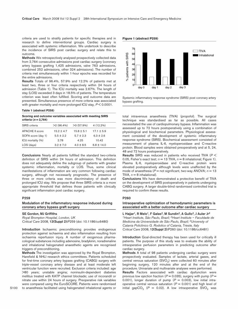
S100
criteria are used to stratify patients for specific therapies and inresearch to define interventional groups. Cardiac surgery isassociated with systemic inflammation. We undertook to describethe incidence of SIRS post cardiac surgery and relate this tooutcome.Methods We retrospectively analysed prospectively collected datafrom 2,764 consecutive admissions post cardiac surgery (coronaryartery bypass grafting 1,425 admissions, valve 763 admissions,combined 252 admissions, other 324 admissions). The number ofcriteria met simultaneously within 1-hour epochs was recorded forthe entire admissions.Results Totals of 96.4%, 57.9% and 12.2% of patients met atleast two, three or four criteria respectively within 24 hours ofadmission (Table 1). The ICU mortality was 2.67%. The length ofstay (LOS) exceeded 3 days in 18.5% of patients. The temperaturecriterion was least often fulfilled. Scoring and outcome data arepresented. Simultaneous presence of more criteria was associatedwith greater mortality and more prolonged ICU stay, P < 0.0001.
Table 1 (abstract P258)
Scoring and outcome variables associated with meeting SIRScriteria (n = 2,764)
SIRS criteria >2 (96.4%) >3 (57.9%) 4 (12.2%)
APACHE II score 15.2 ± 4.7 15.8 ± 5.1 17.1 ± 5.9
SOFA score (day 1) 5.5 ± 2.2 5.7 ± 2.3 6.3 ± 2.6
ICU mortality (%) 2.78 4.25 10.42
LOS (days) 3.2 ± 7.0 4.0 ± 8.5 6.8 ± 14.0
Conclusions Nearly all patients fulfilled the standard two-criteriadefinition of SIRS within 24 hours of admission. This definitiondoes not adequately define the subgroup of patients with greatersystemic inflammation, mortality or LOS. Thus, some clinicalmanifestations of inflammation are very common following cardiacsurgery, although not necessarily prognostic. The presence ofthree or more criteria was more discriminatory of death andprolonged ICU stay. We propose that three SIRS criteria is a moreappropriate threshold that defines those patients with clinicallysignificant inflammation post cardiac surgery.
P259Modulation of the inflammatory response induced duringcoronary artery bypass graft surgery
SE Gordon, MJ GriffithsRoyal Brompton Hospital, London, UKCritical Care 2008, 12(Suppl 2):P259 (doi: 10.1186/cc6480)
Introduction Ischaemic preconditioning provides endogenousprotection against ischaemia and also inflammation resulting fromischaemia reperfusion injury. A number of exogenous pharma-cological substances including adenosine, bradykinin, noradrenalineand inhalational halogenated anaesthetic agents are recognisedtriggers of preconditioning.Methods The investigation was approved by the Royal Brompton,Harefield & NHLI research ethics committees. Patients scheduledfor first-time coronary artery bypass grafting (CABG) surgery withtriple-vessel coronary artery disease and at least moderate leftventricular function were recruited. Exclusion criteria included: age>80 years; unstable angina; noninsulin-dependent diabetesmellitus treated with KATP channel blockade; use of nicorandil ornitrate use within 24 hours of surgery. Preoperative risk variableswere compared using the EuroSCORE. Patients were randomisedto anaesthesia facilitated using halogenated inhalational agents or
total intravenous anaesthesia (TIVA) (propofol). The surgicaltechnique was standardised as far as possible. All casesnecessitated the use of cardiopulmonary bypass. Inflammation wasassessed up to 72 hours postoperatively using a combination ofphysiological and biochemical parameters. Physiological assess-ment consisted of the development of systemic inflammatoryresponse syndrome (SIRS). Biochemical assessment consisted ofmeasurement of plasma IL-6, myeloperoxidase and C-reactiveprotein. Blood samples were obtained preoperatively and at 5, 24,48 and 72 hours postoperatively.Results SIRS was reduced in patients who received TIVA (P <0.05, Fisher’s exact test; n = 13 TIVA, n = 8 inhalational; Figure 1).Plasma IL-6, myeloperoxidase and C-reactive protein wereelevated postoperatively although levels were unaffected by themode of anaesthesia (P = not significant, two-way ANOVA; n = 13TIVA, n = 8 inhalational).Conclusions We have demonstrated a protective benefit of TIVAon the development of SIRS postoperatively in patients undergoingCABG surgery. A larger double-blind randomised controlled trial isrequired to confirm these results.
P260Intraoperative optimization of hemodynamic parameters isassociated with a better outcome after cardiac surgery
L Hajjar1, R Melo1, F Galas2, M Sundin2, A Gullo3, J Auler Jr2
1Heart Institute, São Paulo, Brazil; 2Heart Institute – Faculdade deMedicina da Universidade de São Paulo, Brazil; 3University ofCatania Policlinico G. Rodolico of Catania, Trieste, ItalyCritical Care 2008, 12(Suppl 2):P260 (doi: 10.1186/cc6481)
Introduction Goal-directed therapy has been used for critically illpatients. The purpose of this study was to evaluate the ability ofintraoperative perfusion parameters in predicting outcome aftercardiac surgery.Methods A total of 98 patients undergoing cardiac surgery wereprospectively evaluated. Samples of lactate, arterial gases, andcentral venous saturation (SVO2) were collected 60 minutes afterbeginning surgery, 120 minutes after and at the end of theprocedure. Univariate and multivariate analyses were performed.Results Factors associated with cardiac dysfunction wereprevious low ejection fraction (P = 0.035), surgery with pump (P =0.001), longer duration of pump (P = 0.003), low initial intra-operative central venous saturation (P = 0.001) and high level ofinitial gapCO2 (P = 0.02). A low intraoperative SVO2 was
Critical Care March 2008 Vol 12 Suppl 2 28th International Symposium on Intensive Care and Emergency Medicine
Figure 1 (abstract P259)
Systemic inflammatory response syndrome (SIRS) post coronary arterybypass grafting.
0
10
20
30
40
50
60
70
day 0 day 1 day 2 day 3
TIVAInhalational
% p
atie
nts
with
SIR
S
S101
independently associated with a sevenfold increase (95% CI,2–21) and a initial high level of CO2gap with a 4.5-fold increase(95% CI, 1.6–12) in cardiac dysfunction after cardiac surgery.Associated factors with renal dysfunction were age (P = 0.045),longer duration of pump (P = 0.01) and low initial intraoperativecentral venous saturation (P = 0.001). A low intraoperative SVO2was associated with a 12-fold increase and a low level of initialbase excess with a 27-fold increase in rates of infection aftercardiac surgery. A high level of final arterial lactate predicted alonger time of mechanical ventilation (OR, 4.56; 95% CI,1.4–11.2). There were no relations of perfusion parameters withlonger time of stay in the ICU or mortality.Conclusions In this observational study, a low intraoperative levelof SVO2 is an independent predictor of cardiac dysfunction, renalfailure and infection after cardiac surgery and a high level of lactateis associated with a longer time of mechanical ventilation. Thesefindings suggest that these parameters may be markers ofprognosis after cardiac surgery.Reference1. Rivers E, Nguyen B, et al.: Early goal-directed therapy in the
treatment of severe sepsis and septic shock. N Engl J Med2001, 345:1368-1377.
P261Effects of inhaled iloprost on right ventriculovascularcoupling and ventricular interdependence in acutepulmonary hypertension
S Rex1, C Missant2, P Claus2, W Buhre3, R Rossaint1, PF Wouters4
1University Hospital Aachen, Germany; 2Katholieke UniversiteitLeuven, Belgium; 3University of Witten-Herdecke, Hospital Köln-Merheim, Cologne, Germany; 4Ghent University Hospital, Ghent,BelgiumCritical Care 2008, 12(Suppl 2):P261 (doi: 10.1186/cc6482)
Introduction Prostacyclin inhalation is increasingly used to treatacute pulmonary hypertension (PHT) and right ventricular (RV)failure. Prostacyclins not only affect vasomotor tone, but may alsohave cyclic adenosine 3′,5′-monophosphate-mediated positiveinotropic effects and modulate autonomic nervous system (ANS)tone. We studied the role of these different mechanisms in theoverall hemodynamic effects produced by iloprost (ILO) inhalationin an experimental model of acute PHT.Methods Twenty-six pigs were instrumented with biventricularconductance catheters, a pulmonary artery (PA) flow probe and ahigh-fidelity PA-pressure catheter. The effects of 50 μg inhaled ILOwere studied in healthy animals with and without blockade of theANS, and in animals with acute hypoxia-induced PHT.Results ILO had minimal hemodynamic effects in healthy animalsand produced no direct effects on myocardial contractility afterpharmacological ANS blockade. During PHT, ILO resulted in a51% increase in cardiac output when compared with placebo (5.6± 0.7 vs 3.7 ± 0.8 l/min, P = 0.0013), a selective reduction of RVafterload (effective PA-elastance (PA-Ea): from 0.6 ± 0.3 vs 1.2 ±0.5 mmHg/ml; P = 0.0005) and a significant increase in leftventricular (LV) end-diastolic volume (91 ± 12 vs 70 ± 20 ml, P =0.006). Interestingly, RV contractility was reduced after ILO (slopeof preload recruitable stroke work: 3.4 ± 0.8 vs 2.2 ± 0.5 mW/ml;P = 0.0002), while ventriculovascular coupling remained essentiallypreserved (ratio of RV end-systolic elastance over PA-Ea: 0.97 ±0.33 vs 1.03 ± 0.15).
Conclusions In acute PHT, ILO improved global hemodynamicsprimarily via selective pulmonary vasodilation and a restoration ofLV preload. The reduction of RV afterload was associated with aparadoxical decrease in RV contractility. This appears to reflect anindirect mechanism serving to maintain ventriculovascular couplingat the lowest possible energetic cost, since no evidence for adirect negative inotropic effect of ILO was found.Reference:1. Rex S, et al.: Epoprostenol treatment of acute pulmonary
hypertension is associated with a paradoxical decrease inright ventricular contractility. Intensive Care Med 2008, 34:179-189.
P262Dobutamine in acute myocardial infarction: should we useit for reduction of pulmonary hypertension and pulmonarycapillary wedge pressure in acute myocardial infarction?
A Macas, G Baksyte, A Pikciunas, A SemetaiteKaunas University of Medicine, Kaunas, LithuaniaCritical Care 2008, 12(Suppl 2):P262 (doi: 10.1186/cc6483)
Introduction Enthusiasm for application of dobutamine hasretreated after the introduction to clinical practice of new medica-tions such as levosimendan. Such treatment, however, is ratherexpensive as well as remote outcomes still being under discussion.Owing to this, dobutamine still has an appropriate place in thetreatment of patients with complicated acute myocardial infarction(AMI) where signs of acute heart failure and pulmonaryhypertension should be treated immediately. The study objectivewas to evaluate the hemodynamic effect of dobutamine using thepulmonary artery catheterization technique in patients with AMIcomplicated by cardiogenic shock and pulmonary hypertension.Methods Dobutamine was infused continuously for patients withAMI complicated by cardiogenic shock and with verified pulmonaryhypertension. Only low doses not exceeding 4 μg/kg/mindobutamine were continuously infused. Data were obtained usinga pulmonary artery catheter. Hemodynamic indices including thecardiac output (CO), pulmonary pressures and pulmonary arterycapillary wedge pressure (PAWP) were measured.Results Nineteen patients were investigated according to thestudy protocol, 11 (57.9%) men and eight (42.1%) women.Average age was 65.1 ± 11.2 years. Anterior AMI was diagnosedfor 14 (73.7%) patients, inferior for five (26.3%). The inhospitalmortality rate was 52.6% (10 patients). The initial CO was 3.3 ±0.9 (range from 1.8 to 5.4 l/min), the mean pulmonary arterypressure (MPAP) was 34.8 ± 13.4 mmHg (maximum 50 mmHg),and the PAWP was 25.7 ± 10.4 mmHg (maximum 42 mmHg).After the first day of continuous dobutamine infusion, the CO was4.2 ± 1.2 (range from 2.5 to 6.4 l/min), the MPAP was 31.1 ± 7.9mmHg (maximum 43 mmHg), and the PAWP was 16.2 ± 3.8mmHg (maximum 21 mmHg). After the termination of dobutamine(after 48 hours), the CO was 4.2 ± 0.9 (range from 3.1 to 6.1l/min), the MPAP was 30.2 ± 8.1 mmHg (maximum 46 mmHg),and the PAWP was 16.4 ± 3.4 mmHg (maximum 21 mmHg). Theincrease of the initial CO and reduction of PAWP after the first dayof continuous dobutamine infusion were statistically significant(P < 0.05).Conclusions Application of dobutamine showed a positive benefitin reduction of pulmonary hypertension and pulmonary capillarywedge pressure as well as in the increase of cardiac output forpatients with AMI complicated by cardiogenic shock andpulmonary hypertension.
Available online http://ccforum.com/supplements/12/S2
S102
P263Dobutamine protects lymphocytes against staurosporin-induced apoptosis: investigation of the antioxidative actionof the dobutamine molecule
J Van Melkebeek1, S Vandeput2, M Roesslein3, F Jans1
1Ziekenhuis Oost-Limburg, Genk, Belgium; 2University of Hasselt,Diepenbeek, Belgium; 3University Hospital, Freiburg, GermanyCritical Care 2008, 12(Suppl 2):P263 (doi: 10.1186/cc6484)
Introduction Catecholamines have been shown to modulatevarious immunological functions. In previous experiments wedemonstrated that dobutamine pretreatment protects T cells fromstaurosporin-induced apoptosis [1]. In the current study weplanned to investigate whether antioxidative properties of thedobutamine molecule might be responsible for its protective effect.Methods Jurkat T-cell passages 1–12 were used.Results Experiments with a caspase-activity assay confirmedprevious results: pretreatment (4 hours) with dobutamine 0.1 mMand 0.5 mM decreased staurosporin-induced apoptosis in Jurkat Tcells from 14.0% to 11.6% and 8.7%, respectively (P < 0.01).Other catecholamines such as epinephrine and norepinephrinehad no protective effect. To investigate whether production ofROS could be measured, Jurkat T cells were loaded with CM-H2DCFDA. After washing steps, the cells were exposed to 0 μM,1 μM, 10 μM and 100 μM H2O2 for 6 hours. The fluorescencesignal (ex 480/em 520 nm) measured was 36.7 U, 37.7 U, 38.1 Uand 54.3 U, respectively, demonstrating the relation between ROSand the fluorescence signal. Next, production of ROS due tostaurosporin treatment (2 μM) was measured: ROS productionincreased minimally after exposure for 2 hours. Only after 6 hoursof staurosporin treatment, the ROS signal increased from 36.7 Uto 42.1 U (P < 0.05). Subsequently, the ROS-scavenging effect ofdobutamine was investigated. CM-H2DCFDA-loaded cells wereexposed to staurosporin (2 μM) for 2 hours, with or withoutdobutamine pretreatment (0.1 mM and 0.5 mM): the ROS-scavenging effect was very pronounced in the 0.1 mM group(decrease in fluorescence signal from 56.1 U to 22.5 U, P < 0.01),and increased further in the 0.5 mM group (17.8 U, P < 0.01).Control experiments with unstained cells showed that addition ofdobutamine did not change the autofluorescence signal.Conclusions These experiments demonstrate that dobutamineacts as a ROS scavenger. Whether this scavenging effect isresponsible for the protective properties of dobutamine againststaurosporin-induced apoptosis is currently under investigation.Reference1. Jans F, et al.: Crit Care 2007, 11(Suppl 2):31.
P264Hemodynamic effects of levosimendan in patients withlow-output heart failure
H Gharsallah, M Bel Hadj Amor, W Koubaa, F Gargouri, K Lamine, M FerjaniMilitary Hospital of Tunis, TunisiaCritical Care 2008, 12(Suppl 2):P264 (doi: 10.1186/cc6485)
Introduction Levosimendan is a positive inotropic drug agent thatincreases the sensibility of contractile proteins to calcium [1]. Thedrug is used in patients with decompensated low-output heart failure[2]. The aim of our study is to analyze the hemodynamic effects oflevosimendan in patients with refractory cardiogenic shock.Methods After approval by our institutional ethics committee, 16patients who had a refractory cardiogenic shock after myocardialinfarction (n = 10), peripartum cardiomyopathy (n = 3) and
cardiomyopathy (n = 3) were prospectively included in our study.Levosimendan was added to conventional inotropic agents(dobutamine) with a bolus dose of 12 μg/kg for 30 minutesfollowed by a continuous infusion at a rate of 0.1 μg/kg/min for24 hours. Hemodynamic data (continuous cardiac output andvenous oxygen saturation) were obtained by a Swan–Ganzcatheter at T0, 30 minutes, 90 minutes, 2 hours, 4 hours, 8 hours,12 hours, 24 hours and 48 hours. A transoesophagealechocardiography was performed at T0, day 1, day 2, day 7 andday 15. SPSS version 10 was used for all statistical analyses.Results After levosimendan administration, a significant reductionof pulmonary and vascular resistances values was followed by asignificant increase of the cardiac index and venous oxygensaturation. Changes in heart rates and mean arterial bloodpressure were not significant. The left ventricular ejection fractionwas increased from 24% (T0) to 40% (day 2).Conclusions This study showed that levosimendan improveshemodynamic parameters and left ventricular ejection fraction inpatients with cardiogenic shock. Controlled trials of sufficient sizeare needed, however, to confirm these results.References1. Figgitt DP, Gillies PS, Goa KL: Drugs 2001, 61:613-627.2. Follath F, Cleland JGF, Just H, et al.: Lancet 2002, 360:196-
202.
P265Levosimendan does not improve resuscitation successafter hypovolemic cardiac arrest
E Semenas, L WiklundUppsala University Hospital, Uppsala, SwedenCritical Care 2008, 12(Suppl 2):P265 (doi: 10.1186/cc6486)
Introduction Resuscitation from hemorrhagic shock andsubsequent cardiac arrest (CA) is a major clinical challenge in thecare of trauma patients. Levosimendan, a new calcium sensitizer,exerts positive inotropic and lusitropic effects in failing humanmyocardium without increase in energy expenditure [1]. The aim ofthis study was to evaluate possible beneficial effects oflevosimendan in combination with vasopressin in hypovolemic CAand subsequent cardiopulmonary resuscitation.Methods Five anesthetized male piglets (26.5 ± 1.1 kg) were bled(25.1 ± 3.4% of calculated total blood volume) to a mean arterialblood pressure of 35 mmHg during 12.9 ± 0.2 minutes. After-wards the piglets were subject to 4 minutes untreated ventricularfibrillation followed by 12 minutes open-chest cardiopulmonaryresuscitation (CPR). At 5 minutes of CA, 0.4 U/kg vasopressin and12 μg/kg levosimendan were given intravenously and an infusion of3 ml/kg hypertonic saline and dextran (7.5% saline, 6% dextran70) was given in 20 minutes. Internal defibrillation was attemptedfrom 7 minutes of CA to achieve restoration of spontaneouscirculation (ROSC). If necessary, at 8 minutes of CA, 0.4 U/kgvasopressin was repeated intravenously. Hemodynamic variables,continuous cerebral cortical blood flow and blood gas parameterswere measured during CPR and up to 180 minutes after ROSC.Blood samples for 8-iso-PGF2α, 15-keto-dihydro-PGF2α, protein S-100β and troponin I were taken.Results ROSC was achieved in two out of five piglets. Only one ofthese piglets survived the whole experiment. Another piglet died60 minutes after ROSC due to a new episode of ventricularfibrillation. It was difficult to achieve ROSC due to persistentventricular fibrillation during CPR. The mean number of defibril-lation attempts was 14.2 (range: 8–21). The mean coronary per-fusion pressure was 19–21 mmHg during CPR. Piglets thatachieved ROSC needed a constant dobutamine infusion for
Critical Care March 2008 Vol 12 Suppl 2 28th International Symposium on Intensive Care and Emergency Medicine
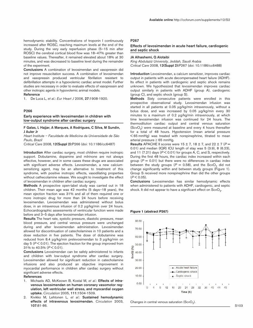
S103
hemodynamic stability. Concentrations of troponin I continuouslyincreased after ROSC, reaching maximum levels at the end of thestudy. During the very early reperfusion phase (5–15 min afterROSC) the cerebral cortical blood flow was 18–47% greater thanbaseline values. Thereafter, it remained elevated about 18% at 30minutes, and was decreased to baseline level during the remainderof the experiment.Conclusions A combination of levosimendan and vasopressin didnot improve resuscitation success. A combination of levosimendanand vasopressin produced ventricular fibrillation resistant todefibrillation attempts in a hypovolemic cardiac arrest model. Furtherstudies are necessary in order to evaluate effects of vasopressin andother inotropic agents in hypovolemic animal models.Reference1. De Luca L, et al.: Eur Heart J 2006, 27:1908-1920.
P266Early experience with levosimendan in children with low-output syndrome after cardiac surgery
F Galas, L Hajjar, A Marques, A Rodrigues, C Silva, M Sundin, J Auler JrHeart Institute – Faculdade de Medicina da Universidade de SãoPaulo, BrazilCritical Care 2008, 12(Suppl 2):P266 (doi: 10.1186/cc6487)
Introduction After cardiac surgery, most children require inotropicsupport. Dobutamine, dopamine and milrinone are not alwayseffective, however, and in some cases these drugs are associatedwith significant adverse effects. Levosimendan, a new calcium-sensitizing agent, may be an alternative to treatment of thissyndrome, with positive inotropic effects, vasodilating propertieswithout cathecolamine release. We sought to investigate the effectof levosimendan in children after cardiac surgery.Methods A prospective open-label study was carried out in 18children. Their mean age was 42 months (5 days–18 years), themean ejection fraction was 31% and all of them required one ormore inotropic drug for more than 24 hours before receivinglevosimendan. Levosimendan was administered without bolusdose, in an intravenous infusion of 0.2 μg/kg/min over 24 hours.Echocardiographic assessments of ventricular function were madebefore and 3–5 days after levosimendan infusion.Results The heart rate, systolic pressure, diastolic pressure, meanblood pressure, and central venous pressure were unchangedduring and after levosimendan administration. Levosimendanallowed for discontinuation of catecholamines in 10 patients and adose reduction in five patients. The dose of dobutamine wasreduced from 8.4 μg/kg/min prelevosimendan to 3 μg/kg/min onday 5 (P < 0.01). The ejection fraction for the group improved from31% to 40.5% (P < 0.01).Conclusions Levosimendan can be safely administered to infantsand children with low-output syndrome after cardiac surgery.Levosimendan allowed for significant reduction in catecholamineinfusions and also produced an objective improvement inmyocardial performance in children after cardiac surgery withoutsignificant adverse effects.References1. Michaels AD, McKeown B, Kostal M, et al.: Effects of intra-
venous levosimendan on human coronary vasomotor reg-ulation, left ventricular wall stress, and myocardial oxygenuptake. Circulation 2005, 111:1504-1509.
2. Kivikko M, Lehtonen L, et al.: Sustained hemodynamiceffects of intravenous levosimendan. Circulation 2003,107:81-86.
P267Effects of levosimendan in acute heart failure, cardiogenicand septic shock
JA Alhashemi, Q AlotaibiKing Abdulaziz University, Jeddah, Saudi ArabiaCritical Care 2008, 12(Suppl 2):P267 (doi: 10.1186/cc6488)
Introduction Levosimendan, a calcium sensitizer, improves cardiacoutput in patients with acute decompensated heart failure (ADHF).Its effect in patients with cardiogenic and septic shock remainsunknown. We hypothesized that levosimendan improves cardiacoutput similarly in patients with ADHF (group A), cardiogenic(group C), and septic shock (group S).Methods Sixty consecutive patients were enrolled in thisprospective observational study. Levosimendan infusion wasstarted in all patients at 0.05 μg/kg/min intravenously, without abolus dose, and was increased by 0.05 μg/kg/min every 30minutes to a maximum of 0.2 μg/kg/min intravenously, at whichtime levosimendan infusion was continued for 24 hours. Thethermodilution cardiac output and central venous saturation(ScvO2) were measured at baseline and every 4 hours thereafterfor a total of 48 hours. Hypotension (mean arterial pressure< 65 mmHg) was treated with norepinephrine, titrated to meanarterial pressure ≥ 65 mmHg.Results APACHE II scores were 15 ± 7, 18 ± 7, and 22 ± 7 (P =0.01) and median (IQR) ICU length of stay was 5 (3.9), 8 (6.23),and 11 (7.21) days (P < 0.01) for groups A, C, and S, respectively.During the first 48 hours, the cardiac index increased within eachgroup (P = 0.01) but there were no differences in cardiac indexbetween the study groups (P = 0.58), and the ScvO2 did notchange significantly within and between study groups (Figure 1).Group S received more norepinephrine than did the other groups(P < 0.05).Conclusions Levosimendan has similar hemodynamic effectswhen administered to patients with ADHF, cardiogenic, and septicshock. It did not appear to have a significant effect on ScvO2.
Available online http://ccforum.com/supplements/12/S2
Figure 1 (abstract P267)
Changes in central venous saturation (ScvO2).
S104
P268Sedation during mechanical ventilation: a comparison ofsedatonarcosis and awake sedation
B Völgyes, MM Mezei, PG GolopenczaBajcsy Zs. Kórház, Budapest, HungaryCritical Care 2008, 12(Suppl 2):P268 (doi: 10.1186/cc6489)
Introduction The most important goal during mechanicalventilation in the ICU is to achieve patient comfort and patient–ventilator synchrony. Once proper analgesia has been established,an infusion of a sedative should be added. The goal of this studywas to investigate whether continuously awake sedation duringmechanical ventilation (MV) decreased the days of ventilation andcomplications.Methods All patients with MV – based on the abovementionedcriteria – were included (age: 20–70 years; community-acquiredpneumonia; two quadrant infiltrates; PaO2/FiO2 < 200; no otherorgan dysfunction). From June 2001 to February 2004, patientswith MV received deep sedation (midazolam 0.03–0.20 mg/kgbody weight/hour and propofol 0.5–2.0 mg/kg body weight/hour).This is the ‘sedatonarcosis’ group. From March 2004 to July 2007,patients were treated with ‘awake sedation’ (alprasolam 1.5–2.0mg/day).Results All of the patients received low tidal volume ventilation, de-escalation antibiotics, continuous correction of homeostasis,management of enteral feeding and pulmonary physiotherapy. Inboth groups we applied noninvasive respiratory therapy afterextubation. It was possible to mobilise patients earlier – before theextubation – in the awake sedation group. See Table 1.Conclusions Adopting awake sedation during MV (compared withcontinuous sedatonarcosis) decreased the days on ventilation, andthe lengths of ICU and hospital stay.
P269Multiparametric evaluation of sedation in the ICU
A Donati, L Botticelli, L Romagnoli, M Romanelli, S Marzocchini, C Anastasi, P PelaiaUniversità Politecnica delle Marche, Torrette di Ancona, ItalyCritical Care 2008, 12(Suppl 2):P269 (doi: 10.1186/cc6490)
Introduction A problem for patients submitted to extendedsedation is evidence of a liver dysfunction. We wanted to verifywhether this dysfunction is either correlated with the splanchnichypoperfusion or with the extended administration of drugs.Methods During March–September 2007, four patients with purecranial trauma (age 34–50 years) were treated with midazolam,propofol, sodium thiopental and fentatienil. Parameters evaluated:burst suppression ratio (BSR) with electroencephalography(Aespect; GE Health Care), functional capillary density (FCD) andmean velocity with Microscan and MAS (MicroVision Medical,Amsterdam, The Netherlands), plasma disappearance ratio (PDR)
with Pulsion LIMON (SEDA; Milano), intramucosal pH and regionalPCO2 with Tonocap (GE Health Care), electrocardiography, meanarterial pressure and hematochemical examinations (transaminases,γ-glutamyl transpeptidase (GGT), serum bilirubin, serum amylases,serum lactates and drugs dosages). Exclusion criteria: hepatopathyat admission, age <18 years and >60 years, BMI > 30, clinicalfactors favouring splanchnic hypoperfusion. All the parameters areanalysed at t0 (admission to the ICU) and then every 48 hoursduring the sedation (t1, t2, t3) and at its end (t4, t5) through theFriedman test (P < 0.05) and the Spearman test (P < 0.05 andR > 0.6).Results Increase of transaminases at the end of sedation in threeof the four patients. Earlier increase of GGT in all patients. Serumbilirubin always in range. Increase of serum amylases in three of thefour patients is correlated to propofol dosage. PDR always>16%/ml (cutoff of hepatic hypoperfusion). BSR always >0%during t1, t2, t3. FCD always steady and mean velocities alwayshigh. Hepatic cytonecrosis indexes, GGT, serum amylases are wellcorrelated to splanchnic perfusion indexes (serum lactates, FCDand regional PCO2) so their increase apparently is not due tosplanchnic hypoperfusion. Patient 1 (propofol, fentatienil earlyreplaced with sodium thiopental, midazolam), increase of trans-aminases when midazolam was stopped. Because of the beginningof an epileptic status, the patient was eliminated from the study.Patient 2 (propofol, midazolam, fentatienil), increase of trans-aminases associated with the paradoxical increase of splanchnicperfusion. Patient 3 (propofol, midazolam, fentatienil), increase oftransaminases and serum amylases is not associated with thesplanchnic hypoperfusion. Serum amylases increase according tothe increase of propofol dosage. Patient 4 (propofol, fentatienil),late increase of transaminases, serum amylases and GGT isassociated with the propofol dosage.Conclusions Drugs used for the analgosedation seem responsiblefor the increase of transaminases but not for the decrease ofsplanchnic perfusion. This study has to be confirmed by otherstudies recruiting more patients and with more precise exclusioncriteria.Reference1. Jacobi J, et al.: Crit Care Med 2002, 30:119-141.
P270Short-term sevoflurane sedation using the anaestheticconserving device AnaConDa® after cardiac surgery:feasibility, recovery and clinical issues
KD Röhm, MW Wolf, J Boldt, T Schöllhorn, A Schellhaass, SN PiperKlinikum Ludwigshafen, GermanyCritical Care 2008, 12(Suppl 2):P270 (doi: 10.1186/cc6491)
Introduction With the approval of the anaesthetic conservingdevice (AnaConDa®), inhalative sedation in the ICU has becomefeasible [1]. Isoflurane has been investigated in postoperative and
Critical Care March 2008 Vol 12 Suppl 2 28th International Symposium on Intensive Care and Emergency Medicine
Table 1 (abstract P268)
Results
MV Length of ICU stay Length of hospital stay Died Exclusion Patients (n) (average, days) (average, days) (average, days) (n) (n)
Sedatonarcosis group (n = 21) 6.37 9.06 18.66 4 1
Awake sedation group (n = 23) 4.38 7.22 15.16 3 2
t0.05 1.8357 1.28725 1.2940
P value <0.05 <0.05 <0.05 Not significant
S105
critically ill patients using AnaConDa® [2,3], whereas sevofluranesedation has only been reported in small observations [4,5]. Thisrandomised, single-blinded, BIS-controlled study was to evaluatefor the first time sevoflurane via AnaConDa® compared withpropofol, with regard to recovery, sedation quality and consump-tion of anaesthetics.Methods Seventy patients scheduled for elective coronary arterybypass graft surgery were randomised at admission to the ICU toeither receive sevoflurane (n = 35) or propofol (n = 35) for post-operative sedation. The primary endpoint was recovery time fromtermination of sedation (extubation time, spontaneous eye openingand hand grip). Sedation quality (using the Richmond AgitationSedation Scale, RASS), sevoflurane consumption, duration of ICUand hospital stays, and adverse side effects were documented.Results Median recovery times were significantly shorter (P <0.002) with sevoflurane than with propofol (extubation time:21.5 min (2–259) vs 150.5 min (22–910)). Mean sevofluraneconsumption was 3.2 ± 1.4 ml/hour to obtain end-tidal concen-trations of 0.5–1 vol%; mean administration of propofol was 2.4 ±1.1 mg/kg/hour. Sedation quality was comparable in both groups(RASS –3 to –4), and no serious complications includinghaemodynamics related to either sedative drug occurred. Length ofstay in the ICU was similar in both groups, whereas patientsreceiving sevoflurane were discharged significantly (P < 0.03)earlier from hospital (10.6 ± 3.3 days vs 14 ± 7.7 days).Conclusions Sevoflurane administration via AnaConDa® is anefficacious and easy titratable way to provide postoperativesedation in the ICU. Recovery from sedation was facilitated withsevoflurane compared with propofol, and resulted in a shorterventilation time. Sevoflurane sedated patients left hospital a mean3 days earlier compared with a propofol-based regimen.References1. Enlund M: Anaesthesia 2001, 56:429-432.2. Sackey PV: Crit Care Med 2004, 32:2241-2246.3. Hanafy M: Egyptian J Anaesth 2005, 21:237-242.4. Soukup J: Intensiv- und Notfallbehandlung 2007, 32:29-36.5. Berton J: Anesth Analg 2007, 104:130-134.
P271Thoracal epidural analgesia in upper abdominal surgery
S Vukosavljevic, T Randjelovic, D PavlovicKBC Bezanijska Kosa, Belgrade, SerbiaCritical Care 2008, 12(Suppl 2):P271 (doi: 10.1186/cc6492)
Introduction Surgery of upper abdominal organs is painful andmutilating, joined with possible serious postoperative complica-tions – pulmonary, abdominal (anastomoses related), cardiological,and thromboembolic.Methods During past year 100 patients had upper abdominalsurgery. According to analgesia type they were divided into twogroups. The first group (G1) was administered a first dose of localanesthetic (bupivacain 0.25% 5 ml) prior to total anesthesiathrough a thoracic epidural catheter. After that they underwentclassical total anesthesia (midazolam, diprivan, fentanil, relaxant),followed by anesthesia with diprivan 6 mg/kg/hour and analgesiawith local anesthetic epidurally. Postoperatively they wereadministered through the thoracic epidural catheter a combinationof opioids (morphine 2 mg) and local anesthetic (bupivacain0.125% 6–8 ml) every 8 hours. The other group (G2) underwentclassical total anesthesia followed by classical proportion of oxiduland oxygen, and analgesia by fentanyl, with postoperative systemicanalgesia by nonsteroid anti-inflammatory drugs, paracetamol, andmetamisol sodium. The parameters followed during surgery werearterial tension, heart rate, gas analysis, diuresis, and operating
field bleeding. The postoperatively followed parameters wereVisual Analog Scale, arterial tension, heart rate, gas analysis,beginning of peristalsis, and pulmonary complications.Results Thoracic epidural analgesia during surgery provides betterhemodynamic patient stability and lower blood loss due tointraoperative bleeding, statistically and clinically significantly betteranalgesia in the first postoperative 72 hours, compared withsystemic analgesia (Visual Analog Scale, G1 < 8 in movement vsG2 > 30 in movement), reduces the period of postoperative ileus(for 1.06 days), reduces pulmonary and cardiologic complications,provides early patient mobilization and decreases the number ofintensive postoperative care days (for 3.9 days).Conclusions Our experience shows that thoracic epiduralanalgesia is the right choice, because it provides effective pain reliefin patients, prevention of postoperative complications, providesearly patient mobilization and reduces the length of stay in the ICU.References1. Bromage PR: Br J Anaesth 1975, 47:199-212.2. Bonica JJ, et al.: Anesthesiology 1970, 33:619-626.
P272Etomidate and relative adrenal insufficiency incardiopulmonary bypass surgery: impact on thepostoperative hemodynamic status
L Lorenzo, M Brouard, J Iribarren, J Jimenez, L Lorente, R Perez, S Palmero, R Santacreu, R Martinez, M MoraHospital Universitario de Canarias, La Laguna, Tenerife, SpainCritical Care 2008, 12(Suppl 2):P272 (doi: 10.1186/cc6493)
Introduction Use of etomidate in cardiopulmonary bypass (CPB)surgery is usual practice during the anesthetic induction. Theobjective of this study was to determine the incidence of relativeadrenal insufficiency (RAI) in CPB patients after etomidateadministration and the impact on hemodynamic status.Methods A prospective cohort study was performed on CPBpatients who received etomidate or not during the anestheticinduction. Patients were excluded if they had received systemic orinhaled corticosteroids or immunosuppressants, and activepreoperative infection. RAI was defined as a rise in serum cortisol≤9 μg/dl after the administration of 250 μg cosyntropin. Cortisollevels were measured preoperatively, immediately before and30 minutes, 60 minutes and 90 minutes after the administration ofcosyntropin (250 μg). We used SPSS version 15.Results We studied 65 patients (74% men), mean age 68 ± 11years. The incidence of RAI was 89.4% in these patients comparedwith 50% in patients who did not receive etomidate (P = 0.01) (Table1). Higher postoperative cortisol levels were associated with lowerdoses of norepinephrine at 4 hours post CPB. Levels of cortisol inetomidate patients were inversely proportional to the needs ofnorepinephrine. Finally the maximum increase of cortisol levels afterACTH stimulation was directly associated with the systemicresistance index in nonetomidate patients at four postoperative hours.
Table 1 (abstract P272)
Incidence of relative adrenal insufficiency
RAI Etomidate (n = 47) Others (n = 18) P value
Yes (%) 89.4 50 0.01
No (%) 10.6 50
Conclusions The use of etomidate was associated with RAI postCPB surgery. Cortisol levels were related to the postoperativehemodynamic profile and the need for vasopressor drugs.
Available online http://ccforum.com/supplements/12/S2
S106
P273Economic evaluation of remifentanil-based versusconventional sedation for patients with an anticipatedmechanical ventilation duration of 2–3 days in Germany
MJ Al1, J Martin2, J Bakker1, R Welte3
1Erasmus MC, University Medical Center, Rotterdam, TheNetherlands; 2Klinik am Eichert, Goeppingen, Germany;3GlaxoSmithKline, Munich, GermanyCritical Care 2008, 12(Suppl 2):P273 (doi: 10.1186/cc6494)
Introduction Hospitals are increasingly forced to consider theeconomics of technology use. We estimated the incremental cost-consequences of remifentanil-based sedation (RS) versus conven-tional sedation (CS) in ICU patients with an anticipated mechanicalventilation (MV) time of 2–3 days for Germany.Methods A probabilistic Markov model was utilized that describesthe patient flow on the ICU using eight model states: MV – main-tenance, MV– eligible start weaning, MV – actual weaning started,MV– eligible for extubation, ICU – extubated, ICU – eligible fordischarge, Discharged from ICU, and Death. At every hour,patients stay at the current state, move to the next state, or die. Therespective transition probabilities and the utilization of sedationdrugs were derived from UltiSAFE, a Dutch open-label trial with205 critically ill patients. In UltiSAFE, patients either received CS(predominantly morphine or fentanyl combined with propofol ormidazolam) or RS (remifentanil, combined with propofol ifrequired). Unit prices for drugs and total costs per ICU-hour withand without MV were collected in one 12-bed adult mixed ICU in ageneral German hospital. Material, staff and overhead costs wereconsidered. All costs were measured from a hospital perspectivewith 2006 as the reference year. According to the UltiSAFE targetpopulation, only patients who started weaning within 72 hours ofthe start of treatment were included.Results The average duration of MV and of ICU stay was shorter,0.9 and 0.8 days respectively, in the RS group compared with theCS group, while the average costs per patient were €6,157 in theRS group versus €7,160 in the CS group. The savings caused bythe shorter length of stay therefore more than offset the additionaldrug acquisition costs, leading to €1,003 savings per patient inthe RS group. The probability of RS being cost-saving wasestimated at 91%.Conclusions RS seems to be the economically preferred optionfor patients with an anticipated MV time of 2–3 days: RSdecreases the length of stay in the ICU, the total costs per patientand the duration of MV, which is a risk factor for ventilator-associated morbidity.
P274Propofol pharmacokinetics in preterm and term neonates:the relevance of both postmenstrual and postnatal age
K Allegaert1, M Peeters2, R Verbesselt1, D Tibboel3, G Naulaers1, J De Hoon1, C Knibbe2
1University Hospitals Leuven, Division of Woman and Child,Leuven, Belgium; 2Leiden/Amsterdam Centre for Drug Research,Leiden, The Netherlands; 3Sophia Children Hospital, Erasmus MC,Rotterdam, The NetherlandsCritical Care 2008, 12(Suppl 2):P274 (doi: 10.1186/cc6495)
Introduction Although disposition of propofol has been extensivelystudied in different populations of adult and paediatric age, dataare still very limited. Preliminary data in neonates suggested thatpropofol clearance is significantly different compared with toddlersand children, with important interindividual variability in propofol
clearance in neonates [1]. We therefore wanted to documentcovariates that contribute to interindividual variability in propofolpharmacokinetics in preterm and term neonates.Methods Population pharmacokinetics were estimated (nonlinearmixed effects model) based on arterial blood samples collected in(pre)term neonates after intravenous bolus administration ofpropofol (3 mg/kg, 10 s). Covariate analysis included post-menstrual age (PMA), postnatal age (PNA), gestational age, weightand creatinaemia.Results Two hundred and thirty-five arterial concentration–timepoints were collected in 25 neonates. The median weight was2,930 (range 680–4,030) g, PMA 38 (27–43) weeks and PNA 8(1–25) days. In a three-compartment model, PMA was the mostpredictive covariate for clearance (P < 0.001) when parameterizedas [CLstd x (PMA / 38)11.5]. The standardized propofol clearance(CLstd) at 38 weeks PMA was 0.029 l/min. The addition of a fixedvalue in neonates with a postnatal age ≥10 days further improvedthe model (P < 0.001) and resulted in the equation [CLstd x(PMA / 38)11.5 + 0.03] for neonates ≥10 days old. Values for thecentral volume (1.32 l), peripheral volume 1 (15.4 l) and peripheralvolume 2 (1.29 l) were not significantly influenced by any of thecovariates (P > 0.001).Conclusions PMA and PNA contribute to the interindividualvariability of propofol clearance with very fast maturation ofclearance in neonatal life. This implicates that preterm neonatesand neonates in the first week of postnatal life are at an increasedrisk for accumulation during either intermittent bolus or continuousadministration of propofol.Reference1. Allegaert K, et al.: Crit Care 2007, 11:S170.
P275Comparison of sedation with dexmedetomidine versuslorazepam in septic ICU patients
P Pandharipande1, TD Girard1, RD Sanders2, JL Thompson1, M Maze2, EW Ely1
1Vanderbilt University, Nashville, TN, USA; 2Imperial College ofLondon, UKCritical Care 2008, 12(Suppl 2):P275 (doi: 10.1186/cc6496)
Introduction New strategies for sedation in mechanically ventilated(MV) patients have yielded improvements in patient outcomes,including acute brain dysfunction, but the differential effect ofsedation regimens across patient diagnosis categories is notknown. In this pilot project, we evaluated the impact of sedationusing dexmedetomidine versus lorazepam, in an a priori determinedsubgroup of septic patients enrolled in the MENDS trial [1].Methods The MENDS study enrolled 103 adult medical/surgicalMV patients and excluded those with neurological disease, severeliver failure, active coronary ischemia, and seizures. Patients wererandomized in a double-blind fashion to receive dexmedetomidine(DEX)-based (maximum 1.5 μg/kg/hour) or lorazepam (LZ)-based(maximum 10 mg/hour) sedation for up to 5 days, titrated to atarget Richmond Agitation-Sedation Scale score. Patients weremonitored for delirium daily with the Confusion AssessmentMethod for the ICU.Results Thirty-nine patients in the MENDS study were admittedwith sepsis, with 19 in the DEX group and 20 in the LZ group.Baseline demographics, ICU type and admission diagnoses of thisseptic subgroup were balanced between DEX and LZ, with themedian (interquartile range) age being 57 (49, 66) vs 55 (44, 65)years, P = 0.66 and APACHE II scores of 30 (24, 32) vs 28.5 (25,32), P = 0.86, respectively. The median DEX dose was0.9 μg/kg/hour and the median LZ dose was 3 mg/hour. DEX
Critical Care March 2008 Vol 12 Suppl 2 28th International Symposium on Intensive Care and Emergency Medicine
S107
patients had greater delirium and coma-free days (8 (4, 10) vs 1.5(0.7, 5) days, P = 0.002), delirium-free days (10 (7.5, 11) vs 7.4 (4,8.2) days, P = 0.007), MV-free days (9.5 (0, 11.6) vs 2 (0, 8.5)days, P = 0.037) and a reduction in the risk of dying at 28 days(hazard ratio 0.3 (0.1, 0.9), P = 0.036) as compared with the LZpatients.Conclusions In this subgroup analysis of severe sepsis patientsfrom the MENDS trial, sedation incorporating dexmedetomidinereduced the duration of delirium and coma and the length of timeon the ventilator, and decreased the risk of dying as compared withlorazepam. This serves as a hypothesis-generating analysis to helpdirect further prospective study in such patients.Reference1. Pandharipande P, et al.: Effect of sedation with dexmedeto-
midine vs lorazepam on acute brain dysfunction in criti-cally ill. The MENDS randomized controlled trial. JAMA2007, 298:2645-2653.
P276Dexmedetomidine-based sedation for noninvasiveventilation failure
O Senturk, O Demirkiran, T Utku, S Urkmez, Y DikmenIstanbul University Cerrahpasa Medical School, Istanbul, TurkeyCritical Care 2008, 12(Suppl 2):P276 (doi: 10.1186/cc6497)
Introduction Noninvasive ventilation (NIV) reduces the intubationand mortality in patients with acute on chronic respiratory failure.NIV is associated with a large number of failures, and with patientrefusal. The purpose of the study was to assess the feasibility andsafety of dexmedetomidine-based sedation during NIV.Methods In this prospective, randomised controlled study, patientson NIV support with agitation and ventilatory discomfort wereincluded. The patients were allocated randomly into two groups:dexmedetomidine (Dex) and control. In the Dex group, the infusionrate was 0.2–0.7 μg/kg/hour to reach a Ramsey sedation score(RSS) between 3 and 4. Haemodynamic and respiratorycharacteristics, and the RSS were documented at 1 minute, 10minutes, 30 minutes, 1 hour, 4 hours and 24 hours after Dex infu-sion was started. When additional sedation was needed0.02–0.03 mg/kg intravenous midazolam was used. Spontaneousventilation and NIV support durations, the total infusion time(hours), the total Dex consumption, the reason for infusion therapycessation, additional sedative agent requirements, and the durationof the mechanical ventilation were documented.Results Thirty patients under NIV support with agitation andventilatory discomfort were included in this study. The results in theDex group are summarized in Table 1. Additional sedative agentrequirement was significantly higher in the control group. Sideeffects such as hypotension and hypoglycemia were found in theDex group.
Conclusions Dexmedetomidine is safe and effective for thesedation of the patients under NIV support.Reference1. Cooper AB, Thornley KS, Young GB, et al.: Chest 2000, 117:
809-881.
P277Dexmedetomidine for endovascular neurosurgery
Y Kase, T ObataJikei University School of Medicine, Tokyo, JapanCritical Care 2008, 12(Suppl 2):P277 (doi: 10.1186/cc6498)
Introduction Perioperative managements for endovascularneurosurgery require some considerations such as minimizinghemodynamic changes and especially avoiding blood pressureelevation accompanying extubation, immobilizing the trunk andlower limbs until bleeding from the site of femoral artery sheathremoval has stopped, and monitoring patients in the ICU for atleast 12 hours to notice neurological deterioration promptly. Theaim of this study was to assess the usefulness of dexmedetomidineas a postoperative sedative drug for endovascular neurosurgery.Methods The study included 182 patients with endovascularneurosurgery admitted to the ICU in 2006. The authors evaluatedthe postoperative sedative state and hemodynamics with theRichmond Agitation Sedation Scale (RASS) [1], heart rate (HR)and mean arterial pressure (MAP). To examine the time-dependentchanges of the RASS, MAP and HR, data were collected frommedical records, including at the start and end of dexmedeto-midine infusion and at the time of extubation.Results The surgical indications in patients with endovascularneurosurgery were unruptured cerebral aneurism (57%), sub-arachnoid hemorrhage (20%), arteriovenous malformation (5%),arteriovenous fistula (3%) and others (15%). One hundred andeighteen patients (85.3%) received dexmedetomidine. The RASSshowed patients with dexmedetomidine experienced RASS –1 to–3 states, which was arousable with verbal stimulation. There wereno dexmedetomidine-induced HR and MAP deteriorations; further-more, dexmedetomidine prevented the blood pressure elevationaccompanying extubation. The overall morbidity and mortality ratesrelating to endovascular neurosurgery were 1.6% and 0.54%,respectively.Conclusions Application of sedative drugs for the postoperativemanagement for neurovascular disease may be controversial;however, the use of dexmedetomidine facilitates postoperativemanagement of endovascular neurosurgery. Compared with thereported endovascular neurosurgery morbidity (3.7–5%) andmortality (1.1–1.5%), our morbidity and mortality rate showed thatdexmedetomidine did not cause neurological deteriorations.Reference1. Ely EW, Truman B, Shintani A, et al.: JAMA 2003, 289:2983-
2991.
P278Single-centre audit on the use of intravenous paracetamolin neonates
C Vanhole, K Allegaert, F van Beek, A Debeer, T de Rijdt, G NaulaersUniversity Hospitals Leuven, BelgiumCritical Care 2008, 12(Suppl 2):P278 (doi: 10.1186/cc6499)
Introduction An intravenous formulation of paracetamol isavailable, but remains off-label in patients below 10 kg althoughpharmacokinetics during repeated intravenous administration were
Available online http://ccforum.com/supplements/12/S2
Table 1 (abstract P276)
Respiratory, haemodynamic characteristics and Ramsey sedationscore variables
1 minute 1 hour P value
PaO2/FiO2 159 ± 47 239 ± 80 <0.01SaO2 95.8 ± 4 98.8 ± 1 <0.05PaO2 83.6 ± 27 130 ± 31 <0.05Respiratory rate 30.4 ± 5 24.5 ± 6 <0.05Mean arterial pressure 86.6 ± 12 77.7 ± 11 <0.01Heart rate 108 ± 23 98.9 ± 18 <0.05RSS 1.0 ± 0 2.6 ± 0.8 <0.01
S108
documented and dosing regimes suggested [1,2]. We thereforeretrospectively evaluated aspects of the administration ofintravenous paracetamol in neonates.Methods A single-centre retrospective study. Data were collectedin neonates born and admitted between 1 January 2006 and 1October 2007 to whom intravenous paracetamol was adminis-tered. In these patients, clinical data (age, duration of treatment,switch to oral treatment, liver enzymes during and up to 2 daysafter intravenous treatment) were retrieved. Correlations (Spear-man rank) of hepatic enzymes with duration of treatment (hours)and differences in liver enzymes during/after (Mann–Whitney Utest) were investigated.Results Information on 2,360 administrations in 189 cases(postmenstrual age 38 (range 30–55) weeks, postnatal age 5(1–182) days) was available. The median duration of administrationwas 48 (6–480) hours. The indication for initiation of intravenousparacetamol was postoperative analgesia in about 50% of cases,of whom the most frequent surgical interventions were cardiacsurgery (39 cases), abdominal surgery (31 cases), thoracic surgery(16 cases) or neurosurgery (seven cases). Switch to oral treatmentwas only documented in 68/189 cases, end of paracetamoladministration in 84 cases and insufficient analgesia (unscheduledinitiation of opioids) in 23 cases. Six hundred and forty-nineobservations on liver enzymes (ALT 280, AST 284, γGT 85) duringand 174 (74, 75 and 25, respectively) after intravenousadministration were available. No significant correlations betweenliver enzymes and duration of administration were observed andthere was no significant difference in liver enzymes during versusafter intravenous administration.Conclusions The current observations in 189 (pre)term neonatessuggest that intravenous paracetamol does not alter hepaticenzymes profiles during or after intravenous administration in thisspecific population, and therefore seems to be a safe drug. Theswitch to oral treatment has only been observed in a relativelylimited number of patients, probably reflecting the need toimplement strategies to facilitate this switch.References1. Anderson B, et al.: Pediatr Anesth 2005, 15:282-292.2. Allegaert K, et al.: Pediatr Anesth 2007, 17:811-812.
P279Postmenstrual age and CYP2D6 polymorphisms determineurinary tramadol O-demethylation excretion in critically illneonates and infants
K Allegaert1, R Verbesselt1, R Van Schaik2, J Van den Anker2, C Vanhole1, A Debeer1, J De Hoon1
1University Hospitals Leuven, Belgium; 2Erasmus MC, Rotterdam,The NetherlandsCritical Care 2008, 12(Suppl 2):P279 (doi: 10.1186/cc6500)
Introduction Interindividual variability in drug metabolism is basedon constitutional, environmental and genetic factors but mainlyreflects ontogeny in early neonatal life. We therefore wanted todocument determinants of O-demethylation activity in critically illneonates and young infants.Methods Tramadol (M) and O-demethyltramadol (M1) concen-trations were determined in 24-hour urine collections in neonatesin whom continuous intravenous tramadol was administered [1].Samples were analysed by a HPLC methodology described earlier[2]. The log M/M1 in 24-hour urine collections was calculated andcorrelations with clinical characteristics and CYP2D6 poly-morphisms were investigated.
Results Based on 86 urine collections, a significant correlationbetween urine log M/M1 (0.98, SD 0.66) and postmenstrual age(PMA) (r = –0.69) was observed. One-way analysis of variancedocumented a significant decrease in log M/M1 with an increasingnumber of active CYP2D6 alleles. In a forward multiple regressionmodel, PMA and the number of active CYP2D6 alleles remainedindependent determinants of the urine log M/M1.Conclusions Both ontogeny (PMA) and CYP2D6 polymorphismsalready contribute to the interindividual variability of phenotypic O-demethylation activity of tramadol in critically ill (pre)term neonatesand young infants. The current observations are of pharmaco-dynamic relevance. In addition, we hereby are the first to illustratethe simultaneous impact of both age and genetic polymorphismson drug metabolism in early life.References1. Allegaert K, et al.: Br J Anaesth 2005, 95:231-239.2. Allegaert K, et al.: Eur J Clin Pharmacol 2005, 61:837-842.
P280Pressure support ventilation improves oxygenation byredistribution of pulmonary blood flow in experimentallung injury
A Carvalho1, P Spieth1, P Pelosi2, B Neykova1, A Heller1, T Koch1, M Gama de Abreu1
1University Clinic Carl Gustav Carus, Dresden, Germany;2University of Insubria, Varese, ItalyCritical Care 2008, 12(Suppl 2):P280 (doi: 10.1186/cc6501)
Introduction The presence of spontaneous breathing (SB) activitymay improve gas exchange during mechanical ventilation. Pressuresupport ventilation (PSV) is one of the most frequently used modesof assisted mechanical ventilation, but little is known about themechanisms of improvement of lung function during PSV. To shedlight into this issue, we evaluated the regional distribution ofaeration and pulmonary blood flow (PBF) during controlled andassisted mechanical ventilation with PSV in experimental acutelung injury.Methods In five anesthetized, controlled mechanically ventilatedpigs, acute lung injury was induced by surfactant depletion. Theventilatory mode was switched to biphasic intermittent positiveairway pressure ventilation and the depth of anesthesia reduced toresume SB. When SB represented 20% of the minute ventilation,animals were ventilated with PSV during 1 hour. Measurements oflung mechanics, gas exchange and hemodynamics, as well aswhole lung computed tomography at mean airway pressure, wereobtained at baseline, injury and during assisted ventilation withPSV. In addition, PBF was marked with intravenously administeredfluorescent microspheres and spatial cluster analysis was used todetermine the effects of interventions in the distribution of PBF.Statistical analysis was performed with Wilcoxon’s test and P <0.05 was considered significant.Results In injured lungs under controlled mechanical ventilation,impairment of oxygenation was associated with a significantincrease of poorly aerated and nonaerated areas in dependent lungregions. Resuming of SB and assisted mechanical ventilation withPSV led to a decrease in mean airway pressures and improvementin oxygenation, but not in total and dependent lung aeration.However, redistribution of PBF toward well aerated nondependentregions was observed.Conclusions The improvement of oxygenation during PSV seemsnot to result from recruitment of dependent lung areas, but ratherfrom redistribution of PBF from dependent, less aerated lung zonestoward better aerated, nondependent lung regions.
Critical Care March 2008 Vol 12 Suppl 2 28th International Symposium on Intensive Care and Emergency Medicine
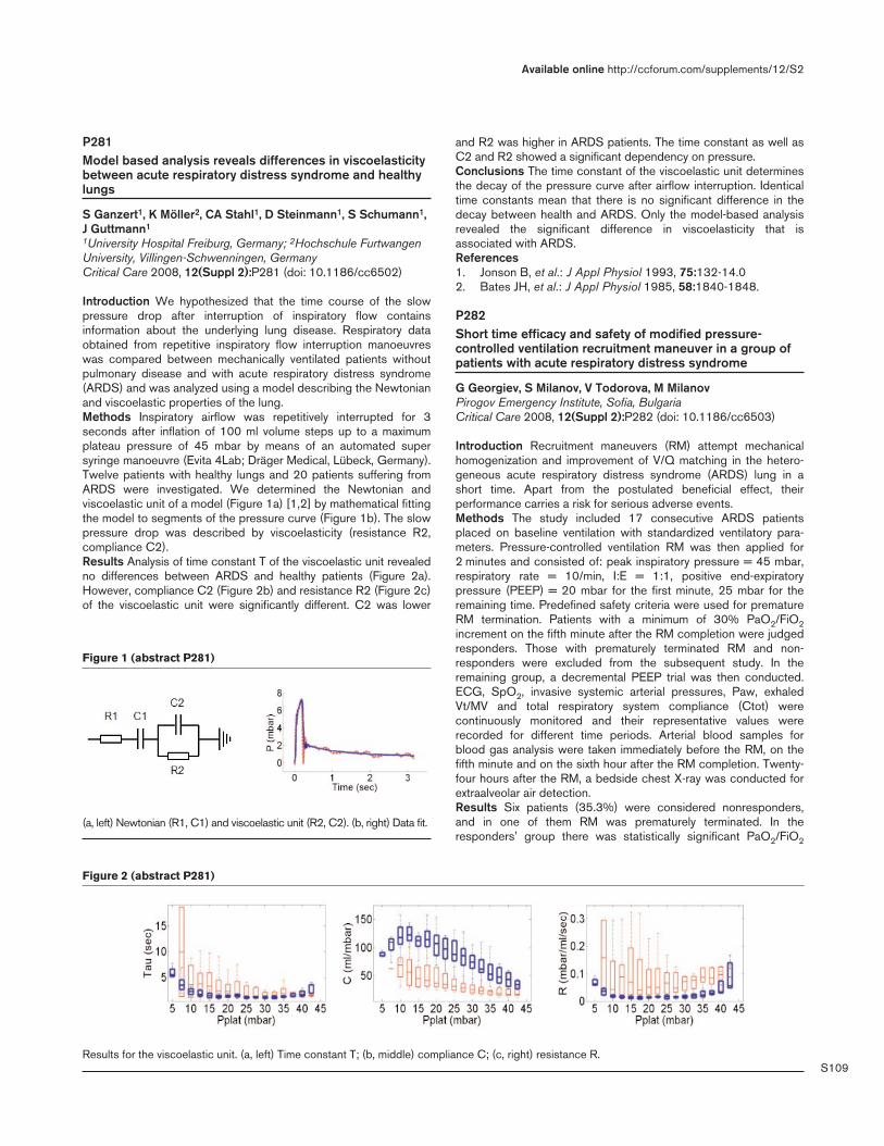
S109
P281Model based analysis reveals differences in viscoelasticitybetween acute respiratory distress syndrome and healthylungs
S Ganzert1, K Möller2, CA Stahl1, D Steinmann1, S Schumann1,J Guttmann1
1University Hospital Freiburg, Germany; 2Hochschule FurtwangenUniversity, Villingen-Schwenningen, GermanyCritical Care 2008, 12(Suppl 2):P281 (doi: 10.1186/cc6502)
Introduction We hypothesized that the time course of the slowpressure drop after interruption of inspiratory flow containsinformation about the underlying lung disease. Respiratory dataobtained from repetitive inspiratory flow interruption manoeuvreswas compared between mechanically ventilated patients withoutpulmonary disease and with acute respiratory distress syndrome(ARDS) and was analyzed using a model describing the Newtonianand viscoelastic properties of the lung.Methods Inspiratory airflow was repetitively interrupted for 3seconds after inflation of 100 ml volume steps up to a maximumplateau pressure of 45 mbar by means of an automated supersyringe manoeuvre (Evita 4Lab; Dräger Medical, Lübeck, Germany).Twelve patients with healthy lungs and 20 patients suffering fromARDS were investigated. We determined the Newtonian andviscoelastic unit of a model (Figure 1a) [1,2] by mathematical fittingthe model to segments of the pressure curve (Figure 1b). The slowpressure drop was described by viscoelasticity (resistance R2,compliance C2).Results Analysis of time constant T of the viscoelastic unit revealedno differences between ARDS and healthy patients (Figure 2a).However, compliance C2 (Figure 2b) and resistance R2 (Figure 2c)of the viscoelastic unit were significantly different. C2 was lower
and R2 was higher in ARDS patients. The time constant as well asC2 and R2 showed a significant dependency on pressure.Conclusions The time constant of the viscoelastic unit determinesthe decay of the pressure curve after airflow interruption. Identicaltime constants mean that there is no significant difference in thedecay between health and ARDS. Only the model-based analysisrevealed the significant difference in viscoelasticity that isassociated with ARDS.References1. Jonson B, et al.: J Appl Physiol 1993, 75:132-14.02. Bates JH, et al.: J Appl Physiol 1985, 58:1840-1848.
P282Short time efficacy and safety of modified pressure-controlled ventilation recruitment maneuver in a group ofpatients with acute respiratory distress syndrome
G Georgiev, S Milanov, V Todorova, M MilanovPirogov Emergency Institute, Sofia, BulgariaCritical Care 2008, 12(Suppl 2):P282 (doi: 10.1186/cc6503)
Introduction Recruitment maneuvers (RM) attempt mechanicalhomogenization and improvement of V/Q matching in the hetero-geneous acute respiratory distress syndrome (ARDS) lung in ashort time. Apart from the postulated beneficial effect, theirperformance carries a risk for serious adverse events.Methods The study included 17 consecutive ARDS patientsplaced on baseline ventilation with standardized ventilatory para-meters. Pressure-controlled ventilation RM was then applied for2 minutes and consisted of: peak inspiratory pressure = 45 mbar,respiratory rate = 10/min, I:E = 1:1, positive end-expiratorypressure (PEEP) = 20 mbar for the first minute, 25 mbar for theremaining time. Predefined safety criteria were used for prematureRM termination. Patients with a minimum of 30% PaO2/FiO2increment on the fifth minute after the RM completion were judgedresponders. Those with prematurely terminated RM and non-responders were excluded from the subsequent study. In theremaining group, a decremental PEEP trial was then conducted.ECG, SpO2, invasive systemic arterial pressures, Paw, exhaledVt/MV and total respiratory system compliance (Ctot) werecontinuously monitored and their representative values wererecorded for different time periods. Arterial blood samples forblood gas analysis were taken immediately before the RM, on thefifth minute and on the sixth hour after the RM completion. Twenty-four hours after the RM, a bedside chest X-ray was conducted forextraalveolar air detection.Results Six patients (35.3%) were considered nonresponders,and in one of them RM was prematurely terminated. In theresponders’ group there was statistically significant PaO2/FiO2
Available online http://ccforum.com/supplements/12/S2
Figure 1 (abstract P281)
(a, left) Newtonian (R1, C1) and viscoelastic unit (R2, C2). (b, right) Data fit.
Figure 2 (abstract P281)
Results for the viscoelastic unit. (a, left) Time constant T; (b, middle) compliance C; (c, right) resistance R.
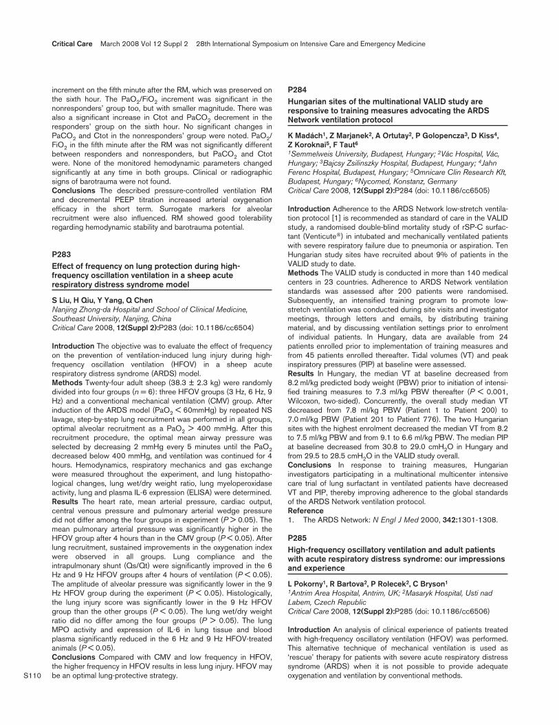
S110
increment on the fifth minute after the RM, which was preserved onthe sixth hour. The PaO2/FiO2 increment was significant in thenonresponders’ group too, but with smaller magnitude. There wasalso a significant increase in Ctot and PaCO2 decrement in theresponders’ group on the sixth hour. No significant changes inPaCO2 and Ctot in the nonresponders’ group were noted. PaO2/FiO2 in the fifth minute after the RM was not significantly differentbetween responders and nonresponders, but PaCO2 and Ctotwere. None of the monitored hemodynamic parameters changedsignificantly at any time in both groups. Clinical or radiographicsigns of barotrauma were not found.Conclusions The described pressure-controlled ventilation RMand decremental PEEP titration increased arterial oxygenationefficacy in the short term. Surrogate markers for alveolarrecruitment were also influenced. RM showed good tolerabilityregarding hemodynamic stability and barotrauma potential.
P283Effect of frequency on lung protection during high-frequency oscillation ventilation in a sheep acuterespiratory distress syndrome model
S Liu, H Qiu, Y Yang, Q ChenNanjing Zhong-da Hospital and School of Clinical Medicine,Southeast University, Nanjing, ChinaCritical Care 2008, 12(Suppl 2):P283 (doi: 10.1186/cc6504)
Introduction The objective was to evaluate the effect of frequencyon the prevention of ventilation-induced lung injury during high-frequency oscillation ventilation (HFOV) in a sheep acuterespiratory distress syndrome (ARDS) model.Methods Twenty-four adult sheep (38.3 ± 2.3 kg) were randomlydivided into four groups (n = 6): three HFOV groups (3 Hz, 6 Hz, 9Hz) and a conventional mechanical ventilation (CMV) group. Afterinduction of the ARDS model (PaO2 < 60mmHg) by repeated NSlavage, step-by-step lung recruitment was performed in all groups,optimal alveolar recruitment as a PaO2 > 400 mmHg. After thisrecruitment procedure, the optimal mean airway pressure wasselected by decreasing 2 mmHg every 5 minutes until the PaO2decreased below 400 mmHg, and ventilation was continued for 4hours. Hemodynamics, respiratory mechanics and gas exchangewere measured throughout the experiment, and lung histopatho-logical changes, lung wet/dry weight ratio, lung myeloperoxidaseactivity, lung and plasma IL-6 expression (ELISA) were determined.Results The heart rate, mean arterial pressure, cardiac output,central venous pressure and pulmonary arterial wedge pressuredid not differ among the four groups in experiment (P > 0.05). Themean pulmonary arterial pressure was significantly higher in theHFOV group after 4 hours than in the CMV group (P < 0.05). Afterlung recruitment, sustained improvements in the oxygenation indexwere observed in all groups. Lung compliance and theintrapulmonary shunt (Qs/Qt) were significantly improved in the 6Hz and 9 Hz HFOV groups after 4 hours of ventilation (P < 0.05).The amplitude of alveolar pressure was significantly lower in the 9Hz HFOV group during the experiment (P < 0.05). Histologically,the lung injury score was significantly lower in the 9 Hz HFOVgroup than the other groups (P < 0.05). The lung wet/dry weightratio did no differ among the four groups (P > 0.05). The lungMPO activity and expression of IL-6 in lung tissue and bloodplasma significantly reduced in the 6 Hz and 9 Hz HFOV-treatedanimals (P < 0.05).Conclusions Compared with CMV and low frequency in HFOV,the higher frequency in HFOV results in less lung injury. HFOV maybe an optimal lung-protective strategy.
P284Hungarian sites of the multinational VALID study areresponsive to training measures advocating the ARDSNetwork ventilation protocol
K Madách1, Z Marjanek2, A Ortutay2, P Golopencza3, D Kiss4, Z Koroknai5, F Taut6
1Semmelweis University, Budapest, Hungary; 2Vác Hospital, Vác,Hungary; 3Bajcsy Zsilinszky Hospital, Budapest, Hungary; 4JahnFerenc Hospital, Budapest, Hungary; 5Omnicare Clin Research Kft,Budapest, Hungary; 6Nycomed, Konstanz, GermanyCritical Care 2008, 12(Suppl 2):P284 (doi: 10.1186/cc6505)
Introduction Adherence to the ARDS Network low-stretch ventila-tion protocol [1] is recommended as standard of care in the VALIDstudy, a randomised double-blind mortality study of rSP-C surfac-tant (Venticute®) in intubated and mechanically ventilated patientswith severe respiratory failure due to pneumonia or aspiration. TenHungarian study sites have recruited about 9% of patients in theVALID study to date.Methods The VALID study is conducted in more than 140 medicalcenters in 23 countries. Adherence to ARDS Network ventilationstandards was assessed after 200 patients were randomised.Subsequently, an intensified training program to promote low-stretch ventilation was conducted during site visits and investigatormeetings, through letters and emails, by distributing trainingmaterial, and by discussing ventilation settings prior to enrolmentof individual patients. In Hungary, data are available from 24patients enrolled prior to implementation of training measures andfrom 45 patients enrolled thereafter. Tidal volumes (VT) and peakinspiratory pressures (PIP) at baseline were assessed.Results In Hungary, the median VT at baseline decreased from8.2 ml/kg predicted body weight (PBW) prior to initiation of intensi-fied training measures to 7.3 ml/kg PBW thereafter (P < 0.001,Wilcoxon, two-sided). Concurrently, the overall study median VTdecreased from 7.8 ml/kg PBW (Patient 1 to Patient 200) to7.0 ml/kg PBW (Patient 201 to Patient 776). The two Hungariansites with the highest enrolment decreased the median VT from 8.2to 7.5 ml/kg PBW and from 9.1 to 6.6 ml/kg PBW. The median PIPat baseline decreased from 30.8 to 29.0 cmH2O in Hungary andfrom 29.5 to 28.5 cmH2O in the VALID study overall.Conclusions In response to training measures, Hungarianinvestigators participating in a multinational multicenter intensivecare trial of lung surfactant in ventilated patients have decreasedVT and PIP, thereby improving adherence to the global standardsof the ARDS Network ventilation protocol.Reference1. The ARDS Network: N Engl J Med 2000, 342:1301-1308.
P285High-frequency oscillatory ventilation and adult patientswith acute respiratory distress syndrome: our impressionsand experience
L Pokorny1, R Bartova2, P Rolecek2, C Bryson1
1Antrim Area Hospital, Antrim, UK; 2Masaryk Hospital, Usti nadLabem, Czech RepublicCritical Care 2008, 12(Suppl 2):P285 (doi: 10.1186/cc6506)
Introduction An analysis of clinical experience of patients treatedwith high-frequency oscillatory ventilation (HFOV) was performed.This alternative technique of mechanical ventilation is used as‘rescue’ therapy for patients with severe acute respiratory distresssyndrome (ARDS) when it is not possible to provide adequateoxygenation and ventilation by conventional methods.
Critical Care March 2008 Vol 12 Suppl 2 28th International Symposium on Intensive Care and Emergency Medicine
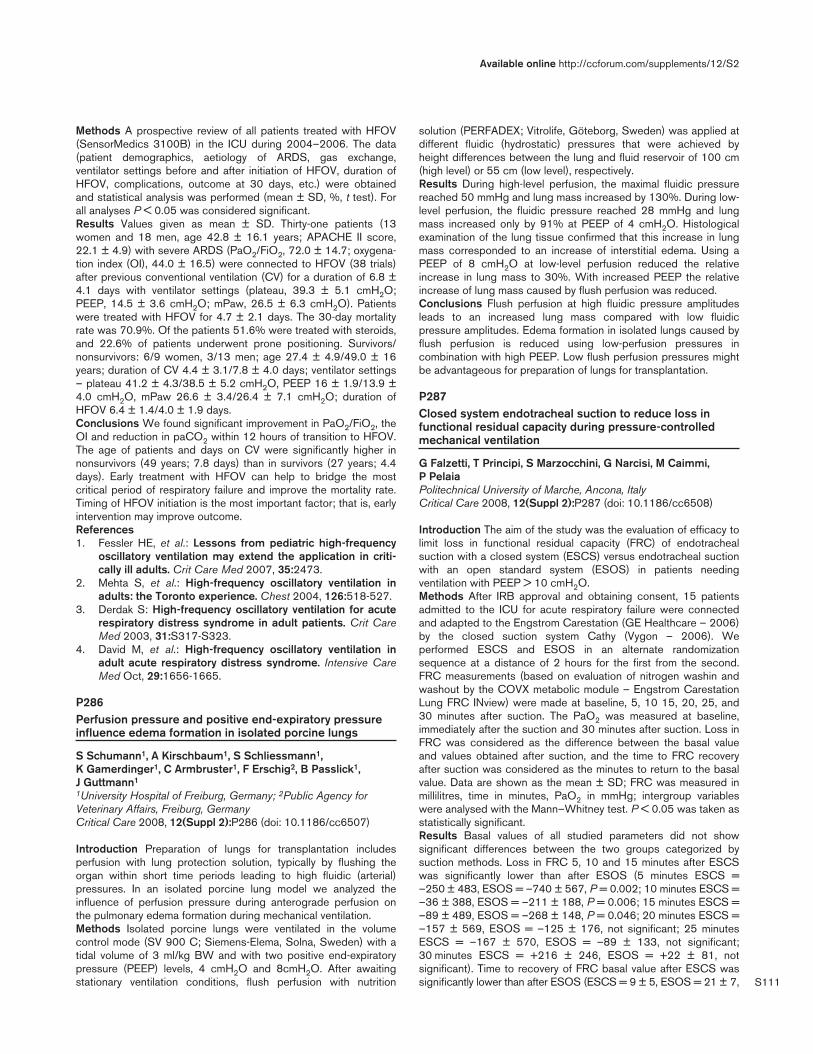
S111
Methods A prospective review of all patients treated with HFOV(SensorMedics 3100B) in the ICU during 2004–2006. The data(patient demographics, aetiology of ARDS, gas exchange,ventilator settings before and after initiation of HFOV, duration ofHFOV, complications, outcome at 30 days, etc.) were obtainedand statistical analysis was performed (mean ± SD, %, t test). Forall analyses P < 0.05 was considered significant.Results Values given as mean ± SD. Thirty-one patients (13women and 18 men, age 42.8 ± 16.1 years; APACHE II score,22.1 ± 4.9) with severe ARDS (PaO2/FiO2, 72.0 ± 14.7; oxygena-tion index (OI), 44.0 ± 16.5) were connected to HFOV (38 trials)after previous conventional ventilation (CV) for a duration of 6.8 ±4.1 days with ventilator settings (plateau, 39.3 ± 5.1 cmH2O;PEEP, 14.5 ± 3.6 cmH2O; mPaw, 26.5 ± 6.3 cmH2O). Patientswere treated with HFOV for 4.7 ± 2.1 days. The 30-day mortalityrate was 70.9%. Of the patients 51.6% were treated with steroids,and 22.6% of patients underwent prone positioning. Survivors/nonsurvivors: 6/9 women, 3/13 men; age 27.4 ± 4.9/49.0 ± 16years; duration of CV 4.4 ± 3.1/7.8 ± 4.0 days; ventilator settings– plateau 41.2 ± 4.3/38.5 ± 5.2 cmH2O, PEEP 16 ± 1.9/13.9 ±4.0 cmH2O, mPaw 26.6 ± 3.4/26.4 ± 7.1 cmH2O; duration ofHFOV 6.4 ± 1.4/4.0 ± 1.9 days.Conclusions We found significant improvement in PaO2/FiO2, theOI and reduction in paCO2 within 12 hours of transition to HFOV.The age of patients and days on CV were significantly higher innonsurvivors (49 years; 7.8 days) than in survivors (27 years; 4.4days). Early treatment with HFOV can help to bridge the mostcritical period of respiratory failure and improve the mortality rate.Timing of HFOV initiation is the most important factor; that is, earlyintervention may improve outcome.References1. Fessler HE, et al.: Lessons from pediatric high-frequency
oscillatory ventilation may extend the application in criti-cally ill adults. Crit Care Med 2007, 35:2473.
2. Mehta S, et al.: High-frequency oscillatory ventilation inadults: the Toronto experience. Chest 2004, 126:518-527.
3. Derdak S: High-frequency oscillatory ventilation for acuterespiratory distress syndrome in adult patients. Crit CareMed 2003, 31:S317-S323.
4. David M, et al.: High-frequency oscillatory ventilation inadult acute respiratory distress syndrome. Intensive CareMed Oct, 29:1656-1665.
P286Perfusion pressure and positive end-expiratory pressureinfluence edema formation in isolated porcine lungs
S Schumann1, A Kirschbaum1, S Schliessmann1, K Gamerdinger1, C Armbruster1, F Erschig2, B Passlick1, J Guttmann1
1University Hospital of Freiburg, Germany; 2Public Agency forVeterinary Affairs, Freiburg, GermanyCritical Care 2008, 12(Suppl 2):P286 (doi: 10.1186/cc6507)
Introduction Preparation of lungs for transplantation includesperfusion with lung protection solution, typically by flushing theorgan within short time periods leading to high fluidic (arterial)pressures. In an isolated porcine lung model we analyzed theinfluence of perfusion pressure during anterograde perfusion onthe pulmonary edema formation during mechanical ventilation.Methods Isolated porcine lungs were ventilated in the volumecontrol mode (SV 900 C; Siemens-Elema, Solna, Sweden) with atidal volume of 3 ml/kg BW and with two positive end-expiratorypressure (PEEP) levels, 4 cmH2O and 8cmH2O. After awaitingstationary ventilation conditions, flush perfusion with nutrition
solution (PERFADEX; Vitrolife, Göteborg, Sweden) was applied atdifferent fluidic (hydrostatic) pressures that were achieved byheight differences between the lung and fluid reservoir of 100 cm(high level) or 55 cm (low level), respectively.Results During high-level perfusion, the maximal fluidic pressurereached 50 mmHg and lung mass increased by 130%. During low-level perfusion, the fluidic pressure reached 28 mmHg and lungmass increased only by 91% at PEEP of 4 cmH2O. Histologicalexamination of the lung tissue confirmed that this increase in lungmass corresponded to an increase of interstitial edema. Using aPEEP of 8 cmH2O at low-level perfusion reduced the relativeincrease in lung mass to 30%. With increased PEEP the relativeincrease of lung mass caused by flush perfusion was reduced.Conclusions Flush perfusion at high fluidic pressure amplitudesleads to an increased lung mass compared with low fluidicpressure amplitudes. Edema formation in isolated lungs caused byflush perfusion is reduced using low-perfusion pressures incombination with high PEEP. Low flush perfusion pressures mightbe advantageous for preparation of lungs for transplantation.
P287Closed system endotracheal suction to reduce loss infunctional residual capacity during pressure-controlledmechanical ventilation
G Falzetti, T Principi, S Marzocchini, G Narcisi, M Caimmi, P PelaiaPolitechnical University of Marche, Ancona, ItalyCritical Care 2008, 12(Suppl 2):P287 (doi: 10.1186/cc6508)
Introduction The aim of the study was the evaluation of efficacy tolimit loss in functional residual capacity (FRC) of endotrachealsuction with a closed system (ESCS) versus endotracheal suctionwith an open standard system (ESOS) in patients needingventilation with PEEP > 10 cmH2O.Methods After IRB approval and obtaining consent, 15 patientsadmitted to the ICU for acute respiratory failure were connectedand adapted to the Engstrom Carestation (GE Healthcare – 2006)by the closed suction system Cathy (Vygon – 2006). Weperformed ESCS and ESOS in an alternate randomizationsequence at a distance of 2 hours for the first from the second.FRC measurements (based on evaluation of nitrogen washin andwashout by the COVX metabolic module – Engstrom CarestationLung FRC INview) were made at baseline, 5, 10 15, 20, 25, and30 minutes after suction. The PaO2 was measured at baseline,immediately after the suction and 30 minutes after suction. Loss inFRC was considered as the difference between the basal valueand values obtained after suction, and the time to FRC recoveryafter suction was considered as the minutes to return to the basalvalue. Data are shown as the mean ± SD; FRC was measured inmillilitres, time in minutes, PaO2 in mmHg; intergroup variableswere analysed with the Mann–Whitney test. P < 0.05 was taken asstatistically significant.Results Basal values of all studied parameters did not showsignificant differences between the two groups categorized bysuction methods. Loss in FRC 5, 10 and 15 minutes after ESCSwas significantly lower than after ESOS (5 minutes ESCS =–250 ± 483, ESOS = –740 ± 567, P = 0.002; 10 minutes ESCS =–36 ± 388, ESOS = –211 ± 188, P = 0.006; 15 minutes ESCS =–89 ± 489, ESOS = –268 ± 148, P = 0.046; 20 minutes ESCS =–157 ± 569, ESOS = –125 ± 176, not significant; 25 minutesESCS = –167 ± 570, ESOS = –89 ± 133, not significant;30 minutes ESCS = +216 ± 246, ESOS = +22 ± 81, notsignificant). Time to recovery of FRC basal value after ESCS wassignificantly lower than after ESOS (ESCS = 9 ± 5, ESOS = 21 ± 7,
Available online http://ccforum.com/supplements/12/S2
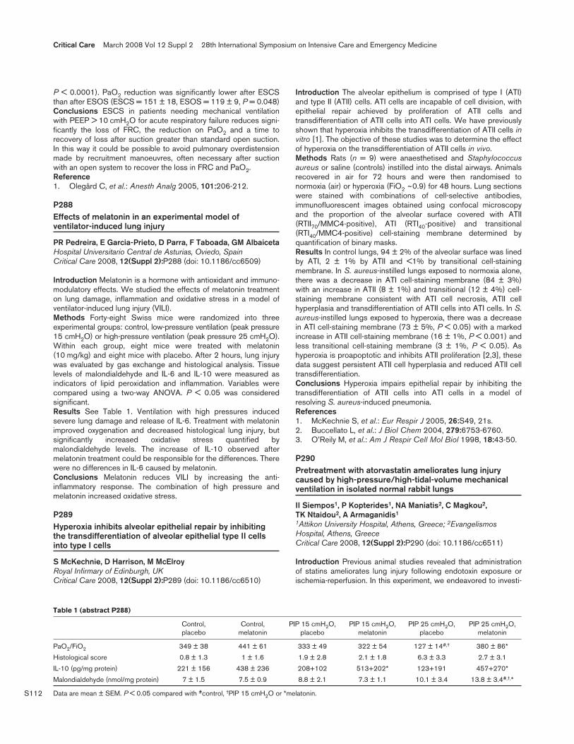
S112
P < 0.0001). PaO2 reduction was significantly lower after ESCSthan after ESOS (ESCS = 151 ± 18, ESOS = 119 ± 9, P = 0.048)Conclusions ESCS in patients needing mechanical ventilationwith PEEP > 10 cmH2O for acute respiratory failure reduces signi-ficantly the loss of FRC, the reduction on PaO2 and a time torecovery of loss after suction greater than standard open suction.In this way it could be possible to avoid pulmonary overdistensionmade by recruitment manoeuvres, often necessary after suctionwith an open system to recover the loss in FRC and PaO2.Reference1. Olegård C, et al.: Anesth Analg 2005, 101:206-212.
P288Effects of melatonin in an experimental model ofventilator-induced lung injury
PR Pedreira, E Garcia-Prieto, D Parra, F Taboada, GM AlbaicetaHospital Universitario Central de Asturias, Oviedo, SpainCritical Care 2008, 12(Suppl 2):P288 (doi: 10.1186/cc6509)
Introduction Melatonin is a hormone with antioxidant and immuno-modulatory effects. We studied the effects of melatonin treatmenton lung damage, inflammation and oxidative stress in a model ofventilator-induced lung injury (VILI).Methods Forty-eight Swiss mice were randomized into threeexperimental groups: control, low-pressure ventilation (peak pressure15 cmH2O) or high-pressure ventilation (peak pressure 25 cmH2O).Within each group, eight mice were treated with melatonin(10 mg/kg) and eight mice with placebo. After 2 hours, lung injurywas evaluated by gas exchange and histological analysis. Tissuelevels of malondialdehyde and IL-6 and IL-10 were measured asindicators of lipid peroxidation and inflammation. Variables werecompared using a two-way ANOVA. P < 0.05 was consideredsignificant.Results See Table 1. Ventilation with high pressures inducedsevere lung damage and release of IL-6. Treatment with melatoninimproved oxygenation and decreased histological lung injury, butsignificantly increased oxidative stress quantified bymalondialdehyde levels. The increase of IL-10 observed aftermelatonin treatment could be responsible for the differences. Therewere no differences in IL-6 caused by melatonin.Conclusions Melatonin reduces VILI by increasing the anti-inflammatory response. The combination of high pressure andmelatonin increased oxidative stress.
P289Hyperoxia inhibits alveolar epithelial repair by inhibitingthe transdifferentiation of alveolar epithelial type II cellsinto type I cells
S McKechnie, D Harrison, M McElroyRoyal Infirmary of Edinburgh, UKCritical Care 2008, 12(Suppl 2):P289 (doi: 10.1186/cc6510)
Introduction The alveolar epithelium is comprised of type I (ATI)and type II (ATII) cells. ATI cells are incapable of cell division, withepithelial repair achieved by proliferation of ATII cells andtransdifferentiation of ATII cells into ATI cells. We have previouslyshown that hyperoxia inhibits the transdifferentiation of ATII cells invitro [1]. The objective of these studies was to determine the effectof hyperoxia on the transdifferentiation of ATII cells in vivo.Methods Rats (n = 9) were anaesthetised and Staphylococcusaureus or saline (controls) instilled into the distal airways. Animalsrecovered in air for 72 hours and were then randomised tonormoxia (air) or hyperoxia (FiO2 ~0.9) for 48 hours. Lung sectionswere stained with combinations of cell-selective antibodies,immunofluorescent images obtained using confocal microscopyand the proportion of the alveolar surface covered with ATII(RTII70/MMC4-positive), ATI (RTI40-positive) and transitional(RTI40/MMC4-positive) cell-staining membrane determined byquantification of binary masks.Results In control lungs, 94 ± 2% of the alveolar surface was linedby ATI, 2 ± 1% by ATII and <1% by transitional cell-stainingmembrane. In S. aureus-instilled lungs exposed to normoxia alone,there was a decrease in ATI cell-staining membrane (84 ± 3%)with an increase in ATII (8 ± 1%) and transitional (12 ± 4%) cell-staining membrane consistent with ATI cell necrosis, ATII cellhyperplasia and transdifferentiation of ATII cells into ATI cells. In S.aureus-instilled lungs exposed to hyperoxia, there was a decreasein ATI cell-staining membrane (73 ± 5%, P < 0.05) with a markedincrease in ATII cell-staining membrane (16 ± 1%, P < 0.001) andless transitional cell-staining membrane (3 ± 1%, P < 0.05). Ashyperoxia is proapoptotic and inhibits ATII proliferation [2,3], thesedata suggest persistent ATII cell hyperplasia and reduced ATII celltransdifferentiation.Conclusions Hyperoxia impairs epithelial repair by inhibiting thetransdifferentiation of ATII cells into ATI cells in a model ofresolving S. aureus-induced pneumonia.References1. McKechnie S, et al.: Eur Respir J 2005, 26:S49, 21s.2. Buccellato L, et al.: J Biol Chem 2004, 279:6753-6760.3. O’Reily M, et al.: Am J Respir Cell Mol Biol 1998, 18:43-50.
P290Pretreatment with atorvastatin ameliorates lung injurycaused by high-pressure/high-tidal-volume mechanicalventilation in isolated normal rabbit lungs
II Siempos1, P Kopterides1, NA Maniatis2, C Magkou2, TK Ntaidou2, A Armaganidis1
1Attikon University Hospital, Athens, Greece; 2EvangelismosHospital, Athens, GreeceCritical Care 2008, 12(Suppl 2):P290 (doi: 10.1186/cc6511)
Introduction Previous animal studies revealed that administrationof statins ameliorates lung injury following endotoxin exposure orischemia-reperfusion. In this experiment, we endeavored to investi-
Critical Care March 2008 Vol 12 Suppl 2 28th International Symposium on Intensive Care and Emergency Medicine
Table 1 (abstract P288)
Control, Control, PIP 15 cmH2O, PIP 15 cmH2O, PIP 25 cmH2O, PIP 25 cmH2O, placebo melatonin placebo melatonin placebo melatonin
PaO2/FiO2 349 ± 38 441 ± 61 333 ± 49 322 ± 54 127 ± 14#,† 380 ± 86*
Histological score 0.8 ± 1.3 1 ± 1.6 1.9 ± 2.8 2.1 ± 1.8 6.3 ± 3.3 2.7 ± 3.1
IL-10 (pg/mg protein) 221 ± 156 438 ± 236 208+102 513+202* 123+191 457+270*
Malondialdehyde (nmol/mg protein) 7 ± 1.5 7.5 ± 0.9 8.8 ± 2.1 7.3 ± 1.1 10.1 ± 3.4 13.8 ± 3.4#,†,*
Data are mean ± SEM. P < 0.05 compared with #control, †PIP 15 cmH2O or *melatonin.
S113
gate whether pretreatment with atorvastatin confers protectionfrom lung injury caused by high-pressure/high-tidal-volume ventilation.Methods Twenty-four isolated sets of normal rabbit lungs wereutilized. Treated animals received atorvastatin (20 mg/kg bodyweight/day per os) for 3 days before surgery. All isolated lungswere perfused (constant flow, 300 ml/min) and ventilated for1 hour with pressure control ventilation at either 23 cmH2O (high-pressure, injurious ventilation) or 11 cmH2O (low-pressure,noninjurious ventilation) peak static pressure and positive end-expiratory pressure of 3 cmH2O. Four groups of six lung sets eachwere examined: HPC (high pressure, no statin), HPS (highpressure, statin pretreatment), LPC (low pressure, no statin), andLPS (low pressure, statin pretreatment). Changes (from baseline tothe end of ventilation) in the ultrafiltration coefficient (ΔKf,c;pulmonary capillary permeability), weight gain (ΔW; edemaformation) and histological lesions (hemorrhage) were used asindices of lung damage.Results At baseline, the compared groups did not differ withregard to Kf,c (P = 0.3). At the end of ventilation, the HPC groupsuffered greater ΔKf,c (P < 0.001) and greater ΔW (P < 0.001)than both the LPC and LPS groups. In contrast, the HPS group didnot differ from both the LPS and LPC groups regarding thesevariables (P > 0.4). In the HPC and HPS groups, lungs with asopposed to those without statin pretreatment experienced asignificantly lower ΔKf,c (–0.013 ± 0.016 versus 1.723 ± 0.495g/min/cmH2O/100 g; P = 0.0006) and lower ΔW (4.62 ± 1.50versus 17.75 ± 4.71 g; P = 0.007). This was also the case for thehistological lesions.Conclusions In an ex vivo model of ventilator-induced lung injury,pretreatment with atorvastatin attenuates lung injury following high-pressure/high-tidal-volume ventilation.References1. Jacobson JR, et al.: Am J Physiol Lung Cell Mol Physiol
2005, 288:L1026-L1032.2. Pirat A, et al.: Anesth Analg 2006, 102:225-232.
P291Systemic markers of inflammation in mechanicallyventilated brain-injured patients in the absence of sepsisand acute lung injury: the effect of positive end-expiratorypressure
I Korovesi1, A Kotanidou1, E Papadomichelakis2, E Giamarellos-Bourboulis2, A Pelekanou2, O Livaditi1, C Sotiropoulou1, A Koutsoukou1, I Dimopoulou2, A Armaganidis2, C Roussos1, N Marczin3, S Orfanos2
1University of Athens Medical School, Evangelismos Hospital,Athens, Greece; 2Attikon Hospital, University of Athens MedicalSchool, Haidari, Athens, Greece; 3Imperial College London, UKCritical Care 2008, 12(Suppl 2):P291 (doi: 10.1186/cc6512)
Introduction In mechanically ventilated (MV) brain-injured patients,pulmonary complications appear to be the leading cause of non-neurological morbidity, suggesting that the local and systemicinflammatory responses associated with brain damage and/or themechanical ventilation itself may contribute to the pulmonary injury(that is, one or two hit model). We recently provided evidence forlung inflammation in brain-injured, mechanically ventilated patientswith neither acute lung injury nor sepsis [1]. We now investigatewhether positive end-expiratory pressure (PEEP) application in thesame cohort (27 MV brain-injured subjects) is associated withsystemic inflammatory changes that could probably contribute tothe lung inflammation observed in these patients.Methods Patients were ventilated with 8 ml/kg tidal volume andwere put on either zero PEEP (ZEEP, n = 12) or 8 cmH2O PEEP
(PEEP, n = 15). The following markers of systemic inflammationwere recorded or measured in blood, on the first, third, and fifthdays of MV: temperature, leukocyte and neutrophil counts, albumin,soluble triggering receptor expressed on myeloid cells (sTREM-1),C-reactive protein, procalcitonin, IL-10, IL-1β, IL-6, IL-8, IL-12p70,and TNFα.Results Upon entry, the two groups were well balanced clinicallyand demographically. Significant differences between the twopatient groups were observed in leukocyte counts, IL-6 andsTREM-1; all three parameters were constantly higher on ZEEP(P < 0.05; two-way ANOVARM), while the former two markersdecreased with time in both groups (P < 0.05).Conclusions In our population of MV brain-injured patients, ZEEPis associated with increases in systemic inflammatory markers thatare present early on and throughout the first 5 days of MV. Ourfindings suggest that PEEP application influences the systemicinflammatory response observed in the absence of sepsis andacute lung injury, and probably points to significant IL-6 andsTREM-1 roles in the systemic and pulmonary inflammationobserved in this patient setting.Reference1. Korovesi I, et al.: Eur Respir J 2007, 30(Suppl 51):444S.
P292Shotgun proteomics reveals biochemistry of normal andacute respiratory distress syndrome bronchoalveolarlavage fluid proteome
D GoodlettUniversity of Washington, Seattle, WA, USACritical Care 2008, 12(Suppl 2):P292 (doi: 10.1186/cc6513)
Introduction Recently, so-called label-free quantitative proteomicmethods have gained acceptance for protein expression analysis viamass spectrometry (MS). These methods allow better experimentdesign by circumventing the need to establish pair-wisecomparisons required by popular stable isotope dilution methods (forexample, isotope coded affinity tag, isobaric tags for relative andabsolute quantitation and stable-isotope labelling by amino acids incell culture) and are thus fundamentally better suited for proteomestudies where sample number is large. Here we discuss the use ofshotgun proteomics (that is, no prior protein fractionation) and label-free quantitative methods to characterize human bronchoalveolarlavage fluid (BALF) proteomes of six normal healthy volunteers andthree patients with acute respiratory distress syndrome (ARDS) [1].Methods Our proteomic profiling technology for BALF involves: (i)removal of cells by centrifugation followed by precipitation of theremaining soluble protein, (ii) denaturation and proteolysis ofsoluble proteins, (iii) analysis in quadruplicate of each sample byLC-MS/MS using Fourier transform-mass spectrometry (FT-MS),(iv) identification of proteins by database search of tandem massspectra (MS2 data), and (v) extraction of changes in proteinrelative expression directly from peptide ion intensity values (MS1data) as recently described [2]. Notably, data for each sample areacquired independent of all other samples, allowing any patient’sdata to be compared directly with all others in silico. We alsomeasured levels of several proteins of interest (identified in LC-MS/MS experiments) by ELISA in ARDS BALF from 69 patientsranging from those at risk to 14 days post diagnosis and sixnormals. Enriched functional categories within BALF proteinsrelative to the entire human proteome were determined usingExpression Analysis Systematic Explorer (EASE) software [3].Results Using BALF from three patients, we identified a total of870 different proteins, a nearly 10-fold increase from previousreports. Among the proteins identified were known markers of lung
Available online http://ccforum.com/supplements/12/S2
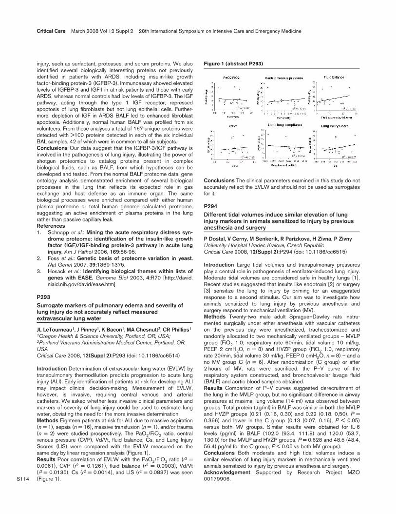
S114
injury, such as surfactant, proteases, and serum proteins. We alsoidentified several biologically interesting proteins not previouslyidentified in patients with ARDS, including insulin-like growthfactor-binding protein-3 (IGFBP-3). Immunoassay showed elevatedlevels of IGFBP-3 and IGF-I in at-risk patients and those with earlyARDS, whereas normal controls had low levels of IGFBP-3. The IGFpathway, acting through the type 1 IGF receptor, repressedapoptosis of lung fibroblasts but not lung epithelial cells. Further-more, depletion of IGF in ARDS BALF led to enhanced fibroblastapoptosis. Additionally, normal human BALF was profiled from sixvolunteers. From these analyses a total of 167 unique proteins weredetected with >100 proteins detected in each of the six individualBAL samples, 42 of which were in common to all six subjects.Conclusions Our data suggest that the IGFBP-3/IGF pathway isinvolved in the pathogenesis of lung injury, illustrating the power ofshotgun proteomics to catalog proteins present in complexbiological fluids, such as BALF, from which hypotheses can bedeveloped and tested. From the normal BALF proteome data, geneontology analysis demonstrated enrichment of several biologicalprocesses in the lung that reflects its expected role in gasexchange and host defense as an immune organ. The samebiological processes were enriched compared with either humanplasma proteome or total human genome calculated proteome,suggesting an active enrichment of plasma proteins in the lungrather than passive capillary leak.References1. Schnapp et al.: Mining the acute respiratory distress syn-
drome proteome: identification of the insulin-like growthfactor (IGF)/IGF-binding protein-3 pathway in acute lunginjury. Am J Pathol 2006, 169:86-95.
2. Foss et al.: Genetic basis of proteome variation in yeast.Nat Genet 2007, 39:1369-1375.
3. Hosack et al.: Identifying biological themes within lists ofgenes with EASE. Genome Biol 2003, 4:R70 [http://david.niaid.nih.gov/david/ease.htm]
P293Surrogate markers of pulmonary edema and severity oflung injury do not accurately reflect measuredextravascular lung water
JL LeTourneau1, J Pinney1, K Bacon1, MA Chesnutt2, CR Phillips1
1Oregon Health & Science University, Portland, OR, USA;2Portland Veterans Administration Medical Center, Portland, OR,USACritical Care 2008, 12(Suppl 2):P293 (doi: 10.1186/cc6514)
Introduction Determination of extravascular lung water (EVLW) bytranspulmonary thermodilution predicts progression to acute lunginjury (ALI). Early identification of patients at risk for developing ALImay impact clinical decision-making. Measurement of EVLW,however, is invasive, requiring central venous and arterialcatheters. We asked whether less invasive clinical parameters andmarkers of severity of lung injury could be used to estimate lungwater, obviating the need for the more invasive determination.Methods Eighteen patients at risk for ALI due to massive aspiration(n = 1), sepsis (n = 16), massive transfusion (n = 1), and/or trauma(n = 2) were studied prospectively. The PaO2/FiO2 ratio, centralvenous pressure (CVP), Vd/Vt, fluid balance, Cs, and Lung InjuryScores (LIS) were compared with the EVLW measured on thesame day by linear regression analysis (Figure 1).Results Poor correlation of EVLW with the PaO2/FiO2 ratio (r2 =0.0061), CVP (r2 = 0.1261), fluid balance (r2 = 0.0903), Vd/Vt(r2 = 0.0135), Cs (r2 = 0.0014), and LIS (r2 = 0.0837) was seen(Figure 1).
Conclusions The clinical parameters examined in this study do notaccurately reflect the EVLW and should not be used as surrogatesfor it.
P294Different tidal volumes induce similar elevation of lunginjury markers in animals sensitized to injury by previousanesthesia and surgery
P Dostal, V Cerny, M Senkerik, R Parizkova, H Zivna, P ZivnyUniversity Hospital Hradec Kralove, Czech RepublicCritical Care 2008, 12(Suppl 2):P294 (doi: 10.1186/cc6515)
Introduction Large tidal volumes and transpulmonary pressuresplay a central role in pathogenesis of ventilator-induced lung injury.Moderate tidal volumes are considered safe in healthy lungs [1].Recent studies suggested that insults like endotoxin [2] or surgery[3] sensitize the lung to injury by priming for an exaggeratedresponse to a second stimulus. Our aim was to investigate howanimals sensitized to lung injury by previous anesthesia andsurgery respond to mechanical ventilation (MV).Methods Twenty-two male adult Sprague–Dawley rats instru-mented surgically under ether anesthesia with vascular catheterson the previous day were anesthetized, tracheostomized andrandomly allocated to two mechanically ventilated groups – MVLPgroup (FiO2 1.0, respiratory rate 60/min, tidal volume 10 ml/kg,PEEP 2 cmH2O, n = 8) and HVZP group (FiO2 1.0, respiratoryrate 20/min, tidal volume 30 ml/kg, PEEP 0 cmH2O, n = 8) – and ano MV group C (n = 6). After randomization (C group) or after2 hours of MV, rats were sacrificed, the P–V curve of therespiratory system constructed, and bronchoalveolar lavage fluid(BALF) and aortic blood samples obtained.Results Comparison of P–V curves suggested derecruitment ofthe lung in the MVLP group, but no significant difference in airwaypressures at maximal lung volume (14 ml) was observed betweengroups. Total protein (μg/ml) in BALF was similar in both the MVLPand HVZP groups (0.21 (0.16, 0.30) and 0.22 (0.18, 0,50), P =0.366) and lower in the C group (0.13 (0.07, 0.16), P < 0.05)versus both MV groups. Similar results were obtained for IL-6levels (pg/ml) in BALF (102.0 (93.4, 111.8) and 120.0 (53.7,130.0) for the MVLP and HVZP groups, P = 0.628 and 48.5 (43.4,56.4) pg/ml for the C group, P < 0.05 vs both MV groups).Conclusions Both moderate and high tidal volumes induce asimilar elevation of lung injury markers in mechanically ventilatedanimals sensitized to injury by previous anesthesia and surgery.Acknowledgement Supported by Research Project MZO00179906.
Critical Care March 2008 Vol 12 Suppl 2 28th International Symposium on Intensive Care and Emergency Medicine
Figure 1 (abstract P293)
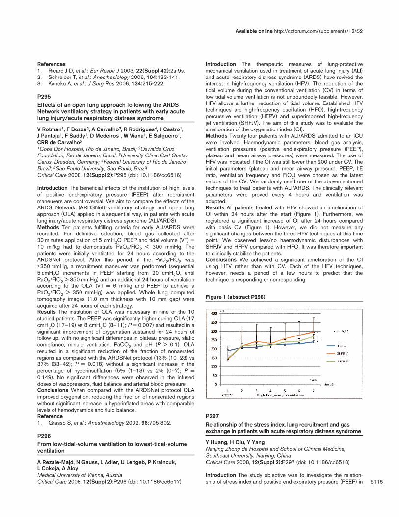
S115
References1. Ricard J-D, et al.: Eur Respir J 2003, 22(Suppl 42):2s-9s.2. Schreiber T, et al.: Anesthesiology 2006, 104:133-141.3. Kaneko A, et al.: J Surg Res 2006, 134:215-222.
P295Effects of an open lung approach following the ARDSNetwork ventilatory strategy in patients with early acutelung injury/acute respiratory distress syndrome
V Rotman1, F Bozza2, A Carvalho3, R Rodrigues4, J Castro1, J Pantoja1, F Saddy1, D Medeiros1, W Viana1, E Salgueiro1, CRR de Carvalho5
1Copa Dor Hospital, Rio de Janeiro, Brazil; 2Oswaldo CruzFoundation, Rio de Janeiro, Brazil; 3University Clinic Carl GustavCarus, Dresden, Germany; 4Federal University of Rio de Janeiro,Brazil; 5São Paulo University, São Paulo, BrazilCritical Care 2008, 12(Suppl 2):P295 (doi: 10.1186/cc6516)
Introduction The beneficial effects of the institution of high levelsof positive end-expiratory pressure (PEEP) after recruitmentmaneuvers are controversial. We aim to compare the effects of theARDS Network (ARDSNet) ventilatory strategy and open lungapproach (OLA) applied in a sequential way, in patients with acutelung injury/acute respiratory distress syndrome (ALI/ARDS).Methods Ten patients fulfilling criteria for early ALI/ARDS wererecruited. For definitive selection, blood gas collected after30 minutes application of 5 cmH2O PEEP and tidal volume (VT) =10 ml/kg had to demonstrate PaO2/FIO2 < 300 mmHg. Thepatients were initially ventilated for 24 hours according to theARDSNet protocol. After this period, if the PaO2/FIO2 was≤350 mmHg, a recruitment maneuver was performed (sequential5 cmH2O increments in PEEP starting from 20 cmH2O, untilPaO2/FIO2 > 350 mmHg) and an additional 24 hours of ventilationaccording to the OLA (VT = 6 ml/kg and PEEP to achieve aPaO2/FIO2 > 350 mmHg) was applied. Whole lung computedtomography images (1.0 mm thickness with 10 mm gap) wereacquired after 24 hours of each strategy.Results The institution of OLA was necessary in nine of the 10studied patients. The PEEP was significantly higher during OLA (17cmH2O (17–19) vs 8 cmH2O (8–11); P = 0.007) and resulted in asignificant improvement of oxygenation sustained for 24 hours offollow-up, with no significant differences in plateau pressure, staticcompliance, minute ventilation, PaCO2 and pH (P > 0.1). OLAresulted in a significant reduction of the fraction of nonaeratedregions as compared with the ARDSNet protocol (13% (10–23) vs37% (33–42); P = 0.018) without a significant increase in thepercentage of hyperinsufflation (5% (1–13) vs 2% (0–7); P =0.149). No significant differences were observed in the infuseddoses of vasopressors, fluid balance and arterial blood pressure.Conclusions When compared with the ARDSNet protocol OLAimproved oxygenation, reducing the fraction of nonaerated regionswithout significant increase in hyperinflated areas with comparablelevels of hemodynamics and fluid balance.Reference1. Grasso S, et al.: Anesthesiology 2002, 96:795-802.
P296From low-tidal-volume ventilation to lowest-tidal-volumeventilation
A Rezaie-Majd, N Gauss, L Adler, U Leitgeb, P Kraincuk, L Cokoja, A AloyMedical University of Vienna, AustriaCritical Care 2008, 12(Suppl 2):P296 (doi: 10.1186/cc6517)
Introduction The therapeutic measures of lung-protectivemechanical ventilation used in treatment of acute lung injury (ALI)and acute respiratory distress syndrome (ARDS) have revived theinterest in high-frequency ventilation (HFV). The reduction of thetidal volume during the conventional ventilation (CV) in terms oflow-tidal-volume ventilation is not unboundedly feasible. However,HFV allows a further reduction of tidal volume. Established HFVtechniques are high-frequency oscillation (HFO), high-frequencypercussive ventilation (HFPV) and superimposed high-frequencyjet ventilation (SHFJV). The aim of this study was to evaluate theamelioration of the oxygenation index (OI).Methods Twenty-four patients with ALI/ARDS admitted to an ICUwere involved. Haemodynamic parameters, blood gas analysis,ventilation pressures (positive end-expiratory pressure (PEEP),plateau and mean airway pressures) were measured. The use ofHFV was indicated if the OI was still lower than 200 under CV. Theinitial parameters (plateau and mean airway pressure, PEEP, I:Eratio, ventilation frequency and FiO2) were chosen as the latestsetups of the CV. We randomly used one of the abovementionedtechniques to treat patients with ALI/ARDS. The clinically relevantparameters were proved every 4 hours and ventilation wasadopted.Results All patients treated with HFV showed an amelioration ofOI within 24 hours after the start (Figure 1). Furthermore, weregistered a significant increase of OI after 24 hours comparedwith basis CV (Figure 1). However, we did not measure anysignificant changes between the three HFV techniques at this timepoint. We observed less/no haemodynamic disturbances withSHFJV and HFPV compared with HFO. It was therefore importantto clinically stabilize the patients.Conclusions We achieved a significant amelioration of the OIusing HFV rather than with CV. Each of the HFV techniques,however, needs a period of a few hours to predict that thetechnique is responding or nonresponding.
P297Relationship of the stress index, lung recruitment and gasexchange in patients with acute respiratory distress syndrome
Y Huang, H Qiu, Y YangNanjing Zhong-da Hospital and School of Clinical Medicine,Southeast University, Nanjing, ChinaCritical Care 2008, 12(Suppl 2):P297 (doi: 10.1186/cc6518)
Introduction The study objective was to investigate the relation-ship of stress index and positive end-expiratory pressure (PEEP) in
Available online http://ccforum.com/supplements/12/S2
Figure 1 (abstract P296)
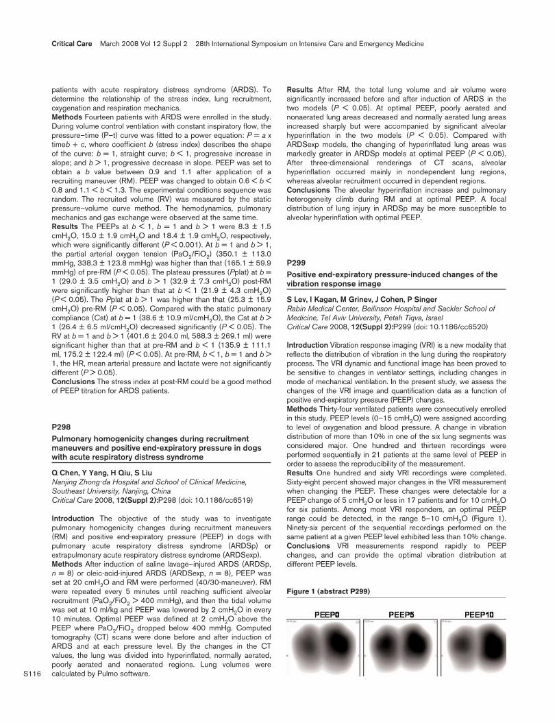
S116
patients with acute respiratory distress syndrome (ARDS). Todetermine the relationship of the stress index, lung recruitment,oxygenation and respiration mechanics.Methods Fourteen patients with ARDS were enrolled in the study.During volume control ventilation with constant inspiratory flow, thepressure–time (P–t) curve was fitted to a power equation: P = a xtimeb + c, where coefficient b (stress index) describes the shapeof the curve: b = 1, straight curve; b < 1, progressive increase inslope; and b > 1, progressive decrease in slope. PEEP was set toobtain a b value between 0.9 and 1.1 after application of arecruiting maneuver (RM). PEEP was changed to obtain 0.6 < b <0.8 and 1.1 < b < 1.3. The experimental conditions sequence wasrandom. The recruited volume (RV) was measured by the staticpressure–volume curve method. The hemodynamics, pulmonarymechanics and gas exchange were observed at the same time.Results The PEEPs at b < 1, b = 1 and b > 1 were 8.3 ± 1.5cmH2O, 15.0 ± 1.9 cmH2O and 18.4 ± 1.9 cmH2O, respectively,which were significantly different (P < 0.001). At b = 1 and b > 1,the partial arterial oxygen tension (PaO2/FiO2) (350.1 ± 113.0mmHg, 338.3 ± 123.8 mmHg) was higher than that (165.1 ± 59.9mmHg) of pre-RM (P < 0.05). The plateau pressures (Pplat) at b =1 (29.0 ± 3.5 cmH2O) and b > 1 (32.9 ± 7.3 cmH2O) post-RMwere significantly higher than that at b < 1 (21.9 ± 4.3 cmH2O)(P < 0.05). The Pplat at b > 1 was higher than that (25.3 ± 15.9cmH2O) pre-RM (P < 0.05). Compared with the static pulmonarycompliance (Cst) at b = 1 (38.6 ± 10.9 ml/cmH2O), the Cst at b >1 (26.4 ± 6.5 ml/cmH2O) decreased significantly (P < 0.05). TheRV at b = 1 and b > 1 (401.6 ± 204.0 ml, 588.3 ± 269.1 ml) weresignificant higher than that at pre-RM and b < 1 (135.9 ± 111.1ml, 175.2 ± 122.4 ml) (P < 0.05). At pre-RM, b < 1, b = 1 and b >1, the HR, mean arterial pressure and lactate were not significantlydifferent (P > 0.05).Conclusions The stress index at post-RM could be a good methodof PEEP titration for ARDS patients.
P298Pulmonary homogenicity changes during recruitmentmaneuvers and positive end-expiratory pressure in dogswith acute respiratory distress syndrome
Q Chen, Y Yang, H Qiu, S LiuNanjing Zhong-da Hospital and School of Clinical Medicine,Southeast University, Nanjing, ChinaCritical Care 2008, 12(Suppl 2):P298 (doi: 10.1186/cc6519)
Introduction The objective of the study was to investigatepulmonary homogenicity changes during recruitment maneuvers(RM) and positive end-expiratory pressure (PEEP) in dogs withpulmonary acute respiratory distress syndrome (ARDSp) orextrapulmonary acute respiratory distress syndrome (ARDSexp).Methods After induction of saline lavage–injured ARDS (ARDSp,n = 8) or oleic-acid-injured ARDS (ARDSexp, n = 8), PEEP wasset at 20 cmH2O and RM were performed (40/30-maneuver). RMwere repeated every 5 minutes until reaching sufficient alveolarrecruitment (PaO2/FiO2 > 400 mmHg), and then the tidal volumewas set at 10 ml/kg and PEEP was lowered by 2 cmH2O in every10 minutes. Optimal PEEP was defined at 2 cmH2O above thePEEP where PaO2/FiO2 dropped below 400 mmHg. Computedtomography (CT) scans were done before and after induction ofARDS and at each pressure level. By the changes in the CTvalues, the lung was divided into hyperinflated, normally aerated,poorly aerated and nonaerated regions. Lung volumes werecalculated by Pulmo software.
Results After RM, the total lung volume and air volume weresignificantly increased before and after induction of ARDS in thetwo models (P < 0.05). At optimal PEEP, poorly aerated andnonaerated lung areas decreased and normally aerated lung areasincreased sharply but were accompanied by significant alveolarhyperinflation in the two models (P < 0.05). Compared withARDSexp models, the changing of hyperinflated lung areas wasmarkedly greater in ARDSp models at optimal PEEP (P < 0.05).After three-dimensional renderings of CT scans, alveolarhyperinflation occurred mainly in nondependent lung regions,whereas alveolar recruitment occurred in dependent regions.Conclusions The alveolar hyperinflation increase and pulmonaryheterogeneity climb during RM and at optimal PEEP. A focaldistribution of lung injury in ARDSp may be more susceptible toalveolar hyperinflation with optimal PEEP.
P299Positive end-expiratory pressure-induced changes of thevibration response image
S Lev, I Kagan, M Grinev, J Cohen, P SingerRabin Medical Center, Beilinson Hospital and Sackler School ofMedicine, Tel Aviv University, Petah Tiqva, IsraelCritical Care 2008, 12(Suppl 2):P299 (doi: 10.1186/cc6520)
Introduction Vibration response imaging (VRI) is a new modality thatreflects the distribution of vibration in the lung during the respiratoryprocess. The VRI dynamic and functional image has been proved tobe sensitive to changes in ventilator settings, including changes inmode of mechanical ventilation. In the present study, we assess thechanges of the VRI image and quantification data as a function ofpositive end-expiratory pressure (PEEP) changes.Methods Thirty-four ventilated patients were consecutively enrolledin this study. PEEP levels (0–15 cmH2O) were assigned accordingto level of oxygenation and blood pressure. A change in vibrationdistribution of more than 10% in one of the six lung segments wasconsidered major. One hundred and thirteen recordings wereperformed sequentially in 21 patients at the same level of PEEP inorder to assess the reproducibility of the measurement.Results One hundred and sixty VRI recordings were completed.Sixty-eight percent showed major changes in the VRI measurementwhen changing the PEEP. These changes were detectable for aPEEP change of 5 cmH2O or less in 17 patients and for 10 cmH2Ofor six patients. Among most VRI responders, an optimal PEEPrange could be detected, in the range 5–10 cmH2O (Figure 1).Ninety-six percent of the sequential recordings performed on thesame patient at a given PEEP level exhibited less than 10% change.Conclusions VRI measurements respond rapidly to PEEPchanges, and can provide the optimal vibration distribution atdifferent PEEP levels.
Critical Care March 2008 Vol 12 Suppl 2 28th International Symposium on Intensive Care and Emergency Medicine
Figure 1 (abstract P299)
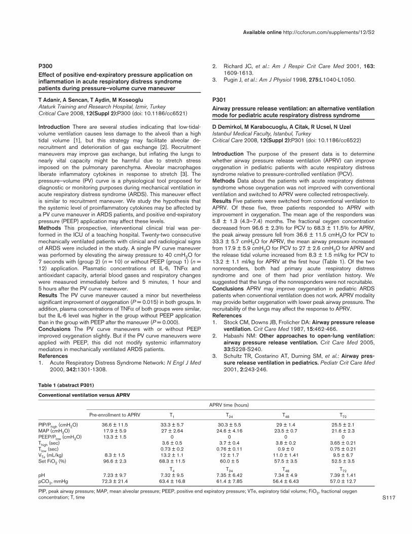
S117
P300Effect of positive end-expiratory pressure application oninflammation in acute respiratory distress syndromepatients during pressure–volume curve maneuver
T Adanir, A Sencan, T Aydin, M KoseogluAtaturk Training and Research Hospital, Izmir, TurkeyCritical Care 2008, 12(Suppl 2):P300 (doi: 10.1186/cc6521)
Introduction There are several studies indicating that low-tidal-volume ventilation causes less damage to the alveoli than a hightidal volume [1], but this strategy may facilitate alveolar de-recruitment and deterioration of gas exchange [2]. Recruitmentmaneuvers may improve gas exchange, but inflating the lungs tonearly vital capacity might be harmful due to stretch stressimposed on the pulmonary parenchyma. Alveolar macrophagesliberate inflammatory cytokines in response to stretch [3]. Thepressure–volume (PV) curve is a physiological tool proposed fordiagnostic or monitoring purposes during mechanical ventilation inacute respiratory distress syndrome (ARDS). This maneuver effectis similar to recruitment maneuver. We study the hypothesis thatthe systemic level of proinflammatory cytokines may be affected bya PV curve maneuver in ARDS patients, and positive end-expiratorypressure (PEEP) application may affect these levels.Methods This prospective, interventional clinical trial was per-formed in the ICU of a teaching hospital. Twenty-two consecutivemechanically ventilated patients with clinical and radiological signsof ARDS were included in the study. A single PV curve maneuverwas performed by elevating the airway pressure to 40 cmH2O for7 seconds with (group 2) (n = 10) or without PEEP (group 1) (n =12) application. Plasmatic concentrations of IL-6, TNFα andantioxidant capacity, arterial blood gases and respiratory changeswere measured immediately before and 5 minutes, 1 hour and5 hours after the PV curve maneuver.Results The PV curve maneuver caused a minor but neverthelesssignificant improvement of oxygenation (P = 0.015) in both groups. Inaddition, plasma concentrations of TNFα of both groups were similar,but the IL-6 level was higher in the group without PEEP applicationthan in the group with PEEP after the maneuver (P = 0.000).Conclusions The PV curve maneuvers with or without PEEPimproved oxygenation slightly. But if the PV curve maneuvers wereapplied with PEEP, this did not modify systemic inflammatorymediators in mechanically ventilated ARDS patients.References1. Acute Respiratory Distress Syndrome Network: N Engl J Med
2000, 342:1301-1308.
2. Richard JC, et al.: Am J Respir Crit Care Med 2001, 163:1609-1613.
3. Pugin J, et al.: Am J Physiol 1998, 275:L1040-L1050.
P301Airway pressure release ventilation: an alternative ventilationmode for pediatric acute respiratory distress syndrome
D Demirkol, M Karabocuoglu, A Citak, R Ucsel, N UzelIstanbul Medical Faculty, Istanbul, TurkeyCritical Care 2008, 12(Suppl 2):P301 (doi: 10.1186/cc6522)
Introduction The purpose of the present data is to determinewhether airway pressure release ventilation (APRV) can improveoxygenation in pediatric patients with acute respiratory distresssyndrome relative to pressure-controlled ventilation (PCV).Methods Data about the patients with acute respiratory distresssyndrome whose oxygenation was not improved with conventionalventilation and switched to APRV were collected retrospectively.Results Five patients were switched from conventional ventilation toAPRV. Of these five, three patients responded to APRV withimprovement in oxygenation. The mean age of the responders was5.8 ± 1.3 (4.3–7.4) months. The fractional oxygen concentrationdecreased from 96.6 ± 2.3% for PCV to 68.3 ± 11.5% for APRV,the peak airway pressure fell from 36.6 ± 11.5 cmH2O for PCV to33.3 ± 5.7 cmH2O for APRV, the mean airway pressure increasedfrom 17.9 ± 5.9 cmH2O for PCV to 27 ± 2.6 cmH2O for APRV andthe release tidal volume increased from 8.3 ± 1.5 ml/kg for PCV to13.2 ± 1.1 ml/kg for APRV at the first hour (Table 1). Of the twononresponders, both had primary acute respiratory distresssyndrome and one of them had prior ventilation history. Wesuggested that the lungs of the nonresponders were not recruitable.Conclusions APRV may improve oxygenation in pediatric ARDSpatients when conventional ventilation does not work. APRV modalitymay provide better oxygenation with lower peak airway pressure. Therecruitability of the lungs may affect the response to APRV.References1. Stock CM, Downs JB, Frolicher DA: Airway pressure release
ventilation. Crit Care Med 1987, 15:462-466.2. Habashi NM: Other approaches to open-lung ventilation:
airway pressure release ventilation. Crit Care Med 2005,33:S228-S240.
3. Schultz TR, Costarino AT, Durning SM, et al.: Airway pres-sure release ventilation in pediatrics. Pediatr Crit Care Med2001, 2:243-246.
Available online http://ccforum.com/supplements/12/S2
Table 1 (abstract P301)
Conventional ventilation versus APRV
APRV time (hours)
Pre-enrollment to APRV T1 T24 T48 T72
PIP/Phigh (cmH2O) 36.6 ± 11.5 33.3 ± 5.7 30.3 ± 5.5 29 ± 1.4 25.5 ± 2.1MAP (cmH2O) 17.9 ± 5.9 27 ± 2.64 24.6 ± 4.16 23.5 ± 0.7 21.6 ± 2.3 PEEP/Plow (cmH2O) 13.3 ± 1.5 0 0 0 0Thigh (sec) 3.6 ± 0.5 3.7 ± 0.4 3.8 ± 0.2 3.65 ± 0.21Tlow (sec) 0.73 ± 0.2 0.76 ± 0.11 0.9 ± 0 0.75 ± 0.21VTe (mL/kg) 8.3 ± 1.5 13.2 ± 1.1 12 ± 1.7 11.0 ± 1.41 9.5 ± 6.7Set FiO2 (%) 96.6 ± 2.3 68.3 ± 11.5 60.0 ± 5 57.5 ± 3.5 52.5 ± 3.5
T4 T24 T48 T72pH 7.23 ± 9.7 7.32 ± 9.5 7.35 ± 6.42 7.34 ± 4.9 7.39 ± 1.41pCO2, mmHg 72.3 ± 21.4 63.4 ± 16.8 61.4 ± 7.85 56.4 ± 6.43 57.0 ± 12.7
PIP, peak airway pressure; MAP, mean alveolar pressure; PEEP, positive end expiratory pressure; VTe, expiratory tidal volume; FiO2, fractional oxygenconcentration; T, time
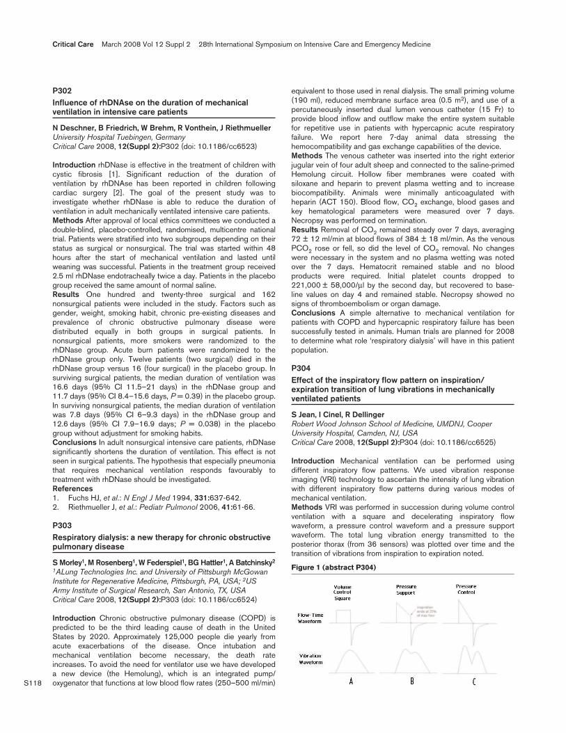
S118
P302Influence of rhDNAse on the duration of mechanicalventilation in intensive care patients
N Deschner, B Friedrich, W Brehm, R Vonthein, J RiethmuellerUniversity Hospital Tuebingen, GermanyCritical Care 2008, 12(Suppl 2):P302 (doi: 10.1186/cc6523)
Introduction rhDNase is effective in the treatment of children withcystic fibrosis [1]. Significant reduction of the duration ofventilation by rhDNAse has been reported in children followingcardiac surgery [2]. The goal of the present study was toinvestigate whether rhDNase is able to reduce the duration ofventilation in adult mechanically ventilated intensive care patients.Methods After approval of local ethics committees we conducted adouble-blind, placebo-controlled, randomised, multicentre nationaltrial. Patients were stratified into two subgroups depending on theirstatus as surgical or nonsurgical. The trial was started within 48hours after the start of mechanical ventilation and lasted untilweaning was successful. Patients in the treatment group received2.5 ml rhDNase endotracheally twice a day. Patients in the placebogroup received the same amount of normal saline.Results One hundred and twenty-three surgical and 162nonsurgical patients were included in the study. Factors such asgender, weight, smoking habit, chronic pre-existing diseases andprevalence of chronic obstructive pulmonary disease weredistributed equally in both groups in surgical patients. Innonsurgical patients, more smokers were randomized to therhDNase group. Acute burn patients were randomized to therhDNase group only. Twelve patients (two surgical) died in therhDNase group versus 16 (four surgical) in the placebo group. Insurviving surgical patients, the median duration of ventilation was16.6 days (95% CI 11.5–21 days) in the rhDNase group and11.7 days (95% CI 8.4–15.6 days, P = 0.39) in the placebo group.In surviving nonsurgical patients, the median duration of ventilationwas 7.8 days (95% CI 6–9.3 days) in the rhDNase group and12.6 days (95% CI 7.9–16.9 days; P = 0.038) in the placebogroup without adjustment for smoking habits.Conclusions In adult nonsurgical intensive care patients, rhDNasesignificantly shortens the duration of ventilation. This effect is notseen in surgical patients. The hypothesis that especially pneumoniathat requires mechanical ventilation responds favourably totreatment with rhDNase should be investigated.References1. Fuchs HJ, et al.: N Engl J Med 1994, 331:637-642.2. Riethmueller J, et al.: Pediatr Pulmonol 2006, 41:61-66.
P303Respiratory dialysis: a new therapy for chronic obstructivepulmonary disease
S Morley1, M Rosenberg1, W Federspiel1, BG Hattler1, A Batchinsky2
1ALung Technologies Inc. and University of Pittsburgh McGowanInstitute for Regenerative Medicine, Pittsburgh, PA, USA; 2USArmy Institute of Surgical Research, San Antonio, TX, USACritical Care 2008, 12(Suppl 2):P303 (doi: 10.1186/cc6524)
Introduction Chronic obstructive pulmonary disease (COPD) ispredicted to be the third leading cause of death in the UnitedStates by 2020. Approximately 125,000 people die yearly fromacute exacerbations of the disease. Once intubation andmechanical ventilation become necessary, the death rateincreases. To avoid the need for ventilator use we have developeda new device (the Hemolung), which is an integrated pump/oxygenator that functions at low blood flow rates (250–500 ml/min)
equivalent to those used in renal dialysis. The small priming volume(190 ml), reduced membrane surface area (0.5 m2), and use of apercutaneously inserted dual lumen venous catheter (15 Fr) toprovide blood inflow and outflow make the entire system suitablefor repetitive use in patients with hypercapnic acute respiratoryfailure. We report here 7-day animal data stressing thehemocompatibility and gas exchange capabilities of the device.Methods The venous catheter was inserted into the right exteriorjugular vein of four adult sheep and connected to the saline-primedHemolung circuit. Hollow fiber membranes were coated withsiloxane and heparin to prevent plasma wetting and to increasebiocompatibility. Animals were minimally anticoagulated withheparin (ACT 150). Blood flow, CO2 exchange, blood gases andkey hematological parameters were measured over 7 days.Necropsy was performed on termination.Results Removal of CO2 remained steady over 7 days, averaging72 ± 12 ml/min at blood flows of 384 ± 18 ml/min. As the venousPCO2 rose or fell, so did the level of CO2 removal. No changeswere necessary in the system and no plasma wetting was notedover the 7 days. Hematocrit remained stable and no bloodproducts were required. Initial platelet counts dropped to221,000 ± 58,000/μl by the second day, but recovered to base-line values on day 4 and remained stable. Necropsy showed nosigns of thromboembolism or organ damage.Conclusions A simple alternative to mechanical ventilation forpatients with COPD and hypercapnic respiratory failure has beensuccessfully tested in animals. Human trials are planned for 2008to determine what role ‘respiratory dialysis’ will have in this patientpopulation.
P304Effect of the inspiratory flow pattern on inspiration/expiration transition of lung vibrations in mechanicallyventilated patients
S Jean, I Cinel, R DellingerRobert Wood Johnson School of Medicine, UMDNJ, CooperUniversity Hospital, Camden, NJ, USACritical Care 2008, 12(Suppl 2):P304 (doi: 10.1186/cc6525)
Introduction Mechanical ventilation can be performed usingdifferent inspiratory flow patterns. We used vibration responseimaging (VRI) technology to ascertain the intensity of lung vibrationwith different inspiratory flow patterns during various modes ofmechanical ventilation.Methods VRI was performed in succession during volume controlventilation with a square and decelerating inspiratory flowwaveform, a pressure control waveform and a pressure supportwaveform. The total lung vibration energy transmitted to theposterior thorax (from 36 sensors) was plotted over time and thetransition of vibrations from inspiration to expiration noted.
Critical Care March 2008 Vol 12 Suppl 2 28th International Symposium on Intensive Care and Emergency Medicine
Figure 1 (abstract P304)
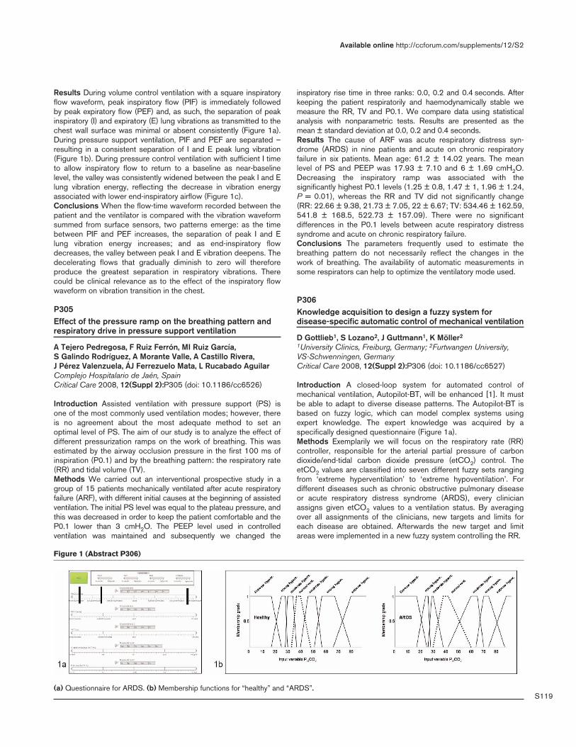
S119
Results During volume control ventilation with a square inspiratoryflow waveform, peak inspiratory flow (PIF) is immediately followedby peak expiratory flow (PEF) and, as such, the separation of peakinspiratory (I) and expiratory (E) lung vibrations as transmitted to thechest wall surface was minimal or absent consistently (Figure 1a).During pressure support ventilation, PIF and PEF are separated –resulting in a consistent separation of I and E peak lung vibration(Figure 1b). During pressure control ventilation with sufficient I timeto allow inspiratory flow to return to a baseline as near-baselinelevel, the valley was consistently widened between the peak I and Elung vibration energy, reflecting the decrease in vibration energyassociated with lower end-inspiratory airflow (Figure 1c).Conclusions When the flow-time waveform recorded between thepatient and the ventilator is compared with the vibration waveformsummed from surface sensors, two patterns emerge: as the timebetween PIF and PEF increases, the separation of peak I and Elung vibration energy increases; and as end-inspiratory flowdecreases, the valley between peak I and E vibration deepens. Thedecelerating flows that gradually diminish to zero will thereforeproduce the greatest separation in respiratory vibrations. Therecould be clinical relevance as to the effect of the inspiratory flowwaveform on vibration transition in the chest.
P305Effect of the pressure ramp on the breathing pattern andrespiratory drive in pressure support ventilation
A Tejero Pedregosa, F Ruiz Ferrón, MI Ruiz García, S Galindo Rodríguez, A Morante Valle, A Castillo Rivera, J Pérez Valenzuela, ÁJ Ferrezuelo Mata, L Rucabado AguilarComplejo Hospitalario de Jaén, SpainCritical Care 2008, 12(Suppl 2):P305 (doi: 10.1186/cc6526)
Introduction Assisted ventilation with pressure support (PS) isone of the most commonly used ventilation modes; however, thereis no agreement about the most adequate method to set anoptimal level of PS. The aim of our study is to analyze the effect ofdifferent pressurization ramps on the work of breathing. This wasestimated by the airway occlusion pressure in the first 100 ms ofinspiration (P0.1) and by the breathing pattern: the respiratory rate(RR) and tidal volume (TV).Methods We carried out an interventional prospective study in agroup of 15 patients mechanically ventilated after acute respiratoryfailure (ARF), with different initial causes at the beginning of assistedventilation. The initial PS level was equal to the plateau pressure, andthis was decreased in order to keep the patient comfortable and theP0.1 lower than 3 cmH2O. The PEEP level used in controlledventilation was maintained and subsequently we changed the
inspiratory rise time in three ranks: 0.0, 0.2 and 0.4 seconds. Afterkeeping the patient respiratorily and haemodynamically stable wemeasure the RR, TV and P0.1. We compare data using statisticalanalysis with nonparametric tests. Results are presented as themean ± standard deviation at 0.0, 0.2 and 0.4 seconds.Results The cause of ARF was acute respiratory distress syn-drome (ARDS) in nine patients and acute on chronic respiratoryfailure in six patients. Mean age: 61.2 ± 14.02 years. The meanlevel of PS and PEEP was 17.93 ± 7.10 and 6 ± 1.69 cmH2O.Decreasing the inspiratory ramp was associated with thesignificantly highest P0.1 levels (1.25 ± 0.8, 1.47 ± 1, 1.96 ± 1.24,P = 0.01), whereas the RR and TV did not significantly change(RR: 22.66 ± 9.38, 21.73 ± 7.05, 22 ± 6.67; TV: 534.46 ± 162.59,541.8 ± 168.5, 522.73 ± 157.09). There were no significantdifferences in the P0.1 levels between acute respiratory distresssyndrome and acute on chronic respiratory failure.Conclusions The parameters frequently used to estimate thebreathing pattern do not necessarily reflect the changes in thework of breathing. The availability of automatic measurements insome respirators can help to optimize the ventilatory mode used.
P306Knowledge acquisition to design a fuzzy system fordisease-specific automatic control of mechanical ventilation
D Gottlieb1, S Lozano2, J Guttmann1, K Möller2
1University Clinics, Freiburg, Germany; 2Furtwangen University,VS-Schwenningen, GermanyCritical Care 2008, 12(Suppl 2):P306 (doi: 10.1186/cc6527)
Introduction A closed-loop system for automated control ofmechanical ventilation, Autopilot-BT, will be enhanced [1]. It mustbe able to adapt to diverse disease patterns. The Autopilot-BT isbased on fuzzy logic, which can model complex systems usingexpert knowledge. The expert knowledge was acquired by aspecifically designed questionnaire (Figure 1a).Methods Exemplarily we will focus on the respiratory rate (RR)controller, responsible for the arterial partial pressure of carbondioxide/end-tidal carbon dioxide pressure (etCO2) control. TheetCO2 values are classified into seven different fuzzy sets rangingfrom ‘extreme hyperventilation’ to ‘extreme hypoventilation’. Fordifferent diseases such as chronic obstructive pulmonary diseaseor acute respiratory distress syndrome (ARDS), every clinicianassigns given etCO2 values to a ventilation status. By averagingover all assignments of the clinicians, new targets and limits foreach disease are obtained. Afterwards the new target and limitareas were implemented in a new fuzzy system controlling the RR.
Available online http://ccforum.com/supplements/12/S2
Figure 1 (Abstract P306)
(a) Questionnaire for ARDS. (b) Membership functions for “healthy” and “ARDS”.
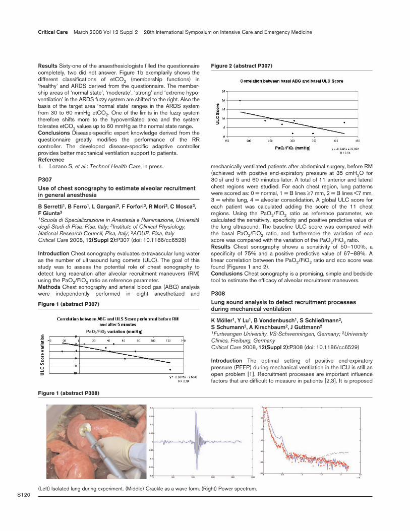
S120
Results Sixty-one of the anaesthesiologists filled the questionnairecompletely, two did not answer. Figure 1b exemplarily shows thedifferent classifications of etCO2 (membership functions) in‘healthy’ and ARDS derived from the questionnaire. The member-ship areas of ‘normal state’, ‘moderate’, ‘strong’ and ‘extreme hypo-ventilation’ in the ARDS fuzzy system are shifted to the right. Also thebasis of the target area ‘normal state’ ranges in the ARDS systemfrom 30 to 60 mmHg etCO2. One of the limits in the fuzzy systemtherefore shifts more to the hypoventilated area and the systemtolerates etCO2 values up to 60 mmHg as the normal state range.Conclusions Disease-specific expert knowledge derived from thequestionnaire greatly modifies the performance of the RRcontroller. The developed disease-specific adaptive controllerprovides better mechanical ventilation support to patients.Reference1. Lozano S, et al.: Technol Health Care, in press.
P307Use of chest sonography to estimate alveolar recruitmentin general anesthesia
B Serretti1, B Ferro1, L Gargani2, F Forfori3, R Mori3, C Mosca3,F Giunta3
1Scuola di Specializzazione in Anestesia e Rianimazione, Universitàdegli Studi di Pisa, Pisa, Italy; 2Institute of Clinical Physiology,National Research Council, Pisa, Italy; 3AOUP, Pisa, ItalyCritical Care 2008, 12(Suppl 2):P307 (doi: 10.1186/cc6528)
Introduction Chest sonography evaluates extravascular lung wateras the number of ultrasound lung comets (ULC). The goal of thisstudy was to assess the potential role of chest sonography todetect lung reaeration after alveolar recruitment maneuvers (RM)using the PaO2/FiO2 ratio as reference parameter.Methods Chest sonography and arterial blood gas (ABG) analysiswere independently performed in eight anesthetized and
mechanically ventilated patients after abdominal surgery, before RM(achieved with positive end-expiratory pressure at 35 cmH2O for30 s) and 5 and 60 minutes later. A total of 11 anterior and lateralchest regions were studied. For each chest region, lung patternswere scored as: 0 = normal, 1 = B lines ≥7 mm, 2 = B lines <7 mm,3 = white lung, 4 = alveolar consolidation. A global ULC score foreach patient was calculated adding the score of the 11 chestregions. Using the PaO2/FiO2 ratio as reference parameter, wecalculated the sensitivity, specificity and positive predictive value ofthe lung ultrasound. The baseline ULC score was compared withthe basal PaO2/FiO2 ratio, and furthermore the variation of ecoscore was compared with the variation of the PaO2/FiO2 ratio.Results Chest sonography shows a sensitivity of 50–100%, aspecificity of 75% and a positive predictive value of 67–88%. Alinear correlation between the PaO2/FiO2 ratio and eco score wasfound (Figures 1 and 2).Conclusions Chest sonography is a promising, simple and bedsidetool to estimate the efficacy of alveolar recruitment maneuvers.
P308Lung sound analysis to detect recruitment processesduring mechanical ventilation
K Möller1, Y Lu1, B Vondenbusch1, S Schließmann2, S Schumann2, A Kirschbaum2, J Guttmann2
1Furtwangen University, VS-Schwenningen, Germany; 2UniversityClinics, Freiburg, GermanyCritical Care 2008, 12(Suppl 2):P308 (doi: 10.1186/cc6529)
Introduction The optimal setting of positive end-expiratorypressure (PEEP) during mechanical ventilation in the ICU is still anopen problem [1]. Recruitment processes are important influencefactors that are difficult to measure in patients [2,3]. It is proposed
Critical Care March 2008 Vol 12 Suppl 2 28th International Symposium on Intensive Care and Emergency Medicine
Figure 1 (abstract P307)
Figure 2 (abstract P307)
Figure 1 (abstract P308)
(Left) Isolated lung during experiment. (Middle) Crackle as a wave form. (Right) Power spectrum.
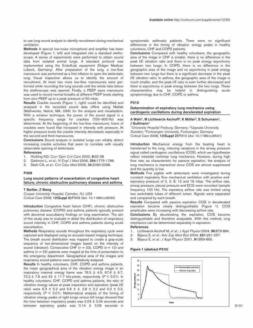
S121
to use lung sound analysis to identify recruitment during mechanicalventilation.Methods A special low-noise microphone and amplifier has beendeveloped (Figure 1, left) and integrated into a standard stetho-scope. A series of experiments were performed to obtain sounddata from isolated animal lungs. A standard protocol wasimplemented using the Evita4Lab equipment (Dräger Medical,Lübeck, Germany). After preparation of the lung, a low-flowmanoeuvre was performed as a first inflation to open the atelectaticlung. Visual inspection allows us to identify the amount ofrecruitment. At most two more low-flow manoeuvres were per-formed while recording the lung sounds until the whole lobe belowthe stethoscope was opened. Finally, a PEEP wave manoeuvrewas used to record normal breaths at different PEEP levels startingfrom zero PEEP up to a peak pressure of 50 mbar.Results Crackle sounds (Figure 1, right) could be identified andanalysed in the recorded sound data offline using Matlab(Mathworks, Natick, MA, USA) for the analysis and visualization.With a window technique, the power of the sound signal in aspecific frequency range for crackles (700–900 Hz) wasdetermined. At the beginning of the low-flow manoeuvre, trains ofcrackles were found that increased in intensity with pressure. Athigher pressure levels the crackle intensity decreased, especially inthe second and third manoeuvres.Conclusions Sound analysis in isolated lungs can reliably detectincreasing crackle activities that seem to correlate with visuallyobservable opening of atelectasis.References1. Hickling KG: Curr Opin Crit Care 2002, 8:32-38.2. Gattinoni L, et al.: N Engl J Med 2006, 354:1775-1786.3. Stahl CA, et al.: Crit Care Med 2006, 34:2090-2098.
P309Lung sound patterns of exacerbation of congestive heartfailure, chronic obstructive pulmonary disease and asthma
T Bartter, Z WangCooper University Hospital, Camden, NJ, USACritical Care 2008, 12(Suppl 2):P309 (doi: 10.1186/cc6530)
Introduction Congestive heart failure (CHF), chronic obstructivepulmonary disease (COPD) and asthma patients typically presentwith abnormal auscultatory findings on lung examination. The aimof this study was to evaluate in detail the distribution of respiratorysound intensity in CHF, COPD and asthma patients during acuteexacerbation.Methods Respiratory sounds throughout the respiratory cycle werecaptured and displayed using an acoustic-based imaging technique.The breath sound distribution was mapped to create a gray-scalesequence of two-dimensional images based on the intensity ofsound (vibration). Consecutive CHF (n = 23), COPD (n = 12) andasthma (n = 22) patients were imaged at the time of presentation tothe emergency department. Geographical area of the images andrespiratory sound patterns were quantitatively analyzed.Results In healthy volunteers, CHF, COPD and asthma patients,the mean geographical area of the vibration energy image in aninspiratory maximal energy frame was 76.2 ± 4.5, 67.6 ± 6.7,72.2 ± 7.6 and 52 ± 11.7 kilo-pixels, respectively (P < 0.01). Inhealthy volunteers, CHF, COPD and asthma patients, the ratio ofvibration energy values at peak inspiration and expiration (peak I/Eratio) were 6.3 ± 5.2 and 5.6 ± 4, 2.8 ± 2.2 and 0.3 ± 0.3,respectively (P < 0.01). Mathematical analysis of the timing ofvibration energy peaks of right lungs versus left lungs showed thatthe time between inspiratory peaks was 0.03 ± 0.04 seconds andbetween expiratory peaks was 0.14 ± 0.09 seconds in
symptomatic asthmatic patients. There were no significantdifferences in the timing of vibration energy peaks in healthyvolunteers, CHF and COPD patients.Conclusions Compared with healthy volunteers, the geographicarea of the image in CHF is smaller, there is no difference in thepeak I/E vibration ratio and there is no peak energy asynchronybetween two lungs; In COPD, there is no difference in thegeographic area of the image and no asynchrony in peak energybetween two lungs but there is a significant decrease in the peakI/E vibration ratio; In asthma, the geographic area of the image ismuch smaller, and the peak I/E ratio is even further decreased andthere is asynchrony in peak energy between the two lungs. Thesecharacteristics may be helpful in distinguishing acutesymptomology due to CHF, COPD or asthma.
P310Determination of expiratory lung mechanics usingcardiogenic oscillations during decelerated expiration
A Wahl1, M Lichtwarck-Aschoff2, K Möller3, S Schumann1, J Guttmann1
1University Hospital Freiburg, Germany; 2Uppsala University,Sweden; 3Furtwangen University, Furtwangen, GermanyCritical Care 2008, 12(Suppl 2):P310 (doi: 10.1186/cc6531)
Introduction Mechanical energy from the beating heart istransferred to the lung, inducing variations in the airway pressuresignal called cardiogenic oscillations (COS), which we hypothesizereflect intratidal nonlinear lung mechanics. However, during highflow rate, as characteristic for passive expiration, the analysis oflung mechanics is impractical since COS are almost suppressedand the quantity is low.Methods Five piglets with atelectasis were investigated duringconstant inspiratory flow mechanical ventilation with positive end-expiratory pressure of 0, 5, 8, 12 and 16 mbar. The airflow rate,airway pressure, pleural pressure and ECG were recorded (samplefrequency 100 Hz). The expiratory airflow rate was limited usingtwo switchable tubes of different lumen. Signals were separatedand compared by each breath.Results Compared with passive expiration COS in deceleratedexpiration became clearly distinguishable (Figure 1). COSamplitudes were increasing with decreasing airflow rate.Conclusions By decelerating the expiration, COS becomedistinguishable and therefore analyzable. With this method, lungmechanics can be determined separately in expiration.References1. Lichtwarck-Aschoff M, et al.: J Appl Physiol 2004, 96:879-884.2. Bijaoui E, et al.: Adv Exp Med Biol 2004, 551:251-257.3. Bijaoui E, et al.: J Appl Physiol 2001, 91:859-865.
Available online http://ccforum.com/supplements/12/S2
Figure 1 (abstract P310)
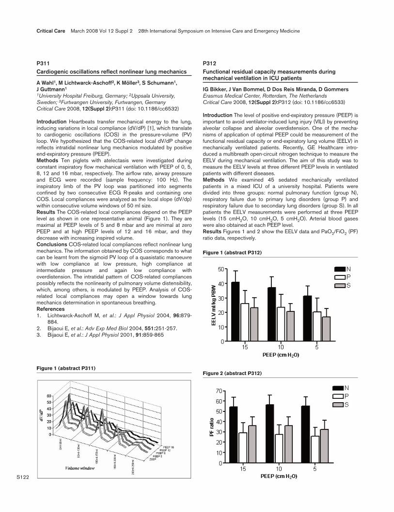
S122
P311Cardiogenic oscillations reflect nonlinear lung mechanics
A Wahl1, M Lichtwarck-Aschoff2, K Möller3, S Schumann1, J Guttmann1
1University Hospital Freiburg, Germany; 2Uppsala University,Sweden; 3Furtwangen University, Furtwangen, GermanyCritical Care 2008, 12(Suppl 2):P311 (doi: 10.1186/cc6532)
Introduction Heartbeats transfer mechanical energy to the lung,inducing variations in local compliance (dV/dP) [1], which translateto cardiogenic oscillations (COS) in the pressure-volume (PV)loop. We hypothesized that the COS-related local dV/dP changereflects intratidal nonlinear lung mechanics modulated by positiveend-expiratory pressure (PEEP).Methods Ten piglets with atelectasis were investigated duringconstant inspiratory flow mechanical ventilation with PEEP of 0, 5,8, 12 and 16 mbar, respectively. The airflow rate, airway pressureand ECG were recorded (sample frequency: 100 Hz). Theinspiratory limb of the PV loop was partitioned into segmentsconfined by two consecutive ECG R-peaks and containing oneCOS. Local compliances were analyzed as the local slope (dV/dp)within consecutive volume windows of 50 ml size.Results The COS-related local compliances depend on the PEEPlevel as shown in one representative animal (Figure 1). They aremaximal at PEEP levels of 5 and 8 mbar and are minimal at zeroPEEP and at high PEEP levels of 12 and 16 mbar, and theydecrease with increasing inspired volume.Conclusions COS-related local compliances reflect nonlinear lungmechanics. The information obtained by COS corresponds to whatcan be learnt from the sigmoid PV loop of a quasistatic manoeuvrewith low compliance at low pressure, high compliance atintermediate pressure and again low compliance withoverdistension. The intratidal pattern of COS-related compliancespossibly reflects the nonlinearity of pulmonary volume distensibility,which, among others, is modulated by PEEP. Analysis of COS-related local compliances may open a window towards lungmechanics determination in spontaneous breathing.References1. Lichtwarck-Aschoff M, et al.: J Appl Physiol 2004, 96:879-
884.2. Bijaoui E, et al.: Adv Exp Med Biol 2004, 551:251-257.3. Bijaoui E, et al.: J Appl Physiol 2001, 91:859-865
P312Functional residual capacity measurements duringmechanical ventilation in ICU patients
IG Bikker, J Van Bommel, D Dos Reis Miranda, D GommersErasmus Medical Center, Rotterdam, The NetherlandsCritical Care 2008, 12(Suppl 2):P312 (doi: 10.1186/cc6533)
Introduction The level of positive end-expiratory pressure (PEEP) isimportant to avoid ventilator-induced lung injury (VILI) by preventingalveolar collapse and alveolar overdistension. One of the mecha-nisms of application of optimal PEEP could be measurement of thefunctional residual capacity or end-expiratory lung volume (EELV) inmechanically ventilated patients. Recently, GE Healthcare intro-duced a multibreath open-circuit nitrogen technique to measure theEELV during mechanical ventilation. The aim of this study was tomeasure the EELV levels at three different PEEP levels in ventilatedpatients with different diseases.Methods We examined 45 sedated mechanically ventilatedpatients in a mixed ICU of a university hospital. Patients weredivided into three groups: normal pulmonary function (group N),respiratory failure due to primary lung disorders (group P) andrespiratory failure due to secondary lung disorders (group S). In allpatients the EELV measurements were performed at three PEEPlevels (15 cmH2O, 10 cmH2O, 5 cmH2O). Arterial blood gaseswere also obtained at each PEEP level.Results Figures 1 and 2 show the EELV data and PaO2/FiO2 (PF)ratio data, respectively.
Critical Care March 2008 Vol 12 Suppl 2 28th International Symposium on Intensive Care and Emergency Medicine
Figure 1 (abstract P311)
Figure 1 (abstract P312)
Figure 2 (abstract P312)
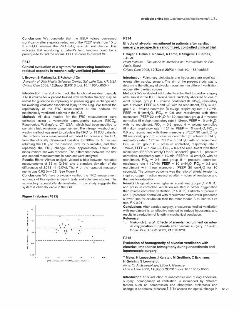
S123
Conclusions We conclude that the EELV values decreasedsignificantly after stepwise reduction of the PEEP levels from 15 to5 cmH2O, whereas the PaO2/FiO2 ratio did not change. Thisindicates that monitoring a patient’s lung function could be aprerequisite to find the optimal PEEP in order to prevent VILI.
P313Clinical evaluation of a system for measuring functionalresidual capacity in mechanically ventilated patients
L Brewer, B Markewitz, E Fulcher, J OrrUniversity of Utah Health Sciences Center, Salt Lake City, UT, USACritical Care 2008, 12(Suppl 2):P313 (doi: 10.1186/cc6534)
Introduction The ability to track the functional residual capacity(FRC) volume for a patient treated with ventilator therapy may beuseful for guidance in improving or preserving gas exchange andfor avoiding ventilator-associated injury to the lung. We tested therepeatability of the FRC measurement at the bedside formechanically ventilated patients in the ICU.Methods All data needed for the FRC measurement werecollected using a volumetric capnography system (NICO2;Respironics, Wallingford, CT, USA), which had been modified tocontain a fast, on-airway oxygen sensor. The nitrogen washout andwashin method was used to calculate the FRC for 13 ICU patients.The protocol for a measurement set called for increasing the FIO2from the clinically determined baseline to 100% for 5 minutes,returning the FIO2 to the baseline level for 5 minutes, and thenrepeating the FIO2 change. After approximately 1 hour, themeasurement set was repeated. The differences between the firstand second measurements in each set were analyzed.Results Bland–Altman analysis yielded a bias between repeatedmeasurements of 85 ml (2.8%) and a standard deviation of thedifferences of ±278 ml (9.0%). The r2 of the repeated measure-ments was 0.92 (n = 28). See Figure 1.Conclusions We have previously verified the FRC measurementaccuracy of this system in bench tests and volunteer studies. Thesatisfactory repeatability demonstrated in this study suggests thesystem is clinically viable in the ICU.
P314Effects of alveolar recruitment in patients after cardiacsurgery: a prospective, randomized, controlled clinical trial
L Hajjar, F Galas, E Nozawa, A Leme, C Shigemi, C Barbas, J AulerHeart Institute – Faculdade de Medicina da Universidade de SãoPaulo, BrazilCritical Care 2008, 12(Suppl 2):P314 (doi: 10.1186/cc6535)
Introduction Pulmonary atelectasis and hypoxemia are significantevents after cardiac surgery. The aim of the present study was todetermine the efficacy of alveolar recruitment in different ventilationmodes after cardiac surgery.Methods We evaluated 480 patients submitted to cardiac surgeryafter arrival in the ICU. Groups were randomly allocated to one ofeight groups: group 1 – volume controlled (8 ml/kg), respiratoryrate if 12/min, PEEP = 5 cmH2O with no recruitment, FiO2 = 0.6;group 2 – volume controlled (8 ml/kg), respiratory rate if 12/min,PEEP = 5 cmH2O, FiO2 = 0.6 and recruitment with threemaneuvers (PEEP 30 cmH2O for 30 seconds); group 3 – volumecontrolled (8 ml/kg), respiratory rate if 12/min, PEEP = 10 cmH2O,with no recruitment, FiO2 = 0.6; group 4 – volume controlled(8 ml/kg), respiratory rate if 12/min, PEEP = 10 cmH2O, FiO2 =0.6 and recruitment with three maneuvers (PEEP 30 cmH2O for30 seconds); group 5 – pressure controlled (to achieve 8 ml/kg),respiratory rate if 12/min, PEEP = 5 cmH2O with no recruitment,FiO2 = 0.6; group 6 – pressure controlled, respiratory rate if12/min, PEEP = 5 cmH2O, FiO2 = 0.6 and recruitment with threemaneuvers (PEEP 30 cmH2O for 30 seconds); group 7 – pressurecontrolled, respiratory rate if 12/min, PEEP = 10 cmH2O, with norecruitment, FiO2 = 0.6; and group 8 – pressure controlled,respiratory rate if 12/min, PEEP = 10 cmH2O, FiO2 = 0.6 andrecruitment with three maneuvers (PEEP 30 cmH2O for 30seconds). The primary outcome was the ratio of arterial tension toinspired oxygen fraction measured after 4 hours of ventilation andthe time for extubation.Results Oxygenation was higher in recruitment groups (P < 0.01),and pressure-controlled ventilation resulted in better oxygenationthan volume-controlled ventilation (P < 0.05). Patients of groups 6and 8 (pressure controlled with recruitment maneuvers) presenteda lower time for extubation than the other modes (280 min vs 476min, P < 0.01).Conclusions After cardiac surgery, pressure-controlled ventilationwith recruitment is an effective method to reduce hypoxemia, andresults in a reduction of length in mechanical ventilation.Reference1. Minkovich L, et al.: Effects of alveolar recruitment on arter-
ial oxygenation in patients after cardiac surgery. J Cardio-thorac Vasc Anesth 2007, 21:375-378.
P315Evaluation of homogeneity of alveolar ventilation withelectrical impedance tomography during anaesthesia andlaparoscopic surgery
T Meier, H Luepschen, J Karsten, M Großherr, C Eckmann, H Gehring, S LeonhardtKlinik für Anästhesiologie, Lübeck, GermanyCritical Care 2008, 12(Suppl 2):P315 (doi: 10.1186/cc6536)
Introduction After induction of anaesthesia and during abdominalsurgery, homogeneity of ventilation is influenced by differentfactors such as compression and absorption atelectasis andchange in abdominal pressure [1]. To assess the spatial change in
Available online http://ccforum.com/supplements/12/S2
Figure 1 (abstract P313)
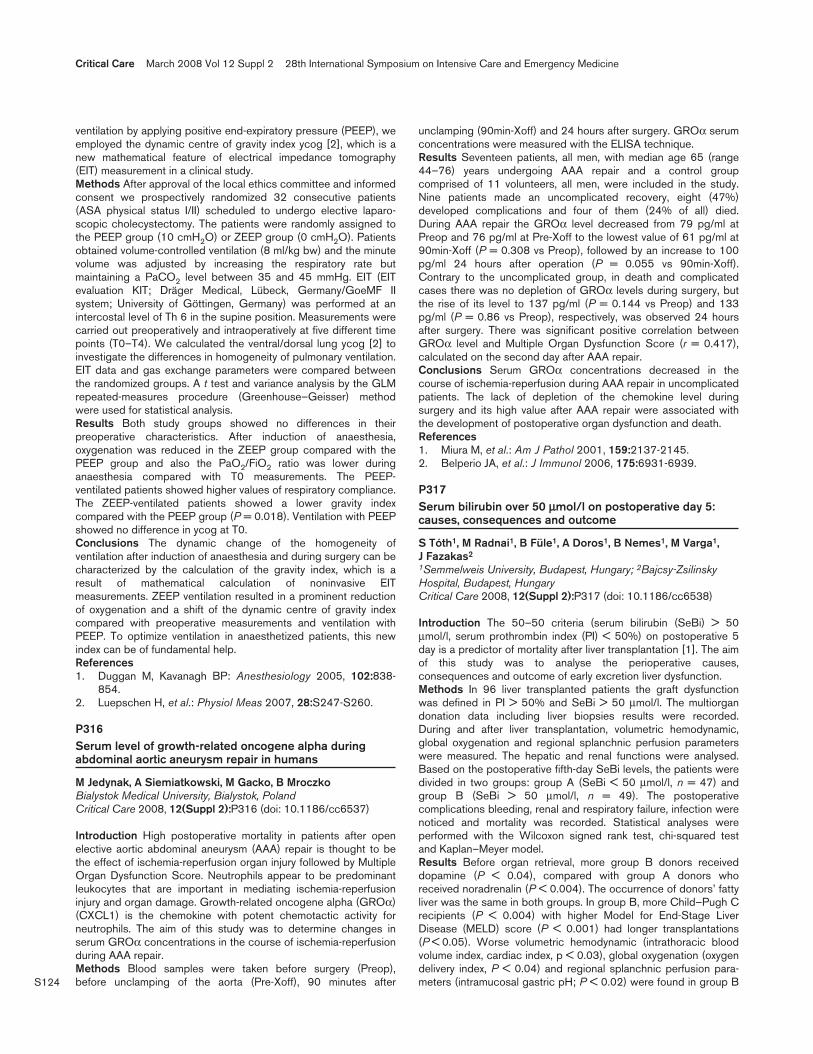
S124
ventilation by applying positive end-expiratory pressure (PEEP), weemployed the dynamic centre of gravity index ycog [2], which is anew mathematical feature of electrical impedance tomography(EIT) measurement in a clinical study.Methods After approval of the local ethics committee and informedconsent we prospectively randomized 32 consecutive patients(ASA physical status I/II) scheduled to undergo elective laparo-scopic cholecystectomy. The patients were randomly assigned tothe PEEP group (10 cmH2O) or ZEEP group (0 cmH2O). Patientsobtained volume-controlled ventilation (8 ml/kg bw) and the minutevolume was adjusted by increasing the respiratory rate butmaintaining a PaCO2 level between 35 and 45 mmHg. EIT (EITevaluation KIT; Dräger Medical, Lübeck, Germany/GoeMF IIsystem; University of Göttingen, Germany) was performed at anintercostal level of Th 6 in the supine position. Measurements werecarried out preoperatively and intraoperatively at five different timepoints (T0–T4). We calculated the ventral/dorsal lung ycog [2] toinvestigate the differences in homogeneity of pulmonary ventilation.EIT data and gas exchange parameters were compared betweenthe randomized groups. A t test and variance analysis by the GLMrepeated-measures procedure (Greenhouse–Geisser) methodwere used for statistical analysis.Results Both study groups showed no differences in theirpreoperative characteristics. After induction of anaesthesia,oxygenation was reduced in the ZEEP group compared with thePEEP group and also the PaO2/FiO2 ratio was lower duringanaesthesia compared with T0 measurements. The PEEP-ventilated patients showed higher values of respiratory compliance.The ZEEP-ventilated patients showed a lower gravity indexcompared with the PEEP group (P = 0.018). Ventilation with PEEPshowed no difference in ycog at T0.Conclusions The dynamic change of the homogeneity ofventilation after induction of anaesthesia and during surgery can becharacterized by the calculation of the gravity index, which is aresult of mathematical calculation of noninvasive EITmeasurements. ZEEP ventilation resulted in a prominent reductionof oxygenation and a shift of the dynamic centre of gravity indexcompared with preoperative measurements and ventilation withPEEP. To optimize ventilation in anaesthetized patients, this newindex can be of fundamental help.References1. Duggan M, Kavanagh BP: Anesthesiology 2005, 102:838-
854.2. Luepschen H, et al.: Physiol Meas 2007, 28:S247-S260.
P316Serum level of growth-related oncogene alpha duringabdominal aortic aneurysm repair in humans
M Jedynak, A Siemiatkowski, M Gacko, B MroczkoBialystok Medical University, Bialystok, PolandCritical Care 2008, 12(Suppl 2):P316 (doi: 10.1186/cc6537)
Introduction High postoperative mortality in patients after openelective aortic abdominal aneurysm (AAA) repair is thought to bethe effect of ischemia-reperfusion organ injury followed by MultipleOrgan Dysfunction Score. Neutrophils appear to be predominantleukocytes that are important in mediating ischemia-reperfusioninjury and organ damage. Growth-related oncogene alpha (GROα)(CXCL1) is the chemokine with potent chemotactic activity forneutrophils. The aim of this study was to determine changes inserum GROα concentrations in the course of ischemia-reperfusionduring AAA repair.Methods Blood samples were taken before surgery (Preop),before unclamping of the aorta (Pre-Xoff), 90 minutes after
unclamping (90min-Xoff) and 24 hours after surgery. GROα serumconcentrations were measured with the ELISA technique.Results Seventeen patients, all men, with median age 65 (range44–76) years undergoing AAA repair and a control groupcomprised of 11 volunteers, all men, were included in the study.Nine patients made an uncomplicated recovery, eight (47%)developed complications and four of them (24% of all) died.During AAA repair the GROα level decreased from 79 pg/ml atPreop and 76 pg/ml at Pre-Xoff to the lowest value of 61 pg/ml at90min-Xoff (P = 0.308 vs Preop), followed by an increase to 100pg/ml 24 hours after operation (P = 0.055 vs 90min-Xoff).Contrary to the uncomplicated group, in death and complicatedcases there was no depletion of GROα levels during surgery, butthe rise of its level to 137 pg/ml (P = 0.144 vs Preop) and 133pg/ml (P = 0.86 vs Preop), respectively, was observed 24 hoursafter surgery. There was significant positive correlation betweenGROα level and Multiple Organ Dysfunction Score (r = 0.417),calculated on the second day after AAA repair.Conclusions Serum GROα concentrations decreased in thecourse of ischemia-reperfusion during AAA repair in uncomplicatedpatients. The lack of depletion of the chemokine level duringsurgery and its high value after AAA repair were associated withthe development of postoperative organ dysfunction and death.References1. Miura M, et al.: Am J Pathol 2001, 159:2137-2145.2. Belperio JA, et al.: J Immunol 2006, 175:6931-6939.
P317Serum bilirubin over 50 μμmol/l on postoperative day 5:causes, consequences and outcome
S Tóth1, M Radnai1, B Füle1, A Doros1, B Nemes1, M Varga1, J Fazakas2
1Semmelweis University, Budapest, Hungary; 2Bajcsy-ZsilinskyHospital, Budapest, HungaryCritical Care 2008, 12(Suppl 2):P317 (doi: 10.1186/cc6538)
Introduction The 50–50 criteria (serum bilirubin (SeBi) > 50μmol/l, serum prothrombin index (PI) < 50%) on postoperative 5day is a predictor of mortality after liver transplantation [1]. The aimof this study was to analyse the perioperative causes,consequences and outcome of early excretion liver dysfunction.Methods In 96 liver transplanted patients the graft dysfunctionwas defined in PI > 50% and SeBi > 50 μmol/l. The multiorgandonation data including liver biopsies results were recorded.During and after liver transplantation, volumetric hemodynamic,global oxygenation and regional splanchnic perfusion parameterswere measured. The hepatic and renal functions were analysed.Based on the postoperative fifth-day SeBi levels, the patients weredivided in two groups: group A (SeBi < 50 μmol/l, n = 47) andgroup B (SeBi > 50 μmol/l, n = 49). The postoperativecomplications bleeding, renal and respiratory failure, infection werenoticed and mortality was recorded. Statistical analyses wereperformed with the Wilcoxon signed rank test, chi-squared testand Kaplan–Meyer model.Results Before organ retrieval, more group B donors receiveddopamine (P < 0.04), compared with group A donors whoreceived noradrenalin (P < 0.004). The occurrence of donors’ fattyliver was the same in both groups. In group B, more Child–Pugh Crecipients (P < 0.004) with higher Model for End-Stage LiverDisease (MELD) score (P < 0.001) had longer transplantations(P < 0.05). Worse volumetric hemodynamic (intrathoracic bloodvolume index, cardiac index, p < 0.03), global oxygenation (oxygendelivery index, P < 0.04) and regional splanchnic perfusion para-meters (intramucosal gastric pH; P < 0.02) were found in group B
Critical Care March 2008 Vol 12 Suppl 2 28th International Symposium on Intensive Care and Emergency Medicine
S125
only after portal and arterial reperfusion. In group B the occurrenceof continuous renal replacement therapy and sepsis were higher(P < 0.02), the ICU therapy was longer (P < 0.02), and the 1-yearmortality was 47% compared with 4% of group A (P < 0.004).Conclusions Early billiary injury after liver transplantation can beconnected with the vasopressor therapy of the donor, the severityof cirrhosis, and the worse oxygenation and hemodynamicparameters after reperfusion.Reference1. Dondero F, et al.: Liver Transplantation 2006, 12(Suppl 5):119.
P318Liver stiffness measurement for diagnosis of portalhypertension-related digestive bleeding in the ICU
L Tual1, R Amathieu1, P Nahon1, D Thabut2, G Dhonneur1
1AP-HP CHU Jean Verdier, Bondy, France; 2AP-HP CHU PitieSalpetriere, Paris, FranceCritical Care 2008, 12(Suppl 2):P318 (doi: 10.1186/cc6539)
Introduction Without past history or clinical signs of cirrhosis, thediagnosis of portal hypertension (PHT)-related digestive haemor-rhage (DH) is often delayed until upper intestinal endoscopyperformance, thus delaying specific medical treatment such asvasoactive drugs. As the liver stiffness measurement (LSM) iscorrelated to hepatic fibrosis [1] and PHT, it could be thereforeuseful for the diagnosis of cirrhosis in this situation. The aim of thisstudy was to assess the predictive value of LSM by FIBROSCAN®
for the diagnosis of cirrhotic PHT-related DH.Methods Between January and May 2006, all consecutive patientsreferred for DH in two ICUs were prospectively included. In allpatients a LSM and an upper gastrointestinal endoscopy wereperformed simultaneously at admission, each operator blinded tothe result of the other examination. Exclusion criteria were thepresence of ascitis or portal thrombosis without cirrhosis.Results Sixty-two patients were included (mean age: 60.2 ± 14.2years; males: 40/62 (65%); BMI: 24.0 ± 11.3 kg/m2; SAPS II: 26.0± 19.3): 27/62 (44%) had non PHT-related DH (gastroduodenalulcer, 16 cases; oesophagitis, seven cases; others, four cases);35/62 (56%) had PHT-related DH (oesophageal varicose, 100%).All of these 35 patients had cirrhosis, either previously known orclinically obvious (18/35, 51%) or biopsy-proven later. The medianLSM was significantly higher in patients with PHT-related DH (54.6kPa (45.0–65.7) vs 5.2 kPa (4.3–6.3), P < 10–6). The AUROC forthe diagnosis of PHT-related DH was 0.97 ± 0.03. A threshold of13.7 kPa was chosen with specificity and a positive predictive valueat 100% (sensitivity, 93%; negative predictive value, 94%).Conclusions LSM is a powerful noninvasive tool for the instantdiagnosis of PHT-related DH. Performed at admission, it couldallow the rapid onset of specific medical management. Theprognostic value assessment of LSM in these patients is ongoing.Reference1. Ganne-Carrie N, et al.: Hepatology 44:1511-1517.
P319Small intestinal transit time in critically ill patients usingendoscopic video capsule
S Rauch1, K Krueger1, DI Sessler2, N Roewer3
1University of Louisville, KY, USA; 2Cleveland Clinic, Cleveland,OH, USA; 3University of Würzburg, GermanyCritical Care 2008, 12(Suppl 2):P319 (doi: 10.1186/cc6540)
Introduction We investigated the small bowel transit time andpathophysiological changes using a video capsule in ICU patients
with head injuries. The gut was only recently recognized as pivotalin the disease process of critical illness; hence, work is needed toimprove our understanding of how bowel dysfunction impactshealing. We hypothesized that new diagnostic technology such aswireless capsule endoscopy, which allows real-time investigationof the small bowel, will show that the small bowel transit time isincreased in critically ill patients with brain injuries.Methods We recruited 32 patients older than 18 years in thisprospective, controlled, IRB-approved trial. Their authorizedrepresentatives gave written, informed consent. Sixteen of themwere neuro-ICU patients with mild-to-moderate brain injury (GCS6–14) who required a feeding tube. The control group consisted of16 ambulatory patients. A small capsule containing a video camera(PillCam™) was positioned in each patient’s proximal smallintestine at the time of endoscopic feeding tube insertion. Sensorson the patient’s abdomen picked up signals the capsule trans-mitted to allow real-time video recording of the gut. Two indepen-dent observers analyzed the data.Results The average small bowel transit time for neuro-ICUpatients ranged from 144 to 480 minutes (median 344 min, mean338 min). For the ambulatory patients, the range was 228–389minutes (median 250 min, mean 279 min). All five patients withsmall bowel transit times greater than 400 minutes were neuro-ICUpatients. The mean difference between the groups was 59 minutes(95% CI: –17 to 135), and according to a Mann–Whitney rank-sum test P = 0.184.Conclusions The current results suggest that the small boweltransit time is not significantly increased in our critical carepatients.
P320Transpulmonary pressure evaluation in an obese patientunder mechanical ventilation
S Delisle1, M Francoeur2, M Albert1
1Hôpital du Sacré-Coeur de Montréal, QC, Canada; 2SummitTechnologies Inc., Montréal, QC, CanadaCritical Care 2008, 12(Suppl 2):P320 (doi: 10.1186/cc6541)
Introduction It is often difficult to manage optimal ventilationparameter settings in patients with low chest wall compliance, as inan obese patient. This case report demonstrates options toventilate such difficult patients.Methods A 34-year-old obese woman was transferred to ouracademic ICU to solve difficult ventilation and oxygenationproblems in the context of septic shock following urinary tractinfection. This patient was transferred to an Avea ventilator (ViasysHealthcare) and an 8 Fr esophageal balloon catheter was insertedin the lower third of the esophagus. An expiratory airway occlusionmaneuver, described by Baydur and colleagues [1], was performedto confirm correct positioning. We performed an inspiratory hold toobtain and compare the airway plateau pressure (Pplat) andtranspulmonary plateau pressure (Ptpplateau), and an end-expiratory hold to obtain and compare airway total positive end-expiratory pressure (PEEPt) and transpulmonary total PEEP(PtpPEEP). Transpulmonary pressures were used to changeventilator parameter settings.Results The patient had a BMI of 58.6. Arterial blood gas showedmetabolic and respiratory acidosis (pH: 7.18, paCO2: 53, HCO3:19) and hypoxemia (paO2: 75, SpO2: 94% with 1.0 FiO2) thatpersisted for 4 days prior to the installation of an esophagealballoon. The Pplat was 52.6 cmH2O and PEEPt was 24.8 cmH2O,with a set PEEP of 8 cmH2O. The Ptpplateau was 14.3 cmH2Oand PtpPEEP was –4.4 cmH2O. The set PEEP was increased to25 cmH2O, which resulted in a PtpPEEP of –1.4 cmH2O.
Available online http://ccforum.com/supplements/12/S2
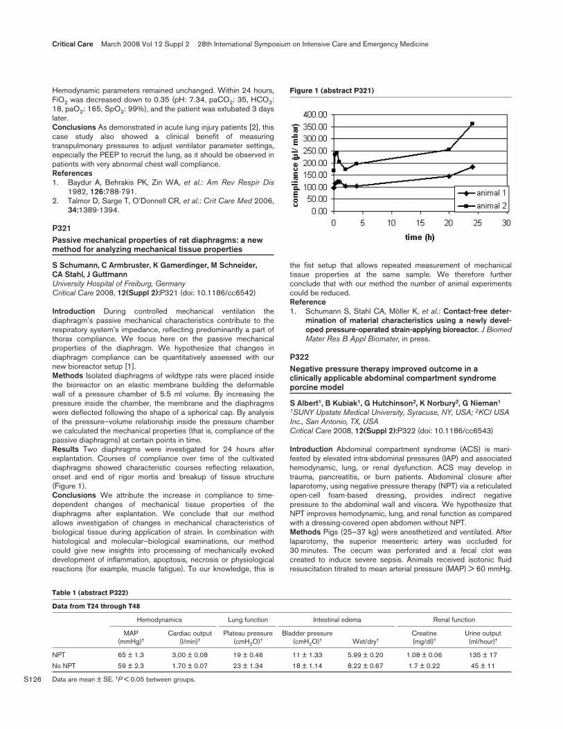
S126
Hemodynamic parameters remained unchanged. Within 24 hours,FiO2 was decreased down to 0.35 (pH: 7.34, paCO2: 35, HCO3:18, paO2: 165, SpO2: 99%), and the patient was extubated 3 dayslater.Conclusions As demonstrated in acute lung injury patients [2], thiscase study also showed a clinical benefit of measuringtranspulmonary pressures to adjust ventilator parameter settings,especially the PEEP to recruit the lung, as it should be observed inpatients with very abnormal chest wall compliance.References1. Baydur A, Behrakis PK, Zin WA, et al.: Am Rev Respir Dis
1982, 126:788-791.2. Talmor D, Sarge T, O’Donnell CR, et al.: Crit Care Med 2006,
34:1389-1394.
P321Passive mechanical properties of rat diaphragms: a newmethod for analyzing mechanical tissue properties
S Schumann, C Armbruster, K Gamerdinger, M Schneider, CA Stahl, J GuttmannUniversity Hospital of Freiburg, GermanyCritical Care 2008, 12(Suppl 2):P321 (doi: 10.1186/cc6542)
Introduction During controlled mechanical ventilation thediaphragm’s passive mechanical characteristics contribute to therespiratory system’s impedance, reflecting predominantly a part ofthorax compliance. We focus here on the passive mechanicalproperties of the diaphragm. We hypothesize that changes indiaphragm compliance can be quantitatively assessed with ournew bioreactor setup [1].Methods Isolated diaphragms of wildtype rats were placed insidethe bioreactor on an elastic membrane building the deformablewall of a pressure chamber of 5.5 ml volume. By increasing thepressure inside the chamber, the membrane and the diaphragmswere deflected following the shape of a spherical cap. By analysisof the pressure–volume relationship inside the pressure chamberwe calculated the mechanical properties (that is, compliance of thepassive diaphragms) at certain points in time.Results Two diaphragms were investigated for 24 hours afterexplantation. Courses of compliance over time of the cultivateddiaphragms showed characteristic courses reflecting relaxation,onset and end of rigor mortis and breakup of tissue structure(Figure 1).Conclusions We attribute the increase in compliance to time-dependent changes of mechanical tissue properties of thediaphragms after explantation. We conclude that our methodallows investigation of changes in mechanical characteristics ofbiological tissue during application of strain. In combination withhistological and molecular–biological examinations, our methodcould give new insights into processing of mechanically evokeddevelopment of inflammation, apoptosis, necrosis or physiologicalreactions (for example, muscle fatigue). To our knowledge, this is
the fist setup that allows repeated measurement of mechanicaltissue properties at the same sample. We therefore furtherconclude that with our method the number of animal experimentscould be reduced.Reference1. Schumann S, Stahl CA, Möller K, et al.: Contact-free deter-
mination of material characteristics using a newly devel-oped pressure-operated strain-applying bioreactor. J BiomedMater Res B Appl Biomater, in press.
P322Negative pressure therapy improved outcome in aclinically applicable abdominal compartment syndromeporcine model
S Albert1, B Kubiak1, G Hutchinson2, K Norbury2, G Nieman1
1SUNY Upstate Medical University, Syracuse, NY, USA; 2KCI USAInc., San Antonio, TX, USACritical Care 2008, 12(Suppl 2):P322 (doi: 10.1186/cc6543)
Introduction Abdominal compartment syndrome (ACS) is mani-fested by elevated intra-abdominal pressures (IAP) and associatedhemodynamic, lung, or renal dysfunction. ACS may develop intrauma, pancreatitis, or burn patients. Abdominal closure afterlaparotomy, using negative pressure therapy (NPT) via a reticulatedopen-cell foam-based dressing, provides indirect negativepressure to the abdominal wall and viscera. We hypothesize thatNPT improves hemodynamic, lung, and renal function as comparedwith a dressing-covered open abdomen without NPT.Methods Pigs (25–37 kg) were anesthetized and ventilated. Afterlaparotomy, the superior mesenteric artery was occluded for30 minutes. The cecum was perforated and a fecal clot wascreated to induce severe sepsis. Animals received isotonic fluidresuscitation titrated to mean arterial pressure (MAP) > 60 mmHg.
Critical Care March 2008 Vol 12 Suppl 2 28th International Symposium on Intensive Care and Emergency Medicine
Figure 1 (abstract P321)
Table 1 (abstract P322)
Data from T24 through T48
Hemodynamics Lung function Intestinal edema Renal function
MAP Cardiac output Plateau pressure Bladder pressure Creatine Urine output (mmHg)† (l/min)† (cmH2O)† (cmH2O)† Wet/dry† (mg/dl)† (ml/hour)†
NPT 65 ± 1.3 3.00 ± 0.08 19 ± 0.46 11 ± 1.33 5.99 ± 0.20 1.08 ± 0.06 135 ± 17
No NPT 59 ± 2.3 1.70 ± 0.07 23 ± 1.34 18 ± 1.14 8.22 ± 0.67 1.7 ± 0.22 45 ± 11
Data are mean ± SE. †P < 0.05 between groups.
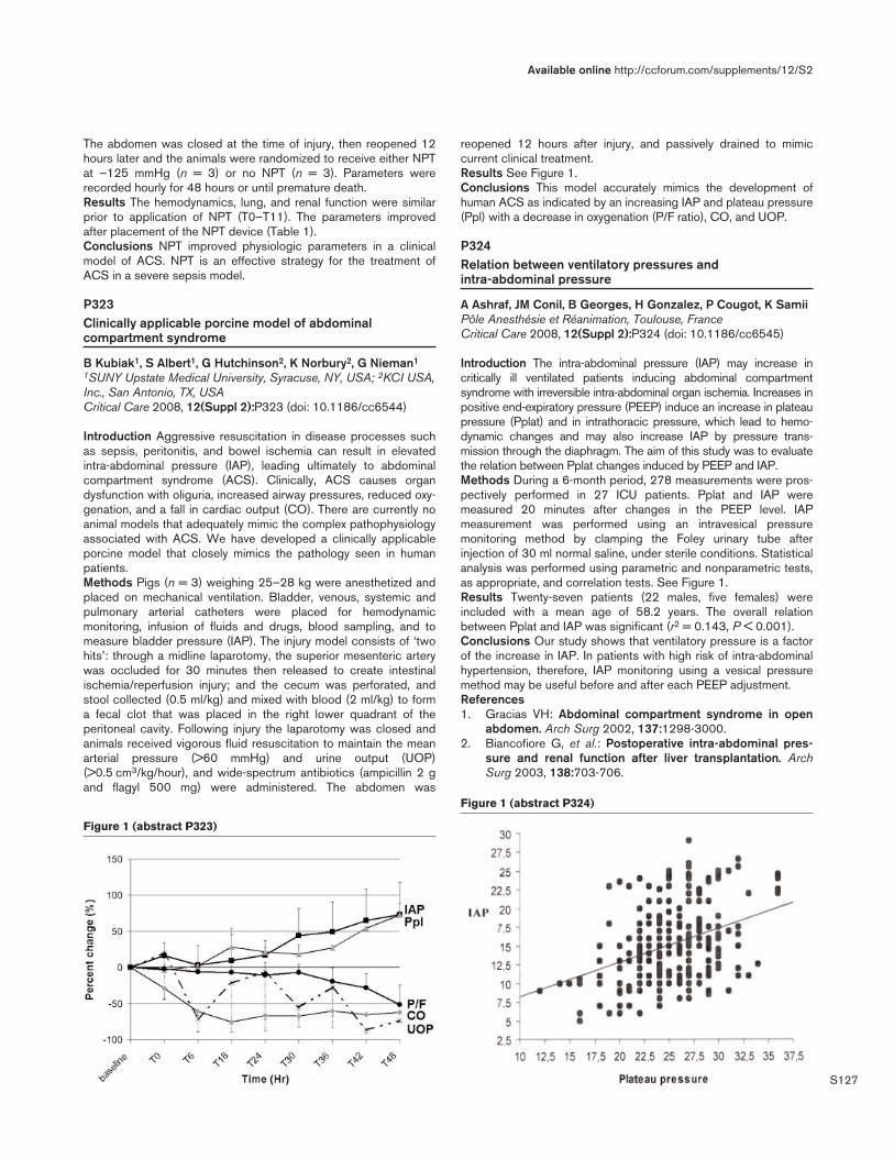
S127
The abdomen was closed at the time of injury, then reopened 12hours later and the animals were randomized to receive either NPTat –125 mmHg (n = 3) or no NPT (n = 3). Parameters wererecorded hourly for 48 hours or until premature death.Results The hemodynamics, lung, and renal function were similarprior to application of NPT (T0–T11). The parameters improvedafter placement of the NPT device (Table 1).Conclusions NPT improved physiologic parameters in a clinicalmodel of ACS. NPT is an effective strategy for the treatment ofACS in a severe sepsis model.
P323Clinically applicable porcine model of abdominalcompartment syndrome
B Kubiak1, S Albert1, G Hutchinson2, K Norbury2, G Nieman1
1SUNY Upstate Medical University, Syracuse, NY, USA; 2KCI USA,Inc., San Antonio, TX, USACritical Care 2008, 12(Suppl 2):P323 (doi: 10.1186/cc6544)
Introduction Aggressive resuscitation in disease processes suchas sepsis, peritonitis, and bowel ischemia can result in elevatedintra-abdominal pressure (IAP), leading ultimately to abdominalcompartment syndrome (ACS). Clinically, ACS causes organdysfunction with oliguria, increased airway pressures, reduced oxy-genation, and a fall in cardiac output (CO). There are currently noanimal models that adequately mimic the complex pathophysiologyassociated with ACS. We have developed a clinically applicableporcine model that closely mimics the pathology seen in humanpatients.Methods Pigs (n = 3) weighing 25–28 kg were anesthetized andplaced on mechanical ventilation. Bladder, venous, systemic andpulmonary arterial catheters were placed for hemodynamicmonitoring, infusion of fluids and drugs, blood sampling, and tomeasure bladder pressure (IAP). The injury model consists of ‘twohits’: through a midline laparotomy, the superior mesenteric arterywas occluded for 30 minutes then released to create intestinalischemia/reperfusion injury; and the cecum was perforated, andstool collected (0.5 ml/kg) and mixed with blood (2 ml/kg) to forma fecal clot that was placed in the right lower quadrant of theperitoneal cavity. Following injury the laparotomy was closed andanimals received vigorous fluid resuscitation to maintain the meanarterial pressure (>60 mmHg) and urine output (UOP)(>0.5 cm3/kg/hour), and wide-spectrum antibiotics (ampicillin 2 gand flagyl 500 mg) were administered. The abdomen was
reopened 12 hours after injury, and passively drained to mimiccurrent clinical treatment.Results See Figure 1.Conclusions This model accurately mimics the development ofhuman ACS as indicated by an increasing IAP and plateau pressure(Ppl) with a decrease in oxygenation (P/F ratio), CO, and UOP.
P324Relation between ventilatory pressures andintra-abdominal pressure
A Ashraf, JM Conil, B Georges, H Gonzalez, P Cougot, K SamiiPôle Anesthésie et Réanimation, Toulouse, FranceCritical Care 2008, 12(Suppl 2):P324 (doi: 10.1186/cc6545)
Introduction The intra-abdominal pressure (IAP) may increase incritically ill ventilated patients inducing abdominal compartmentsyndrome with irreversible intra-abdominal organ ischemia. Increases inpositive end-expiratory pressure (PEEP) induce an increase in plateaupressure (Pplat) and in intrathoracic pressure, which lead to hemo-dynamic changes and may also increase IAP by pressure trans-mission through the diaphragm. The aim of this study was to evaluatethe relation between Pplat changes induced by PEEP and IAP.Methods During a 6-month period, 278 measurements were pros-pectively performed in 27 ICU patients. Pplat and IAP weremeasured 20 minutes after changes in the PEEP level. IAPmeasurement was performed using an intravesical pressuremonitoring method by clamping the Foley urinary tube afterinjection of 30 ml normal saline, under sterile conditions. Statisticalanalysis was performed using parametric and nonparametric tests,as appropriate, and correlation tests. See Figure 1.Results Twenty-seven patients (22 males, five females) wereincluded with a mean age of 58.2 years. The overall relationbetween Pplat and IAP was significant (r2 = 0.143, P < 0.001).Conclusions Our study shows that ventilatory pressure is a factorof the increase in IAP. In patients with high risk of intra-abdominalhypertension, therefore, IAP monitoring using a vesical pressuremethod may be useful before and after each PEEP adjustment.References1. Gracias VH: Abdominal compartment syndrome in open
abdomen. Arch Surg 2002, 137:1298-3000.2. Biancofiore G, et al.: Postoperative intra-abdominal pres-
sure and renal function after liver transplantation. ArchSurg 2003, 138:703-706.
Available online http://ccforum.com/supplements/12/S2
Figure 1 (abstract P323)
Figure 1 (abstract P324)
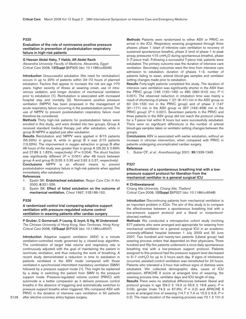
S128
P325Evaluation of the role of noninvasive positive pressureventilation in prevention of postextubation respiratoryfailure in high-risk patients
G Hassan Abdel Naby, T Habib, AR Abdel RazikAlexandria University Faculty of Medicine, Alexandria, EgyptCritical Care 2008, 12(Suppl 2):P325 (doi: 10.1186/cc6546)
Introduction Unsuccessful extubation (the need for reintubation)occurs in up to 20% of patients within 24–72 hours of plannedextubation. Factors that appear to increase the risk are age >70years, higher severity of illness at weaning onset, use of intra-venous sedation, and longer duration of mechanical ventilationprior to extubation [1]. Reintubation is associated with increasedhospital stay and mortality [2]. Noninvasive positive pressureventilation (NIPPV) has been proposed in the management ofacute respiratory failure occurring in the postextubation period. Theuse of NIPPV to prevent postextubation respiratory failure musttherefore be considered.Methods Thirty high-risk patients for postextubation failure wereenrolled in this study, and were divided into two groups. Group Areceived standard medical therapy just after extubation, while ingroup B NIPPV is applied just after extubation.Results Reintubation and NIPPV were applied in 8/15 patients(55.33%) in group A, while in group B it was 2/15 patients(13.33%). The improvement in oxygen extraction in group B after48 hours of the study was greater than in group A (25.32 ± 0.69%and 27.89 ± 1.82%, respectively) (P = 0.004). The shunt fractionwas significantly different (P = 0.001) after 48 hours betweengroup A and group B (3.55 ± 0.35 and 2.92 ± 0.37, respectively).Conclusions NIPPV is an efficient means to preventpostextubation respiratory failure in high-risk patients when appliedimmediately after extubation.References1. Epstin SK: Endotracheal extubation. Respir Care Clin N Am
2000, 6:321-326.2. Epstin SK: Effect of failed extubation on the outcome of
mechanical ventilation. Chest 1997, 112:186-192.
P326A randomized control trial comparing adaptive supportventilation with pressure-regulated volume controlventilation in weaning patients after cardiac surgery
P Gruber, C Gomersall, P Leung, G Joynt, S Ng, M UnderwoodThe Chinese University of Hong Kong, New Territories, Hong KongCritical Care 2008, 12(Suppl 2):P326 (doi: 10.1186/cc6547)
Introduction Adaptive support ventilation (ASV) is a minuteventilation-controlled mode governed by a closed-loop algorithm.The combination of target tidal volume and respiratory rate iscontinuously adjusted with the goal of maintaining the patient inisominute ventilation, and thus reducing the work of breathing. Arecent study demonstrated a reduction in time to extubation inpatients ventilated in the ASV mode compared with thoseventilated in synchronized intermittent mandatory ventilation (SIMV)followed by a pressure support mode [1]. This might be explainedby a delay in switching the patient from SIMV to the pressuresupport mode. Pressure-regulated volume control (PRVC) withautomode is a better comparator as it delivers pressure controlbreaths in the absence of triggering and automatically switches topressure support breaths when triggered. We compared ASV withPRVC in the duration of intensive care ventilation in 50 patientsafter elective coronary artery bypass surgery.
Methods Patients were randomized to either ASV or PRVC onarrival in the ICU. Respiratory weaning progressed through threephases: phase 1 (start of intensive care ventilation to recovery ofsustained spontaneous breaths), phase 2 (end of phase 1 to peakairway pressures <15 cmH2O during spontaneous breaths), phase3 (T-piece trial). Following a successful T-piece trial, patients wereextubated. The primary outcome was the duration of intensive careventilation. Secondary outcomes were the time from intensive careadmission to extubation, duration of phases 1–3, number ofpatients failing to wean, arterial blood-gas samples and ventilatorsetting changes made prior to extubation.Results Forty-eight patients completed the study. The duration ofintensive care ventilation was significantly shorter in the ASV thanthe PRVC group (165 (120–195) vs 480 (360–510) min; P <0.001). The observed reduction in intubation time was mainly aresult of shortening of phase 1 (21 (6–41) min in the ASV group vs60 (24–153) min in the PRVC group) and of phase 2 (147(91–171) min in the ASV group vs 357 (163–458) min in thePRVC group) (P < 0.001). Seventeen patients in the PRVC andthree patients in the ASV group did not reach the protocol criteriafor a T-piece trial within 8 hours but were successfully extubated.There were no significant differences in the number of arterialblood-gas samples taken or ventilator setting changes between thegroups.Conclusions ASV is associated with earlier extubation, without anincrease in clinician intervention, when compared with PRVC inpatients undergoing uncomplicated cardiac surgery.Reference1. Sulzer CF, et al.: Anesthesiology 2001, 95:1339-1345.
P327Effectiveness of a spontaneous breathing trial with a low-pressure support protocol for liberation from themechanical ventilator in a general surgical ICU
K ChittawatanaratChiang Mai University, Chiang Mai, ThailandCritical Care 2008, 12(Suppl 2):P327 (doi: 10.1186/cc6548)
Introduction Discontinuing patients from mechanical ventilation isan important problem in ICUs. The aim of this study is to comparethe effectiveness between a spontaneous breathing trial with alow-pressure support protocol and a liberal or nonprotocol-directed method.Methods We conducted a retrospective cohort study involving577 patients who were arranged and appropriate for weaning frommechanical ventilation on a general surgical ICU in an academicuniversity-affiliated hospital between 1 July 2003 and 30 June2007. Two hundred and twenty-two patients (Liberal group) hadweaning process orders that depended on their physicians. Threehundred and fifty-five patients underwent a once-daily spontaneousbreathing trial with a low-pressure support protocol. Patientsassigned to this protocol had the pressure support level decreasedto 5–7 cmH2O for up to 2 hours each day. If signs of intoleranceoccurred, assisted control ventilation was reinstituted for 24 hours.Patients who tolerated a 2-hour trial without signs of distress wereextubated. We collected demographic data, cause of ICUadmission, APACHE II score at arranged time of weaning, theweaning process time, ventilator days and ICU length of stay.Results There were no statistical differences between liberal andprotocol groups in age (59.2 ± 19.3 vs 55.6 ± 19.8 years; P =0.03), gender (male 74.3 vs 67.9%; P = 0.2) and APACHE IIscore at arranged time of weaning (14.7 ± 7.4 vs 15.3 ± 6.3; P =0.2). The mean duration of the weaning process was 72.1 ± 101.3
Critical Care March 2008 Vol 12 Suppl 2 28th International Symposium on Intensive Care and Emergency Medicine
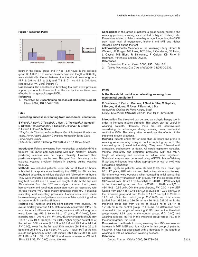
S129
hours in the liberal group and 7.7 ± 16.8 hours in the protocolgroup (P < 0.01). The mean ventilator days and length of ICU staywere statistically different between the liberal and protocol groups(5.7 ± 2.8 vs 2.7 ± 2.3; and 7.3 ± 7.1 vs 4.4 ± 3.4 days,respectively; P < 0.01) (Figure 1).Conclusions The spontaneous breathing trial with a low-pressuresupport protocol for liberation from the mechanical ventilator waseffective in the general surgical ICU.Reference1. MacIntyre N: Discontinuing mechanical ventilatory support.
Chest 2007, 132:1049-1056.
P328Predicting success in weaning from mechanical ventilation
S Vieira1, A Savi2, C Teixeira3, L Nasi1, C Trevisan1, A Guntzel1,R Oliveira2, R Cremonesi2, T Tonietto2, J Hervé1, S Brodt2, F Alves2, J Horer3, N Silva2
1Hospital de Clínicas de Porto Alegre, Brazil; 2Hospital Monihos deVento, Porto Alegre, Brazil; 3Complexo Hospitalar Santa Casa,Porto Alegre, BrazilCritical Care 2008, 12(Suppl 2):P328 (doi: 10.1186/cc6549)
Introduction Failure in weaning from mechanical ventilation (MV) isfrequent (25–30%) and associated with high mortality. Indexespredicting success can be helpful clinically. However, theirpredictive capacity can be low. The goal from this study is toevaluate weaning predictor indexes in patients during weaningfrom MV.Methods We included patients under MV for at least 48 hours,submitted to a spontaneous breathing trial (SBT) for 30 minutes,extubated according to clinical decision and followed for 48 hours.They were evaluated concerning age, sex, clinical characteristics,length of hospital and ICU stays and length of MV. At the first and30th minutes from the SBT we analyzed: arterial blood gases,hemodynamic and respiratory parameters such as respiratory rate(f), tidal volume (VT), rapid shallow breathing index (f/VT), maximalinspiratory and expiratory pressures. Comparisons were madebetween two groups of patients: success vs failure, defining failureas return to MV in the first 48 hours.Results Four hundred and fifty-eight patients were studied. Theoverall mortality rate was 14%. Return to MV occurred in 21%. Themost important differences comparing success with failure groupswere: lower age (56 ± 19 vs 62 ± 17 years, P < 0.01), lowermortality rate (10% vs 31%, P < 0.001), shorter length of ICU stay(15 ± 12 vs 19 ± 13 days, P < 0.01), higher oxygen saturation atthe first and 30th minutes (97 ± 3 vs 96 ± 6 and 95 ± 4 vs 94 ± 4,P < 0.05), lower f at the first and 30th minutes (24 ± 6 vs 26 ± 6bpm and 25 ± 6 vs 28 ± 7 bpm, P < 0.001), lower f/VT at the firstminute and principally in the 30th minute (56 ± 32 vs 69 ± 38 and62 ± 39 vs 84 ± 55, P < 0.001), and lower increase in f/VT (4 ±28 vs 12 ± 38, P < 0.05) during the test.
Conclusions In this group of patients a great number failed in theweaning process, showing, as expected, a higher mortality rate.Parameters related to failure were higher age, longer length of ICUstay, lower level of oxygenation, higher f and f/VT and higherincrease in f/VT during the test.Acknowledgements Members of the Weaning Study Group: RWickert, LG Borges, ME Alves, ACT Silva, R Condessa, CE Hahn,L Cassel, MB Blom, R Zancanaro, F Callefe, KB Pinto, KHartmann, P Pinheiro, and ES Oliveira.References1. Frutos-Vivar F, et al.: Chest 2006, 130:1664-1671.2. Tanios MA, et al.: Crit Care Med 2006, 34:2530-2535.
P329Is the threshold useful in accelerating weaning frommechanical ventilation?
R Condessa, S Vieira, J Brauner, A Saul, A Silva, M Baptista, L Borges, M Moura, M Alves, F Kutchak, L BizHospital de Clinicas de Porto Alegre, BrazilCritical Care 2008, 12(Suppl 2):P329 (doi: 10.1186/cc6550)
Introduction The threshold can be used as a physiotherapy tool inorder to increase muscle strength. This effect can be useful inweaning patients. However, there are still controversiesconsidering its advantages during weaning from mechanicalventilation (MV). This study aims to evaluate the effects of thethreshold in such situations.Methods Patients under MV for more than 48 hours and prone toweaning were randomly assigned to the control group or to thethreshold group (trained twice daily). They were followed untilextubation, tracheotomy or death. All cardiorespiratory variables,maximal inspiratory and expiratory pressures (MIP and MEP),length of weaning and success or failure were registered.Statistical analysis was performed using ANOVA, Mann–WhitneyU test and chi-square test, where appropriate. A level of 0.05 wasconsidered significant.Results Eighty-six patients were studied (52% men, mean age63 ± 17 years, 48% with chronic obstructive pulmonary disease).No differences were observed when comparing initial versus finalcardiorespiratory variables in both groups, with the exception of theMIP (varied from –33.72 ± 13.5 cmH2O to –40.81 ± 12.67 cmH2Oin the threshold group and from –37.67 ± 10.49 cmH2O to–34.19 ± 10.85 cmH2O in the control group, P < 0.001), the MEP(varied from 25.47 ± 12.48 cmH2O to 29.65 ± 12.02 cmH2O inthe threshold group and from 29.65 ± 11.97 cmH2O to 26.86 ±11.6 cmH2O in the control group, P < 0.05) and tidal volume(varied from 386.16 ± 236.56 ml to 436.16 ± 228.39 ml in thethreshold group and from 361.91 ± 168.81 ml to 357.14 ±121.35 ml in the control group, P < 0.05). No differences wereobserved in the length of weaning (1.36 days in the thresholdgroup versus 1.98 days in the control group, P > 0.05) andweaning success (83.7% in the threshold group versus 76.7% inthe control group, P > 0.05).Conclusions The threshold during weaning from MV can cause anincrease in MIP, MEP and tidal volume. In this group of patients,however, it was not associated with a decrease in the length ofweaning or with an increase in weaning success.Reference1. Caruso P, et al.: Clinics 2005, 60:479-484.
Available online http://ccforum.com/supplements/12/S2
Figure 1 (abstract P327)
S130
P330Addition of a spontaneous awakening trial improvesoutcome in mechanically ventilated medical ICU patients
H Mann1, M Lupei1, C Weinert2
1University of Minnesota College of Pharmacy and 2University ofMinnesota College of Medicine, Center for Excellence in CriticalCare, Minneapolis, MN, USACritical Care 2008, 12(Suppl 2):P330 (doi: 10.1186/cc6551)
Introduction Delayed discontinuation of mechanical ventilation isassociated with increased mortality. The Sixth InternationalConsensus Conference on Intensive Care Medicine recommendsspontaneous breathing trials (SBT) as best practice for mechanicalventilation weaning. Daily spontaneous awakening trials (SAT) arealso correlated with reduced ventilation duration and ICU length ofstay. The aim of our study was to implement the SBT and SAT asbest practices in the ICU and to assess the outcome of using theSAT and SBT combined.Methods We collected information on medical ICU patients for12 weeks in 2006 after implementing a SBT protocol and in 2007after adding a SAT protocol to the SBT. We compared thelikelihood of passing the SBT, extubation after a complete SBT,reasons for not extubating after a passed SBT, and the medianventilator days. Statistical comparison included the chi-square testand Mann–Whitney test (two-tailed with P < 0.05 consideredsignificant).Results Fifty-three patients were enrolled in the SBT-only groupand 44 patients were included in the SAT + SBT group. In the SAT+ SBT group the likelihood of passing both a safety screen (38%vs 47%; P < 0.05) and 30-minute SBT (73% vs 85%, P < 0.05)were lower than in the SBT-only group. The decreased likelihoodof passing the safety screen in the SAT + SBT group wasassociated with an increased incidence of physician override to theprotocol. The number of SBT trials performed decreased from 6.1to 5.7 per patient with the addition of the SAT. The likelihood ofextubation following a complete SBT increased in the SAT + SBTgroup versus the SBT-only group (42% versus 29%, P = 0.143).The likelihood of not extubating following a passed SBT due tosedation is decreased in the SAT + SBT group (10% vs 36%, P =0.002). The median ventilator days was reduced in the SAT + SBTgroup versus the SBT-only group (5 days versus 6 days, P = 0.18).Conclusions Implementation of a best practice protocol for SAT toan SBT in the medical ICU improved patient outcome bydecreasing the days on the ventilator and increasing the likelihoodof extubation.
P331Early versus late tracheotomy in the ICU
K Mitka, V Soulountsi, I Houris, E Massa, G Vasiliadou, M BitzaniG. Papanikolaou, Thessaloniki, GreeceCritical Care 2008, 12(Suppl 2):P331 (doi: 10.1186/cc6552)
Introduction This study was conducted to compare early versuslate tracheotomy in ICU patients.Methods A total of 103 patients (81 men, 22 women) wereincluded in this study. They were classified into two groups: GroupA – early tracheotomy (≤7 days) included 36 patients (mean age55.14 ± 17.698 years), and Group B – late tracheotomy (>7 days)included 67 patients (mean age 56.55 ± 19.078 years). Westudied the impact of timing of tracheotomy on the duration ofmechanical ventilation, duration of weaning, length of stay in the
ICU (LOS), outcome in 28 days, incidence of ventilator-associatedpneumonia (VAP), and days of sedation administration. Severity ofillness and organ dysfunction were assessed by APACHE II, SAPSand SOFA scores. Statistical analysis was performed using thePearson x2, independent t test, Levene significance control,Mann–Whitney U test, and paired t test. The control criterion wasP (significance) ≤ a (significance level), a = 5%.Results The two groups were comparable in terms of age,APACHE II score and SAPS. There was a statistically significantdifference in the admission SOFA score (P << a), the SOFA scoreof the tracheotomy day (P = 0.003) and in SOFA max (P << a), aswell as the total days of mechanical ventilation (Group A 18.36 ±12.059 vs Group B 24.19 ± 14.27, P = 0.05) and the LOS(Group A 16.75 ± 7.038 vs Group B 22.51 ± 10.726, P = 0.007).No difference was observed regarding the days of weaning aftertracheotomy (Group A 7.56 ± 6.135 vs Group B 9.19 ± 9.24) andmortality (25% vs 23.9%, respectively). The prevalence of VAPwas evaluated in 58 patients. In Group A VAP developed in23.1%, vs 76.9% of patients in Group B (P = 0.099). There wasno difference in the day VAP was diagnosed (P = 0.959). Asignificant difference in the days of sedative administration beforeand after tracheotomy was observed in both groups (before:7.49 ± 5.34 days, after: 4.76 ± 8.05 days, P = 0.005). Days ofsedative administration before tracheotomy were significantly differ-ent (Group A 4.32 ± 2.083 vs Group B 9 ± 5.690, P = 0.003).Conclusions Our results reinforce the findings of previous studiesshowing that early tracheotomy decreases significantly the durationof mechanical ventilation, ICU LOS and total days of sedativeadministration, and may provide a benefit in reducing theoccurrence of VAP.
P332Precocious tracheotomy versus prolonged intubation in amedical ICU
B Charra, A Hachimi, A Benslama, S MotaouakkilIbn Rochd University Hospital, Casablanca, MoroccoCritical Care 2008, 12(Suppl 2):P332 (doi: 10.1186/cc6553)
Introduction The main purpose of our study was to assess whetherprecocious tracheotomy, compared with prolonged intubation,reduces the duration of ventilation, the frequency of nosocomialpneumopathy, the duration of hospitalization and the mortality.Methods A retrospective and comparative study between twogroups who present a neurologic or respiratory pathology andrequire mechanical ventilation for more than 3 weeks. The studycovered 7 years and was about 60 patients divided into twogroups: tracheotomy group (TG, n = 30), where the tracheotomywas realized between the eighth day and the 15th day, after thefirst period of intubation; and intubation group (IG, n = 30), wherethe patients are still intubated during the whole period ofhospitalization until extubation or death. We determined theduration of ventilation, the frequency of nosocomial pneumopathy,the mean duration of hospitalization and the mortality. Thestatistical study was based on the chi-squared test for qualitativevariables and on Student’s test for quantitative variables. P < 0.05was considered significant. The two groups contain a similarnumber of cases that have the same diagnosis. They have thesame data about age, the sex and the gravity score: SAPS II andAPACHE II score.Results There was a significant statistical decrease of the wholeduration of mechanical ventilation for the TG, 27.03 ± 3.31 daysversus 31.63 ± 6.05 days for the IG, with P = 0.001. However,
Critical Care March 2008 Vol 12 Suppl 2 28th International Symposium on Intensive Care and Emergency Medicine
S131
there was no significant difference between the two groups for thefrequency of nosocomial pneumopathy (P = 0.18). The meanduration of hospitalization did not differ between the two groups,and was about 30.96 ± 9.47 days for the TG versus 34.26 ± 9.74days for the IG with P = 0.10. The study of the evolution showsthat there was no statistically significant difference between thetwo groups regarding the mortality, 26.7% in the TG versus 46.7%for the IG with P = 0.10.Conclusions It seems that precocious tracheotomy in theresuscitation of patients leads to a decrease of the duration ofventilation and delayed the incidence of nosocomial pneumopathywithout a modification of the frequency of the mean duration ofhospitalization in the resuscitation ward, or of death.
P333Ciaglia Blue Dolphin: a new technique for percutaneoustracheostomy using balloon dilation
C Byhahn1, M Zgoda2, O Birkelbach3, C Hofstetter1, T Gromann3
1JW Goethe Medical School, Frankfurt, Germany; 2CarolinasMedical Center, Charlotte, NC, USA; 3German Heart Institute,Berlin, GermanyCritical Care 2008, 12(Suppl 2):P333 (doi: 10.1186/cc6554)
Introduction Percutaneous tracheostomy (PcT) has reached ahigh level of safety; however, significant perioperativecomplications still occur. The most feared complication is posteriortracheal wall injury during insertion of the dilational device into thetrachea applying downward pressure. Ciaglia Blue Dolphin (CBD)is a new technique for PcT using radial balloon dilation, therebyeliminating downward pressure during insertion and dilation.Methods An observational, clinical trial was conducted in 20 adultICU patients undergoing elective PcT with the CBD technique(Cook Inc., Bloomington, IN, USA). After a 15 mm skin incision,tracheal puncture, and predilation of the puncture channel with a14 F punch dilator, a balloon-cannula apparatus was passed over aguidewire until the tip of the balloon mounted at the distal end ofthe apparatus was seen in the trachea. The balloon was inflatedwith saline solution to 11 atm for a few seconds, then deflated, andthe 8.0 mm ID tracheostomy tube preloaded onto a customizedstylet, which formed the proximal portion of the apparatus, wasplaced by advancing the entire apparatus further into the trachea.The apparatus and guidewire were then removed, leaving only thecannula in place.Results Twenty patients underwent CBD PcT under broncho-scopic control. All procedures were successfully completed in amean time of 3.8 ± 1.7 minutes. Even though six patients wereunder continuous therapeutic anticoagulation therapy, blood losswas classified as ‘none’ (n = 14), ‘marginal’ (n = 5), or ‘moderate’(n = 1). In the latter patient, bleeding occurred from a sub-cutaneous vein, but ceased without further intervention once thetracheostomy tube was in place. No other complications of eithermedical or technical nature were noted.Conclusions Based on the data of this first clinical report, the newCBD device allows for quick, reliable, and safe dilation andsubsequent cannula placement with one single apparatus. Eventhough the operators had no previous experience with CBD, nocomplications were noted. Randomized trials need now to beconducted to confirm the promising results of our study and todetermine both advantages and disadvantages of the CBDtechnique when compared with other techniques of PcT.Reference1. Zgoda M, Berger R: Chest 2005, 128:3688-3690.
P334Percutaneous dilatation tracheostomy in critically illpatients with documented coagulopathy
P Kakar, D Govil, S Gupta, S Srinivasan, P Mehta, A Malhotra,O Prakash, D Arora, S Das, P GovilMax Super Speciality Hospital, New Delhi, IndiaCritical Care 2008, 12(Suppl 2):P334 (doi: 10.1186/cc6555)
Introduction Percutaneous tracheostomy techniques are gaininggreater popularity in ICUs. Refinement of the percutaneoustracheostomy technique has made this a straightforward and safeprocedure in appropriately selected patients. Generally, coagulo-pathy is a relative contraindication for surgical tracheotomy. Wesought to determine its usage in high-risk patients withdocumented coagulopathy.Methods Twenty critically ill patients with coagulopathy (Inter-national Normalized Ratio (INR) ≥ 1.5) underwent elective per-cutaneous tracheostomy using a Portex percutaneous tracheos-tomy kit (Ultraperc). The Ciaglia Blue Rhino single-stage dilator setwas used in all cases and the same intensivists performed all of thetracheotomiesResults There were 17 patients with an INR > 1.5, two patientswere on a heparin drip, and one patient had a platelet count<20,000. One patient included in the study met requirements fortwo categories with a platelet count of 17,000 and an INR of 1.7.The procedural times ranged from 3 to 5 minutes. Apart from minorbleeding episodes during and after the procedures in threepatients, which were controlled promptly, no other complicationsoccurred; average estimated blood loss was around 5–10 ml.Conclusions In trained hands with careful precautions, we believethat percutaneous tracheostomy is safe even in patients withdocumented coagulopathy.Reference1. Aoyagi M: Percutaneous tracheostomy to the patient with
coagulopathy. Jpn J Anesthesiol 2005, 54:153-155.
P335An audit of perioperative staffing and complications duringpercutaneous and surgical tracheostomy insertion
P O’Neil, D NobleAberdeen Royal Infirmary, Aberdeen, UKCritical Care 2008, 12(Suppl 2):P335 (doi: 10.1186/cc6556)
Introduction Percutaneous tracheostomy (PDT) has beenestablished as a safe technique in the critically ill, with an equivalentcomplication rate to surgical tracheostomy (ST). However, PDTinsertion may result in unrecognised hypercarbia, and has beenassociated with an increased perioperative complication rate. Wetherefore decided to audit current practice within our ICU.Methods Over a 3-month period, prospective data were collectedon 25 patients within a 14-bed regional ICU. A single observercollected data on staff present, cardiovascular recordings and end-tidal carbon dioxide.Results PDT was performed on 15 patients within the ICU, and STwas performed on 10 patients. Indication for tracheostomy wasprolonged mechanical ventilation in 16 patients, poor neurologicalstatus in eight patients and sputum retention in one patient.Cardiovascular instability, defined as a greater than 20% deviationfrom normal blood pressure, occurred in nine (60%) patientsduring PDT. For ST, eight (80%) patients were cardiovascularlyunstable. Hypercarbia, as detected by an end-tidal CO2 rise ofmore than 20%, occurred in six (40%) patients during PDT and inone (10%) patient during ST. See Table 1.
Available online http://ccforum.com/supplements/12/S2
S132
Conclusions This audit has shown that assistance for PDT isinferior to that provided in the operating theatre, and this haspotential safety implications particularly when junior staff areanaesthetising. Perioperative complication rates were similaroverall, confirming the safety of PDT as a technique. Hypercarbiaoccurred relatively frequently during PDT, however, which mayhave deleterious effects in the brain-injured patient. From this audit,we would recommend that within our ICU more attention befocused on adequate staffing during the performance of thisoperative procedure on critically ill patients. Also, end-tidal carbondioxide should be monitored carefully and treated if elevated.Reference1. Rana S, et al.: Tracheostomy in critically ill patients. Mayo
Clin Proc 2005, 80:1632-1638.
P336Severe airway compromise after percutaneous dilatationaltracheostomy
AT Bos, BI Cleffken, P BreedveldUniversity Hospital Maastricht, The NetherlandsCritical Care 2008, 12(Suppl 2):P336 (doi: 10.1186/cc6557)
Introduction Percutaneous dilatational tracheostomy (PDT) isconsidered a safe alternative to open surgical tracheotomy, withcomparable complication rates. Major complications are reportedto be <1.5%, with a mortality rate of 0.3% [1].Methods A 52-year-old male was admitted to our ICU followingcraniotomy for an intracranial hemorrhage. Prior history revealedhypertension and morbid obesity (BMI 46).PDT was performed on the fourth day after intubation, because ofpersisting low Glasgow Coma Score and failure to clearsecretions. PDT was performed with a Seldinger technique. Withbronchoscopic guidance, endotracheal placement was confirmed.Initial airway pressure was high, but normalized quickly. Althoughoxygenation was maintained, saturation was 84% at the end of theprocedure.Results After 3 days a subcutaneous swelling occurred around thetracheostomy tube (TT), compromising the airway. An abscess wasexpected but could not be confirmed by stab incision or CAT scan.A rise of airway pressure with loss of tidal volume was seen in thenext hours. On oral and transtracheostomy bronchoscopy, adiffusely swollen larynx with narrowing of the proximal trachea wasseen. The TT was exchanged for a Bivona TT.On retrospection, the CAT scan revealed a dislocated cuffvisualized as a double bubble. This was caused by tissue swelling,gradually enlarging the distance between skin and trachea. In thismorbid obese patient, the standard TT was too short anddislocation could occur. A second CAT scan confirmed anadequate position of the Bivona TT. After 1 week, a TT withincreased skin-to-trachea length was inserted and the patient wassuccessfully weaned from ventilation.Conclusions Since the complication rate is increased whenperforming a PDT in the obese [2], we suggest the following. First,
PDT should be guided by fiberoptic bronchoscopy. Second, a TTof adequate diameter and length should be used. Inadequate skin-to-trachea length of the TT can result in improper placement withcuff dislocation not necessarily resulting in air leak with ventilation.An experienced team should perform the procedure: one persondoing a bronchoscopy, another placing the TT.References1. Marx WH, et al.: Chest 1996, 110:762-766.2. Byhahn C, et al.: Anaesthesia 2005, 60:12-15.
P337Incidence of postoperative sore throat and cough:comparison of a polyvinylchloride tube and an armoured tube
J Saleem, S AtharHameed Latif Hospital, Lahore, PakistanCritical Care 2008, 12(Suppl 2):P337 (doi: 10.1186/cc6558)Introduction One of the common postoperative complications issore throat and cough. A number of factors may be responsible,namely direct trauma by the airway, the endotracheal tube, andmucosal damage caused by pressure by the cuff hyperinflated byN2O. This complication is minor but distressing to an otherwisehealthy patient; different strategies have been proposed to preventit. They include changes in the technique of intubation, in theendotracheal tube material, and lower cuff pressure.Methods Eighty female patients ASA status I and II, scheduled forelective caesarean sections, were divided randomly into twogroups. See Table 1. The same anaesthetist performed all theintubations and extubations. The patients were interviewed on theday of operation and on the following 2 days about cough and sorethroat. See Table 2.Results The frequency of postoperative sore throat and cough wasgreater with the use of the polyvinylchloride endotracheal tube.Conclusions The study demonstrates that the use of a poly-vinylchloride endotracheal tube was associated with a significantlyhigher incidence of postoperative sore throat and cough.
References1. Mandoe H, Nikolajsen L: Sore throat after endotracheal
intubation. Anesth Analg 1992, 74:897-900.2. Stout DM, et al.: Correlation of endotracheal size with sore
throat and hoarseness following general anaesthesia.Anesthesiology 1987, 67:419-421.
3. Hahnel J, Treiber H: A comparison of diffeent endotrachealtubes. Tracheal cuff seal, peak centering and the inci-dence of post operative sore throat [in German]. Anaes-thetist 1993, 42:232-237.
Critical Care March 2008 Vol 12 Suppl 2 28th International Symposium on Intensive Care and Emergency Medicine
Table 1 (abstract P335)
Staff involved in PDT and ST
PDT (%) ST (%)
Assistants ≥2 8 (53) 10 (100)
Operator SpR3+ 12 (80) 4 (40)
Anaesthetist SpR3+ 6 (40) 9 (90)
Table 1 (abstract P337)
Demographic data
Tube Number Mean age (years) Mean weight (kg)
Polyvinylchloride 40 25.2 67.27
Armoured 40 28.8 65.95
Table 2 (abstract P337)
Frequency of cough and sore throat
Tube Cough Sore throat P value
Armoured 10 (12.5%) 26 (32.5%) 0.0704
Polyvinylchloride 26 (32.5%) 58 (72.5%) 0.0129
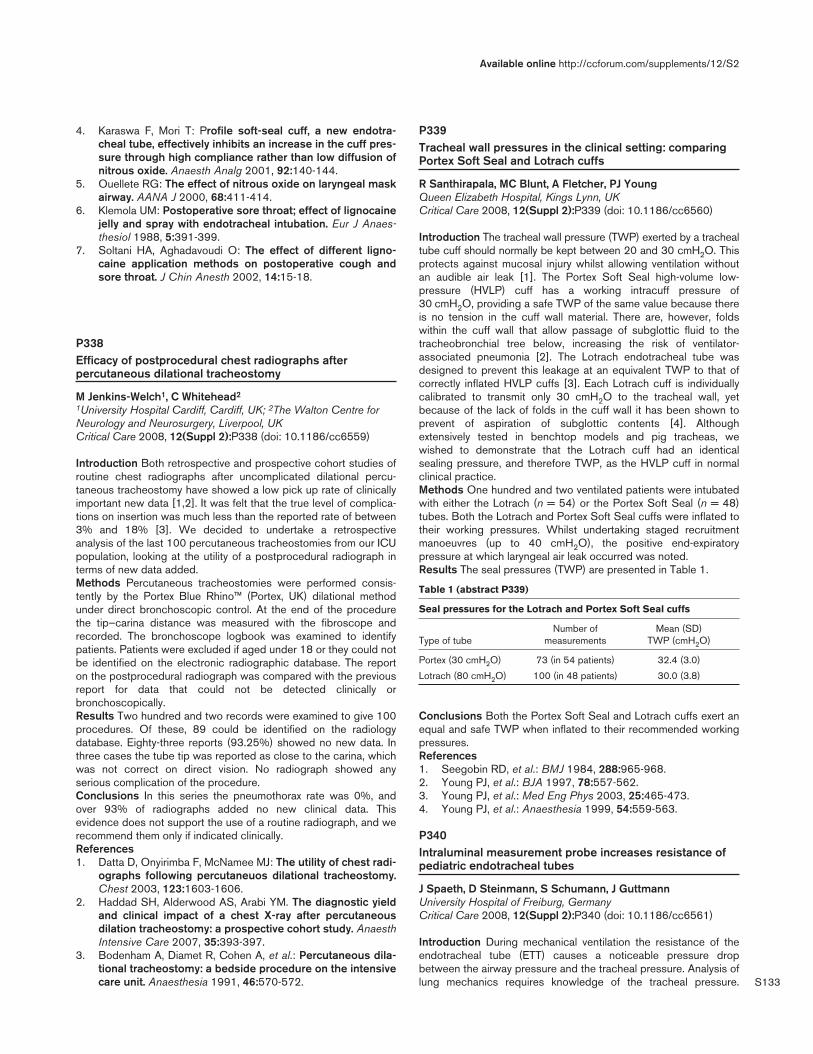
S133
4. Karaswa F, Mori T: Profile soft-seal cuff, a new endotra-cheal tube, effectively inhibits an increase in the cuff pres-sure through high compliance rather than low diffusion ofnitrous oxide. Anaesth Analg 2001, 92:140-144.
5. Ouellete RG: The effect of nitrous oxide on laryngeal maskairway. AANA J 2000, 68:411-414.
6. Klemola UM: Postoperative sore throat; effect of lignocainejelly and spray with endotracheal intubation. Eur J Anaes-thesiol 1988, 5:391-399.
7. Soltani HA, Aghadavoudi O: The effect of different ligno-caine application methods on postoperative cough andsore throat. J Chin Anesth 2002, 14:15-18.
P338Efficacy of postprocedural chest radiographs afterpercutaneous dilational tracheostomy
M Jenkins-Welch1, C Whitehead2
1University Hospital Cardiff, Cardiff, UK; 2The Walton Centre forNeurology and Neurosurgery, Liverpool, UKCritical Care 2008, 12(Suppl 2):P338 (doi: 10.1186/cc6559)
Introduction Both retrospective and prospective cohort studies ofroutine chest radiographs after uncomplicated dilational percu-taneous tracheostomy have showed a low pick up rate of clinicallyimportant new data [1,2]. It was felt that the true level of complica-tions on insertion was much less than the reported rate of between3% and 18% [3]. We decided to undertake a retrospectiveanalysis of the last 100 percutaneous tracheostomies from our ICUpopulation, looking at the utility of a postprocedural radiograph interms of new data added.Methods Percutaneous tracheostomies were performed consis-tently by the Portex Blue Rhino™ (Portex, UK) dilational methodunder direct bronchoscopic control. At the end of the procedurethe tip–carina distance was measured with the fibroscope andrecorded. The bronchoscope logbook was examined to identifypatients. Patients were excluded if aged under 18 or they could notbe identified on the electronic radiographic database. The reporton the postprocedural radiograph was compared with the previousreport for data that could not be detected clinically orbronchoscopically.Results Two hundred and two records were examined to give 100procedures. Of these, 89 could be identified on the radiologydatabase. Eighty-three reports (93.25%) showed no new data. Inthree cases the tube tip was reported as close to the carina, whichwas not correct on direct vision. No radiograph showed anyserious complication of the procedure.Conclusions In this series the pneumothorax rate was 0%, andover 93% of radiographs added no new clinical data. Thisevidence does not support the use of a routine radiograph, and werecommend them only if indicated clinically.References1. Datta D, Onyirimba F, McNamee MJ: The utility of chest radi-
ographs following percutaneuos dilational tracheostomy.Chest 2003, 123:1603-1606.
2. Haddad SH, Alderwood AS, Arabi YM. The diagnostic yieldand clinical impact of a chest X-ray after percutaneousdilation tracheostomy: a prospective cohort study. AnaesthIntensive Care 2007, 35:393-397.
3. Bodenham A, Diamet R, Cohen A, et al.: Percutaneous dila-tional tracheostomy: a bedside procedure on the intensivecare unit. Anaesthesia 1991, 46:570-572.
P339Tracheal wall pressures in the clinical setting: comparingPortex Soft Seal and Lotrach cuffs
R Santhirapala, MC Blunt, A Fletcher, PJ YoungQueen Elizabeth Hospital, Kings Lynn, UKCritical Care 2008, 12(Suppl 2):P339 (doi: 10.1186/cc6560)
Introduction The tracheal wall pressure (TWP) exerted by a trachealtube cuff should normally be kept between 20 and 30 cmH2O. Thisprotects against mucosal injury whilst allowing ventilation withoutan audible air leak [1]. The Portex Soft Seal high-volume low-pressure (HVLP) cuff has a working intracuff pressure of30 cmH2O, providing a safe TWP of the same value because thereis no tension in the cuff wall material. There are, however, foldswithin the cuff wall that allow passage of subglottic fluid to thetracheobronchial tree below, increasing the risk of ventilator-associated pneumonia [2]. The Lotrach endotracheal tube wasdesigned to prevent this leakage at an equivalent TWP to that ofcorrectly inflated HVLP cuffs [3]. Each Lotrach cuff is individuallycalibrated to transmit only 30 cmH2O to the tracheal wall, yetbecause of the lack of folds in the cuff wall it has been shown toprevent of aspiration of subglottic contents [4]. Althoughextensively tested in benchtop models and pig tracheas, wewished to demonstrate that the Lotrach cuff had an identicalsealing pressure, and therefore TWP, as the HVLP cuff in normalclinical practice.Methods One hundred and two ventilated patients were intubatedwith either the Lotrach (n = 54) or the Portex Soft Seal (n = 48)tubes. Both the Lotrach and Portex Soft Seal cuffs were inflated totheir working pressures. Whilst undertaking staged recruitmentmanoeuvres (up to 40 cmH2O), the positive end-expiratorypressure at which laryngeal air leak occurred was noted.Results The seal pressures (TWP) are presented in Table 1.
Table 1 (abstract P339)
Seal pressures for the Lotrach and Portex Soft Seal cuffs
Number of Mean (SD) Type of tube measurements TWP (cmH2O)
Portex (30 cmH2O) 73 (in 54 patients) 32.4 (3.0)
Lotrach (80 cmH2O) 100 (in 48 patients) 30.0 (3.8)
Conclusions Both the Portex Soft Seal and Lotrach cuffs exert anequal and safe TWP when inflated to their recommended workingpressures.References1. Seegobin RD, et al.: BMJ 1984, 288:965-968.2. Young PJ, et al.: BJA 1997, 78:557-562.3. Young PJ, et al.: Med Eng Phys 2003, 25:465-473.4. Young PJ, et al.: Anaesthesia 1999, 54:559-563.
P340Intraluminal measurement probe increases resistance ofpediatric endotracheal tubes
J Spaeth, D Steinmann, S Schumann, J GuttmannUniversity Hospital of Freiburg, GermanyCritical Care 2008, 12(Suppl 2):P340 (doi: 10.1186/cc6561)
Introduction During mechanical ventilation the resistance of theendotracheal tube (ETT) causes a noticeable pressure dropbetween the airway pressure and the tracheal pressure. Analysis oflung mechanics requires knowledge of the tracheal pressure.
Available online http://ccforum.com/supplements/12/S2
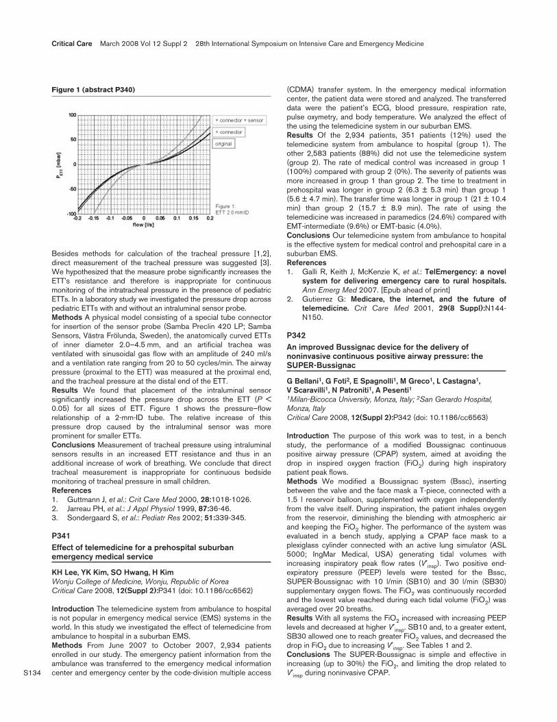
S134
Besides methods for calculation of the tracheal pressure [1,2],direct measurement of the tracheal pressure was suggested [3].We hypothesized that the measure probe significantly increases theETT’s resistance and therefore is inappropriate for continuousmonitoring of the intratracheal pressure in the presence of pediatricETTs. In a laboratory study we investigated the pressure drop acrosspediatric ETTs with and without an intraluminal sensor probe.Methods A physical model consisting of a special tube connectorfor insertion of the sensor probe (Samba Preclin 420 LP; SambaSensors, Västra Frölunda, Sweden), the anatomically curved ETTsof inner diameter 2.0–4.5 mm, and an artificial trachea wasventilated with sinusoidal gas flow with an amplitude of 240 ml/sand a ventilation rate ranging from 20 to 50 cycles/min. The airwaypressure (proximal to the ETT) was measured at the proximal end,and the tracheal pressure at the distal end of the ETT.Results We found that placement of the intraluminal sensorsignificantly increased the pressure drop across the ETT (P <0.05) for all sizes of ETT. Figure 1 shows the pressure–flowrelationship of a 2-mm-ID tube. The relative increase of thispressure drop caused by the intraluminal sensor was moreprominent for smaller ETTs.Conclusions Measurement of tracheal pressure using intraluminalsensors results in an increased ETT resistance and thus in anadditional increase of work of breathing. We conclude that directtracheal measurement is inappropriate for continuous bedsidemonitoring of tracheal pressure in small children.References1. Guttmann J, et al.: Crit Care Med 2000, 28:1018-1026.2. Jarreau PH, et al.: J Appl Physiol 1999, 87:36-46.3. Sondergaard S, et al.: Pediatr Res 2002; 51:339-345.
P341Effect of telemedicine for a prehospital suburbanemergency medical service
KH Lee, YK Kim, SO Hwang, H KimWonju College of Medicine, Wonju, Republic of KoreaCritical Care 2008, 12(Suppl 2):P341 (doi: 10.1186/cc6562)
Introduction The telemedicine system from ambulance to hospitalis not popular in emergency medical service (EMS) systems in theworld. In this study we investigated the effect of telemedicine fromambulance to hospital in a suburban EMS.Methods From June 2007 to October 2007, 2,934 patientsenrolled in our study. The emergency patient information from theambulance was transferred to the emergency medical informationcenter and emergency center by the code-division multiple access
(CDMA) transfer system. In the emergency medical informationcenter, the patient data were stored and analyzed. The transferreddata were the patient’s ECG, blood pressure, respiration rate,pulse oxymetry, and body temperature. We analyzed the effect ofthe using the telemedicine system in our suburban EMS.Results Of the 2,934 patients, 351 patients (12%) used thetelemedicine system from ambulance to hospital (group 1). Theother 2,583 patients (88%) did not use the telemedicine system(group 2). The rate of medical control was increased in group 1(100%) compared with group 2 (0%). The severity of patients wasmore increased in group 1 than group 2. The time to treatment inprehospital was longer in group 2 (6.3 ± 5.3 min) than group 1(5.6 ± 4.7 min). The transfer time was longer in group 1 (21 ± 10.4min) than group 2 (15.7 ± 8.9 min). The rate of using thetelemedicine was increased in paramedics (24.6%) compared withEMT-intermediate (9.6%) or EMT-basic (4.0%).Conclusions Our telemedicine system from ambulance to hospitalis the effective system for medical control and prehospital care in asuburban EMS.References1. Galli R, Keith J, McKenzie K, et al.: TelEmergency: a novel
system for delivering emergency care to rural hospitals.Ann Emerg Med 2007. [Epub ahead of print]
2. Gutierrez G: Medicare, the internet, and the future oftelemedicine. Crit Care Med 2001, 29(8 Suppl):N144-N150.
P342An improved Bussignac device for the delivery ofnoninvasive continuous positive airway pressure: theSUPER-Bussignac
G Bellani1, G Foti2, E Spagnolli1, M Greco1, L Castagna1, V Scaravilli1, N Patroniti1, A Pesenti11Milan-Bicocca University, Monza, Italy; 2San Gerardo Hospital,Monza, ItalyCritical Care 2008, 12(Suppl 2):P342 (doi: 10.1186/cc6563)
Introduction The purpose of this work was to test, in a benchstudy, the performance of a modified Boussignac continuouspositive airway pressure (CPAP) system, aimed at avoiding thedrop in inspired oxygen fraction (FiO2) during high inspiratorypatient peak flows.Methods We modified a Boussignac system (Bssc), insertingbetween the valve and the face mask a T-piece, connected with a1.5 l reservoir balloon, supplemented with oxygen independentlyfrom the valve itself. During inspiration, the patient inhales oxygenfrom the reservoir, diminishing the blending with atmospheric airand keeping the FiO2 higher. The performance of the system wasevaluated in a bench study, applying a CPAP face mask to aplexiglass cylinder connected with an active lung simulator (ASL5000; IngMar Medical, USA) generating tidal volumes withincreasing inspiratory peak flow rates (V′insp). Two positive end-expiratory pressure (PEEP) levels were tested for the Bssc,SUPER-Boussignac with 10 l/min (SB10) and 30 l/min (SB30)supplementary oxygen flows. The FiO2 was continuously recordedand the lowest value reached during each tidal volume (FiO2) wasaveraged over 20 breaths.Results With all systems the FiO2 increased with increasing PEEPlevels and decreased at higher V′insp. SB10 and, to a greater extent,SB30 allowed one to reach greater FiO2 values, and decreased thedrop in FiO2 due to increasing V′insp. See Tables 1 and 2.Conclusions The SUPER-Boussignac is simple and effective inincreasing (up to 30%) the FiO2, and limiting the drop related toV′insp during noninvasive CPAP.
Critical Care March 2008 Vol 12 Suppl 2 28th International Symposium on Intensive Care and Emergency Medicine
Figure 1 (abstract P340)
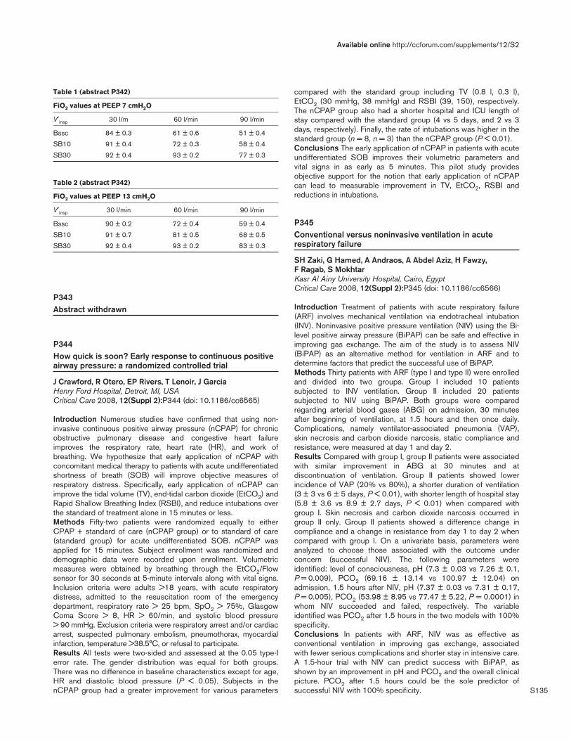
S135
P343Abstract withdrawn
P344How quick is soon? Early response to continuous positiveairway pressure: a randomized controlled trial
J Crawford, R Otero, EP Rivers, T Lenoir, J GarciaHenry Ford Hospital, Detroit, MI, USACritical Care 2008, 12(Suppl 2):P344 (doi: 10.1186/cc6565)
Introduction Numerous studies have confirmed that using non-invasive continuous positive airway pressure (nCPAP) for chronicobstructive pulmonary disease and congestive heart failureimproves the respiratory rate, heart rate (HR), and work ofbreathing. We hypothesize that early application of nCPAP withconcomitant medical therapy to patients with acute undifferentiatedshortness of breath (SOB) will improve objective measures ofrespiratory distress. Specifically, early application of nCPAP canimprove the tidal volume (TV), end-tidal carbon dioxide (EtCO2) andRapid Shallow Breathing Index (RSBI), and reduce intubations overthe standard of treatment alone in 15 minutes or less.Methods Fifty-two patients were randomized equally to eitherCPAP + standard of care (nCPAP group) or to standard of care(standard group) for acute undifferentiated SOB. nCPAP wasapplied for 15 minutes. Subject enrollment was randomized anddemographic data were recorded upon enrollment. Volumetricmeasures were obtained by breathing through the EtCO2/Flowsensor for 30 seconds at 5-minute intervals along with vital signs.Inclusion criteria were adults >18 years, with acute respiratorydistress, admitted to the resuscitation room of the emergencydepartment, respiratory rate > 25 bpm, SpO2 > 75%, GlasgowComa Score > 8, HR > 60/min, and systolic blood pressure> 90 mmHg. Exclusion criteria were respiratory arrest and/or cardiacarrest, suspected pulmonary embolism, pneumothorax, myocardialinfarction, temperature >38.5°C, or refusal to participate.Results All tests were two-sided and assessed at the 0.05 type-Ierror rate. The gender distribution was equal for both groups.There was no difference in baseline characteristics except for age,HR and diastolic blood pressure (P < 0.05). Subjects in thenCPAP group had a greater improvement for various parameters
compared with the standard group including TV (0.8 l, 0.3 l),EtCO2 (30 mmHg, 38 mmHg) and RSBI (39, 150), respectively.The nCPAP group also had a shorter hospital and ICU length ofstay compared with the standard group (4 vs 5 days, and 2 vs 3days, respectively). Finally, the rate of intubations was higher in thestandard group (n = 8, n = 3) than the nCPAP group (P < 0.01).Conclusions The early application of nCPAP in patients with acuteundifferentiated SOB improves their volumetric parameters andvital signs in as early as 5 minutes. This pilot study providesobjective support for the notion that early application of nCPAPcan lead to measurable improvement in TV, EtCO2, RSBI andreductions in intubations.
P345Conventional versus noninvasive ventilation in acuterespiratory failure
SH Zaki, G Hamed, A Andraos, A Abdel Aziz, H Fawzy, F Ragab, S MokhtarKasr Al Ainy University Hospital, Cairo, EgyptCritical Care 2008, 12(Suppl 2):P345 (doi: 10.1186/cc6566)
Introduction Treatment of patients with acute respiratory failure(ARF) involves mechanical ventilation via endotracheal intubation(INV). Noninvasive positive pressure ventilation (NIV) using the Bi-level positive airway pressure (BiPAP) can be safe and effective inimproving gas exchange. The aim of the study is to assess NIV(BiPAP) as an alternative method for ventilation in ARF and todetermine factors that predict the successful use of BiPAP.Methods Thirty patients with ARF (type I and type II) were enrolledand divided into two groups. Group I included 10 patientssubjected to INV ventilation. Group II included 20 patientssubjected to NIV using BiPAP. Both groups were comparedregarding arterial blood gases (ABG) on admission, 30 minutesafter beginning of ventilation, at 1.5 hours and then once daily.Complications, namely ventilator-associated pneumonia (VAP),skin necrosis and carbon dioxide narcosis, static compliance andresistance, were measured at day 1 and day 2.Results Compared with group I, group II patients were associatedwith similar improvement in ABG at 30 minutes and atdiscontinuation of ventilation. Group II patients showed lowerincidence of VAP (20% vs 80%), a shorter duration of ventilation(3 ± 3 vs 6 ± 5 days, P < 0.01), with shorter length of hospital stay(5.8 ± 3.6 vs 8.9 ± 2.7 days, P < 0.01) when compared withgroup I. Skin necrosis and carbon dioxide narcosis occurred ingroup II only. Group II patients showed a difference change incompliance and a change in resistance from day 1 to day 2 whencompared with group I. On a univariate basis, parameters wereanalyzed to choose those associated with the outcome underconcern (successful NIV). The following parameters wereidentified: level of consciousness, pH (7.3 ± 0.03 vs 7.26 ± 0.1,P = 0.009), PCO2 (69.16 ± 13.14 vs 100.97 ± 12.04) onadmission, 1.5 hours after NIV, pH (7.37 ± 0.03 vs 7.31 ± 0.17,P = 0.005), PCO2 (53.98 ± 8.95 vs 77.47 ± 5.22, P = 0.0001) inwhom NIV succeeded and failed, respectively. The variableidentified was PCO2 after 1.5 hours in the two models with 100%specificity.Conclusions In patients with ARF, NIV was as effective asconventional ventilation in improving gas exchange, associatedwith fewer serious complications and shorter stay in intensive care.A 1.5-hour trial with NIV can predict success with BiPAP, asshown by an improvement in pH and PCO2 and the overall clinicalpicture. PCO2 after 1.5 hours could be the sole predictor ofsuccessful NIV with 100% specificity.
Available online http://ccforum.com/supplements/12/S2
Table 1 (abstract P342)
FiO2 values at PEEP 7 cmH2O
V′insp 30 l/m 60 l/min 90 l/min
Bssc 84 ± 0.3 61 ± 0.6 51 ± 0.4
SB10 91 ± 0.4 72 ± 0.3 58 ± 0.4
SB30 92 ± 0.4 93 ± 0.2 77 ± 0.3
Table 2 (abstract P342)
FiO2 values at PEEP 13 cmH2O
V′insp 30 l/min 60 l/min 90 l/min
Bssc 90 ± 0.2 72 ± 0.4 59 ± 0.4
SB10 91 ± 0.7 81 ± 0.5 68 ± 0.5
SB30 92 ± 0.4 93 ± 0.2 83 ± 0.3
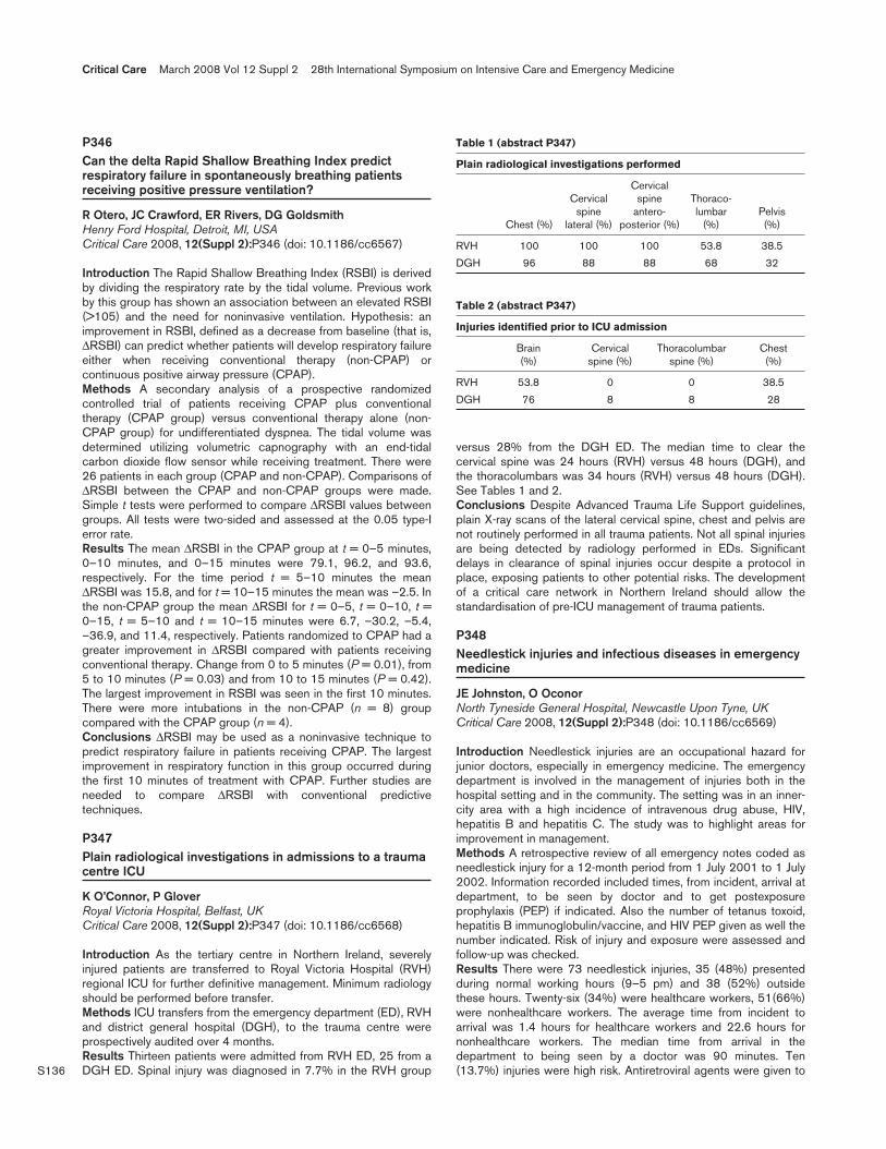
S136
P346Can the delta Rapid Shallow Breathing Index predictrespiratory failure in spontaneously breathing patientsreceiving positive pressure ventilation?
R Otero, JC Crawford, ER Rivers, DG GoldsmithHenry Ford Hospital, Detroit, MI, USACritical Care 2008, 12(Suppl 2):P346 (doi: 10.1186/cc6567)
Introduction The Rapid Shallow Breathing Index (RSBI) is derivedby dividing the respiratory rate by the tidal volume. Previous workby this group has shown an association between an elevated RSBI(>105) and the need for noninvasive ventilation. Hypothesis: animprovement in RSBI, defined as a decrease from baseline (that is,ΔRSBI) can predict whether patients will develop respiratory failureeither when receiving conventional therapy (non-CPAP) orcontinuous positive airway pressure (CPAP).Methods A secondary analysis of a prospective randomizedcontrolled trial of patients receiving CPAP plus conventionaltherapy (CPAP group) versus conventional therapy alone (non-CPAP group) for undifferentiated dyspnea. The tidal volume wasdetermined utilizing volumetric capnography with an end-tidalcarbon dioxide flow sensor while receiving treatment. There were26 patients in each group (CPAP and non-CPAP). Comparisons ofΔRSBI between the CPAP and non-CPAP groups were made.Simple t tests were performed to compare ΔRSBI values betweengroups. All tests were two-sided and assessed at the 0.05 type-Ierror rate.Results The mean ΔRSBI in the CPAP group at t = 0–5 minutes,0–10 minutes, and 0–15 minutes were 79.1, 96.2, and 93.6,respectively. For the time period t = 5–10 minutes the meanΔRSBI was 15.8, and for t = 10–15 minutes the mean was –2.5. Inthe non-CPAP group the mean ΔRSBI for t = 0–5, t = 0–10, t =0–15, t = 5–10 and t = 10–15 minutes were 6.7, –30.2, –5.4,–36.9, and 11.4, respectively. Patients randomized to CPAP had agreater improvement in ΔRSBI compared with patients receivingconventional therapy. Change from 0 to 5 minutes (P = 0.01), from5 to 10 minutes (P = 0.03) and from 10 to 15 minutes (P = 0.42).The largest improvement in RSBI was seen in the first 10 minutes.There were more intubations in the non-CPAP (n = 8) groupcompared with the CPAP group (n = 4).Conclusions ΔRSBI may be used as a noninvasive technique topredict respiratory failure in patients receiving CPAP. The largestimprovement in respiratory function in this group occurred duringthe first 10 minutes of treatment with CPAP. Further studies areneeded to compare ΔRSBI with conventional predictivetechniques.
P347Plain radiological investigations in admissions to a traumacentre ICU
K O’Connor, P GloverRoyal Victoria Hospital, Belfast, UKCritical Care 2008, 12(Suppl 2):P347 (doi: 10.1186/cc6568)
Introduction As the tertiary centre in Northern Ireland, severelyinjured patients are transferred to Royal Victoria Hospital (RVH)regional ICU for further definitive management. Minimum radiologyshould be performed before transfer.Methods ICU transfers from the emergency department (ED), RVHand district general hospital (DGH), to the trauma centre wereprospectively audited over 4 months.Results Thirteen patients were admitted from RVH ED, 25 from aDGH ED. Spinal injury was diagnosed in 7.7% in the RVH group
versus 28% from the DGH ED. The median time to clear thecervical spine was 24 hours (RVH) versus 48 hours (DGH), andthe thoracolumbars was 34 hours (RVH) versus 48 hours (DGH).See Tables 1 and 2.Conclusions Despite Advanced Trauma Life Support guidelines,plain X-ray scans of the lateral cervical spine, chest and pelvis arenot routinely performed in all trauma patients. Not all spinal injuriesare being detected by radiology performed in EDs. Significantdelays in clearance of spinal injuries occur despite a protocol inplace, exposing patients to other potential risks. The developmentof a critical care network in Northern Ireland should allow thestandardisation of pre-ICU management of trauma patients.
P348Needlestick injuries and infectious diseases in emergencymedicine
JE Johnston, O OconorNorth Tyneside General Hospital, Newcastle Upon Tyne, UKCritical Care 2008, 12(Suppl 2):P348 (doi: 10.1186/cc6569)
Introduction Needlestick injuries are an occupational hazard forjunior doctors, especially in emergency medicine. The emergencydepartment is involved in the management of injuries both in thehospital setting and in the community. The setting was in an inner-city area with a high incidence of intravenous drug abuse, HIV,hepatitis B and hepatitis C. The study was to highlight areas forimprovement in management.Methods A retrospective review of all emergency notes coded asneedlestick injury for a 12-month period from 1 July 2001 to 1 July2002. Information recorded included times, from incident, arrival atdepartment, to be seen by doctor and to get postexposureprophylaxis (PEP) if indicated. Also the number of tetanus toxoid,hepatitis B immunoglobulin/vaccine, and HIV PEP given as well thenumber indicated. Risk of injury and exposure were assessed andfollow-up was checked.Results There were 73 needlestick injuries, 35 (48%) presentedduring normal working hours (9–5 pm) and 38 (52%) outsidethese hours. Twenty-six (34%) were healthcare workers, 51(66%)were nonhealthcare workers. The average time from incident toarrival was 1.4 hours for healthcare workers and 22.6 hours fornonhealthcare workers. The median time from arrival in thedepartment to being seen by a doctor was 90 minutes. Ten(13.7%) injuries were high risk. Antiretroviral agents were given to
Critical Care March 2008 Vol 12 Suppl 2 28th International Symposium on Intensive Care and Emergency Medicine
Table 1 (abstract P347)
Plain radiological investigations performed
Cervical Cervical spine Thoraco-
spine antero- lumbar Pelvis Chest (%) lateral (%) posterior (%) (%) (%)
RVH 100 100 100 53.8 38.5
DGH 96 88 88 68 32
Table 2 (abstract P347)
Injuries identified prior to ICU admission
Brain Cervical Thoracolumbar Chest (%) spine (%) spine (%) (%)
RVH 53.8 0 0 38.5
DGH 76 8 8 28
S137
15 (20.1%) patients and the average time from door to HIV PEPwas 141 minutes.Conclusions Emergency medicine staff should be aware of therisks of blood-borne viral transmission as they have greaterexposure than other healthcare groups. They are at higher risk ofpercutaneous injury and therefore should adopt universalprecautions; shield and sheath devices would also reduce the riskof sharp injury. The HIV PEP is effective if given early, so thesepatients must be assessed urgently and antiretroviral agents givenas soon as possible if indicated. Emergency medicine has had anincreasing role in management of needlestick injuries in healthcareworkers occurring outside working hours, out-of-hospital injuriesand other attendances for HIV PEP. Greater education ofemergency staff, other healthcare workers and the general public isrequired for optimal management of needlestick injuries.
P349Impact of a pandemic triage tool on intensive care admission
A Bailey, I Leditschke, J Ranse, K GroveThe Canberra Hospital, Canberra, AustraliaCritical Care 2008, 12(Suppl 2):P349 (doi: 10.1186/cc6570)
Introduction The issues of pandemic preparedness and the use ofcritical care resources in a pandemic have been of increasedinterest recently [1]. We assessed the effect of a proposedpandemic critical care triage tool [2] on admissions to the ICU. Thetool aims to identify patients who will most benefit from admissionto the ICU and excludes patients considered ‘too well’, ‘too sick’,or with comorbidities likely to limit survival in the shorter term.Methods To assess the impact of the pandemic tool on our currentpractice, we performed a retrospective observational study of theapplication of the pandemic triage tool described by Christian andcolleagues [2] to all admissions to our 14-bed general medical–surgical ICU over a 1-month period.Results One hundred and nineteen patients were admitted to theICU. Using the pandemic triage tool, 91 of these patients (76%)did not meet the triage inclusion criteria on admission. As requiredby the triage tool, patients were reassessed at 48 and 120 hours,with only one of the 91 patients becoming eligible for admission onreassessment. Further assessment of the 29 patients (24%) whomet the triage inclusion criteria revealed that 17 of these met thetriage exclusion criteria, leaving 12 patients (10%) from the original119 as qualifying for ICU. One of these 12 was deemed ‘too sick’by the triage tool and therefore was also excluded, leaving 11patients (9%).Conclusions Application of this triage tool to our current ICUpatient population would radically change practice, and wouldgenerate substantial capacity that could be used in the situation ofa pandemic. In addition, as the triage tool aims to exclude patientswho are less likely to benefit from admission to the ICU, theseresults also have implications for ICU resource management in thenonpandemic situation.References1. Challen K, et al: Crit Care 2007, 11:212.2. Christian MD, et al: CMAJ 2006, 175:1377.
P350Pneumonia Severity Index: a validation and assessmentstudy for its use as a triage tool in the emergencydepartment
S JacobsAlice Springs Hospital, Alice Springs, AustraliaCritical Care 2008, 12(Suppl 2):P350 (doi: 10.1186/cc6571)
Introduction Our rural 10-bed ICU serves a 167-bed hospitalsituated in a small town at the centre of the Australian Continent.The hospital is utilised mainly by Indigenous patients. We wereinterested to estimate the Pneumonia Severity Index (PSI) amongall patients presenting to our emergency department (ED) withcommunity-acquired pneumonia (CAP) during 2006 to ascertainwhether this scoring system could be validated among our uniquepatient population and to ascertain whether the score could havebeen helpful to support a clinical decision to transfer patients tothe medical ward or ICU.Methods All patients presenting to the ED during 2006 wereidentified and their demographic and parameters details noted tocalculate their PSI scores, which were performed retrospectively.Triage was performed clinically by the ED doctors. The followingcomplications were noted: requirement for artificial ventilation,septic shock, acute renal failure, requirement for percutaneoustracheostomy and mortality. All patients transferred to tertiarycentres because of staff or bed shortages were also noted. Micro-biological data were collected on all ICU patients and most of theward patients.Results Four hundred and seventy-six CAP patients presented tothe ED in 2006, of which 91% (436/476) were Indigenous and12% (57/476) were transferred to the ICU. Admission charac-teristics of ICU patients revealed high incidences of alcoholism(76%) and chronic illness (70%). Artificial ventilation rates of these57 patients were defined according to PSI severity: no patient witha score <91 required artificial ventilation, whereas 64% of patientswith a score of 91–130 and 75% of patients with a score >130required artificial ventilation. Using PSI < 91 for predictingabsence of the need for artificial ventilation, specificity of 100%and sensitivity of 91% were demonstrated.Conclusions The CAP rate among the central AustralianIndigenous population is unacceptably high. This high rate isassociated with high incidences of alcoholism, chronic ill healthand poor social conditions. The PSI has been validated in thisstudy, accurately predicting mortality and the need for artificialventilation. The PSI could be a useful tool to support a clinicaldecision to transfer patients from the ED to the general medicalwards (PSI < 91) or to the ICU (PSI ≥ 91).
P351Injuries sustained by patients with behavioral disturbancesbrought to an emergency department in police custody
J Metzger, P Maher, M Wainscott, PE Pepe, K DelaneyUT Southwestern Medical School, Dallas, TX, USACritical Care 2008, 12(Suppl 2):P351 (doi: 10.1186/cc6572)
Introduction Although it is widely recognized that physicalrestraint of violent persons can result in death or serious injury,formal reports documenting the incidence or rate of such injuriesare lacking. The rate and nature of injuries for emotionally disturbedpatients brought in police custody to a major urban emergencydepartment (ED) were therefore recorded.Methods All medical records of persons brought by police to thepublic hospital ED for a large metropolitan county (population 2.5million) are electronically marked for subsequent rapid searches.Excluding those arriving following commission of a crime or forevaluation of medical conditions or sexual assaults, all patientsbrought with the transport assistance of paramedic crews to theED in police custody for psychiatric evaluation as an emotionallydisturbed person from 12 December 1999 through 31 August2003 were studied. Patients were classified specifically as‘agitated’ if they were described as violent, psychotic, aggressive,combative, hostile, threatening, homicidal or dangerous.
Available online http://ccforum.com/supplements/12/S2
S138
Results Of the total 24,895 police custody visits, 17,733 met theinclusion criteria for receiving psychiatric evaluation as emotionallydisturbed persons. Of these, 10,173 (57%) could be classified asagitated. A potentially lethal weapon was confiscated in 447cases. Of the 17,733 studied, 511 (3.3%) sustained injuries – 398(78%) of which were self-inflicted stab wounds, wrist lacerationsand hangings. Mace exposure resulted in 34 minor injuries, whilenone were attributable to conductive electrical devices. Overall,the rate of self-inflicted injuries over 3 years and 9 months was2.2% (n = 398) while it was 0.6% (n = 113) for those inflicted byothers. Only four of these patients (approximately one per year)required admission to a surgical service. Of note, 29% of injuriessustained by agitated patients were not self-inflicted, comparedwith 13.6% in nonagitated patients (70/238 vs 37/273, P =0.0001).Conclusions With the assistance of transporting paramedics,police officers were able to restrain violent, emotionally disturbedpatients with a very low risk of serious injury.Reference1. Morrison A, et al.: Death of a psychiatric patient during
physical restraint. Med Sci Law 2001, 41:46-50.
P352A study on the reliability and validity of two four-levelemergency triage systems
N Parenti1, L Ferrara1, D Sangiorgi2, M Bacchi Reggiani2, R Manfredi1, T Lenzi11Hospital Santa Maria della Scaletta Imola, Bologna, Italy;2Policlinico Sant’Orsola, Bologna, ItalyCritical Care 2008, 12(Suppl 2):P352 (doi: 10.1186/cc6573)
Introduction We compared the reliability and validity of two four-level emergency triage systems: the ETS1, used in our emergencydepartment (ED); and the ETS2, a triage algorithm derived from theEmergency Severity Index [1].Methods This is an observational retrospective study of 189patients admitted to our ED in 2 weeks of October 2006. Triagescenarios were designed with medical records. Ten trained triagenurses were randomly assigned either to training in ETS2, or torefresh the ETS1. They independently assigned triage scores tothe scenarios, at time zero and after 6 months. Both triage systemshave four urgency categories (UC): UC1 = immediate response;UC2, UC3, UC4 = assessment within 20, 60, 120 minutes. Wecollected demographic and clinical characteristics, nurse triagecategory, admission status and site, nurse triage forms withpresenting complaint, mode and time of arrival, past diseases, vitalsigns, and pain score. For each scenario we considered ‘truetriage’ the mode of the UC assigned by the nurses. Weightedkappa (Κ) was used to calculate inter-rater and intra-rater reliabilityin each of the two groups of nurses. The relationships between the‘true triage’ and admission, and admission site were assessed.Results The UCs assigned were similar in two groups: 20%versus 21% with UC4, 50% versus 48% with UC3, 28% versus28% with UC2, 2% versus 3% with UC1. A complete disagree-ment in UC was found in 3% and 5% cases of ETS1 and ETS2; acomplete agreement in 52% and 56% cases of ETS1 and ETS2.Inter-rater reliability among nurses using ETS1 and ETS2 was Κ =0.73 (95% CI: 0.59–0.87) and Κ = 0.79 (95% CI: 0.65–0.93),respectively; intra-rater reliability was: Κ = 0.82 (95% CI:0.67–0.96) and Κ = 0.78 (95% CI: 0.62–0.93) in ETS1 andETS2. Hospital admission by ETS1 and ETS2 was similar for UC2(39% vs 37%), UC3 (5% vs 8%), and UC4 (3% vs 0%); 100% ofpatients with UC1 in ETS1 and 60% in ETS2 were admitted to theICU.
Conclusions The two emergency triage systems showed similarreliability and validity. ETS2 is easier to consult but worse inprediction of ICU admission. To our knowledge this is the firststudy on the intra-rater reliability of two four-level emergency triagesystems.Reference1. Wuerz RC, Milne LW, Eitel DR, et al.: Reliability and validity
of a new five-level triage instrument. Acad Emerg Med2000, 7:236-242.
P353Patterns of medical illness and injuries in emotionallydisturbed patients brought to an emergency department inpolice custody
J Metzger, P Maher, M Wainscott, P Pepe, K DelaneyUT Southwestern Medical School, Dallas, TX, USACritical Care 2008, 12(Suppl 2):P353 (doi: 10.1186/cc6574)
Introduction Emotionally disturbed persons brought to the emer-gency department (ED) are at risk of surgical and medical illness.The patterns of medical and surgical illness for emotionallydisturbed patients brought in police custody to a major urban EDwere recorded.Methods Our electronic ED medical records include the chiefcomplaint, final diagnosis and disposition. A smaller number havedetailed nursing notes. All patients with detailed nursing notes whowere brought between 1 December 1999 and 31 August 2003 inpolice custody for psychiatric evaluation were studied. Patientswere classified as ‘agitated’ if they were described as violent,psychotic, aggressive, combative, hostile, threatening, homicidal ordangerous.Results In total, 17,733 were brought for psychiatric evaluation. Ofthese 6,432 had complete nursing notes. Rates of injury andillness were low. Of 1,985 nonagitated patients, 194 (9.8%) wereinjured: 180 (93%) self-inflected and none was attributed to arestraint process. For 4,447 agitated patients, 227 (3.5%) wereinjured, 160 (70%) were self-inflicted and 39 (0.9%) wereattributed to the restraint process. Of these, 31 were exposed tomace, six had minor head or soft-tissue injuries, one had apneumothorax, and one had an airway injury. For the subgroup self-inflicted injuries included lacerations (245), head injuries (60),hangings (13), penetrating wounds (10), hand fractures orinfection (three), and other minor (nine). Overall 2,903 (45.8%)were admitted to a psychiatric service, 110 (1.7%) to a medicalservice and seven (0.12%) to a surgical service. Admitted surgicaldiagnoses were abdominal stab wound (one), tibial fracture (two),pneumothorax (one), airway injury (one), infected human bite (one),complicated lacerations (three), and penile foreign body (one). Thepredominant medical admission diagnoses were alcoholwithdrawal (16 patients), overdose (24 patients), andrhabdomyolysis (12 patients). Forty-five patients had complicationsof chronic medical illness and eight patients had dementia with bypsychosis.Conclusions Although serious medical illnesses and injuriesoccurred, there was a low rate of medical and surgical illness thatrequired admission. There was a high rate of psychiatric admission.Reference1. Morrison A, et al.: Death of a psychiatric patient during
physical restraint. Med Sci Law 2001, 41:46-50.
Critical Care March 2008 Vol 12 Suppl 2 28th International Symposium on Intensive Care and Emergency Medicine
S139
P354Factors of hospitalization for mild heat illness
M Todani, T Ryosuke, T Kaneko, Y Kawamura, S Kasaoka, T MaekawaYamaguchi University Hospital, Ube Yamaguchi, JapanCritical Care 2008, 12(Suppl 2):P354 (doi: 10.1186/cc6575)
Introduction Untreated mild heat illness (heat exhaustion) becomesprogressively more severe (heat stroke). Although the prognosis andrisk factors for hospital mortality in patients with severe heat illnessare often reported, the epidemiologic data for mild heat illness havebeen rarely reported. We therefore investigated the hospitalizationpredictive factors in the patients with mild heat illness.Methods Questionnaire sheets were sent to our affiliated hospitalsin Yamaguchi prefecture, located at 34° north latitude with a popula-tion of 1.5 million, to identify the patients who received medicalattentions with heat illness from 1 July to 31 August 2007. Thequestionnaire included symptoms, vital signs, laboratory data andpresence or absence of hospitalization (decided by each doctor).Results We analyzed the data from 114 of the 126 patients withmild heat illness. Twelve patients were excluded because ofinsufficient description. The total number of hospitalizations was44 (35%) and all patients were discharged without subsequentcomplications. The significant differences were shown in bodytemperature, consciousness disturbance, dysfunction of centralnerve system including convulsion or cerebellar ataxia, age, levelsof serum C-reactive protein, blood urea nitrogen (BUN) and whiteblood cell count between hospitalized and nonhospitalized patientswith mild heat illness. Independent predictive factors forhospitalization were Glasgow Coma Scale < 15 (P = 0.04, OR =3.56, 95% CI = 1.05–12.01), age ≥ 60 years (P < 0.01, OR =4.44, 95% CI =1.50–13.08), and BUN ≥21 mg/dl (P = 0.03,OR = 3.35, 95% CI = 1.16–9.67).Conclusions Based on the present findings, the factors ofhospitalization for mild heat illness were identified as presence ofconsciousness disturbance, seniority and high BUN level. Thesefactors did not directly show the severity of heat illness, but they dohelp us to determine the patients who should be hospitalized.References1. Bouchama A, Knochel JP: N Engl J Med 2002, 346:1978-
1988.2. Misset B, et al: Crit Care Med 2006, 34:1087-1092.
P355Missed opportunities to facilitate the diagnosis of coloncancer in emergency department patients
K DelaneyUT Southwestern Medical School, Dallas, TX, USACritical Care 2008, 12(Suppl 2):P355 (doi: 10.1186/cc6576)
Introduction We have been aware that colon cancer in our publicinstitution is too often diagnosed at a late stage. We evaluated thepattern of visits of these patients to the emergency department(ED) prior to the diagnosis of cancer.Methods All patients in the hospital cancer registry with a diagnosisof colon cancer between January 2001 and December 2004 wereevaluated. The prior ED visits were tracked in the searchable EDelectronic medical record back to January 1999. Chief complaintsand hematocrits were recorded. Anemia was defined as a hematocrit<37%. Symptoms were unexplained anorectal or abdominal pain orbleeding, chronic constipation, diarrhea or vomiting.
Results Two hundred and thirteen patients (116 men and 97women) with colon cancer diagnosed during the 3-year period hadvisited the ED at least once prior to diagnosis. 37.6% werediagnosed within 1 week of the first symptomatic ED visit and 50%within the first 40 days. The median time to diagnosis after the firstsymptomatic ED visit was 46 days while the average was 256days. Diagnoses were delayed more than 1 year in 51 patients.The median time to diagnosis after the first detection of anemiawas 84 days. Forty-one patients were diagnosed at greater than 1year. Women with anemia (n = 27) were nearly twice as likely asmen (n = 16) to have ≥1-year delays in diagnosis following thedetection of anemia.Conclusions Opportunities to facilitate early diagnosis of coloncancer were missed in some cases. Focused interaction of EDproviders with outpatient care providers to facilitate evaluation ofsuspected colon cancer is necessary to improve early detection.Women with anemia are less likely to be evaluated for agastrointestinal source of blood loss.
P356When are chest X-ray scans performed in the emergencydepartment useful?
JE JohnstonNorth Tyneside General Hospital, Newcastle Upon Tyne, UKCritical Care 2008, 12(Suppl 2):P356 (doi: 10.1186/cc6577)
Introduction This study was designed to demonstrate use of chestX-ray scans in a district general hospital emergency departmentand to highlight areas of inappropriate use.Methods A retrospective chart review of 62 consecutive chest X-ray exposures from emergency patients in the department. Thefrequency of temperature, pulse, respiratory rate and oxygensaturation parameters were recorded, and those subsequentlyabnormal. The indication for the X-ray scan, information aboutprevious chest X-ray scans and the record of the observed resultwere reviewed, as well as whether the patient’s management wasaltered by the result of the X-ray scan.Results Only 50% of the X-ray scans provided information thatwould potentially change the patient’s management, although 68%had positive findings recorded. Twenty-nine per cent wererequested for investigation of chest pain (pleuritic in 8.1%), 11.3%for investigation of abdominal pain, and 5% for transient ischemicattacks.Conclusions Approximately 50% of chest X-ray scans requestedfrom the emergency department are inappropriate. They are oftenrequested unnecessarily for chest pain, transient ischaemicattacks, mild chest infections, head injury, haematemesis and minorinjuries.
P357Management of acute organophosphorus poisoning at auniversity hospital
JM ShaikhLiaquat University of Medical & Health Sciences, Jamshoro,PakistanCritical Care 2008, 12(Suppl 2):P357 (doi: 10.1186/cc6578)
Introduction Organophosphorus (OP) insecticides are widelyused in agriculture, usually as pesticides, and frequently cause ill
Available online http://ccforum.com/supplements/12/S2
S140
health and death for hundreds of thousands of people each year.The majority of deaths occur following deliberate self-poisoning.They are common suicidal agents in Pakistan, India, Sri Lanka andother South Asian countries. The Accident & EmergencyDepartment of Liaquat University Hospital Hyderabad routinelyreceives victims of OP poisoning from the farming communities allaround. Our objective was to document the management, complica-tions and subsequent outcome of patients with acute organo-phosphorus poisoning in the ICU of Liaquat University Hospital.Methods All victims of OP poisoning admitted to the ICU ofLiaquat University Hospital admitted from May 2004 to October2006 were included in the study. Diagnosis of OP poisoning wasconfirmed from history and clinical findings. Management,complications and subsequent outcome were noted. Statisticalanalysis was performed using SPSS 10.Results A total of 111 patients of OP poisoning were admitted tothe ICU during the study period; 60.4% of patients were males.The mean age was 25.26 ± 8.52 years; 85.6% were within theage limit of 12–30 years. Of patients, 89.2% were a suicidalattempt. In 94.6% of patients, ingestion was the route of exposure.The mean ICU stay was 2.3 ± 3.2 days. Twenty (18%) patientsneeded mechanical ventilatory support. The overall mortality ratiowas 9% (n = 10). The mortality rate for the patients who requiredmechanical ventilation was 40% (n = 8), but the rate was 2.2%(n = 2) for the patients who were not mechanically ventilated.Conclusions Because of widespread use of OP pesticides byfarming communities of the developing world, it is very difficult toreduce mortality by primary prevention. Immediate shifting of thevictim to a well-equipped and well-staffed ICU, careful resusci-tation with appropriate use of antidotes and good supportive careand observation can help reduce the number of deaths in theperiod after admission to the hospital. Awareness and education ofgeneral practitioners in the rural areas regarding emergencymanagement, as well as prompt referral to an appropriate facility, isalso recommended to reduce the mortality rate.References1. World Health Organization in collaboration with the United
Nations Environmental Programme: Public Impact of Pesti-cides Used in Agriculture. Geneva: WHO; 1990.
2. Bairy KL, Vidyasagar S, Sharma A, Sammad V: Controversiesin the management of organophosphate poisoning. Ind JPharmacol 2007, 39:71-74.
3. Dreisbach RH: Cholinesterase inhibitor pesticides. In HandBook of Poisoning. 11th edition. CA: Longe Medical Publica-tions; 1983:106-114.
4. Phillips MR, Li X, Zhang Y: Suicide rates in China, 1995–99.Lancet 2002, 359:835-840.
5. Niwaz A, Faridi MA: Organophosphate insecticide poison-ing [case report]. Anaesth Pain Intensive Care 1999, 3:34-36.
6. Vijayakumar L: Suicide prevention: the urgent need indeveloping countries. World Psychiatry 2004, 3:158-159.
7. Eddleston M, Nick AB, Andrew HD: The need for transla-tional research on antidotes for pesticide poisoning. ClinExp Pharmacol Physiol 2005, 32:999-1005.
8. Eddleston M, Phillips MR: Self poisoning with pesticides.BMJ 2004, 328:42-44.
9. Gunnell D, Eddleston M: Suicide by intentional ingestion ofpesticides: a continuing tragedy in developing countries.Int J Epidemiol 2003, 32:902-909.
10. Eddleston M: Patterns and problems of deliberate self-poi-soning in the developing world. Q J Med 2000, 93:715-731.
11. Karalliedde L, Eddelston M, Murray V: The global picture oforganophosphate insecticide poisoning. In Karalliedde,
Feldman F, Henry J, Marrs T, editors. Organophosphates andHealth. London: Imperial Press; 2001:432-471.
12. Jamil H: Organophosphorus insecticide poisoning. J PakMed Assoc 1989, 39:27-31.
13. Singh S, Wig N, Chaudhary D, Sood NK, Sharma BK: Chang-ing pattern of acute poisoning in adults: experience of alarge North-West Indian hospital (1970–89). J Assoc PhyscIndia 1997, 45:194-197.
14. Ganesvaran T, Subramaniam S, Mhadevan K: Suicide in anorthern town of Sri Lanka. Acta Psychiatr Scand 1984, 69:420-425.
P358Suicidal intoxication by the black stone in Tunisia
I El Amri1, K Garrab1, Y Youssef2, S Slaheddine2
1CHU Hached, Kalaa-Sghira, Tunisia; 2CHU Hached, Sousse,TunisiaCritical Care 2008, 12(Suppl 2):P358 (doi: 10.1186/cc6579)
Introduction Paraphenylenediamine (PPD) is frequently used bywomen in certain countries such as Tunisia, Morocco, India,Pakistan and Sudan to dye their hair black. Knowledge of itssystemic toxicity led to use for a purpose of autolysis, but globalstudies including a medico-legal and toxicological investigationremain very rare. In Tunisia the sporadic cases of suicide by theingestion of this substance were recorded in the regions of thesouth and the center.Methods A retrospective study concerning a series of 10 cases ofvoluntary acute intoxications with PPD brought together in theLaboratory of Toxicology of CHU Farhat Hached Sousse. Thesamples of blood, urine and gastric contents were realized duringthe clinical examination and autopsy in the hospitals of Sfax,Sousse and Kairouan. The characterization of PPD in the aqueousbiological circles was able to be made after the clarification of aseparation technique.Results The sex ratio (male/female) was equal to 0.25. Theaverage age was 28 years. Seven subjects were single and threewere married. The socioeconomic level was low in all of the cases.Death was noted in 9/10 cases. The most common clinicalevidence was cervicofacial oedema (nine cases), diffuse myalgia(three cases), and blackish urine (six cases). The analysistoxicology brought conclusive evidence of the ingestion of PPD byrevealing the level of the gastric contents and the urine in almost allof the cases (10 cases).Conclusions The study shows that intoxication by the black stoneis relatively rare but there is a potentially burning absence of a fastand effective medical intervention. The prognosis for survivalinvolves the initial phase characterized by cervicofacial oedemarequiring a tracheotomy of rescue for lack of intubation, oftendifficult. It is this logic that makes the role for the Laboratory ofToxicology bringing, in the shortest time period, proof of an acuteintoxication, for which the diagnosis is not to be underestimated.References1. Zeggwagh AA, Aboukal R, Madani R, et al.: Myocardite
toxique due à la paraphénylène diamine, à propos dedeux cas. Rea Urg 1996, 5:699-703.
2. Brasch J: ‘New’ contact allergens. Curr Opin Allergy ClinImmunol 2007, 7:409-412.
Critical Care March 2008 Vol 12 Suppl 2 28th International Symposium on Intensive Care and Emergency Medicine
S141
P359Prognosis factors of poisonings treated withextracorporeal life support in the ICU
B Mégarbane1, N Deye1, S Mohebbi-Amoli1, J Théodore1, P Brun1, I Malissin1, P Le Prince2, F Baud1
1Hôpital Lariboisière, Paris, France; 2Hôpital Pitié Salpétrière, Paris,FranceCritical Care 2008, 12(Suppl 2):P359 (doi: 10.1186/cc6580)
Introduction Massive drug ingestions may be associated withrefractory cardiac failure, which reversibility makes extracorporeal lifesupport (ECLS) promising despite prolonged arrest. Our objectivewas to determine the prognosis factors of ECLS-treated patients.Methods A prospective study including all poisoned patients treatedwith ECLS during 2003–2007; surgical cannulation of femoralvessels in the ICU to perform ECLS (Rotaflow®; Jostra-Maquet SA)in collaboration with a cardiosurgical team of a neighboring hospital.Descriptive analysis (median, (percentiles 10–90%)); univariatecomparisons using chi-squared and Mann–Whitney tests.Results Fifty-seven poisoned patients (19 males/38 females, 41years (21–59); SAPS II, 75 (49–94)) were treated with ECLS overa 4-year period in relation to cardiac failure (26/57) and arrest(31/57). Patients had ingested high doses of cardiotoxicants in49/57 cases (chloroquine 19%, class I antiarrhythmic drugs 19%,β-blockers 14%, calcium channel blockers 11%). Sixteen patients(28%) survived, including five to prolonged cardiac arrest (maximalduration: 180 min). Death was consecutive to multiorgan failure,anoxic encephalopathy or capillary leak syndrome if ECLS wasperformed under cardiac massage. Four patients presenteddocumented brain death, allowing organ donation in two cases.Among these patients, the heart of one flecainide-poisoned patientwas successfully transplanted, after normalization of ECG andmyocardial function as well as toxicant elimination under ECLS.Prognosis factors in ECLS-treated poisoned patients were asfollows: QRS enlargement on admission (P = 0.009), SAPS IIscore on admission (P = 0.005), ECLS performance undermassage (P = 0.008), arterial pH (P < 0.001), lactate concen-tration (10.7 (6.6–19.6) versus 15.0 mmol/l (6.2–29.5), P =0.003), as well as red cell (P = 0.008), fresh plasma (P = 0.003),and platelet (P = 0.03) transfusions within the first 24 hours.Conclusions ECLS appears to be an efficient salvage technique incases of refractory toxic cardiac failure or arrest.
P360Prognostic factors of acute calcium-channel blockerpoisonings
S Karyo1, B Mégarbane1, K Abidi2, P Sauder2, A Jaeger3, F Baud1
1Hôpital Lariboisière, Paris, France; 2Hôpital Civil, Strasbourg,France; 3Hôpital Hautepierre, Strasbourg, FranceCritical Care 2008, 12(Suppl 2):P360 (doi: 10.1186/cc6581)
Introduction The incidence of acute calcium-channel blocker(CCB) poisonings is increasing. Our objectives were to describethe CCB-poisoned patients admitted to the ICU and to determinethe prognostic factors.Methods Retrospective collection of clinical data in three ICU in2000–2006; determination of plasma concentration using HPLC(REMEDI). Description (median, (25–75% percentiles)); compari-sons using Mann–Whitney and chi-squared tests; multivariateanalysis using a step-by-step logistic regression model.Results Eighty-four patients (47 males/36 females, 44 years(31–56); SAPS II, 15 (8–25)) were included. Verapamil (39/83),diltiazem (13/83), nifedipine (11/83), nicardipin (9/83), and amlo-
pdipine (8/83) were involved. On admission, systolic bloodpressure was 105 mmHg (86–118), heart rate 76/min (67–91),QRS duration 85 ms (80–110), and plasma lactate concentration2.86 mmol/l (1.79–5.98). Poisoning features included shock(42/83), atrioventricular block (34/83), asystole (8/83), and/orventricular arrhythmia (4/83). All patients received fluid replace-ment, 50/83 epinephrine infusion (maximal rate: 3.0 mg/hour(1.4–8.0)), and 27/83 norepinephrine (5.0 mg/hour (2.9–15.0)).Thirty-three out of 83 were mechanically ventilated. Treatmentsincluded calcium salts (22/83), glucagon (18/83), dobutamine(18./33), 8.4% sodium bicarbonate (16/83), isoprenaline (14/83),insulin + glucose (13/83), terlipressin (4/83), electrosystolicstimulation (2/83), and extracorporeal life support (5/83). Elevenpatients (13%) died in the ICU. The plasma verapamil concen-tration was significantly different on admission regarding survival(800 versus 2,522 mg/l, P < 0.05). If excluding SAPS II from themodel, multivariate analysis showed that QRS duration (>100 ms;OR, 5.3; 95% CI, 1.1–27.0) and maximal epinephrine rate(>5 mg/hour; OR, 27.6; 9%% CI, 5.3–144.7) were the only twopredictive factors of death (P = 0.007). Shock was refractory ifepinephrine + norepinephrine was ≥8 mg/hour with renal(creatinine > 150 μmol/l) or respiratory failure (PaO2/FiO2 > 150mmHg) (sensitivity, 100%; specificity, 89%).Conclusions Despite optimal management in the ICU, the CCBpoisoning mortality remains high (13%), encouraging developmentof extracorporeal life support and new antidotes.
P361Retrospective study of patients following deliberate self-poisoning admitted to Cardiff and Vale NHS critical careservices between April 2006 and December 2007
A Yates, C Weaver, J Parry-JonesUniversity Hospital of Wales, Cardiff, UKCritical Care 2008, 12(Suppl 2):P361 (doi: 10.1186/cc6582)
Introduction There is a paucity of data regarding the demo-graphics and the type of drugs ingested by patients who requireadmission to critical care following deliberate self-poisoning (DSP).Critical care admissions place a large economic burden on thehealth care system, and potential measures to prevent critical careadmissions should be taken. Ingestion of antihypertensivemedication, coma upon presentation, and presentation to theemergency department less than 2 hours after ingestion arepredictive of ICU admission. Our aim was to establish theincidence of DSP, to assess the demographics, and to identifyfactors that may contribute to multiple admissions for DSP.Methods Data were retrieved from the Riyadh Intensive CareProgramme (RIP) database for each case of DSP between April2006 and October 2007 that required critical care. Case noteswere reviewed and the following data recorded: type of poisoningested, past medical history, past psychiatric history, previousICU admission for DSP and demographics.Results The RIP database identified 64 episodes of DSP involving55 patients. The mean age was 40 years (26 males, 29 females).Forty-eight episodes required level 3 care. Forty-one patientsrequired intubation. The average length of stay was 2.5 days. Ninepatients (16.3% admissions) had more than one admission to theICU during the study period. Of these, seven were female, averageage 35 years, and two male, mean age 50 years. The commonestdrug in multiple DSP was alcohol, followed by benzodiazepines. Sixpatients (75%) had a known psychiatric history. Three patients diedduring this period, one male and two female. Their average age was46 years. None of these patients had previously presented to criticalcare. One of the deceased patients had a psychiatric history.
Available online http://ccforum.com/supplements/12/S2
S142
Conclusions Most of the ICU admissions to Cardiff and Vale NHStrust following multiple DSP episodes involve young females. Themost frequently ingested drug is alcohol, and then benzodia-zepines. Further targeted psychiatric involvement with youngwomen with a known psychiatric history may be warrantedfinancially to prevent multiple critical care admissions. This mayhave no impact on overall successful DSP suicides.Reference1. Novack V, et al.: Eur J Intern Med 2006, 17:485-489.
P362Immunological manifestations in paraphenylenediaminepoisoning
B Charra, A Hachimi, A Benslama, S MotaouakkilIbn Rochd University Hospital, Casablanca, MoroccoCritical Care 2008, 12(Suppl 2):P362 (doi: 10.1186/cc6583)
Introduction The classic physiopathologic approach of themanifestations after paraphenylenediamine (PPD) intoxicationpresents many limits. Recently, the immunogenic role of PPD(particularly its derivatives of oxidation) in the genesis of contactdermatitis and immunologic perturbations has been revealed. Ouraim is to establish the immunologic profile of PPD-intoxicatedpersons based on monitoring of the inflammatory reaction.Methods A prospective study of 21 patients treated in the medicalresuscitation unit in Ibn Rochd University Hospital of Casablancadue to PPD intoxication, realized during 2005. A follow-up of thedemographic, clinical, paraclinical, therapeutic and evolutiveparameters as well as valuation of the scores of gravity (SAPS II,APACHE II, OSF) was carried out in all our patients, and aninflammatory check-up including the white corpuscles, C-reactiveprotein, C3 and C4 fractions of complement and lymphocytesubpopulations CD3, CD4, CD8 and CD19 were realized for all ofthem. A follow-up was realized, and the kinetics compared withthose of the clinical and paraclinical evolution of the patients.Results The monitoring of the inflammatory reaction in our patientsshows evolution at three times for this reaction, with the first time ofinflammatory stress during the first 3 days after the intoxicationcharacterized by a relative immunodepression, the second time fromthe third day when the rhabdomyolysis exerts its proinflammatorypower, and the third time (from the sixth day) corresponds to theimmunomodulative action of PPD and to its oxidative metabolism. It isa systemic inflammatory reaction specific to a cytotoxic cell support,which would explain the secondary worsening of the clinical andparaclinical parameters of our patients (the nature of the state relativeto hemodynamic shock, the cause of multivisceral failures, etc.).Conclusions It seems that the immunological aspect may presentthe answer to several questions that rhabdomyolysis alone couldnot answer. This study tried to establish a first immunologic profileof PPD-intoxicated persons, and to correlate it with their evolution.
P363Introducing rapid response teams in Slovenia
G Lokajner, D Radolic, A Zagar, P EcimovicInstitute of Oncology, Ljubljana, SloveniaCritical Care 2008, 12(Suppl 2):P363 (doi: 10.1186/cc6584)
Introduction Hospitals in Slovenia lack an organized approach tomedical emergencies and clinical deterioration in hospitalized
patients. The Institute of Oncology (IO) is the first hospital inSlovenia that is considering implementation of rapid responseteams (RRT) with the intention of improving patient safety and care.The IO is a teaching hospital and tertiary national cancer centre forSlovenia. On average, 350 outpatients and 210 inpatients aretreated every day.Methods A cross-sectional study of emergencies and clinical deteri-orations in our wards from August to October 2007 with intent toaccess the situation and provide the optimal basis for introduction ofRRT. Data were collected through a report form that was filled out bydoctors and nurses on the ward at the time of the emergency orclinical deterioration. All hospital wards were included.Results A total of 3,140 patients were hospitalized during this 3-month period and 43 reports were returned. Most emergenciesand clinical deteriorations were linked to active patient treatment –surgical and systemic therapy (chemotherapy and targettherapies). The most common complications were: sepsis (34.8%),serious hypersensitivity to therapy (30.2%), pulmonary embolism(9.3%), bleeding (4.6%), followed by single cases of ileus, acuterespiratory failure, cardiac arrest, spinal cord compression, stroke,pneumothorax, high intracranial pressure, peritonitis, and acuterenal failure. There were seven fatal outcomes (16.3%), alltransferred and treated in the ICU, caused mainly by lateidentification and treatment of sepsis, and were possiblypreventable.Conclusions The incidence of emergencies and clinicaldeteriorations in the IO was somewhat lower than reported, whichcan be ascribed to under-reporting [1]. The results are useful forproviding basis for planning the most efficient and appropriate formof RRT but also to provide better education for ward staff with theintent to improve their awareness and immediate management ofthese conditions. As a result we hope to introduce RRT by the endof 2008, as the first hospital in Slovenia. We intend to continuewith assessment of emergencies on the wards also as a part ofquality assessment after the introduction of RRT.Reference1. Vincent C, et al.: Adverse events in British hospitals: pre-
liminary retrospective record review. BMJ 2001, 322:517-519.
P364Emergency call system in the hospital
M Sugiyama, Y Moriwaki, H Toyoda, T Kosuge, M Iwashita, J Ishikawa, Y Tahara, N Harunari, S Arata, N SuzukiYokohama City University Medical Center, Yokohama, JapanCritical Care 2008, 12(Suppl 2):P364 (doi: 10.1186/cc6585)
Introduction Although there are many people of poor condition inthe hospital, they have not been properly managed compared without-of-hospital patients by the out-of-hospital emergency medicalservice system. The system of an inhospital medical emergencyteam (MET) is desired to be set up. Our staff of the Critical Careand Emergency Center functioned as a voluntary MET in responseto the inhospital whole paging system. Since 2006, our hospitalhas adopted the emergency call system as a regular system. Theobjective of this study was to clarify the usefulness and problemsof our MET system.Methods We examined the medical records of our MET system(Doctor Call (DC)) for the past 1 year and 8 months.Results The data of 34 cases were enrolled. Events occurred inwards or diagnostic and treatment rooms in the outpatients’department in 29% of cases, examination room in 6%, and othernonmedical areas in 65%. Patients were found by the doctor in21%, nurse in 18%, patients’ family in 6%, nonhospital staff
Critical Care March 2008 Vol 12 Suppl 2 28th International Symposium on Intensive Care and Emergency Medicine
S143
including other patients in 12%, and other nonmedical staff in43%. The reasons why bystanders decided to start the DC systemwere cardiac arrest in 12%, unresponsiveness in 26%, convulsionin 12%, falling down in 29%, lying in 15%, and others in 6%. Intwo cases, who were inpatients in their ward, a bystander whofound their abnormality (unresponsive and no respiration) at firstcalled a doctor on night duty for the ward before starting up theDC system, with hesitation for using this system because theseevents occurred at night. We experienced six cases ofcardiopulmonary arrest in the DC system including these twocases, 33% of whom were survived without any functionaldisturbance, 49% died after temporary return of spontaneouscirculation, and 18% died without return of spontaneous circula-tion because of acute aortic dissection in the outpatient depart-ment of cardiac surgery during consultation by a specialist. Exceptfor these two cases, patients were managed at first by bystandingdoctors before DC in 25%, by MET in 53% within 3 minutes, andby other generalists and specialists in 22% within 2 minutes.Conclusions Although our MET system is thought to work well, itneeds to be helped by other doctors working nearer the scenethan the MET. It is thought necessary to educate the importance ofthe emergency call and the MET system even at night for allhospital staff.
P365Relevance of a cardiac arrest team in an Indian cancerhospital ICU
S Nainan Myatra, J Divatia, R SareenTata Memorial Hospital, Mumbai, IndiaCritical Care 2008, 12(Suppl 2):P365 (doi: 10.1186/cc6586)
Introduction Cardiopulmonary resuscitation (CPR) after cardiacarrest in cancer patients is often discouraged as it is associatedwith very poor outcomes. In our 560-bed tertiary cancer hospital inMumbai, India, the ICU runs a cardiac arrest team (CAT). Wereviewed our data to determine outcomes in our patients andwhether it is justified to continue the CAT.Methods All inhospital patients from June 2005 to July 2007 withunanticipated cardiorespiratory arrests and other emergencies forwhom the CAT was called were included. Data were recordedusing the Utstein template. Patients with anticipated progressiontowards arrest, and those with seizures, hypotension withoutdysarrythmias or dysarrythmias without hypotension, wereexcluded. The outcome studied was survival on hospital discharge(SOHD). Binary logistic regression analysis was performed todetermine factors associated with SOHD.Results Three hundred and sixty patients (227 males, 133 females,mean age 45.2 ± 18.3 years) were studied. The mean time intervalbetween collapse and onset of resuscitation was 2.3 ± 2.1 minutes.The overall SOHD was 25.3%. Sixteen out of 244 patients (6.6%)with cardiac arrest and 75/116 (64.7%) patients with respiratoryarrest or other emergencies had SOHD. The initial rhythm recordedduring CPR was asystole in 189 patients, pulseless electricalactivity in 31 patients and ventricular fibrillation/tachycardia in24patients, while 116 patients had other rhythms. SOHD for theserhythms was 1.6%, 3.2%, 54% and 65.6%, respectively. Cardiacarrest in medical oncology patients was associated with significantlyworse SOHD than in other patients (3/117, 2.6% vs 13/127,10.1%, P = 0.02). On univariate analysis, the age, medical oncologyadmission and monitored arrest were not associated with SOHD.On multivariate analysis, only asystole (OR = 97.5, 95% CI =29.0–327.5) and time to resuscitation (OR = 1.4, 95% CI =1.17–1.67) were significantly associated with mortality (P < 0.000),while witnessed arrest and cardiac arrest were not.
Conclusions The overall survival was 25.3%. Nearly one-third ofpatients suffer from respiratory arrest or other emergencies withgood (64.7%) SOHD. A reduced response time is associated withimproved SOHD. These considerations justify the presence of aCAT in our cancer hospital. Asystolic patients should not beresuscitated.
P366Outcome after ICU admission following out-of-hospitalcardiac arrest in a UK teaching hospital
H Roberts, M SmithiesUniversity Hospital of Wales, Cardiff, UKCritical Care 2008, 12(Suppl 2):P366 (doi: 10.1186/cc6587)
Introduction Outcome after out-of-hospital cardiac arrest is poor[1]. We examined the records of patients admitted to the UniversityHospital of Wales ICU to compare our outcomes with publishedliterature and to identify risk factors for poor outcome.Methods All patients aged >18 years who were admitted between1 January 2004 and 31 December 2006 after out-of-hospitalcardiac arrest were identified from computerised records and casenotes. Patients admitted between 1 January 2007 and 31 May2007 were studied prospectively. Demographic and outcome datawere collected as well as information related to the cardiac arrestepisode.Results Sixty patients were admitted over 41 months. Twenty-oneout of 60 were female (male:female ratio 2:1). The mean age was61.8 ± 15.2 years. There were six patients >80 years old.Bystander cardiopulmonary resuscitation (CPR) was attempted in73% of cases. The response time of medical services ranged from3 to 45 minutes (mean 10.5, median 7 min). The longest responsetime for a surviving patient was 6 minutes. No patient survived witha total duration of cardiac arrest >15 minutes or time without CPR>6 minutes. There were no survivors with any initial rhythm otherthan ventricular fibrillation/ventricular tachycardia (VF/VT). Themean ICU length of stay was 3.3 days for nonsurvivors (range1–15 days) and 12.9 days (range 1–35 days) for survivors. Meanhospital length of stay was 4.4 days for nonsurvivors (range 1–35days) and 31.4 days (range 1–91 days) for survivors. Overallsurvivals to ICU/ home discharge were 38.3%/33.3%, respec-tively. Survival in the >80-year-old group was 0% compared with40% in those aged <80 years (P = 0.024). Survival in males was38.5% and in females 23.8% (P = 0.25). Information onneurological outcome was available for seven out of 20 survivors.All seven received therapeutic hypothermia treatment. Five (71%)had ‘good’ neurological outcomes. One had minor cognitive deficitand one required long-term nursing home care.Conclusions The high male:female ratio may reflect the higherincidence of ischaemic heart disease in males. Gender does notaffect outcome after ICU admission. Our survival rates of 33.3%are higher than the national average of 28.6% [1], but we have hadno survivors over the age of 80 years or with any initial rhythm otherthan VF/VT. Delay to initiation of CPR (>6 min) and prolongedCPR (>15 min) were also universally associated with death in thispatient cohort. Neurological outcomes of survivors appear good.Reference1. Nolan JP, Laver SR, Welch C, Harrison D, Gupta V, Rowan K:
Case mix, outcome and activity for admissions to adultgeneral ICUs following a cardiac arrest: a secondaryanalysis of the ICNARC Case Mix Programme Database.J Intens Care Soc 2007, 8:38.
Available online http://ccforum.com/supplements/12/S2
S144
P367Comparison of the characteristics and outcome betweenpatients suffering from out-of-hospital primary cardiacarrest and drowning victims with cardiac arrest: an analysisof variables based on the Utstein Style for Drowning
S Grmec, M Strnad, D Podgorsek, A JusZD dr. Adolfa Drolca Maribor, SloveniaCritical Care 2008, 12(Suppl 2):P367 (doi: 10.1186/cc6588)
Introduction In 2003, ILCOR published the Utstein Style forDrowning (UFSD) to improve the understanding of epidemiology,treatment and outcome prediction after drowning. Characteristicsand outcome among patients with out-of-hospital primary cardiacarrest (OHPCA) compared with drowning victims with cardiacarrest (DCA) patients were described with application andevaluation of UFSD data for outcome analysis.Methods All patients with OHPCA and DCA from February 1998to February 2007 were included in the research and analysis. Dataon patients with OHPCA and DCA were collected prospectivelyusing the Utstein method. Data on patients with DCA were thencompared with data of patients with OHPCA.Results During the study period 788 cardiac arrests withresuscitation attempts were identified: 528 of them were OHPCA(67%) and 32 (4%) were DCA. The differences between patientswith DCA and patients with OHPCA were: the patients with DCAwere younger (46.5 ± 21.4 vs 62.5 ±15.8 years; P = 0.01), theysuffered a witnessed cardiac arrest less frequently (9/32 vs343/528; P = 0.03), they were more often found in a non-shockable rhythm (29/3 vs 297/231; P < 0.0001), they had aprolonged ambulance response time (11 vs 6 min; P = 0.001),they had a relatively better (but not statistically significant) return ofspontaneous circulation in the field (22/32 (65%) vs 301/528(57%); P = 0.33) and more of them were admitted to hospital(19/32 (60%) vs 253/528 (48%); P = 0.27) and they also had asignificantly higher survival rate – discharge from hospital (14/32(44%) vs 116/528 (22%); P = 0.01). Patients with DCA hadhigher values of initial partial pressure of end-tidal carbon dioxide(petCO2) (53.2 ± 16.8 vs 15.8 ± 8.3 mmHg; P < 0.0001) andaverage petCO2 (43.5 ±13.8 vs 23.5 ± 8.2; P = 0.002). Thesevalues of petCO2 suggest asphyxial mechanism of cardiac arrest.The analysis showed that survived patients with DCA wereyounger, had more bystander cardiopulmonary resuscitation,shorter call–arrival interval, higher values of petCO2 after 1 minuteof cardiopulmonary resuscitation, higher average and final values ofpetCO2, a lower value of initial serum K+ and more of themreceived vasopressin (P < 0.05) in comparison with DCA patientswho did not survive.Conclusions Patients with DCA had a better survival rate(discharge from hospital) and higher initial and average petCO2values, and more of them had a nonshockable initial rhythm.
P368Trends (1998–2006) in hospital mortality for admissions toUK critical care units following cardiopulmonary resuscitation
JP Nolan1, DA Harrison2, CA Welch2, SR Laver1, K Rowan1
1Royal United Hospital, Bath, UK; 2Intensive Care National Auditand Research Centre, London, UKCritical Care 2008, 12(Suppl 2):P368 (doi: 10.1186/cc6589)
Introduction The objective of this study was to investigate trendsin hospital mortality for admissions to UK critical care unitsfollowing cardiopulmonary resuscitation (CPR) for the period1998–2006.
Methods A retrospective analysis of the Intensive Care NationalAudit and Research Centre Case Mix Programme Database of480,433 admissions to 178 units in England, Wales and NorthernIreland. Admissions, mechanically ventilated in the first 24 hours inthe critical care unit and admitted following CPR in the 24 hoursbefore admission, were identified for the period 1 January1998–31 December 2006.Results Mechanically ventilated survivors following CPR accountedfor 26,722 (5.6%) of admissions. In total 15,143 (56.7%) died onthe admitting critical care unit and 18,573 (70.7%) died beforeultimate discharge from acute hospital. Over the 9 years, relative toall admissions, the proportion of patients admitted following CPRdecreased from 6.6% to 5.0%; this reduction occurred mainlyamong those admitted following inhospital CPR. The mean age ofadmissions following CPR has increased (from 62 to 65 yearsfollowing inhospital CPR (P < 0.001) and from 58 to 62 years forout-of-hospital CPR (P < 0.001)). Hospital mortality decreasedsignificantly from 70.5% to 69.0% (trend analysis odds ratio (95%confidence interval); 0.98 per year (0.97–0.99) P < 0.001). Afteradjustment for case mix, the reduction in hospital mortalityfollowing inhospital CPR remained significant (0.97 per year(0.96–0.99) P = 0.001) but did not for out-of-hospital CPR (0.99per year (0.97–1.01) P = 0.43).Conclusions In the period 1998–2006, the crude hospitalmortality for admissions to UK critical care units following CPR hasdecreased significantly, and for inhospital CPR this decreaseremained significant after adjustment for case mix.
P369Return of spontaneous circulation after cardiac arrest usingmechanical chest compressions with the Lund CardiacArrest System compared with manual chest compressions
M Beukema, P Van der Weyden, R De Wilde, MS Arbous, HIHarinckLeiden University Medical Center, Leiden, The NetherlandsCritical Care 2008, 12(Suppl 2):P369 (doi: 10.1186/cc6590)
Introduction Experimental studies have shown improved organperfusion with mechanical chest compression with the LundCardiac Arrest System (LUCAS) compared with conventionalcardiopulmonary resuscitation (CPR). Few data exist on the effectson clinical outcome. From September 2006 onwards, all out-of-hospital resuscitations for cardiac arrest in the Leiden area wereperformed using the LUCAS in combination with continuous flowinsufflations of oxygen (CFI). We studied the effect of the LUCAS-CFI on the return of spontaneous circulation (ROSC) on arrival atthe hospital compared with conventional CPR.Methods From January 2007 to September 2007, data on ROSCon arrival at the hospital were collected prospectively, and werecompared with historical controls, manual CPR, for January2006–September 2006. Only patients with primary cardiacdisease (ischemia or arrhythmias) were included in the analysis.Groups were compared using the chi-square test and theMann–Whitney test. Potential confounders of the effect of theLUCAS on ROSC were tested in a univariate logistic regressionmodel. In a multivariate logistic regression model, the effect ofLUCAS was tested, corrected for confounders.Results From January 2007 to September 2007, 57 patients wereresuscitated using LUCAS-CFI. Fifty-six patients were used ashistorical controls. Groups were comparable (Table 1) with theexception of bystander CPR. ROSC occurred in 20 (35%) patientsin the LUCAS-CFI group versus 14 (25%) in the control group. Inthe univariate analysis, asystole significantly decreased the chanceof ROSC (OR = 0.21, 95% CI = 0.05–0.96). Corrected for
Critical Care March 2008 Vol 12 Suppl 2 28th International Symposium on Intensive Care and Emergency Medicine
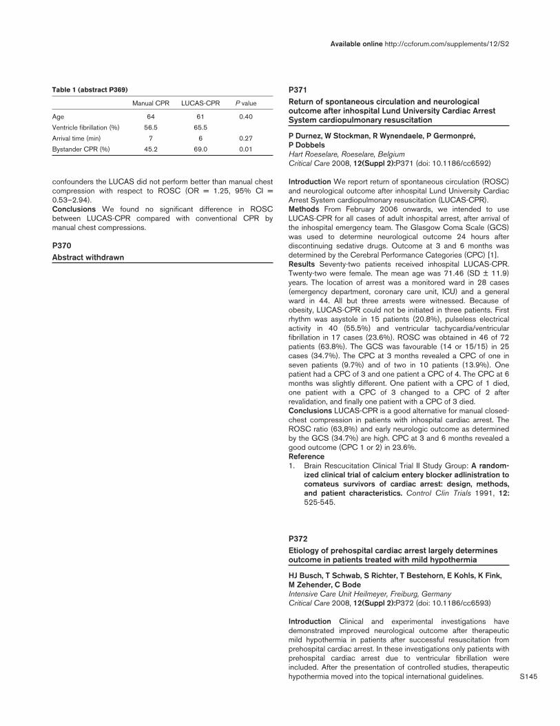
S145
confounders the LUCAS did not perform better than manual chestcompression with respect to ROSC (OR = 1.25, 95% CI =0.53–2.94).Conclusions We found no significant difference in ROSCbetween LUCAS-CPR compared with conventional CPR bymanual chest compressions.
P370Abstract withdrawn
P371Return of spontaneous circulation and neurologicaloutcome after inhospital Lund University Cardiac ArrestSystem cardiopulmonary resuscitation
P Durnez, W Stockman, R Wynendaele, P Germonpré, P DobbelsHart Roeselare, Roeselare, BelgiumCritical Care 2008, 12(Suppl 2):P371 (doi: 10.1186/cc6592)
Introduction We report return of spontaneous circulation (ROSC)and neurological outcome after inhospital Lund University CardiacArrest System cardiopulmonary resuscitation (LUCAS-CPR).Methods From February 2006 onwards, we intended to useLUCAS-CPR for all cases of adult inhospital arrest, after arrival ofthe inhospital emergency team. The Glasgow Coma Scale (GCS)was used to determine neurological outcome 24 hours afterdiscontinuing sedative drugs. Outcome at 3 and 6 months wasdetermined by the Cerebral Performance Categories (CPC) [1].Results Seventy-two patients received inhospital LUCAS-CPR.Twenty-two were female. The mean age was 71.46 (SD ± 11.9)years. The location of arrest was a monitored ward in 28 cases(emergency department, coronary care unit, ICU) and a generalward in 44. All but three arrests were witnessed. Because ofobesity, LUCAS-CPR could not be initiated in three patients. Firstrhythm was asystole in 15 patients (20.8%), pulseless electricalactivity in 40 (55.5%) and ventricular tachycardia/ventricularfibrillation in 17 cases (23.6%). ROSC was obtained in 46 of 72patients (63.8%). The GCS was favourable (14 or 15/15) in 25cases (34.7%). The CPC at 3 months revealed a CPC of one inseven patients (9.7%) and of two in 10 patients (13.9%). Onepatient had a CPC of 3 and one patient a CPC of 4. The CPC at 6months was slightly different. One patient with a CPC of 1 died,one patient with a CPC of 3 changed to a CPC of 2 afterrevalidation, and finally one patient with a CPC of 3 died.Conclusions LUCAS-CPR is a good alternative for manual closed-chest compression in patients with inhospital cardiac arrest. TheROSC ratio (63,8%) and early neurologic outcome as determinedby the GCS (34.7%) are high. CPC at 3 and 6 months revealed agood outcome (CPC 1 or 2) in 23.6%.Reference1. Brain Rescucitation Clinical Trial II Study Group: A random-
ized clinical trial of calcium entery blocker adlinistration tocomateus survivors of cardiac arrest: design, methods,and patient characteristics. Control Clin Trials 1991, 12:525-545.
P372Etiology of prehospital cardiac arrest largely determinesoutcome in patients treated with mild hypothermia
HJ Busch, T Schwab, S Richter, T Bestehorn, E Kohls, K Fink,M Zehender, C BodeIntensive Care Unit Heilmeyer, Freiburg, GermanyCritical Care 2008, 12(Suppl 2):P372 (doi: 10.1186/cc6593)
Introduction Clinical and experimental investigations havedemonstrated improved neurological outcome after therapeuticmild hypothermia in patients after successful resuscitation fromprehospital cardiac arrest. In these investigations only patients withprehospital cardiac arrest due to ventricular fibrillation wereincluded. After the presentation of controlled studies, therapeutichypothermia moved into the topical international guidelines.
Available online http://ccforum.com/supplements/12/S2
Table 1 (abstract P369)
Manual CPR LUCAS-CPR P value
Age 64 61 0.40
Ventricle fibrillation (%) 56.5 65.5
Arrival time (min) 7 6 0.27
Bystander CPR (%) 45.2 69.0 0.01
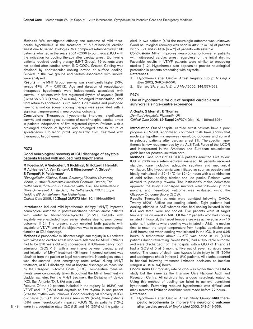
S146
Methods We investigated efficacy and outcome of mild thera-peutic hypothermia in the treatment of out-of-hospital cardiacarrest due to varied etiologies. We compared retrospectively 168patients admitted in the years 2001–2006 to our medical ICU withthe indication for cooling therapy after cardiac arrest. Eighty-ninepatients received cooling therapy (MHT Group), 79 patients werenot cooled after cardiac arrest (NO-COOL Group). Cooling wasobtained by endovascular cooling device or surface cooling.Survival in the two groups and factors associated with survivalwere analysed.Results In the MHT Group, survival was significantly higher (53%versus 47%, P = 0.0012). Age and duration of resuscitationtherapeutic hypothermia were independently associated withsurvival. In patients with first registered rhythm of asystole (8/25(32%) vs 2/13 (15%), P = 0.06), prolonged resuscitation, timefrom return to spontaneous circulation >20 minutes and prolongedtime to arrival on scene, cooling therapy was associated with asignificant improvement in neurological outcome.Conclusions Therapeutic hypothermia improves significantlysurvival and neurological outcome of out-of-hospital cardiac arrestin patients independent of first registered rhythm. Patients with aprolonged episode of hypoxia and prolonged time to return ofspontaneous circulation profit significantly from treatment withtherapeutic hypothermia.
P373Good neurological recovery at ICU discharge of asystolepatients treated with induced mild hypothermia
M Foedisch1, A Viehoefer1, N Richling2, M Holzer2, I Herold3, M Adriaanse4, A Van Zanten4, E Rijnsburger5, A Girbes5, S Tompot6, K Polderman3
1Evangelische Kliniken, Bonn, Germany; 2Medical University,Vienna, Austria; 3Universitair Medisch Centrum, Utrecht, TheNetherlands; 4Ziekenhuis Gelderse Vallei, Ede, The Netherlands;5Vrije Universiteit, Amsterdam, The Netherlands; 6KCI EuropeHolding BV, Amstelveen, The NetherlandsCritical Care 2008, 12(Suppl 2):P373 (doi: 10.1186/cc6594)
Introduction Induced mild hypothermia therapy (MHyT) improvesneurological outcome in postresuscitation cardiac arrest patientswith ventricular fibrillation/tachycardia (VF/VT). Patients withasystole were excluded from earlier studies due to poor overalloutcome [1,2]. The present study enrolled both patients withasystole or VT/VF; one of the objectives was to assess neurologicalfunction at ICU discharge.Methods A prospective multicenter single-arm registry in 49 patientswith witnessed cardiac arrest who were selected for MHyT. Patientshad to be ≥18 years old and unconscious at ICU/emergency roomadmission (GCS < 8), with a time interval between cardiac arrestand initiation of MHyT treatment <6 hours. Informed consent wasobtained from the patient or legal representative. Neurological statuswas documented upon emergency room arrival, during MHyTtreatment, at ICU discharge and at hospital discharge as measuredby the Glasgow Outcome Scale (GOS). Temperature measure-ments were continuously taken throughout the MHyT treatment viabladder catheter. For the MHyT treatment the Deltatherm® device(KCI, San Antonio, TX, USA) was used.Results Of the 49 patients included in the registry 31 (63%) hadVF/VT and 17 (35%) had asystole as first rhythm. In one patient(2%) the rhythm was unknown. Good neurological recovery at ICUdischarge (GOS 5 and 4) was seen in 22 (45%), three patients(6%) were neurologically impaired (GOS 3), six patients (12%)were in a vegetative state (GOS 2) and 16 (33%) of the patients
died. In two patients (4%) the neurologic outcome was unknown.Good neurological recovery was seen in 48% (n = 15) of patientswith VF/VT and in 41% (n = 7) of patients with asystole.Conclusions MHyT improves neurological outcome in patientswith witnessed cardiac arrest regardless of the initial rhythm.Favorable results in VT/VF patients were similar to precedingstudies [1,2]. Hypothermia also appears to provide neurologicalprotection in patients presenting with asystole.References1. Hypothermia after Cardiac Arrest Registry Group: N Engl J
Med 2002, 346:549-556.2. Bernard SA, et al.: N Engl J Med 2002, 346:557-563.
P374Use of hypothermia for out-of-hospital cardiac arrestsurvivors: a single-centre experience
A Gupta, S Morrish, E ThomasDerriford Hospitals, Plymouth, UKCritical Care 2008, 12(Suppl 2):P374 (doi: 10.1186/cc6595)
Introduction Out-of-hospital cardiac arrest patients have a poorprognosis. Recent randomised controlled trials have shown thatmoderate hypothermia improves neurologic outcome and survivalin selected patients after cardiac arrest [1]. Therapeutic hypo-thermia is now recommended by the ALS Task Force of the ILCORand incorporated in the American and European resuscitationguidelines for postresuscitation care.Methods Case notes of all OHCA patients admitted alive to ourICU in 2006 were retrospectively analysed. All patients receivedstandard care including adequate sedation and mechanicalventilation. Mild hypothermia was initiated as soon as possible andideally maintained at 32–34°C for 12–24 hours with a combinationof cold saline, cooling blanket and ice packs. Patients wereallowed to passively rewarm. The institution’s ethics committeeapproved the study. Discharged survivors were followed up for 6months, and neurologic outcome was evaluated using theGlasgow Outcome Score (GOS).Results Twenty-five patients were admitted following OHCA.Twenty (80%) fulfilled our cooling criteria. Eight patients hadcooling initiated in A&E whereas nine had cooling initiated in theICU and three were not cooled. Five patients were at targettemperature on arrival in A&E. Of the 17 patients who had coolinginitiated in hospital, the target temperature was achieved in only 15patients. In patients where cooling was initiated in A&E, the mediantime to reach the target temperature from hospital admission was4.25 hours; and when cooling was initiated in the ICU, it was 6.25hours. A temperature above 37.5°C was noted in 12 (48%)patients during rewarming. Seven (28%) had a favourable outcomeand were discharged from the hospital with a GCS of 15 and allhad a GOS of 5 at 6 months. Five out of seven survivors werecooled. The cause of death was hypoxic brain injury in 15 (60%)and cardiogenic shock in three (12%) patients. All deaths occurredin hospital following treatment limitation decisions at (median(range)) 41 (9.5–94) hours.Conclusions Our mortality rate of 72% was higher than the HACAstudy but the same as the Intensive Care National Audit andResearch Centre. All survivors had a good neurologic outcome.Using this method of cooling we failed to achieve consistenthypothermia. Preventing rebound hyperthermia was difficult andmany treatment limitation decisions were made before 72 hours.Reference1. Hypothermia after Cardiac Arrest Study Group: Mild thera-
peutic hypothermia to improve the neurologic outcomeafter cardiac arrest. N Engl J Med 2002, 346:549-556.
Critical Care March 2008 Vol 12 Suppl 2 28th International Symposium on Intensive Care and Emergency Medicine
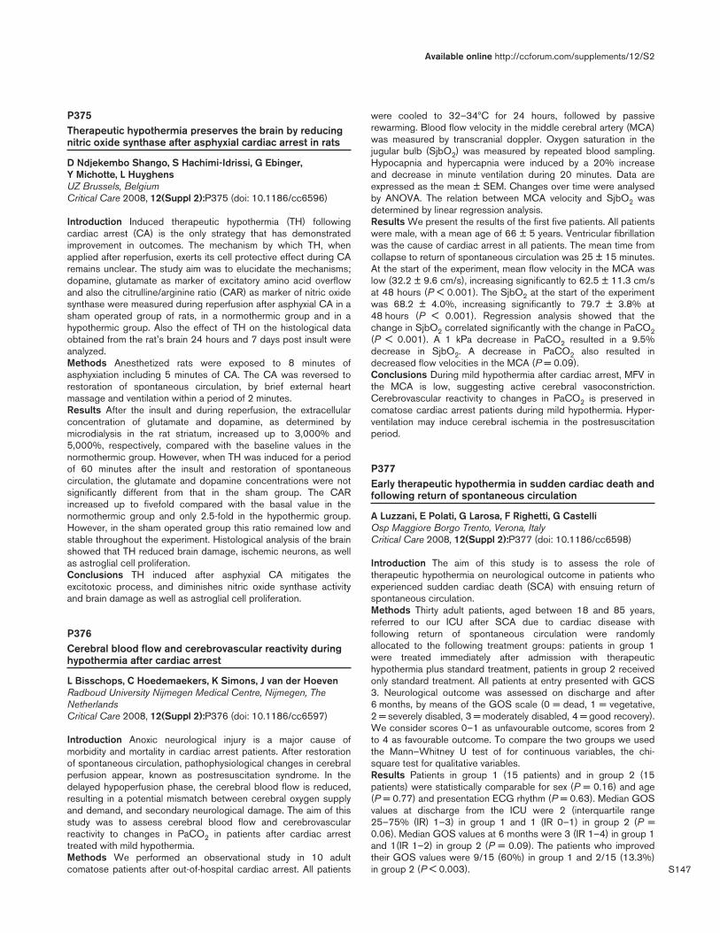
S147
P375Therapeutic hypothermia preserves the brain by reducingnitric oxide synthase after asphyxial cardiac arrest in rats
D Ndjekembo Shango, S Hachimi-Idrissi, G Ebinger, Y Michotte, L HuyghensUZ Brussels, BelgiumCritical Care 2008, 12(Suppl 2):P375 (doi: 10.1186/cc6596)
Introduction Induced therapeutic hypothermia (TH) followingcardiac arrest (CA) is the only strategy that has demonstratedimprovement in outcomes. The mechanism by which TH, whenapplied after reperfusion, exerts its cell protective effect during CAremains unclear. The study aim was to elucidate the mechanisms;dopamine, glutamate as marker of excitatory amino acid overflowand also the citrulline/arginine ratio (CAR) as marker of nitric oxidesynthase were measured during reperfusion after asphyxial CA in asham operated group of rats, in a normothermic group and in ahypothermic group. Also the effect of TH on the histological dataobtained from the rat’s brain 24 hours and 7 days post insult wereanalyzed.Methods Anesthetized rats were exposed to 8 minutes ofasphyxiation including 5 minutes of CA. The CA was reversed torestoration of spontaneous circulation, by brief external heartmassage and ventilation within a period of 2 minutes.Results After the insult and during reperfusion, the extracellularconcentration of glutamate and dopamine, as determined bymicrodialysis in the rat striatum, increased up to 3,000% and5,000%, respectively, compared with the baseline values in thenormothermic group. However, when TH was induced for a periodof 60 minutes after the insult and restoration of spontaneouscirculation, the glutamate and dopamine concentrations were notsignificantly different from that in the sham group. The CARincreased up to fivefold compared with the basal value in thenormothermic group and only 2.5-fold in the hypothermic group.However, in the sham operated group this ratio remained low andstable throughout the experiment. Histological analysis of the brainshowed that TH reduced brain damage, ischemic neurons, as wellas astroglial cell proliferation.Conclusions TH induced after asphyxial CA mitigates theexcitotoxic process, and diminishes nitric oxide synthase activityand brain damage as well as astroglial cell proliferation.
P376Cerebral blood flow and cerebrovascular reactivity duringhypothermia after cardiac arrest
L Bisschops, C Hoedemaekers, K Simons, J van der HoevenRadboud University Nijmegen Medical Centre, Nijmegen, TheNetherlandsCritical Care 2008, 12(Suppl 2):P376 (doi: 10.1186/cc6597)
Introduction Anoxic neurological injury is a major cause ofmorbidity and mortality in cardiac arrest patients. After restorationof spontaneous circulation, pathophysiological changes in cerebralperfusion appear, known as postresuscitation syndrome. In thedelayed hypoperfusion phase, the cerebral blood flow is reduced,resulting in a potential mismatch between cerebral oxygen supplyand demand, and secondary neurological damage. The aim of thisstudy was to assess cerebral blood flow and cerebrovascularreactivity to changes in PaCO2 in patients after cardiac arresttreated with mild hypothermia.Methods We performed an observational study in 10 adultcomatose patients after out-of-hospital cardiac arrest. All patients
were cooled to 32–34ºC for 24 hours, followed by passiverewarming. Blood flow velocity in the middle cerebral artery (MCA)was measured by transcranial doppler. Oxygen saturation in thejugular bulb (SjbO2) was measured by repeated blood sampling.Hypocapnia and hypercapnia were induced by a 20% increaseand decrease in minute ventilation during 20 minutes. Data areexpressed as the mean ± SEM. Changes over time were analysedby ANOVA. The relation between MCA velocity and SjbO2 wasdetermined by linear regression analysis.Results We present the results of the first five patients. All patientswere male, with a mean age of 66 ± 5 years. Ventricular fibrillationwas the cause of cardiac arrest in all patients. The mean time fromcollapse to return of spontaneous circulation was 25 ± 15 minutes.At the start of the experiment, mean flow velocity in the MCA waslow (32.2 ± 9.6 cm/s), increasing significantly to 62.5 ± 11.3 cm/sat 48 hours (P < 0.001). The SjbO2 at the start of the experimentwas 68.2 ± 4.0%, increasing significantly to 79.7 ± 3.8% at48 hours (P < 0.001). Regression analysis showed that thechange in SjbO2 correlated significantly with the change in PaCO2(P < 0.001). A 1 kPa decrease in PaCO2 resulted in a 9.5%decrease in SjbO2. A decrease in PaCO2 also resulted indecreased flow velocities in the MCA (P = 0.09).Conclusions During mild hypothermia after cardiac arrest, MFV inthe MCA is low, suggesting active cerebral vasoconstriction.Cerebrovascular reactivity to changes in PaCO2 is preserved incomatose cardiac arrest patients during mild hypothermia. Hyper-ventilation may induce cerebral ischemia in the postresuscitationperiod.
P377Early therapeutic hypothermia in sudden cardiac death andfollowing return of spontaneous circulation
A Luzzani, E Polati, G Larosa, F Righetti, G CastelliOsp Maggiore Borgo Trento, Verona, ItalyCritical Care 2008, 12(Suppl 2):P377 (doi: 10.1186/cc6598)
Introduction The aim of this study is to assess the role oftherapeutic hypothermia on neurological outcome in patients whoexperienced sudden cardiac death (SCA) with ensuing return ofspontaneous circulation.Methods Thirty adult patients, aged between 18 and 85 years,referred to our ICU after SCA due to cardiac disease withfollowing return of spontaneous circulation were randomlyallocated to the following treatment groups: patients in group 1were treated immediately after admission with therapeutichypothermia plus standard treatment, patients in group 2 receivedonly standard treatment. All patients at entry presented with GCS3. Neurological outcome was assessed on discharge and after6 months, by means of the GOS scale (0 = dead, 1 = vegetative,2 = severely disabled, 3 = moderately disabled, 4 = good recovery).We consider scores 0–1 as unfavourable outcome, scores from 2to 4 as favourable outcome. To compare the two groups we usedthe Mann–Whitney U test of for continuous variables, the chi-square test for qualitative variables.Results Patients in group 1 (15 patients) and in group 2 (15patients) were statistically comparable for sex (P = 0.16) and age(P = 0.77) and presentation ECG rhythm (P = 0.63). Median GOSvalues at discharge from the ICU were 2 (interquartile range25–75% (IR) 1–3) in group 1 and 1 (IR 0–1) in group 2 (P =0.06). Median GOS values at 6 months were 3 (IR 1–4) in group 1and 1(IR 1–2) in group 2 (P = 0.09). The patients who improvedtheir GOS values were 9/15 (60%) in group 1 and 2/15 (13.3%)in group 2 (P < 0.003).
Available online http://ccforum.com/supplements/12/S2
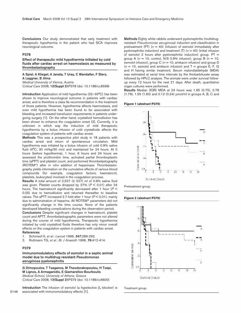
S148
Conclusions Our study demonstrated that early treatment withtherapeutic hypothermia in the patient who had SCA improvesneurological outcome.
P378Effect of therapeutic mild hypothermia initiated by coldfluids after cardiac arrest on haemostasis as measured bythrombelastography
A Spiel, A Kliegel, A Janata, T Uray, C Wandaller, F Sterz, A Laggner, B JilmaMedical University of Vienna, AustriaCritical Care 2008, 12(Suppl 2):P378 (doi: 10.1186/cc6599)
Introduction Application of mild hypothermia (32–33°C) has beenshown to improve neurological outcome in patients with cardiacarrest, and is therefore a class IIa recommendation in the treatmentof those patients. However, hypothermia affects haemostasis, andeven mild hypothermia has been found to be associated withbleeding and increased transfusion requirements in patients under-going surgery [1]. On the other hand, crystalloid hemodilution hasbeen shown to enhance the coagulation onset [2]. Currently, it isunknown in which way the induction of mild therapeutichypothermia by a bolus infusion of cold crystalloids affects thecoagulation system of patients with cardiac arrest.Methods This was a prospective pilot study in 18 patients withcardiac arrest and return of spontaneous circulation. Mildhypothermia was initiated by a bolus infusion of cold 0.9% salinefluid (4°C; 30 ml/kg/30 min) and maintained for 24 hours. At 0hours (before hypothermia), 1 hour, 6 hours and 24 hours weassessed the prothrombin time, activated partial thromboplastintime (aPPT) and platelet count, and performed thrombelastography(ROTEM®) after in vitro addition of heparinase. Thrombelasto-graphy yields information on the cumulative effects of various bloodcompounds (for example, coagulation factors, haematocrit,platelets, leukocytes) involved in the coagulation process.Results A total amount of 2,527 (± 527) ml of 0.9% saline fluidwas given. Platelet counts dropped by 27% (P < 0.01) after 24hours. The haematocrit significantly decreased after 1 hour (P <0.05) due to hemodilution and returned thereafter to baselinevalues. The aPTT increased 2.7-fold after 1 hour (P < 0.01), mainlydue to administration of heparins. All ROTEM® parameters did notsignificantly change in the time course. None of the patientsdeveloped bleeding complications during the observation period.Conclusions Despite significant changes in haematocrit, plateletcount and APTT, thrombelastographic parameters were not alteredduring the course of mild hypothermia. Therapeutic hypothermiainitiated by cold crystalloid fluids therefore has only minor overalleffects on the coagulation system in patients with cardiac arrest.References1. Schmied H, et al.: Lancet 1996, 347:289-292.2. Ruttmann TG, et al.: Br J Anaesth 1996, 76:412-414.
P379Immunomodulatory effects of esmolol in a septic animalmodel due to multidrug-resistant Pseudomonasaeruginosa pyelonephritis
G Dimopoulos, T Tsaganos, M Theodorakopoulou, H Tzepi, M Lignos, A Armaganidis, E Giamarellos-BourboulisMedical School, University of Athens, GreeceCritical Care 2008, 12(Suppl 2):P379 (doi: 10.1186/cc6600)
Introduction The infusion of esmolol (a hypelective β1-blocker) isassociated with immunomodulatory effects [1].
Methods Eighty white rabbits underwent pyelonephritis (multidrug-resistant Pseudomonas aeruginosa) induction and classification inpretreatment (PT) (n = 40) (infusion of esmolol immediately afterpyelonephritis induction) and treatment (T) (n = 40) (initial infusionof esmolol 2 hours after pyelonephritis induction) group. PT =group A (n = 10, control, N/S 0.9% infusion), group B (n = 10,esmolol infusion), group C (n = 10, amikacin infusion) and group D(n = 10, esmolol and amikacin infusion) and T = groups E, F, Gand H having similar treatment. Serum malondialdehyde (MDA)was estimated at serial time intervals by the thiobarbiturate assayfollowed by HPLC analysis. The animals were under survival follow-up every 12 hours for the next 21 days. After death, quantitativeorgan cultures were performed.Results Median (IQR) MDA at 24 hours was 1.95 (0.75), 0.78(1.79), 1.55 (1.60) and 0.12 (0.24) μmol/ml in groups A, B, C and
Critical Care March 2008 Vol 12 Suppl 2 28th International Symposium on Intensive Care and Emergency Medicine
Figure 1 (abstract P379)
Pretreatment group.
Figure 2 (abstract P379)
Treatment group.
S149
D, respectively. Respective values at 48 hours were 2.60 (2.00),1.40 (2.36), 3.15 (3.00) and 0.25 (0.20) μmol/ml. At 24 hours, themedian (IQR) MDA of groups E, F, G and H were 2.80 (5.74), 0.32(0.87), 0.61 (5.83) and 0.19 (2.75) μmol/ml, respectively. Tissuebacterial load was similar within groups. See Figures 1 and 2.Conclusions In the present septic model, esmolol prolongedsurvival probably by exerting an immunomodulatory effect asassessed by reduced oxidative stress without any effect on tissuebacterial load.Reference1. Suzuki T, et al.: Crit Care Med 2005, 33:2294-2300.
P380Applying the 2003 SCCM/ESICM/ACCP/ATS/SIS insteadof the 1992 ACCP/SCCM sepsis definitions increases thenumbers of patients with systemic inflammatory responsesyndrome shock and septic shock but decreases mortalityrates
M Weiss, M Taenzer, K Traeger, J Altherr, B Hay, M Kron, M Huber-Lang, M SchneiderUniversity Hospital, Ulm, GermanyCritical Care 2008, 12(Suppl 2):P380 (doi: 10.1186/cc6601)
Introduction To compare the prevalence of patients suffering fromdifferent stages of systemic inflammatory response syndrome (SIRS)and sepsis applying the original 1992 ACCP/SCCM and the revised2003 SCCM/ESICM/ACCP/ATS/SIS sepsis definitions.Methods Set in a university adult ICU. Patients werepostoperative/post-traumatic critically ill patients admitted to theICU from October 2006 to October 2007. No interventions wereused. From October 2006 to October 2007, 714 patients weresurveyed using computer assistance with respect to differentstages of SIRS and sepsis using the 1992 and the 2003 sepsisdefinitions, respectively.Results Within the same patient collective, applying the 2003definitions instead of the 1992 definitions, the prevalence of noSIRS (11 vs 110, respectively), no SIRS due to infection (sepsis)(0 vs 12), SIRS (129 vs 169) and sepsis (18 vs 52) decreased,and the prevalence of severe SIRS (169 vs 86), SIRS shock (121vs 65) and septic shock (216 vs 168) increased. Prevalence ofsevere sepsis was comparable with both definitions (50 vs 52).Applying the 2003 definitions in patients with SIRS shock andseptic shock, the mortality rates of 17% and 25% were markedlylower than those of 23% and 30%, respectively, under the 1992definitions. Compared with the patients classified to be withoutSIRS shock and septic shock, the risk of mortality of those patientswas markedly elevated that were classified to be in SIRS shock orseptic shock with the 2003 definitions but not with the 1992definitions (odds ratio = 5.0, CI = 2.2–11.2, P < 0.0001).Conclusions Replacing the original 1992 sepsis definitions withthe 2003 revised sepsis definitions may result in increasedprevalence of severe SIRS, SIRS shock and septic shock.However, the mortality rates of patients with SIRS shock andseptic shock will be lower.References1. American College of Chest Physicians/Society of Critical Care
Medicine Consensus Conference: Definitions for sepsis andorgan failure and guidelines for the use of innovative ther-apies in sepsis. Crit Care Med 1992, 20:864-874.
2. Levy MM, Fink MP, Marshall JC, et al.: 2001 SCCM/ESICM/ACCP/ATS/SIS International Sepsis Definitions Confer-ence. Intensive Care Med 2003, 29:530-538.
P381Systemic inflammatory response syndrome as a clinicaldetection tool and inclusion criterion in sepsis trials: tooblunt an instrument?
P Gille-Johnson, K Hansson, B GårdlundKarolinska Hospital, Stockholm, SwedenCritical Care 2008, 12(Suppl 2):P381 (doi: 10.1186/cc6602)
Introduction The term systemic inflammatory response syndrome(SIRS) was introduced in 1992 [1] and has frequently served as acriterion for enrollment in sepsis trials. In the present study, theprevalence of SIRS in patients with significant bacterial infectionsand in patients with septic shock was assessed.Methods A cohort of 404 adult patients admitted to theDepartment of Infectious Diseases from the emergency room (ER)for suspected severe infection was studied prospectively. Of theSIRS variables, white blood cells (WBC) were measured on arrivalwhile the physiological variables (temperature, heart rate (HR) andrespiratory rate (RR)) were recorded on arrival to the ER and every4 hours for 24 hours. In another cohort of 36 consecutive adultswith vasopressor-dependent septic shock, the presence of SIRScriteria during 24 hours around the start of vasopressors wasevaluated.Results Bacterial infections requiring antibiotic treatment werediagnosed in 306 patients in the ER cohort. Nonbacterial infectionor noninfection was diagnosed in 82 patients. In 16 patients, nodiagnosis could be verified. Significant bacteremia was detected in68 patients; the most common pathogens were pneumococci andEscherichia coli. Of the 306 patients with a verified bacterialinfection and of the 68 with verified bacteremia, 26% and 21%,respectively, failed to meet two or more of the SIRS criteria onarrival in the ER. SIRS on arrival correlated significantly withbacterial infection, but not with bacteremia. Only RR and WBCcontributed significantly to this finding, HR and temperature did not.In the septic shock group, all patients eventually fulfilled the HRand RR criteria but only 23/36 (64%) reached the temperaturecriterion and 25/36 (69%) the WBC criterion during 24 hours.Conclusions SIRS correlated with a subsequently verifiedbacterial infection requiring antibiotic treatment, but only the RRand WBC criteria contributed to this finding. As a tool for definingsepsis and selecting patients for enrollment in clinical sepsis trials,SIRS is nonspecific, and for ≥3 fulfilled criteria it lacks sensitivity. Itmay be time to abandon the SIRS criteria in selecting patients forsepsis trials and instead focus on more strict definitions ofunderlying infections in association with sepsis-relatedhypoperfusion and organ dysfunction.Reference1. Bone RC, et al.: Chest 1992, 101:1644-1655.
P382Pharmacokinetic–pharmacodynamic analysis of humanpurified C1-esterase inhibitor in patients with sepsis
L Dolzhenkova, N Lazareva, A IgoninSechenov Medical Academy, Moscow, Russian FederationCritical Care 2008, 12(Suppl 2):P382 (doi: 10.1186/cc6603)
Introduction Several randomized prospective studies showed somebeneficial protective effects of exogenous human purified C1-esterase inhibitor (C1INH) in patients with sepsis [1,2]. Our purposewas to evaluate influence of systemic inflammation on the pharmaco-kinetic–pharmacodynamic of C1INH in patients with sepsis.Methods C1INH (Bicizar®; BioGenius LLC, Russia) wasadministered at the total dosage of 12,000 U in 48 hours (scheme
Available online http://ccforum.com/supplements/12/S2
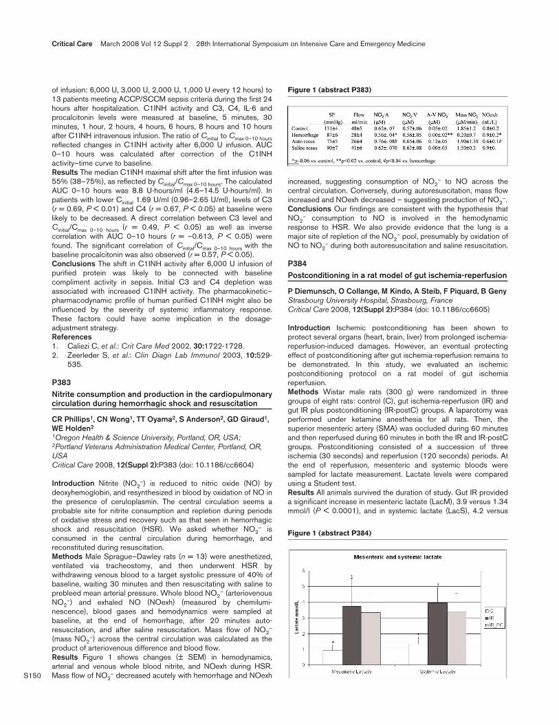
S150
of infusion: 6,000 U, 3,000 U, 2,000 U, 1,000 U every 12 hours) to13 patients meeting ACCP/SCCM sepsis criteria during the first 24hours after hospitalization. C1INH activity and C3, C4, IL-6 andprocalcitonin levels were measured at baseline, 5 minutes, 30minutes, 1 hour, 2 hours, 4 hours, 6 hours, 8 hours and 10 hoursafter C1INH intravenous infusion. The ratio of Cinitial to Cmax 0–10 hoursreflected changes in C1INH activity after 6,000 U infusion. AUC0–10 hours was calculated after correction of the C1INHactivity–time curve to baseline.Results The median C1INH maximal shift after the first infusion was55% (38–75%), as reflected by Cinitial/Cmax 0–10 hours. The calculatedAUC 0–10 hours was 8.8 U-hours/ml (4.6–14.5 U-hours/ml). Inpatients with lower Cinitial 1.69 U/ml (0.96–2.65 U/ml), levels of C3(r = 0.69, P < 0.01) and C4 (r = 0.67, P < 0.05) at baseline werelikely to be decreased. A direct correlation between C3 level andCinitial/Cmax 0–10 hours (r = 0.49, P < 0.05) as well as inversecorrelation with AUC 0–10 hours (r = –0.613, P < 0.05) werefound. The significant correlation of Cinitial/Cmax 0–10 hours with thebaseline procalcitonin was also observed (r = 0.57, P < 0.05).Conclusions The shift in C1INH activity after 6,000 U infusion ofpurified protein was likely to be connected with baselinecompliment activity in sepsis. Initial C3 and C4 depletion wasassociated with increased C1INH activity. The pharmacokinetic–pharmacodynamic profile of human purified C1INH might also beinfluenced by the severity of systemic inflammatory response.These factors could have some implication in the dosage-adjustment strategy.References1. Caliezi C, et al.: Crit Care Med 2002, 30:1722-1728.2. Zeerleder S, et al.: Clin Diagn Lab Immunol 2003, 10:529-
535.
P383Nitrite consumption and production in the cardiopulmonarycirculation during hemorrhagic shock and resuscitation
CR Phillips1, CN Wong1, TT Oyama2, S Anderson2, GD Giraud1,WE Holden2
1Oregon Health & Science University, Portland, OR, USA;2Portland Veterans Administration Medical Center, Portland, OR,USACritical Care 2008, 12(Suppl 2):P383 (doi: 10.1186/cc6604)
Introduction Nitrite (NO2–) is reduced to nitric oxide (NO) by
deoxyhemoglobin, and resynthesized in blood by oxidation of NO inthe presence of ceruloplasmin. The central circulation seems aprobable site for nitrite consumption and repletion during periodsof oxidative stress and recovery such as that seen in hemorrhagicshock and resuscitation (HSR). We asked whether NO2
– isconsumed in the central circulation during hemorrhage, andreconstituted during resuscitation.Methods Male Sprague–Dawley rats (n = 13) were anesthetized,ventilated via tracheostomy, and then underwent HSR bywithdrawing venous blood to a target systolic pressure of 40% ofbaseline, waiting 30 minutes and then resuscitating with saline toprebleed mean arterial pressure. Whole blood NO2
– (arteriovenousNO2
–) and exhaled NO (NOexh) (measured by chemilumi-nescence), blood gases and hemodynamics were sampled atbaseline, at the end of hemorrhage, after 20 minutes auto-resuscitation, and after saline resuscitation. Mass flow of NO2
–
(mass NO2–) across the central circulation was calculated as the
product of arteriovenous difference and blood flow.Results Figure 1 shows changes (± SEM) in hemodynamics,arterial and venous whole blood nitrite, and NOexh during HSR.Mass flow of NO2
– decreased acutely with hemorrhage and NOexh
increased, suggesting consumption of NO2– to NO across the
central circulation. Conversely, during autoresuscitation, mass flowincreased and NOexh decreased – suggesting production of NO2
–.Conclusions Our findings are consistent with the hypothesis thatNO2
– consumption to NO is involved in the hemodynamicresponse to HSR. We also provide evidence that the lung is amajor site of repletion of the NO2
– pool, presumably by oxidation ofNO to NO2
– during both autoresuscitation and saline resuscitation.
P384Postconditioning in a rat model of gut ischemia-reperfusion
P Diemunsch, O Collange, M Kindo, A Steib, F Piquard, B GenyStrasbourg University Hospital, Strasbourg, FranceCritical Care 2008, 12(Suppl 2):P384 (doi: 10.1186/cc6605)
Introduction Ischemic postconditioning has been shown toprotect several organs (heart, brain, liver) from prolonged ischemia-reperfusion-induced damages. However, an eventual protectingeffect of postconditioning after gut ischemia-reperfusion remains tobe demonstrated. In this study, we evaluated an ischemicpostconditioning protocol on a rat model of gut ischemiareperfusion.Methods Wistar male rats (300 g) were randomized in threegroups of eight rats: control (C), gut ischemia-reperfusion (IR) andgut IR plus postconditioning (IR-postC) groups. A laparotomy wasperformed under ketamine anesthesia for all rats. Then, thesuperior mesenteric artery (SMA) was occluded during 60 minutesand then reperfused during 60 minutes in both the IR and IR-postCgroups. Postconditioning consisted of a succession of threeischemia (30 seconds) and reperfusion (120 seconds) periods. Atthe end of reperfusion, mesenteric and systemic bloods weresampled for lactate measurement. Lactate levels were comparedusing a Student test.Results All animals survived the duration of study. Gut IR provideda significant increase in mesenteric lactate (LacM), 3.9 versus 1.34mmol/l (P < 0.0001), and in systemic lactate (LacS), 4.2 versus
Critical Care March 2008 Vol 12 Suppl 2 28th International Symposium on Intensive Care and Emergency Medicine
Figure 1 (abstract P383)
Figure 1 (abstract P384)
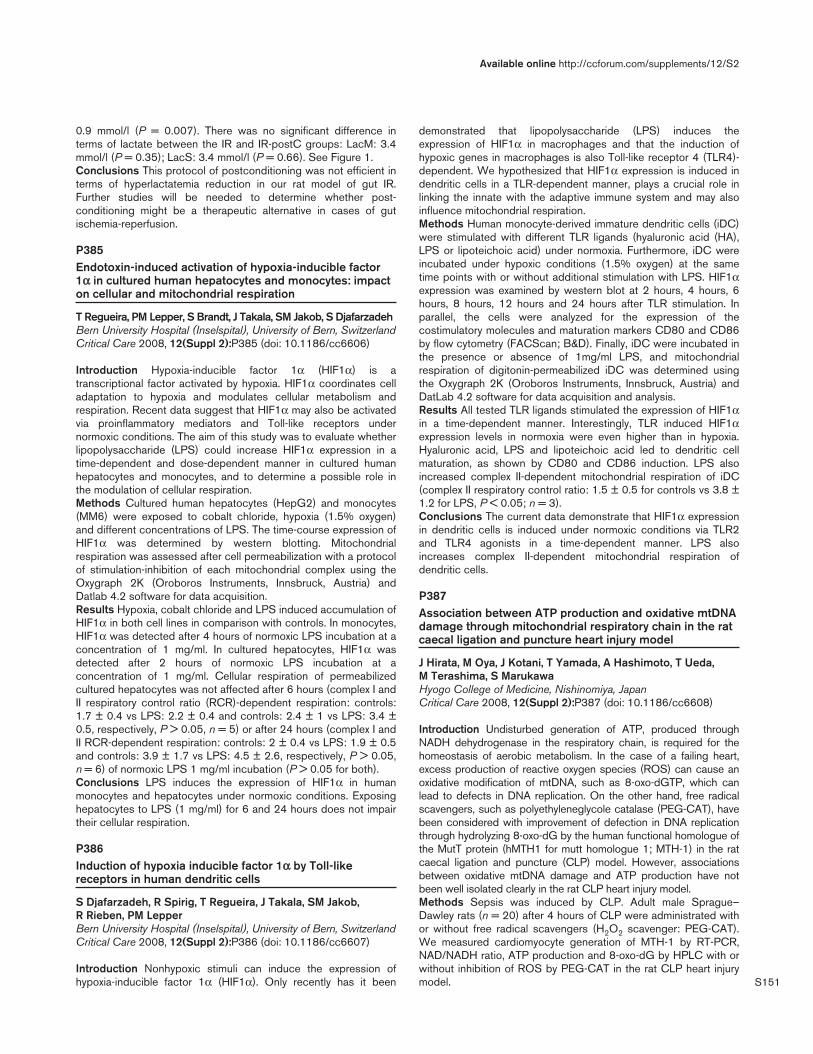
S151
0.9 mmol/l (P = 0.007). There was no significant difference interms of lactate between the IR and IR-postC groups: LacM: 3.4mmol/l (P = 0.35); LacS: 3.4 mmol/l (P = 0.66). See Figure 1.Conclusions This protocol of postconditioning was not efficient interms of hyperlactatemia reduction in our rat model of gut IR.Further studies will be needed to determine whether post-conditioning might be a therapeutic alternative in cases of gutischemia-reperfusion.
P385Endotoxin-induced activation of hypoxia-inducible factor1αα in cultured human hepatocytes and monocytes: impacton cellular and mitochondrial respiration
T Regueira, PM Lepper, S Brandt, J Takala, SM Jakob, S DjafarzadehBern University Hospital (Inselspital), University of Bern, SwitzerlandCritical Care 2008, 12(Suppl 2):P385 (doi: 10.1186/cc6606)
Introduction Hypoxia-inducible factor 1α (HIF1α) is atranscriptional factor activated by hypoxia. HIF1α coordinates celladaptation to hypoxia and modulates cellular metabolism andrespiration. Recent data suggest that HIF1α may also be activatedvia proinflammatory mediators and Toll-like receptors undernormoxic conditions. The aim of this study was to evaluate whetherlipopolysaccharide (LPS) could increase HIF1α expression in atime-dependent and dose-dependent manner in cultured humanhepatocytes and monocytes, and to determine a possible role inthe modulation of cellular respiration.Methods Cultured human hepatocytes (HepG2) and monocytes(MM6) were exposed to cobalt chloride, hypoxia (1.5% oxygen)and different concentrations of LPS. The time-course expression ofHIF1α was determined by western blotting. Mitochondrialrespiration was assessed after cell permeabilization with a protocolof stimulation-inhibition of each mitochondrial complex using theOxygraph 2K (Oroboros Instruments, Innsbruck, Austria) andDatlab 4.2 software for data acquisition.Results Hypoxia, cobalt chloride and LPS induced accumulation ofHIF1α in both cell lines in comparison with controls. In monocytes,HIF1α was detected after 4 hours of normoxic LPS incubation at aconcentration of 1 mg/ml. In cultured hepatocytes, HIF1α wasdetected after 2 hours of normoxic LPS incubation at aconcentration of 1 mg/ml. Cellular respiration of permeabilizedcultured hepatocytes was not affected after 6 hours (complex I andII respiratory control ratio (RCR)-dependent respiration: controls:1.7 ± 0.4 vs LPS: 2.2 ± 0.4 and controls: 2.4 ± 1 vs LPS: 3.4 ±0.5, respectively, P > 0.05, n = 5) or after 24 hours (complex I andII RCR-dependent respiration: controls: 2 ± 0.4 vs LPS: 1.9 ± 0.5and controls: 3.9 ± 1.7 vs LPS: 4.5 ± 2.6, respectively, P > 0.05,n = 6) of normoxic LPS 1 mg/ml incubation (P > 0.05 for both).Conclusions LPS induces the expression of HIF1α in humanmonocytes and hepatocytes under normoxic conditions. Exposinghepatocytes to LPS (1 mg/ml) for 6 and 24 hours does not impairtheir cellular respiration.
P386Induction of hypoxia inducible factor 1αα by Toll-likereceptors in human dendritic cells
S Djafarzadeh, R Spirig, T Regueira, J Takala, SM Jakob, R Rieben, PM LepperBern University Hospital (Inselspital), University of Bern, SwitzerlandCritical Care 2008, 12(Suppl 2):P386 (doi: 10.1186/cc6607)
Introduction Nonhypoxic stimuli can induce the expression ofhypoxia-inducible factor 1α (HIF1α). Only recently has it been
demonstrated that lipopolysaccharide (LPS) induces theexpression of HIF1α in macrophages and that the induction ofhypoxic genes in macrophages is also Toll-like receptor 4 (TLR4)-dependent. We hypothesized that HIF1α expression is induced indendritic cells in a TLR-dependent manner, plays a crucial role inlinking the innate with the adaptive immune system and may alsoinfluence mitochondrial respiration.Methods Human monocyte-derived immature dendritic cells (iDC)were stimulated with different TLR ligands (hyaluronic acid (HA),LPS or lipoteichoic acid) under normoxia. Furthermore, iDC wereincubated under hypoxic conditions (1.5% oxygen) at the sametime points with or without additional stimulation with LPS. HIF1αexpression was examined by western blot at 2 hours, 4 hours, 6hours, 8 hours, 12 hours and 24 hours after TLR stimulation. Inparallel, the cells were analyzed for the expression of thecostimulatory molecules and maturation markers CD80 and CD86by flow cytometry (FACScan; B&D). Finally, iDC were incubated inthe presence or absence of 1mg/ml LPS, and mitochondrialrespiration of digitonin-permeabilized iDC was determined usingthe Oxygraph 2K (Oroboros Instruments, Innsbruck, Austria) andDatLab 4.2 software for data acquisition and analysis.Results All tested TLR ligands stimulated the expression of HIF1αin a time-dependent manner. Interestingly, TLR induced HIF1αexpression levels in normoxia were even higher than in hypoxia.Hyaluronic acid, LPS and lipoteichoic acid led to dendritic cellmaturation, as shown by CD80 and CD86 induction. LPS alsoincreased complex II-dependent mitochondrial respiration of iDC(complex II respiratory control ratio: 1.5 ± 0.5 for controls vs 3.8 ±1.2 for LPS, P < 0.05; n = 3).Conclusions The current data demonstrate that HIF1α expressionin dendritic cells is induced under normoxic conditions via TLR2and TLR4 agonists in a time-dependent manner. LPS alsoincreases complex II-dependent mitochondrial respiration ofdendritic cells.
P387Association between ATP production and oxidative mtDNAdamage through mitochondrial respiratory chain in the ratcaecal ligation and puncture heart injury model
J Hirata, M Oya, J Kotani, T Yamada, A Hashimoto, T Ueda, M Terashima, S MarukawaHyogo College of Medicine, Nishinomiya, JapanCritical Care 2008, 12(Suppl 2):P387 (doi: 10.1186/cc6608)
Introduction Undisturbed generation of ATP, produced throughNADH dehydrogenase in the respiratory chain, is required for thehomeostasis of aerobic metabolism. In the case of a failing heart,excess production of reactive oxygen species (ROS) can cause anoxidative modification of mtDNA, such as 8-oxo-dGTP, which canlead to defects in DNA replication. On the other hand, free radicalscavengers, such as polyethyleneglycole catalase (PEG-CAT), havebeen considered with improvement of defection in DNA replicationthrough hydrolyzing 8-oxo-dG by the human functional homologue ofthe MutT protein (hMTH1 for mutt homologue 1; MTH-1) in the ratcaecal ligation and puncture (CLP) model. However, associationsbetween oxidative mtDNA damage and ATP production have notbeen well isolated clearly in the rat CLP heart injury model.Methods Sepsis was induced by CLP. Adult male Sprague–Dawley rats (n = 20) after 4 hours of CLP were administrated withor without free radical scavengers (H2O2 scavenger: PEG-CAT).We measured cardiomyocyte generation of MTH-1 by RT-PCR,NAD/NADH ratio, ATP production and 8-oxo-dG by HPLC with orwithout inhibition of ROS by PEG-CAT in the rat CLP heart injurymodel.
Available online http://ccforum.com/supplements/12/S2
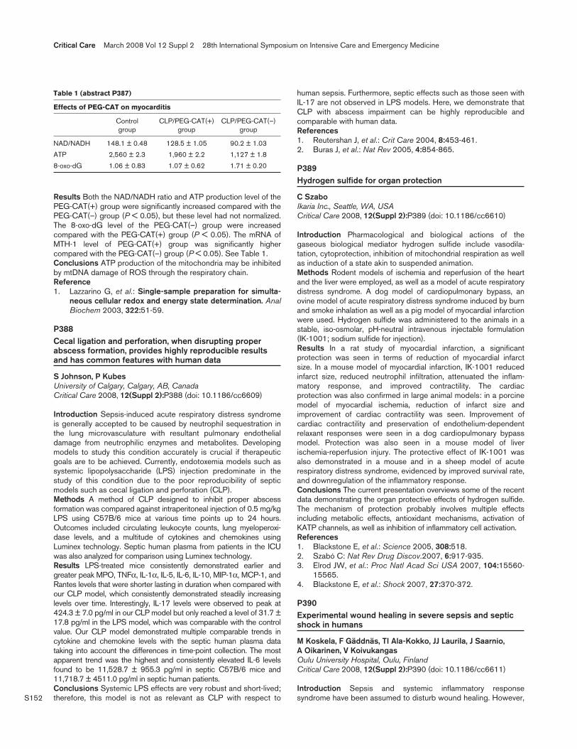
S152
Results Both the NAD/NADH ratio and ATP production level of thePEG-CAT(+) group were significantly increased compared with thePEG-CAT(–) group (P < 0.05), but these level had not normalized.The 8-oxo-dG level of the PEG-CAT(–) group were increasedcompared with the PEG-CAT(+) group (P < 0.05). The mRNA ofMTH-1 level of PEG-CAT(+) group was significantly highercompared with the PEG-CAT(–) group (P < 0.05). See Table 1.Conclusions ATP production of the mitochondria may be inhibitedby mtDNA damage of ROS through the respiratory chain.Reference1. Lazzarino G, et al.: Single-sample preparation for simulta-
neous cellular redox and energy state determination. AnalBiochem 2003, 322:51-59.
P388Cecal ligation and perforation, when disrupting properabscess formation, provides highly reproducible resultsand has common features with human data
S Johnson, P KubesUniversity of Calgary, Calgary, AB, CanadaCritical Care 2008, 12(Suppl 2):P388 (doi: 10.1186/cc6609)
Introduction Sepsis-induced acute respiratory distress syndromeis generally accepted to be caused by neutrophil sequestration inthe lung microvasculature with resultant pulmonary endothelialdamage from neutrophilic enzymes and metabolites. Developingmodels to study this condition accurately is crucial if therapeuticgoals are to be achieved. Currently, endotoxemia models such assystemic lipopolysaccharide (LPS) injection predominate in thestudy of this condition due to the poor reproducibility of septicmodels such as cecal ligation and perforation (CLP).Methods A method of CLP designed to inhibit proper abscessformation was compared against intraperitoneal injection of 0.5 mg/kgLPS using C57B/6 mice at various time points up to 24 hours.Outcomes included circulating leukocyte counts, lung myeloperoxi-dase levels, and a multitude of cytokines and chemokines usingLuminex technology. Septic human plasma from patients in the ICUwas also analyzed for comparison using Luminex technology.Results LPS-treated mice consistently demonstrated earlier andgreater peak MPO, TNFα, IL-1α, IL-5, IL-6, IL-10, MIP-1α, MCP-1, andRantes levels that were shorter lasting in duration when compared withour CLP model, which consistently demonstrated steadily increasinglevels over time. Interestingly, IL-17 levels were observed to peak at424.3 ± 7.0 pg/ml in our CLP model but only reached a level of 31.7 ±17.8 pg/ml in the LPS model, which was comparable with the controlvalue. Our CLP model demonstrated multiple comparable trends incytokine and chemokine levels with the septic human plasma datataking into account the differences in time-point collection. The mostapparent trend was the highest and consistently elevated IL-6 levelsfound to be 11,528.7 ± 955.3 pg/ml in septic C57B/6 mice and11,718.7 ± 4511.0 pg/ml in septic human patients.Conclusions Systemic LPS effects are very robust and short-lived;therefore, this model is not as relevant as CLP with respect to
human sepsis. Furthermore, septic effects such as those seen withIL-17 are not observed in LPS models. Here, we demonstrate thatCLP with abscess impairment can be highly reproducible andcomparable with human data.References1. Reutershan J, et al.: Crit Care 2004, 8:453-461.2. Buras J, et al.: Nat Rev 2005, 4:854-865.
P389Hydrogen sulfide for organ protection
C SzaboIkaria Inc., Seattle, WA, USACritical Care 2008, 12(Suppl 2):P389 (doi: 10.1186/cc6610)
Introduction Pharmacological and biological actions of thegaseous biological mediator hydrogen sulfide include vasodila-tation, cytoprotection, inhibition of mitochondrial respiration as wellas induction of a state akin to suspended animation.Methods Rodent models of ischemia and reperfusion of the heartand the liver were employed, as well as a model of acute respiratorydistress syndrome. A dog model of cardiopulmonary bypass, anovine model of acute respiratory distress syndrome induced by burnand smoke inhalation as well as a pig model of myocardial infarctionwere used. Hydrogen sulfide was administered to the animals in astable, iso-osmolar, pH-neutral intravenous injectable formulation(IK-1001; sodium sulfide for injection).Results In a rat study of myocardial infarction, a significantprotection was seen in terms of reduction of myocardial infarctsize. In a mouse model of myocardial infarction, IK-1001 reducedinfarct size, reduced neutrophil infiltration, attenuated the inflam-matory response, and improved contractility. The cardiacprotection was also confirmed in large animal models: in a porcinemodel of myocardial ischemia, reduction of infarct size andimprovement of cardiac contractility was seen. Improvement ofcardiac contractility and preservation of endothelium-dependentrelaxant responses were seen in a dog cardiopulmonary bypassmodel. Protection was also seen in a mouse model of liverischemia-reperfusion injury. The protective effect of IK-1001 wasalso demonstrated in a mouse and in a sheep model of acuterespiratory distress syndrome, evidenced by improved survival rate,and downregulation of the inflammatory response.Conclusions The current presentation overviews some of the recentdata demonstrating the organ protective effects of hydrogen sulfide.The mechanism of protection probably involves multiple effectsincluding metabolic effects, antioxidant mechanisms, activation ofKATP channels, as well as inhibition of inflammatory cell activation.References1. Blackstone E, et al.: Science 2005, 308:518.2. Szabó C: Nat Rev Drug Discov.2007, 6:917-935.3. Elrod JW, et al.: Proc Natl Acad Sci USA 2007, 104:15560-
15565.4. Blackstone E, et al.: Shock 2007, 27:370-372.
P390Experimental wound healing in severe sepsis and septicshock in humans
M Koskela, F Gäddnäs, TI Ala-Kokko, JJ Laurila, J Saarnio, A Oikarinen, V KoivukangasOulu University Hospital, Oulu, FinlandCritical Care 2008, 12(Suppl 2):P390 (doi: 10.1186/cc6611)
Introduction Sepsis and systemic inflammatory responsesyndrome have been assumed to disturb wound healing. However,
Critical Care March 2008 Vol 12 Suppl 2 28th International Symposium on Intensive Care and Emergency Medicine
Table 1 (abstract P387)
Effects of PEG-CAT on myocarditis
Control CLP/PEG-CAT(+) CLP/PEG-CAT(–) group group group
NAD/NADH 148.1 ± 0.48 128.5 ± 1.05 90.2 ± 1.03
ATP 2,560 ± 2.3 1,960 ± 2.2 1,127 ± 1.8
8-oxo-dG 1.06 ± 0.83 1.07 ± 0.62 1.71 ± 0.20
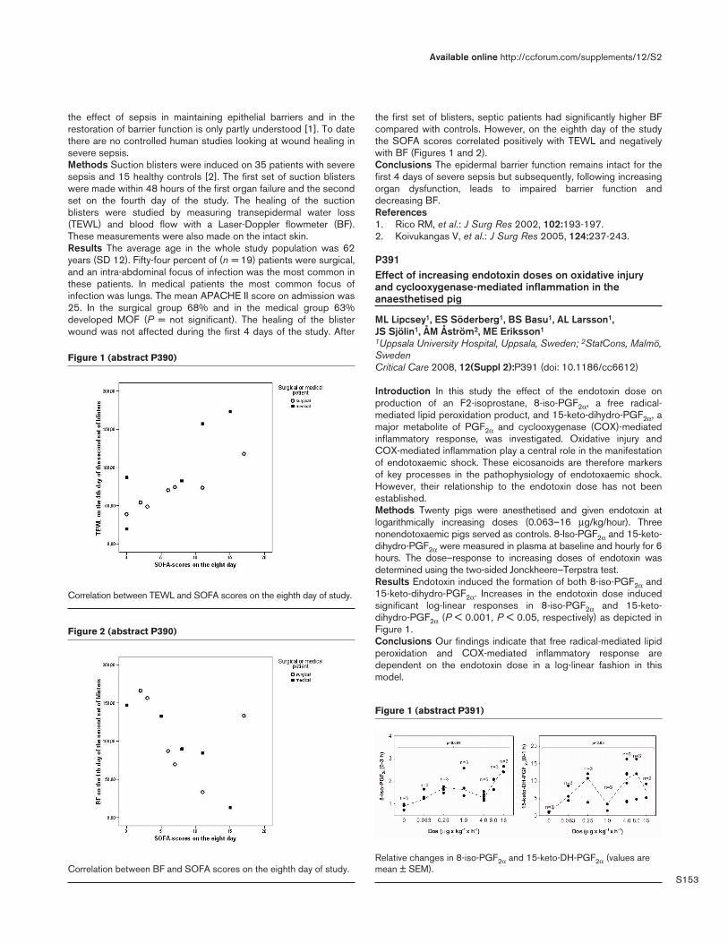
S153
the effect of sepsis in maintaining epithelial barriers and in therestoration of barrier function is only partly understood [1]. To datethere are no controlled human studies looking at wound healing insevere sepsis.Methods Suction blisters were induced on 35 patients with severesepsis and 15 healthy controls [2]. The first set of suction blisterswere made within 48 hours of the first organ failure and the secondset on the fourth day of the study. The healing of the suctionblisters were studied by measuring transepidermal water loss(TEWL) and blood flow with a Laser-Doppler flowmeter (BF).These measurements were also made on the intact skin.Results The average age in the whole study population was 62years (SD 12). Fifty-four percent of (n = 19) patients were surgical,and an intra-abdominal focus of infection was the most common inthese patients. In medical patients the most common focus ofinfection was lungs. The mean APACHE II score on admission was25. In the surgical group 68% and in the medical group 63%developed MOF (P = not significant). The healing of the blisterwound was not affected during the first 4 days of the study. After
the first set of blisters, septic patients had significantly higher BFcompared with controls. However, on the eighth day of the studythe SOFA scores correlated positively with TEWL and negativelywith BF (Figures 1 and 2).Conclusions The epidermal barrier function remains intact for thefirst 4 days of severe sepsis but subsequently, following increasingorgan dysfunction, leads to impaired barrier function anddecreasing BF.References1. Rico RM, et al.: J Surg Res 2002, 102:193-197.2. Koivukangas V, et al.: J Surg Res 2005, 124:237-243.
P391Effect of increasing endotoxin doses on oxidative injuryand cyclooxygenase-mediated inflammation in theanaesthetised pig
ML Lipcsey1, ES Söderberg1, BS Basu1, AL Larsson1, JS Sjölin1, ÅM Åström2, ME Eriksson1
1Uppsala University Hospital, Uppsala, Sweden; 2StatCons, Malmö,SwedenCritical Care 2008, 12(Suppl 2):P391 (doi: 10.1186/cc6612)
Introduction In this study the effect of the endotoxin dose onproduction of an F2-isoprostane, 8-iso-PGF2α, a free radical-mediated lipid peroxidation product, and 15-keto-dihydro-PGF2α, amajor metabolite of PGF2α and cyclooxygenase (COX)-mediatedinflammatory response, was investigated. Oxidative injury andCOX-mediated inflammation play a central role in the manifestationof endotoxaemic shock. These eicosanoids are therefore markersof key processes in the pathophysiology of endotoxaemic shock.However, their relationship to the endotoxin dose has not beenestablished.Methods Twenty pigs were anesthetised and given endotoxin atlogarithmically increasing doses (0.063–16 μg/kg/hour). Threenonendotoxaemic pigs served as controls. 8-Iso-PGF2α and 15-keto-dihydro-PGF2α were measured in plasma at baseline and hourly for 6hours. The dose–response to increasing doses of endotoxin wasdetermined using the two-sided Jonckheere–Terpstra test.Results Endotoxin induced the formation of both 8-iso-PGF2α and15-keto-dihydro-PGF2α. Increases in the endotoxin dose inducedsignificant log-linear responses in 8-iso-PGF2α and 15-keto-dihydro-PGF2α (P < 0.001, P < 0.05, respectively) as depicted inFigure 1.Conclusions Our findings indicate that free radical-mediated lipidperoxidation and COX-mediated inflammatory response aredependent on the endotoxin dose in a log-linear fashion in thismodel.
Available online http://ccforum.com/supplements/12/S2
Figure 1 (abstract P390)
Correlation between TEWL and SOFA scores on the eighth day of study.
Figure 2 (abstract P390)
Correlation between BF and SOFA scores on the eighth day of study.
Figure 1 (abstract P391)
Relative changes in 8-iso-PGF2α and 15-keto-DH-PGF2α (values aremean ± SEM).
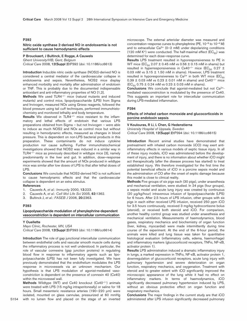
S154
P392Nitric oxide synthase 2-derived NO in endotoxemia is notsufficient to cause hemodynamic effects
P Brouckaert, J Bultinck, E Rogge, A CauwelsGhent University/VIB, Gent, BelgiumCritical Care 2008, 12(Suppl 2):P392 (doi: 10.1186/cc6613)
Introduction Inducible nitric oxide synthase (NOS2)-derived NO isconsidered a central mediator of the cardiovascular collapse inendotoxemia and sepsis. Nevertheless, NOS2 mice displayenhanced morbidity and mortality after administration of endotoxinor TNF. This is probably due to the documented indispensableantioxidant and anti-inflammatory properties of NO [1,2].Methods We used TLR4–/– mice (natural mutants and inducedmutants) and control mice, lipopolysaccharide (LPS) from Sigmaand Invivogen, measured NOx using Griess reagents, followed theblood pressure using tail cuff techniques, performed immunohisto-chemistry and monitored lethality and body temperature.Results We observed in TLR4–/– mice resistant to the inflam-matory and lethal effects of endotoxin that various LPSpreparations obtained from Sigma – but not Invivogen – were ableto induce as much NOS2 and NOx as control mice but withoutresulting in hemodynamic effects, measured as changes in bloodpressure. This is dependent on non-LPS bacterial products in thispreparation. These contaminants do not induce cytokineproduction nor cause suffering. Further immunohistochemicalinvestigations showed that NOS2 was induced in a similar way inTLR4–/– mice as previously described in wildtype mice [3], namelypredominantly in the liver and gut. In addition, dose–responseexperiments showed that the amount of NOx produced in wildtypemice was similar after low doses of LPS to that after lethal doses ofLPS.Conclusions We conclude that NOS2-derived NO is not sufficientto cause hemodynamic effects and that the cardiovascularcollapse is dependent on an extra stimulus.References1. Cauwels A, et al.: Immunity 2000, 13:223.2. Cauwels A, et al.: Cell Mol Life Sci 2005, 62:1362.3. Bultinck J, et al.: FASEB J 2006, 20:2363.
P393Lipopolysaccharide modulation of phenylephrine-dependentvasoconstriction is dependent on intercellular communication
Y OuelletteMayo Clinic, Rochester, MN, USACritical Care 2008, 12(Suppl 2):P393 (doi: 10.1186/cc6614)
Introduction The role of gap junctional intercellular communicationbetween endothelial cells and vascular smooth muscle cells duringthe inflammatory process is not well understood. In particular, therole of vascular connexins (gap junction proteins) in regulatingblood flow in response to inflammatory agents such as lipo-polysaccharide (LPS) has not been fully investigated. We havepreviously demonstrated that the endothelium modulates the LPSresponse in microvessels via an unknown mechanism. Ourhypothesis is that LPS modulation of agonist-mediated vaso-constriction is dependent on the presence of connexin 40 (Cx40)within the microvessel wall.Methods Wildtype (WT) and Cx40 knockout (Cx40–/–) animalswere treated with LPS (15 mg/kg intraperitoneally) or saline for 18hours. Small mesenteric resistance arteries (190–220 μm) wereisolated, mounted on glass cannulae, pressurized at 60 mmHgwith no lumen flow and placed on the stage of an inverted
microscope. The external arteriolar diameter was measured andconcentration–response curves to phenylephrine (PE, 10–9 to 10–4 M)and to extracellular Ca2+ (0–2 mM) under depolarizing conditions(120 mM K+) were conducted. The half-maximal effect (EC50) wasdetermined for each dose–response curve.Results LPS treatment resulted in hyporesponsiveness to PE inWT mice (EC50 2.07 ± 0.45 mM vs 0.56 ± 0.15 mM in shams) butresulted in hyperresponsiveness in Cx40–/– mice (EC50 0.27 ±0.03 mM vs 3.15 ± 1.50 mM in shams). However, LPS treatmentresulted in hyporesponsiveness to Ca2+ in both WT mice (EC500.39 ± 0.03 mM vs 0.23 ± 0.01 mM in shams) and Cx40–/– mice(EC50 0.75 ± 0.24 mM vs 0.23 ± 0.03 mM in shams).Conclusions We conclude that agonist-mediated but not Ca2+-mediated vasoconstriction is modulated by the presence of Cx40.This suggests an important role for intercellular communicationduring LPS-mediated inflammation.
P394Effects of inhaled carbon monoxide and glucocorticoids inporcine endotoxin sepsis
V Koulouras, R Li, L Chen, G HedenstiernaUniversity Hospital of Uppsala, SwedenCritical Care 2008, 12(Suppl 2):P394 (doi: 10.1186/cc6615)
Introduction Recent animal studies have demonstrated thatpretreatment with inhaled carbon monoxide (iCO) may exert anti-inflammatory effects in various models of septic tissue injury. In allof these injury models, iCO was administered before the develop-ment of injury, and there is no information about whether iCO mightact therapeutically (after the disease process has started) to treatestablished injury. We therefore investigated for the first time thepotential beneficial effects of iCO in a porcine sepsis model andthe administration of CO after the onset of septic damage becausethis model is close to clinical reality.Methods Five groups of six pigs each (30 pigs), under anaesthesiaand mechanical ventilation, were studied. In 24 pigs (four groups),a sepsis model and acute lung injury was created by continuous(20 μg/kg/hour) intravenous infusion of lipopolysaccharide (LPS)for 6 hours. After 2.5 hours of LPS infusion, other groups with sixpigs in each either received LPS infusion, received 250 ppm iCOfor 3.5 hours continuously, received 3 mg/kg hydrocortisone bolus(steroid), or received both steroid and iCO. For comparison,another healthy control group was studied under anaesthesia andmechanical ventilation. Measurements of haemodynamics, bloodgases, respiratory mechanics and biochemistry of organ function(liver, kidney, myocardial) were made intermittently during timecourse of the experiment. At the end of the 6-hour period, theanimals were killed and lung tissue was taken for quantitativehistological evaluation (inflammatory cells, edema, haemorrhage)and inflammatory markers (glucocorticoid receptors, TNFα, NF-κB,activator protein 1).Results LPS administration induced a dramatic inflammatory injuryin lungs, a marked expression in TNFα, NF-κB, activator protein 1,downregulation of glucocorticoid receptors, acute lung injury withpulmonary hypertension and severe deterioration on organfunction, respiratory mechanics, and oxygenation. Treatment withsteroid and to greater extent with iCO significantly improved themicroscopic appearance of the lung while it had no effect oninflammatory markers. In terms of haemodynamics, iCOsignificantly decreased pulmonary hypertension induced by LPS,without an obvious protective effect on organ function andrespiratory mechanics.Conclusions The major findings in the current study are that iCOadministered after LPS infusion significantly decreased pulmonary
Critical Care March 2008 Vol 12 Suppl 2 28th International Symposium on Intensive Care and Emergency Medicine
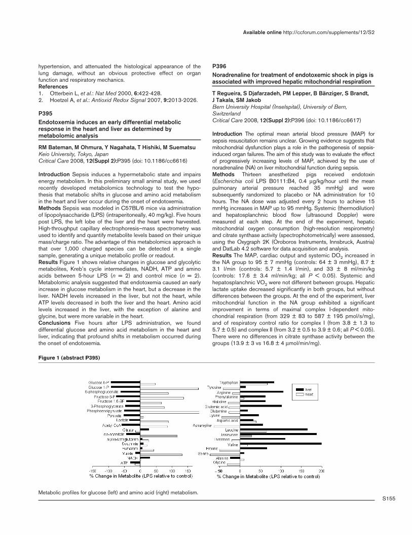
S155
hypertension, and attenuated the histological appearance of thelung damage, without an obvious protective effect on organfunction and respiratory mechanics.References1. Otterbein L, et al.: Nat Med 2000, 6:422-428.2. Hoetzel A, et al.: Antioxid Redox Signal 2007, 9:2013-2026.
P395Endotoxemia induces an early differential metabolicresponse in the heart and liver as determined bymetabolomic analysis
RM Bateman, M Ohmura, Y Nagahata, T Hishiki, M SuematsuKeio University, Tokyo, JapanCritical Care 2008, 12(Suppl 2):P395 (doi: 10.1186/cc6616)
Introduction Sepsis induces a hypermetabolic state and impairsenergy metabolism. In this preliminary small animal study, we usedrecently developed metabolomics technology to test the hypo-thesis that metabolic shifts in glucose and amino acid metabolismin the heart and liver occur during the onset of endotoxemia.Methods Sepsis was modeled in C57BL/6 mice via administrationof lipopolysaccharide (LPS) (intraperitoneally, 40 mg/kg). Five hourspost LPS, the left lobe of the liver and the heart were harvested.High-throughput capillary electrophoresis–mass spectrometry wasused to identify and quantify metabolite levels based on their uniquemass/charge ratio. The advantage of this metabolomics approach isthat over 1,000 charged species can be detected in a singlesample, generating a unique metabolic profile or readout.Results Figure 1 shows relative changes in glucose and glycolyticmetabolites, Kreb’s cycle intermediates, NADH, ATP and aminoacids between 5-hour LPS (n = 2) and control mice (n = 2).Metabolomic analysis suggested that endotoxemia caused an earlyincrease in glucose metabolism in the heart, but a decrease in theliver. NADH levels increased in the liver, but not the heart, whileATP levels decreased in both the liver and the heart. Amino acidlevels increased in the liver, with the exception of alanine andglycine, but were more variable in the heart.Conclusions Five hours after LPS administration, we founddifferential glucose and amino acid metabolism in the heart andliver, indicating that profound shifts in metabolism occurred duringthe onset of endotoxemia.
P396Noradrenaline for treatment of endotoxemic shock in pigs isassociated with improved hepatic mitochondrial respiration
T Regueira, S Djafarzadeh, PM Lepper, B Bänziger, S Brandt, J Takala, SM JakobBern University Hospital (Inselspital), University of Bern,SwitzerlandCritical Care 2008, 12(Suppl 2):P396 (doi: 10.1186/cc6617)
Introduction The optimal mean arterial blood pressure (MAP) forsepsis resuscitation remains unclear. Growing evidence suggests thatmitochondrial dysfunction plays a role in the pathogenesis of sepsis-induced organ failures. The aim of this study was to evaluate the effectof progressively increasing levels of MAP, achieved by the use ofnoradrenaline (NA) on liver mitochondrial function during sepsis.Methods Thirteen anesthetized pigs received endotoxin(Escherichia coli LPS B0111:B4, 0.4 μg/kg/hour until the meanpulmonary arterial pressure reached 35 mmHg) and weresubsequently randomized to placebo or NA administration for 10hours. The NA dose was adjusted every 2 hours to achieve 15mmHg increases in MAP up to 95 mmHg. Systemic (thermodilution)and hepatosplanchnic blood flow (ultrasound Doppler) weremeasured at each step. At the end of the experiment, hepaticmitochondrial oxygen consumption (high-resolution respirometry)and citrate synthase activity (spectrophotometrically) were assessed,using the Oxygraph 2K (Oroboros Instruments, Innsbruck, Austria)and DatLab 4.2 software for data acquisition and analysis.Results The MAP, cardiac output and systemic DO2 increased inthe NA group to 95 ± 7 mmHg (controls: 64 ± 3 mmHg), 8.7 ±3.1 l/min (controls: 5.7 ± 1.4 l/min), and 33 ± 8 ml/min/kg(controls: 17.6 ± 3.4 ml/min/kg; all P < 0.05). Systemic andhepatosplanchnic VO2 were not different between groups. Hepaticlactate uptake decreased significantly in both groups, but withoutdifferences between the groups. At the end of the experiment, livermitochondrial function in the NA group exhibited a significantimprovement in terms of maximal complex I-dependent mito-chondrial respiration (from 329 ± 83 to 587 ± 195 pmol/s/mg),and of respiratory control ratio for complex I (from 3.8 ± 1.3 to5.7 ± 0.5) and complex II (from 3.2 ± 0.5 to 3.9 ± 0.6; all P < 0.05).There were no differences in citrate synthase activity between thegroups (13.9 ± 3 vs 16.8 ± 4 μmol/min/mg).
Available online http://ccforum.com/supplements/12/S2
Figure 1 (abstract P395)
Metabolic profiles for glucose (left) and amino acid (right) metabolism.
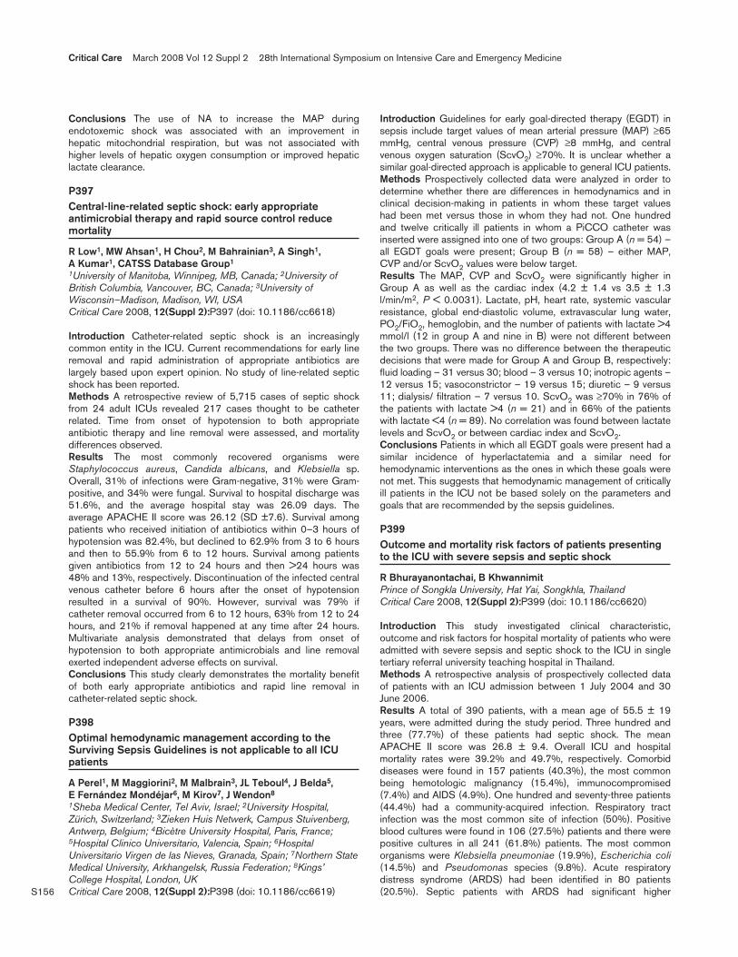
S156
Conclusions The use of NA to increase the MAP duringendotoxemic shock was associated with an improvement inhepatic mitochondrial respiration, but was not associated withhigher levels of hepatic oxygen consumption or improved hepaticlactate clearance.
P397Central-line-related septic shock: early appropriateantimicrobial therapy and rapid source control reducemortality
R Low1, MW Ahsan1, H Chou2, M Bahrainian3, A Singh1, A Kumar1, CATSS Database Group1
1University of Manitoba, Winnipeg, MB, Canada; 2University ofBritish Columbia, Vancouver, BC, Canada; 3University ofWisconsin–Madison, Madison, WI, USACritical Care 2008, 12(Suppl 2):P397 (doi: 10.1186/cc6618)
Introduction Catheter-related septic shock is an increasinglycommon entity in the ICU. Current recommendations for early lineremoval and rapid administration of appropriate antibiotics arelargely based upon expert opinion. No study of line-related septicshock has been reported.Methods A retrospective review of 5,715 cases of septic shockfrom 24 adult ICUs revealed 217 cases thought to be catheterrelated. Time from onset of hypotension to both appropriateantibiotic therapy and line removal were assessed, and mortalitydifferences observed.Results The most commonly recovered organisms wereStaphylococcus aureus, Candida albicans, and Klebsiella sp.Overall, 31% of infections were Gram-negative, 31% were Gram-positive, and 34% were fungal. Survival to hospital discharge was51.6%, and the average hospital stay was 26.09 days. Theaverage APACHE II score was 26.12 (SD ±7.6). Survival amongpatients who received initiation of antibiotics within 0–3 hours ofhypotension was 82.4%, but declined to 62.9% from 3 to 6 hoursand then to 55.9% from 6 to 12 hours. Survival among patientsgiven antibiotics from 12 to 24 hours and then >24 hours was48% and 13%, respectively. Discontinuation of the infected centralvenous catheter before 6 hours after the onset of hypotensionresulted in a survival of 90%. However, survival was 79% ifcatheter removal occurred from 6 to 12 hours, 63% from 12 to 24hours, and 21% if removal happened at any time after 24 hours.Multivariate analysis demonstrated that delays from onset ofhypotension to both appropriate antimicrobials and line removalexerted independent adverse effects on survival.Conclusions This study clearly demonstrates the mortality benefitof both early appropriate antibiotics and rapid line removal incatheter-related septic shock.
P398Optimal hemodynamic management according to theSurviving Sepsis Guidelines is not applicable to all ICUpatients
A Perel1, M Maggiorini2, M Malbrain3, JL Teboul4, J Belda5, E Fernández Mondéjar6, M Kirov7, J Wendon8
1Sheba Medical Center, Tel Aviv, Israel; 2University Hospital,Zürich, Switzerland; 3Zieken Huis Netwerk, Campus Stuivenberg,Antwerp, Belgium; 4Bicètre University Hospital, Paris, France;5Hospital Clinico Universitario, Valencia, Spain; 6HospitalUniversitario Virgen de las Nieves, Granada, Spain; 7Northern StateMedical University, Arkhangelsk, Russia Federation; 8Kings’College Hospital, London, UKCritical Care 2008, 12(Suppl 2):P398 (doi: 10.1186/cc6619)
Introduction Guidelines for early goal-directed therapy (EGDT) insepsis include target values of mean arterial pressure (MAP) ≥65mmHg, central venous pressure (CVP) ≥8 mmHg, and centralvenous oxygen saturation (ScvO2) ≥70%. It is unclear whether asimilar goal-directed approach is applicable to general ICU patients.Methods Prospectively collected data were analyzed in order todetermine whether there are differences in hemodynamics and inclinical decision-making in patients in whom these target valueshad been met versus those in whom they had not. One hundredand twelve critically ill patients in whom a PiCCO catheter wasinserted were assigned into one of two groups: Group A (n = 54) –all EGDT goals were present; Group B (n = 58) – either MAP,CVP and/or ScvO2 values were below target.Results The MAP, CVP and ScvO2 were significantly higher inGroup A as well as the cardiac index (4.2 ± 1.4 vs 3.5 ± 1.3l/min/m2, P < 0.0031). Lactate, pH, heart rate, systemic vascularresistance, global end-diastolic volume, extravascular lung water,PO2/FiO2, hemoglobin, and the number of patients with lactate >4mmol/l (12 in group A and nine in B) were not different betweenthe two groups. There was no difference between the therapeuticdecisions that were made for Group A and Group B, respectively:fluid loading – 31 versus 30; blood – 3 versus 10; inotropic agents –12 versus 15; vasoconstrictor – 19 versus 15; diuretic – 9 versus11; dialysis/ filtration – 7 versus 10. ScvO2 was ≥70% in 76% ofthe patients with lactate >4 (n = 21) and in 66% of the patientswith lactate <4 (n = 89). No correlation was found between lactatelevels and ScvO2 or between cardiac index and ScvO2.Conclusions Patients in which all EGDT goals were present had asimilar incidence of hyperlactatemia and a similar need forhemodynamic interventions as the ones in which these goals werenot met. This suggests that hemodynamic management of criticallyill patients in the ICU not be based solely on the parameters andgoals that are recommended by the sepsis guidelines.
P399Outcome and mortality risk factors of patients presentingto the ICU with severe sepsis and septic shock
R Bhurayanontachai, B KhwannimitPrince of Songkla University, Hat Yai, Songkhla, ThailandCritical Care 2008, 12(Suppl 2):P399 (doi: 10.1186/cc6620)
Introduction This study investigated clinical characteristic,outcome and risk factors for hospital mortality of patients who wereadmitted with severe sepsis and septic shock to the ICU in singletertiary referral university teaching hospital in Thailand.Methods A retrospective analysis of prospectively collected dataof patients with an ICU admission between 1 July 2004 and 30June 2006.Results A total of 390 patients, with a mean age of 55.5 ± 19years, were admitted during the study period. Three hundred andthree (77.7%) of these patients had septic shock. The meanAPACHE II score was 26.8 ± 9.4. Overall ICU and hospitalmortality rates were 39.2% and 49.7%, respectively. Comorbiddiseases were found in 157 patients (40.3%), the most commonbeing hemotologic malignancy (15.4%), immunocompromised(7.4%) and AIDS (4.9%). One hundred and seventy-three patients(44.4%) had a community-acquired infection. Respiratory tractinfection was the most common site of infection (50%). Positiveblood cultures were found in 106 (27.5%) patients and there werepositive cultures in all 241 (61.8%) patients. The most commonorganisms were Klebsiella pneumoniae (19.9%), Escherichia coli(14.5%) and Pseudomonas species (9.8%). Acute respiratorydistress syndrome (ARDS) had been identified in 80 patients(20.5%). Septic patients with ARDS had significant higher
Critical Care March 2008 Vol 12 Suppl 2 28th International Symposium on Intensive Care and Emergency Medicine
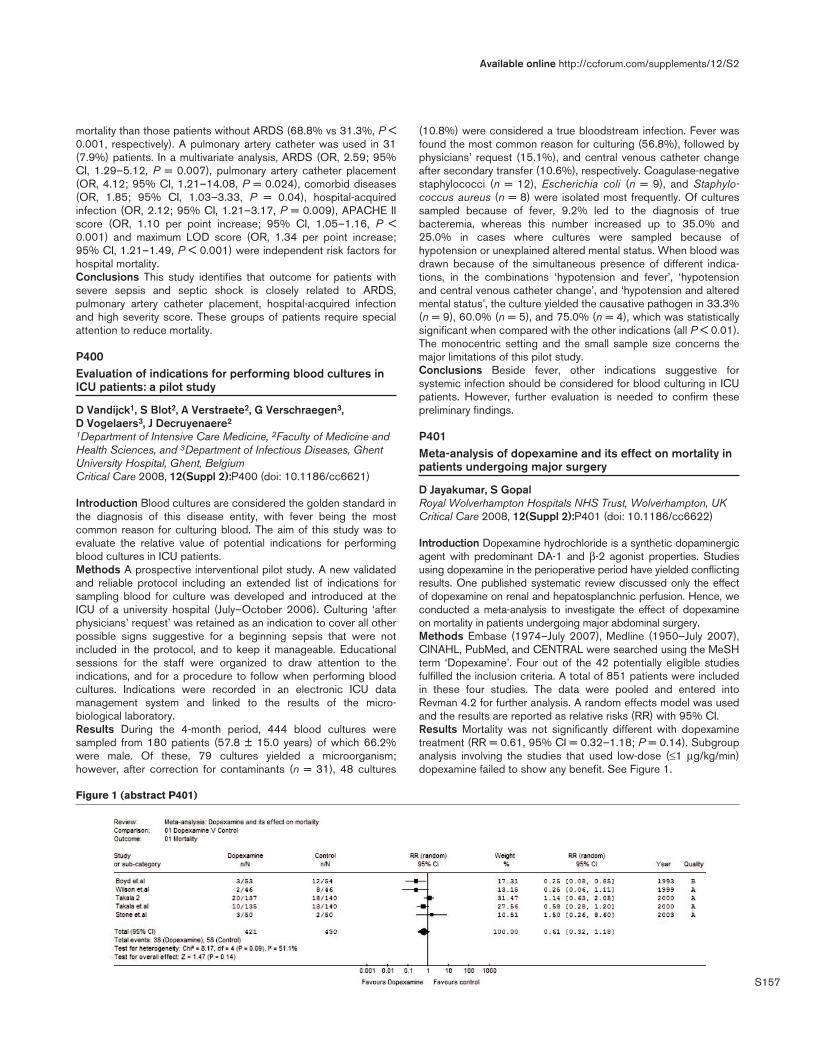
S157
mortality than those patients without ARDS (68.8% vs 31.3%, P <0.001, respectively). A pulmonary artery catheter was used in 31(7.9%) patients. In a multivariate analysis, ARDS (OR, 2.59; 95%CI, 1.29–5.12, P = 0.007), pulmonary artery catheter placement(OR, 4.12; 95% CI, 1.21–14.08, P = 0.024), comorbid diseases(OR, 1.85; 95% CI, 1.03–3.33, P = 0.04), hospital-acquiredinfection (OR, 2.12; 95% CI, 1.21–3.17, P = 0.009), APACHE IIscore (OR, 1.10 per point increase; 95% CI, 1.05–1.16, P <0.001) and maximum LOD score (OR, 1.34 per point increase;95% CI, 1.21–1.49, P < 0.001) were independent risk factors forhospital mortality.Conclusions This study identifies that outcome for patients withsevere sepsis and septic shock is closely related to ARDS,pulmonary artery catheter placement, hospital-acquired infectionand high severity score. These groups of patients require specialattention to reduce mortality.
P400Evaluation of indications for performing blood cultures inICU patients: a pilot study
D Vandijck1, S Blot2, A Verstraete2, G Verschraegen3, D Vogelaers3, J Decruyenaere2
1Department of Intensive Care Medicine, 2Faculty of Medicine andHealth Sciences, and 3Department of Infectious Diseases, GhentUniversity Hospital, Ghent, BelgiumCritical Care 2008, 12(Suppl 2):P400 (doi: 10.1186/cc6621)
Introduction Blood cultures are considered the golden standard inthe diagnosis of this disease entity, with fever being the mostcommon reason for culturing blood. The aim of this study was toevaluate the relative value of potential indications for performingblood cultures in ICU patients.Methods A prospective interventional pilot study. A new validatedand reliable protocol including an extended list of indications forsampling blood for culture was developed and introduced at theICU of a university hospital (July–October 2006). Culturing ‘afterphysicians’ request’ was retained as an indication to cover all otherpossible signs suggestive for a beginning sepsis that were notincluded in the protocol, and to keep it manageable. Educationalsessions for the staff were organized to draw attention to theindications, and for a procedure to follow when performing bloodcultures. Indications were recorded in an electronic ICU datamanagement system and linked to the results of the micro-biological laboratory.Results During the 4-month period, 444 blood cultures weresampled from 180 patients (57.8 ± 15.0 years) of which 66.2%were male. Of these, 79 cultures yielded a microorganism;however, after correction for contaminants (n = 31), 48 cultures
(10.8%) were considered a true bloodstream infection. Fever wasfound the most common reason for culturing (56.8%), followed byphysicians’ request (15.1%), and central venous catheter changeafter secondary transfer (10.6%), respectively. Coagulase-negativestaphylococci (n = 12), Escherichia coli (n = 9), and Staphylo-coccus aureus (n = 8) were isolated most frequently. Of culturessampled because of fever, 9.2% led to the diagnosis of truebacteremia, whereas this number increased up to 35.0% and25.0% in cases where cultures were sampled because ofhypotension or unexplained altered mental status. When blood wasdrawn because of the simultaneous presence of different indica-tions, in the combinations ‘hypotension and fever’, ‘hypotensionand central venous catheter change’, and ‘hypotension and alteredmental status’, the culture yielded the causative pathogen in 33.3%(n = 9), 60.0% (n = 5), and 75.0% (n = 4), which was statisticallysignificant when compared with the other indications (all P < 0.01).The monocentric setting and the small sample size concerns themajor limitations of this pilot study.Conclusions Beside fever, other indications suggestive forsystemic infection should be considered for blood culturing in ICUpatients. However, further evaluation is needed to confirm thesepreliminary findings.
P401Meta-analysis of dopexamine and its effect on mortality inpatients undergoing major surgery
D Jayakumar, S GopalRoyal Wolverhampton Hospitals NHS Trust, Wolverhampton, UKCritical Care 2008, 12(Suppl 2):P401 (doi: 10.1186/cc6622)
Introduction Dopexamine hydrochloride is a synthetic dopaminergicagent with predominant DA-1 and β-2 agonist properties. Studiesusing dopexamine in the perioperative period have yielded conflictingresults. One published systematic review discussed only the effectof dopexamine on renal and hepatosplanchnic perfusion. Hence, weconducted a meta-analysis to investigate the effect of dopexamineon mortality in patients undergoing major abdominal surgery.Methods Embase (1974–July 2007), Medline (1950–July 2007),CINAHL, PubMed, and CENTRAL were searched using the MeSHterm ‘Dopexamine’. Four out of the 42 potentially eligible studiesfulfilled the inclusion criteria. A total of 851 patients were includedin these four studies. The data were pooled and entered intoRevman 4.2 for further analysis. A random effects model was usedand the results are reported as relative risks (RR) with 95% CI.Results Mortality was not significantly different with dopexaminetreatment (RR = 0.61, 95% CI = 0.32–1.18; P = 0.14). Subgroupanalysis involving the studies that used low-dose (≤1 μg/kg/min)dopexamine failed to show any benefit. See Figure 1.
Available online http://ccforum.com/supplements/12/S2
Figure 1 (abstract P401)
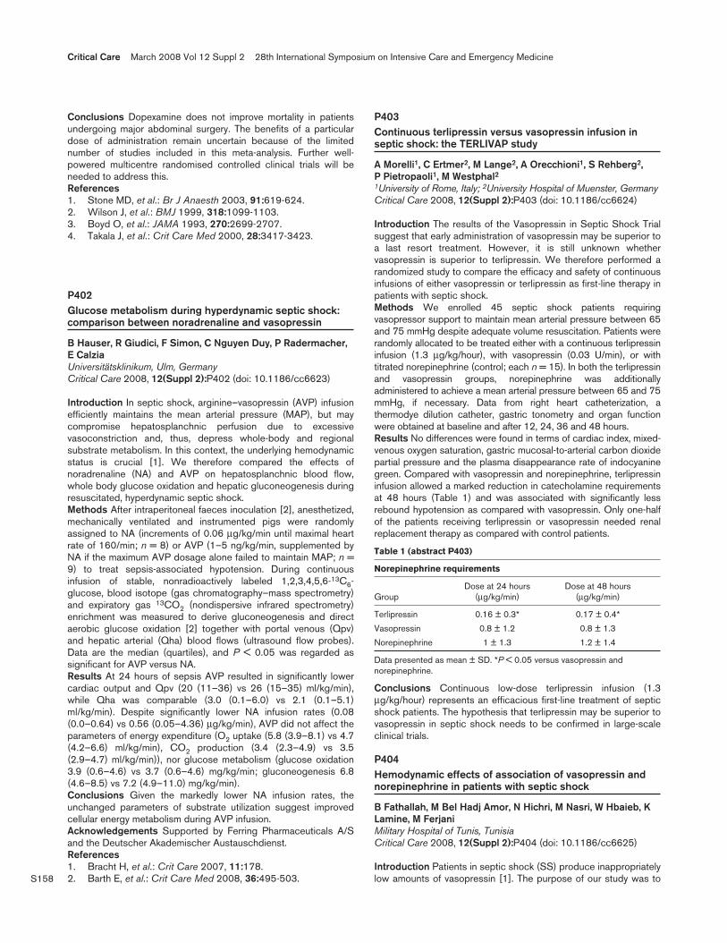
S158
Conclusions Dopexamine does not improve mortality in patientsundergoing major abdominal surgery. The benefits of a particulardose of administration remain uncertain because of the limitednumber of studies included in this meta-analysis. Further well-powered multicentre randomised controlled clinical trials will beneeded to address this.References1. Stone MD, et al.: Br J Anaesth 2003, 91:619-624.2. Wilson J, et al.: BMJ 1999, 318:1099-1103.3. Boyd O, et al.: JAMA 1993, 270:2699-2707.4. Takala J, et al.: Crit Care Med 2000, 28:3417-3423.
P402Glucose metabolism during hyperdynamic septic shock:comparison between noradrenaline and vasopressin
B Hauser, R Giudici, F Simon, C Nguyen Duy, P Radermacher,E CalziaUniversitätsklinikum, Ulm, GermanyCritical Care 2008, 12(Suppl 2):P402 (doi: 10.1186/cc6623)
Introduction In septic shock, arginine–vasopressin (AVP) infusionefficiently maintains the mean arterial pressure (MAP), but maycompromise hepatosplanchnic perfusion due to excessivevasoconstriction and, thus, depress whole-body and regionalsubstrate metabolism. In this context, the underlying hemodynamicstatus is crucial [1]. We therefore compared the effects ofnoradrenaline (NA) and AVP on hepatosplanchnic blood flow,whole body glucose oxidation and hepatic gluconeogenesis duringresuscitated, hyperdynamic septic shock.Methods After intraperitoneal faeces inoculation [2], anesthetized,mechanically ventilated and instrumented pigs were randomlyassigned to NA (increments of 0.06 μg/kg/min until maximal heartrate of 160/min; n = 8) or AVP (1–5 ng/kg/min, supplemented byNA if the maximum AVP dosage alone failed to maintain MAP; n =9) to treat sepsis-associated hypotension. During continuousinfusion of stable, nonradioactively labeled 1,2,3,4,5,6-13C6-glucose, blood isotope (gas chromatography–mass spectrometry)and expiratory gas 13CO2 (nondispersive infrared spectrometry)enrichment was measured to derive gluconeogenesis and directaerobic glucose oxidation [2] together with portal venous (Qpv)and hepatic arterial (Qha) blood flows (ultrasound flow probes).Data are the median (quartiles), and P < 0.05 was regarded assignificant for AVP versus NA.Results At 24 hours of sepsis AVP resulted in significantly lowercardiac output and Qpv (20 (11–36) vs 26 (15–35) ml/kg/min),while Qha was comparable (3.0 (0.1–6.0) vs 2.1 (0.1–5.1)ml/kg/min). Despite significantly lower NA infusion rates (0.08(0.0–0.64) vs 0.56 (0.05–4.36) μg/kg/min), AVP did not affect theparameters of energy expenditure (O2 uptake (5.8 (3.9–8.1) vs 4.7(4.2–6.6) ml/kg/min), CO2 production (3.4 (2.3–4.9) vs 3.5(2.9–4.7) ml/kg/min)), nor glucose metabolism (glucose oxidation3.9 (0.6–4.6) vs 3.7 (0.6–4.6) mg/kg/min; gluconeogenesis 6.8(4.6–8.5) vs 7.2 (4.9–11.0) mg/kg/min).Conclusions Given the markedly lower NA infusion rates, theunchanged parameters of substrate utilization suggest improvedcellular energy metabolism during AVP infusion.Acknowledgements Supported by Ferring Pharmaceuticals A/Sand the Deutscher Akademischer Austauschdienst.References1. Bracht H, et al.: Crit Care 2007, 11:178.2. Barth E, et al.: Crit Care Med 2008, 36:495-503.
P403Continuous terlipressin versus vasopressin infusion inseptic shock: the TERLIVAP study
A Morelli1, C Ertmer2, M Lange2, A Orecchioni1, S Rehberg2, P Pietropaoli1, M Westphal21University of Rome, Italy; 2University Hospital of Muenster, GermanyCritical Care 2008, 12(Suppl 2):P403 (doi: 10.1186/cc6624)
Introduction The results of the Vasopressin in Septic Shock Trialsuggest that early administration of vasopressin may be superior toa last resort treatment. However, it is still unknown whethervasopressin is superior to terlipressin. We therefore performed arandomized study to compare the efficacy and safety of continuousinfusions of either vasopressin or terlipressin as first-line therapy inpatients with septic shock.Methods We enrolled 45 septic shock patients requiringvasopressor support to maintain mean arterial pressure between 65and 75 mmHg despite adequate volume resuscitation. Patients wererandomly allocated to be treated either with a continuous terlipressininfusion (1.3 μg/kg/hour), with vasopressin (0.03 U/min), or withtitrated norepinephrine (control; each n = 15). In both the terlipressinand vasopressin groups, norepinephrine was additionallyadministered to achieve a mean arterial pressure between 65 and 75mmHg, if necessary. Data from right heart catheterization, athermodye dilution catheter, gastric tonometry and organ functionwere obtained at baseline and after 12, 24, 36 and 48 hours.Results No differences were found in terms of cardiac index, mixed-venous oxygen saturation, gastric mucosal-to-arterial carbon dioxidepartial pressure and the plasma disappearance rate of indocyaninegreen. Compared with vasopressin and norepinephrine, terlipressininfusion allowed a marked reduction in catecholamine requirementsat 48 hours (Table 1) and was associated with significantly lessrebound hypotension as compared with vasopressin. Only one-halfof the patients receiving terlipressin or vasopressin needed renalreplacement therapy as compared with control patients.
Table 1 (abstract P403)
Norepinephrine requirements
Dose at 24 hours Dose at 48 hours Group (μg/kg/min) (μg/kg/min)
Terlipressin 0.16 ± 0.3* 0.17 ± 0.4*
Vasopressin 0.8 ± 1.2 0.8 ± 1.3
Norepinephrine 1 ± 1.3 1.2 ± 1.4
Data presented as mean ± SD. *P < 0.05 versus vasopressin andnorepinephrine.
Conclusions Continuous low-dose terlipressin infusion (1.3μg/kg/hour) represents an efficacious first-line treatment of septicshock patients. The hypothesis that terlipressin may be superior tovasopressin in septic shock needs to be confirmed in large-scaleclinical trials.
P404Hemodynamic effects of association of vasopressin andnorepinephrine in patients with septic shock
B Fathallah, M Bel Hadj Amor, N Hichri, M Nasri, W Hbaieb, KLamine, M FerjaniMilitary Hospital of Tunis, TunisiaCritical Care 2008, 12(Suppl 2):P404 (doi: 10.1186/cc6625)
Introduction Patients in septic shock (SS) produce inappropriatelylow amounts of vasopressin [1]. The purpose of our study was to
Critical Care March 2008 Vol 12 Suppl 2 28th International Symposium on Intensive Care and Emergency Medicine
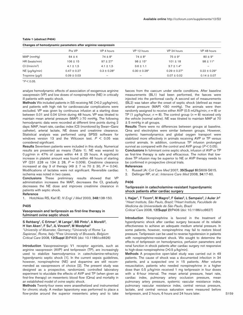
S159
analyze hemodynamic effects of association of exogenous argininevasopressin (VP) and low doses of norepinephrine (NE) in criticallyill patients with septic shock.Methods We included patients in SS receiving NE (>0.2 μg/kg/min),and patients with high risk for cardiovascular complications wereexcluded. VP was given by continuous infusion at a starting dosebetween 0.01 and 0.04 U/min during 48 hours. VP was titrated tomaintain mean arterial pressure (MAP) ≥ 70 mmHg. The followinghemodynamic data were recorded at different time points during 2days: MAP, heart rate, cardiac index (CI) (monitored by Swan–Ganzcatheter), arterial lactate, NE doses and creatinine clearance.Statistical analysis was performed using SPSS software forwindows version 13 and the Wilcoxon test. P < 0.05 wasconsidered significant.Results Seventeen patients were included in this study. Numericalresults are presented as means (Table 1). NE was weaned to0 μg/min in 47% of cases within 44 ± 25 hours. A significantincrease in platelet amount was found within 48 hours of startingVP (231 ±28 vs 134 ± 28, P = 0.009). Creatinine clearanceincreased at day 3 of therapy (49 ± 7 vs 70 ± 30, P = 0.04).Modifications of lactates were not significant. Reversible cardiacischemia was noted in two cases.Conclusions These preliminary results showed that VPadministration increases the MAP, decreases the CI, graduallydecreases the NE dose and improves creatinine clearance inpatients with septic shock.Reference1. Hotchkiss RS, Karl IE: N Engl J Med 2003, 348:138-150.
P405Vasopressin and terlipressin as first-line therapy infulminant ovine septic shock
S Rehberg1, C Ertmer1, M Lange1, BB Pinto1, A Morelli2, H Van Aken1, F Su3, JL Vincent3, M Westphal11University of Muenster, Germany; 2University of Rome ‘LaSapienza’, Rome, Italy; 3Free University of Brussels, BelgiumCritical Care 2008, 12(Suppl 2):P405 (doi: 10.1186/cc6626)
Introduction Vasopressinergic V1 receptor agonists, such asarginine vasopressin (AVP) and terlipressin (TP), are increasinglyused to stabilize haemodynamics in catecholamine-refractoryhyperdynamic septic shock [1]. In the current sepsis guidelines,however, norepinephrine (NE) and dopamine are still recom-mended as vasopressors of choice [2]. The present study wasdesigned as a prospective, randomized, controlled laboratoryexperiment to elucidate the effects of AVP and TP (when given asfirst-line therapy) on mesenteric blood flow (Qma) and mortality inan established model of ovine septic shock.Methods Twenty-four ewes were anaesthetized and instrumentedfor chronic study. A median laparotomy was performed to place aflow-probe around the superior mesenteric artery and to take
faeces from the caecum under sterile conditions. After baselinemeasurements (BL1) had been performed, the faeces wereinjected into the peritoneal cavity. A second set of measurements(BL2) was taken after the onset of septic shock (defined as meanarterial pressure (MAP) <60 mmHg). The animals were thenrandomly assigned to receive either AVP (0.5 mU/kg/min; n = 8) orTP (1 μg/kg/hour; n = 8). The control group (n = 8) received onlythe vehicle (normal saline). NE was titrated to maintain MAP at 70± 5 mmHg in all groups.Results There were no differences between groups at baseline.Qma and electrolytes were similar between groups. However,systemic haemodynamics and global oxygen transport werestabilized more effectively in animals receiving AVP or TP versuscontrol animals. In addition, continuous TP infusion prolongedsurvival as compared with the control and AVP group (P < 0.05).Conclusions In fulminant ovine septic shock, infusion of AVP or TPas first-line therapy is safe and efficacious. The notion that low-dose TP infusion may be superior to NE or AVP therapy needs tobe confirmed in prospective clinical trials.References1. Russell JA: Crit Care Med 2007, 35(Suppl 9):S609-S615.2. Dellinger RP, et al.: Intensive Care Med 2008, 34:17-60.
P406Terlipressin in catecholamine-resistant hyperdynamicshock patients after cardiac surgery
L Hajjar1, T Ticom2, M Magro2, F Galas2, L Sampaio2, J Auler Jr2
1Heart Institute, São Paulo, Brazil; 2Heart Institute, Faculdade deMedicina da Universidade de São Paulo, BrazilCritical Care 2008, 12(Suppl 2):P406 (doi: 10.1186/cc6627)
Introduction Norepinephrine is favored in the treatment ofhyperdynamic shock after cardiac surgery because of its reliableeffectiveness to achieve an adequate medium arterial pressure. Insome patients, however, norepinephrine may fail to restore bloodpressure. Terlipressin can be used to reverse hypotension in patientswith norepinephrine-resistant shock. We sought to determine theeffects of terlipressin on hemodynamics, perfusion parameters andrenal function in shock patients after cardiac surgery not responsiveto high-dose norepinephrine (>0.5 μ/kg/min).Methods A prospective open-label study was carried out in 49patients. The cause of shock was a documented infection in 34patients, and a suspected one in 15 patients. After volumeresuscitation, patients who needed norepinephrine in a higherdose than 0.5 μ/kg/min received 1 mg terlipressin in four doseswith a 6-hour interval. The mean arterial pressure, heart rate,cardiac index, pulmonary artery occlusion pressure, meanpulmonary artery pressure, systemic vascular resistance index,pulmonary vascular resistance index, central venous pressure,lactate, and central venous saturation were measured beforeterlipressin, and 2 hours, 6 hours and 24 hours later.
Available online http://ccforum.com/supplements/12/S2
Table 1 (abstract P404)
Changes of hemodynamic parameters after arginine vasopressin
Pre VP VP 4 hours VP 12 hours VP 24 hours VP 48 hours
MAP (mmHg) 64 ± 4 74 ± 8* 74 ± 8* 75 ± 9* 80 ± 9*
HR (beats/min) 106 ± 15 97 ± 27* 98 ± 15* 101 ± 18 98 ± 11*
CI (l/min/m2) 4.1 ± 1,5 4.1 ± 1,5 3.6 ± 1.1 3.7 ± 1.4* –
NE (μg/kg/min) 0.47 ± 0.27 0.3 ± 0.28* 0.30 ± 0.28* 0.29 ± 0.27* 0.22 ± 0.29*
Tropinine (μg/l) 0.09 ± 0.03 – – 0.07 ± 0.02 0.14 ± 0.07
*P < 0.05.
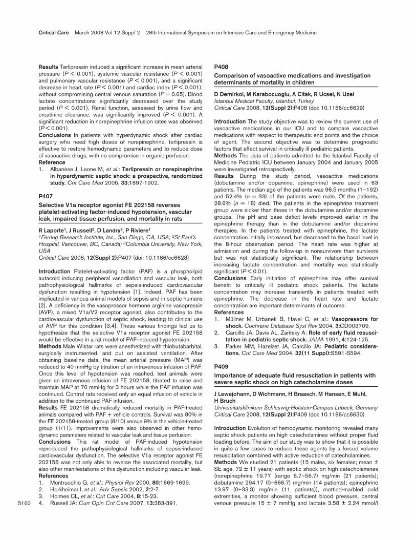
S160
Results Terlipressin induced a significant increase in mean arterialpressure (P < 0.001), systemic vascular resistance (P < 0.001)and pulmonary vascular resistance (P < 0.001), and a significantdecrease in heart rate (P < 0.001) and cardiac index (P < 0.001),without compromising central venous saturation (P = 0.65). Bloodlactate concentrations significantly decreased over the studyperiod (P < 0.001). Renal function, assessed by urine flow andcreatinine clearance, was significantly improved (P < 0.001). Asignificant reduction in norepinephrine infusion rates was observed(P < 0.001).Conclusions In patients with hyperdynamic shock after cardiacsurgery who need high doses of norepinephrine, terlipressin iseffective to restore hemodynamic parameters and to reduce doseof vasoactive drugs, with no compromise in organic perfusion.Reference1. Albanèse J, Leone M, et al.: Terlipressin or norepinephrine
in hyperdynamic septic shock: a prospective, randomizedstudy. Crit Care Med 2005, 33:1897-1902.
P407Selective V1a receptor agonist FE 202158 reversesplatelet-activating factor-induced hypotension, vascularleak, impaired tissue perfusion, and mortality in rats
R Laporte1, J Russell2, D Landry3, P Riviere1
1Ferring Research Institute, Inc., San Diego, CA, USA; 2St Paul’sHospital, Vancouver, BC, Canada; 3Columbia University, New York,USACritical Care 2008, 12(Suppl 2):P407 (doi: 10.1186/cc6628)
Introduction Platelet-activating factor (PAF) is a phospholipidautacoid inducing peripheral vasodilation and vascular leak, bothpathophysiological hallmarks of sepsis-induced cardiovasculardysfunction resulting in hypotension [1]. Indeed, PAF has beenimplicated in various animal models of sepsis and in septic humans[2]. A deficiency in the vasopressor hormone arginine vasopressin(AVP), a mixed V1a/V2 receptor agonist, also contributes to thecardiovascular dysfunction of septic shock, leading to clinical useof AVP for this condition [3,4]. These various findings led us tohypothesize that the selective V1a receptor agonist FE 202158would be effective in a rat model of PAF-induced hypotension.Methods Male Wistar rats were anesthetized with thiobutabarbital,surgically instrumented, and put on assisted ventilation. Afterobtaining baseline data, the mean arterial pressure (MAP) wasreduced to 40 mmHg by titration of an intravenous infusion of PAF.Once this level of hypotension was reached, test animals weregiven an intravenous infusion of FE 202158, titrated to raise andmaintain MAP at 70 mmHg for 3 hours while the PAF infusion wascontinued. Control rats received only an equal infusion of vehicle inaddition to the continued PAF infusion.Results FE 202158 dramatically reduced mortality in PAF-treatedanimals compared with PAF + vehicle controls. Survival was 80% inthe FE 202158-treated group (8/10) versus 9% in the vehicle-treatedgroup (1/11). Improvements were also observed in other hemo-dynamic parameters related to vascular leak and tissue perfusion.Conclusions This rat model of PAF-induced hypotensionreproduced the pathophysiological hallmarks of sepsis-inducedcardiovascular dysfunction. The selective V1a receptor agonist FE202158 was not only able to reverse the associated mortality, butalso other manifestations of this dysfunction including vascular leak.References1. Montrucchio G, et al.: Physiol Rev 2000, 80:1669-1699.2. Horkheimer I, et al.: Adv Sepsis 2002, 2:2-7.3. Holmes CL, et al.: Crit Care 2004, 8:15-23.4. Russell JA: Curr Opin Crit Care 2007, 13:383-391.
P408Comparison of vasoactive medications and investigationdeterminants of mortality in children
D Demirkol, M Karabocuoglu, A Citak, R Ucsel, N UzelIstanbul Medical Faculty, Istanbul, TurkeyCritical Care 2008, 12(Suppl 2):P408 (doi: 10.1186/cc6629)
Introduction The study objective was to review the current use ofvasoactive medications in our ICU and to compare vasoactivemedications with respect to therapeutic end points and the choiceof agent. The second objective was to determine prognosticfactors that effect survival in critically ill pediatric patients.Methods The data of patients admitted to the Istanbul Faculty ofMedicine Pediatric ICU between January 2004 and January 2005were investigated retrospectively.Results During the study period, vasoactive medications(dobutamine and/or dopamine, epinephrine) were used in 63patients. The median age of the patients was 96.5 months (1–192)and 52.4% (n = 33) of the patients were male. Of the patients,28.6% (n = 18) died. The patients in the epinephrine treatmentgroup were sicker than those in the dobutamine and/or dopaminegroups. The pH and base deficit levels improved earlier in theepinephrine therapy than in the dobutamine and/or dopaminetherapies. In the patients treated with epinephrine, the lactateconcentration initially increased, but decreased to the basal level inthe 8-hour observation period. The heart rate was higher atadmission and during the follow-up in nonsurvivors than survivorsbut was not statistically significant. The relationship betweenincreasing lactate concentration and mortality was statisticallysignificant (P < 0.01).Conclusions Early initiation of epinephrine may offer survivalbenefit to critically ill pediatric shock patients. The lactateconcentration may increase transiently in patients treated withepinephrine. The decrease in the heart rate and lactateconcentration are important determinants of outcome.References1. Müllner M, Urbanek B, Havel C, et al.: Vasopressors for
shock. Cochrane Database Syst Rev 2004, 3:CD003709.2. Carcillo JA, Davis AL, Zaritsky A: Role of early fluid resusci-
tation in pediatric septic shock. JAMA 1991, 4:124-125.3. Parker MM, Hazelzet JA, Carcillo JA: Pediatric considera-
tions. Crit Care Med 2004, 32(11 Suppl):S591-S594.
P409Importance of adequate fluid resuscitation in patients withsevere septic shock on high catecholamine doses
J Lewejohann, D Wichmann, H Braasch, M Hansen, E Muhl, H BruchUniversitätsklinikum Schleswig-Holstein-Campus Lübeck, GermanyCritical Care 2008, 12(Suppl 2):P409 (doi: 10.1186/cc6630)
Introduction Evolution of hemodynamic monitoring revealed manyseptic shock patients on high catecholamines without proper fluidloading before. The aim of our study was to show that it is possiblein quite a few cases to reduce these agents by a forced volumeresuscitation combined with active reduction of catecholamines.Methods We studied 21 patients (15 males, six females; mean ±SE age, 72 ± 11 years) with septic shock on high catecholamines(norepinephrine 19.77 (range 6.7–56.7) mg/min (21 patients);dobutamine 294.17 (0–666.7) mg/min (14 patients); epinephrine13.97 (0–33.3) mg/min (11 patients)), mottled-marbled coldextremities, a monitor showing sufficient blood pressure, centralvenous pressure 15 ± 7 mmHg and lactate 3.58 ± 2.24 mmol/l
Critical Care March 2008 Vol 12 Suppl 2 28th International Symposium on Intensive Care and Emergency Medicine
S161
after major surgery (surgical ICU, university hospital). Intervention:forced volume challenge combined with an active inducedreduction of catecholamines to achieve an adequate fluid loadingstatus guided by passive leg-raising test, course of central venouspressure and in 14 cases by hemodynamic monitoring (Vigilance IIn = 12; Vigileo n = 2 (Edwards)). It was stopped after clinicalimprovement with rewarmed extremities, increasing diuresis andlack of improvement by passive leg-raising test. Data collection:baseline characteristics, individual hemodynamic parameters,PAO2/FiO2, course of catecholamines, administered volume,lactate, time needed to wean from catecholamines, and outcome.Results Mean catecholamine doses decreased significantly in allpatients: norepinephrine 2.0 ± 0.17 (0–10); dobutamine 166.7 ±15.83 (0–333.3); epinephrine 1.8 ± 0.28 (0–6.7) mg/min (mean ±SEM, range; P < 0.05 (t test)). Volume challenge: 4,476 ± 2,976ml Ringer (range up to 12,500 ml) and 1,062 ± 946 mlhydroxyethyl starch (range up to 3,500 ml) (mean ± SE), fluidbalance during intervention 6,724 ± 3,971 ml (range 2,040–18,410ml). Mean weaning time from catecholamines: 10.35 ± 6.57 hours(range 3–23 hours). All patients showed rewarmed extremities,decrease of mean lactate levels (2.44 ± 1.33 mmol/l (1.10–5.4)).Hemodynamic constellations were dishomogeneous withoutcardiac deterioration or mean PAO2/FiO2 deterioration (253 ± 122to 284 ± 86 mmHg). Fifteen patients survived, six died.Conclusions It is possible to wean quite a few septic shockpatients from high catecholamines. Adequate fluid loadingpreceding the use of high catecholamine doses should be a mainsubject of discussion in patients with severe septic shock.
P410Low cardiac function index predicts ICU mortality inpatients with severe sepsis or septic shock
S Ritter, A Rudiger, M MaggioriniUniversity Hospital, Zurich, SwitzerlandCritical Care 2008, 12(Suppl 2):P410 (doi: 10.1186/cc6631)
Introduction Cardiac dysfunction is an important organmanifestation of severe sepsis, and its occurrence increases withdisease severity. Similarly, patients suffering from acute heartfailure are characterised by a poor cardiac performance and a highmortality. We compared the cardiac function index (CFI), a markerof myocardial contractility and other hemodynamic variables, inpatients with acute heart failure and severe sepsis or septic shockand studied their relationship with ICU mortality.Methods Twenty-one patients requiring invasive hemodynamicmonitoring were included. Diagnoses were severe sepsis or septicshock in nine patients (with no history of impaired ventricularfunction) and acute heart failure in 12 patients. In each patient, fourhemodynamic measurements were performed during 24 hours ofcombined monitoring with a pulmonary artery catheter and aPiCCO catheter. The following parameters were simultaneouslyassessed: cardiac index by pulmonary artery catheter, CFI,pulmonary artery occlusion pressure, right atrial pressure, andglobal end-diastolic volume index. A nonparametric Mann–WhitneyU test and Fisher’s exact test were performed, as appropriate.Results presented as the median (interquartile range). Statisticalsignificance defined as P < 0.05.Results The ICU length of stay was 17 (14–30) days in septicpatients and 12 (5–19) days in heart failure patients (P = 0.13).Overall ICU mortality was 44% among patients with sepsis and25% among those with heart failure (P = 0.40). In septic patients,the cardiac index in survivors was 5.2 (4.4–6.3) l/min/m2
compared with 3.6 (3.3–3.8) l/min/m2 in nonsurvivors (P < 0.001).In patients with heart failure, the cardiac index in survivors andnonsurvivors was 2.6 (2.1–3.0) l/min/m2 and 2.8 (2.3–3.3)l/min/m2, respectively (P = 0.54). The CFI in septic survivors was6.7 (6.0–7.4) min–1 compared with 3.5 (3.0–5.4) min–1 innonsurvivors (P < 0.001). The CFI in patients with heart failure whosurvived was 2.6 (2.0–3.0) min–1 and 2.9 (2.3–3.2) min–1 in thosewho died (P = 0.46). In both septic and heart failure patients, thepulmonary artery occlusion pressure, right atrial pressure andglobal end-diastolic volume index did not differ between survivorsand non-survivors.Conclusions The cardiac index and CFI both provide prognosticinformation in patients with severe sepsis or septic shock. Norelationship with ICU mortality was found in patients with acuteheart failure.
P411Australasian Resuscitation in Sepsis Evaluation study
ARISE Study InvestigatorsANZICS Clinical Trials Group, Australia and New ZealandCritical Care 2008, 12(Suppl 2):P411 (doi: 10.1186/cc6632)
Introduction The role of early-goal directed therapy (EGDT) insevere sepsis (SS) is unclear. A 3-month prospective, multicentre,ANZICS CTG-endorsed observational study supported by theANZIC Research Centre was performed to determine currentresuscitation practices and outcomes in patients presenting to theemergency department (ED) with SS in 32 Australian and NewZealand (ANZ) hospitals from September 2006 to January 2007.Methods Adult patients with ≥2 systemic inflammatory responsecriteria and either hypotension (systolic blood pressure <90 mmHgor mean arterial pressure <65 mmHg after a 500 ml fluid challengeor the requirement for vasoactive agents) or metabolic acidosis(blood lactate >4.0 mmol/l or anion gap >20 mEq/l) presenting tothe ED were identified.Results Three hundred and twenty-four patients (52.5% male) ofmedian age 66 years (IQR 50, 79) and APACHE II score 18.0(IQR 13.0, 24.5) were enrolled. Pneumonia (42.6%) and urinarytract infection (30.2%) were the commonest causes of severesepsis. Between T0 (enrolment) and T+6 hours, 44.4% receivedan intra-arterial, 37% a central venous and no patients acontinuous central venous oxygen saturation catheter. Between T0and T+6 hours, a vasoactive infusion was commenced in 30.2%, ared blood cell transfusion in 7.7%, and a dobutamine infusion in2.5%. In the same time period, 52.6% were admitted to the ICUand 18.8% had invasive ventilation. Overall hospital mortality was20.1% and was not different between patients admitted to the ICU(18.9%) and the general ward (20.4%).Conclusions The management of patients presenting to the EDwith SS in ANZ does not include EGDT. Hospital mortality waslower than previously reported and the baseline mortality in ANZwas much lower than in Rivers and colleagues’ trial [1]. Reliableestimates of sample size and recruitment rate for a proposedmulticentre trial in ANZ were obtained, confirming that there aresufficient patient numbers to perform a randomised controlled trialin ANZ. The present observational study has resulted in asuccessful NHMRC grant application to fund a large multicentretrial of EGDT in SS in Australasia.Acknowledgements Supported by Intensive Care Foundation andEdwards Life Sciences.Reference1. Rivers E et al.: N Eng J Med 2001, 345:1368-1377.
Available online http://ccforum.com/supplements/12/S2
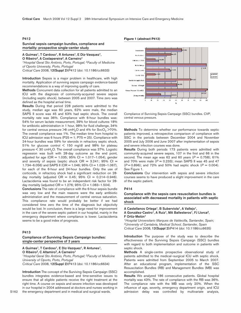
S162
P412Survival sepsis campaign bundles, compliance andmortality: prospective single-center study
A Guimas1, T Cardoso1, R Antunes1, E Diz-Vasquez1, O Ribeiro2, A Costapereira2, A Carneiro1
1Hospital Geral Sto António, Porto, Portugal; 2Faculty of Medicineof Oporto University, Porto, PortugalCritical Care 2008, 12(Suppl 2):P412 (doi: 10.1186/cc6633)
Introduction Sepsis is a major problem in healthcare, with highmortality. Application of surviving sepsis campaign evidence-basedrecommendations is a way of improving quality of care.Methods Concurrent data collection for all patients admitted to anICU with the diagnosis of community-acquired severe sepsis(including septic shock), between 2005 and 2007. Time zero wasdefined as the hospital arrival time.Results During that period 228 patients were admitted to thestudy, median age was 60 years, 62% were male, the medianSAPS II score was 45 and 63% had septic shock. The overallmortality rate was 36%. Compliance with 6-hour bundles was:54% for serum lactate measurement, 39% for blood cultures 18%for antibiotic administration in 1 hour, 98% for fluid challenge, 34%for central venous pressure >8 cmH2O and 4% for SvcO2 >70%.The overall compliance was 1%. The median time from hospital toICU admission was 6 hours (P25 = 1; P75 = 25). Compliance with24-hour bundles was 59% for steroids in refractory septic shock,51% for glucose control < 150 mg/dl and 98% for plateaupressure < 30 cmH2O. The overall compliance was 37%. Logisticregression was built with 28-day outcome as the end point,adjusted for age (OR = 1.035; 95% CI = 1.017–1.054), genderand severity of sepsis (septic shock OR = 3.241; 95% CI =1.734–6.056) and SAPS II (OR = 1.046; 95% CI = 1.026–1.067),for each of the 6-hour and 24-hour bundles. Only the use ofcorticoids, in refractory shock had a significant reduction on 28-day mortality (adjusted OR = 0.45; 95% CI = 0.214–0.946).Lactacidemia was found to be an independent risk factor for 28-day mortality (adjusted OR = 1.276; 95% CI = 1.083–1.504).Conclusions The rate of compliance with the 6-hour sepsis bundlewas very low and the main reasons were the early antibioticadministration and the measurement of central venous saturation.This compliance rate would probably be better if we hadconsidered time zero the time of the diagnosis but objectivitywould be lost. In conclusion, there is a large need for improvementin the care of the severe septic patient in our hospital, mainly in theemergency department where compliance is lower. Lactacidemiaseems to be a good index of prognosis.
P413Compliance of Surviving Sepsis Campaign bundles:single-center perspective of 3 years
A Guimas1, T Cardoso1, E Diz-Vazquez1, R Antunes1, O Ribeiro2, C Altamiro2, A Carneiro1
1Hospital Geral Sto António, Porto, Portugal; 2Faculty of MedicineUniversity of Oporto, Porto, PortugalCritical Care 2008, 12(Suppl 2):P413 (doi: 10.1186/cc6634)
Introduction The concept of the Surviving Sepsis Campaign (SSC)bundles integrates evidence-based and time-sensitive issues toensure that all eligible patients receive the right treatment at theright time. A course on sepsis and severe infection was developedin our hospital in 2004 addressed at doctors and nurses working inthe emergency department and in medical and surgical wards.
Methods To determine whether our performance towards septicpatients improved, a retrospective comparison of compliance withSSC in the periods between December 2004 and November2005 and July 2006 and June 2007 after implementation of sepsisand severe infection courses was done.Results During both periods 173 patients were admitted withcommunity-acquired severe sepsis, 107 in the first and 66 in thesecond. The mean age was 62 and 60 years (P = 0.758); 61%and 70% were male (P = 0.233); mean SAPS II was 45 and 47(P = 0.896); and 72% and 50% had septic shock (P = 0.004).See Figure 1.Conclusions Our intervention with sepsis and severe infectioncourses seems to have produced a slight improvement in the careof the septic patient.
P414Compliance with the sepsis care resuscitation bundles isassociated with decreased mortality in patients with septicshock
A Castellanos Ortega1, B Suberviola1, A Vallejo1, A González-Castro1, A Ruiz1, MA Ballesteros1, FJ Llorca2, F Ortíz-Melón1
1Hospital Universitario Marques de Valdecilla, Santander, Spain;2University of Cantabria, Medical School, Santander, SpainCritical Care 2008, 12(Suppl 2):P414 (doi: 10.1186/cc6635)
Introduction The purpose of the study was to describe theeffectiveness of the Surviving Sepsis Campaign (SSC) bundleswith regard to both implementation and outcome in patients withseptic shock.Methods A single-centre prospective observational study ofpatients admitted to the medical–surgical ICU with septic shock.Patients were admitted from September 2005 to March 2007.After an educational program, implementation of the SSCResuscitation Bundles (RB) and Management Bundles (MB) wasaccomplished.Results We analyzed 186 consecutive patients. Global hospitalmortality was 43%. The rate of compliance with the RB was 36%.The compliance rate with the MB was only 20%. When theinfluence of age, severity, emergency department origin, and ICUadmission delay was controlled by multivariate analysis,
Critical Care March 2008 Vol 12 Suppl 2 28th International Symposium on Intensive Care and Emergency Medicine
Figure 1 (abstract P413)
Compliance of Surviving Sepsis Campaign (SSC) bundles. CVP,central venous pressure.
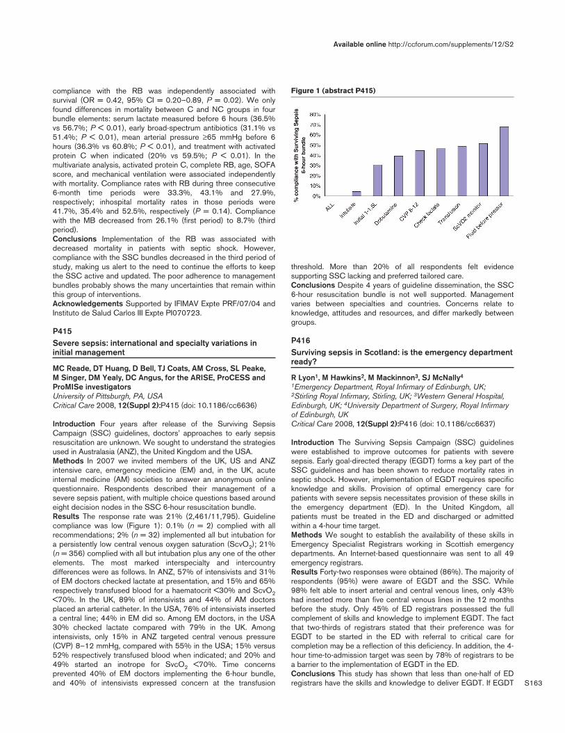
S163
compliance with the RB was independently associated withsurvival (OR = 0.42, 95% CI = 0.20–0.89, P = 0.02). We onlyfound differences in mortality between C and NC groups in fourbundle elements: serum lactate measured before 6 hours (36.5%vs 56.7%; P < 0.01), early broad-spectrum antibiotics (31.1% vs51.4%; P < 0.01), mean arterial pressure ≥65 mmHg before 6hours (36.3% vs 60.8%; P < 0.01), and treatment with activatedprotein C when indicated (20% vs 59.5%; P < 0.01). In themultivariate analysis, activated protein C, complete RB, age, SOFAscore, and mechanical ventilation were associated independentlywith mortality. Compliance rates with RB during three consecutive6-month time periods were 33.3%, 43.1% and 27.9%,respectively; inhospital mortality rates in those periods were41.7%, 35.4% and 52.5%, respectively (P = 0.14). Compliancewith the MB decreased from 26.1% (first period) to 8.7% (thirdperiod).Conclusions Implementation of the RB was associated withdecreased mortality in patients with septic shock. However,compliance with the SSC bundles decreased in the third period ofstudy, making us alert to the need to continue the efforts to keepthe SSC active and updated. The poor adherence to managementbundles probably shows the many uncertainties that remain withinthis group of interventions.Acknowledgements Supported by IFIMAV Expte PRF/07/04 andInstituto de Salud Carlos III Expte PI070723.
P415Severe sepsis: international and specialty variations ininitial management
MC Reade, DT Huang, D Bell, TJ Coats, AM Cross, SL Peake, M Singer, DM Yealy, DC Angus, for the ARISE, ProCESS andProMISe investigatorsUniversity of Pittsburgh, PA, USACritical Care 2008, 12(Suppl 2):P415 (doi: 10.1186/cc6636)
Introduction Four years after release of the Surviving SepsisCampaign (SSC) guidelines, doctors’ approaches to early sepsisresuscitation are unknown. We sought to understand the strategiesused in Australasia (ANZ), the United Kingdom and the USA.Methods In 2007 we invited members of the UK, US and ANZintensive care, emergency medicine (EM) and, in the UK, acuteinternal medicine (AM) societies to answer an anonymous onlinequestionnaire. Respondents described their management of asevere sepsis patient, with multiple choice questions based aroundeight decision nodes in the SSC 6-hour resuscitation bundle.Results The response rate was 21% (2,461/11,795). Guidelinecompliance was low (Figure 1): 0.1% (n = 2) complied with allrecommendations; 2% (n = 32) implemented all but intubation fora persistently low central venous oxygen saturation (ScvO2); 21%(n = 356) complied with all but intubation plus any one of the otherelements. The most marked interspecialty and intercountrydifferences were as follows. In ANZ, 57% of intensivists and 31%of EM doctors checked lactate at presentation, and 15% and 65%respectively transfused blood for a haematocrit <30% and ScvO2<70%. In the UK, 89% of intensivists and 44% of AM doctorsplaced an arterial catheter. In the USA, 76% of intensivists inserteda central line; 44% in EM did so. Among EM doctors, in the USA30% checked lactate compared with 79% in the UK. Amongintensivists, only 15% in ANZ targeted central venous pressure(CVP) 8–12 mmHg, compared with 55% in the USA; 15% versus52% respectively transfused blood when indicated; and 20% and49% started an inotrope for SvcO2 <70%. Time concernsprevented 40% of EM doctors implementing the 6-hour bundle,and 40% of intensivists expressed concern at the transfusion
threshold. More than 20% of all respondents felt evidencesupporting SSC lacking and preferred tailored care.Conclusions Despite 4 years of guideline dissemination, the SSC6-hour resuscitation bundle is not well supported. Managementvaries between specialties and countries. Concerns relate toknowledge, attitudes and resources, and differ markedly betweengroups.
P416Surviving sepsis in Scotland: is the emergency departmentready?
R Lyon1, M Hawkins2, M Mackinnon3, SJ McNally4
1Emergency Department, Royal Infirmary of Edinburgh, UK;2Stirling Royal Infirmary, Stirling, UK; 3Western General Hospital,Edinburgh, UK; 4University Department of Surgery, Royal Infirmaryof Edinburgh, UKCritical Care 2008, 12(Suppl 2):P416 (doi: 10.1186/cc6637)
Introduction The Surviving Sepsis Campaign (SSC) guidelineswere established to improve outcomes for patients with severesepsis. Early goal-directed therapy (EGDT) forms a key part of theSSC guidelines and has been shown to reduce mortality rates inseptic shock. However, implementation of EGDT requires specificknowledge and skills. Provision of optimal emergency care forpatients with severe sepsis necessitates provision of these skills inthe emergency department (ED). In the United Kingdom, allpatients must be treated in the ED and discharged or admittedwithin a 4-hour time target.Methods We sought to establish the availability of these skills inEmergency Specialist Registrars working in Scottish emergencydepartments. An Internet-based questionnaire was sent to all 49emergency registrars.Results Forty-two responses were obtained (86%). The majority ofrespondents (95%) were aware of EGDT and the SSC. While98% felt able to insert arterial and central venous lines, only 43%had inserted more than five central venous lines in the 12 monthsbefore the study. Only 45% of ED registrars possessed the fullcomplement of skills and knowledge to implement EGDT. The factthat two-thirds of registrars stated that their preference was forEGDT to be started in the ED with referral to critical care forcompletion may be a reflection of this deficiency. In addition, the 4-hour time-to-admission target was seen by 78% of registrars to bea barrier to the implementation of EGDT in the ED.Conclusions This study has shown that less than one-half of EDregistrars have the skills and knowledge to deliver EGDT. If EGDT
Available online http://ccforum.com/supplements/12/S2
Figure 1 (abstract P415)
S164
is to be implemented within the ED, appropriately skilled personneland equipment must be available. Early referral to critical care isessential if the SSC aim of reducing mortality from sepsis is to beachieved.Reference1. Rivers E, Nguyen B, Havstad S, et al.: Early goal directed
therapy in the treatment of severe sepsis and septicshock. N Engl J Med 2001, 345:1368-1377.
P417A multicentre study on early goal-directed therapy ofsevere sepsis and septic shock patients in the ICU:collaborative study group on early goal-directed therapy inZhejiang Province, China
J Yan, G CaiZhejiang Hospital, Hangzhou, ChinaCritical Care 2008, 12(Suppl 2):P417 (doi: 10.1186/cc6638)
Introduction The use of early goal-directed therapy (EGDT) insevere sepsis and septic shock has been shown to decreasemorbidity and mortality rates significantly when given to patientsprior to ICU admission, especially in the emergency department(ED). The aim of this study was to assess the efficacy of EGDT insevere sepsis and septic shock patients in the ICU.Methods A muticentre, prospective, randomized and controlledstudy. We randomly assigned patients admitted to the ICU withsevere sepsis or septic shock to receive either 6 hours of EGDT orstandard therapy (as a control). The primary end point (28-daymortality for any cause) and secondary end points (ICU stay days,mechanical ventilation duration, APACHE II scores and MODSscores) were obtained serially for 28 days and compared betweenthe two groups.Results Of the 313 enrolled patients, 162 were randomlyassigned to EGDT and 151 to standard therapy; there were nosignificant differences between the groups with respect tobaseline characteristics. The 28-day mortality was 24.8% in thegroup assigned to EGDT, as compared with 42.5% in the groupassigned to standard therapy (P = 0.001). During the interval fromthe first hour to 28 days stay in the ICU, the patients assigned toEGDT had a significantly lower APACHE II score (14.44 ± 8.46 vs18.00 ± 7.04, P = 0.043) and MODS score (4.53 ± 3.55 vs 7.09± 4.95, P = 0.009) than those assigned to standard therapy; therewere no differences in ICU stay days (20.6 ± 1.9 vs 19.9 ± 2.2, P= 0.82) and mechanical ventilation duration (13.2 ± 1.5 vs 14.4 ±1.6, P = 0.6) between the two groups.Conclusions EGDT provides significant benefits with respect tooutcome and scores in patients with severe sepsis and septicshock in the ICU.References1. Varpula M, et al.: Hemodynamic variables related to out-
come in septic shock. Intensive Care Med 2005, 31:1066-1071.
2. Rivers E, et al.: Early goal-directed therapy in the treatmentof severe sepsis and septic shock. N Engl J Med 2001,345:1368-1377.
P418Effect of prolonged emergency department length of stayon inpatient length of stay and inhospital mortality insevere sepsis and septic shock
K Haji, D Haji, V Le Blanc, J BothaPeninsula Health Care Network, Frankston, Victoria, AustraliaCritical Care 2008, 12(Suppl 2):P418 (doi: 10.1186/cc6639)
Introduction With overcrowding in our emergency departmentsand an access block issue, the emergency department (ED) lengthof stay (LOS) is becoming a growing concern. Over the pastdecade, despite decreasing mortality in sepsis and septic shock inAustralia and NZ hospitals, the reported incidence of sepsis andseptic shock in ICU patients presenting to the ED has increased. Inthis study we postulated that, in severe sepsis and septic shockpatients, prolonged ED LOS is associated with increased ICU andhospital LOS as well as increased inhospital mortality.Methods A retrospective observational study. Data were collectedfrom 1 July 2004 to 31 October 2007. The setting was a cosmo-politan 360-bed teaching hospital with a nine-bed general ICU.The ED has 50,000 presentations per year.Results There were 120 patients with diagnosis of severe sepsisand septic shock admitted to the ICU. They were equallydistributed males and females with a mean age of 64.75 years. Weexcluded three patients due to incomplete data. The mean ED LOSwas 10.05 hours and the median was 8.33 hours. The mean ICULOS and hospital LOS were 5.54 and 15.02 days, respectively.Twenty-six patients (22.2%) died in the hospital. Based on EDLOS, the patients were divided into three groups of <6 hours, 6–8hours and >10 hours. There was no association between ED LOSand inhospital mortality (P = 0.23) between the three groups.Similarly, no association was found between ED LOS and ICULOS or hospital LOS (P = 0.31 and P = 0.28, respectively).Gender, age and SAP score did not affect outcomes. However, asignificant association was found between APACHE II score andICU LOS (P = 0.009), with no effect on hospital LOS or mortality.Conclusions Our study showed that, in the severe sepsis andseptic shock patient group, a prolonged ED LOS had no effect onICU and hospital LOS or on inhospital mortality.References1. ARISE investigators: Crit Care Resusc 2007, 9:8-18.2. Chalfin D, et al.: Crit Care Med 2007, 35:1477-1483.3. Soukkonen KA et al.: J Internal Med 2006, 260:586-591.4. Richardson DB, et al.: Med J Aust 2002, 177:492-495.
P419Does early antibiotic administration affect disposition orlength of stay in patients with cryptic shock? A retrospective pilot study
H Chung, R Otero, S Rivers, S Sankey, E RiversHenry Ford Hospital, Detroit, MI, USACritical Care 2008, 12(Suppl 2):P419 (doi: 10.1186/cc6640)
Introduction Recent published literature supports therecommendations of early antibiotic administration for patients withsepsis. However, certain patients with suspected sepsis do notmanifest typical symptoms, such as perturbations in bloodpressure and heart rate. To identify these high-risk patients,surrogate evidence of hypoperfusion such as hyperlactatemia anddecreased central venous oxygen saturation may be employed.These patients are described as being in compensated or ‘crypticshock’. The current study investigates whether early administrationof antibiotics affects the ICU or hospital length of stay (LOS) inpatients with ‘cryptic shock’.Methods The current investigation is a retrospective analysis(January 2000–December 2005) of patients evaluated in theemergency department with evidence of cryptic shock. Twohundred and sixty patients were identified. Cryptic shock diagnosiswas based upon the lactate level, central venous oxygen saturationand mean arterial pressure (MAP) at the time of presentation toevaluate the impact of early antibiotic administration (antibiotics <1hour, or antibiotics <3 hours) on disposition and length of stay.
Critical Care March 2008 Vol 12 Suppl 2 28th International Symposium on Intensive Care and Emergency Medicine
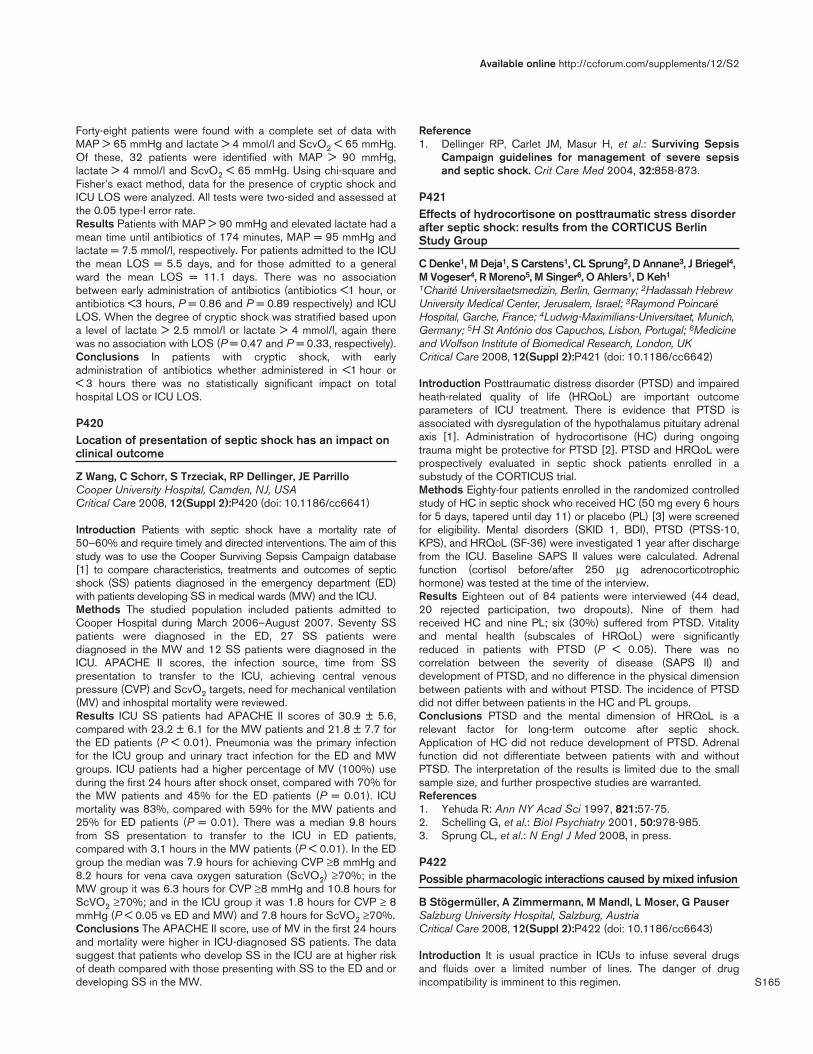
S165
Forty-eight patients were found with a complete set of data withMAP > 65 mmHg and lactate > 4 mmol/l and ScvO2 < 65 mmHg.Of these, 32 patients were identified with MAP > 90 mmHg,lactate > 4 mmol/l and ScvO2 < 65 mmHg. Using chi-square andFisher’s exact method, data for the presence of cryptic shock andICU LOS were analyzed. All tests were two-sided and assessed atthe 0.05 type-I error rate.Results Patients with MAP > 90 mmHg and elevated lactate had amean time until antibiotics of 174 minutes, MAP = 95 mmHg andlactate = 7.5 mmol/l, respectively. For patients admitted to the ICUthe mean LOS = 5.5 days, and for those admitted to a generalward the mean LOS = 11.1 days. There was no associationbetween early administration of antibiotics (antibiotics <1 hour, orantibiotics <3 hours, P = 0.86 and P = 0.89 respectively) and ICULOS. When the degree of cryptic shock was stratified based upona level of lactate > 2.5 mmol/l or lactate > 4 mmol/l, again therewas no association with LOS (P = 0.47 and P = 0.33, respectively).Conclusions In patients with cryptic shock, with earlyadministration of antibiotics whether administered in <1 hour or< 3 hours there was no statistically significant impact on totalhospital LOS or ICU LOS.
P420Location of presentation of septic shock has an impact onclinical outcome
Z Wang, C Schorr, S Trzeciak, RP Dellinger, JE ParrilloCooper University Hospital, Camden, NJ, USACritical Care 2008, 12(Suppl 2):P420 (doi: 10.1186/cc6641)
Introduction Patients with septic shock have a mortality rate of50–60% and require timely and directed interventions. The aim of thisstudy was to use the Cooper Surviving Sepsis Campaign database[1] to compare characteristics, treatments and outcomes of septicshock (SS) patients diagnosed in the emergency department (ED)with patients developing SS in medical wards (MW) and the ICU.Methods The studied population included patients admitted toCooper Hospital during March 2006–August 2007. Seventy SSpatients were diagnosed in the ED, 27 SS patients werediagnosed in the MW and 12 SS patients were diagnosed in theICU. APACHE II scores, the infection source, time from SSpresentation to transfer to the ICU, achieving central venouspressure (CVP) and ScvO2 targets, need for mechanical ventilation(MV) and inhospital mortality were reviewed.Results ICU SS patients had APACHE II scores of 30.9 ± 5.6,compared with 23.2 ± 6.1 for the MW patients and 21.8 ± 7.7 forthe ED patients (P < 0.01). Pneumonia was the primary infectionfor the ICU group and urinary tract infection for the ED and MWgroups. ICU patients had a higher percentage of MV (100%) useduring the first 24 hours after shock onset, compared with 70% forthe MW patients and 45% for the ED patients (P = 0.01). ICUmortality was 83%, compared with 59% for the MW patients and25% for ED patients (P = 0.01). There was a median 9.8 hoursfrom SS presentation to transfer to the ICU in ED patients,compared with 3.1 hours in the MW patients (P < 0.01). In the EDgroup the median was 7.9 hours for achieving CVP ≥8 mmHg and8.2 hours for vena cava oxygen saturation (ScVO2) ≥70%; in theMW group it was 6.3 hours for CVP ≥8 mmHg and 10.8 hours forScVO2 ≥70%; and in the ICU group it was 1.8 hours for CVP ≥ 8mmHg (P < 0.05 vs ED and MW) and 7.8 hours for ScVO2 ≥70%.Conclusions The APACHE II score, use of MV in the first 24 hoursand mortality were higher in ICU-diagnosed SS patients. The datasuggest that patients who develop SS in the ICU are at higher riskof death compared with those presenting with SS to the ED and ordeveloping SS in the MW.
Reference1. Dellinger RP, Carlet JM, Masur H, et al.: Surviving Sepsis
Campaign guidelines for management of severe sepsisand septic shock. Crit Care Med 2004, 32:858-873.
P421Effects of hydrocortisone on posttraumatic stress disorderafter septic shock: results from the CORTICUS BerlinStudy Group
C Denke1, M Deja1, S Carstens1, CL Sprung2, D Annane3, J Briegel4,M Vogeser4, R Moreno5, M Singer6, O Ahlers1, D Keh1
1Charité Universitaetsmedizin, Berlin, Germany; 2Hadassah HebrewUniversity Medical Center, Jerusalem, Israel; 3Raymond PoincaréHospital, Garche, France; 4Ludwig-Maximilians-Universitaet, Munich,Germany; 5H St António dos Capuchos, Lisbon, Portugal; 6Medicineand Wolfson Institute of Biomedical Research, London, UKCritical Care 2008, 12(Suppl 2):P421 (doi: 10.1186/cc6642)
Introduction Posttraumatic distress disorder (PTSD) and impairedheath-related quality of life (HRQoL) are important outcomeparameters of ICU treatment. There is evidence that PTSD isassociated with dysregulation of the hypothalamus pituitary adrenalaxis [1]. Administration of hydrocortisone (HC) during ongoingtrauma might be protective for PTSD [2]. PTSD and HRQoL wereprospectively evaluated in septic shock patients enrolled in asubstudy of the CORTICUS trial.Methods Eighty-four patients enrolled in the randomized controlledstudy of HC in septic shock who received HC (50 mg every 6 hoursfor 5 days, tapered until day 11) or placebo (PL) [3] were screenedfor eligibility. Mental disorders (SKID 1, BDI), PTSD (PTSS-10,KPS), and HRQoL (SF-36) were investigated 1 year after dischargefrom the ICU. Baseline SAPS II values were calculated. Adrenalfunction (cortisol before/after 250 μg adrenocorticotrophichormone) was tested at the time of the interview.Results Eighteen out of 84 patients were interviewed (44 dead,20 rejected participation, two dropouts). Nine of them hadreceived HC and nine PL; six (30%) suffered from PTSD. Vitalityand mental health (subscales of HRQoL) were significantlyreduced in patients with PTSD (P < 0.05). There was nocorrelation between the severity of disease (SAPS II) anddevelopment of PTSD, and no difference in the physical dimensionbetween patients with and without PTSD. The incidence of PTSDdid not differ between patients in the HC and PL groups.Conclusions PTSD and the mental dimension of HRQoL is arelevant factor for long-term outcome after septic shock.Application of HC did not reduce development of PTSD. Adrenalfunction did not differentiate between patients with and withoutPTSD. The interpretation of the results is limited due to the smallsample size, and further prospective studies are warranted.References1. Yehuda R: Ann NY Acad Sci 1997, 821:57-75.2. Schelling G, et al.: Biol Psychiatry 2001, 50:978-985.3. Sprung CL, et al.: N Engl J Med 2008, in press.
P422Possible pharmacologic interactions caused by mixed infusion
B Stögermüller, A Zimmermann, M Mandl, L Moser, G PauserSalzburg University Hospital, Salzburg, AustriaCritical Care 2008, 12(Suppl 2):P422 (doi: 10.1186/cc6643)
Introduction It is usual practice in ICUs to infuse several drugsand fluids over a limited number of lines. The danger of drugincompatibility is imminent to this regimen.
Available online http://ccforum.com/supplements/12/S2
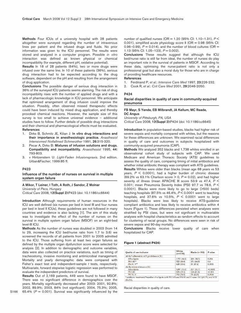
S166
Methods Four ICUs of a university hospital with 28 patientsaltogether were surveyed regarding the number of intravenouslines per patient and the infused drugs and fluids. No priorinformation was given to the ICU personnel. The results werestored and analyzed in a computer program. Possible in vitrointeraction was defined as known physical or chemicalincompatibility (for example, different pH, oxidative potential).Results In 18 of 28 patients (64%), two or more drugs wereinfused over the same line. In 10 of these patients (36%), seriousdrug interaction had to be expected according to the drugsoftware, dependent on the pH and resulting from the arrangementof drug application.Conclusions The possible danger of serious drug interaction in36% of the surveyed ICU patients seems alarming. The risk of drugincompatibility rises with the number of applied drugs and with alack of pharmacologic knowledge in ICU personnel. We anticipatethat optimized arrangement of drug infusion could improve thesituation. Possibly, often observed missed therapeutic effectscould have been induced by mixed drug application, and therebyprovoked chemical reactions. However, the sample size of thissurvey is too small to achieve universal evidence – additionalstudies have to follow. Further details of possible drug interactionsand their chemical and pharmacological effects must be evaluated.References1. Dirks B, Schmitz JE, Kilian J: In vitro drug interactions and
their importance in anesthesiologic practice. AnasthesiolIntensivmed Notfallmed Schmerzther 1991, 26:315-320.
2. Pecar A, Dirks B: Mixtures of infusion solutions and drugs.Compatibility and incompatibility. Anaesthesist 1995, 44:793-803.
3. v Hintzenstern U: Light-Faden Infusionspraxis. 2nd edition.Urban&Fischer; 1999:85 ff.
P423Influence of the number of nurses on survival in multiplesystem organ failure
A Mikor, T Leiner, I Toth, A Roth, J Sandor, Z MolnarUniversity of Pecs, HungaryCritical Care 2008, 12(Suppl 2):P423 (doi: 10.1186/cc6644)
Introduction Although requirements of human resources in theICU are well defined (six nurses per bed in level III and four nursesper bed in level II ICUs), these guidelines are not followed in manycountries and evidence is also lacking [1]. The aim of this studywas to investigate the effect of the number of nurses on thesurvival in multiple system organ failure (MSOF) on an eight-bedlevel-III ICU.Methods As the number of nurses was doubled in 2003 (from 14to 29, increasing the ICU bed/nurse ratio from 1.7 to 3.6) wescreened the records of all patients from 2001 to 2005 admittedto the ICU. Those suffering from at least two organ failures asdefined by the multiple organ dysfunction score were selected foranalysis [2]. In addition to demographic and outcome variables,data were also collected on practice variations, such as timing oftracheostomy, invasive monitoring and antimicrobial management.Mortality and yearly demographic data were compared withFisher’s exact test and independent-sample t tests, respectively.Multivariate, forward stepwise logistic regression was performed toevaluate the independent predictors of survival.Results Out of 2,169 patients, 449 were found to have MSOF.There was no significant difference in demographics over theyears. Mortality significantly decreased after 2003: 2001, 92.8%;2002, 89.9%; 2003, 84% (not significant); 2004, 75.3%; 2005,65.4% (P = 0.001). Survival was significantly influenced by the
number of qualified nurses (OR = 1.20 (95% CI: 1.10–1.31), P <0.001), simplified acute physiology score II (OR = 0.98 (95% CI:0.96–0.99), P = 0.014), and the number of blood cultures (OR =1.13 (95% CI: 1.05–123), P = 0.002).Conclusions These results suggest that although the ICUbed/nurse ratio is still far from ideal, the number of nurses do playan important role in the survival of patients in MSOF. According tothese data, optimising the nurse:patient ratio is not only aprofessional goal but also a moral duty for those who are in chargeof providing healthcare resources.References1. Ferdinand P, et al.: Intensive Care Med 1997, 23:226-232.2. Cook R, et al.: Crit Care Med 2001, 29:2046-2050.
P424Racial disparities in quality of care in community-acquiredpneumonia
FB Mayr, S Yende, EB Milbrandt, JA Kellum, MC Reade, DC AngusUniversity of Pittsburgh, PA, USACritical Care 2008, 12(Suppl 2):P424 (doi: 10.1186/cc6645)
Introduction In population-based studies, blacks had higher risk ofsevere sepsis and mortality compared with whites, but the reasonsfor these differences are unknown. We examined racial differencesin quality of care and outcomes in subjects hospitalized withcommunity-acquired pneumonia (CAP).Methods We analyzed 352 blacks and 1,738 whites enrolled in anobservational cohort study of subjects with CAP. We usedMedicare and American Thoracic Society (ATS) guidelines toassess the quality of care, comparing timing of initial antibiotics andwhether initial antibiotic therapy was compliant with ATS guidelines.Results Whites were older than blacks (mean age 68 years vs 53years, P < 0.0001), had a higher burden of chronic disease(69.2% vs 63.1% Charlson score > 0, P = 0.02), and had higherseverity of illness (mean APACHE III score 53.9 vs 47.4, P <0.001; mean Pneumonia Severity Index (PSI) 97.7 vs 78.6, P <0.0001). Blacks were more likely to go to large (>500 beds)teaching hospitals (87.5% vs 46.4%, P < 0.0001 went to teachinghospitals, and 37.6% vs 10.5%, P < 0.0001 went to largehospitals). Blacks were less likely to receive ATS-guidelinecompliant antibiotics and less likely to receive antibiotics within 4hours (Figure 1). These differences persisted when analyses werestratified by PSI class, but were not significant in multivariableanalyses with hospital characteristics as random effects to accountfor clustering of racial groups. No differences were seen in risk ofsevere sepsis and 90-day mortality.Conclusions Blacks receive lower quality of care whenhospitalized for CAP.
Critical Care March 2008 Vol 12 Suppl 2 28th International Symposium on Intensive Care and Emergency Medicine
Figure 1 (abstract P424)
Racial disparities in quality of care.
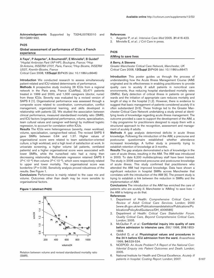
S167
Acknowledgements Supported by T32HL00782010 andR01GM61992.
P425Global assessment of performance of ICUs: a Frenchexperience
A Faye1, P Aegerter1, A Boumendil2, E Minvielle3, B Guidet2
1Hopital Ambroise Paré (AP-HP), Boulogne, France; 2HopSt-Antoine, INSERM U707, Paris, France; 3Hop Bicetre, INSERMU537, Kremlin-Bicetre, FranceCritical Care 2008, 12(Suppl 2):P425 (doi: 10.1186/cc6646)
Introduction We conducted research to assess simultaneouslypatient-related and ICU-related determinants of performance.Methods A prospective study involving 26 ICUs from a regionalnetwork in the Paris area, France (CubRéa), 33,471 patientstreated in 1999 and 2000, and 1,000 caregivers (doctor, nurse)from these ICUs. Severity was evaluated by a revised version ofSAPS II [1]. Organisational performance was assessed through acomposite score related to coordination, communication, conflictmanagement, organizational learning, and skills developed inrelationship with patients [2]. We studied the association betweenclinical performance, measured standardised mortality ratio (SMR),and ICU factors (organisational performance, volume, specialisation,team cultural values and caregiver well-being) by multilevel logisticregression, to account for correlation within ICUs.Results The ICUs were heterogeneous (severity, mean workload,volume, specialisation, caregiver/bed ratios). The revised SAPS IIgave SMRs between 0.64 and 1.37. Higher values oforganisational score were related to team satisfaction-orientedculture, a high workload, and a high level of satisfaction at work. Inunivariate screening, a higher volume (all patients, ventilatedpatients) and a higher organisational score were associated withlower SMRs, while the nurse/bed ratio had a rising thendecreasing relationship. Multivariate regression retained SAPS II(P < 10–4) then volume (P < 10–3), which were respectively relatedto upper and lower mortality. The organisational score wasborderline (P = 0.09). Sensitivity analysis proved robustness of theresults. See Figure 1.Conclusions Performance is mainly related to the case mix andvolume. Outcomes other than death may be more sensible toorganisational factors.
References1. Aegerter P, et al.: Intensive Care Med 2005. 31:416-423.2. Minvielle E, et al.: J Crit Care in press.
P426AIMing to save lives
A Berry, A StevensGreater Manchester Critical Care Network, Manchester, UKCritical Care 2008, 12(Suppl 2):P426 (doi: 10.1186/cc6647)
Introduction This poster guides us through the process ofunderstanding how the Acute Illness Management Course (AIM)originated and its effectiveness in enabling practitioners to providequality care to acutely ill adult patients in noncritical careenvironments, thus reducing hospital standardised mortality rates(SMRs). Early detection of critical illness in patients on generalwards and the initiation of appropriate care reduces mortality andlength of stay in the hospital [1,2]. However, there is evidence tosuggest that basic management of patients considered acutely ill isoften substandard [3-5]. These findings led to the Greater Man-chester Critical Care Network undertaking a study aimed at identi-fying levels of knowledge regarding acute illness management. Theoutcome provided a case to support the development of the AIM, a1-day programme for practitioners designed to equip them with astructured approach to the recognition, assessment and manage-ment of acutely ill adults.Methods A gap analysis determined deficits in acute illnessknowledge. Following the introduction of the AIM, a precourse andpostcourse questionnaire established whether attendanceincreased knowledge. A further study is presently trying toestablish retention of knowledge at 3 months.Results The gap analysis demonstrated a lack of knowledge in thecare of acute illness, resulting in the AIM. The AIM was introducedin 2003. To date 6,200 multidisciplinary staff have been trained.The study in 2006 examined precourse and postcourse knowledgeof acute illness. This study confirmed that practitioners whoattended the AIM had improved knowledge. Data have shown asignificant reduction in hospital SMRs across Manchester thatcorrelates with the introduction of the AIM [6]. The present study istrying to establish a link between the reduction in SMRs and theimplementation of AIM.Conclusions The introduction of the AIM has enriched the care ofpatients who are acutely ill. Manchester is ‘AIMing’ to save lives –the AIM is helping us do that.References1. Department of Health: Comprehensive Critical Care; A
Review of Adult Critical Care Services. London; 2000[www.dh.gov.uk/en/Publicationsandstatistics/Publications/PublicationsPolicyAndGuidance/DH_4006585]
2. Department of Health: Critical Care Stakeholder Forum.Quality Critical Care, Beyond Comprehensive Critical Care.London; 2005.
3. McQuillan P, et al.: Confidential inquiry into quality of carebefore admission to intensive care. BMJ 1998, 316:1853-1858.
4. Goldhill D, et al. Physiological values and procedures inthe 24 h before ICU admission from the ward. Anaesthesia1999, 54:529-534.
5. NCEPOD: An Acute Problem? A Report of the National Con-fidential Enquiry into Patient Outcomes and Death. London;2005.
6. National Institute for Health and Clinical Excellence. Acutely illpatients in hospital. Costing Report. London; 2007.
Available online http://ccforum.com/supplements/12/S2
Figure 1 (abstract P425)
Relation between volume and revised standardised mortality ratio(SMR).
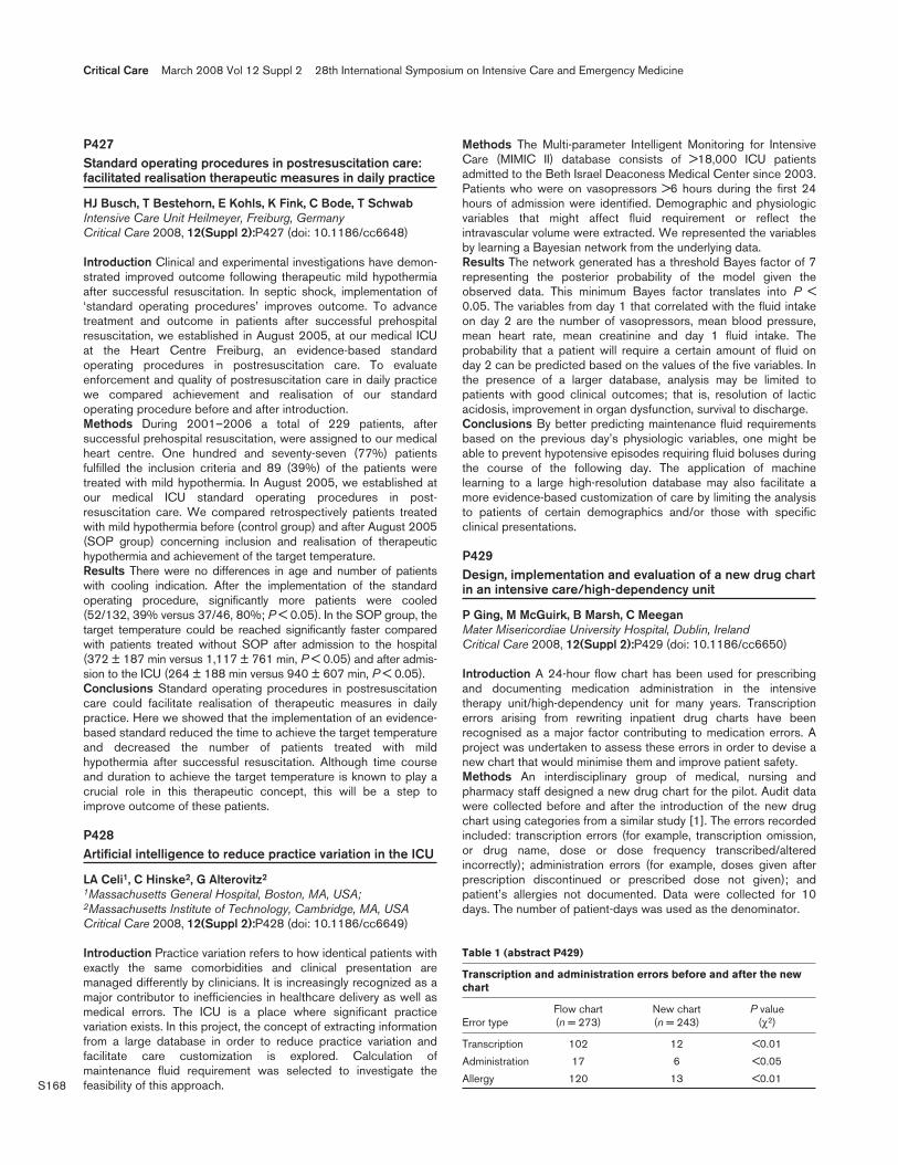
S168
P427Standard operating procedures in postresuscitation care:facilitated realisation therapeutic measures in daily practice
HJ Busch, T Bestehorn, E Kohls, K Fink, C Bode, T SchwabIntensive Care Unit Heilmeyer, Freiburg, GermanyCritical Care 2008, 12(Suppl 2):P427 (doi: 10.1186/cc6648)
Introduction Clinical and experimental investigations have demon-strated improved outcome following therapeutic mild hypothermiaafter successful resuscitation. In septic shock, implementation of‘standard operating procedures’ improves outcome. To advancetreatment and outcome in patients after successful prehospitalresuscitation, we established in August 2005, at our medical ICUat the Heart Centre Freiburg, an evidence-based standardoperating procedures in postresuscitation care. To evaluateenforcement and quality of postresuscitation care in daily practicewe compared achievement and realisation of our standardoperating procedure before and after introduction.Methods During 2001–2006 a total of 229 patients, aftersuccessful prehospital resuscitation, were assigned to our medicalheart centre. One hundred and seventy-seven (77%) patientsfulfilled the inclusion criteria and 89 (39%) of the patients weretreated with mild hypothermia. In August 2005, we established atour medical ICU standard operating procedures in post-resuscitation care. We compared retrospectively patients treatedwith mild hypothermia before (control group) and after August 2005(SOP group) concerning inclusion and realisation of therapeutichypothermia and achievement of the target temperature.Results There were no differences in age and number of patientswith cooling indication. After the implementation of the standardoperating procedure, significantly more patients were cooled(52/132, 39% versus 37/46, 80%; P < 0.05). In the SOP group, thetarget temperature could be reached significantly faster comparedwith patients treated without SOP after admission to the hospital(372 ± 187 min versus 1,117 ± 761 min, P < 0.05) and after admis-sion to the ICU (264 ± 188 min versus 940 ± 607 min, P < 0.05).Conclusions Standard operating procedures in postresuscitationcare could facilitate realisation of therapeutic measures in dailypractice. Here we showed that the implementation of an evidence-based standard reduced the time to achieve the target temperatureand decreased the number of patients treated with mildhypothermia after successful resuscitation. Although time courseand duration to achieve the target temperature is known to play acrucial role in this therapeutic concept, this will be a step toimprove outcome of these patients.
P428Artificial intelligence to reduce practice variation in the ICU
LA Celi1, C Hinske2, G Alterovitz2
1Massachusetts General Hospital, Boston, MA, USA;2Massachusetts Institute of Technology, Cambridge, MA, USACritical Care 2008, 12(Suppl 2):P428 (doi: 10.1186/cc6649)
Introduction Practice variation refers to how identical patients withexactly the same comorbidities and clinical presentation aremanaged differently by clinicians. It is increasingly recognized as amajor contributor to inefficiencies in healthcare delivery as well asmedical errors. The ICU is a place where significant practicevariation exists. In this project, the concept of extracting informationfrom a large database in order to reduce practice variation andfacilitate care customization is explored. Calculation ofmaintenance fluid requirement was selected to investigate thefeasibility of this approach.
Methods The Multi-parameter Intelligent Monitoring for IntensiveCare (MIMIC II) database consists of >18,000 ICU patientsadmitted to the Beth Israel Deaconess Medical Center since 2003.Patients who were on vasopressors >6 hours during the first 24hours of admission were identified. Demographic and physiologicvariables that might affect fluid requirement or reflect theintravascular volume were extracted. We represented the variablesby learning a Bayesian network from the underlying data.Results The network generated has a threshold Bayes factor of 7representing the posterior probability of the model given theobserved data. This minimum Bayes factor translates into P <0.05. The variables from day 1 that correlated with the fluid intakeon day 2 are the number of vasopressors, mean blood pressure,mean heart rate, mean creatinine and day 1 fluid intake. Theprobability that a patient will require a certain amount of fluid onday 2 can be predicted based on the values of the five variables. Inthe presence of a larger database, analysis may be limited topatients with good clinical outcomes; that is, resolution of lacticacidosis, improvement in organ dysfunction, survival to discharge.Conclusions By better predicting maintenance fluid requirementsbased on the previous day’s physiologic variables, one might beable to prevent hypotensive episodes requiring fluid boluses duringthe course of the following day. The application of machinelearning to a large high-resolution database may also facilitate amore evidence-based customization of care by limiting the analysisto patients of certain demographics and/or those with specificclinical presentations.
P429Design, implementation and evaluation of a new drug chartin an intensive care/high-dependency unit
P Ging, M McGuirk, B Marsh, C MeeganMater Misericordiae University Hospital, Dublin, IrelandCritical Care 2008, 12(Suppl 2):P429 (doi: 10.1186/cc6650)
Introduction A 24-hour flow chart has been used for prescribingand documenting medication administration in the intensivetherapy unit/high-dependency unit for many years. Transcriptionerrors arising from rewriting inpatient drug charts have beenrecognised as a major factor contributing to medication errors. Aproject was undertaken to assess these errors in order to devise anew chart that would minimise them and improve patient safety.Methods An interdisciplinary group of medical, nursing andpharmacy staff designed a new drug chart for the pilot. Audit datawere collected before and after the introduction of the new drugchart using categories from a similar study [1]. The errors recordedincluded: transcription errors (for example, transcription omission,or drug name, dose or dose frequency transcribed/alteredincorrectly); administration errors (for example, doses given afterprescription discontinued or prescribed dose not given); andpatient’s allergies not documented. Data were collected for 10days. The number of patient-days was used as the denominator.
Critical Care March 2008 Vol 12 Suppl 2 28th International Symposium on Intensive Care and Emergency Medicine
Table 1 (abstract P429)
Transcription and administration errors before and after the newchart
Flow chart New chart P value Error type (n = 273) (n = 243) (χ2)
Transcription 102 12 <0.01
Administration 17 6 <0.05
Allergy 120 13 <0.01
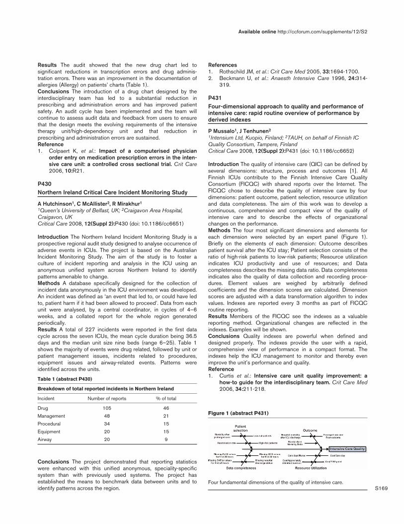
S169
Results The audit showed that the new drug chart led tosignificant reductions in transcription errors and drug adminis-tration errors. There was an improvement in the documentation ofallergies (Allergy) on patients’ charts (Table 1).Conclusions The introduction of a drug chart designed by theinterdisciplinary team has led to a substantial reduction inprescribing and administration errors and has improved patientsafety. An audit cycle has been implemented and the team willcontinue to assess audit data and feedback from users to ensurethat the design meets the evolving requirements of the intensivetherapy unit/high-dependency unit and that reduction inprescribing and administration errors are sustained.Reference1. Colpaert K, et al.: Impact of a computerised physician
order entry on medication prescription errors in the inten-sive care unit: a controlled cross sectional trial. Crit Care2006, 10:R21.
P430Northern Ireland Critical Care Incident Monitoring Study
A Hutchinson1, C McAllister2, R Mirakhur1
1Queen’s University of Belfast, UK; 2Craigavon Area Hospital,Craigavon, UKCritical Care 2008, 12(Suppl 2):P430 (doi: 10.1186/cc6651)
Introduction The Northern Ireland Incident Monitoring Study is aprospective regional audit study designed to analyse occurrence ofadverse events in ICUs. The project is based on the AustralianIncident Monitoring Study. The aim of the study is to foster aculture of incident reporting and analysis in the ICU using ananonymous unified system across Northern Ireland to identifypatterns amenable to change.Methods A database specifically designed for the collection ofincident data anonymously in the ICU environment was developed.An incident was defined as ‘an event that led to, or could have ledto, patient harm if it had been allowed to proceed’. Data from eachunit were analysed, by a central coordinator, in cycles of 4–6weeks, and a collated report for the whole region generatedperiodically.Results A total of 227 incidents were reported in the first datacycle across the seven ICUs, the mean cycle duration being 36.5days and the median unit size nine beds (range 6–25). Table 1shows the majority of events were drug related, followed by unit orpatient management issues, incidents related to procedures,equipment issues and airway-related events. Patterns wereidentified across the units.
Table 1 (abstract P430)
Breakdown of total reported incidents in Northern Ireland
Incident Number of reports % of total
Drug 105 46
Management 48 21
Procedural 34 15
Equipment 20 15
Airway 20 9
Conclusions The project demonstrated that reporting statisticswere enhanced with this unified anonymous, speciality-specificsystem than with previously used systems. The project hasestablished the means to benchmark data between units and toidentify patterns across the region.
References1. Rothschild JM, et al.: Crit Care Med 2005, 33:1694-1700.2. Beckmann U, et al.: Anaesth Intensive Care 1996, 24:314-
319.
P431Four-dimensional approach to quality and performance ofintensive care: rapid routine overview of performance byderived indexes
P Mussalo1, J Tenhunen2
1Intensium Ltd, Kuopio, Finland; 2TAUH, on behalf of Finnish ICQuality Consortium, Tampere, FinlandCritical Care 2008, 12(Suppl 2):P431 (doi: 10.1186/cc6652)
Introduction The quality of intensive care (QIC) can be defined byseveral dimensions: structure, process and outcomes [1]. AllFinnish ICUs contribute to the Finnish Intensive Care QualityConsortium (FICQC) with shared reports over the Internet. TheFICQC chose to describe the quality of intensive care by fourdimensions: patient outcome, patient selection, resource utilizationand data completeness. The aim of this work was to develop acontinuous, comprehensive and compact view of the quality ofintensive care and to describe the effects of organizationalchanges on the performance.Methods The four most significant dimensions and elements foreach dimension were selected by an expert panel (Figure 1).Briefly on the elements of each dimension: Outcome describespatient survival after the ICU stay; Patient selection consists of theratio of high-risk patients to low-risk patients; Resource utilizationindicates ICU productivity and use of resources; and Datacompleteness describes the missing data ratio. Data completenessindicates also the quality of data collection and recording proce-dures. Element values are weighed by arbitrarily definedcoefficients and the dimension scores are calculated. Dimensionscores are adjusted with a data transformation algorithm to indexvalues. Indexes are reported every 3 months as part of FICQCroutine reporting.Results Members of the FICQC see the indexes as a valuablereporting method. Organizational changes are reflected in theindexes. Examples will be shown.Conclusions Quality indexes are powerful when defined anddesigned properly. The indexes provide the user with a rapid,comprehensive view of performance in a compact format. Theindexes help the ICU management to monitor and thereby evenimprove the unit’s performance and quality.Reference1. Curtis et al.: Intensive care unit quality improvement: a
how-to guide for the interdisciplinary team. Crit Care Med2006, 34:211-218.
Available online http://ccforum.com/supplements/12/S2
Figure 1 (abstract P431)
Four fundamental dimensions of the quality of intensive care.
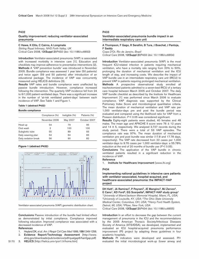
S170
P432Quality improvement: reducing ventilator-associatedpneumonia
C Hawe, K Ellis, C Cairns, A LongmateStirling Royal Infirmary, NHS Forth Valley, UKCritical Care 2008, 12(Suppl 2):P432 (doi: 10.1186/cc6653)
Introduction Ventilator-associated pneumonia (VAP) is associatedwith increased morbidity in intensive care [1]. Education andchecklists may improve adherence to preventative interventions [2].Methods A ‘VAP prevention bundle’ was introduced in November2005. Bundle compliance was assessed 1 year later (50 patients)and twice again (59 and 50 patients) after introduction of aneducational package. The incidence of VAP was concurrentlymeasured using HELICS definitions [3].Results VAP rates and bundle compliance were unaffected bypassive bundle introduction. However, compliance increasedfollowing the intervention. The quarterly VAP incidence fell from 24to 6/1,000 patient-ventilated days. There was a significant increasein the number of ‘at-risk ventilated patient-days’ between eachincidence of VAP. See Table 1 and Figure 1.
Table 1 (abstract P432)
Bundle compliance
Compliance (%) Ineligible (%) Patients (%)
November 2006 May 2007 October 2007
Head up 54 79 95Oral wash 2 100 100Subglottic tube 55 89 93Daily weaning plan 54 64 65Daily sedation break 56 90 82
Conclusions Passive introduction of the bundle had limited effectas demonstrated by initial compliance. Compliance improvedfollowing education. Improved compliance was associated with adecreased incidence of VAP.References1. Heyland DK, et al.: Am J Respir Crit Care Med 1999, 159:1249-1256.2. Evidence Report/Technology Assessment [http://www.
ahrq.gov/downloads/pub/evidence/pdf/qualgap6/hainfgap.pdf]3. HELICS [http://helics.univ-lyon1.fr/home.htm]
P433Ventilator-associated pneumonia bundle impact in anintermediate respiratory care unit
A Thompson, F Gago, R Serafim, B Tura, J Boechat, J Pantoja,F SaddyCopa D’Or, Rio de Janeiro, BrazilCritical Care 2008, 12(Suppl 2):P433 (doi: 10.1186/cc6654)
Introduction Ventilator-associated pneumonia (VAP) is the mostfrequent ICU-related infection in patients requiring mechanicalventilation, who have a mortality rate ranging from 20% to 50%,prolonging the duration of mechanical ventilation and the ICUlength of stay, and increasing costs. We describe the impact ofVAP bundle use in an intermediate respiratory care unit (IRCU) toprevent VAP in patients requiring prolonged mechanical ventilation.Methods A prospective observational study enrolled alltracheotomized patients admitted to a seven-bed IRCU of a tertiarycare hospital between March 2005 and October 2007. The dailyVAP bundle checklist as described by the Institute for HealthcareImprovement [1] was performed since March 2006 to evaluatecompliance. VAP diagnosis was supported by the ClinicalPulmonary Index Score and microbiological quantitative criteria.The mean duration of mechanical ventilation and VAP rate per1,000 ventilator-days pre and post the bundle period wasevaluated and compared using the likelihood ratio for VAP from aPoisson distribution. P < 0.05 was considered significant.Results Eighty-eight patients were studied, 40 females and 48males. The mean age and APACHE II score were 76 ± 12 yearsand 14 ± 5, respectively. We analyzed 3,727 records during thestudy period. There were a total of 53 VAP episodes. Thecompliance rate was 97%. The mean duration of mechanicalventilation pre and post bundle was similar (17.8 and 17.78 days,respectively). The VAP rate decreased from 22 cases per 1,000ventilator-days to 9.76 cases per 1,000 ventilator-days: a 55.78%reduction at the end of 20 months of bundle use (P < 0.05).Conclusions The application of the VAP bundle in chronicventilated patients resulted in a significant reduction in theincidence of VAP.Reference1. Institute for Healthcare Improvement [www.ihi.org]
P434Implementing national guidelines in intensive care patientswith ventilator-associated, hospital-acquired, andhealthcare-associated pneumonia: the IMPACT-HAPproject
DH Kett1, JA Ramirez2, P Peyrani2, JE Mangino3, MJ Zervos4, E Cano1, KD Ford5, EG Scerpella5, IMPACT-HAP study group1
1University of Miami/Jackson Memorial Hospital, Miami, FL, USA;2University of Louisville, KY, USA; 3The Ohio State UniversityMedical Center, Columbus, OH, USA; 4Henry Ford Health System,Detroit, MI, USA; 5Pfizer, New York, USACritical Care 2008, 12(Suppl 2):P434 (doi: 10.1186/cc6655)
Introduction In an effort to decrease the gap between the currentmanagement of pneumonia in the ICU and the recommendationsby the 2005 American Thoracic Society/Infectious DiseasesSociety of America (ATS/IDSA), we developed, implemented andevaluated an ICU hospital-acquired pneumonia performanceimprovement (PI) project by adapting these guidelines in fouracademic hospitals.Methods PI indicators were developed and assessed. PI1evaluated the initial microbiological work-up (lower airway and
Critical Care March 2008 Vol 12 Suppl 2 28th International Symposium on Intensive Care and Emergency Medicine
Figure 1 (abstract P432)
Ventilator-associated pneumonia (VAP) geometric distribution chart.
S171
blood cultures within 48 hours). PI2 assessed empiric antibiotics inpatients with risk factors for multidrug-resistant organisms. PI3evaluated patients for short-course therapy (clinical pulmonaryinfection score ≤6 on day 0 and day 3 and no hemodynamicinstability or severe sepsis). PI4 assessed whether patients werecandidates for de-escalation and had their antibiotics adjusted onday 3. Data were collected prospectively and reviewed by theprincipal investigators prior to submission to the database.Results Analysis was performed on the first 164 patients meetingthe clinical criteria of pneumonia. Respiratory cultures were obtainedin 94% and blood cultures were obtained in 88% of patients. Onehundred and twenty-five patients received empiric therapy. Theempiric therapy was compliant with the ATS/IDSA guidelines in only31% of the patients, with failure to use a second agent to covermultidrug-resistant Gram-negative pathogens in 55% patients.Nineteen patients were candidates for short-course therapy, and thiswas implemented in one patient. De-escalation criteria was met in106 patients and occurred in 75% of candidates.Conclusions Adherence to ATS/IDSA guidelines for diagnosisand management of hospital-acquired pneumonia was less thanexpected, but initial work-up with appropriate cultures wasperformed in the majority of patients. In patients at risk formultidrug-resistant organisms, empiric antibiotics were compliantwith the guidelines only one-third of the time. De-escalation ofantibiotic therapy, while not optimal, did occur in most candidates.In patients who were candidates for short-course therapy, thisoption was rarely chosen.Reference1. American Thoracic Society and Infectious Diseases Society of
America: Am J Respir Crit Care Med 2005, 71:388-416.
P435Prevention of unplanned extubations in the ICU: results ofa nurse-driven endotracheal tube care protocol
R Delvaux, K Tavernier, M Sevenois, M Van Hemelrijck, L Huyghens, H SpapenUZ Brussels, BelgiumCritical Care 2008, 12(Suppl 2):P435 (doi: 10.1186/cc6656)
Introduction Unplanned extubations (UEs) can be accidental orintentional (autoextubation). The incidence of UE is 1–4/100ventilator-days and is considered a quality indicator for nursingcare in the ICU. We studied the effect of a predominantly nurse-driven endotracheal tube care (ETTC) protocol on incidence,circumstances, and outcome of UEs in orotracheally intubatedmechanically ventilated patients.Methods A 1-year prospective observational study in a 24-bedmixed medicosurgical ICU in a university hospital. The ETTC protocolincludes: endotracheal tube (ETT) fixation with waterproof adhesiveplaster and/or adapted tubeholder; wrist fixation in all patients;nurse-driven sedation and analgesia supervised by a dedicated ICUphysician; pressure support weaning using a flow trigger and tubecompensation when possible, actively steered by a nurse and arespiratory physiotherapist; special care to avoid ETT dislocationduring mobilisation or transport of the patient; and promptpharmacological treatment of the restless or agitated patient.Results Six hundred and eighty-eight patients were ventilatedduring 4,605 days. The incidence of UE was 0.32/100 ventilation-days. Autoextubation occurred in 11 (73%) patients, mostly (82%)during the evening or night shift, and in the absence of a nurse inthe patient’s room. All patients had been successfully weaned andwere free of sedation. No patient had to be reintubated. Allaccidental extubations (n = 4; 27%) occurred in the immediate
postoperative phase following cardiac bypass surgery and in thepresence of a nurse and/or physiotherapist at the bedside.Seventy-five percent took place during morning shifts in full-sedated patients and were due to ETT dysfunction. All patientswere promptly reintubated. UEs never occurred during patientmobilisation or transport. No patient died as a result of UE.Conclusions The implementation of a nurse-driven ETTC protocolfor prevention of UE resulted in a very low incidence of UE, in a lownumber of accidental UEs occurring in relatively controlledcircumstances, in no UEs during patient mobilisation, and in noexcess morbidity and mortality.
P436Evidence-based guidelines for preventing ventilator-associated pneumonia: results of a knowledge test among3329 European intensive care nurses
SO Labeau1, DM Vandijck2, J Rello3, S Adam4, KH Vandewoude2, SI Blot2
1University College Ghent, Belgium; 2University Hospital Ghent,Belgium; 3Joan XXIII University Hospital, Tarragona, Spain;4University College London Hospitals Trust, London, UKCritical Care 2008, 12(Suppl 2):P436 (doi: 10.1186/cc6657)
Introduction Although evidence-based guidelines for preventingventilator-associated pneumonia (VAP) are available, non-adherence by ICU clinicians seems common. This may, at leastpartly, be due to a lack of knowledge of the recommendations. Ourstudy aimed to assess ICU nurses’ knowledge of evidence-basedVAP prevention recommendations.Methods A European survey by means of a validated and reliablemultiple-choice questionnaire concerning nine evidence-basedstrategies for VAP prevention [1,2]. Data gathered were gender,years of ICU experience (<1 years, 1–5 years, 6–10 years, >10years), number of ICU beds (<8 beds, 8–15 beds, >15 beds), andwhether they hold a specialized qualification in intensive care.Results Between November 2006 and April 2007, 3,329questionnaires were gathered from 22 European countries. Thenurses’ mean score was 4.06/9 (45.1%). No differences werefound between males and females. Nurses with a longer ICUworking experience scored significantly better than their lessexperienced colleagues (P < 0.001 for <1 year vs >1 year and for<5 years vs >5 years of experience; P = 0.001 for <10 years vs>10 years of experience). Respondents from larger ICUs obtainedsignificantly lower scores than those from smaller units (P < 0.001for <8 beds vs >8 beds and P = 0.048 for <15 beds vs >15beds). Linear regression analysis demonstrated knowledge to beindependently associated with years of ICU experience, and withthe number of ICU beds (both P < 0.001).Conclusions European nurses’ knowledge of VAP preventionguidelines is poor. We recommend including VAP preventionguidelines in the core nurse education curriculum and in continuingrefresher nursing education programs.References1. Labeau S, et al.: Critical care nurses’ knowledge of evi-
dence-based guidelines for preventing ventilator-associ-ated pneumonia: development and validation of anevaluation questionnaire. Am J Crit Care 2007, 16:371-377.
2. Labeau S, et al.: Evidence-based guidelines for the preven-tion of ventilator-associated pneumonia. Results of aknowledge test among intensive care nurses. IntensiveCare Med 2007, 33:1463-1467.
Available online http://ccforum.com/supplements/12/S2
S172
P437Preliminary investigation into the response of free cortisolto the low-dose corticotrophin test in patients with septicshock
J Cohen, J Prins, R Deans, M Lassig-Smith, C Bertenshaw, B VenkateshRoyal Brisbane Hospital, Queensland, AustraliaCritical Care 2008, 12(Suppl 2):P437 (doi: 10.1186/cc6658)
Introduction Controversy exists around the use of total plasmacortisol and the high-dose adrenocorticotrophic hormone (ACTH)test for diagnosing relative adrenal insufficiency in sepsis [1,2].Our aim was to examine the response of bioactive free cortisollevels to stimulation with 1 μg synthetic ACTH in a cohort ofpatients with septic shock.Methods A prospective observational study at a tertiary intensivecare facility. Patients with septic shock (n = 14) were enrolled.Measurements of free cortisol and total plasma cortisol wereperformed at baseline and at 30 and 60 minutes following injectionof 1 μg synthetic ACTH (synacthen).Results Free plasma cortisol and total plasma cortisol at baselineon day 1 were 39.7 (±44.7) and 424.6 (±232.8) nmol/l, respec-tively. The mean increments at 30 minutes of total cortisol and freecortisol were 157.3 (±106) and 54.4(±36) nmol/l, respectively.The mean percentage changes in total and free cortisol were29.6% and 82.8%, respectively; P = 0.07. Relative adrenal insuffi-ciency diagnosed by a total plasma cortisol response <250 nmol/lwas present in 12 patients (85%), and in 13 patients (92%) by afree cortisol increment of less than 110 nmol/l.Conclusions To our knowledge these data represent the first setof information on free cortisol response to 1 μg synacthen.Although preliminary, the proportionally larger change in freecortisol in response to a low-dose synacthen stimulus may indicatea greater sensitivity for secondary adrenal insufficiency, and isconsistent with previous observations [1]. These data support thesuggestion that the traditional relative adrenal insufficiency criteriamight need reconsideration. Further study is warranted todetermine the clinical utility of this investigation.References1. Ho JT, et al: Septic shock and sepsis: a comparison of
total and free plasma cortisol levels. J Clin EndocrinolMetab 2006, 91:105-114.
2. Marik PE, et al: Adrenal insufficiency during septic shock.Crit Care Med 2003, 31:141-145.
P438Copeptin as a marker of shock and predictor of adverseoutcome in critically ill patients
B Meyer1, P Wexberg1, NG Morgenthaler2, A Bergmann2, G Heinz1, J Struck2, R Pacher1, M Huelsmann1
1Medical University of Vienna, Austria; 2BRAHMS AG,Biotechnology Centre, Hennigsdorf, Berlin, GermanyCritical Care 2008, 12(Suppl 2):P438 (doi: 10.1186/cc6659)
Introduction Arginine vasopressin (AVP) is a peptide hormone thatis released in response to osmotic and haemodynamic changes inorder to maintain fluid volume and vascular tone. As a ‘stresshormone’, AVP is significantly increased in acute haemodynamicinstability [1]. Copeptin is a stable fragment of pre-pro-vasopressinthat is released in equimolar quantities as AVP, and thus reflectslevels of released AVP. Unlike AVP, copeptin is highly stable exvivo and is thus used for analysis. We aimed to study whethercopeptin is elevated in any form of acute haemodynamic instability
(that is, shock state) and whether copeptin is a predictor ofoutcome in critically ill patients.Methods A total of 352 consecutive patients (65% male, 64 ± 15years, SAPS 2 52 ± 23) admitted to the ICU of the Department ofCardiology/Medical University of Vienna were included. Bloodsamples were obtained on admission in all patients. Copeptin wasdetermined using a recently developed immunoassay in thechemiluminescence/coated tube format [2].Results Two hundred and seventy-seven patients survived to ICUdischarge and 75 patients died. Copeptin plasma levels weresignificantly higher in ICU nonsurvivors than in ICU survivors (194± 205 pmol/l vs 101 ± 100 pmol/l; P < 0.0001). The area underthe ROC curve for prediction of ICU survival was 0.678 forcopeptin. Patients with diagnosis of shock (n = 132) hadsignificantly higher copeptin plasma levels than patients withoutshock (174 ± 169 pmol/l vs 93 ± 100 pmol/l; P < 0.0001). Therewas no statistically significant difference in copeptin plasma levelsbetween patients with different shock forms. Using a Kaplan–Meiermodel for 28-day survival, patients with copeptin plasma levelsabove the mean had significantly lower survival rates compared withpatients with copeptin plasma levels below the mean (P = 0.03).Conclusions In critically ill patients, elevated plasma levels ofcopeptin are a strong and independent predictor of adverseoutcome. Copeptin is significantly increased in shock patients,independent of the shock category.References1. Luckner G, et al.: Arginine vasopressin in 316 patients with
advanced vasodilatory shock. Crit Care Med 2005,33:2659-2666.
2. Morgenthaler NG, et al.: Assay for the measurement ofcopeptin, a stable peptide derived from the precursor ofvasopressin. Clin Chem 2006, 52:112-119.
P439Copeptin and acute renal failure in critically ill patients
B Meyer1, A Bergmann2, P Wexberg1, J Struck2, NG Morgenthaler2, G Heinz1, R Pacher1, M Huelsmann1
1Medical University of Vienna, Austria; 2BRAHMS AG,Biotechnology Centre, Hennigsdorf, Berlin, GermanyCritical Care 2008, 12(Suppl 2):P439 (doi: 10.1186/cc6660)
Introduction Acute renal failure (ARF) is a common problem incritically ill patients and associated with an adverse prognosis [1].Vasopressin is a peptide hormone that is released in response toosmotic and haemodynamic changes. An elevation of vasopressinhas been suggested to decrease the glomerular filtration rate [2].In the present study we aimed to assess whether an elevation ofvasopressin is associated with the development of ARF in criticallyill patients. As vasopressin is highly unstable, copeptin – a stablefragment of the precursor molecule pre-pro-vasopressin that issecreted in equimolar quantities [3] – is used for analysis.Methods Three hundred and fifty-two consecutive patients (228males, age 64 ± 15 years, SAPS 2 52 ± 23) admitted to the ICUof the Department of Cardiology/Medical University of Vienna wereincluded. In addition to routine laboratory assessment, bloodsamples for determination of copeptin were obtained in all patientson admission. Copeptin was determined by use of thechemiluminescence/coated tube format [3].Results Two hundred and seventy-seven patients survived to ICUdischarge and 75 patients died. There was a significant inversecorrelation between copeptin and glomerular filtration rate(–0.266; P < 0.0001). Patients who developed ARF (n = 114) hadhigher copeptin plasma levels (180 ± 167pmol/l vs 95 ± 107pmol/l; P < 0.0001), a lower mean blood pressure (48 ± 28
Critical Care March 2008 Vol 12 Suppl 2 28th International Symposium on Intensive Care and Emergency Medicine
S173
mmHg vs 60 ± 24 mmHg; P < 0.0001), were more often in needof vasopressor support (81% vs 63%; P < 0.001) and were moreoften in shock (72% vs 20%; P < 0.0001) than patients withpreserved renal function. Entering all these variables in a stepwiselogistic regression model, only shock (P < 0.0001) and copeptin(P < 0.005) were independent predictors for development of ARF.Conclusions In critically ill patients, an elevation of plasmacopeptin is strongly associated with development of ARF. Thetherapeutic impact of this observation is still to be elucidated.References1. Ostermann M, et al.: Acute kidney injury in the intensive
care unit according to RIFLE. Crit Care Med 2007, 35:1837-1843.
2. Roald AB, et al.: The effect of AVP-V receptor stimulationon local GFR in the rat kidney. Acta Physiol Scand 2004,182:197-204.
3. Jochberger S, et al.: Copeptin and arginine vasopressinconcentrations in critically ill patients. J Clin EndocrinolMetab 2006, 91:4381-4386.
P440Prognostic value of N-terminal B-type natriuretic peptide inpatients at high risk of systemic inflammatory responsesyndrome/sepsis
A Rezaie-Majd1, K Hellemann1, A Khorsand2, L Adler1, J Zuber1,T Szekeres3, A Aloy1
1Department of Anaesthesia and General Intensive Care/ICU13C1, 2Department of Internal Medicine II, and 3Department ofMedical and Clinical Laboratory Diagnostics, Medical University ofVienna, AustriaCritical Care 2008, 12(Suppl 2):P440 (doi: 10.1186/cc6661)
Introduction Patients undergoing major surgery are at high risk ofpostoperative sepsis. Early markers of septic complications would beuseful for diagnosis and therapy of patients with sepsis. In contrastto the rather uniform results of studies dealing with other markers ofsepsis such as C-reactive protein (CRP), the impact of raised n-terminal B-type natriuretic peptide (BNP) levels in the postoperativeperiod is less clear. Furthermore, growing evidence supports thehypothesis that BNP could be an early predictor of sepsis in the ICU.We evaluated the time courses of BNP, CRP and APACHE II scoreand investigated their role as early markers of systemic inflammatoryresponse syndrome (SIRS)/sepsis after major surgery.Methods Twenty-nine patients were prospectively included: 20patients had major oncological surgery of the head and neck(Group 1). The second group included patients with manifestcardiovascular disease (n = 9) undergoing cardiac surgery. TheAPACHE II score and serum concentrations of BNP and CRPwere determined before and daily after surgery. The AmericanCollege of Chest Physicians Classification was used to diagnoseSIRS, and organ system failure to define sepsis.Results Mean baseline levels of BNP were different in both groups(Group 1: 435.2 ± 906.1 ng/ml vs Group 2: 2,083.4 ± 3,694.9ng/ml). The maximum values of BNP achieved at postoperative day3 were significantly higher compared with the baseline levels. Therewas also a significant difference between both groups (Group 1:1,500.0 ± 1,963.8 ng/ml vs 3,806.6 ± 3,822.0 ng/ml; P < 0.05;r2 = 0.84). The APACHE II score was significantly higher at themaximum time point (day 3) for all patients (P < 0.05, r2 = 0.45).However, the CRP levels were not significantly different within thisperiod. Six patients developed SIRS. The average maximum BNPwas 2,286.98 ± 2,943.58 ng/ml. Within this group, two patientsdied of severe sepsis and had significantly higher BNP levels ascompared with the maximum postoperative values (P < 0.05).
Conclusions Our results show that BNP levels are significantlyhigher in patients after undergoing major surgery at risk ofSIRS/sepsis. We also observed significantly higher BNP levels inpatients developing SIRS/sepsis. BNP may therefore be anappropriate prognostic marker indicating the early development ofpostoperative severe sepsis after major surgery.
P441Incremental power of the combination of brain natriureticpeptide and tumoral antigen carbohydrate 125 for riskstratification in patients with acute heart failure
H Michalopoulou, A Bakhal, J Vaitsis, N Stravodimou, D Nastou, D Pragastis, N Stravodimou‘Metaxa’ Hospital, Athens, GreeceCritical Care 2008, 12(Suppl 2):P441 (doi: 10.1186/cc6662)
Introduction Brain natriuretic peptide (BNP) is a biochemicalmarker for risk stratification in heart failure, and carbohydrate (CA)125 has been shown to correlate with heart failure severity.However, few data are available about the combination of thesetwo biomarkers in the evaluation of short-term mortality in patientsadmitted for acute heart failure (AHF).Methods We studied 184 consecutive patients admitted to ourICU for AHF. The CA 125 and BNP levels were measured, and fourcategories were formed: CI = BNP < 350 ng/l, CA 125 < 65 U/ml;CII = BNP ≥ 350 ng/l, CA 125 < 65 U/ml; CIII = BNP < 350 ng/l,CA 125 ≥ 65 U/ml; and CIV = BNP ≥ 350 ng/l, CA 125 ≥ 65 U/ml.The independent association between the aforementionedcategories and survival was assessed with Cox regression analysis.Results At 6 months follow-up, 39 deaths (21.2%) were identified.The mortality rate was lower in CI (8.2%), intermediate in CII andCIII (27% and 25.2%, respectively) and higher in CIV (41.8%).After adjusting for age, gender, NYHA class, ejection fraction,systolic blood pressure on admission, sodium and creatinine, themortality rate remained unaltered. The higher mortality wasobserved when both biomarkers were elevated (CIV vs CI: heartrate = 3.6, 95% CI: 1.5–8.84). Intermediate risk when only one ofthem was elevated (CII vs CI: heart rate = 2.2, 95% CI: 0.94–6.4).The lower mortality was observed in the group with none of thebiomarkers elevated (CI = control group).Conclusions CA 125 added prognostic value to BNP, and thecombination of these biomarkers affords a better risk stratificationin patients admitted with AHF.
P442Pro-brain natriuretic peptide as a marker of successfulthrombolysis in patients with submassive pulmonarythromboembolism
R Castro, M Andresen, C Ruiz, A Gonzalez, O Diaz, M Mercado,L Meneses, M Fava, S CordovaPontificia Universidad Catolica De Chile, Santiago Centro, ChileCritical Care 2008, 12(Suppl 2):P442 (doi: 10.1186/cc6663)
Introduction Pulmonary embolism (PE) is frequently under-diagnosed and associated with high lethality despite progress inprophylaxis and treatment. Aggressive management may improveoutcome in patients with high risk of right ventricle (RV) failuresecondary to pulmonary hypertension. CT scan angiography,cardiac echo and biomarkers (pro-brain natriuretic peptide(proBNP) and troponin) have been used to identify these high-riskpatients. We wanted to evaluate relationship between inter-ventional treatment of acute PE with cardiac biomarkers changes.
Available online http://ccforum.com/supplements/12/S2
S174
Methods Patients with PE < 24 hours confirmed by thoracic CTscan angiography, pulmonary circulation obstruction > 50% andechographic or CT evidence of RV dysfunction underwent invasiveangiography and thrombolysis with r-tPA. Reperfusion wasevaluated by Miller–Walsh’s score and a second angiograph wasperformed 6 hours later to re-evaluate. Plasmatic proBNP andtroponin were measured before and 6 hours after angiography.Results Thirteen patients, four men (31%), age 60.4 ± 19.2 years.ProBNP pre/post angiography in whole group = 3,672 ± 2,894/2,954 ± 2,648 (P = 0.055); in successful reperfusion group =4,225 ± 3,050/3,233 ± 2,910 (pre/post) (P < 0.013) and inunsuccessful reperfusion group = 1,830 ± 1,320/2,024 ± 1,512(pre/post) (P = not significant). Troponin pre/post angiography =0.31 ± 0.27/0.23 ± 0.28 (P = not significant), without a significantlowering with regards to reperfusion. A significant lowering wasseen in pulmonary arterial mean pressure before and afterangiography in the whole group = 34.5 ± 8.9/23.4 ± 6.9 (P <0.002), as in successfully reperfused patients (P < 0.005). Nocomplications were derived from the procedure. Twenty-eight-daymortality was 8.3% (one patient from the reperfused group).Conclusions Biomarkers may help to define a high-risk PE group,susceptible to benefit from more aggressive management. ProBNPhas a dynamic and sensitive correlation with pulmonary bedcirculatory status, and with major variations in pulmonaryhemodynamics, as we observed. Cardiopulmonary overloadimproves after successful direct thrombolysis. Angiographic reper-fusion correlates directly with a significant lowering in proBNPlevels and a significant improvement on pulmonary pressures. Wethink this aggressive approach may be recommended: if indirectevidence of reperfusion is present (for example, significantlowering on plasmatic proBNP levels), a control invasiveangiography could be avoided. Also, these results could beextrapolated to intravenous thrombolysis.
P443Serum measurement of N-terminal pro-brain peptideamong septic patients
M Èubrilo-Turek, V Pilas, D Striniæ, K ÈalaSveti Duh General Hospital, Zagreb, CroatiaCritical Care 2008, 12(Suppl 2):P443 (doi: 10.1186/cc6664)
Introduction Elevated serum N-terminal pro-brain natriureticpeptide (NT-proBNP) levels occur in severe sepsis or septicshock. Studies on the value of NT-proBNP testing among septicpatients revealed conflicting results. The aim of the study was toinvestigate NT-proBNP among septic patients admitted to the ICU.Methods Serum NT-proBNP measurements were carried out in 10consecutive patients (seven males/three females, age 66 ± 10.67years) with sepsis within 6 hours of admission to the IXU. NT-proBNP was determined with a sandwich immunoassay on anElecsys 2010 (Roche Diagnostics, Mannheim, Germany). Logarith-mic transformation of data was required because of the skeweddistributions of the NT-proBNP.Results Elevated levels of NT-proBNP were found in eightpatients, while two were in the ‘grey’ zone (NT-proBNP < 400pg/l). The median NT-proBNP level was 3,930 pg/ml (range,307–16,800 pg/ml). Inhospital mortality was high, five patientsdied (50%). NT-proBNP showed a weak and inverse correlationwith systolic blood pressure (r = –0.49, P < 0.05) and with bodytemperature (r = –0.57, P < 0.02), while no correlation was foundfor other analyzed parameters (age, diastolic blood pressure, bodytemperature, leucocytes, C-reactive protein, fibrinogen, lactates,procalcitonin). At admission, the mean log NT-proBNP levels weresignificantly higher in hospital nonsurvivors (3.66 ± 0.63 pg/ml)
compared with survivors (3.36 ± 0.65 pg/ml), but withoutstatistical significance (P < 0.54). Mean baseline levels of log NT-proBNP were different in septic patients with proved bacterio-logical infection than in patients without proved infection. Higherconcentrations were found in proved infection (X ± SD) (3.66 ±0.71 pg/ml) than in the bacteriological negative patients (3.24 ±0.52 pg/ml), but were statistically insignificant (P < 0.32).Conclusions Our results showed that NT-proBNP levels can beelevated in critically ill patients presenting with sepsis. Values arehigher in nonsurvivors than in survivors and in the patients withproved bacteriological infection, but with no statistical significance.More research is needed to accentuate the positive value of NT-proBNP in the ICU.Reference1. Bluth MH: Brain natriuretic peptides in sepsis: ideal bio-
marker or just along for the ride? Crit Care Med 2007, 35:1421-1422.
P444Plasma brain natriuretic peptide and cardiac troponin Iconcentrations after adult cardiac surgery: association withcardiovascular death, postoperative cardiac, renal andpulmonary dysfunction
L Hajjar1, F Galas2, S Grande2, M Campos2, R Silva2, A Marques2, W Leao2, J Auler Jr2
1Heart Institute, São Paulo, Brazil; 2Heart Institute, Faculdade deMedicina da Universidade de São Paulo, BrazilCritical Care 2008, 12(Suppl 2):P444 (doi: 10.1186/cc6665)
Introduction B-type natriuretic peptide (BNP) and troponin levelscan predict outcome after acute myocardial infarction and afteracute heart failure. The purpose of this study was to evaluate theprognostic implication of high postoperative BNP and cardiactroponin I concentrations in adult patients undergoing cardiacsurgery.Methods A prospective study was carried out in a total of 181consecutive patients undergoing elective coronary artery bypass orvalve surgery patients. BNP and cardiac troponin I concentrationswere measured immediately after surgery (day 0), and at day 1after surgery (day 1). Postoperative cardiac dysfunction wasdefined as low output cardiac or hemodynamic instability requiringinotropic support for >24 hours. Pulmonary dysfunction wasdefined as postoperative hypoxemia (rate PO2/FiO2 < 200) or timeto extubation longer than 360 minutes. Univariate and multivariateanalyses were performed.Results A significant secretion of BNP was observed after cardiacsurgery. Independent predictors of cardiac dysfunction werehigher values of BNP and troponin measured on day 0 and on day1 (P = 0.0001), higher levels of CKMB (P = 0.006), and lowerejection fraction (P = 0.05). Independent predictors of pulmonarydysfunction were elevation of BNP on day 0 (P = 0.0005) and onday 1 (P = 0.02). BNP levels higher than 300 pg/ml wereassociated with higher rates of renal failure (P = 0.0001). A BNPlevel higher than 300 pg/ml and a troponin level higher than 15ng/ml was associated with a higher risk of mortality (P = 0.0001).Conclusions Postoperative BNP and cardiac troponin areindependent predictors of mortality and cardiac and pulmonarydysfunction after cardiac surgery. Higher levels of BNP are alsocorrelated with higher rates of renal failure after cardiac surgery.Reference1. Provenchère S, et al.: Plasma brain natriuretic peptide and
cardiac troponin I concentrations after adult cardiacsurgery: association with postoperative dysfunction and1-year mortality. Crit Care Med 2006, 34:995-1000.
Critical Care March 2008 Vol 12 Suppl 2 28th International Symposium on Intensive Care and Emergency Medicine
S175
P445Prognostic value of plasma amino-terminal pro-brainnatriuretic peptide in a large, representative ICUpopulation
A Kotanidou1, P Karsaliakos1, M Tzanela1, I Mavrou2, P Kopterides2, E Papadomichelakis2, M Theodorakopoulou2, I Tsagaris2, M Lignos2, I Ikonomidis2, I Ilias3, A Armaganidis2, S Orfanos2, I Dimopoulou2
1Evangelismos Hospital, Athens, Greece; 2’Attiko’ University Hospital,Athens, Greece; 3Elena Venizelou Hospital, Athens, GreeceCritical Care 2008, 12(Suppl 2):P445 (doi: 10.1186/cc6666)
Introduction We sought to determine whether amino-terminal pro-brain natriuretic peptide (NT-proBNP) predicts ICU outcome in arepresentative cohort of mechanically ventilated, critically ill patients.Methods A total of 234 consecutive patients (109 men – medianage: 60 years) with a wide range of admitting diagnoses (medical,surgical, and multiple trauma cases) were included in thisprospective study that was conducted in two ICUs of tertiaryhospitals over an 18-month period. The following data wererecorded upon ICU entry: age, sex, admitting diagnosis, severity ofcritical illness according to the APACHE II score, degree of organdysfunction quantified by the SOFA score and the presence ofsepsis using established criteria. Exclusion criteria includedchronic heart failure defined by a left ventricular ejection fraction<50% on admission echocardiography, and pre-existent renalinsufficiency (history of serum creatinine >1.8 mg/dl before ICUentry). The primary end point was ICU outcome.Results Nonsurvivors (n = 98) had significantly higher NT-proBNPlevels on ICU admission (2,074 vs 283 pg/ml, P < 0.001), on day1 (2,197 vs 221 pg/ml, P < 0.001) and on day 2 (2,726 vs 139pg/ml, P < 0.001) than survivors (n = 135). Receiver operatedcharacteristics (ROC) analysis showed that the area under theROC curve with regard to ICU mortality prediction was 0.70 forAPACHE II and 0.77 for admission NT-proBNP. The cutoff value inadmission NT-proBNP that best predicted outcome was 941pg/ml. Multiple logistic regression analysis revealed that APACHEII score (OR = 1.06, 95% CI = 1.01–1.11, P = 0.007) and thebest cutoff point in NT-proBNP (OR = 7.74, 95% CI = 4.00–14.9,P < 0.0001) independently predicted ICU outcome.Conclusions In a representative mixed ICU population, non-survivors have consistently higher NT-proBNP levels comparedwith survivors. Admission NT-proBNP concentrations provideindependent prognostic information for ICU mortality.
P446N-terminal B-type natriuretic peptide and renal functionparameters in cerebral salt wasting syndrome
V Spatenkova1, A Kazda2, P Skrabalek1, P Suchomel11Regional Hospital, Liberec, Czech Republic; 2Charles University,Prague, Czech RepublicCritical Care 2008, 12(Suppl 2):P446 (doi: 10.1186/cc6667)
Introduction Elevated levels of B-type natriuretic peptides arereported in hypo-osmolal hyponatraemia with natriuresis in cerebralsalt wasting (CSW) syndrome. The aim of our study was toevaluate serum N-terminal pro-B-type natriuretic peptide (NT-proBNP) and its relationship with renal function parameters inpatients with acute brain diseases who developed CSW.Methods We prospectively measured NT-proBNP in 12 patients(mean age 46 ± 17 years, seven men) with acute brain diseases(four subarachnoid haemorrhage, five tumour, three trauma, meanGlasgow Coma Scale 13 ± 2.7). All patients were classified as
New York Heart Association (NYHA) I and none had pulmonaryoedema. The mean Glasgow Outcome Scale upon discharge fromthe neurologic–neurosurgical ICU was 4 ± 1.2. Diagnoses of CSW(mean serum sodium 129 ± 3.6 mmol/l) were stated by evaluationof renal function parameters – only with clearance of creatinineabove 1.15 ml/s. Urine was collected within 24 hours. The controlgroup was made up of patients with acute brain diseases,normonatraemia, NYHA I and normal renal parameters (n = 20patients).Results We found significantly higher levels of NT-proBNP inpatients with CSW (430.4 ± 706.4 pg/ml) compared not only withthe reference range (125 pg/ml, P = 0.001) but also with thecontrol group (268.3 ± 203.9, P < 0.001). There were nodifferences in fluid intake (P = 0.440) between the two groups, butpatients with CSW had higher sodium intake per kilogram of bodyweight (P = 0.024), diuresis (P = 0.019), daily sodium output (P =0.036), electrolyte clearance (P = 0.001) and sodium clearance(P = 0.007). Further analysis of the control group did not show anyrelationship between NT-proBNP and measured parameters, incontrast to CSW, where we found a negative relation between NT-proBNP and serum sodium (P = 0.022), fluid intake (P = 0.047),osmotically active substances clearance (P = 0.031) and electro-lyte clearance (P = 0.047). No further correlations were found.Conclusions Our results showed that natriuresis with hypo-osmolal hyponatraemia in CSW were related to significant higherserum levels of NT-proBNP with a negative correlation to serumsodium and fluid intake, compared with patients with acute braindisease and normonatraemia.Reference1. Berendes E, et al.: Secretion of brain natriuretic peptide in
patients with aneurysmal subarachnoid haemorrhage.Lancet 1997, 349:245-249.
P447Sepsis-associated diastolic dysfunction without elevatedplasma B-type natriuretic peptide
DJ Sturgess, BA Haluska, P Masci, M Jones, B VenkateshUniversity of Queensland, Brisbane, AustraliaCritical Care 2008, 12(Suppl 2):P447 (doi: 10.1186/cc6668)
Introduction Plasma B-type natriuretic peptide concentration(BNP) is a marker of cardiac dysfunction [1]. It is unclear whyextremely high concentrations have been reported in sepsis withpreserved systolic function [2]. This study sought to evaluaterelationships between BNP and in vivo diastolic function in a ratcecal ligation and perforation (CLP) model of sepsis.Methods With institutional ethics committee approval (UQ AECProtocol 675/05), 24 male Sprague–Dawley rats (518 ± 56 g)were studied. Rats were assigned to CLP (n = 12), sham surgery(sham, n = 6) or anaesthesia without surgery (control, n = 6).Echocardiography (15 MHz rodent probe) and venous bloodsampling (BNP enzyme immunoassay) were performed prior tointervention (baseline; T0) and 18–22 hours following intervention(final; T2).Results Prior to final evaluation, three CLP rats (25%) and onesham rat (during anaesthesia) died. Baseline differences betweengroups (ANOVA) were demonstrated for heart rate (HR) (CLP 340± 36 bpm, sham 351 ± 19 bpm, control 300 ± 12 bpm; P = 0.02),peak passive to active diastolic transmitral velocity ratio (E/A) (CLP1.7 ± 0.65, sham 1.26 ± 0.27, control 2.34 ± 0.59; P = 0.048)and A velocity (CLP 0.574 ± 0.13 m/s, sham 0.733 ± 0.33 m/s,control 0.411 ± 0.07 m/s; P = 0.03). BNP was not significantlydifferent between groups (CLP 0.676 ± 0.179 ng/ml; sham0.719 ± 0.202 ng/ml; control 0.503 ± 0.006 ng/ml; P = 0.07). At
Available online http://ccforum.com/supplements/12/S2
S176
T2, CLP was compared with sham and control (ANCOVA withadjustment for baseline values). Compared with control rats, CLPrats demonstrated higher HR (CLP 402 ± 46 bpm, control 305 ±11 bpm; P = 0.003), higher A velocity (CLP 0.802 ± 0.152 m/s,control 0.501 ± 0.103 m/s; P = 0.01), and lower E/A (CLP 1.02 ±0.23, control 1.74 ± 0.46; P = 0.004). BNP was not significantlydifferent in the CLP group (CLP 0.743 ± 0.225 ng/ml, sham 0.756± 0.213 ng/ml (P = 0.3), control 0.509 ± 0.026 ng/ml (P = 0.7)).At T2, multiple linear regression with backward elimination yieldedHR as the only independent predictor of BNP (adjusted r2 = 0.56,P < 0.001).Conclusions In this model, sepsis was associated withechocardiographic evidence of diastolic dysfunction resemblingclinical sepsis without an associated increase in BNP. HR was anindependent predictor of BNP, accounting for 56% of variation.References1. Sturgess DJ, et al.: Anaesth Intensive Care 2006, 34:151-
163.2. Maeder M, et al.: Eur J Heart Fail 2005, 7:1164-1167.
P448Methemoglobin level as an indicator for disease severity insepsis
T Schuerholz, J Irmer, TP Simon, K Reinhart, G MarxFriedrich-Schiller University, Jena, GermanyCritical Care 2008, 12(Suppl 2):P448 (doi: 10.1186/cc6669)
Introduction The objective of our study was to investigate howmethemoglobin (MetHb) levels reflect the occurrence and severityof sepsis. MetHb is generated of nitric oxide (NO) throughinteraction with hemoglobin in a variety of patients [1]. Thisincludes patients in sepsis with a potentially higher NO generation.Methods Six hundred and sixty-five patients of a university ICUwere included in a retrospective study to analyze data after onsetof sepsis (day 0) and 24 hours later (day 1). Eligible for inclusionwere sepsis or severe sepsis patients as defined by ACCP/SCCMconsensus conference and control ICU patients, both withmeasured MetHb levels and age >18 years. The recorded SOFAscore on day 0 was used to stratify the study population in lessseverely (≤10 points) and more severely (>10 points) patients.After verification of skewness, septic and nonseptic patients andSOFA groups were compared using the Mann–Whitney U test.P < 0.05 was considered significant.Results Of 665 patients, 71% (n = 469) had a SOFA score ≤10.Forty-five percent (n = 211) of them had sepsis. One hundred andsixty-four out of 196 severely ill patients (SOFA score > 10) hadsepsis. MetHb levels were significantly higher in patients withsepsis compared with nonseptic patients on day 0 (SOFA ≤ 10:0.72 ± 0.27% vs 0.65 ± 0.15% (P < 0.01); SOFA > 10: 0.79 ±0.21% vs 0.62 ± 0.17% (P < 0.01)). On day 1 similar resultscomparing septic and nonseptic patients were obtained (SOFA≤ 10: 0.73 ± 0.24% vs 0.63 ± 0.16% (P < 0.01); SOFA > 10:0.80 ± 0.24% vs 0.69 ± 0.20% (P < 0.01)). If sepsis was moresevere, as defined by SOFA score > 10, significantly higher MetHblevels were observed compared with sepsis patients with SOFAscores ≤10 on day 0 and day 1 (P < 0.01).Conclusions Our data suggest that increased MetHb levels reflectthe severity of disease defined by SOFA score. Furthermore,MetHb levels may have the potential to contribute to the diagnosisof sepsis. This potential of MetHb levels needs prospectivevalidation.Reference1. Kosaka H, et al.: Free Radic Biol Med 1989, 7:653-658.
P449Effects of postoperative hyperlactatemia on patients’intensive therapy unit length of stay
S Thanthulage, S StaceyLondon Chest Hospital, London, UKCritical Care 2008, 12(Suppl 2):P449 (doi: 10.1186/cc6670)
Introduction Lactate is one of the most crucial intermediates ofcarbohydrate and nonessential amino acid metabolism. Moderatehyperlactatemia in the immediate postoperative cardiac surgicalperiod is associated with a high risk of major complications [1,2].Methods We audited 246 unselected, consecutive postoperativepatients who were admitted post cardiac surgery over 3 months viainterrogation of the blood gas analysis machine at the LondonChest Hospital. Patients were divided into three groups accordingto lactate levels for their first 24 hour stay on the intensive therapyunit (ITU): normal up to 2 mmol/l (NoHL), moderate 2.1–4.9 mmol/l(MHL), severe hyperlactatemia >5 mmol/l (SHL) [3]. The length ofstay in the ITU was also identified.Results See Tables 1 and 2.
Table 1 (abstract P449)
Normal, moderate and severe hyperlactatemia groups
Lactatemia Average age level Total Male (%) Female (%) (years)
NoHL 121 100 (40.6) 21 (8.5) 66
MHL 112 89 (36.2) 23 (9.3) 68
SHL 13 10 (4) 3 (1.2) 72
Table 2 (abstract P449)
Patient length of stay in ITU related to maximum lactate levels inthe first 24 hours
Length of stay NoHL (%) MHL (%) SHL (%)
≤24 hours 100 (82.6) 72 (64.3) 4 (30.7)
≤48 hours 14 (11.6) 23 (20.5) 3 (23.1)
>48 hours 7 (5.7) 17 (15.1) 6 (46.2)
Conclusions We can confirm that hyperlactatemia during the first 24hours post cardiac surgery is associated with a prolonged ITU stay.References1. Micheal JM, et al.: Frequency, risk factors and outcome of
hyperlactatemia after cardiac surgery. Chest 2003, 123:1361-1366.
2. Iqbal M, et al.: Effect of cardio pulmonary bypass on lactatemetabolism. Intensive Care Med 2003, 29:1279-1285.
3. Brandis K: Acid–Base Physiology [www.anaesthesiaMCQ.com]
P450Intraoperative base excess and carbon dioxide gap predictlength of intensive care stay after cardiac surgery
L Hajjar, F Galas, F Maeda, T Yamaguti, A Roquim, M Fortim, J AulerHeart Institute – Faculdade de Medicina da Universidade de SãoPaulo, BrazilCritical Care 2008, 12(Suppl 2):P450 (doi: 10.1186/cc6671)
Introduction Perfusion parameters are goals of standard treatmentin shock states and have prognostic significance in critically ill
Critical Care March 2008 Vol 12 Suppl 2 28th International Symposium on Intensive Care and Emergency Medicine
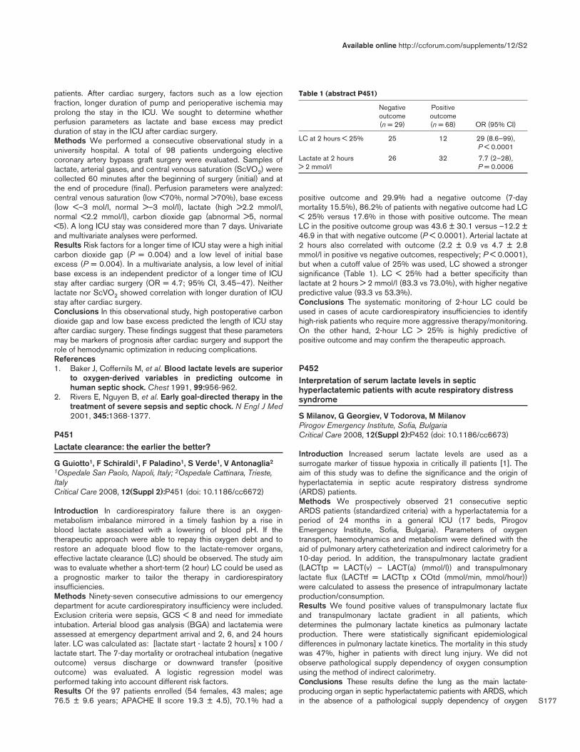
S177
patients. After cardiac surgery, factors such as a low ejectionfraction, longer duration of pump and perioperative ischemia mayprolong the stay in the ICU. We sought to determine whetherperfusion parameters as lactate and base excess may predictduration of stay in the ICU after cardiac surgery.Methods We performed a consecutive observational study in auniversity hospital. A total of 98 patients undergoing electivecoronary artery bypass graft surgery were evaluated. Samples oflactate, arterial gases, and central venous saturation (ScVO2) werecollected 60 minutes after the beginning of surgery (initial) and atthe end of procedure (final). Perfusion parameters were analyzed:central venous saturation (low <70%, normal >70%), base excess(low <–3 mol/l, normal >–3 mol/l), lactate (high >2.2 mmol/l,normal <2.2 mmol/l), carbon dioxide gap (abnormal >5, normal<5). A long ICU stay was considered more than 7 days. Univariateand multivariate analyses were performed.Results Risk factors for a longer time of ICU stay were a high initialcarbon dioxide gap (P = 0.004) and a low level of initial baseexcess (P = 0.004). In a multivariate analysis, a low level of initialbase excess is an independent predictor of a longer time of ICUstay after cardiac surgery (OR = 4.7; 95% CI, 3.45–47). Neitherlactate nor ScVO2 showed correlation with longer duration of ICUstay after cardiac surgery.Conclusions In this observational study, high postoperative carbondioxide gap and low base excess predicted the length of ICU stayafter cardiac surgery. These findings suggest that these parametersmay be markers of prognosis after cardiac surgery and support therole of hemodynamic optimization in reducing complications.References1. Baker J, Coffernils M, et al. Blood lactate levels are superior
to oxygen-derived variables in predicting outcome inhuman septic shock. Chest 1991, 99:956-962.
2. Rivers E, Nguyen B, et al. Early goal-directed therapy in thetreatment of severe sepsis and septic chock. N Engl J Med2001, 345:1368-1377.
P451Lactate clearance: the earlier the better?
G Guiotto1, F Schiraldi1, F Paladino1, S Verde1, V Antonaglia2
1Ospedale San Paolo, Napoli, Italy; 2Ospedale Cattinara, Trieste,ItalyCritical Care 2008, 12(Suppl 2):P451 (doi: 10.1186/cc6672)
Introduction In cardiorespiratory failure there is an oxygen-metabolism imbalance mirrored in a timely fashion by a rise inblood lactate associated with a lowering of blood pH. If thetherapeutic approach were able to repay this oxygen debt and torestore an adequate blood flow to the lactate-remover organs,effective lactate clearance (LC) should be observed. The study aimwas to evaluate whether a short-term (2 hour) LC could be used asa prognostic marker to tailor the therapy in cardiorespiratoryinsufficiencies.Methods Ninety-seven consecutive admissions to our emergencydepartment for acute cardiorespiratory insufficiency were included.Exclusion criteria were sepsis, GCS < 8 and need for immediateintubation. Arterial blood gas analysis (BGA) and lactatemia wereassessed at emergency department arrival and 2, 6, and 24 hourslater. LC was calculated as: [lactate start - lactate 2 hours] x 100 /lactate start. The 7-day mortality or orotracheal intubation (negativeoutcome) versus discharge or downward transfer (positiveoutcome) was evaluated. A logistic regression model wasperformed taking into account different risk factors.Results Of the 97 patients enrolled (54 females, 43 males; age76.5 ± 9.6 years; APACHE II score 19.3 ± 4.5), 70.1% had a
positive outcome and 29.9% had a negative outcome (7-daymortality 15.5%), 86.2% of patients with negative outcome had LC< 25% versus 17.6% in those with positive outcome. The meanLC in the positive outcome group was 43.6 ± 30.1 versus –12.2 ±46.9 in that with negative outcome (P < 0.0001). Arterial lactate at2 hours also correlated with outcome (2.2 ± 0.9 vs 4.7 ± 2.8mmol/l in positive vs negative outcomes, respectively; P < 0.0001),but when a cutoff value of 25% was used, LC showed a strongersignificance (Table 1). LC < 25% had a better specificity thanlactate at 2 hours > 2 mmol/l (83.3 vs 73.0%), with higher negativepredictive value (93.3 vs 53.3%).Conclusions The systematic monitoring of 2-hour LC could beused in cases of acute cardiorespiratory insufficiencies to identifyhigh-risk patients who require more aggressive therapy/monitoring.On the other hand, 2-hour LC > 25% is highly predictive ofpositive outcome and may confirm the therapeutic approach.
P452Interpretation of serum lactate levels in septichyperlactatemic patients with acute respiratory distresssyndrome
S Milanov, G Georgiev, V Todorova, M MilanovPirogov Emergency Institute, Sofia, BulgariaCritical Care 2008, 12(Suppl 2):P452 (doi: 10.1186/cc6673)
Introduction Increased serum lactate levels are used as asurrogate marker of tissue hypoxia in critically ill patients [1]. Theaim of this study was to define the significance and the origin ofhyperlactatemia in septic acute respiratory distress syndrome(ARDS) patients.Methods We prospectively observed 21 consecutive septicARDS patients (standardized criteria) with a hyperlactatemia for aperiod of 24 months in a general ICU (17 beds, PirogovEmergency Institute, Sofia, Bulgaria). Parameters of oxygentransport, haemodynamics and metabolism were defined with theaid of pulmonary artery catheterization and indirect calorimetry for a10-day period. In addition, the transpulmonary lactate gradient(LACTtp = LACT(v) – LACT(a) (mmol/l)) and transpulmonarylactate flux (LACTtf = LACTtp x COtd (mmol/min, mmol/hour))were calculated to assess the presence of intrapulmonary lactateproduction/consumption.Results We found positive values of transpulmonary lactate fluxand transpulmonary lactate gradient in all patients, whichdetermines the pulmonary lactate kinetics as pulmonary lactateproduction. There were statistically significant epidemiologicaldifferences in pulmonary lactate kinetics. The mortality in this studywas 47%, higher in patients with direct lung injury. We did notobserve pathological supply dependency of oxygen consumptionusing the method of indirect calorimetry.Conclusions These results define the lung as the main lactate-producing organ in septic hyperlactatemic patients with ARDS, whichin the absence of a pathological supply dependency of oxygen
Available online http://ccforum.com/supplements/12/S2
Table 1 (abstract P451)
Negative Positive outcome outcome (n = 29) (n = 68) OR (95% CI)
LC at 2 hours < 25% 25 12 29 (8.6–99), P < 0.0001
Lactate at 2 hours 26 32 7.7 (2–28), > 2 mmol/l P = 0.0006
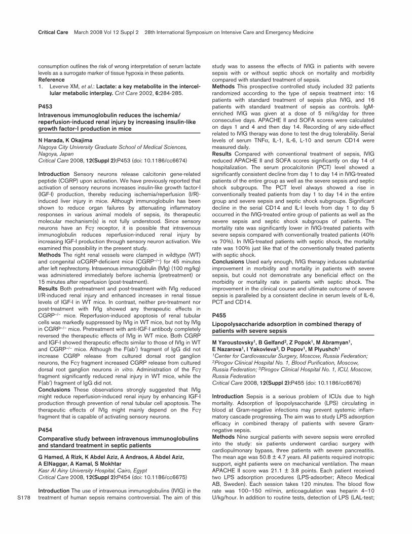
S178
consumption outlines the risk of wrong interpretation of serum lactatelevels as a surrogate marker of tissue hypoxia in these patients.Reference1. Leverve XM, et al.: Lactate: a key metabolite in the intercel-
lular metabolic interplay. Crit Care 2002, 6:284-285.
P453Intravenous immunoglobulin reduces the ischemia/reperfusion-induced renal injury by increasing insulin-likegrowth factor-I production in mice
N Harada, K OkajimaNagoya City University Graduate School of Medical Sciences,Nagoya, JapanCritical Care 2008, 12(Suppl 2):P453 (doi: 10.1186/cc6674)
Introduction Sensory neurons release calcitonin gene-relatedpeptide (CGRP) upon activation. We have previously reported thatactivation of sensory neurons increases insulin-like growth factor-I(IGF-I) production, thereby reducing ischemia/reperfusion (I/R)-induced liver injury in mice. Although immunoglobulin has beenshown to reduce organ failures by attenuating inflammatoryresponses in various animal models of sepsis, its therapeuticmolecular mechanism(s) is not fully understood. Since sensoryneurons have an Fcγ receptor, it is possible that intravenousimmunoglobulin reduces reperfusion-induced renal injury byincreasing IGF-I production through sensory neuron activation. Weexamined this possibility in the present study.Methods The right renal vessels were clamped in wildtype (WT)and congenital αCGRP-deficient mice (CGRP–/–) for 45 minutesafter left nephrectomy. Intravenous immunoglobulin (IVIg) (100 mg/kg)was administered immediately before ischemia (pretreatment) or15 minutes after reperfusion (post-treatment).Results Both pretreatment and post-treatment with IVIg reducedI/R-induced renal injury and enhanced increases in renal tissuelevels of IGF-I in WT mice. In contrast, neither pre-treatment norpost-treatment with IVIg showed any therapeutic effects inCGRP–/– mice. Reperfusion-induced apoptosis of renal tubularcells was markedly suppressed by IVIg in WT mice, but not by IVIgin CGRP–/– mice. Pretreatment with anti-IGF-I antibody completelyreversed the therapeutic effects of IVIg in WT mice. Both CGRPand IGF-I showed therapeutic effects similar to those of IVIg in WTand CGRP–/– mice. Although the F(ab′) fragment of IgG did notincrease CGRP release from cultured dorsal root ganglionneurons, the Fcγ fragment increased CGRP release from cultureddorsal root ganglion neurons in vitro. Administration of the Fcγfragment significantly reduced renal injury in WT mice, while theF(ab′) fragment of IgG did not.Conclusions These observations strongly suggested that IVIgmight reduce reperfusion-induced renal injury by enhancing IGF-Iproduction through prevention of renal tubular cell apoptosis. Thetherapeutic effects of IVIg might mainly depend on the Fcγfragment that is capable of activating sensory neurons.
P454Comparative study between intravenous immunoglobulinsand standard treatment in septic patients
G Hamed, A Rizk, K Abdel Aziz, A Andraos, A Abdel Aziz, A ElNaggar, A Kamal, S MokhtarKasr Al Ainy University Hospital, Cairo, EgyptCritical Care 2008, 12(Suppl 2):P454 (doi: 10.1186/cc6675)
Introduction The use of intravenous immunoglobulins (IVIG) in thetreatment of human sepsis remains controversial. The aim of this
study was to assess the effects of IVIG in patients with severesepsis with or without septic shock on mortality and morbiditycompared with standard treatment of sepsis.Methods This prospective controlled study included 32 patientsrandomized according to the type of sepsis treatment into: 16patients with standard treatment of sepsis plus IVIG, and 16patients with standard treatment of sepsis as controls. IgM-enriched IVIG was given at a dose of 5 ml/kg/day for threeconsecutive days. APACHE II and SOFA scores were calculatedon days 1 and 4 and then day 14. Recording of any side-effectrelated to IVIG therapy was done to test the drug tolerability. Seriallevels of serum TNFα, IL-1, IL-6, L-10 and serum CD14 weremeasured daily.Results Compared with conventional treatment of sepsis, IVIGreduced APACHE II and SOFA scores significantly on day 14 ofhospitalization. The serum procalcitonin (PCT) level showed asignificantly consistent decline from day 1 to day 14 in IVIG-treatedpatients of the entire group as well as the severe sepsis and septicshock subgroups. The PCT level always showed a rise inconventionally treated patients from day 1 to day 14 in the entiregroup and severe sepsis and septic shock subgroups. Significantdecline in the serial CD14 and IL-I levels from day 1 to day 5occurred in the IVIG-treated entire group of patients as well as thesevere sepsis and septic shock subgroups of patients. Themortality rate was significantly lower in IVIG-treated patients withsevere sepsis compared with conventionally treated patients (40%vs 70%). In IVIG-treated patients with septic shock, the mortalityrate was 100% just like that of the conventionally treated patientswith septic shock.Conclusions Used early enough, IVIG therapy induces substantialimprovement in morbidity and mortality in patients with severesepsis, but could not demonstrate any beneficial effect on themorbidity or mortality rate in patients with septic shock. Theimprovement in the clinical course and ultimate outcome of severesepsis is paralleled by a consistent decline in serum levels of IL-6,PCT and CD14.
P455Lipopolysaccharide adsorption in combined therapy ofpatients with severe sepsis
M Yaroustovsky1, B Gelfand2, Z Popok1, M Abramyan1, E Nazarova1, I Yakovleva3, D Popov1, M Plyushch1
1Center for Cardiovascular Surgery, Moscow, Russia Federation;2Pirogov Clinical Hospital No. 1, Blood Purification, Moscow,Russia Federation; 3Pirogov Clinical Hospital No. 1, ICU, Moscow,Russia FederationCritical Care 2008, 12(Suppl 2):P455 (doi: 10.1186/cc6676)
Introduction Sepsis is a serious problem of ICUs due to highmortality. Adsorption of lipopolysaccharide (LPS) circulating inblood at Gram-negative infections may prevent systemic inflam-matory cascade progressing. The aim was to study LPS adsorptionefficacy in combined therapy of patients with severe Gram-negative sepsis.Methods Nine surgical patients with severe sepsis were enrolledinto the study: six patients underwent cardiac surgery withcardiopulmonary bypass, three patients with severe pancreatitis.The mean age was 50.8 ± 4.7 years. All patients required inotropicsupport, eight patients were on mechanical ventilation. The meanAPACHE II score was 21.1 ± 3.8 points. Each patient receivedtwo LPS adsorption procedures (LPS-adsorber; Alteco MedicalAB, Sweden). Each session takes 120 minutes. The blood flowrate was 100–150 ml/min, anticoagulation was heparin 4–10U/kg/hour. In addition to routine tests, detection of LPS (LAL-test;
Critical Care March 2008 Vol 12 Suppl 2 28th International Symposium on Intensive Care and Emergency Medicine
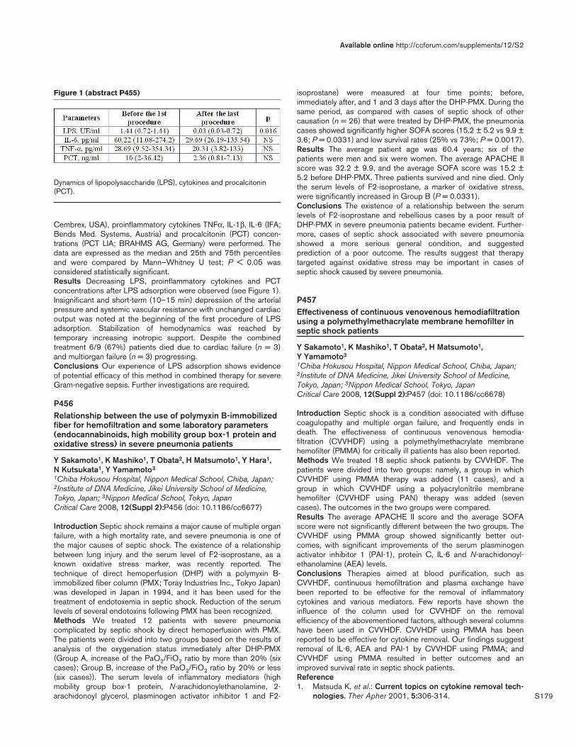
S179
Cembrex, USA), proinflammatory cytokines TNFα, IL-1β, IL-6 (IFA;Bends Med. Systems, Austria) and procalcitonin (PCT) concen-trations (PCT LIA; BRAHMS AG, Germany) were performed. Thedata are expressed as the median and 25th and 75th percentilesand were compared by Mann–Whitney U test; P < 0.05 wasconsidered statistically significant.Results Decreasing LPS, proinflammatory cytokines and PCTconcentrations after LPS adsorption were observed (see Figure 1).Insignificant and short-term (10–15 min) depression of the arterialpressure and systemic vascular resistance with unchanged cardiacoutput was noted at the beginning of the first procedure of LPSadsorption. Stabilization of hemodynamics was reached bytemporary increasing inotropic support. Despite the combinedtreatment 6/9 (67%) patients died due to cardiac failure (n = 3)and multiorgan failure (n = 3) progressing.Conclusions Our experience of LPS adsorption shows evidenceof potential efficacy of this method in combined therapy for severeGram-negative sepsis. Further investigations are required.
P456Relationship between the use of polymyxin B-immobilizedfiber for hemofiltration and some laboratory parameters(endocannabinoids, high mobility group box-1 protein andoxidative stress) in severe pneumonia patients
Y Sakamoto1, K Mashiko1, T Obata2, H Matsumoto1, Y Hara1, N Kutsukata1, Y Yamamoto3
1Chiba Hokusou Hospital, Nippon Medical School, Chiba, Japan;2Institute of DNA Medicine, Jikei University School of Medicine,Tokyo, Japan; 3Nippon Medical School, Tokyo, JapanCritical Care 2008, 12(Suppl 2):P456 (doi: 10.1186/cc6677)
Introduction Septic shock remains a major cause of multiple organfailure, with a high mortality rate, and severe pneumonia is one ofthe major causes of septic shock. The existence of a relationshipbetween lung injury and the serum level of F2-isoprostane, as aknown oxidative stress marker, was recently reported. Thetechnique of direct hemoperfusion (DHP) with a polymyxin B-immobilized fiber column (PMX; Toray Industries Inc., Tokyo Japan)was developed in Japan in 1994, and it has been used for thetreatment of endotoxemia in septic shock. Reduction of the serumlevels of several endotoxins following PMX has been recognized.Methods We treated 12 patients with severe pneumoniacomplicated by septic shock by direct hemoperfusion with PMX.The patients were divided into two groups based on the results ofanalysis of the oxygenation status immediately after DHP-PMX(Group A, increase of the PaO2/FiO2 ratio by more than 20% (sixcases); Group B, increase of the PaO2/FiO2 ratio by 20% or less(six cases)). The serum levels of inflammatory mediators (highmobility group box-1 protein, N-arachidonoylethanolamine, 2-arachidonoyl glycerol, plasminogen activator inhibitor 1 and F2-
isoprostane) were measured at four time points; before,immediately after, and 1 and 3 days after the DHP-PMX. During thesame period, as compared with cases of septic shock of othercausation (n = 26) that were treated by DHP-PMX, the pneumoniacases showed significantly higher SOFA scores (15.2 ± 5.2 vs 9.9 ±3.6; P = 0.0331) and low survival rates (25% vs 73%; P = 0.0017).Results The average patient age was 60.4 years; six of thepatients were men and six were women. The average APACHE IIscore was 32.2 ± 9.9, and the average SOFA score was 15.2 ±5.2 before DHP-PMX. Three patients survived and nine died. Onlythe serum levels of F2-isoprostane, a marker of oxidative stress,were significantly increased in Group B (P = 0.0331).Conclusions The existence of a relationship between the serumlevels of F2-isoprostane and rebellious cases by a poor result ofDHP-PMX in severe pneumonia patients became evident. Further-more, cases of septic shock associated with severe pneumoniashowed a more serious general condition, and suggestedprediction of a poor outcome. The results suggest that therapytargeted against oxidative stress may be important in cases ofseptic shock caused by severe pneumonia.
P457Effectiveness of continuous venovenous hemodiafiltrationusing a polymethylmethacrylate membrane hemofilter inseptic shock patients
Y Sakamoto1, K Mashiko1, T Obata2, H Matsumoto1, Y Yamamoto3
1Chiba Hokusou Hospital, Nippon Medical School, Chiba, Japan;2Institute of DNA Medicine, Jikei University School of Medicine,Tokyo, Japan; 3Nippon Medical School, Tokyo, JapanCritical Care 2008, 12(Suppl 2):P457 (doi: 10.1186/cc6678)
Introduction Septic shock is a condition associated with diffusecoagulopathy and multiple organ failure, and frequently ends indeath. The effectiveness of continuous venovenous hemodia-filtration (CVVHDF) using a polymethylmethacrylate membranehemofilter (PMMA) for critically ill patients has also been reported.Methods We treated 18 septic shock patients by CVVHDF. Thepatients were divided into two groups: namely, a group in whichCVVHDF using PMMA therapy was added (11 cases), and agroup in which CVVHDF using a polyacrylonitrile membranehemofilter (CVVHDF using PAN) therapy was added (sevencases). The outcomes in the two groups were compared.Results The average APACHE II score and the average SOFAscore were not significantly different between the two groups. TheCVVHDF using PMMA group showed significantly better out-comes, with significant improvements of the serum plasminogenactivator inhibitor 1 (PAI-1), protein C, IL-6 and N-arachidonoyl-ethanolamine (AEA) levels.Conclusions Therapies aimed at blood purification, such asCVVHDF, continuous hemofiltration and plasma exchange havebeen reported to be effective for the removal of inflammatorycytokines and various mediators. Few reports have shown theinfluence of the column used for CVVHDF on the removalefficiency of the abovementioned factors, although several columnshave been used in CVVHDF. CVVHDF using PMMA has beenreported to be effective for cytokine removal. Our findings suggestremoval of IL-6, AEA and PAI-1 by CVVHDF using PMMA; andCVVHDF using PMMA resulted in better outcomes and animproved survival rate in septic shock patients.Reference1. Matsuda K, et al.: Current topics on cytokine removal tech-
nologies. Ther Apher 2001, 5:306-314.
Available online http://ccforum.com/supplements/12/S2
Figure 1 (abstract P455)
Dynamics of lipopolysaccharide (LPS), cytokines and procalcitonin(PCT).
S180
P458Polymyxin B hemoperfusion in high endotoxin activity levelseptic shock patients
G Monti, V Terzi, M Mininni, S Colombo, S Vesconi, G CasellaNiguarda Hospital, Milan, ItalyCritical Care 2008, 12(Suppl 2):P458 (doi: 10.1186/cc6679)
Introduction The aim of the study was to evaluate the clinicalimpact of polymyxin-B hemoperfusion (HP-PMX) as adjuvant therapyversus conventional treatment in septic shock (SS) patients withhigh endotoxemia.Methods A retrospective analysis of the clinical profile andevolution related to the treatment strategy of SS patients with ahigh endotoxin activity (EA) level (>0.6 units) attending our ICUfrom January to August 2007. All patients (n = 16) were treatedwith standard therapy (ST) according to the Surviving SepsisCampaign. According to our ICU practice, adjuvant therapy withHP-PMX (twice, 2 hours/session with an interval of 24 hours) wasperformed only in SS patients with known or presumed Gram-negative and Gram-positive infections, worsening of haemo-dynamic instability in the next 6 hours of diagnosis and ≥3 organfailures (PMX group, n = 8). The clinical profile was evaluated inthe two groups (ST vs PMX group) at T0 (SS diagnosis, start ofHP-PMX) and at T1 (at 48 hours). The Student t test for pairedvalues was used for statistical purpose (P < 0.05 significant).Results At T0, the clinical profile of the two groups of high EA levelSS patients was similar (EA level, age, SOFA score, mean arterialpressure, norepinephrine, lactates, PaO2/FiO2, continuous renalreplacement therapy, used of activated protein C). Otherwise thePMX group showed a significant improvement of clinical conditionscompared with the ST group at T1 (Table 1). The ICU length ofstay was significantly longer in the PMX group versus the ST group(21.5 ± 21.3 vs 53.6 ± 67 days, P < 0.05). Further PMX-HPresulted in a reduction of ICU mortality when compared with ST inhigh EA level SS patients, probably due to endotoxin removal(45% vs 16%, not significant due to small sample size).
Table 1 (abstract P458)
Evolution of high endotoxin activity level in septic shock patients
T0 T1 P value
Norepinephrine, ST group 0.58 0.33 Not significant
Norepinephrine, PMX group 0.85 0.28 <0.05
SOFA score, ST group 11.5 11.1 Not significant
SOFA score, PMX group 14.15 12.17 <0.05
Lactate, ST group 5.41 4.71 Not significant
Lactate, PMX group 7.94 5.35 <0.05
Conclusions We can question whether the EA level could be auseful guide to early institution of specific anti-lipopolysaccharidetreatments.
P459Efficacy of the CTR-001 direct hemoperfusion adsorptioncolumn in sepsis
Y Suzuki, S Kikuchi, M Kojika, N Sato, S EndoIwate Medical University, Morioka, JapanCritical Care 2008, 12(Suppl 2):P459 (doi: 10.1186/cc6680)
Introduction Despite developing therapeutic strategies, mortalityof sepsis and septic shock has actually remained at a level of about
35–50%. The objective was to study the safety and effectivenessof a newly developed cytokine adsorption column in patients withsepsis and septic shock.Methods This study was a prospective, randomized, controlledclinical trial. The newly developed column contains microporouscellulose beads with a hexadecyl alkyl chain as the ligand. Eighteenpatients with early septic shock or septic organ dysfunction wereenrolled. Nine of 18 patients were randomized to directhemoperfusion (DHP). All patients received supportive intensivecare, and those randomized to DHP received direct hemoperfusionfor 4 hours more than two times up to 14 times during 14 days.Results The decrease of the APACHE II score from thepretreatment level at the seventh day was significantly larger in thetreatment group than in the control group (P = 0.0189;Mann–Whitney test). Adsorption-column-related serious adverseevents were not observed in the DHP group. The concentration ofIL-6 and IL-8 in the plasma decreased from the pretreatment levelin the DHP group significantly (P = 0.0464 and P = 0.0464,respectively; Wilcoxon test).Conclusions The newly developed DHP column improved theseptic shock better than the ordinary supportive intensive care.
P460Coupled plasma filtration adsorption in peritonitis-inducedporcine hyperdynamic septic shock
R Sykora, J Chvojka, A Krouzecky, J Radej, V Varnerova, T Karvunidis, I Novak, M MatejovicCharles University Medical School and Teaching Hospital Plzen,Plzen, Czech RepublicCritical Care 2008, 12(Suppl 2):P460 (doi: 10.1186/cc6681)
Introduction Extracorporeal blood purification techniques for thetreatment of sepsis remain conceptually sound but are still anunresolved issue. Coupled plasma filtration adsorption (CPFA)enabling higher nonselective elimination of septic mediatorsrepresents a promising concept [1]. Its efficacy, however, has notbeen tested in a clinically relevant large animal model of septicshock. We therefore evaluated the biological effects of CPFA in aporcine long-term, hyperdynamic, peritonitis-induced septic shockmodel.Methods In 16 mechanically ventilated and instrumented pigs,fecal peritonitis was induced by inoculating autologue feces.Twelve hours after induction of sepsis, the pigs were randomlyallocated to 10 hours of either CPFA (n = 8) or standard treatment(Control, n = 8). Before, 12, 18 and 22 hours after the induction ofperitonitis, we measured, in addition to systemic and regionalhemodynamics, ileal mucosal and renal cortex microvascularperfusion (OPS and laser Doppler flowmetry). Energy metabolismwas determined by measuring arterial, hepatic, portal and renalvenous lactate/pyruvate (L/P) ratios and hepatic venous ketonebody (KBR) ratios. Plasma IL-6 and TNFα levels were alsodetermined.Results All animals in both groups required noadrenaline infusionto maintain the mean arterial pressure above 70 mmHg. Neither thedose of vasopressor support nor the median time to developmentof arterial hypotension differed between the groups. In the controlgroup, hyperdynamic septic shock resulted in a progressivedeterioration of intestinal mucosal and renal cortex microvascularperfusion despite well-maintained regional blood flows. Alteredmicrocirculation was paralleled by gradually increased systemicand regional L/P and KBR. Although CPFA blunted the otherwiseprogressive increase in plasma TNFα levels, it failed to beneficiallyinfluence sepsis-induced alterations in microvascular perfusion andenergy metabolism.
Critical Care March 2008 Vol 12 Suppl 2 28th International Symposium on Intensive Care and Emergency Medicine
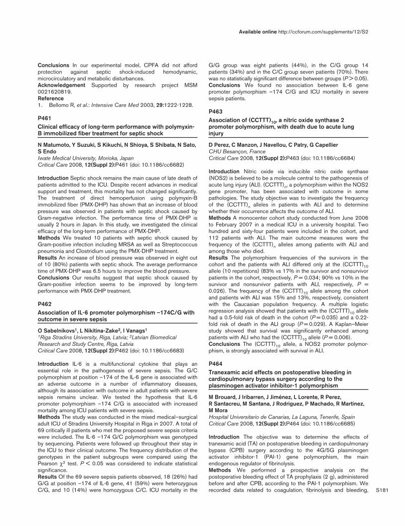
S181
Conclusions In our experimental model, CPFA did not affordprotection against septic shock-induced hemodynamic,microcirculatory and metabolic disturbances.Acknowledgement Supported by research project MSM0021620819.Reference1. Bellomo R, et al.: Intensive Care Med 2003, 29:1222-1228.
P461Clinical efficacy of long-term performance with polymyxin-B immobilized fiber treatment for septic shock
N Matumoto, Y Suzuki, S Kikuchi, N Shioya, S Shibata, N Sato,S EndoIwate Medical University, Morioka, JapanCritical Care 2008, 12(Suppl 2):P461 (doi: 10.1186/cc6682)
Introduction Septic shock remains the main cause of late death ofpatients admitted to the ICU. Despite recent advances in medicalsupport and treatment, this mortality has not changed significantly.The treatment of direct hemoperfusion using polymyxin-Bimmobilized fiber (PMX-DHP) has shown that an increase of bloodpressure was observed in patients with septic shock caused byGram-negative infection. The performance time of PMX-DHP isusually 2 hours in Japan. In this study, we investigated the clinicalefficacy of the long-term performance of PMX-DHP.Methods We treated 10 patients with septic shock caused byGram-positive infection including MRSA as well as Streptococcuspneumonia and Clostridium using the PMX-DHP treatment.Results An increase of blood pressure was observed in eight outof 10 (80%) patients with septic shock. The average performancetime of PMX-DHP was 6.5 hours to improve the blood pressure.Conclusions Our results suggest that septic shock caused byGram-positive infection seems to be improved by long-termperformance with PMX-DHP treatment.
P462Association of IL-6 promoter polymorphism –174C/G withoutcome in severe sepsis
O Sabelnikovs1, L Nikitina-Zake2, I Vanags1
1Riga Stradins University, Riga, Latvia; 2Latvian BiomedicalResearch and Study Centre, Riga, LatviaCritical Care 2008, 12(Suppl 2):P462 (doi: 10.1186/cc6683)
Introduction IL-6 is a multifunctional cytokine that plays anessential role in the pathogenesis of severe sepsis. The G/Cpolymorphism at position –174 of the IL-6 gene is associated withan adverse outcome in a number of inflammatory diseases,although its association with outcome in adult patients with severesepsis remains unclear. We tested the hypothesis that IL-6promoter polymorphism –174 C/G is associated with increasedmortality among ICU patients with severe sepsis.Methods The study was conducted in the mixed medical–surgicaladult ICU of Stradins University Hospital in Riga in 2007. A total of69 critically ill patients who met the proposed severe sepsis criteriawere included. The IL-6 –174 G/C polymorphism was genotypedby sequencing. Patients were followed up throughout their stay inthe ICU to their clinical outcome. The frequency distribution of thegenotypes in the patient subgroups were compared using thePearson χ2 test. P < 0.05 was considered to indicate statisticalsignificance.Results Of the 69 severe sepsis patients observed, 18 (26%) hadG/G at position –174 of IL-6 gene, 41 (59%) were heterozygousC/G, and 10 (14%) were homozygous C/C. ICU mortality in the
G/G group was eight patients (44%), in the C/G group 14patients (34%) and in the C/C group seven patients (70%). Therewas no statistically significant difference between groups (P > 0.05).Conclusions We found no association between IL-6 genepromoter polymorphism –174 C/G and ICU mortality in severesepsis patients.
P463Association of (CCTTT)10, a nitric oxide synthase 2promoter polymorphism, with death due to acute lunginjury
D Perez, C Manzon, J Navellou, C Patry, G CapellierCHU Besançon, FranceCritical Care 2008, 12(Suppl 2):P463 (doi: 10.1186/cc6684)
Introduction Nitric oxide via inducible nitric oxide synthase(NOS2) is believed to be a molecule central to the pathogenesis ofacute lung injury (ALI). (CCTTT)n, a polymorphism within the NOS2gene promoter, has been associated with outcome in somepathologies. The study objective was to investigate the frequencyof the (CCTTT)n alleles in patients with ALI and to determinewhether their occurrence affects the outcome of ALI.Methods A monocenter cohort study conducted from June 2006to February 2007 in a medical ICU in a university hospital. Twohundred and sixty-four patients were included in the cohort, and112 patients with ALI. The main outcome measures were thefrequency of the (CCTTT)n alleles among patients with ALI andamong those who died.Results The polymorphism frequencies of the survivors in thecohort and the patients with ALI differed only at the (CCTTT)10allele (10 repetitions) (83% vs 17% in the survivor and nonsurvivorpatients in the cohort, respectively, P = 0.034; 90% vs 10% in thesurvivor and nonsurvivor patients with ALI, respectively, P =0.026). The frequency of the (CCTTT)10 allele among the cohortand patients with ALI was 15% and 13%, respectively, consistentwith the Caucasian population frequency. A multiple logisticregression analysis showed that patients with the (CCTTT)10 allelehad a 0.5-fold risk of death in the cohort (P = 0.035) and a 0.22-fold risk of death in the ALI group (P = 0.029). A Kaplan–Meierstudy showed that survival was significantly enhanced amongpatients with ALI who had the (CCTTT)10 allele (P = 0.006).Conclusions The (CCTTT)10 allele, a NOS2 promoter polymor-phism, is strongly associated with survival in ALI.
P464Tranexamic acid effects on postoperative bleeding incardiopulmonary bypass surgery according to theplasminogen activator inhibitor-1 polymorphism
M Brouard, J Iribarren, J Jiménez, L Lorente, R Perez, R Santacreu, M Santana, J Rodriguez, P Machado, R Martinez,M MoraHospital Universitario de Canarias, La Laguna, Tenerife, SpainCritical Care 2008, 12(Suppl 2):P464 (doi: 10.1186/cc6685)
Introduction The objective was to determine the effects oftranexamic acid (TA) on postoperative bleeding in cardiopulmonarybypass (CPB) surgery according to the 4G/5G plasminogenactivator inhibitor-1 (PAI-1) gene polymorphism, the mainendogenous regulator of fibrinolysis.Methods We performed a prospective analysis on thepostoperative bleeding effect of TA prophylaxis (2 g), administeredbefore and after CPB, according to the PAI-1 polymorphism. Werecorded data related to coagulation, fibrinolysis and bleeding,
Available online http://ccforum.com/supplements/12/S2
S182
preoperatively, at admission (0 hours) to the ICU, and 4 hours and24 hours postoperatively. SPSS version 15 was used.Results We studied 50 patients (24 with TA and 26 without TA).In patients not receiving TA, significant differences were foundbetween PAI-1 genotype groups (4G/4G; 4G/5G; 5G/5G) inchest-tube blood loss at 0 hours (P = 0.03), at 4 hours (P =0.001) and at 24 hours (P = 0.009). Fresh frozen plasma wasrequired during the ICU stay in 50% of 5G/5G, 25% of 4G/5Gand none of the 4G/4G carriers (P = 0.021). 4G/4G patients didnot show significant differences in blood loss between the TA andplacebo groups. 4G/5G patients receiving TA had lower bloodloss than the placebo group at 0 hours (P = 0.012) and at 24hours after surgery (P = 0.014). In contrast, 5G/5G patientsreceiving TA had significantly lower blood loss compared with theplacebo group at 0 hours (P = 0.028), at 4 hours (P = 0.008) andat 24 hours (P = 0.004) after surgery. Fifty-five percent of 5G/Gpatients in the placebo group received fresh frozen plasma duringthe ICU stay compared with no patients in the TA group (P =0.014).Conclusions PAI-1 5G/5G homozygotes who did not receive TAshowed significantly greater postoperative bleeding than patientswith other PAI-1 genotypes. The 5G/5G homozygotes whoreceived TA showed the greatest blood-sparing benefit.
P465Effect of the IL-6 promoter polymorphism –174 G/C onrisk and outcome of pneumonia
I Martín-Loeches1, J Solé Violan1, J Blanquer2, J Aspa3, C Rodríguez Gallego1, F Rodríguez de Castro1, I GarcíaLaorden1, O Rajas2, A García-Saavedra1
1Hospital General de Gran Canaria Dr Negrin, Las Palmas de GranCanaria, Spain; 2Hospital Clínico de Valencia, Valencia, Spain;3Hospital de la Princesa, Madrid, SpainCritical Care 2008, 12(Suppl 2):P465 (doi: 10.1186/cc6686)
Introduction IL-6 is a pleiotropic cytokine expressed in manytissues. A polymorphism in the IL-6 gene, associated with differ-ences in the IL-6 transcription rate, has been recently described[1]. The influence of genetic polymorphisms of IL-6 gene promoter–174 G/C on the severity of systemic inflammatory responsesyndrome associated with community-acquired pneumonia (CAP)was studied.Methods A prospective, multicentric case–control study in threeuniversity hospitals in Spain. In 852 consecutive patients whopresented with CAP and 923 controls, using PCR-RFLP analysis,we analyzed the –174G/C single nucleotide polymorphism of theIL-6 gene. The pneumonia severity was assessed on the day ofadmission and stratified according to the PORT score andcomplications recorded such as respiratory failure, renalimpairment, severe sepsis, shock and multiorgan failure. Outcomesevaluated were the duration of hospital stay, ICU admittance, andinhospital and 28-day mortality.Results See Table 1. The distribution of the G/C 174 genotypewas similar in CAP patients and controls. The genotype distributionof the polymorphism was 45.07% for GG and 43.89% and
11.03% for GC and CC, respectively. In patients who wereadmitted with CAP, no significant differences were observedcompared with progression between groups.Multivariate analysis (which included genotype, age, sex, andclassical risk factors for CAP) identified the C/C genotype as theonly independent predictor of renal failure.Conclusions Our findings show that the 174G/C polymorphism isnot associated with risk and outcome of CAP in the Spanish whiteCaucasian population.Reference1. Schluter B, Raufhake C, Erren M, et al.: Effect of the inter-
leukin-6 promoter polymorphism (-174 G/C) on the inci-dence and outcome of sepsis. Crit Care Med 2002, 30:32-37.
P466Relationship between angiotensin-converting enzymegene polymorphism (insertion/deletion) and the clinicalcondition of sepsis
R YildizdasCukurova University, Adana, TurkeyCritical Care 2008, 12(Suppl 2):P466 (doi: 10.1186/cc6687)
Introduction It has been postulated that genetic predispositionmay influence the susceptibility to infection and disease outcome.The D allele of the angiotensin-converting enzyme (ACE) gene isassociated with many diseases. However, there are only fewreports available about infection. We have investigated theassociation between ACE I/D polymorphism and sepsis, its clinicalfeatures such as shock, acute respiratory distress syndrome(ARDS), multiorgan dysfunction syndrome (MODS) and mortality.Methods Ninety-eight patients who had been diagnosed withsepsis and 100 healthy individuals were included. The patientswere divided into groups based on the presence of shock, ARDS,MODS and mortality. The ACE gene polymorphism was analyzedby PCR.Results There was no statistical difference between the controls’and patients’ genotype (P = 0.29). No evidence emergedregarding the association of the ACE I/D polymorphism withMODS, but there was evidence of an association with sepsis-related hypotension, mortality and ARDS. While the I/I genotypewas observed to increase the sepsis-related mortality risk 11.5-fold(P = 0.008), it increased the risk for hypotension 9.5-fold (P <0.001). On the other hand, it was found that carrying D/Dgenotypes increased the risk of the having ARDS 4.5-fold (0R =4.5, 95% CI 1.15–19.6) (P = 0.028). Meanwhile, in multivariatelogistic analysis, shock is the only factor associated with mortality.Conclusions The insertion polymorphism in angiotensin-convertingenzyme gene is associated with an increase in the risk of sepsis-related mortality and shock, but the deletion polymorphism isassociated with an increase in the risk of sepsis-related ARDS.
P467Incidence of renal failure following orthotopic livertransplantation is lower in the Asian population
J Read, P Bearfield, B AgarwalRoyal Free Hospital, London, UKCritical Care 2008, 12(Suppl 2):P467 (doi: 10.1186/cc6688)
Introduction Renal failure is reported as a frequent complicationfollowing orthotopic liver transplantation (OLT) and is associatedwith a higher morbidity and mortality [1]. Acute kidney injury andthe need for renal replacement therapy (RRT) are known to be
Critical Care March 2008 Vol 12 Suppl 2 28th International Symposium on Intensive Care and Emergency Medicine
Table 1 (abstract P465)
C/C genotype G/C genotype G/G genotype
No sepsis 10 (13.51%) 32 (43.24%) 32 (43.24%)
Sepsis 53 (10.08%) 236 (44.87%) 237 (45.06%)
Severe sepsis 18 (13.14%) 56 (40.88%) 63 (45.99%)
Septic shock 13 (11.30%) 50 (43.48%) 52 (45.22%)
S183
generally greater in the Asian population for a number of proposedreasons, such as higher incidence of diabetes mellitus andhypertension. In our study the incidence of renal failure and theneed for RRT in the early postoperative period following OLT wasactually lower in this group.Methods A retrospective case-note review was undertaken on348 consecutive elective OLTs performed in the period2000–2007 at our institution. The primary outcome was therequirement for RRT and renal impairment as per the RIFLE andAKIN criteria. Patients on RRT pretransplant, acute hepatic failure,regraft and combined organ transplant were excluded.Results Two hundred and seventy-one patients fulfilled theinclusion criteria, of which 45 (17%) were of Asian ethnicity. A totalof 32 patients required RRT in the early postoperative period, ofwhich only three (9.4%) were Asian. The change in serumcreatinine in the first 48 hours was also examined and is presentedin Table 1. The baseline pretransplant serum creatinine was similarbetween the groups: The median serum creatinine was 86 μmol/lfor both the Asian (range 60–179) and non-Asian groups (range34–264).
Table 1 (abstract P467)
Change in serum creatinine following OLT as per RIFLE/AKINcriteria
Non-Asian Asian ΔCr 48 hours group (n = 22) group (n = 45)
Creatinine > 150% 77 (34%) 19 (42%)
Creatinine > 200% 57 (25%) 11 (24%)
Creatinine > 300%/RRT 36 (16%) 3 (7%)
Conclusions The burden of RRT following liver transplantationwas less in the Asian population. Examination of perioperativeserum creatinine showed that the Asian group actually sufferedmild renal dysfunction to a greater extent (42% vs 34%) but didnot progress to ‘failure’ stage as often as non-Asians (7% vs 16%,P < 0.05). The reason for this reversal may be partly due to farfewer Asian transplant recipients with alcoholic liver disease, aknown predisposing risk factor.Reference1. Bilbao I, et al.: Risk factors for acute renal failure requiring
dialysis after liver transplantation. Clin Transplant 1998,12:123-129.
P468Renal protection in the ICU: role of fenoldopam
P Bertini1, F Pratesi1, T Angelini1, F Forfori2, G Licitra2, F Giunta2
1Scuola di Specializzazione in Anestesia e Rianimazione, Universitàdegli Studi di Pisa, AOUP, Pisa, Italy; 2Department of Surgery,AOUP, Pisa, ItalyCritical Care 2008, 12(Suppl 2):P468 (doi: 10.1186/cc6689)
Introduction Acute renal failure (ARF) is one of the most commonproblems of the critical patient, often resulting in acute tubularnecrosis following an impairment in renal hemodynamics. Fenoldo-pam is a selective dopaminergic agonist specific for dopaminergicreceptor DA-1 that, provoking an increase in renal blood flow,diuresis and natriuresis, seems to prevent ARF – exerting a nephro-protective effect.Methods Patients in the ICU without ARF but at risk of renaldamage (sepsis, nephrotoxic drug administration) were recruited.Fenoldopam was administered by intravenous infusion (0.05–0.1
μg/kg/min) for 24 hours. Specific tubular enzymatic biomarkers(β2-microglobulin, microalbuminuria, retinal binding protein, γGT,alkaline phosphatase, n-acetyl-glucosaminidase) have been dosedin urine before fenoldopam administration (basal value), at the endof the infusion (T1) and 24 hours later (T2). Diuresis, bloodcreatinine and cystatin C were also measured in the same interval.All data were compared using a t test and logistic regressionanalysis.Results See Figure 1. Of the 23 patients studied, after informedwritten consent, 10 patients received fenoldopam. At T1 astatistically significant reduction of the tubular biomarkers, cystatinC and creatinine, and an increase in diuresis were found comparedwith basal. At T2 a nonstatistically significant reduction of γGT andmicroalbumin and cystatin C associated with an increased urinaryoutput were observed, showing a benefit on kidney function lastingeven after 48 hours.Conclusions Our study shows a significant protective effect offenoldopam measured by tubular biomarkers after a 24 hour infusion.Reference1. Morelli A: Crit Care Med 2005, 33:2451-2456.
P469Renal hemodynamic, microcirculatory and metabolicresponses to peritonitis-induced acute kidney injury in pigs
J Chvojka1, R Sykora1, A Krouzecky1, J Radej1, V Varnerova2, T Karvunidis1, I Novak1, M Matejovic1
1University Hospital, Plzen, Czech Republic; 2Medical Faculty,Charles University, Plzen, Czech RepublicCritical Care 2008, 12(Suppl 2):P469 (doi: 10.1186/cc6690)
Introduction The widely held paradigm that renal vasoconstrictionmay not be a prerequisite for septic acute kidney injury (AKI) hasrecently been challenged [1]. Further research is needed toestablish whether this concept is valid in other clinically relevantmodels of sepsis-induced AKI [2]. The aim of our study wastherefore to dynamically assess the pattern of renal hemodynamics,microcirculation and energy balance during a hyperdynamicporcine model of progressive septic shock.
Available online http://ccforum.com/supplements/12/S2
Figure 1 (abstract P468)
Markers of renal function. NAG, n-acetyl-glucosaminidase; B2M, β2-microglobulin; Microalb, microalbuminuria; PLR, retinal binding protein;ALP, alkaline phosphatase; GGT, γGT.
S184
Methods In eight anesthetized, mechanically ventilated andinstrumented pigs, fecal peritonitis was induced by inoculatingautologous feces. Six sham-operated animals served as time-matched controls. Hyperdynamic circulation was achieved usinghydroxyethylstarch, and norepinephrine was administered tomaintain mean arterial pressure ≥70 mmHg. Before and at 12, 18and 24 hours of peritonitis, we measured, in addition to systemichemodynamics, renal blood flow, renal cortex microvascularperfusion, renal venous pressure, renal oxygen kinetics andregional acid–base balance and lactate/pyruvate (L/P) ratios.Results All pigs developed hyperdynamic septic shock associatedwith the development of AKI as evidenced by increased plasmacreatinine levels to 30% of baseline. Although the renal blood flowslightly decreased by the end of the experiment, renal vascularresistance remained unchanged. Despite maintained regionalhemodynamics, there was significant and early decline in renalcortex microvascular perfusion. Although renal oxygen consump-tion did not change, renal venous acidosis and an increased L/Pratio developed.Conclusions Septic AKI was not associated with renalvasoconstriction. While early alterations in renal cortexmicrocirculation preceded a minor decline in renal blood flow,unchanged renal oxygen consumption together with signs ofsignificant metabolic stress suggests that the basis for septic AKImay be attributable not only to changes in microcirculatoryperfusion, but also to disturbed cellular energy machineryindependent of tissue oxygen availability.Acknowledgement Supported by MSM 0021620819.References1. Langenberg C, et al.: Kidney Int 2006, 69:1996-2002.2. Matejovic M, et al.: Intensive Care Med 2007, 33:1498-1500.
P470Risk factors and outcome of rhabdomyolysis after cardiacsurgery
L Hajjar, S Grande, F Galas, A Roquim, L Sampaio, J Auler JrHeart Institute, São Paulo, BrazilCritical Care 2008, 12(Suppl 2):P470 (doi: 10.1186/cc6691)
Introduction Rhabdomyolysis is a dissolution of skeletal musclescausing extravasation of toxic intracellular contents from themyocytes into the circulatory system. It leads to electrolyte distur-bances, hypovolemia, and renal failure. The incidence of rhabdo-myolysis after cardiac surgery and the impact on prognosis are notknown. The purpose of this study was to determine the incidence ofrhabdomyolysis, the associated risk factors and to assess theprognostic impact in patients undergoing cardiac surgery.Methods We performed a prospective, observational study in atertiary surgical ICU in a university hospital. A total of 200 patientsundergoing elective surgery were evaluated. Creatine kinase (CK)samples were collected daily (rhabdomyolysis, CK > 2,500 U/l) asplasma creatinine, CK-MB and troponin. Patients were followedduring the ICU stay and clinical outcomes were evaluated. Renalfailure was defined as >50% increase in serum creatinine frombaseline, and myocardial ischemia as an increase of CK-MB ortroponin greater than five times.Results Rhabdomyolysis was present in 38 patients (19%). Therewere no relations between rhabdomyolysis and hypothermia, sex,age, cardioprotection or use of vasoactive drugs. Risk factorswere: previous use of statin (P = 0.002), intraoperativehemodynamic instability (P = 0.045), longer duration of surgery(P < 0.001), and longer duration of pump (P < 0.0001). Theprevious use of statin and a longer duration of surgery wereindependent predictors of rhabdomyolysis (statin OR = 3.68, 95%
CI = 1.38–9.83 and longer duration of surgery (minutes) OR =1.01, 95% CI = 1.006–1.02). The occurrence of rhabdomyolysiswas not associated with higher incidence of renal failure,myocardial ischemia, mortality and longer time of ICU stay.Conclusions In this observational study, rhabdomyolysis was afrequent event after cardiac surgery, present most in patients withprevious use of statin or who presented hemodynamic instability inthe intraoperative period. However, this had no implications withrespect to midterm prognosis.References1. Gabow PA, Kachny WD, Kelleber SP: The spectrum of
rhabdomyolysis. Medicine 1982, 61:141-152.2. Slater MD, Mullins RJ: Rhabdomyolysis and myoglobinuric
renal failure in trauma and surgical patients: a review. JAm Coll Surg 1998, 186:693-716.
P471Requirement for renal replacement therapy followingorthotopic liver transplantation in adults is associated withprolonged mechanical ventilation and higher incidence ofpneumonia.
P Bearfield, J Read, S Shaw, B AgarwalRoyal Free Hospital, London, UKCritical Care 2008, 12(Suppl 2):P471 (doi: 10.1186/cc6692)
Introduction Acute renal failure requiring renal replacementtherapy (RRT) is common following orthotopic liver transplantation(OLT) and is associated with increased morbidity and mortality [1].Prolonged mechanical ventilation in intensive therapy unit (ITU)patients (>3 days) is associated with increases in length of stay,costs, mortality, risk of ventilator-associated pneumonia andsedation requirements [2].Methods A retrospective cohort study was conducted on 348consecutive OLTs in adults performed at the Royal Free Hospital(London, UK) between January 2000 and January 2007. Patientson RRT pretransplant, acute or fulminant hepatic failure, regraft orcombined organ transplant were excluded. Primary outcomes werepostoperative RRT and renal impairment as defined by the RIFLEcriteria. Data also examined included the number of ventilatedpostoperative days, and the presence of a clinical diagnosis ofpneumonia by the ITU physician.Results Two hundred and seventy-one patients were included.Thirty-two of them required postoperative RRT. The mean durationof ventilation, prolonged ventilation, length of ITU stay, incidence ofpneumonia, and ITU mortality were higher in the RRT group (Table 1).
Table 1 (abstract P471)
RRT Non-RRT
n 32 239
MV days (mean (SD)) 20 (19) 5 (12)
Prolonged MV (>3 days) 30 (94%) 51 (21%)
ITU days (median (IQR)) 13(7–24) 2 (1–4)
Pneumonia 15 (47%) 29 (12%)
ITU mortality 34% 4%
MV, mechanical ventilation.
Conclusions RRT after OLT is associated with prolonged mechanicalventilation, increased risk of pneumonia, and higher ITU mortality.References1. Bilbao I, et al.: Risk factors for acute renal failure requiring
dialysis after liver transplantation. Clin Transplant 1998,12:123-129.
Critical Care March 2008 Vol 12 Suppl 2 28th International Symposium on Intensive Care and Emergency Medicine
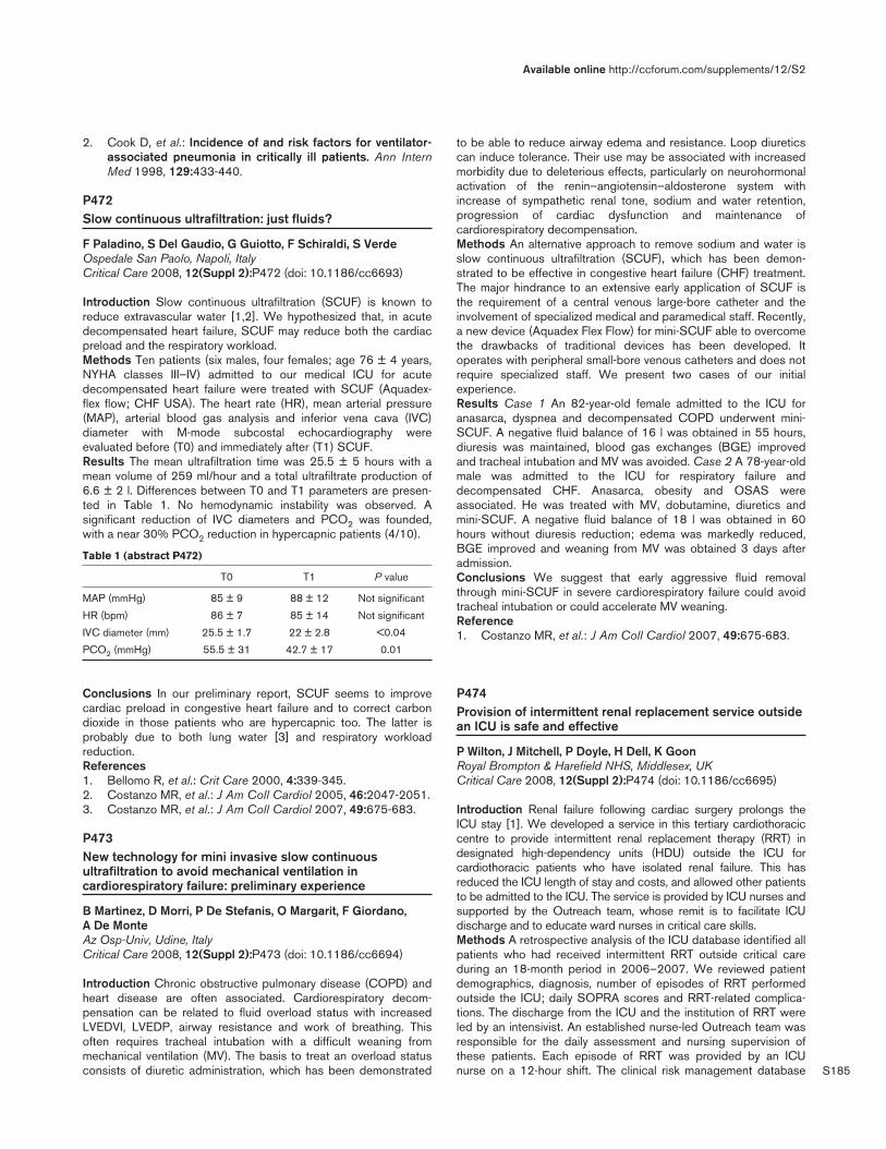
S185
2. Cook D, et al.: Incidence of and risk factors for ventilator-associated pneumonia in critically ill patients. Ann InternMed 1998, 129:433-440.
P472Slow continuous ultrafiltration: just fluids?
F Paladino, S Del Gaudio, G Guiotto, F Schiraldi, S VerdeOspedale San Paolo, Napoli, ItalyCritical Care 2008, 12(Suppl 2):P472 (doi: 10.1186/cc6693)
Introduction Slow continuous ultrafiltration (SCUF) is known toreduce extravascular water [1,2]. We hypothesized that, in acutedecompensated heart failure, SCUF may reduce both the cardiacpreload and the respiratory workload.Methods Ten patients (six males, four females; age 76 ± 4 years,NYHA classes III–IV) admitted to our medical ICU for acutedecompensated heart failure were treated with SCUF (Aquadex-flex flow; CHF USA). The heart rate (HR), mean arterial pressure(MAP), arterial blood gas analysis and inferior vena cava (IVC)diameter with M-mode subcostal echocardiography wereevaluated before (T0) and immediately after (T1) SCUF.Results The mean ultrafiltration time was 25.5 ± 5 hours with amean volume of 259 ml/hour and a total ultrafiltrate production of6.6 ± 2 l. Differences between T0 and T1 parameters are presen-ted in Table 1. No hemodynamic instability was observed. Asignificant reduction of IVC diameters and PCO2 was founded,with a near 30% PCO2 reduction in hypercapnic patients (4/10).
Table 1 (abstract P472)
T0 T1 P value
MAP (mmHg) 85 ± 9 88 ± 12 Not significant
HR (bpm) 86 ± 7 85 ± 14 Not significant
IVC diameter (mm) 25.5 ± 1.7 22 ± 2.8 <0.04
PCO2 (mmHg) 55.5 ± 31 42.7 ± 17 0.01
Conclusions In our preliminary report, SCUF seems to improvecardiac preload in congestive heart failure and to correct carbondioxide in those patients who are hypercapnic too. The latter isprobably due to both lung water [3] and respiratory workloadreduction.References1. Bellomo R, et al.: Crit Care 2000, 4:339-345.2. Costanzo MR, et al.: J Am Coll Cardiol 2005, 46:2047-2051.3. Costanzo MR, et al.: J Am Coll Cardiol 2007, 49:675-683.
P473New technology for mini invasive slow continuousultrafiltration to avoid mechanical ventilation incardiorespiratory failure: preliminary experience
B Martinez, D Morri, P De Stefanis, O Margarit, F Giordano, A De MonteAz Osp-Univ, Udine, ItalyCritical Care 2008, 12(Suppl 2):P473 (doi: 10.1186/cc6694)
Introduction Chronic obstructive pulmonary disease (COPD) andheart disease are often associated. Cardiorespiratory decom-pensation can be related to fluid overload status with increasedLVEDVI, LVEDP, airway resistance and work of breathing. Thisoften requires tracheal intubation with a difficult weaning frommechanical ventilation (MV). The basis to treat an overload statusconsists of diuretic administration, which has been demonstrated
to be able to reduce airway edema and resistance. Loop diureticscan induce tolerance. Their use may be associated with increasedmorbidity due to deleterious effects, particularly on neurohormonalactivation of the renin–angiotensin–aldosterone system withincrease of sympathetic renal tone, sodium and water retention,progression of cardiac dysfunction and maintenance ofcardiorespiratory decompensation.Methods An alternative approach to remove sodium and water isslow continuous ultrafiltration (SCUF), which has been demon-strated to be effective in congestive heart failure (CHF) treatment.The major hindrance to an extensive early application of SCUF isthe requirement of a central venous large-bore catheter and theinvolvement of specialized medical and paramedical staff. Recently,a new device (Aquadex Flex Flow) for mini-SCUF able to overcomethe drawbacks of traditional devices has been developed. Itoperates with peripheral small-bore venous catheters and does notrequire specialized staff. We present two cases of our initialexperience.Results Case 1 An 82-year-old female admitted to the ICU foranasarca, dyspnea and decompensated COPD underwent mini-SCUF. A negative fluid balance of 16 l was obtained in 55 hours,diuresis was maintained, blood gas exchanges (BGE) improvedand tracheal intubation and MV was avoided. Case 2 A 78-year-oldmale was admitted to the ICU for respiratory failure anddecompensated CHF. Anasarca, obesity and OSAS wereassociated. He was treated with MV, dobutamine, diuretics andmini-SCUF. A negative fluid balance of 18 l was obtained in 60hours without diuresis reduction; edema was markedly reduced,BGE improved and weaning from MV was obtained 3 days afteradmission.Conclusions We suggest that early aggressive fluid removalthrough mini-SCUF in severe cardiorespiratory failure could avoidtracheal intubation or could accelerate MV weaning.Reference1. Costanzo MR, et al.: J Am Coll Cardiol 2007, 49:675-683.
P474Provision of intermittent renal replacement service outsidean ICU is safe and effective
P Wilton, J Mitchell, P Doyle, H Dell, K GoonRoyal Brompton & Harefield NHS, Middlesex, UKCritical Care 2008, 12(Suppl 2):P474 (doi: 10.1186/cc6695)
Introduction Renal failure following cardiac surgery prolongs theICU stay [1]. We developed a service in this tertiary cardiothoraciccentre to provide intermittent renal replacement therapy (RRT) indesignated high-dependency units (HDU) outside the ICU forcardiothoracic patients who have isolated renal failure. This hasreduced the ICU length of stay and costs, and allowed other patientsto be admitted to the ICU. The service is provided by ICU nurses andsupported by the Outreach team, whose remit is to facilitate ICUdischarge and to educate ward nurses in critical care skills.Methods A retrospective analysis of the ICU database identified allpatients who had received intermittent RRT outside critical careduring an 18-month period in 2006–2007. We reviewed patientdemographics, diagnosis, number of episodes of RRT performedoutside the ICU; daily SOPRA scores and RRT-related complica-tions. The discharge from the ICU and the institution of RRT wereled by an intensivist. An established nurse-led Outreach team wasresponsible for the daily assessment and nursing supervision ofthese patients. Each episode of RRT was provided by an ICUnurse on a 12-hour shift. The clinical risk management database
Available online http://ccforum.com/supplements/12/S2
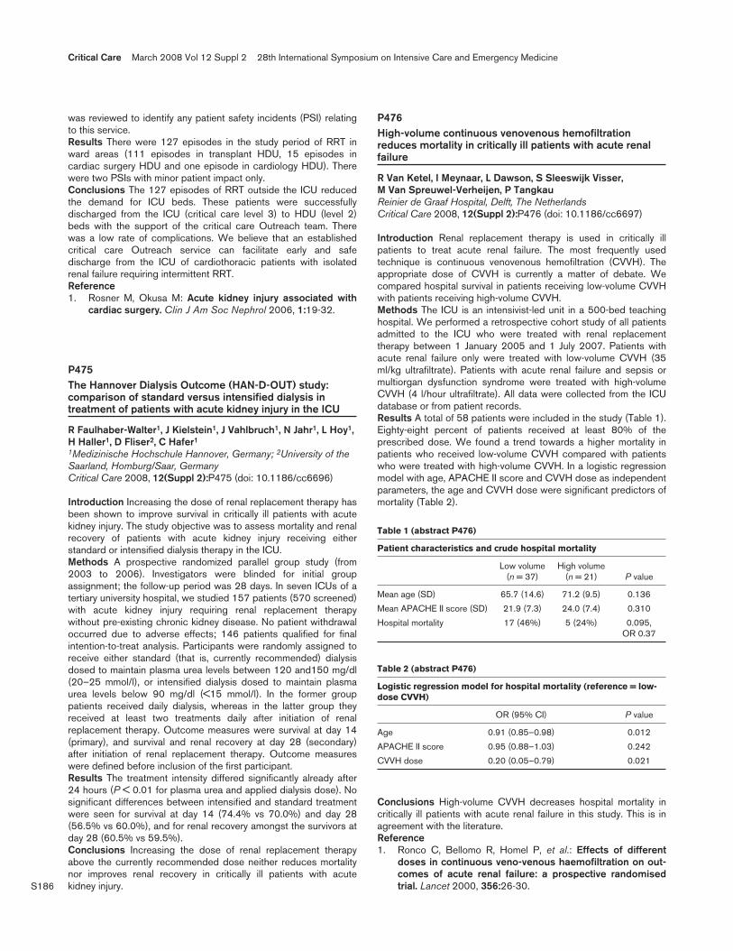
S186
was reviewed to identify any patient safety incidents (PSI) relatingto this service.Results There were 127 episodes in the study period of RRT inward areas (111 episodes in transplant HDU, 15 episodes incardiac surgery HDU and one episode in cardiology HDU). Therewere two PSIs with minor patient impact only.Conclusions The 127 episodes of RRT outside the ICU reducedthe demand for ICU beds. These patients were successfullydischarged from the ICU (critical care level 3) to HDU (level 2)beds with the support of the critical care Outreach team. Therewas a low rate of complications. We believe that an establishedcritical care Outreach service can facilitate early and safedischarge from the ICU of cardiothoracic patients with isolatedrenal failure requiring intermittent RRT.Reference1. Rosner M, Okusa M: Acute kidney injury associated with
cardiac surgery. Clin J Am Soc Nephrol 2006, 1:19-32.
P475The Hannover Dialysis Outcome (HAN-D-OUT) study:comparison of standard versus intensified dialysis intreatment of patients with acute kidney injury in the ICU
R Faulhaber-Walter1, J Kielstein1, J Vahlbruch1, N Jahr1, L Hoy1,H Haller1, D Fliser2, C Hafer1
1Medizinische Hochschule Hannover, Germany; 2University of theSaarland, Homburg/Saar, GermanyCritical Care 2008, 12(Suppl 2):P475 (doi: 10.1186/cc6696)
Introduction Increasing the dose of renal replacement therapy hasbeen shown to improve survival in critically ill patients with acutekidney injury. The study objective was to assess mortality and renalrecovery of patients with acute kidney injury receiving eitherstandard or intensified dialysis therapy in the ICU.Methods A prospective randomized parallel group study (from2003 to 2006). Investigators were blinded for initial groupassignment; the follow-up period was 28 days. In seven ICUs of atertiary university hospital, we studied 157 patients (570 screened)with acute kidney injury requiring renal replacement therapywithout pre-existing chronic kidney disease. No patient withdrawaloccurred due to adverse effects; 146 patients qualified for finalintention-to-treat analysis. Participants were randomly assigned toreceive either standard (that is, currently recommended) dialysisdosed to maintain plasma urea levels between 120 and150 mg/dl(20–25 mmol/l), or intensified dialysis dosed to maintain plasmaurea levels below 90 mg/dl (<15 mmol/l). In the former grouppatients received daily dialysis, whereas in the latter group theyreceived at least two treatments daily after initiation of renalreplacement therapy. Outcome measures were survival at day 14(primary), and survival and renal recovery at day 28 (secondary)after initiation of renal replacement therapy. Outcome measureswere defined before inclusion of the first participant.Results The treatment intensity differed significantly already after24 hours (P < 0.01 for plasma urea and applied dialysis dose). Nosignificant differences between intensified and standard treatmentwere seen for survival at day 14 (74.4% vs 70.0%) and day 28(56.5% vs 60.0%), and for renal recovery amongst the survivors atday 28 (60.5% vs 59.5%).Conclusions Increasing the dose of renal replacement therapyabove the currently recommended dose neither reduces mortalitynor improves renal recovery in critically ill patients with acutekidney injury.
P476High-volume continuous venovenous hemofiltrationreduces mortality in critically ill patients with acute renalfailure
R Van Ketel, I Meynaar, L Dawson, S Sleeswijk Visser, M Van Spreuwel-Verheijen, P TangkauReinier de Graaf Hospital, Delft, The NetherlandsCritical Care 2008, 12(Suppl 2):P476 (doi: 10.1186/cc6697)
Introduction Renal replacement therapy is used in critically illpatients to treat acute renal failure. The most frequently usedtechnique is continuous venovenous hemofiltration (CVVH). Theappropriate dose of CVVH is currently a matter of debate. Wecompared hospital survival in patients receiving low-volume CVVHwith patients receiving high-volume CVVH.Methods The ICU is an intensivist-led unit in a 500-bed teachinghospital. We performed a retrospective cohort study of all patientsadmitted to the ICU who were treated with renal replacementtherapy between 1 January 2005 and 1 July 2007. Patients withacute renal failure only were treated with low-volume CVVH (35ml/kg ultrafiltrate). Patients with acute renal failure and sepsis ormultiorgan dysfunction syndrome were treated with high-volumeCVVH (4 l/hour ultrafiltrate). All data were collected from the ICUdatabase or from patient records.Results A total of 58 patients were included in the study (Table 1).Eighty-eight percent of patients received at least 80% of theprescribed dose. We found a trend towards a higher mortality inpatients who received low-volume CVVH compared with patientswho were treated with high-volume CVVH. In a logistic regressionmodel with age, APACHE II score and CVVH dose as independentparameters, the age and CVVH dose were significant predictors ofmortality (Table 2).
Table 1 (abstract P476)
Patient characteristics and crude hospital mortality
Low volume High volume (n = 37) (n = 21) P value
Mean age (SD) 65.7 (14.6) 71.2 (9.5) 0.136
Mean APACHE II score (SD) 21.9 (7.3) 24.0 (7.4) 0.310
Hospital mortality 17 (46%) 5 (24%) 0.095, OR 0.37
Table 2 (abstract P476)
Logistic regression model for hospital mortality (reference = low-dose CVVH)
OR (95% CI) P value
Age 0.91 (0.85–0.98) 0.012
APACHE II score 0.95 (0.88–1.03) 0.242
CVVH dose 0.20 (0.05–0.79) 0.021
Conclusions High-volume CVVH decreases hospital mortality incritically ill patients with acute renal failure in this study. This is inagreement with the literature.Reference1. Ronco C, Bellomo R, Homel P, et al.: Effects of different
doses in continuous veno-venous haemofiltration on out-comes of acute renal failure: a prospective randomisedtrial. Lancet 2000, 356:26-30.
Critical Care March 2008 Vol 12 Suppl 2 28th International Symposium on Intensive Care and Emergency Medicine
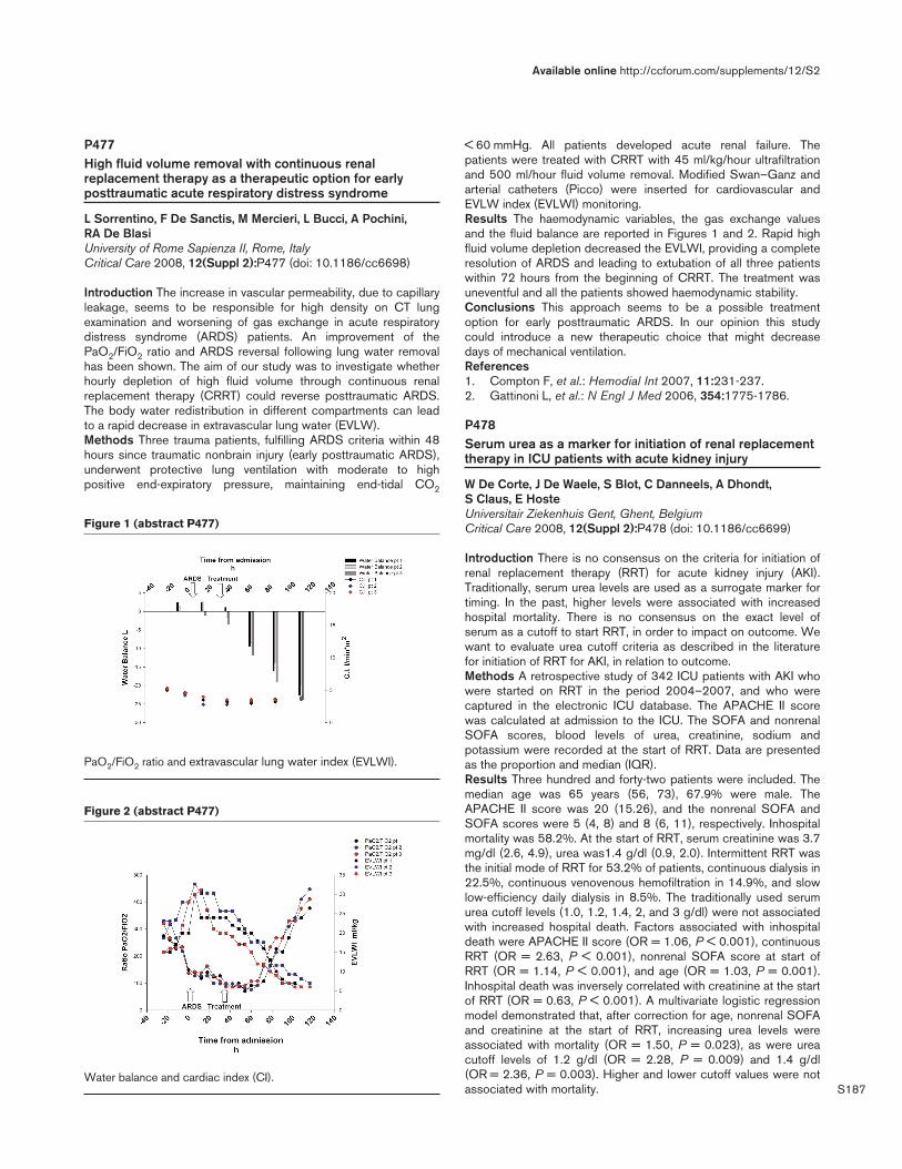
S187
P477High fluid volume removal with continuous renalreplacement therapy as a therapeutic option for earlyposttraumatic acute respiratory distress syndrome
L Sorrentino, F De Sanctis, M Mercieri, L Bucci, A Pochini, RA De BlasiUniversity of Rome Sapienza II, Rome, ItalyCritical Care 2008, 12(Suppl 2):P477 (doi: 10.1186/cc6698)
Introduction The increase in vascular permeability, due to capillaryleakage, seems to be responsible for high density on CT lungexamination and worsening of gas exchange in acute respiratorydistress syndrome (ARDS) patients. An improvement of thePaO2/FiO2 ratio and ARDS reversal following lung water removalhas been shown. The aim of our study was to investigate whetherhourly depletion of high fluid volume through continuous renalreplacement therapy (CRRT) could reverse posttraumatic ARDS.The body water redistribution in different compartments can leadto a rapid decrease in extravascular lung water (EVLW).Methods Three trauma patients, fulfilling ARDS criteria within 48hours since traumatic nonbrain injury (early posttraumatic ARDS),underwent protective lung ventilation with moderate to highpositive end-expiratory pressure, maintaining end-tidal CO2
< 60 mmHg. All patients developed acute renal failure. Thepatients were treated with CRRT with 45 ml/kg/hour ultrafiltrationand 500 ml/hour fluid volume removal. Modified Swan–Ganz andarterial catheters (Picco) were inserted for cardiovascular andEVLW index (EVLWI) monitoring.Results The haemodynamic variables, the gas exchange valuesand the fluid balance are reported in Figures 1 and 2. Rapid highfluid volume depletion decreased the EVLWI, providing a completeresolution of ARDS and leading to extubation of all three patientswithin 72 hours from the beginning of CRRT. The treatment wasuneventful and all the patients showed haemodynamic stability.Conclusions This approach seems to be a possible treatmentoption for early posttraumatic ARDS. In our opinion this studycould introduce a new therapeutic choice that might decreasedays of mechanical ventilation.References1. Compton F, et al.: Hemodial Int 2007, 11:231-237.2. Gattinoni L, et al.: N Engl J Med 2006, 354:1775-1786.
P478Serum urea as a marker for initiation of renal replacementtherapy in ICU patients with acute kidney injury
W De Corte, J De Waele, S Blot, C Danneels, A Dhondt, S Claus, E HosteUniversitair Ziekenhuis Gent, Ghent, BelgiumCritical Care 2008, 12(Suppl 2):P478 (doi: 10.1186/cc6699)
Introduction There is no consensus on the criteria for initiation ofrenal replacement therapy (RRT) for acute kidney injury (AKI).Traditionally, serum urea levels are used as a surrogate marker fortiming. In the past, higher levels were associated with increasedhospital mortality. There is no consensus on the exact level ofserum as a cutoff to start RRT, in order to impact on outcome. Wewant to evaluate urea cutoff criteria as described in the literaturefor initiation of RRT for AKI, in relation to outcome.Methods A retrospective study of 342 ICU patients with AKI whowere started on RRT in the period 2004–2007, and who werecaptured in the electronic ICU database. The APACHE II scorewas calculated at admission to the ICU. The SOFA and nonrenalSOFA scores, blood levels of urea, creatinine, sodium andpotassium were recorded at the start of RRT. Data are presentedas the proportion and median (IQR).Results Three hundred and forty-two patients were included. Themedian age was 65 years (56, 73), 67.9% were male. TheAPACHE II score was 20 (15.26), and the nonrenal SOFA andSOFA scores were 5 (4, 8) and 8 (6, 11), respectively. Inhospitalmortality was 58.2%. At the start of RRT, serum creatinine was 3.7mg/dl (2.6, 4.9), urea was1.4 g/dl (0.9, 2.0). Intermittent RRT wasthe initial mode of RRT for 53.2% of patients, continuous dialysis in22.5%, continuous venovenous hemofiltration in 14.9%, and slowlow-efficiency daily dialysis in 8.5%. The traditionally used serumurea cutoff levels (1.0, 1.2, 1.4, 2, and 3 g/dl) were not associatedwith increased hospital death. Factors associated with inhospitaldeath were APACHE II score (OR = 1.06, P < 0.001), continuousRRT (OR = 2.63, P < 0.001), nonrenal SOFA score at start ofRRT (OR = 1.14, P < 0.001), and age (OR = 1.03, P = 0.001).Inhospital death was inversely correlated with creatinine at the startof RRT (OR = 0.63, P < 0.001). A multivariate logistic regressionmodel demonstrated that, after correction for age, nonrenal SOFAand creatinine at the start of RRT, increasing urea levels wereassociated with mortality (OR = 1.50, P = 0.023), as were ureacutoff levels of 1.2 g/dl (OR = 2.28, P = 0.009) and 1.4 g/dl(OR = 2.36, P = 0.003). Higher and lower cutoff values were notassociated with mortality.
Available online http://ccforum.com/supplements/12/S2
Figure 1 (abstract P477)
PaO2/FiO2 ratio and extravascular lung water index (EVLWI).
Figure 2 (abstract P477)
Water balance and cardiac index (CI).
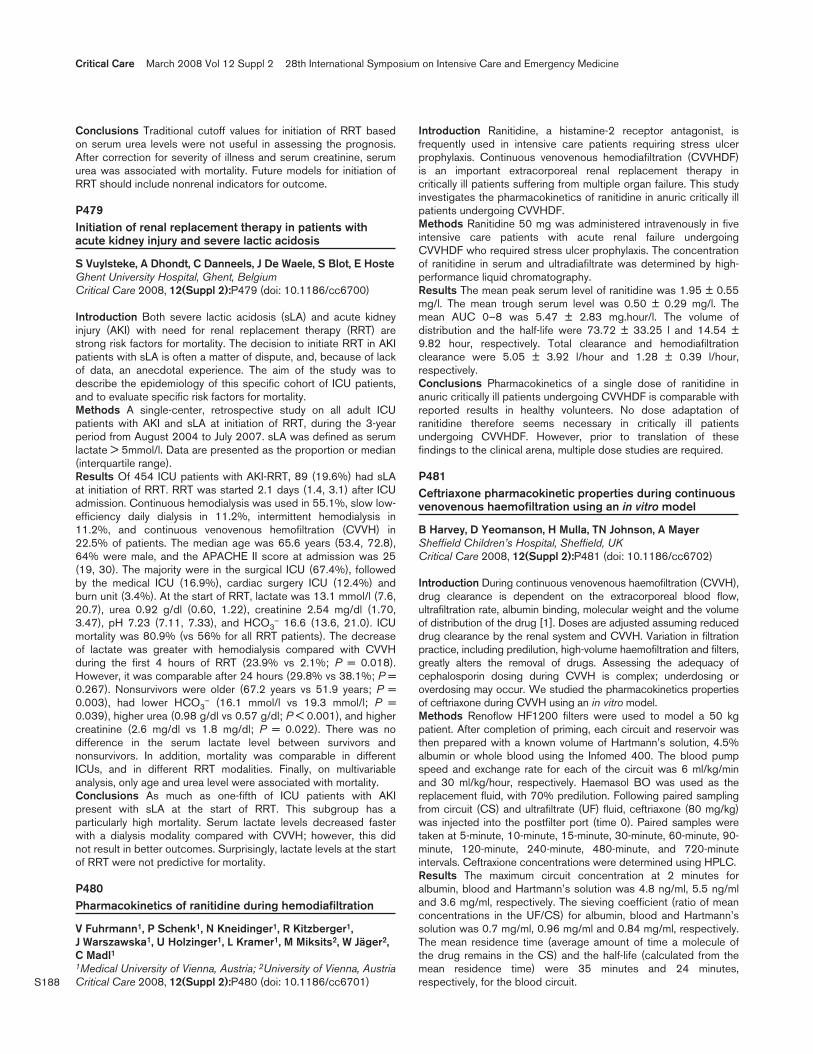
S188
Conclusions Traditional cutoff values for initiation of RRT basedon serum urea levels were not useful in assessing the prognosis.After correction for severity of illness and serum creatinine, serumurea was associated with mortality. Future models for initiation ofRRT should include nonrenal indicators for outcome.
P479Initiation of renal replacement therapy in patients withacute kidney injury and severe lactic acidosis
S Vuylsteke, A Dhondt, C Danneels, J De Waele, S Blot, E HosteGhent University Hospital, Ghent, BelgiumCritical Care 2008, 12(Suppl 2):P479 (doi: 10.1186/cc6700)
Introduction Both severe lactic acidosis (sLA) and acute kidneyinjury (AKI) with need for renal replacement therapy (RRT) arestrong risk factors for mortality. The decision to initiate RRT in AKIpatients with sLA is often a matter of dispute, and, because of lackof data, an anecdotal experience. The aim of the study was todescribe the epidemiology of this specific cohort of ICU patients,and to evaluate specific risk factors for mortality.Methods A single-center, retrospective study on all adult ICUpatients with AKI and sLA at initiation of RRT, during the 3-yearperiod from August 2004 to July 2007. sLA was defined as serumlactate > 5mmol/l. Data are presented as the proportion or median(interquartile range).Results Of 454 ICU patients with AKI-RRT, 89 (19.6%) had sLAat initiation of RRT. RRT was started 2.1 days (1.4, 3.1) after ICUadmission. Continuous hemodialysis was used in 55.1%, slow low-efficiency daily dialysis in 11.2%, intermittent hemodialysis in11.2%, and continuous venovenous hemofiltration (CVVH) in22.5% of patients. The median age was 65.6 years (53.4, 72.8),64% were male, and the APACHE II score at admission was 25(19, 30). The majority were in the surgical ICU (67.4%), followedby the medical ICU (16.9%), cardiac surgery ICU (12.4%) andburn unit (3.4%). At the start of RRT, lactate was 13.1 mmol/l (7.6,20.7), urea 0.92 g/dl (0.60, 1.22), creatinine 2.54 mg/dl (1.70,3.47), pH 7.23 (7.11, 7.33), and HCO3
– 16.6 (13.6, 21.0). ICUmortality was 80.9% (vs 56% for all RRT patients). The decreaseof lactate was greater with hemodialysis compared with CVVHduring the first 4 hours of RRT (23.9% vs 2.1%; P = 0.018).However, it was comparable after 24 hours (29.8% vs 38.1%; P =0.267). Nonsurvivors were older (67.2 years vs 51.9 years; P =0.003), had lower HCO3
– (16.1 mmol/l vs 19.3 mmol/l; P =0.039), higher urea (0.98 g/dl vs 0.57 g/dl; P < 0.001), and highercreatinine (2.6 mg/dl vs 1.8 mg/dl; P = 0.022). There was nodifference in the serum lactate level between survivors andnonsurvivors. In addition, mortality was comparable in differentICUs, and in different RRT modalities. Finally, on multivariableanalysis, only age and urea level were associated with mortality.Conclusions As much as one-fifth of ICU patients with AKIpresent with sLA at the start of RRT. This subgroup has aparticularly high mortality. Serum lactate levels decreased fasterwith a dialysis modality compared with CVVH; however, this didnot result in better outcomes. Surprisingly, lactate levels at the startof RRT were not predictive for mortality.
P480Pharmacokinetics of ranitidine during hemodiafiltration
V Fuhrmann1, P Schenk1, N Kneidinger1, R Kitzberger1, J Warszawska1, U Holzinger1, L Kramer1, M Miksits2, W Jäger2,C Madl11Medical University of Vienna, Austria; 2University of Vienna, AustriaCritical Care 2008, 12(Suppl 2):P480 (doi: 10.1186/cc6701)
Introduction Ranitidine, a histamine-2 receptor antagonist, isfrequently used in intensive care patients requiring stress ulcerprophylaxis. Continuous venovenous hemodiafiltration (CVVHDF)is an important extracorporeal renal replacement therapy incritically ill patients suffering from multiple organ failure. This studyinvestigates the pharmacokinetics of ranitidine in anuric critically illpatients undergoing CVVHDF.Methods Ranitidine 50 mg was administered intravenously in fiveintensive care patients with acute renal failure undergoingCVVHDF who required stress ulcer prophylaxis. The concentrationof ranitidine in serum and ultradiafiltrate was determined by high-performance liquid chromatography.Results The mean peak serum level of ranitidine was 1.95 ± 0.55mg/l. The mean trough serum level was 0.50 ± 0.29 mg/l. Themean AUC 0–8 was 5.47 ± 2.83 mg.hour/l. The volume ofdistribution and the half-life were 73.72 ± 33.25 l and 14.54 ±9.82 hour, respectively. Total clearance and hemodiafiltrationclearance were 5.05 ± 3.92 l/hour and 1.28 ± 0.39 l/hour,respectively.Conclusions Pharmacokinetics of a single dose of ranitidine inanuric critically ill patients undergoing CVVHDF is comparable withreported results in healthy volunteers. No dose adaptation ofranitidine therefore seems necessary in critically ill patientsundergoing CVVHDF. However, prior to translation of thesefindings to the clinical arena, multiple dose studies are required.
P481Ceftriaxone pharmacokinetic properties during continuousvenovenous haemofiltration using an in vitro model
B Harvey, D Yeomanson, H Mulla, TN Johnson, A MayerSheffield Children’s Hospital, Sheffield, UKCritical Care 2008, 12(Suppl 2):P481 (doi: 10.1186/cc6702)
Introduction During continuous venovenous haemofiltration (CVVH),drug clearance is dependent on the extracorporeal blood flow,ultrafiltration rate, albumin binding, molecular weight and the volumeof distribution of the drug [1]. Doses are adjusted assuming reduceddrug clearance by the renal system and CVVH. Variation in filtrationpractice, including predilution, high-volume haemofiltration and filters,greatly alters the removal of drugs. Assessing the adequacy ofcephalosporin dosing during CVVH is complex; underdosing oroverdosing may occur. We studied the pharmacokinetics propertiesof ceftriaxone during CVVH using an in vitro model.Methods Renoflow HF1200 filters were used to model a 50 kgpatient. After completion of priming, each circuit and reservoir wasthen prepared with a known volume of Hartmann’s solution, 4.5%albumin or whole blood using the Infomed 400. The blood pumpspeed and exchange rate for each of the circuit was 6 ml/kg/minand 30 ml/kg/hour, respectively. Haemasol BO was used as thereplacement fluid, with 70% predilution. Following paired samplingfrom circuit (CS) and ultrafiltrate (UF) fluid, ceftriaxone (80 mg/kg)was injected into the postfilter port (time 0). Paired samples weretaken at 5-minute, 10-minute, 15-minute, 30-minute, 60-minute, 90-minute, 120-minute, 240-minute, 480-minute, and 720-minuteintervals. Ceftraxione concentrations were determined using HPLC.Results The maximum circuit concentration at 2 minutes foralbumin, blood and Hartmann’s solution was 4.8 ng/ml, 5.5 ng/mland 3.6 mg/ml, respectively. The sieving coefficient (ratio of meanconcentrations in the UF/CS) for albumin, blood and Hartmann’ssolution was 0.7 mg/ml, 0.96 mg/ml and 0.84 mg/ml, respectively.The mean residence time (average amount of time a molecule ofthe drug remains in the CS) and the half-life (calculated from themean residence time) were 35 minutes and 24 minutes,respectively, for the blood circuit.
Critical Care March 2008 Vol 12 Suppl 2 28th International Symposium on Intensive Care and Emergency Medicine
S189
Conclusions Estimates of a high sieving coefficient and shortcircuit half-life from this in vitro model suggest ceftriaxone is rapidlycleared during CVVH (almost entirely cleared by 240 min). Thishas important implications for dosing schedules during in vivohaemofiltration. Overall the albumin circuit had the lowest sievingcoefficient and longest terminal half-life, reflecting protein bindingof drug and suggesting ceftriaxone clearance may increase inhypoalbuminaemic patients. The maximum circuit concentrationwas lower in circuits primed with Hartmann’s solution. This mayreflect precipitation of the drug with calcium in this solution.Reference1. Golper T: Kidney Int 1988, 53:165-168.
P482Intermittent versus continuous enoxaparine foranticoagulation during continuous venovenous hemofiltration
A Koroljov, I Kordjukova, M Magi, A ReintamEast Tallinn Central Hospital, Tallinn, EstoniaCritical Care 2008, 12(Suppl 2):P482 (doi: 10.1186/cc6703)
Introduction The exact dosage of anticoagulation during continuousvenovenous hemofiltration (CVVH) is crucial for maximal effect andsafety of the procedure. Enoxaparine is one possible choice ofanticoagulant drug. The aim of our study was to compare the meandose of enoxaparine depending on whether a bolus or continuousadministration scheme was used.Methods We performed retrospective analysis of prospectivelycollected data for 36 consecutive patients (24 male and 12female) receiving CVVH in the ICU of East Tallinn Central Hospital.Twenty-three patients received boluses of enoxaparine every 6hours, and 13 patients received continuous infusion ofenoxaparine. The dosage of anticoagulation was guided accordingto the results of anti-Xa measurements.Results The mean age of the patients (61.4 ± 15.7 years) and themean duration of the procedure (76.2 ± 29.2 hours) did not differsignificantly between the two groups. The mean dose ofenoxaparine was 0.12 ± 0.05 mg/kg/hour in the bolus group and0.08 ± 0.03 mg/kg/hour in the continuous group (P = 0.027). Themean anti-Xa did not differ significantly between the groups(1.03 ± 0.42 vs 0.88 ± 0.24 units/ml, respectively, P = 0.252).Filter clotting was observed 0.39 ± 0.58 times per procedure inthe bolus group and 0.38 ± 0.77 times in the continuous group(P = 0.977). One patient in the bolus group developed cerebralhemorrhage.Conclusions Continuous administration allows the maintenance ofa necessary level of anticoagulation with lower doses ofenoxaparine during CVVH as compared with bolus dosing. It stillneeds to be clarified whether this difference in the mean dose ofthe anticoagulant may be important in reducing the complications.
P483Mechanical ventilation in a critical care unit in southernBrazil: mortality risk factors
L Fialkow, M Bozzetti, M Farenzena, R Sens, L Sehn, R Cardoso, A Wolmeister, A Milani, M Ficanha, G Machado, A Güntzel, S Vieira, J BraunerHospital de Clínicas de Porto Alegre, BrazilCritical Care 2008, 12(Suppl 2):P483 (doi: 10.1186/cc6704)
Introduction Patients requiring mechanical ventilation (MV) havehigh mortality rates. In Latin America, the knowledge of mortality riskfactors in patients on MV is scarce. Identification of such factors isessential to improve outcomes. The objective of our study was to
determine the mortality risk factors in patients that required MV inthe ICU of a general university hospital in southern Brazil.Methods A prospective cohort study of 1,115 adult patientsadmitted to the ICU who needed MV for at least 24 hours,between March 2004 and April 2007. Data were collected oneach patient at the inclusion in the study and daily during thecourse of MV for up to 28 days. Multivariate analysis by logisticconditional regression was used.Results The frequency of MV was 46%; the overall and specificmortality rates were 23% and 51%, respectively. The mean (±SD)age was 57 ± 18 years; 52% were males; the mean APACHE IIscore was 22 ± 8.3; 69% were medical patients; the meanduration of MV was 10 ± 7.9 days; 93% were on invasive MV. Thevariables independently associated with increased mortality were(1) conditions present at beginning of MV: age (P = 0.04),APACHE II score (P < 0.001), acute lung injury/acute respiratorydistress syndrome (ALI/ARDS) as cause of MV (P = 0.04), andgastrointestinal failure (P = 0.01); and (2) conditions occurringover the course of MV: ALI/ARDS (P < 0.001), sepsis (P = 0.007),cardiovascular (P = 0.002), renal (P < 0.001), and hepatic failure(P = 0.009), use of vasoactive drugs (P < 0.001) and opioids (P =0.04), and duration of MV (P < 0.001). It is of note that ventilatorymonitored variables included in the multivariate model were notassociated with mortality.Conclusions In our study, the risk factors for mortality 28 daysafter the beginning of MV were conditions present at the beginningof MV (age, APACHE II score, ALI/ARDS as a cause of MV, andgastrointestinal failure) and conditions occurring over the course ofMV (ALI/ARDS, sepsis, cardiovascular, renal and hepatic failure,use of vasoactive drugs and opioids, and duration of MV). Thesurvival of patients on MV therefore involves conditions present atthe start of MV, and conditions that occur during the MV period,including aspects related to patient management. Knowledge ofthese factors may permit the evolvement of early interventions thatmight decrease poor outcomes of patients that require MV.References1. Esteban A, et al.: JAMA 2002, 287:345-355.2. Esteban A, et al.: Am J Respir Crit Care Med 2008, 177:170-
177.
P484Long-stay critically ill patients’ characteristics andoutcomes: a cohort study
J Carmeli, S BurszteinCarmel Medical Center, Haifa, IsraelCritical Care 2008, 12(Suppl 2):P484 (doi: 10.1186/cc6705)
Introduction Long-stay critically ill patients represent a smallsubgroup of ICU patients with huge resource consumption. Thegoal of this study was to identify outcomes and characteristics ofthese patients.Methods Data were collected retrospectively from consecutivepatients requiring at least 60 days of ICU care admitted over sevencalendar years (2000–2006) to a general ICU in a university-affiliated 427-bed hospital.Results A total of 27 patients met the inclusion criteria. Long-stayICU patients represented 1.2% of total admissions. The mean agewas 69 years (26–92 years). The median ICU stay in overall ICUpatients was 7 days, while in long-stay ICU patients it was 98 days(62–202 days). General ICU mortality was 17.4%, in the studiedgroup the mortality rate was 44.4%. A further eight patients died inthe hospital after discharge from the ICU, representing anadditional 26% of all long-stay ICU patients. Two years after thedischarge 22.2% of these patients were alive, and 14.8% are alive
Available online http://ccforum.com/supplements/12/S2
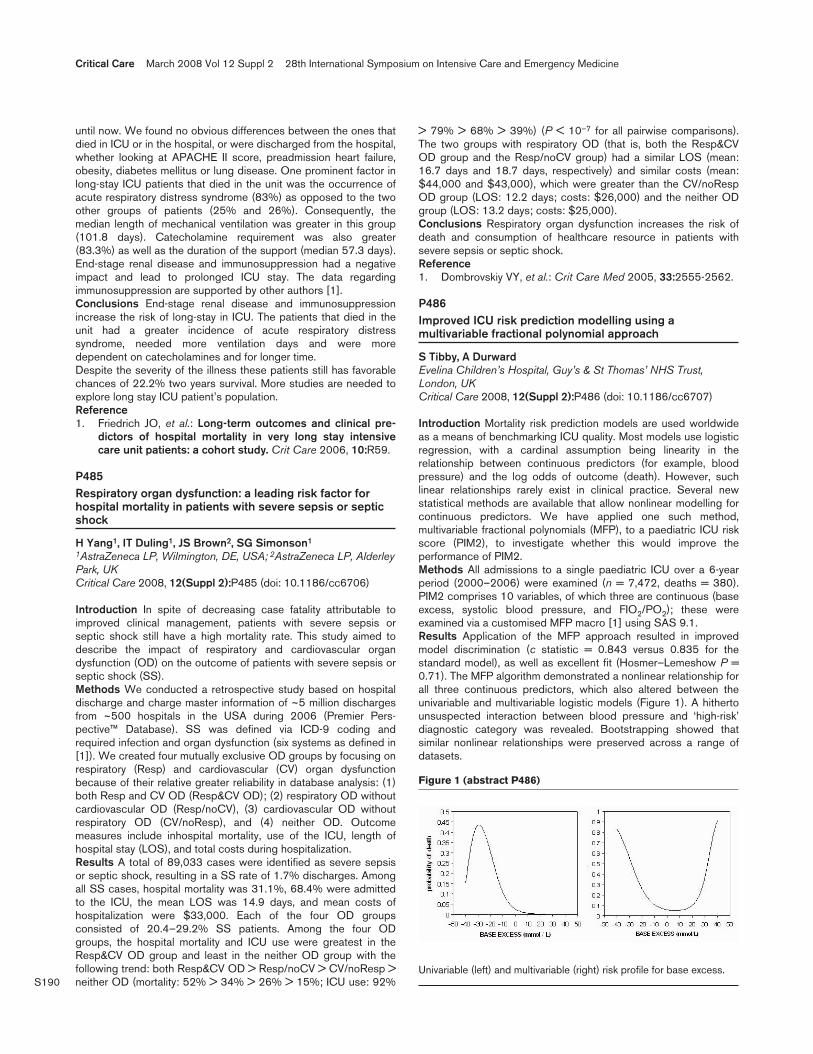
S190
until now. We found no obvious differences between the ones thatdied in ICU or in the hospital, or were discharged from the hospital,whether looking at APACHE II score, preadmission heart failure,obesity, diabetes mellitus or lung disease. One prominent factor inlong-stay ICU patients that died in the unit was the occurrence ofacute respiratory distress syndrome (83%) as opposed to the twoother groups of patients (25% and 26%). Consequently, themedian length of mechanical ventilation was greater in this group(101.8 days). Catecholamine requirement was also greater(83.3%) as well as the duration of the support (median 57.3 days).End-stage renal disease and immunosuppression had a negativeimpact and lead to prolonged ICU stay. The data regardingimmunosuppression are supported by other authors [1].Conclusions End-stage renal disease and immunosuppressionincrease the risk of long-stay in ICU. The patients that died in theunit had a greater incidence of acute respiratory distresssyndrome, needed more ventilation days and were moredependent on catecholamines and for longer time.Despite the severity of the illness these patients still has favorablechances of 22.2% two years survival. More studies are needed toexplore long stay ICU patient’s population.Reference1. Friedrich JO, et al.: Long-term outcomes and clinical pre-
dictors of hospital mortality in very long stay intensivecare unit patients: a cohort study. Crit Care 2006, 10:R59.
P485Respiratory organ dysfunction: a leading risk factor forhospital mortality in patients with severe sepsis or septicshock
H Yang1, IT Duling1, JS Brown2, SG Simonson1
1AstraZeneca LP, Wilmington, DE, USA; 2AstraZeneca LP, AlderleyPark, UKCritical Care 2008, 12(Suppl 2):P485 (doi: 10.1186/cc6706)
Introduction In spite of decreasing case fatality attributable toimproved clinical management, patients with severe sepsis orseptic shock still have a high mortality rate. This study aimed todescribe the impact of respiratory and cardiovascular organdysfunction (OD) on the outcome of patients with severe sepsis orseptic shock (SS).Methods We conducted a retrospective study based on hospitaldischarge and charge master information of ~5 million dischargesfrom ~500 hospitals in the USA during 2006 (Premier Pers-pective™ Database). SS was defined via ICD-9 coding andrequired infection and organ dysfunction (six systems as defined in[1]). We created four mutually exclusive OD groups by focusing onrespiratory (Resp) and cardiovascular (CV) organ dysfunctionbecause of their relative greater reliability in database analysis: (1)both Resp and CV OD (Resp&CV OD); (2) respiratory OD withoutcardiovascular OD (Resp/noCV), (3) cardiovascular OD withoutrespiratory OD (CV/noResp), and (4) neither OD. Outcomemeasures include inhospital mortality, use of the ICU, length ofhospital stay (LOS), and total costs during hospitalization.Results A total of 89,033 cases were identified as severe sepsisor septic shock, resulting in a SS rate of 1.7% discharges. Amongall SS cases, hospital mortality was 31.1%, 68.4% were admittedto the ICU, the mean LOS was 14.9 days, and mean costs ofhospitalization were $33,000. Each of the four OD groupsconsisted of 20.4–29.2% SS patients. Among the four ODgroups, the hospital mortality and ICU use were greatest in theResp&CV OD group and least in the neither OD group with thefollowing trend: both Resp&CV OD > Resp/noCV > CV/noResp >neither OD (mortality: 52% > 34% > 26% > 15%; ICU use: 92%
> 79% > 68% > 39%) (P < 10–7 for all pairwise comparisons).The two groups with respiratory OD (that is, both the Resp&CVOD group and the Resp/noCV group) had a similar LOS (mean:16.7 days and 18.7 days, respectively) and similar costs (mean:$44,000 and $43,000), which were greater than the CV/noRespOD group (LOS: 12.2 days; costs: $26,000) and the neither ODgroup (LOS: 13.2 days; costs: $25,000).Conclusions Respiratory organ dysfunction increases the risk ofdeath and consumption of healthcare resource in patients withsevere sepsis or septic shock.Reference1. Dombrovskiy VY, et al.: Crit Care Med 2005, 33:2555-2562.
P486Improved ICU risk prediction modelling using amultivariable fractional polynomial approach
S Tibby, A DurwardEvelina Children’s Hospital, Guy’s & St Thomas’ NHS Trust,London, UKCritical Care 2008, 12(Suppl 2):P486 (doi: 10.1186/cc6707)
Introduction Mortality risk prediction models are used worldwideas a means of benchmarking ICU quality. Most models use logisticregression, with a cardinal assumption being linearity in therelationship between continuous predictors (for example, bloodpressure) and the log odds of outcome (death). However, suchlinear relationships rarely exist in clinical practice. Several newstatistical methods are available that allow nonlinear modelling forcontinuous predictors. We have applied one such method,multivariable fractional polynomials (MFP), to a paediatric ICU riskscore (PIM2), to investigate whether this would improve theperformance of PIM2.Methods All admissions to a single paediatric ICU over a 6-yearperiod (2000–2006) were examined (n = 7,472, deaths = 380).PIM2 comprises 10 variables, of which three are continuous (baseexcess, systolic blood pressure, and FIO2/PO2); these wereexamined via a customised MFP macro [1] using SAS 9.1.Results Application of the MFP approach resulted in improvedmodel discrimination (c statistic = 0.843 versus 0.835 for thestandard model), as well as excellent fit (Hosmer–Lemeshow P =0.71). The MFP algorithm demonstrated a nonlinear relationship forall three continuous predictors, which also altered between theunivariable and multivariable logistic models (Figure 1). A hithertounsuspected interaction between blood pressure and ‘high-risk’diagnostic category was revealed. Bootstrapping showed thatsimilar nonlinear relationships were preserved across a range ofdatasets.
Critical Care March 2008 Vol 12 Suppl 2 28th International Symposium on Intensive Care and Emergency Medicine
Figure 1 (abstract P486)
Univariable (left) and multivariable (right) risk profile for base excess.
S191
Conclusions The MFP approach offers several advantages overlinear modelling, both in model fit and a better elucidation of riskprofiles for individual predictors. This requires confirmation in anational dataset.Reference1. Multivariable Fractional Polynomial Approach [http://www.
imbi.uni-freiburg.de/biom/mfp/]
P487Risk factors for the development of acute lung injury inpatients with septic shock: an observational cohort study
R Iscimen1, M Yilmaz2, R Cartin-Ceba3, R Hubmayr3, B Afessa3,O Gajic3, J Farmer3
1Uludag University, Bursa, Turkey; 2Akdeniz University, Antalya,Turkey; 3Mayo Clinic, Rochester, NY, USACritical Care 2008, 12(Suppl 2):P487 (doi: 10.1186/cc6708)
Introduction Almost one-half of patients with septic shock developacute lung injury (ALI). The understanding of why some patients doand others do not develop ALI is limited. The objective of this studywas to test the hypothesis that delayed treatment of septic shockis associated with the development of ALI.Methods An observational cohort study in a medical ICU in atertiary medical center. Patients were prospectively identified withseptic shock who did not have ALI at the outset, excluding thosewho denied research authorization. There were no interventions.Results High-frequency cardiorespiratory monitoring, arterial gasanalysis and portable chest radiographs were reviewed to identifythe timing of ALI development. Risk factors present before ALIdevelopment were identified by review of electronic medicalrecords and were analyzed in univariate and multivariate analyses.Seventy-one out of 160 patients (44%) developed ALI at a medianof 5 (range 2–94) hours after the onset of septic shock.Multivariate logistic regression analysis identified the followingpredictors of ALI development: delayed goal-directed resuscitation(OR = 3.55, 95% CI = 1.52–8.63, P = 0.004), delayed antibiotics(OR = 2.39, 95% CI = 1.06–5.59, P = 0.039), transfusion (OR =2.75, 95% CI = 1.22–6.37, P = 0.016), alcohol abuse (OR =2.09, 95% CI = 0.88–5.10, P = 0.098), recent chemotherapy(OR = 6.47, 95% CI = 1.99–24.9, P = 0.003), diabetes mellitus(OR = 0.44, 95% CI = 0.17–1.07, P = 0.076) and baselinerespiratory rate (OR 2.03 per standard deviation, 95% CI =1.38–3.08, P < 0.001).Conclusions When adjusted for known modifiers of ALIexpression, delayed treatment of shock and infection wereassociated with development of ALI.References1. Rubenfeld GD, et al: Chest 2007, 131:554-562.2. Gong MN, et al: Crit Care Med 2005, 33:1191-1198.3. Hudson LD, et al: Am J Respir Crit Care Med 1995, 151:293-
301.
P488Outcome in severe sepsis and septic shock patients withhematological malignancies: impact of recent intravenouschemotherapeutic treatment
D Vandijck1, D Benoit1, P Depuydt1, E Steel2, F Offner2, S Blot3,J Decruyenaere3
1Department of Intensive Care Medicine, 2Department ofHematology and 3Faculty of Medicine and Health Sciences, GhentUniversity Hospital, Ghent, BelgiumCritical Care 2008, 12(Suppl 2):P488 (doi: 10.1186/cc6709)
Introduction Developing severe sepsis or septic shock afterchemotherapeutic treatment is commonly perceived as the worst-case scenario in hematological patients. This study compared thecharacteristics and outcome of patients with hematologicalmalignancies who were referred to the ICU because of severesepsis and septic shock who had received versus those who hadnot received intravenous chemotherapeutic treatment within 3weeks prior to ICU admission.Methods A prospective observational cohort study was conductedat the medical ICU of a tertiary care centre. All consecutivepatients with hematological malignancies admitted to this unitbetween 2000 and 2006, and who were diagnosed with eithersevere sepsis or septic shock, were considered for analysis.Results Seventy-seven patients were admitted with severe sepsisand 109 with septic shock. Ninety-one patients (49%) hadreceived recent intravenous chemotherapy within 3 weeks prior toICU admission. As compared with those without, patients withrecent chemotherapy more often had a high-grade malignancy (P <0.001), were more often neutropenic (P < 0.001), less often hadpulmonary infiltrates (P = 0.007), and less often requiredmechanical ventilation (P = 0.04). The ICU, 28-day, inhospital, and6-month mortality rates were 33% versus 48.4% (P = 0.037),40.7% versus 57.4% (P = 0.027), 45.1% versus 58.9% (P =0.076), and 50.5% versus 63.2% (P = 0.103), in patients with andwithout recent chemotherapy, respectively. Logistic regressionidentified four variables independently associated with 28-daymortality: SOFA score at ICU admission (P < 0.001), pulmonarysite of infection (P = 0.005), and fungal infection (P = 0.003) wereassociated with worse outcome, whereas intravenous chemo-therapy within 3 weeks prior to ICU admission was protective atborderline significance (P = 0.049). After more complete adjust-ment with a propensity score for recent chemotherapy, chemo-therapy was no longer associated with outcome (P = 0.079).Conclusions Patients referred to the ICU with severe sepsis andseptic shock complicating active chemotherapeutic treatment havean overall crude mortality that is comparable with septic shock ingeneral ICU patients, and have a better prognosis than commonlyperceived.
P489Outcome of patients with haematological malignancyadmitted to the general ICU
I Ketchley, N Al-Subaie, R Hewitt, M Grounds, A Rhodes, A RhodesSt George’s Hospital, London, UKCritical Care 2008, 12(Suppl 2):P489 (doi: 10.1186/cc6710)
Introduction Previous studies have demonstrated a widely variableprognosis in critical illness amongst patients with haematologicalneoplastic disease; with inhospital mortality documented as high as78% [1]. We sought to describe the outcomes in our population.Methods We conducted a retrospective study of consecutiveadmissions with a primary diagnosis of haematological malignancyover a 4-year period. Patients admitted recurrently as separateadmissions were recorded as multiple events. Neutropenia wasdefined as <1.5 x 10⁹/l [2]. Outcome was defined as ICU mortality,inhospital mortality, length of ICU stay and requirement formechanical ventilation or renal replacement therapy (RRT).Results Of 5,142 admissions, 33 had a primary diagnosis ofhaematological neoplasm (24 males). The average age was 55.8 ±13.6 years. The median length of ICU stay was 5.4 days. The ICUmortality rate of all ICU admissions was 15.2%, compared with39.4% in patients with haematological malignancy (P < 0.01). Theinhospital mortality was 63.6%. Eleven patients were neutropenic
Available online http://ccforum.com/supplements/12/S2
S192
on admission. In the neutropenic subgroup, ICU mortality was45.5% (vs 36.3% in non-neutropenic patients, P = 0.46) andinhospital mortality was 45.5% (vs 72.7% in non-neutropenicpatients). Of 22 patients requiring mechanical ventilation, the ICUmortality was 50% (vs 18.1%, P = 0.13) and inhospital mortalitywas 72.7% (vs 45%, P = 0.44). Of six patients requiring RRT, theICU mortality was 50% (vs 37% in those patients not requiringRRT, P = 0.65) and inhospital mortality was 66.7% (vs 63%).Conclusions The mortality rate is high amongst patients in whomhaematological malignancy is complicated by critical illness.Although significantly higher than our remaining ICU population, itwas lower than reported elsewhere in the literature. Neutropeniaon admission was not significantly associated with a pooreroutcome. Although mortality was higher in those requiringmechanical ventilation or RRT, this did not reach statisticalsignificance. This may be a result of our small sample size.References1. Gordon AC, et al.: Anaesthesia 2005, 60:340-347.2. Murphy MF, Wainscoat J, Colvin BT: Haematological
disease. In Kumar P, Clark M, editors. Clinical Medicine. 5thedition. Saunders; 2002:452.
P490Prognostic factors of cancer patients admitted to the ICUin Brazil: a prospective cohort.
DN Rocha Cardoso1, JM Meira Teles1, DS Gaudêncio dos Santos1,AC Da Hora Pereira1, EB Maia1, FS Batista1, DG Flores1, RA Passos1, AA Camelier1, OH Coelho Messeder1, AS Neto2
1Hospital Português, Salvador, Brazil; 2Universidade Federal daBahia, Salvador, BrazilCritical Care 2008, 12(Suppl 2):P490 (doi: 10.1186/cc6711)
Introduction The objective of this study was to identify mortality incritically ill oncologic patients.Methods Data were collected prospectively from 120 consecutivepatients who were admitted to the combined medical and surgicalICU. The primary outcome studied was overall ICU mortality.Results Overall, 49.2% of patients were older than 66 years old(our study median) and 55.8% were men. The median time for thelength of stay was 3 days and the median time for the follow-upwas 8 days. The ICU mortality and the hospital mortality were35.8% and 46.7%, respectively. Through the Kaplan–Meiersurvival curve, there was significant difference among the patients’mortality with terminal stages of cancer and the patients’ mortalitywith response to treatment. The median survival times were 13.47and 29.4 days, respectively. The survival of patients with a need forinvasive mechanical ventilation was fourfold to fivefold lower (P =0.005). Through the Cox multiple regression, the variables relatedto ICU mortality were a higher length of hospital stay before theICU admission (P = 0.003) and coagulopathy over the ICU stay(P = 0.035) (Table 1).Conclusions Our study suggest that patients in terminal stages ofcancer who presented a higher length of hospital stay before ICUadmission, coagulopathy and mechanical ventilation over the ICUstay have higher ICU mortality.
References1. Azoulay E, et al.: Intensive Care Med 2000, 26:1817-1823.2. Brenner H, et al.: Lancet 2002, 360:1131-1135.
P491Organ dysfunction influences outcome in medicaloncology patients without multiorgan failure
J Divatia, V Agarwal, R HawaldarTata Memorial Hospital, Mumbai, IndiaCritical Care 2008, 12(Suppl 2):P491 (doi: 10.1186/cc6712)
Introduction In a previous audit, patients with ≤1 organ failure(OF) on day 1 or day 2 had high ICU mortality (63% and 52%,respectively). We examined the impact of organ dysfunction (OD)patients with ≤1 OF.Methods One hundred and seventy-nine consecutive adultadmissions (111 males, 68 females) from February 2006 to June2007 with an ICU stay ≥3 days were prospectively studied. TheAPACHE II score, day 1 SOFA score, highest SOFA score of thefirst three days (MAX3), and changes in SOFA score between day2 and day 1 (Δ1) and between day 3 and day 1 (Δ2) werecalculated. For each organ, a SOFA score of 1–2 or 3–4 definedOD or OF, respectively. The total number of ODs and OFs on ICUdays 1–3 was calculated. Binary logistic regression (backwardconditional) was used to determine factors associated withmortality.Results The mean age was 44.8 ± 15.4 years, APACHE II score20.1 ± 6.5 and SOFA score 16.8 ± 3.8. The ICU mortality was66%. On days 1 and 2, 65% and 77% patients had ≤1 OF. Ondays 1–3, an increasing number of ODs were significantlyassociated with increasing mortality in patients with ≤1 OF, but notin patients with ≥2 OFs (Table 1). On multivariate analysis, Δ2(OR = 1.26, 95% CI = 1.12–1.41, P = 0.000), ODs on day 1(OR = 1.54, CI = 1.06–2.23, P = 0.03) and OFs on day 2 (OR =2.67, CI = 1.76–4.06) were independently associated with ICUmortality.
Table 1 (abstract P491)
Number Day 1, Day 1, Day 2, Day 2, of ODs ≤1 OF ≥2 OF ≤1 OF ≥2 OF
0 9/16, 56% 8/14, 57% 13/47, 28% 8/11, 73%
1–2 40/72, 56% 31/40, 78% 36/67, 54% 46/54, 87%
>2 23/28, 82%* 7/9, 78%† 12/17, 71%** 11/13, 85%††
Data presented as (deaths/ total patients, %). *P = 0.04; †P = 0.31; **P = 0.002; ††P = 0.59.
Conclusions An increasing number of ODs worsens the outcomein critically ill medical oncology patients with ≤1 OF. WorseningOD scores are associated with mortality. Early recognition andmanagement of OD before the onset of multiple OF may helpimprove outcome.
Critical Care March 2008 Vol 12 Suppl 2 28th International Symposium on Intensive Care and Emergency Medicine
Table 1 (abstract P490)
Results of the final model of the Cox multiple regression (n = 120)
P value in 95% CI of Variable β β standard likelihood ratio Hazard ratio hazard ratio
Length of hospital stay before ICU admission 1.226 0.473 0.003 2.930 0.161–0.534
Coagulopathy 1.565 0.686 0.035 4.785 1.867–12.260
S193
P492Causes and risk factors of maternal mortality in the ICU
C Kaddour, R Souissi, Z Haddad, Z Zaghdoudi, M Magouri, M Soussi, S AbbassiInstitute of Neurology, La Rabta, TunisiaCritical Care 2008, 12(Suppl 2):P492 (doi: 10.1186/cc6713)
Introduction Maternal mortality is considered a basic healthindicator that reflects the adequacy of healthcare; in our countrythe overall maternal mortality is 69.8 per 100,000 live births(Tunisian National Enquiry into Maternal Deaths 1993–1994). Inthis study we tried to identify causes of maternal death in our ICU.Methods A prospective study of patients first managed in a tertiaryreferral maternity center for high-risk pregnancies. Collected datawere age, gravity, parity, admission diagnosis and delivery mode.The APACHE II score, APACHE III score and SAPS Obst scorewere calculated. The study period was January 2004–December2004. The outcome measure was mortality. We used theInternational Classification of Diseases definition of a maternaldeath: ‘the death of a woman while pregnant or within 42 days oftermination of pregnancy, from any cause related to or aggravatedby the pregnancy or its management, but not from accidental orincidental causes’.Results From 131 obstetrics admissions, 18 patients died (14%).Advanced age (33 ± 9), high gravity (2.8 ± 1.7) and high parity(2.77 ± 1.7) scores are common to all death. As compared withvaginal delivery (n = 9), caesarean delivery (n = 9) was notassociated with a significantly increased risk for postpartummaternal death; the majority of maternal deaths are due to directobstetric causes (16–89%). At admission all patients had highseverity of illness scores, APACHE II (18.76 ± 16.65), APACHE III(71.58 ± 30.19) and SAPS Obst (30.29 ± 8.24), which indicateshow grave our patients’ health condition is. The most commoncause of death was hypertensive disorders in five patients,infection in four patients and hemorrhage in three cases, othercauses were pulmonary embolisms in two cases, one case ofamniotic fluid embolism, one patient with peripartum cardio-myopathy, one patient with intracerebral hemorrhage and onepatient with anaphylactic shock.Conclusions The knowledge of the specific causes involved in thematernal mortality risk could help to elaborate adequate preventivestrategies to reduce maternal death.
P493Epidemiology and outcome in acute respiratory failure
E Bermejo López, J Sotillo Díaz, S Arenal López, J VillanuevaFlórez, P García OlivaresGregorio Marañón, Madrid, SpainCritical Care 2008, 12(Suppl 2):P493 (doi: 10.1186/cc6714)
Introduction Acute respiratory failure (ARF) is the most commonorgan failure in the ICU, and mortality is high. The outcomeworsens in association with any other organ failure. The objectiveof this study was to describe the aetiology, mortality rate andcauses of ARF and to evaluate the outcome of ARF with/withoutconcomitant organ failure.Methods A prospective cohort study that includes all patientsadmitted to the ICU of a tertiary hospital from January to July 2007.ARF was defined based on the SOFA score criteria, SOFArespiratory ≥3, PaO2/FiO2 <200 and mechanical ventilation.Results Two hundred and ninety-six patients were admitted, 48%with ARF (142 patients), 59.6% males. Primary diagnoses were52.5% respiratory insufficiency, 27.7% neurological disease,
14.2% cardiologic/septic shock, and 5.7% cardiac arrest. Causesof ARF were 47.5% pneumonia, 11.5% pulmonary oedema, 11.5%chronic pulmonary disease, 10.8% nonpulmonary ARDS, 7.9%atelectasis, 3.5% bronchospasm, 3.5% pleural effusion, 2.1%pulmonary fibrosis. Severity was APACHE II score 23.17 ± 7.1,SAPS II score 50.5 ± 15.4, SAPS III score 66.5 ± 15.5, SOFAscore at admission 8.6 ± 4.1, SOFA maximum score 10.23 ± 4.7,lactic acid 4.01 ± 3.3, and lung injury score (LIS) 1.7 ± 0.7(0.66–3.5). Of patients, 21.6% received noninvasive ventilationand 78.4% invasive ventilation. The mean stay in the ICU was 9.6± 10.5 days, hospital stay was 19.8 ± 18.29 days. In the ICU,73.8% developed multiorgan failure syndrome (MOFS). Themortality rate in the ICU was 39.7% and in the hospital was50.4%. Mortality in patients without ARF was 15.6%, and inpatients with ARF was 50.4% (RR = 5.53, 95% CI, 3.2–9.5). Themortality rate was lowest in the subgroup with ARF as a singleorgan failure (16.7%) and it increased with additional organs infailure (ARF + 1, 37.8%; ARF + 2, 69%; ARF + 3, 90%; ARF + 4or 5, 91%; χ2 test P = 0.000). The mortality rate of those with ARFat admission was 55.5%, versus 30% for those who developedARF (RR = 2.95, 95% CI = 1.2–6.8). Patients with invasiveventilation had 2.45 times more risk to die than noninvasiveventilation (55% vs 33.3%). The mortality rate in acute respiratorydistress syndrome (ARDS) was 77.8% vs 40.8% without ARDS(RR = 5.1, 95% CI = 2.1–12.2). The mortality rate in MOFS was62.7% versus 16.2% only with ARF. Multivariant logisticregression showed there is no association with mortality rate andage, Barthel, PaO2, HCO3, and PaCO2. The mortality rateassociates with lactic acid, APACHE II and III scores, acutephysiological score, SAPS II and III scores, MODS, SOFA score atadmission, pH, LIS and PaO2/FiO2. Developing MOFS and the LISscore are independent predictors of mortality in ARF patients.Conclusions ARF has a high mortality rate especially if it isassociated with severe failure of other organs. Developing MODSand the LIS score are good predictors of outcome in patients withARF.
P494Outcome for immunocompromised pediatric critical patients
Y Peña, M Pujol, S Cañadas, P Domínguez, J Balcells, J RoquetaHospital Vall d´Hebron, Barcelona, SpainCritical Care 2008, 12(Suppl 2):P494 (doi: 10.1186/cc6715)
Introduction The incidence and prevalence of immuno-compromised patients has increased (higher number of trans-plantations and improved management of primary immuno-deficiencies). Nevertheless, the management strategies of thesepatients in the pediatric ICU (PICU) remain a challenge because oftheir important death rate. The study objective was to analyse themorbimortality and prognosis of immunosuppressed patientsrequiring critical care due to a medical cause.Methods A retrospective study (January 2004–July 2007) in a 15-bed PICU at a university hospital. Chi-square and Fisher exact testanalysis was performed.Results One hundred and thirty-nine immunocompromised patientswere admitted 186 times, which comprised 10% of the totalnumber of admissions to the PICU in this period. The median agewas 63 months (range, 1 month–23 years); male predominance(60.2%). Cause of immunosuppression: postchemotherapy neutro-penia or hematopoietic stem cell transplantation (HSCT; 65.1%),solid organ transplantation (15.6%), primary immunodeficiency(9.7%) and miscellany (8.6%). Initial problem: haemodynamic(43.5%), respiratory (34.4%), neurological (14.0%), other (8.1%).PRISM 24-hour score was 8 (0–40). Evolutive failures (% patients):
Available online http://ccforum.com/supplements/12/S2
S194
hematologic (50.5%), respiratory (40.5%), cardiovascular (40.5%),renal (30.6%), hepatic (19.4%), neurological (14%), gastrointestinal(5.6%). Multisystem organ failure (MOF; ≥3 organs) in 32.2%.Critical care support (% patients): mechanical ventilation (50.5%),sympathicomimetic drugs (36.6%), renal depuration (12%),extracorporeal life support with membrane oxygenator (0.5%). Meanstay in the PICU was 7.3 days (median 3 (1–49)). Mortality was22%; higher in HSCT (26.2%) but without significant differencesamong groups (P = 0.529). Mortality according to initial problem:respiratory (31.2%), neurological (23%), cardiovascular (17.5%),other (6.6%) (P = 0.012). Mortality of patients who underwentmechanical ventilation was 40.4%; if respiratory and haematologicfailure association it was 63.8%; if MOF it was 55% (P > 0.0001).Conclusions Regarding immunocompromised patients in thePICU, respiratory failure at admission or during the evolutioninvolves a high risk of death, especially when associated withhaematological failure. The impact on the policy of admission in thePICU must be investigated in new studies.References1. Díaz de Heredia C, et al.: Bone Marrow Transplant 1999, 24:
163-168.2. Lamas A, et al.: Intensive Care Med 2003, 29:91-96.
P495Immunocompromised pediatric patients with acuterespiratory failure: outcome and prognosis factors
M Pujol, Y Peña, A Vazquez, J Balcells, J Ortega, J RoquetaHospital Vall d´Hebron, Barcelona, SpainCritical Care 2008, 12(Suppl 2):P495 (doi: 10.1186/cc6716)
Introduction Acute respiratory failure (ARF) in immunocompro-mised patients is associated with a high mortality rate. The aim ofthis investigation is to study these patients’ behavior patterns tocollect data that can allow us to improve their survival rate.Methods A retrospective study (January 2004–July 2007) in apediatric ICU. We analyse the morbimortality, the reasons for ARFand the critical care support required. We included medicalinmunocompromised patients with ARF (Wilkinson criteria). Exclusionfactors: postoperative mechanical ventilation; 15 were ruled out dueto upper obstruction airway, pulmonary cardiogenic edema,pulmonary thromboembolism and limitation of therapeutic effort (LTE).Results Fifty-seven patients (64 admissions) were enrolled in thestudy. Median age was 66 months (1 month–17 years). Reason forinmunodepressed situation: haematological 41 (64.1%): postchemo-therapy neutropenia 20, hematopoietic stem cell transplantation(HSCT) 21, primary immunodeficiencies 10 (15.6%); solid organtransplantation 7 (10.9%), miscellany 6 (9.4%). PRISM 24-hour scorewas 12 (0–40). Reason for ARF: infectious pulmonary disease 23(36%), noninfectious pulmonary disease 8 (13%), secondarypulmonary injury 28 (43%), neurological disorders 5 (8%). Evolutiveorgan failures (% patients): hematopoietic (68.8%), cardiovascular(65.6%), renal (54.7%), hepatic (37.5%), neurologic (28.1%),gastrointestinal (6.3%). Multiorgan failure (MOF) in 70.3%. Criticalcare support: conventional mechanical ventilation (96.8%),noninvasive positive pressure ventilation (7.8%; exclusive 2), highfrequency ventilation (HFV) (20.3%), extracorporeal life support withmembrane oxygenator (1.5%) (positive end-expiratory pressure(PEEP) > 7 (62.5%), PaFiO2 < 150 (57.8%), oxygenation index (OI)> 15 (42.2%), barotrauma: 18.8%), sympathomimetic drugs (64.1%),renal replacement therapy (25%). Length of stay was 10 days (1–47).Mortality was 51.6%; greater if PaFiO2 < 150 (67.5%; p = 0.004),PEEP > 7 (65%; P = 0.006), OI > 15 (64%; P = 0.008), HSCT(71.4%; P = 0.024), MOF (60%; P = 0.035), HFV (76.9%; P =0.039). Death with LTE was 91%, and by respiratory failure was 40%.
Conclusions Immunocompromised patients with ARF have a highmortality rate, especially if they have undergone HSCT and presentMOF. Assessing the kind of lung injury by a biopsy sample to begin anearly direct treatment is necessary to improve their survival. Althoughthe respiratory index associated with the risk of mortality is known, thisdoes not allow one to predict these patients’ prognosis with certainty.Reference1. Diaz de Heredia C, et al.: Bone Marrow Transplant 1999, 24:
163-168.
P496ICU patients: does age make any difference?
M Laskou, M Katsiari, E Mainas, A Kotsaimani, I Karampela, A MaginaKonstantopoulion General Hospital, Nea Ionia, Athens, GreeceCritical Care 2008, 12(Suppl 2):P496 (doi: 10.1186/cc6717)
Introduction The aim of our study was to compare thecharacteristics of older patients (Group A, age ≥65 years) andyounger ones (Group B, age <65 years) admitted to the seven-bedmultidisciplinary ICU of our hospital, during a 9-year period.Methods A retrospective observational study of the records of allpatients who were admitted to the ICU from 1998 to 2006. Datacollected included age, gender (male, female), reason foradmission (medical or surgical), APACHE II score on admission,length of ICU stay (LOS) in days and ICU mortality. Data wereanalyzed using Pearson correlation.Results In total, 1,981 patients were admitted to the ICU duringthe aforementioned period. Group A included 1,216 patients(61.4% of total) and Group B 765 patients (38.6%). The annualpercentage of elderly patients ranged from 53.3% (in 1999) to67.2% (in 2005). Male patients outnumbered female ones in allage groups with the exception of the over-90 years age group.Surgical admissions outweighed medical ones only for patientsaged 55–74 years of age. See Table 1.
Table 1 (abstract P496)
Characteristics of the ICU population from 1998 to 2006
Number APACHE Surgical of Male II LOS patients ICU
Age patients (%) score (days) (%) mortality
<65 years 765 63.5 13.1 10.2 50.3 13.3
≥65 years 1,216 57.1 18.3 9.8 52.1 24.1
Conclusions Elderly patients represented the majority of ICU-admitted patients. No correlation was found between ICU LOSand age or severity of disease as measured by APACHE II scoreon admission. ICU mortality was found to be consistently higher inolder patients compared with the younger ones (P < 0.001).
P497How does care differ for neurological patients admitted toa neuro-ICU versus a general ICU? The Greater New YorkHospital Association ICU Prevalence Survey
P Kurtz, V Fitts, Z Sumer, T Straub, J Cooke, K Vladimir, S MayerGreater New York Hospital Association Critical LeadershipComitee, New York, USACritical Care 2008, 12(Suppl 2):P497 (doi: 10.1186/cc6718)
Introduction Outcomes research suggests that neurologicalpatients cared for in specialized neuro-ICUs have lower mortality
Critical Care March 2008 Vol 12 Suppl 2 28th International Symposium on Intensive Care and Emergency Medicine
S195
and better outcomes compared with general ICUs. However, littleis known about how the process of care differs in these two typesof units.Methods The Greater New York Hospital Association conducted acity-wide 24-hour ICU prevalence survey on 15 March 2007. Datawere collected on all patients admitted to 141 ICUs in 68 differenthospitals.Results Of 1,906 ICU patients surveyed, 231 with a primaryneurological diagnosis were analyzed; 52 (22%) were admitted toa neuro-ICU and 179 (78%) to a general ICU. Patients in neuro-ICUs were more likely to have been transferred from an outsidehospital (37% vs 11%, P < 0.0001). Hemorrhagic stroke wasmore frequent in neuro-ICUs (46% vs 16%, P < 0.0001), whereastraumatic brain injury (2% vs 24%, P < 0.0001) and ischemicstroke (0% vs 19%, P = 0.001) were less common. Despite aslightly lower rate of mechanical ventilation (39% vs 50%, P =0.15), the ICU length of stay (LOS ≥10 days) was longer in neuro-ICUs (40% vs 17%, P < 0.0001). Neuro-ICU patients had moreoften undergone tracheostomy (35% vs 15%, P = 0.043), invasivehemodynamic monitoring (40% vs 20%, P = 0.002), and externalventricular drain placement (14% vs 1%, P < 0.001) than patientscared for in general ICUs; the use of intracranial pressure bolts(15% vs 9%, P = 0.18) and electroencephalogram monitoring wassimilar (4% vs 6%, P = 1.00). There was no difference in the useof intravenous insulin (4% vs 9%, P = 0.37) or analgesics (14% vs12%, P = 0.82), but intravenous sedation was less prevalent inneuro-ICUs (12% vs 30%, P = 0.009). Fewer neuro-ICU patientshad received blood transfusions (0% vs 8%, P = 0.03) and morewere receiving nutritional support compared with general ICUs(67% vs 39%, P < 0.001). The frequency of Do-Not-Resuscitateorders was also somewhat lower in neuro-ICUs (3.8% vs 8.4%,P = 0.37).Conclusions Neurological patients cared for in specialty neuro-ICUs had longer ICU LOS, underwent more invasive intracranialand hemodynamic monitoring, tracheostomy, and nutritionalsupport, and received less intravenous sedation than patients ingeneral ICUs. These differences in care may explain previouslyobserved disparities in outcome between neuro-ICUs and general-ICUs.
P498Analysis of morbimortality in patients with multiorgandysfunction
S Arenal López, JC Sotillo Díaz, E Bermejo López, P Garcia Olivares, JM Gómez Garcia, I Rodado MuñozHospital Gregorio Marañon, Madrid, SpainCritical Care 2008, 12(Suppl 2):P498 (doi: 10.1186/cc6719)
Introduction Multiorgan dysfunction syndrome (MODS) is oftenthe final complication in critical patients. The increasing incidenceof MODS has been linked to development of resuscitation andsupport techniques. The study aims were to describe the etiology,morbimortality rate and mortality causes of MODS, and to findfactors associated with this syndrome.Methods A prospective cohort study including 296 nonselectedmedical critically ill patients diagnosed with multiorgan failure(SOFA score ≥3 in at least two organs) and admitted to a 24-bedICU of a tertiary hospital from January to June 2007.Results Two hundred and ninety-six patients were admitted, 124suffered from MODS and 72% of them had it at admission. Fifty-nine percent came from the emergency room. The mean age was59 ± 16.2 years, and 60% were male. Causes of admission: 26%neurologic pathology, 23% sepsis and 17% respiratory pathology.Most frequent cause of MODS was sepsis (55%). Other causes
were respiratory pathology (13%) and neurologic impairment(14%). At admission, 66.2% showed one (19.4%) or two (46.8%)failing organs. Four or more failing organs were found in 10% ofpatients at admission. During their stay in the ICU, 24.4% showedfour or more failing organs and almost 50% of them showed twofailing organs. Severity: APACHE II score 24.6 ± 7, APACHE IIIscore 79.4 ± 29, SAPS II score 55.57 ± 15, SAPS III score 72 ±14; SOFA score at admission 10 ± 3, SOFA score in 24 hours 9.3± 4, SOFA score in 48 hours 8.3 ± 3; SOFA score in 72 hours 7.6± 3.7, SOFA maximum score 12.4 ± 3.8. Outcome complicationsin 62%. Mean stay in the ICU was 9.7 ± 11.1 days and in thehospital was 20 ± 19 days. The mortality rate in the ICU was 50%and in hospital was 59%. Patients with severe respiratory failurehad higher mortality rates (63% vs 40%, χ2 P = 0.05). Themortality rate in the hospital increased with the number of failingorgans (patients with four or more failing organs showed 90%mortality, χ2 P = 0.000). Patients who developed complicationshad higher mortality rates (76.3% vs 30.4%, χ2 P = 0.000), exceptfor nosocomial pneumonia. Severity score systems and sequentialSOFA scores were found to be independent predictor factors ofmortality.Conclusions Critically ill patients who show MODS have a highmortality rate, especially if they suffer from respiratory failure or hadfour or more failing organs. In this study, severity score shows agood discriminatory power.
P499Determination of SpO2/FiO2 thresholds to impute forPaO2/FiO2 ratios in the Sequential Organ FailureAssessment score
P Pandharipande, A Shintani, T Rice, L Ware, G Bernard, EW ElyVanderbilt University, Nashville, TN, USACritical Care 2008, 12(Suppl 2):P499 (doi: 10.1186/cc6720)
Introduction The Sequential Organ Failure Assessment (SOFA)score is a well-validated and effective method by which to measureorgan dysfunction in critically ill patients and is a good indicator ofprognosis. However, repeated arterial blood gas data are oftenlacking for calculating the respiratory component of the SOFAscore on a daily basis. The aim of this study was to deriveSpO2/FiO2 (S/F) ratio correlations with the PaO2/ FiO2 (P/F) ratioin assessing the respiratory parameters of the SOFA score, incor-porating the level of positive end-expiratory pressure (PEEP).Methods Matched measurements of SpO2 (limited to values≤98%) and PaO2 from patients admitted with acute respiratorydistress syndrome were used to determine the relationshipbetween S/F and P/F, using three separate linear regressionmodels, for patients on <8 cmH2O PEEP, 8–12 cmH2O PEEP and>12 cmH2O PEEP. S/F threshold values correlating with P/F ratiosof 100, 200, 300 and 400 were determined, using the equationderived from the model.Results The linear regression coefficients of determination (R2) forthe three models (PEEP < 8 cmH2O, n = 1,107; PEEP 8–12 cmH2O,
Available online http://ccforum.com/supplements/12/S2
Table 1 (abstract P499)
Variable SF ratio SF ratio SF ratio
PF ratio PEEP <8 cmH2O PEEP 8–12 cmH2O PEEP >12 cmH2O
100 115 130 129
200 240 259 234
300 370 387 332
400 502 515 425
S196
n = 1,404 and PEEP >12 cmH2O, n = 405) were 0.58, 0.61 and0.59, respectively. S/F threshold values correlating with P/F ratiosof 100, 200, 300 and 400, according to PEEP, are presented inTable 1.Conclusions S/F ratios categorized by the level of PEEP can beused to impute for P/F ratios in the calculation of the respiratorycomponent of the SOFA score.
P500Validation of APACHE IV in patients with severe acutepancreatitis
P Kakar, D Govil, S Gupta, S Srinivasan, P Mehta, O Prakash, D Arora, S Das, P Govil, A Malhotra, R Tobin, P BajajMax Super Speciality Hospital, New Delhi, IndiaCritical Care 2008, 12(Suppl 2):P500 (doi: 10.1186/cc6721)
Introduction Scoring systems represent classification systems orpoint systems that have been designed for making quantitativestatements regarding the severity of a disease, its prognosis, andits course. These systems are based on physiologic abnormalitiesand have been successful in measuring severity of illness amongcritically ill patients. Furthermore, scores may serve the purposes ofassessing therapies, of quality control and of quality assurance,and of an economic evaluation of intensive care. We validated theAPACHE IV benchmark for a subset of patients with severe acutepancreatitis (SAP).Methods Twenty consecutive patients of SAP (as per Balthazar’sCT classification of SAP) admitted to a surgical ICU betweenFebruary 2007 and December 2007 were enrolled in the study.The length of stay and mortality percentages was predicted usingAPACHE IV and later both the variables were compared with theobserved data.Results Predictive ICU length of stay was 4.69 days (2.6–6.04)whereas the exact ICU length of stay of patients was 25 (9.5–103)days. Observed mortality was 20% (four patients) in our study, ascompared with 0% predicted mortality.Conclusions APACHE IV could not be validated in our subset ofpatients of SAP. The predictive ICU length of stay and mortalitypercentage did not correlate with our experience in such patients.Reference1. Zimmerman JE, et al.: Acute Physiology and Chronic Health
Evaluation (APACHE) IV: hospital mortality assessmentfor today’s critically ill patients. Crit Care Med 2006, 34:1297-1310.
P501Disparity in outcome prediction between APACHE II,APACHE III and APACHE IV
Z Haddad1, BF Falissard2, KC Chokri3, BK Kamel3, BN Nader3,SN Nagi3, SR Riadh3
1CH des 4 Villes, Saint Cloud, France; 2INSERM U669, Paris,France; 3National Institute of Neurology, Tunis, TunisiaCritical Care 2008, 12(Suppl 2):P501 (doi: 10.1186/cc6722)
Introduction The critically ill obstetric population still search for amodel that accurately predicts mortality. The study hypothesis wasthat APACHE IV [1] predicts ICU mortality better than APACHE III[2] and APACHE II [3].Methods A prospective collection of data concerning APACHE IIand APACHE III, and a retrospective analysis of complimentarydata necessary for APACHE IV mortality calculation. Discriminationwas assessed by the area under the receiver operator curve (ROC)and calibration by the Hosmer–Lemeshow (HL) goodness-of-fit
test. Results are expressed as the mean ± SD. P < 0.05 wasconsidered significant.Results The mean age was 31.2 ± 5.9 years. Seventy-five percentwere delivered by caesarean section. Seventy-eight percentneeded mechanical ventilation. Overall mortality was 11.23% (n =71/641). Acute physiology scores (APS) of APACHE II andAPACHE III were significantly different between survivors andnonsurvivors, respectively (7.2 ± 5 vs 20 ± 9 and 23.5 ± 18 vs76 ± 39) (P < 0.001). See Table 1.
Table 1 (abstract P501)
Performance of the scores concerning mortality predictionformulas and acute physiology scores
System ROC HL
APACHE II mortality 0.79 ± 0.033 0.07
APACHE III mortality 0.91 ± 0.018 0.012
APACHE IV mortality 0.93 ± 0.015 0.056
APACHE II APS 0.89 ± 0.02 0.27
APACHE III APS 0.9 ± 0.022 0.75
Conclusions APACHE II mortality prediction is out of date.APACHE III and APACHE IV mortality have excellent discriminationbut poor calibration. Considering the APS alone, the APACHEsystems discriminate and calibrate well. APACHE IV can thereforebe considered the best mortality prediction model. Incorporation ofnew predictor variables such as mechanical ventilation andimportance of respiratory dysfunction explains part of thisimprovement. Regular recalibration of mortality prediction formulasis important and helps improve calibration for aggregate patientsamples. For specific subgroups of patients, however, thismeasure is probably insufficient; we need to incorporate newspecific variables.References1. Zimmerman JE, et al.: Crit Care Med 2006, 34:1297-1310.2. Knaus WA, et al.: Chest 1991, 100:1619-1636.3. Knaus WA, et al.: Crit Care Med 1985, 13:818-829.
P502Assessment of the performance of the SAPS 3, SAPS II,and APACHE II prognostic models in a surgical ICU
C Krauss1, Y Sakr1, A Amaral2, A Rea-Neto1, M Specht1, K Reinhart1, G Marx1
1Friedrich Schiller University Hospital, Jena, Germany; 2HospitalBrasilia, Rede ESHO, Brasilia, BrazilCritical Care 2008, 12(Suppl 2):P502 (doi: 10.1186/cc6723)
Introduction The aim of this study was to assess the comparativeperformance of the SAPS 3 score with that of the APACHE II andSAPS II scores in surgical ICU patients.Methods In a cohort, observational study in a 51-bedpostoperative ICU of a university hospital, we included allconsecutive patients admitted to the ICU between August 2004and December 2005. The SAPS 3 score was retrospectivelycalculated from prospectively collected data. The probability of ICUmortality was calculated for SAPS II, APACHE II, adjustedAPACHE II (adjAPACHE II), SAPS 3, and customized SAPS 3 forWest Europe (C-SAPS3 (Eu)) using standard formulas. A first-levelcustomization was performed using logistic regression on theoriginal scores, and the corresponding probability of ICU deathwas calculated for the customized scores (C-SAPS II, C-SAPS 3,and C-APACHE II).
Critical Care March 2008 Vol 12 Suppl 2 28th International Symposium on Intensive Care and Emergency Medicine
S197
Results The study group constituted 1,851 patients; 1,173 males(63.4%) and 678 females (36.6%), mean age 62 years. Patientswere mostly admitted after cardiac surgery (26.4%). Gastro-intestinal, neurosurgery, vascular, and trauma surgeries contributedto 18.7%, 8.1%, 5.7%, and 7.5%. The overall ICU and hospitalmortality rates were 6.4% and 9%. Hosmer and Lemeshow (H–L)statistics showed poor calibration for SAPS II, APACHE II,adjAPACHE II, SAPS 3, and C-SAPS 3 (Eu) (H–L C-statistics andH-statistics: P > 0.05), whereas C-SAPS II, C-APACHE II, and C-SAPS 3 showed good calibration. Discrimination was generallygood for all models (area under the receiver operator curve(aROC) ranged from 0.78 (C-APACHE II score) to 0.89 (C-SAPS3). APACHE II and C-APACHE II scores had significantly loweraROC compared with other scores. C-SAPS 3 score appears tohave the best calibration curve by visual inspection.Conclusions In this group of surgical ICU patients the perfor-mance of the SAPS 3 score was similar to that of the APACHE IIand SAPS II scores. Customization improved calibration of allprognostic models.
P503Prediction of hospital mortality by support vector machineversus logistic regression in patients with ahaematological malignancy admitted to the ICU
T Verplancke1, S Vanlooy2, D Benoit1, S Vansteelandt3, P Depuydt1, F Deturck2, J Decruyenaere1
1Ghent University Hospital, Ghent, Belgium; 2Faculty ofEngineering, Ghent University, Ghent, Belgium; 3Faculty ofSciences, Ghent University, Ghent, BelgiumCritical Care 2008, 12(Suppl 2):P503 (doi: 10.1186/cc6724)
Introduction Prognostic indicators have been identified in criticallyill patients with haematological malignancies [1,2]. Locallydeveloped risk-prediction models have proven to be equally ormore accurate in predicting hospital mortality for these patientsthan models constructed for the general ICU population such asthe APACHE II or SAPS II [1,2]. The objective of this study is tocompare the accuracy of predicting hospital mortality in patientswith haematological malignancies admitted to the ICU between arisk-prediction model based on multiple logistic regression (MLR)and a support vector machine (SVM)-based risk prediction model.Methods Three hundred and fifty-two patients with haematologicalmalignancies that were admitted to the ICU between 1997 and2006 for a life-threatening complication were included. Twohundred and fifty-two patient records were used for training and100 were used for validation. In a first model, 12 input parameterswere included for comparison between MLR and SVM. In asecond, more complex model, 17 input parameters were used.MLR and SVM analyses were performed independently from eachother. Discrimination was evaluated using the area under thereceiver operating characteristic (ROC) curves (±SE).Results The area under the ROC curve for the MLR and SVM inthe validation set were 0.836 (±0.04) versus 0.802 (±0.04) in thefirst model (P = 0.21) and 0.891 (±0.03) versus 0.808 (±0.04) inthe second, more complex model (P = 0.01), respectively.However, SVM only needed four variables to make its prediction inboth models, whereas MLR needed seven and eight variables,respectively, in the first and second models.Conclusions MLR had better discriminative power for prediction ofhospital mortality in critically ill patients with haematologicalmalignancy as compared with SVM, but to the expense of inclusionof more input variables. The discriminative power in both modelswas sufficient for clinical use. After further validation andoptimization, the application of SVM algorithms might contribute in
the near future to the development of new risk-prediction models inthe ICU.References1. Groeger JS, et al.: J Clin Oncol 1998, 16:761-770.2. Benoit DD, et al.: Crit Care Med 2003, 31:104-112.
P504Incidence, risk factors and outcome of venousthromboembolism in critically ill obstetric patients
C Kaddour, R Souissi, Z Haddad, M Soussi, M Magouri, M Achour, S Abbassi, Z ZaghdoudiInstitute of Neurology, La Rabta, TunisiaCritical Care 2008, 12(Suppl 2):P504 (doi: 10.1186/cc6725)
Introduction Venous thromboembolism (VTE) during pregnancyremains a significant cause of obstetric morbidity and mortality,presenting the leading cause of maternal mortality in the USA andEurope. In the present study we tried to determine the prevalenceof VTE, risk factors and mortality associated with VTE in critically illobstetric patients.Methods A retrospective analysis of data collected prospectively.Charts were reviewed for maternal characteristics of age, parity,body mass index (BMI), presenting clinical symptoms and signs,biological disturbances (deficiencies of protein S, protein C,antithrombin III, etc.), obstetric management modalities and diag-nosis of ICU admission. Objective modalities used to diagnoseVTE included Doppler ultrasound, computed tomography, andmagnetic resonance imaging scanning. High-probability ventilationand perfusion scans were considered confirmatory of pulmonaryembolism, patients with low or intermediate probability scans hadpulmonary angiography for confirmation. SAPS Obst and APACHEIII scores were collected. The main outcome of interest was thevital status at ICU discharge.Results From January 1996 to July 2004, 541 obstetrics patientswere hospitalized in our ICU; 31 presented venous thrombo-embolism (5.73%), 27 of them in the postpartum period (87%).The mean age was 28.7 ± 6.2 (20–40) years, mean gravidity was2.5 ± 1.5 (1–8), mean parity was 1.97 ± 1.27 (0–5) and BMI was>27 in 17 cases. One patient presents a history of intracranialvenous thrombosis, another one with a history of ischemic strokeand a third with Behçet disease. Two patients had haematologicalabnormalities (deficiencies of protein C, antiphospholipidantibodies). Delivery was vaginal in 15 cases and caesarean in 16cases (P = 0.025). Two patients had deep-vein thrombosis(6.45%), 15 with pulmonary embolism (48.3) and 14 withintracranial venous thrombosis (45.1). The APACHE III score was19.4 ± 13.8 and SAPS Obst was 13 ± 6. Five deaths (16.1%)occurred in this study and three patients left the hospital withneurologic deficits, requiring chronic care or rehabilitation.Conclusions We found that vaginal delivery is a risk factor forthromboembolic events in pregnancy. This indicates that we needto review our antithrombotic strategy for preventing VTE events inpregnancy for this mode of delivery.
P505Quality of life after prolonged ICU stay: preliminary resultsof a prospective survey in critically ill patients
R Nakahara, L Weibel, A Zollinger, C HoferTriemli City Hospital, Zurich, SwitzerlandCritical Care 2008, 12(Suppl 2):P505 (doi: 10.1186/cc6726)
Introduction Prolonged ICU stay contributes largely to the overallICU costs. However, only limited data are available on outcome
Available online http://ccforum.com/supplements/12/S2
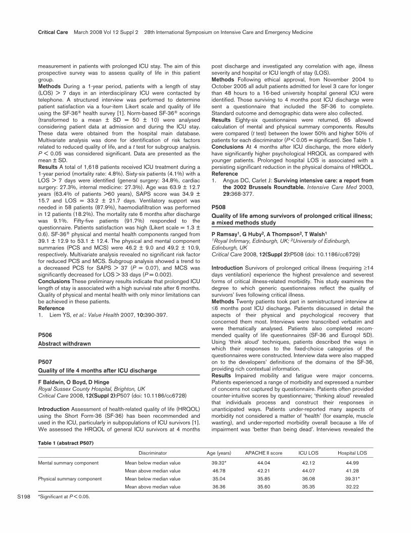
S198
measurement in patients with prolonged ICU stay. The aim of thisprospective survey was to assess quality of life in this patientgroup.Methods During a 1-year period, patients with a length of stay(LOS) > 7 days in an interdisciplinary ICU were contacted bytelephone. A structured interview was performed to determinepatient satisfaction via a four-item Likert scale and quality of lifeusing the SF-36® health survey [1]. Norm-based SF-36® scorings(transformed to a mean ± SD = 50 ± 10) were analysedconsidering patient data at admission and during the ICU stay.These data were obtained from the hospital main database.Multivariate analysis was done for identification of risk factorsrelated to reduced quality of life, and a t test for subgroup analysis.P < 0.05 was considered significant. Data are presented as themean ± SD.Results A total of 1,618 patients received ICU treatment during a1-year period (mortality rate: 4.8%). Sixty-six patients (4.1%) with aLOS > 7 days were identified (general surgery: 34.8%, cardiacsurgery: 27.3%, internal medicine: 27.3%). Age was 63.9 ± 12.7years (63.4% of patients >60 years), SAPS score was 34.9 ±15.7 and LOS = 33.2 ± 21.7 days. Ventilatory support wasneeded in 58 patients (87.9%), haemodiafiltration was performedin 12 patients (18.2%). The mortality rate 6 months after dischargewas 9.1%. Fifty-five patients (91.7%) responded to thequestionnaire. Patients satisfaction was high (Likert scale = 1.3 ±0.6). SF-36® physical and mental health components ranged from39.1 ± 12.9 to 53.1 ± 12.4. The physical and mental componentsummaries (PCS and MCS) were 46.2 ± 9.0 and 49.2 ± 10.9,respectively. Multivariate analysis revealed no significant risk factorfor reduced PCS and MCS. Subgroup analysis showed a trend toa decreased PCS for SAPS > 37 (P = 0.07), and MCS wassignificantly decreased for LOS > 33 days (P = 0.002).Conclusions These preliminary results indicate that prolonged ICUlength of stay is associated with a high survival rate after 6 months.Quality of physical and mental health with only minor limitations canbe achieved in these patients.Reference1. Liem YS, et al.: Value Health 2007, 10:390-397.
P506Abstract withdrawn
P507Quality of life 4 months after ICU discharge
F Baldwin, O Boyd, D HingeRoyal Sussex County Hospital, Brighton, UKCritical Care 2008, 12(Suppl 2):P507 (doi: 10.1186/cc6728)
Introduction Assessment of health-related quality of life (HRQOL)using the Short Form-36 (SF-36) has been recommended andused in the ICU, particularly in subpopulations of ICU survivors [1].We assessed the HRQOL of general ICU survivors at 4 months
post discharge and investigated any correlation with age, illnessseverity and hospital or ICU length of stay (LOS).Methods Following ethical approval, from November 2004 toOctober 2005 all adult patients admitted for level 3 care for longerthan 48 hours to a 16-bed university hospital general ICU wereidentified. Those surviving to 4 months post ICU discharge weresent a questionnaire that included the SF-36 to complete.Standard outcome and demographic data were also collected.Results Eighty-six questionnaires were returned, 65 allowedcalculation of mental and physical summary components. Resultswere compared (t test) between the lower 50% and higher 50% ofpatients for each discriminator (P < 0.05 = significant). See Table 1.Conclusions At 4 months after ICU discharge, the more elderlyhave significantly higher psychological HRQOL as compared withyounger patients. Prolonged hospital LOS is associated with apersisting significant reduction in the physical domains of HRQOL.Reference1. Angus DC, Carlet J: Surviving intensive care: a report from
the 2002 Brussels Roundtable. Intensive Care Med 2003,29:368-377.
P508Quality of life among survivors of prolonged critical illness;a mixed methods study
P Ramsay1, G Huby2, A Thompson2, T Walsh1
1Royal Infirmary, Edinburgh, UK; 2University of Edinburgh,Edinburgh, UKCritical Care 2008, 12(Suppl 2):P508 (doi: 10.1186/cc6729)
Introduction Survivors of prolonged critical illness (requiring ≥14days ventilation) experience the highest prevalence and severestforms of critical illness-related morbidity. This study examines thedegree to which generic questionnaires reflect the quality ofsurvivors’ lives following critical illness.Methods Twenty patients took part in semistructured interview at≤6 months post ICU discharge. Patients discussed in detail theaspects of their physical and psychological recovery thatconcerned them most. Interviews were transcribed verbatim andwere thematically analysed. Patients also completed recom-mended quality of life questionnaires (SF-36 and Euroqol 5D).Using ‘think aloud’ techniques, patients described the ways inwhich their responses to the fixed-choice categories of thequestionnaires were constructed. Interview data were also mappedon to the developers’ definitions of the domains of the SF-36,providing rich contextual information.Results Impaired mobility and fatigue were major concerns.Patients experienced a range of morbidity and expressed a numberof concerns not captured by questionnaire. Patients often providedcounter-intuitive scores by questionnaire; ‘thinking aloud’ revealedthat individuals process and construct their responses inunanticipated ways. Patients under-reported many aspects ofmorbidity not considered a matter of ‘health’ (for example, musclewasting), and under-reported morbidity overall because a life ofimpairment was ‘better than being dead’. Interviews revealed the
Critical Care March 2008 Vol 12 Suppl 2 28th International Symposium on Intensive Care and Emergency Medicine
Table 1 (abstract P507)
Discriminator Age (years) APACHE II score ICU LOS Hospital LOS
Mental summary component Mean below median value 39.32* 44.04 42.12 44.99
Mean above median value 46.78 42.21 44.07 41.28
Physical summary component Mean below median value 35.04 35.85 36.08 39.31*
Mean above median value 36.36 35.60 35.35 32.22
*Significant at P < 0.05.
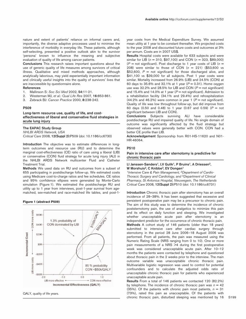
S199
nature and extent of patients’ reliance on informal carers and,importantly, the diverse adaptive processes used to minimise theinterference of morbidity in everyday life. These patients, althoughself-selecting, presented a positive outlook akin to the survivor‘persona’ known to influence the meaning and subjectiveevaluation of quality of life among cancer patients.Conclusions This research raises important questions about theuse of generic quality of life measures among survivors of criticalillness. Qualitative and mixed methods approaches, althoughanalytically laborious, may yield experientially important informationand clinically useful insights into the quality of survivors’ lives thatare inaccessible by questionnaire alone.References1. Mallinson S: Soc Sci Med 2002, 54:11-21.2. Westerman MJ, et al.: Qual Life Res 2007, 16:853-861.3. Zebrack BJ: Cancer Practice 2000, 8:238-242.
P509Long-term resource use, quality of life, and cost-effectiveness of liberal and conservative fluid strategies inacute lung injury
The EAPAC Study GroupNHLBI ARDS Network, USACritical Care 2008, 12(Suppl 2):P509 (doi: 10.1186/cc6730)
Introduction The objective was to estimate differences in long-term outcomes and resource use (RU) and to determine themarginal cost-effectiveness (CE) ratio of care using a liberal (LIB)or conservative (CON) fluid strategy for acute lung injury (ALI) inthe NHLBI ARDS Network multicenter Fluid and CatheterTreatment Trial.Methods We used data on RU and outcomes from a subset of655 participating in postdischarge follow-up. We estimated costsusing Medicare cost-to-charge ratios and fee schedules. CE ratiosand 95% confidence ellipses were generated by Monte-Carlosimulation (Figure 1). We estimated the postdischarge RU andutility up to 1 year from interviews, post-1-year survival from age-matched, sex-matched and race-matched life tables, and post-1-
year costs from the Medical Expenditure Survey. We assumedmean utility at 1 year to be constant thereafter. We projected coststo the year 2008 and discounted future costs and outcomes at 3%per annum. Costs are in 2007 US$.Results Hospital costs were available for 633 subjects and weresimilar for LIB (n = 310, $97,100) and CON (n = 323, $89,000)(P = not significant). Post discharge to 1 year costs of LIB (n =208) were similar to those of CON (n = 221) ($53,600 vs$52,600, P = not significant) for those discharged alive, and$41,100 vs $39,000 for all subjects. Post 1 year costs weresimilar. Mortality increased from 26.9% (LIB) and 24.5% (CON) at60 days to 35.6% and 32.1% at 1 year (P = 0.31). Home oxygenuse was 32.2% and 28.5% for LIB and CON (P = not significant)and 15.4% and 14.0% at 1 year (P = not significant). Admission toa rehabilitation facility (34.1% and 29.4%) and rehospitalization(44.2% and 46.2%) were common in year 1 (P = not significant).Quality of life was low throughout follow-up, but did improve from60 days (0.50 and 0.48) to 1 year (0.67 and 0.59) (P = notsignificant between LIB and CON).Conclusions Subjects surviving ALI have considerablepostdischarge RU and impaired quality of life. No single domain ofoutcome was significantly affected by the fluid strategy butobserved values were generally better with CON. CON had abetter CE profile than LIB.Acknowledgement Sponsorship from R01-HS-11620 and N01-HR-46064.
P510Pain in intensive care after sternotomy is predictive forchronic thoracic pain
LI Janssen-Sanders1, LV Gulik1, P Bruins1, A Driessen2, W Morshuis2, C Knibbe3, EV Dongen1
1Intensive Care & Pain Management, 2Department of Cardio-Thoracic Surgery and Cardiology, and 3Department of ClinicalPharmacy, St Antonius Hospital, Nieuwegein, The NetherlandsCritical Care 2008, 12(Suppl 2):P510 (doi: 10.1186/cc6731)
Introduction Chronic thoracic pain after sternotomy has an overallincidence of 28–38%. It has been suggested that excessive andpersistent postoperative pain may be a precursor to chronic pain.The aim of this study was to determine the incidence of chronicpoststernotomy pain, the use of analgetics to minimize this painand its effect on daily function and sleeping. We investigatedwhether unacceptable acute pain after sternotomy is anindependent predictor for the occurrence of chronic thoracic pain.Methods A cohort study of 146 patients (older than 18 years)submitted to intensive care after cardiac surgery throughsternotomy in the period 28 June 2006–18 August 2006 wasperformed. From all patients, the pain was measured using theNumeric Rating Scale (NRS ranging from 0 to 10). One or morepain measurements of a NRS >4 during the first postoperativeweek was considered unacceptable acute pain. After 10–12months the patients were contacted by telephone and questionedabout thoracic pain in the 2 weeks prior to the interview. The mainoutcome variable was unacceptable chronic thoracic pain.Multivariable logistic regression was used to control for potentialconfounders and to calculate the adjusted odds ratio ofunacceptable chronic thoracic pain for patients who experiencedunacceptable acute pain.Results From a total of 146 patients we contacted 120 (82.2%)by telephone. The incidence of chronic thoracic pain was n = 42(35%). Of the patients with chronic pain most patients, n = 31(73%), rated this pain as unacceptable. Of the patients withchronic thoracic pain, disturbed sleeping was mentioned by 16
Available online http://ccforum.com/supplements/12/S2
Figure 1 (abstract P509)
QALY, quality of life years.
S200
(38%) patients, use of analgetics for thoracic pain by 15 (36%)patients and disturbed daily function by six (14%) patients. Almostall of these patients rated their chronic pain unacceptable. Theadjusted odds ratio of unacceptable chronic thoracic pain forpatients who experienced unacceptable acute pain was 4.6 (95%CI: 1.26–16.4).Conclusions The results of this study confirmed that chronic painis an important complication after sternotomy. Unacceptable acutepain is a strong predictor for the development of chronic thoracicpain.References1. Lahtinen P, Kokki H, Hynynen M: Pain after cardiac surgery:
a prospective cohort study of 1-year incidence and inten-sity. Anesthesiology 2006, 105:794-800.
2. Bruce J, Drury N, Poobalan AS, et al.: The prevalence ofchronic chest and leg pain following cardiac surgery: ahistorical cohort study. Pain 2003, 104:265-273.
3. Ho SC, Royse CF, Royse AG, et al.: Persistent pain aftercardiac surgery: an audit of high thoracic epidural andprimary opioid analgesia therapies. Anesth Analg 2002,95:820-823.
4. Meyerson J, Thelin S, Gordh T, Karlsten R: The incidence ofchronic post-sternotomy pain after cardiac surgery – aprospective study. Acta Anaesthesiol Scand 2001, 45:940-944.
5. Kalso E, Mennander S, Tasmuth T, Nilsson E: Chronic post-sternotomy pain. Acta Anaesthesiol Scand 2001, 45:935-939.
6. Mueller XM, Tinguely F, Tevaearai HT, et al.: Pain location,distribution, and intensity after cardiac surgery. Chest2000, 118:391-396.
7. Pluijms WA, Steegers MAH, Verhagen AFTM, Wilder-SmithOHG: Chronic post-thoracotomy pain: a retrospectivestudy. Acta Anesth Scand 2006, 50:804-808.
8. Gotoda Y, Kambara N, Sakai T, et al.: The morbidity, timecourse and predictive factors for persistent post-thoraco-tomy pain. Eur J Pain 2001, 5:89-96.
9. Norgaard MA, Andersen TC, Lavrsen MJ, Borgeskov S: Theoutcome of sternal wire removal on persistent anteriorchest wall pain after median sternotomy. Eur J Cardiotho-rac Surg 2006, 29:920-924.
10. Fine PG, Karwande SV: Sternal wire-induced persistentchest pain: a possible hypersensitivity reaction. AnnThorac Surg 1990, 49:135-136.
11. Perkins FM, Kehlet H: Chronic pain as an outcome ofsurgery. A review of predictive factors. Anasthesiology2000, 93:1123-1133.
12. Cohen AJ, Moore P, Jones C, et al.: Effect of internalmammary harvest on postoperative pain and pulmonaryfunction. Ann Thorac Surg 1993, 56:1107-1109.
13. Katz J, Jackson M, Kavanagh BP, Sandler AN: Acute painafter thoracic surgery predicts long-term post-thoraco-tomy pain. Clin J Pain 1996, 12:50-55.
14. Woolf CJ, Salter MW: Neural plasticity: increasing the gainin pain. Science 2000, 288:1765-1769.
15. Zalon LM: Nurses’ assessment of postoperative patients inpain. Pain 1993, 54:329-34.
16. Macrae WA: Chronic pain after sternotomy. Acta Anaesthe-siol Scand 2001, 45:927-928.
P511Pharmacokinetics of oral melatonin in patients recoveringfrom critical illness
R BourneSheffield Teaching Hospitals, Sheffield, UKCritical Care 2008, 12(Suppl 2):P511 (doi: 10.1186/cc6732)
Introduction The study was designed to evaluate the pharmaco-kinetics of oral melatonin in patients recovering from critical illness.To re-enforce a patients’ circadian rhythm it is necessary toadminister an appropriate dose at the correct time. However, dataare currently unavailable to guide effective dosing in this population.Methods A randomised double-blind placebo controlled trial wasperformed in 24 critically ill patients weaning from mechanicalventilation. Melatonin 10 mg was administered as an oral solutionat 21:00 hours each night for four nights. Pharmacokinetic analysisof plasma melatonin concentrations was undertaken in the firstnine patients in the active group. Twelve blood samples werecollected from each patient at appropriately spaced intervals. Aftersample dilution, plasma melatonin was measured in duplicate usinga melatonin radioimmunoassay. Plasma concentrations werecorrected for endogenous plasma melatonin concentration bysubtracting the 21:00-hour baseline value. Pharmacokinetic datafrom mean values were described using noncompartmental analysis.Nocturnal sleep quantity was measured using the bispectral index(BIS) and expressed as the sleep efficiency index (SEI) and BIS areaunder the curve (AUC) between 22:00 and 07:00 hours.Results Melatonin was rapidly and extensively absorbed with amean (SD) time to maximum plasma concentration of 0.5 (0) hours,a maximum plasma concentration (Cmax) of 14,974 (3,200) pg/mland the area under the plasma concentration time curve 0–24hours (AUC(0–24)) was 29,979 (8,205) ng.hour/l. Plasma concen-trations declined biexponentially with an overall plasma half-life of1.47 (0.28) hours. Clearance of this liver-metabolised drugresembled cirrhotic patients [1] rather than healthy individuals [2].Both Cmax and AUC(0–24) had a moderately strong correlation withplasma ALT concentrations; r = 0.7 (P = 0.04) and r = 0.62 (P =0.07), respectively. No such association was found with age,gender, weight, creatinine or bilirubin. Morning plasma levels(plasma concentration at 08:00 hours) remained supraphysio-logical at 84 (64) pg/ml. No association was found between thepharmacokinetic parameters Cmax, AUC(0–24) or plasma concen-tration at 08:00 hours and the mean SEI or BIS AUC measure-ments of nocturnal sleep.Conclusions The 10 mg nocturnal dose is excessive in thispopulation, and reduced doses should be used in futurechronotherapeutic studies of exogenous melatonin.References1. Iguchi H, et al.: J Clin Endocrinol Metab 1982, 54:1025-1027.2. Hartter S, et al.: Br J Clin Pharmacol 2003, 56:679-682.
P512Current awareness of delirium in the ICU: a postal surveyin The Netherlands
F Cadogan1, B Riekerk2, R Vreeswijk1, JH Rommes2, A Toornvliet1, PE Spronk2
1Medical Center Alkmaar, The Netherlands; 2Gelre Hospitals,Apeldoorn, The NetherlandsCritical Care 2008, 12(Suppl 2):P512 (doi: 10.1186/cc6733)
Introduction Delirium in the ICU can compromise the recoveryprocess, prolong the ICU and hospital stays and increase mortality.Therefore, recognition of delirium is of utmost importance.
Critical Care March 2008 Vol 12 Suppl 2 28th International Symposium on Intensive Care and Emergency Medicine
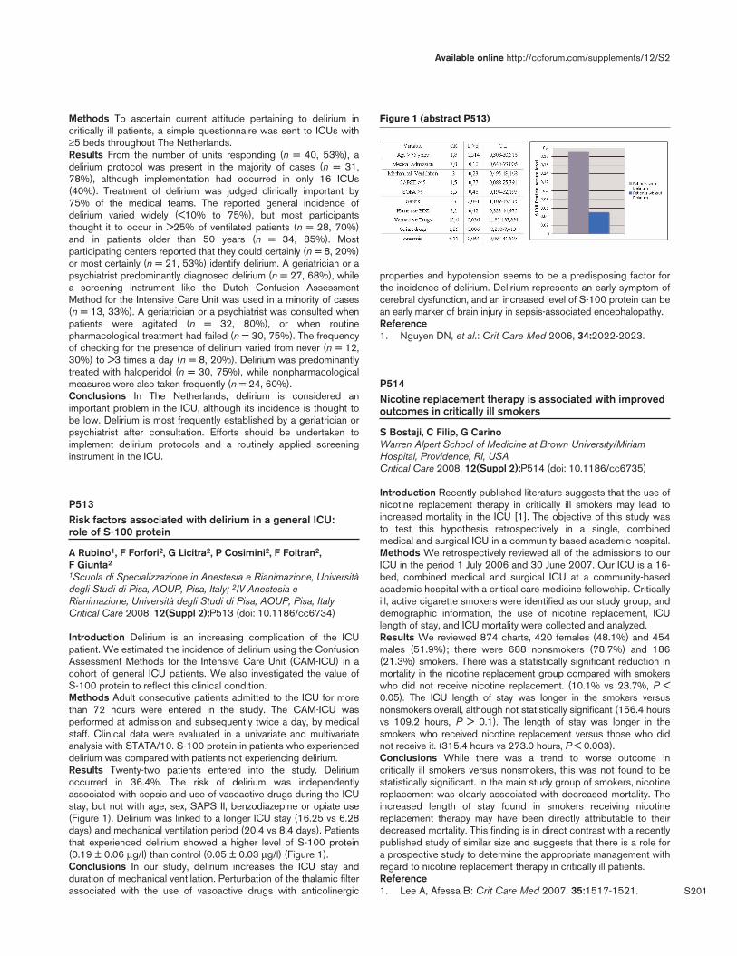
S201
Methods To ascertain current attitude pertaining to delirium incritically ill patients, a simple questionnaire was sent to ICUs with≥5 beds throughout The Netherlands.Results From the number of units responding (n = 40, 53%), adelirium protocol was present in the majority of cases (n = 31,78%), although implementation had occurred in only 16 ICUs(40%). Treatment of delirium was judged clinically important by75% of the medical teams. The reported general incidence ofdelirium varied widely (<10% to 75%), but most participantsthought it to occur in >25% of ventilated patients (n = 28, 70%)and in patients older than 50 years (n = 34, 85%). Mostparticipating centers reported that they could certainly (n = 8, 20%)or most certainly (n = 21, 53%) identify delirium. A geriatrician or apsychiatrist predominantly diagnosed delirium (n = 27, 68%), whilea screening instrument like the Dutch Confusion AssessmentMethod for the Intensive Care Unit was used in a minority of cases(n = 13, 33%). A geriatrician or a psychiatrist was consulted whenpatients were agitated (n = 32, 80%), or when routinepharmacological treatment had failed (n = 30, 75%). The frequencyof checking for the presence of delirium varied from never (n = 12,30%) to >3 times a day (n = 8, 20%). Delirium was predominantlytreated with haloperidol (n = 30, 75%), while nonpharmacologicalmeasures were also taken frequently (n = 24, 60%).Conclusions In The Netherlands, delirium is considered animportant problem in the ICU, although its incidence is thought tobe low. Delirium is most frequently established by a geriatrician orpsychiatrist after consultation. Efforts should be undertaken toimplement delirium protocols and a routinely applied screeninginstrument in the ICU.
P513Risk factors associated with delirium in a general ICU: role of S-100 protein
A Rubino1, F Forfori2, G Licitra2, P Cosimini2, F Foltran2, F Giunta2
1Scuola di Specializzazione in Anestesia e Rianimazione, Universitàdegli Studi di Pisa, AOUP, Pisa, Italy; 2IV Anestesia eRianimazione, Università degli Studi di Pisa, AOUP, Pisa, ItalyCritical Care 2008, 12(Suppl 2):P513 (doi: 10.1186/cc6734)
Introduction Delirium is an increasing complication of the ICUpatient. We estimated the incidence of delirium using the ConfusionAssessment Methods for the Intensive Care Unit (CAM-ICU) in acohort of general ICU patients. We also investigated the value ofS-100 protein to reflect this clinical condition.Methods Adult consecutive patients admitted to the ICU for morethan 72 hours were entered in the study. The CAM-ICU wasperformed at admission and subsequently twice a day, by medicalstaff. Clinical data were evaluated in a univariate and multivariateanalysis with STATA/10. S-100 protein in patients who experienceddelirium was compared with patients not experiencing delirium.Results Twenty-two patients entered into the study. Deliriumoccurred in 36.4%. The risk of delirium was independentlyassociated with sepsis and use of vasoactive drugs during the ICUstay, but not with age, sex, SAPS II, benzodiazepine or opiate use(Figure 1). Delirium was linked to a longer ICU stay (16.25 vs 6.28days) and mechanical ventilation period (20.4 vs 8.4 days). Patientsthat experienced delirium showed a higher level of S-100 protein(0.19 ± 0.06 μg/l) than control (0.05 ± 0.03 μg/l) (Figure 1).Conclusions In our study, delirium increases the ICU stay andduration of mechanical ventilation. Perturbation of the thalamic filterassociated with the use of vasoactive drugs with anticolinergic
properties and hypotension seems to be a predisposing factor forthe incidence of delirium. Delirium represents an early symptom ofcerebral dysfunction, and an increased level of S-100 protein can bean early marker of brain injury in sepsis-associated encephalopathy.Reference1. Nguyen DN, et al.: Crit Care Med 2006, 34:2022-2023.
P514Nicotine replacement therapy is associated with improvedoutcomes in critically ill smokers
S Bostaji, C Filip, G CarinoWarren Alpert School of Medicine at Brown University/MiriamHospital, Providence, RI, USACritical Care 2008, 12(Suppl 2):P514 (doi: 10.1186/cc6735)
Introduction Recently published literature suggests that the use ofnicotine replacement therapy in critically ill smokers may lead toincreased mortality in the ICU [1]. The objective of this study wasto test this hypothesis retrospectively in a single, combinedmedical and surgical ICU in a community-based academic hospital.Methods We retrospectively reviewed all of the admissions to ourICU in the period 1 July 2006 and 30 June 2007. Our ICU is a 16-bed, combined medical and surgical ICU at a community-basedacademic hospital with a critical care medicine fellowship. Criticallyill, active cigarette smokers were identified as our study group, anddemographic information, the use of nicotine replacement, ICUlength of stay, and ICU mortality were collected and analyzed.Results We reviewed 874 charts, 420 females (48.1%) and 454males (51.9%); there were 688 nonsmokers (78.7%) and 186(21.3%) smokers. There was a statistically significant reduction inmortality in the nicotine replacement group compared with smokerswho did not receive nicotine replacement. (10.1% vs 23.7%, P <0.05). The ICU length of stay was longer in the smokers versusnonsmokers overall, although not statistically significant (156.4 hoursvs 109.2 hours, P > 0.1). The length of stay was longer in thesmokers who received nicotine replacement versus those who didnot receive it. (315.4 hours vs 273.0 hours, P < 0.003).Conclusions While there was a trend to worse outcome incritically ill smokers versus nonsmokers, this was not found to bestatistically significant. In the main study group of smokers, nicotinereplacement was clearly associated with decreased mortality. Theincreased length of stay found in smokers receiving nicotinereplacement therapy may have been directly attributable to theirdecreased mortality. This finding is in direct contrast with a recentlypublished study of similar size and suggests that there is a role fora prospective study to determine the appropriate management withregard to nicotine replacement therapy in critically ill patients.Reference1. Lee A, Afessa B: Crit Care Med 2007, 35:1517-1521.
Available online http://ccforum.com/supplements/12/S2
Figure 1 (abstract P513)
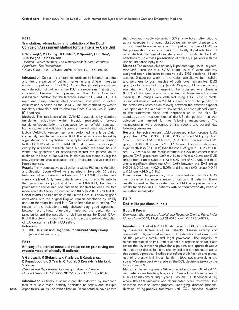
S202
P515Translation, retranslation and validation of the DutchConfusion Assessment Method for the Intensive Care Unit
R Vreeswijk1, M Honing1, K Bakker1, P Spronk2, T De Man1, J De Jonghe1, K Kalisvaart1
1Medical Center Alkmaar, The Netherlands; 2Gelre Ziekenhuis,Apeldoorn, The NetherlandsCritical Care 2008, 12(Suppl 2):P515 (doi: 10.1186/cc6736)
Introduction Delirium is a common problem in hospital settings,and the prevalence of delirium varies among different hospitalinpatient populations (42–87%). As in other patient populations,early detection of delirium in the ICU is a necessary first step forsuccessful treatment and prevention. The Dutch ConfusionAssessment Method for the Intensive Care Unit (CAM-ICU) is arapid and easily administrated screening instrument to detectdelirium and is based on the DSM-IV. The aim of this study was totranslate, retranslate and validate the CAM-ICU for use in DutchICU settings.Methods The translation of the CAM-ICU was done by standardtranslation guidelines, which include preparation, forwardtranslation/reconciliation, retranslation by an official translator,harmonization and validation. Secondly, the validation study of theDutch CAM-ICU version itself was performed in a large Dutchcommunity hospital with a mixed ICU. The patients were tested bya geriatrician or a psychiatrist for symptoms of delirium accordingto the DSM-IV criteria. The CAM-ICU testing was done indepen-dently by a trained research nurse but within the same hour inwhich the geriatrician or psychiatrist tested the patient, as tominimise the bias of fluctuations in delirium symptoms during theday. Agreement was calculated using crosstabs analysis and theKappa statistic.Results Thirty consecutive adult patients with a Richmond Agitationand Sedation Score –3/+4 were included in the study, 60 pairedtests for delirium were carried out and 30 CAM-ICU instrumentswere completed. Only three patients were diagnosed differently bythe geriatrician or psychiatrist and the CAM-ICU; two had apsychiatric disorder and one had been sedated between the twomeasurements. Overall agreement was 90% (Κ > 0.81, P < 0.001).Conclusions The translation of the Dutch CAM-ICU showed goodcorrelation with the original English version developed by W Elyand can therefore be used in a Dutch intensive care setting. Theresults of the validation study showed very good agreementbetween the clinical diagnoses made by the geriatrician orpsychiatrist and the detection of delirium using the Dutch CAM-ICU. It therefore provides the means for early and reliable detectionof ICU delirium in a Dutch ICU setting.Reference1. ICU Delirium and Cognitive Impairment Study Group
[www.icudelirium.org]
P516Efficacy of electrical muscle stimulation on preserving themuscle mass of critically ill patients
V Gerovasili, K Stefanidis, K Vitzilaios, E Karatzanos, E Papadopoulos, G Tzanis, C Routsi, D Zervakis, V Markaki, S NanasNational and Kapodistrian University of Athens, GreeceCritical Care 2008, 12(Suppl 2):P516 (doi: 10.1186/cc6737)
Introduction Critically ill patients are characterized by increasedloss of muscle mass, partially attributed to sepsis and multipleorgan failure, as well as immobilization. Recent studies have shown
that electrical muscle stimulation (EMS) may be an alternative toactive exercise in chronic obstructive pulmonary disease andchronic heart failure patients with myopathy. The role of EMS forthe preservation of muscle mass of critically ill patients has notbeen studied. The aim of our study was to investigate the EMSeffects on muscle mass preservation of critically ill patients with theuse of ultrasonography (US).Methods Ten consecutive critically ill patients (age: 69 ± 16 years,APACHE score: 22 ± 5, SOFA score: 10 ± 3) were randomlyassigned upon admission to receive daily EMS sessions (45-minsession, 5 days per week) of the vastus lateralis, vastus medialisand peroneus longus muscles of both lower extremities (EMSgroup) or to the control group (non-EMS group). Muscle mass wasevaluated with US, by measuring the cross-sectional diameter(CSD) of the quadriceps muscle (rectus femoris–vastus inter-medius). US images were obtained using a GE Vivid 7 modelultrasound scanner with a 7.5 MHz linear probe. The position ofthe probe was selected as midway between the anterior superioriliac spine and the midpoint of the patella, and was placed ventralto the transverse plane and perpendicular to the skin. Tostandardize the measurements of the US, the position that wasselected was marked for the following measurement. Themeasurements were performed on the second and seventh dayfollowing admission.Results The rectus femorist CSD decreased in both groups (EMSgroup: from 1.24 ± 0.33 to 1.16 ± 0.35 cm, non-EMS group: from1.52 ± 0.51 to 1.25 ± 0.43 cm) (P < 0.05); however, the EMSgroup (–0.08 ± 0.05 cm, –7.2 ± 4.1%) was observed to decreasesignificantly less (P < 0.05) than the non-EMS group (–0.26 ± 0.13cm, –17.3 ± 7.6%). The vastus intermedius CSD decreased in bothgroups (EMS group: from 0.87 ± 0.43 to 0.79 ± 0.42 cm, non-EMSgroup: from 1.65 ± 0.65 to 1.23 ± 0.47 cm) (P < 0.05), and therewas a significant difference (P < 0.05) between the EMS group(–0.08 ± 0.02 cm, –10.0 ± 5.0%) and the non-EMS group (–0.41± 0.21 cm, –24.6 ± 5.1%).Conclusions The preliminary data presented suggest that EMSmay preserve the muscle mass of critically ill patients. Theseresults as well as the potential use of EMS as a preventive andrehabilitation tool in ICU patients with polyneuromyopathy need tobe further investigated.
P517End of life practices in India
S Jog, B PawarDeenanath Mangeshkar Hospital and Research Centre, Pune, IndiaCritical Care 2008, 12(Suppl 2):P517 (doi: 10.1186/cc6738)
Introduction ‘End of life’ (EOL) decisions in ICUs are influencedby numerous factors such as patient’s disease severity andreversibility, religious and cultural traits, education and awarenessof the patient’s family and legal provisions. The majority ofpublished studies on EOL reflect either a European or an Americanethos; that is, either the physician’s paternalistic approach aboutthe patient or the patient’s autonomy and self-determination aboutthis sensitive process. Studies that reflect the influence and pivotalrole of a closely knit Indian family in EOL decision-making arescant. We retrospectively analysed the EOL decisions taken by thefamily in our ICU.Methods The setting was a 50-bed multidisciplinary ICU of a 400-bed tertiary care teaching hospital in Pune in India. Case papers ofall ICU admissions during 1 year (1 January–31 December 2006)where the EOL decision was documented were reviewed. Datacollected included demographics, underlying disease process,duration of aggressive treatment until EOL consent, duration
Critical Care March 2008 Vol 12 Suppl 2 28th International Symposium on Intensive Care and Emergency Medicine
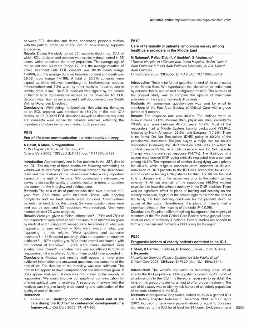
S203
between EOL decision and death, consenting person’s relationwith the patient, organ failure and level of life-sustaining supportsat decision.Results During the study period 524 patients died in our ICU, ofwhich EOL decision and consent was explicitly documented in 95cases, which constitute the study population. The average age ofthe patient was 63 years (range 17–91), the average duration ofactive treatment until EOL consent was 83.35 hours (range1–960), and the average duration between consent and death was29.03 hours (range 1–168). A total of 92.7% consents weresigned by close relatives (son/daughter, brother/sister, spouse,father/mother) and 7.3% were by other relatives (cousins, son inlaw/daughter in law). No EOL decision was signed by the patientor his/her legal representative as well as the physician. No EOLdecision was taken as per a patient’s self-documented own ‘DeathWill’ or ‘Advanced Directive’.Conclusions Withholding nonbeneficial life-sustaining therapiesas an EOL process was practised in 18.12% of the total ICUdeaths. All 95 (100%) EOL decisions as well as directive requestsand consents were signed by patients’ relatives, reflecting theimportance of close family ties in Indian EOL practices.
P518End of life care: communication – a retrospective survey
A Devitt, R Mane, S YoganathanBHR Hospitals NHS Trust, Romford, UKCritical Care 2008, 12(Suppl 2):P518 (doi: 10.1186/cc6739)
Introduction Approximately one in five patients in the USA dies inthe ICU. The majority of these deaths are following withholding orwithdrawal of treatment. Communication between the healthcareteam and the relatives of the patient constitutes a very importantaspect of the end of life care. We conducted a retrospectivesurvey to assess the quality of communication in terms of durationand content of the interview and spiritual care.Methods The next of kin of patients who died over a period of 1year from April 2003 were identified. Those with ongoingcomplaints and no fixed abode were excluded. Seventy-threepatients had died during this period. Sixty-one questionnaires weresent out by post and 32 responses were received. Nine wereincomplete and 23 were analysed.Results Were you given sufficient information? – 74% and 78% ofthe responders were satisfied with the amount of information givenby medical and nursing staff, respectively. Awareness of what washappening to your relative? – 65% were aware of what washappening to their relative. Were questions and concernsanswered? – 74% replied positively. Was the duration of interviewsufficient? – 87% replied yes. Was there overall satisfaction withthe content of interview? – 70% were overall satisfied. Wasspiritual care offered? – spiritual care was not offered to 56% ofresponders; if it was offered, 85% of them would have accepted it.Conclusions Medical and nursing staff appear to have givensufficient information and answered questions and concerns of thenext of kin. The duration of the interview was also sufficient. Thenext of kin appear to have comprehended the information given. Itdoes appear that spiritual care was not offered to the majority ofresponders. We could improve our quality of communication byoffering spiritual care to relatives. A structured interview with therelatives can improve family understanding and satisfaction of thequality of end of life care!Reference1. Curtis et al.: Studying communication about end of life
care during the ICU family conference: development of aframework. J Crit Care 2002, 17:147-160.
P519Care of terminally ill patients: an opinion survey amonghealthcare providers in the Middle East
M Rahman1, F Abu-Zidan2, Y Ibrahim1, S Abuhasna1
1Tawam Hospital in affiliation with Johns Hopkins, Al-Ain, UnitedArab Emirates; 2United Arab Emirates University, Al-Ain, UnitedArab EmiratesCritical Care 2008, 12(Suppl 2):P519 (doi: 10.1186/cc6740)
Introduction There is no formal guideline on end of life care issuesin the Middle East. We hypothesize that decisions are influencedby personal belief, culture and background training. The purpose ofthe present study was to compare the opinion of healthcareproviders on the care of terminally ill patients.Methods An anonymous questionnaire was sent by email tomembers of the Pan Arab Society of Critical Care with a graceperiod of 6 months.Results The response rate was 46.2%. The findings were asfollows: males 91.8%, Muslims 86%, physicians 96%, consultants70.9%, and aged between 40–50 years 47.7%. Most of theresponders had a Middle Eastern training background (33.8%),followed by North American (29.2%) and European (17.6%). Thereis no formal Do Not Resuscitate (DNR) policy in 62.2% of theparticipants’ institutions. Religion played a major role for 59.3%responders in making the DNR decision. DNR was equivalent tocomfort care in 39.5%. In a futile case scenario, Do Not EscalateTherapy was the preferred response (54.7%). The likelihood of apatient once labeled DNR being clinically neglected was a concernamong 46.5%. The importance of comfort during dying was a priorityfor 45.3%, while religious concerns were important in 52.3%.Admission of DNR patients to the ICU was acceptable for 47.7%,and to continue feeding DNR patients for 94%. For 60.5% the besttime to discuss end of life issues was prior to the patient gettingseverely ill. Almost one-half of the responders (46.5%) wantedphysicians to have the ultimate authority in the DNR decision. Therewas no significant effect of place of training and seniority on themanagement plan, neglect of the patient, right to override the right ofthe family, the best defining conditions for the patient’s death orabuse of the code. Nevertheless, the place of training had asignificant effect on the meaning of the code (P < 0.08).Conclusions Despite a different training background, the majority ofmembers of the Pan Arab Critical Care Society have a general agree-ment on care of terminally ill patients. Further studies are needed toform a consensus and formalize a DNR policy for the region.
P520Prognostic factors of elderly patients admitted to an ICU
F Stein, R Barros, F Feitosa, D Toledo, J Silva Junior, A Isola, E RezendeHospital do Servidor Público Estadual de São Paulo, BrazilCritical Care 2008, 12(Suppl 2):P520 (doi: 10.1186/cc6741)
Introduction The world’s population is becoming older, whichaffects the ICU population. Elderly patients constitute 42–52% ofall admissions to the ICU. It is therefore necessary to establish therisks of this group of patients, aiming to offer proper treatment. Theaim of this study was to identify risk factors of an elderly populationof patients admitted to the ICU.Methods A prospective longitudinal cohort study in a general ICUof a tertiary hospital, between 1 December 2006 and 30 April2007. Inclusion criteria were patients above or equal to 65 yearsold, admitted to the ICU for at least for 24 hours. Exclusion criteria
Available online http://ccforum.com/supplements/12/S2
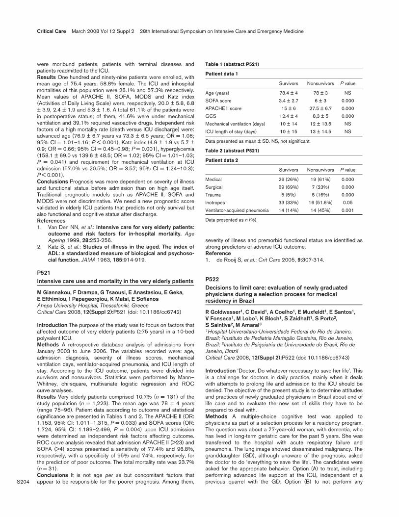
S204
were moribund patients, patients with terminal diseases andpatients readmitted to the ICU.Results One hundred and ninety-nine patients were enrolled, withmean age of 75.4 years, 58.8% female. The ICU and inhospitalmortalities of this population were 28.1% and 57.3% respectively.Mean values of APACHE II, SOFA, MODS and Katz index(Activities of Daily Living Scale) were, respectively, 20.0 ± 5.8, 6.8± 3.9, 2.4 ± 1.9 and 5.3 ± 1.6. A total 61.1% of the patients werein postoperative status; of them, 41.6% were under mechanicalventilation and 39.1% required vasoactive drugs. Independent riskfactors of a high mortality rate (death versus ICU discharge) were:advanced age (76.9 ± 6.7 years vs 73.3 ± 6.5 years; OR = 1.08;95% CI = 1.01–1.16; P < 0.001), Katz index (4.9 ± 1.9 vs 5.7 ±0.9; OR = 0.66; 95% CI = 0.45–0.98; P = 0.001), hyperglycemia(158.1 ± 69.0 vs 139.6 ± 48.5; OR = 1.02; 95% CI = 1.01–1.03;P = 0.041) and requirement for mechanical ventilation at ICUadmission (57.0% vs 20.5%; OR = 3.57; 95% CI = 1.24–10.3);P < 0.001).Conclusions Prognosis was more dependent on severity of illnessand functional status before admission than on high age itself.Traditional prognostic models such as APACHE II, SOFA andMODS were not discriminative. We need a new prognostic scorevalidated in elderly ICU patients that predicts not only survival butalso functional and cognitive status after discharge.References1. Van Den NN, et al.: Intensive care for very elderly patients:
outcome and risk factors for in-hospital mortality. AgeAgeing 1999, 28:253-256.
2. Katz S, et al.: Studies of illness in the aged. The index ofADL: a standardized measure of biological and psychoso-cial function. JAMA 1963, 185:914-919.
P521Intensive care use and mortality in the very elderly patients
M Giannakou, F Drampa, G Tsaousi, E Anastasiou, E Geka, E Efthimiou, I Papageorgiou, K Matsi, E SofianosAhepa University Hospital, Thessaloniki, GreeceCritical Care 2008, 12(Suppl 2):P521 (doi: 10.1186/cc6742)
Introduction The purpose of the study was to focus on factors thataffected outcome of very elderly patients (≥75 years) in a 10-bedpolyvalent ICU.Methods A retrospective database analysis of admissions fromJanuary 2003 to June 2006. The variables recorded were: age,admission diagnosis, severity of illness scores, mechanicalventilation days, ventilator-acquired pneumonia, and ICU length ofstay. According to the ICU outcome, patients were divided intosurvivors and nonsurvivors. Statistics were performed by Mann–Whitney, chi-square, multivariate logistic regression and ROCcurve analyses.Results Very elderly patients comprised 10.7% (n = 131) of thestudy population (n = 1,223). The mean age was 78 ± 4 years(range 75–96). Patient data according to outcome and statisticalsignificance are presented in Tables 1 and 2. The APACHE II (OR:1.153, 95% CI: 1.011–1.315, P = 0.033) and SOFA scores (OR:1.724, 95% CI: 1.189–2.499, P = 0.004) upon ICU admissionwere determined as independent risk factors affecting outcome.ROC curve analysis revealed that admission APACHE II (>23) andSOFA (>4) scores presented a sensitivity of 77.4% and 96.8%,respectively, with a specificity of 95% and 74%, respectively, forthe prediction of poor outcome. The total mortality rate was 23.7%(n = 31).Conclusions It is not age per se but concomitant factors thatappear to be responsible for the poorer prognosis. Among them,
severity of illness and premorbid functional status are identified asstrong predictors of adverse ICU outcome.Reference1. de Rooij S, et al.: Crit Care 2005, 9:307-314.
P522Decisions to limit care: evaluation of newly graduatedphysicians during a selection process for medicalresidency in Brazil
R Goldwasser1, C David1, A Coelho1, E Muxfeldt1, E Santos1, V Fonseca1, M Lobo1, K Bloch1, S Zaidhaft1, S Porto2, S Saintive2, M Amaral31Hospital Universitario-Universidade Federal do Rio de Janeiro,Brazil; 2Instituto de Pediatria Martagão Gesteira, Rio de Janeiro,Brazil; 3Instituto de Psiquiatria da Universidade do Brasil, Rio deJaneiro, BrazilCritical Care 2008, 12(Suppl 2):P522 (doi: 10.1186/cc6743)
Introduction ‘Doctor. Do whatever necessary to save her life’. Thisis a challenge for doctors in daily practice, mainly when it dealswith attempts to prolong life and admission to the ICU should bedenied. The objective of the present study is to determine attitudesand practices of newly graduated physicians in Brazil about end oflife care and to evaluate the new set of skills they have to beprepared to deal with.Methods A multiple-choice cognitive test was applied tophysicians as part of a selection process for a residency program.The question was about a 77-year-old woman, with dementia, whohas lived in long-term geriatric care for the past 5 years. She wastransferred to the hospital with acute respiratory failure andpneumonia. The lung image showed disseminated malignancy. Thegranddaughter (GD), although unaware of the prognosis, askedthe doctor to do ‘everything to save the life’. The candidates wereasked for the appropriate behavior. Option (A) to treat, includingperforming advanced life support at the ICU, independent of aprevious quarrel with the GD; Option (B) to not perform any
Critical Care March 2008 Vol 12 Suppl 2 28th International Symposium on Intensive Care and Emergency Medicine
Table 1 (abstract P521)
Patient data 1
Survivors Nonsurvivors P value
Age (years) 78.4 ± 4 78 ± 3 NS
SOFA score 3.4 ± 2.7 6 ± 3 0.000
APACHE II score 15 ± 6 27.5 ± 6.7 0.000
GCS 12.4 ± 4 8,3 ± 5 0.000
Mechanical ventilation (days) 10 ± 14 12 ± 13.5 NS
ICU length of stay (days) 10 ± 15 13 ± 14.5 NS
Data presented as mean ± SD. NS, not significant.
Table 2 (abstract P521)
Patient data 2
Survivors Nonsurvivors P value
Medical 26 (26%) 19 (61%) 0.000
Surgical 69 (69%) 7 (23%) 0.000
Trauma 5 (5%) 5 (16%) 0.000
Inotropes 33 (33%) 16 (51.6%) 0.05
Ventilator-acquired pneumonia 14 (14%) 14 (45%) 0.001
Data presented as n (%).
S205
diagnostic or therapeutically measure and inform the GD that thiswill no longer bring benefit to her grandmother; Option (C) todefine, together with the GD, possible palliative interventions; orOption (D) to apply protocols based on evidence related topalliative care, independently of the GD’s opinion. The correctanswer was based on two skills: the concern about futile treatmentand ethical issues – Option (C).Results A total of 1,133 physicians participated in the selectionprocess for medical residency and answered the questionnaire. Ofthe respondents, 698 (61.61%) would rather define with the familypossible palliative interventions (correct option – (C)); 312respondents (27.54%) would treat the patient even without thefamily’s opinion (option (A)); 122 respondents (10.77%) wouldapply protocols, again independent of the GD’s opinion (option(D)); and one candidate (0.08%) chose option (B).Conclusions Although the concept of palliative care and theimportance to share decisions with the family has been chosen bythe majority of the newly graduating physicians, 38.31% would stillmake decisions independently of the family’s opinion. Medicalstudents have to be prepared for a new set of skills.References1. Kasper DL, Braunwald E, Fauci AS, et al.: Harrison’s Principle
of Internal Medicine. 16th edition. McGraw-Hill; 2005.2. Tinker J, Browne DRG, Sibbald WJ: Critical Care. Standards,
Audit and Ethics. London: Arnold; 1996.
P523Interdisciplinary ethics consultation on the surgical ICU:indication ‘on a gut level’?
A Chromik, T Wemhöner, D Sülberg, H Laubenthal, U Mittelkötter, W UhlSt Josef University Hospital, Bochum, GermanyCritical Care 2008, 12(Suppl 2):P523 (doi: 10.1186/cc6744)
Introduction Interdisciplinary ethics consultation (EC) on the ICUcan be requested by the clinical team as a result of a subjectiveassessment of the patient’s situation. The aim of this study was toobjectify the initiation of EC by means of the SOFA score and toexamine its impact on the clinical course.Methods Over a 2-year period, all patients receiving an EC on theICU were recorded. The age, hospital stay and mortality werecompared with ICU patients who did not receive an EC. SOFAscore values of EC patients at the time of admission to the ICU andat the time of EC were compared. Furthermore, the effect ofdifferent EC decisions (maximization/limitation of treatment) onhospital stay and mortality were defined.Results An EC was carried out in 52 patients out of the total 764patients (6.8%). Age (76.6 years; range 40–99), hospital stay(20.5 days; range 5–286) and ICU mortality (92.3%) weresignificantly higher in EC patients compared with patients withoutEC (68.3 years; range 10–100; P ≤ 0.001) (3 days; range 2–106;P ≤ 0.001) (7.6%; P ≤ 0.001). The mean SOFA score at the timeof EC (7.52; ±0.48 SEM) was significantly higher compared withthe time of admission to the ICU (4.29; ±0.42 SEM) (P ≤ 0.001).Following maximization of treatment (n = 9), the median hospitalstay was significantly longer (13 days; range 4–254) comparedwith other EC decisions (n = 43) (P ≤ 0.007).Conclusions In critically ill patients, the interdisciplinary ECprovides a meaningful tool for decision-making betweenmaximization and reduction of treatment – facilitating improved endof life care and dignified dying. Indication of an EC at the bedsideshould be supported by the SOFA score.
P524Safety assessment of the direct sequence spread spectrumwireless local area network with a medical device
T Loh, F KwanKK Hospital, SingaporeCritical Care 2008, 12(Suppl 2):P524 (doi: 10.1186/cc6745)
Introduction In conjunction with the Electronic Medical Record’sComputerised Physician Order Entry project, KK Hospital hasparticipated in the SingHealth Wireless Local Area Network(WLAN) Infrastructure project to evaluate the CISCO® Wirelessnetwork technology product. The vendor, medical devicemanufacturers and local medical equipment distributors would notguarantee in writing that there would be no radiofrequency orelectromagnetic interference (EMI) from the 2.4 GHz and 5 GHzdirect sequencing spread spectrum system (DSSS) with medicaldevices. The Standards, Productivity and Innovation Board are ableto conduct onsite electric field measurements but could notguarantee any EMI on medical devices. Senior management at KKHospital requested an EMI test to be conducted inhouse todetermine the potential effects of the WLAN on medical devices.Methods The test was conducted within a vacant room in thewomen’s ICU. Selected medical devices were representative oftechnology used in KK Hospital ICUs and wards. The WLAN andmedical devices were set up to replicate the actual environment asclosely as possible. The test was conducted on both the 2.4 GHzand 5 GHz direct sequencing frequency bandwidth at a powersetting of 50 mW with two PC Notebooks. A command wasexecuted on the two PC Notebooks to generate continuouswireless transmission between the WLAN card of the PCNotebooks and the access point in the room. One women’s ICUnurse was deployed to chart and monitor the parameters of eachmedical device under test. The project team also checked theequipment under test at 1 or 2 hour intervals.Results No electromagnetic interference was detected on any ofthe devices under test.Conclusions The EMI test was only a sampling, bench-top testand should not be taken as conclusive for EMI effects of the DSSSWLAN on each medical device within the hospital. However, duediligence was exercised in conducting the EMI test and thereappears to be no evidence to suggest adverse EMI effects on themedical devices tested.
P525Consumption of ICU resources by long-stay patients doesnot change over time: 10-year observation in a teachinghospital in The Netherlands
A Schoffelen, J Hofhuis, JH Rommes, PE SpronkGelre Hospitals, Apeldoorn, The NetherlandsCritical Care 2008, 12(Suppl 2):P525 (doi: 10.1186/cc6746)
Introduction The steady increase of expected lifetime and meanage of man in the western world can only be accompanied by anincrease in medical care and inherent use of ICU resources. Now-adays, one in five Americans dies using ICU care. We exploredwhether the use of ICU resources by long-stay patients hasincreased over time.Methods Data from all patients admitted to a 10-bedmedical–surgical ICU in a university-affiliated teaching hospital in a10-year period were analyzed. In the study period, organizationalaspects such as the nurse-per-patient ratio and the number ofintensivists remained stable. In the first 24 hours, the type ofadmission and medical history was recorded and APACHE II
Available online http://ccforum.com/supplements/12/S2
S206
scores were calculated. After discharge, patients were divided intofour groups with ICU length of stay (LOS) <3 days, 3–6 days,7–13 days and ≥14 days. Data are shown as the median(interquartile range (IQR)).Results Over the years, the number of patients admitted per yearwas stable at a median 578 (IQR 535–588). Age (69 (65–76)years) was stable over the years and did not differ between LOSgroups. APACHE II scores (12 (8–17)) were stable as well, while alonger ICU LOS was associated with higher scores. Hospitalmortality was 18.5% (range 16.6–19.1%). The proportion ofpatients per year in the different LOS groups did not change overthe years; that is, LOS <3 days: 62.5% (range 61.4–64.1%), LOS3–6 days: 19.1% (range 18.9–20.1%), LOS 7–13 days: 9.8%(range 9.2–10.9%), and LOS ≥14 days: 8.6% (range 7.5–9.6%).The relatively small group of long-stay patients (≥14 days; n = 44(39–51) per year) comprised predominantly medical admissions(65.5%) and consumed 54.6% of the yearly ICU treatment days.Mortality in this group (35.1%) was higher than in the groups withshorter length of ICU stay (14.2% in LOS <3 days, 17.1% in LOS3–6 days and 27.0% in LOS 7–13 days; P < 0.001).Conclusions Consumption of ICU resources by long-stay patientswas considerable, but did not change over time in our setting.Long-stay patients were predominantly medical admissions andshowed the highest mortality. These findings are important in viewof futility and costs.
P526Microcosting study of ICU costs in three European countries
SS Tan1, J Martin2, A Pezzi3, J Bakker1, C Neurohr4, A Pitrelli5, L Hakkaart1, R Welte4
1Erasmus MC, University Medical Center, Rotterdam, TheNetherlands; 2Klinik am Eichert, Goeppingen, Germany; 3Universitàdegli Studi di Milano, Milan, Italy; 4GlaxoSmithKline, Munich,Germany; 5GlaxoSmithKline Italy, Verona, ItalyCritical Care 2008, 12(Suppl 2):P526 (doi: 10.1186/cc6747)
Introduction A recently published study by Negrini and colleaguesin 2006 suggested strongly diverging patient-related costs per ICUday between Western European countries. We tried to verify theseresults by performing a cost analysis in three European countries.Methods We conducted a retrospective cost study in oneGerman, one Italian and three Dutch adult medical–surgery ICUs,from the hospital perspective. A microcosting approach was used;that is, all relevant resources were identified and valued at the mostdetailed level. Resource use was divided into patient-related andnonpatient-related care. Patient-related care comprised diagnostics,consumables, inpatient stay and labour, while nonpatient careincluded overheads and capital. Resource use was primarilyderived from hospital administrative databases, while unit costswere acquired from financial hospital databases and hospitalpharmacy databases, using 2006 as the reference year.Results Average patient-related care costs per ICU day were€1,040 in Germany, €1,333 in Italy and €1,243 in TheNetherlands. Hence these costs are similar in all three countries,although there are differences between the considered patient-related care categories (for example, nursing and medical staff).The total average ICU cost per day (patient-related care +nonpatient-related care) amounted to €1,225 in Germany, €1,472in Italy, and €1,911 in The Netherlands. Variations in overheadsand capital costs might be (partly) caused by differences in theaccounting and financing systems. For example, unlike in Italy andThe Netherlands, in Germany most capital costs of public hospitalsare paid by the states and hence do not represent any cost to thehospital.
Conclusions Our results indicate that the patient-related carecosts of intensive care in Germany, Italy, and The Netherlands aresimilar from the hospital perspective.Reference1. Negrini D, et al.: Acta Anaesthesiol Scand 2006, 50:72-79.
P527Indication and use of chest X-ray scans in the ICU
P Shastri1, G Raghuraman2
1South Warwickshire General Hospital, Warwick, UK; 2HOEFT,Birmingham, UKCritical Care 2008, 12(Suppl 2):P527 (doi: 10.1186/cc6748)
Introduction Chest X-ray scans are frequently performed in ICUs.It is uncertain how much clinical information all chest X-ray scansprovide. There are currently no guidelines or protocols on thissubject. We believe some of the chest X-ray scans performed wereavoidable, limiting the cost for hospitals and radiation exposure topatients.Methods All chest X-ray scans performed in our 23-bed ICU andhigh dependency unit were monitored by asking the personordering the X-ray scan to fill in the form confirming the indicationfor the chest X-ray scan. At the end of 1 month all forms werecollected and analysed.Results A total of 112 X-ray scans were performed in a 4-weekperiod but only 102 forms were completed. Eighty-four out of 102chest X-ray scans were taken for only one indication, with 36/84for diagnosis of chest pathology. Fourteen X-ray scans were takenwithout completing the indication. Twenty-five per cent of X-rayscans were performed looking for two or more parameters, makingthem more cost-effective.Conclusions Routine chest X-ray scans are debatable. Post-procedure X-ray scans are not required if done by an experiencedclinician. The presence of protocols for ordering the chest X-rayscan may reduce the total number of chest X-ray scans performedand so reduce the cost and radiation exposure. A picture archivingand communication system may reduce the overall cost.
P528Technology in the ICU; the nurses’ point of view
O VerhulstHogeschool Antwerpen, BelgiumCritical Care 2008, 12(Suppl 2):P528 (doi: 10.1186/cc6749)
Introduction The objective of this study is to gain a view on howintensive care staff thinks of technology in the ICU. The studyinvestigates the acceptance of technology by intensive care nursesand tries to determine the advantages and disadvantages of theequipment in use in ICUs at this moment.Methods After reviewing some earlier publicised studies on thistopic, an online survey was set up to investigate the nurse’s viewon technology in the ICU. As this survey was set to reach as manyrespondents from Flemish hospitals, it was set up in Dutch.Results The survey was taken by 116 ICU staff members from 12Flemish hospitals, including three academic hospitals, with agesranging from 22 to 58 years. The working experience ranges from<1 year to 38 years. Educational records vary from bachelornursing degrees to specialized nurses and master degrees. Someinteresting results of the survey were the following: 91% of the
Critical Care March 2008 Vol 12 Suppl 2 28th International Symposium on Intensive Care and Emergency Medicine
S207
respondents perceived an increase of technology in the ICU duringthe time they were working in the unit. Sixty-six percent find thatthe complexity of these technological devices is increasing. Only62% think that the devices they have to work with are rather user-friendly. Seventy-seven percent think that the technologicalevolution of the ICU gives them a better view on the patients status.Twenty-five percent say it gives them a better overview on nursingtasks. On the other hand, 22% disagree with this statement andthinks that the technological evolution of the ICU complicates thisoverview. Even more alarming is the fact that 41% of therespondents say that they are kept from giving essential care to thepatient because they have to control and adjust too many devices.Conclusions The results of the survey imply that there is much roomfor improvement of the technological equipment and processes in anICU. Technology seems to have a duality between its advantagesand disadvantages. For example, this survey made clear that somedevices are too complicated, so the advantage they create by givinggood feedback on the patients status results in the disadvantagethat too much attention is kept away from the patient because of thecomplexity of the device and the time spent to set it up.References1. Alasad J: Int J Nurs Studies 2002, 39:407-413.2. Wikström A et al.: Intensive Crit Care Nurs 2007, 23:187-195.
P529Overflow critical care facility in a teaching hospital: how often do we use it?
M Vijaya Kumar, L Cheater, D Raw, SV NagarajaUniversity Hospital Aintree, Liverpool, UKCritical Care 2008, 12(Suppl 2):P529 (doi: 10.1186/cc6750)
Introduction In the United Kingdom there is evidence ofinadequate intensive care provision [1], causing delays in criticalcare admission [2] and interhospital transfer. Recovery rooms areoften used as overflow critical care units.Methods In 2006 an audit was undertaken of critically ill patientsadmitted to recovery. Data were collected on the demography,time/source of admission, duration of stay, destination andmortality. The audit was repeated in 2007, following an expansionfrom 17 to 19 beds in critical care.Results Despite the increase in beds, the number of patientsadmitted to recovery doubled with more medical admissions fromwards/A&E during 2006–2007 (40% versus 31%). The reason foruse of the facility remains a lack of intensive therapy unit (ITU)beds. The majority were ventilated (>70%) and admitted after-hours. There is significantly higher mortality in emergency patientsadmitted to the ITU via the overflow facility (Table 1).Conclusions There is increasing use of the overflow facilityespecially out of hours and for the nonsurgical population. Thesepatients have a higher mortality than those admitted directly to the
ITU. We recommend earlier interhospital transfer of critical carepatients when an internal bed is not available.References1. Evidence of Inadequacy of Intensive Care Provision. South
Thames Regional Intensive Care Committee; 1996.2. National ITU Audit 1992/3 [www.rcoa.ac.uk]
P530Visiting policies in Italian pediatric ICUs: a national survey
A Giannini1, G Miccinesi2, S Leoncino1, L Petrò1
1Fondazione Ospedale Maggiore Policlinico, Mangiagalli e ReginaElena, Milan, Italy; 2Centro per lo Studio e la PrevenzioneOncologica, Firenze, ItalyCritical Care 2008, 12(Suppl 2):P530 (doi: 10.1186/cc6751)
Introduction Until recently no studies had been carried outconcerning visiting policies in adult ICUs in Italy [1]. Moreover,today no data are as yet available on visiting policies in Italy’spediatric ICUs (PICUs). We carried out a national survey toevaluate visiting policies in Italian PICUs.Methods An email questionnaire was sent to the heads of all 34Italian PICUs asking about their visiting policies.Results The response rate was 100%. The median daily visitingtime was 300 minutes (range 30 min–24 hours). Only 12% ofsurveyed PICUs have unrestricted policies where one parent isallowed to be present both day and night, while 59% of PICUs donot allow the constant presence of a parent even during the day.Children were not permitted to visit in 76% of PICUs. In the caseof a dying patient, 6% of PICUs did not alter their policy; 71%extended visiting hours; 62% increased the number of slots; and44% allowed more visitors. A gowning procedure was compulsoryfor visitors in 94% of PICUs. No waiting room was provided by32% of PICUs. In 16 units (48%) a formal process of revision ofthe ward’s visiting policies was underway.Conclusions Despite the widely held conviction that there is nosound scientific basis for restricting visitors in ICUs [2-4], our findingsshow a clear tendency in Italian PICUs to apply restrictive visitingpolicies. They are nevertheless slightly more liberal than policies inItalian adult ICUs [1]. In addition, it should be noted that in about one-half of ICUs a revision of current policies is underway. Our surveycould contribute towards liberalizing visiting policies in Italian PICUsand promoting more attentive care for the patient and his/her family.Acknowledgement The study was supported by Associazione peril Bambino Nefropatico (Milan, Italy).References1. Giannini A, et al.: Crit Care 2007, 11(Suppl 2):P497.2. Burchardi H: Intensive Care Med 2002, 8:1371-1372.3. Berwick DM, et al.: JAMA 2004, 292:736-737.4. Slota M, et al.: Crit Care Med 2003, 31(Suppl):S362-S366.
Available online http://ccforum.com/supplements/12/S2
Table 1 (abstract P529)
Number of Out-of-hours Length of stay Year admissions admissions (minutes) Mode of admission Died Survived Mortality
2005–2006 51 22 (43%) 60–1464 (438) Recovery 25 24 51%
Direct ITU admission 191 543 26%, P < 0.009*
2006–2007 105 65 (62%), P = 0.039* 20–1,650 (391) Recovery 38 58 39.6%
Direct ITU admission 149 501 22.9%, P < 0.0001*
*P < 0.05 by χ2 test.
S208
P531Multidisciplinary educational needs assessment of criticalcare in community hospitals
A Sarti1, S Hayes2, P Cardinal1, S Murray2, R Bouali11Canadian Resuscitation Institute, Critical Care Education Network,Ottawa, ON, Canada; 2AXDEV Group, Brossard, QC, CanadaCritical Care 2008, 12(Suppl 2):P531 (doi: 10.1186/cc6752)
Introduction Performing a needs assessment is essential toensure that educational interventions and materials created willaddress areas of deficit in the learners knowledge/skills. The goalof this Ministry-funded project was to determine the educationalneeds of healthcare professionals from community hospitals in thecontext of Ontario’s Critical Care Strategy (CCS). The studyhypothesis was that a novel model, applying sound principles ofeducation research, must be created to concomitantly assess theneeds of multiple disciplines.Methods A mixed-methods design was employed, combining bothqualitative and quantitative data collection techniques. Numeroussources were triangulated, including a comprehensive literaturereview, expert consultation, questionnaires, and multidisciplinaryand discipline-specific focus groups. Interviews were held with theCCS and Local Health Integrated Networks (LHIN) leaders.Selective sampling was used to ensure greater validity and allowfor a smaller sample. Qualitative data were audio recorded andcoded. Quantitative data were analyzed using descriptive statistics(frequencies, cross-tabulations and gap analysis).Results A high proportion of LHIN (10/13) and CCS leaders (6/7)were sampled. Physicians, nurses, respiratory therapists andpharmacists were represented in seven focus groups. The ABCsof critical care figured prominently in educational needs, as didsepsis, pediatric emergencies, codes, trauma, medications andtechnical skills. The common denominator expressed by profes-sionals was the infrequency of these events, resulting in lack ofexposure, experience, and training through which they couldbecome confident. There was no statistically significant differencebetween educational needs identified between physicians, nursesand respiratory therapists.Conclusions We conducted a multidisciplinary needs assessmentto identify perceived and nonperceived educational needs ofhealthcare providers working in community hospitals. Nodifferences in needs were identified between the major disciplines.
P532Intensive care not confined to the bedspace: successfullyimplemented ventilation and monitoring solution for ICUintrahospital transport that is offering improvements inpatient care
D WilliamsWestmead Hospital, Sydney, AustraliaCritical Care 2008, 12(Suppl 2):P532 (doi: 10.1186/cc6753)
Introduction The redevelopment of the ICU at Westmead Hospitalin 2006 required each of the 39 new bedspaces to have atransportable system that utilised bedside mechanical ventilationand patient monitoring equipment. Clinicians at Westmead ICU feltthat there were certain limitations in patient care with the use oftransport ventilators and monitors. When patients were transferredout of the ICU to other departments (for example, for CT scan,etc.), it was proposed that enhancements to care could beachieved using more sophisticated bedside equipment in place ofbasic transport ventilators and monitors.
Methods A specialised multidisciplinary team was assembled forthe planned project. It was envisaged that the new system wouldbe on a detachable trolley, connected to an articulating ceiling-mounted service pendant. It would be mobile, with the equipmentmounted in such a way that it would also be capable of docking tothe ICU bed. This concept would therefore allow uninterruptedmonitoring and mechanical ventilation of the patient by the unit’sbedside equipment, thereby allowing seamless transfer out of theICU for radiological investigations.Results The decision on the final configuration for the mounting ofthe selected equipment for the trolleys required comprehensivestrategic planning. This was to ensure that the bulky equipment bemanageable during patient transfers. Following installation andimplementation, 826 patients have been transferred toCT/radiology, with some patients transferred multiple times. Therehave been improvements in patient care experienced with the useof the system during these transfers, which include greater patientrespiratory and haemodynamic stability, maintenance of lung-protective strategies, advances in infection control and reducedpreparation times.Conclusions The staff have become extremely competent in theuse of the equipment and confident in the system. The patient-centred benefits observed have justified the expense and timeinvested. After experience in the use of this model, new ideas thatcould be implemented to enhance patient management with use ofthe system have also been gained.References1. A Ventilator Cleared for Take Off, stand 12D51/D52
[www.ihe-online.com]2. Intra-hospital Transport [www.maquet.com]
P533Quantification and characterisation of work done byintensive care doctors outside the ICU
M Mackinnon1, P O’Neil2, S Ramsay3, C Cairns4
1Western General Hospital, Edinburgh, UK; 2Royal Infirmary,Aberdeen, UK; 3Western Infirmary, Glasgow, UK; 4Stirling RoyalInfirmary, Stirling, UKCritical Care 2008, 12(Suppl 2):P533 (doi: 10.1186/cc6754)
Introduction The National Confidential Enquiry into PatientOutcome and Death (NCEPOD) published a report in 2005criticising the care of medical patients being admitted to ICUs inthe United Kingdom. In particular it criticised the low level ofconsultant involvement in management of patients being admittedto the ICU. Little is known, however, about patients referred to theICU but not admitted. These referrals, whether for advice only,requests for practical skills or for decisions on suitability of apatient for the ICU, reflect a large proportion of work done byintensive care doctors outside the ICU.Methods Data on all referrals were collected prospectively from all24 ICUs in Scotland over 2 weeks. All referrals for admission, fromall specialties, were included. Referrals not resulting in admissionwere stratified into five groups: advice only; too well; too sick; skillsought; and other. These data were anonymised and analysed froma national perspective.Results Of the 857 referrals made during the study period, themajority (585, 68%) were not admitted. The reasons for referraland the rationale for nonadmission are shown in Figure 1. Of those132 patients deemed too sick for admission, 43% were cardiacarrest calls. Excluding the cardiac arrest calls, the referring teamconsultant was aware of 60% of cases compared with the ICUconsultants’ 87%.
Critical Care March 2008 Vol 12 Suppl 2 28th International Symposium on Intensive Care and Emergency Medicine
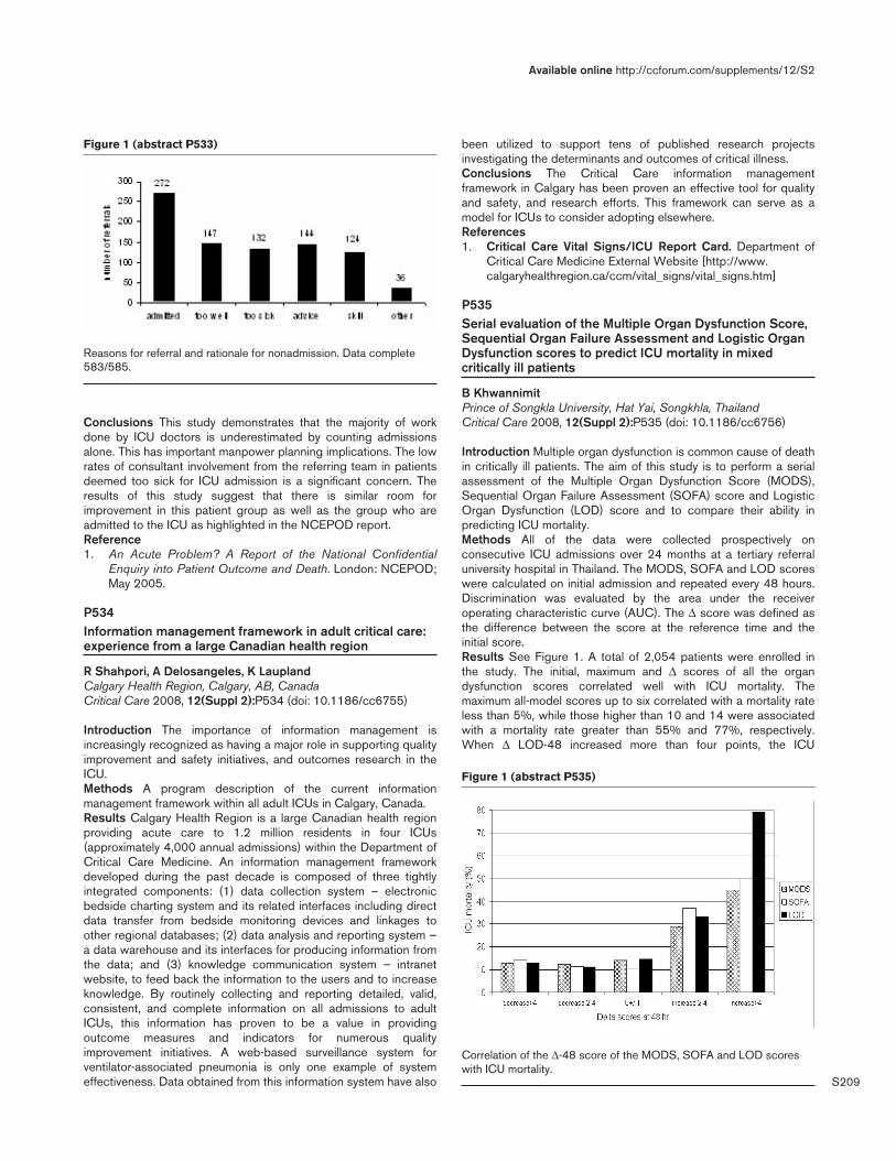
S209
Conclusions This study demonstrates that the majority of workdone by ICU doctors is underestimated by counting admissionsalone. This has important manpower planning implications. The lowrates of consultant involvement from the referring team in patientsdeemed too sick for ICU admission is a significant concern. Theresults of this study suggest that there is similar room forimprovement in this patient group as well as the group who areadmitted to the ICU as highlighted in the NCEPOD report.Reference1. An Acute Problem? A Report of the National Confidential
Enquiry into Patient Outcome and Death. London: NCEPOD;May 2005.
P534Information management framework in adult critical care:experience from a large Canadian health region
R Shahpori, A Delosangeles, K LauplandCalgary Health Region, Calgary, AB, CanadaCritical Care 2008, 12(Suppl 2):P534 (doi: 10.1186/cc6755)
Introduction The importance of information management isincreasingly recognized as having a major role in supporting qualityimprovement and safety initiatives, and outcomes research in theICU.Methods A program description of the current informationmanagement framework within all adult ICUs in Calgary, Canada.Results Calgary Health Region is a large Canadian health regionproviding acute care to 1.2 million residents in four ICUs(approximately 4,000 annual admissions) within the Department ofCritical Care Medicine. An information management frameworkdeveloped during the past decade is composed of three tightlyintegrated components: (1) data collection system – electronicbedside charting system and its related interfaces including directdata transfer from bedside monitoring devices and linkages toother regional databases; (2) data analysis and reporting system –a data warehouse and its interfaces for producing information fromthe data; and (3) knowledge communication system – intranetwebsite, to feed back the information to the users and to increaseknowledge. By routinely collecting and reporting detailed, valid,consistent, and complete information on all admissions to adultICUs, this information has proven to be a value in providingoutcome measures and indicators for numerous qualityimprovement initiatives. A web-based surveillance system forventilator-associated pneumonia is only one example of systemeffectiveness. Data obtained from this information system have also
been utilized to support tens of published research projectsinvestigating the determinants and outcomes of critical illness.Conclusions The Critical Care information managementframework in Calgary has been proven an effective tool for qualityand safety, and research efforts. This framework can serve as amodel for ICUs to consider adopting elsewhere.References1. Critical Care Vital Signs/ICU Report Card. Department of
Critical Care Medicine External Website [http://www.calgaryhealthregion.ca/ccm/vital_signs/vital_signs.htm]
P535Serial evaluation of the Multiple Organ Dysfunction Score,Sequential Organ Failure Assessment and Logistic OrganDysfunction scores to predict ICU mortality in mixedcritically ill patients
B KhwannimitPrince of Songkla University, Hat Yai, Songkhla, ThailandCritical Care 2008, 12(Suppl 2):P535 (doi: 10.1186/cc6756)
Introduction Multiple organ dysfunction is common cause of deathin critically ill patients. The aim of this study is to perform a serialassessment of the Multiple Organ Dysfunction Score (MODS),Sequential Organ Failure Assessment (SOFA) score and LogisticOrgan Dysfunction (LOD) score and to compare their ability inpredicting ICU mortality.Methods All of the data were collected prospectively onconsecutive ICU admissions over 24 months at a tertiary referraluniversity hospital in Thailand. The MODS, SOFA and LOD scoreswere calculated on initial admission and repeated every 48 hours.Discrimination was evaluated by the area under the receiveroperating characteristic curve (AUC). The Δ score was defined asthe difference between the score at the reference time and theinitial score.Results See Figure 1. A total of 2,054 patients were enrolled inthe study. The initial, maximum and Δ scores of all the organdysfunction scores correlated well with ICU mortality. Themaximum all-model scores up to six correlated with a mortality rateless than 5%, while those higher than 10 and 14 were associatedwith a mortality rate greater than 55% and 77%, respectively.When Δ LOD-48 increased more than four points, the ICU
Available online http://ccforum.com/supplements/12/S2
Figure 1 (abstract P533)
Reasons for referral and rationale for nonadmission. Data complete583/585.
Figure 1 (abstract P535)
Correlation of the Δ-48 score of the MODS, SOFA and LOD scoreswith ICU mortality.
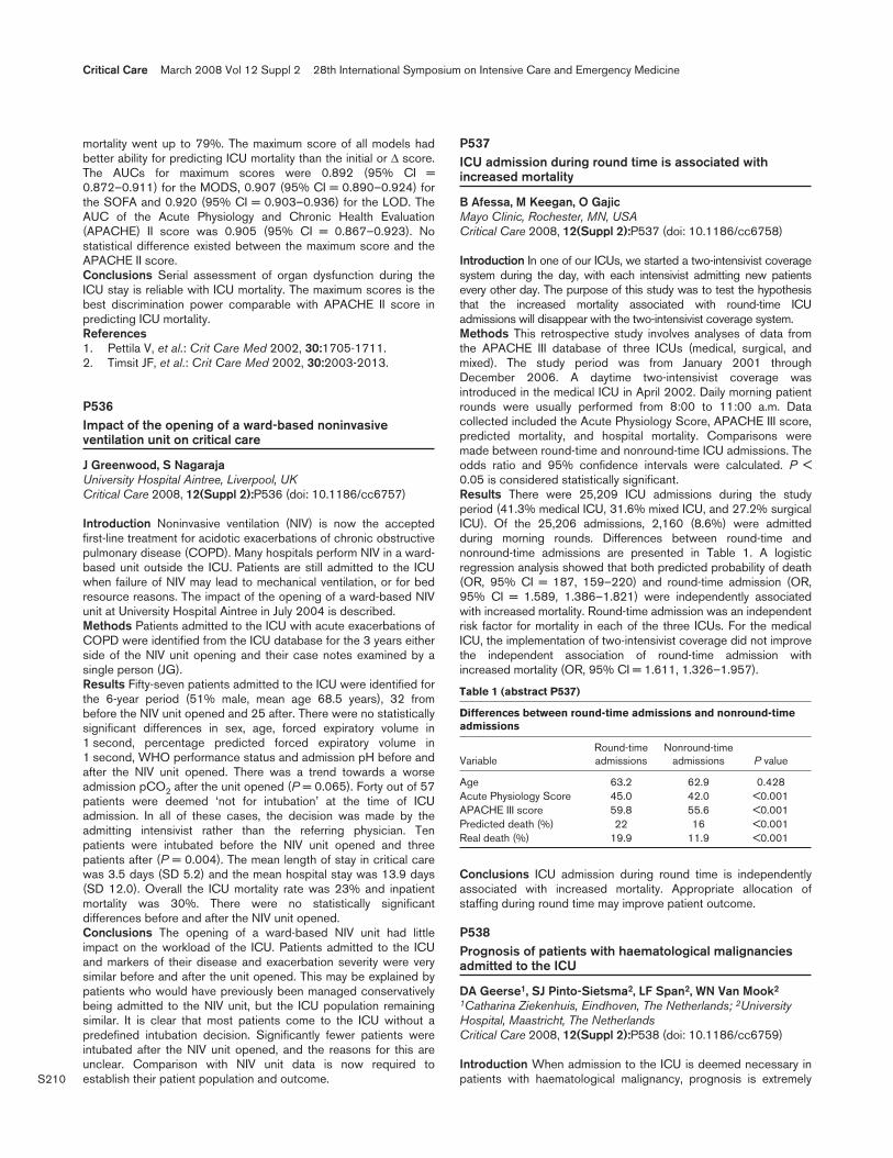
S210
mortality went up to 79%. The maximum score of all models hadbetter ability for predicting ICU mortality than the initial or Δ score.The AUCs for maximum scores were 0.892 (95% CI =0.872–0.911) for the MODS, 0.907 (95% CI = 0.890–0.924) forthe SOFA and 0.920 (95% CI = 0.903–0.936) for the LOD. TheAUC of the Acute Physiology and Chronic Health Evaluation(APACHE) II score was 0.905 (95% CI = 0.867–0.923). Nostatistical difference existed between the maximum score and theAPACHE II score.Conclusions Serial assessment of organ dysfunction during theICU stay is reliable with ICU mortality. The maximum scores is thebest discrimination power comparable with APACHE II score inpredicting ICU mortality.References1. Pettila V, et al.: Crit Care Med 2002, 30:1705-1711.2. Timsit JF, et al.: Crit Care Med 2002, 30:2003-2013.
P536Impact of the opening of a ward-based noninvasiveventilation unit on critical care
J Greenwood, S NagarajaUniversity Hospital Aintree, Liverpool, UKCritical Care 2008, 12(Suppl 2):P536 (doi: 10.1186/cc6757)
Introduction Noninvasive ventilation (NIV) is now the acceptedfirst-line treatment for acidotic exacerbations of chronic obstructivepulmonary disease (COPD). Many hospitals perform NIV in a ward-based unit outside the ICU. Patients are still admitted to the ICUwhen failure of NIV may lead to mechanical ventilation, or for bedresource reasons. The impact of the opening of a ward-based NIVunit at University Hospital Aintree in July 2004 is described.Methods Patients admitted to the ICU with acute exacerbations ofCOPD were identified from the ICU database for the 3 years eitherside of the NIV unit opening and their case notes examined by asingle person (JG).Results Fifty-seven patients admitted to the ICU were identified forthe 6-year period (51% male, mean age 68.5 years), 32 frombefore the NIV unit opened and 25 after. There were no statisticallysignificant differences in sex, age, forced expiratory volume in1 second, percentage predicted forced expiratory volume in1 second, WHO performance status and admission pH before andafter the NIV unit opened. There was a trend towards a worseadmission pCO2 after the unit opened (P = 0.065). Forty out of 57patients were deemed ‘not for intubation’ at the time of ICUadmission. In all of these cases, the decision was made by theadmitting intensivist rather than the referring physician. Tenpatients were intubated before the NIV unit opened and threepatients after (P = 0.004). The mean length of stay in critical carewas 3.5 days (SD 5.2) and the mean hospital stay was 13.9 days(SD 12.0). Overall the ICU mortality rate was 23% and inpatientmortality was 30%. There were no statistically significantdifferences before and after the NIV unit opened.Conclusions The opening of a ward-based NIV unit had littleimpact on the workload of the ICU. Patients admitted to the ICUand markers of their disease and exacerbation severity were verysimilar before and after the unit opened. This may be explained bypatients who would have previously been managed conservativelybeing admitted to the NIV unit, but the ICU population remainingsimilar. It is clear that most patients come to the ICU without apredefined intubation decision. Significantly fewer patients wereintubated after the NIV unit opened, and the reasons for this areunclear. Comparison with NIV unit data is now required toestablish their patient population and outcome.
P537ICU admission during round time is associated withincreased mortality
B Afessa, M Keegan, O GajicMayo Clinic, Rochester, MN, USACritical Care 2008, 12(Suppl 2):P537 (doi: 10.1186/cc6758)
Introduction In one of our ICUs, we started a two-intensivist coveragesystem during the day, with each intensivist admitting new patientsevery other day. The purpose of this study was to test the hypothesisthat the increased mortality associated with round-time ICUadmissions will disappear with the two-intensivist coverage system.Methods This retrospective study involves analyses of data fromthe APACHE III database of three ICUs (medical, surgical, andmixed). The study period was from January 2001 throughDecember 2006. A daytime two-intensivist coverage wasintroduced in the medical ICU in April 2002. Daily morning patientrounds were usually performed from 8:00 to 11:00 a.m. Datacollected included the Acute Physiology Score, APACHE III score,predicted mortality, and hospital mortality. Comparisons weremade between round-time and nonround-time ICU admissions. Theodds ratio and 95% confidence intervals were calculated. P <0.05 is considered statistically significant.Results There were 25,209 ICU admissions during the studyperiod (41.3% medical ICU, 31.6% mixed ICU, and 27.2% surgicalICU). Of the 25,206 admissions, 2,160 (8.6%) were admittedduring morning rounds. Differences between round-time andnonround-time admissions are presented in Table 1. A logisticregression analysis showed that both predicted probability of death(OR, 95% CI = 187, 159–220) and round-time admission (OR,95% CI = 1.589, 1.386–1.821) were independently associatedwith increased mortality. Round-time admission was an independentrisk factor for mortality in each of the three ICUs. For the medicalICU, the implementation of two-intensivist coverage did not improvethe independent association of round-time admission withincreased mortality (OR, 95% CI = 1.611, 1.326–1.957).
Table 1 (abstract P537)
Differences between round-time admissions and nonround-timeadmissions
Round-time Nonround-time Variable admissions admissions P value
Age 63.2 62.9 0.428Acute Physiology Score 45.0 42.0 <0.001APACHE III score 59.8 55.6 <0.001Predicted death (%) 22 16 <0.001Real death (%) 19.9 11.9 <0.001
Conclusions ICU admission during round time is independentlyassociated with increased mortality. Appropriate allocation ofstaffing during round time may improve patient outcome.
P538Prognosis of patients with haematological malignanciesadmitted to the ICU
DA Geerse1, SJ Pinto-Sietsma2, LF Span2, WN Van Mook2
1Catharina Ziekenhuis, Eindhoven, The Netherlands; 2UniversityHospital, Maastricht, The NetherlandsCritical Care 2008, 12(Suppl 2):P538 (doi: 10.1186/cc6759)
Introduction When admission to the ICU is deemed necessary inpatients with haematological malignancy, prognosis is extremely
Critical Care March 2008 Vol 12 Suppl 2 28th International Symposium on Intensive Care and Emergency Medicine
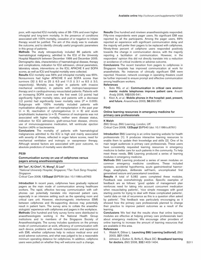
S211
poor, with reported ICU mortality rates of 38–73% and even higherinhospital and long-term mortality. In the presence of conditionsassociated with 100% mortality, cessation of or negation to initiatetherapy would be justified. The aim of this study was to evaluatethe outcome, and to identify clinically useful prognostic parametersin this group of patients.Methods The study retrospectively included 86 patients withhaematological malignancy, admitted to the ICU of the UniversityHospital Maastricht between December 1999 and December 2005.Demographic data, characteristics of haematological disease, therapyand complications, indication for ICU admission, clinical parameters,laboratory values, interventions on the ICU, APACHE II and SOFAscores, as well as ICU and hospital mortality were collected.Results ICU mortality was 56% and inhospital mortality was 65%.Nonsurvivors had higher APACHE II and SOFA scores thansurvivors (32 ± 8.0 vs 25 ± 6.5 and 11.5 ± 3.1 vs 8.5 ± 3.0,respectively). Mortality was higher in patients with invasivemechanical ventilation, in patients with inotropic/vasopressortherapy and in cardiopulmonary resuscitated patients. Patients withan increasing SOFA score over the first week (≥2 points) hadsignificantly higher mortality rates, and patients with a decrease(≥2 points) had significantly lower mortality rates (P = 0.005).Subgroups with 100% mortality included patients withmyeloablative allogeneic stem cell transplantation in the past year(n = 9), reactivation of cytomegalovirus (n = 6) and acutelymphoblastic leukaemia (n = 5). Presence of neutropenia was notassociated with higher mortality, neither were disease status,indication for ICU admission, graft-versus-host disease, chronicuse of immunosuppressive medication, left ventricular ejectionfraction ≤40% or bacteraemia.Conclusions The mortality of patients with haematologicalmalignancies admitted to the ICU is high and mainly associatedwith severity of illness, reflected in organ failure scores, need formechanical ventilation and inotropic or vasopressor therapy.Although several factors are associated with poor outcome, noabsolute predictors of mortality were identified.
P539Communication survey on use of cellphones versuspagers among anesthesiologists
BH Tan1, KJ Chin2, YL Wong2, S Loo2
1National University Hospital, Singapore; 2Tan Tock Seng Hospital,SingaporeCritical Care 2008, 12(Suppl 2):P539 (doi: 10.1186/cc6760)
Introduction In recent years, cellphones have gradually replacedpagers as the main mode of communication among healthcareworkers. The rapid, effective two-way communication with cell-phones can potentially translate into improved patient care,especially in an intensive setting such as the operating room andcritical care unit. However, electromagnetic interference (EMI)between cellphones and life-supporting devices may potentiallyresult in patient harm. The survey aims to collate the anaesthe-siologists’ experience with cellphones and pagers in the workplace.Methods One hundred and forty survey forms were distributed toanaesthesiologists working in the National Health Groupinstitutions and to members of the Singapore Society ofAnaesthesiologists. The survey includes questions on the primarymode of communication at work, efficiency of communication witheach device, problems with network transmission and experiencewith EMI, whether cellphones help to reduce medical error andavoid adverse outcomes, and what was judged to be a reasonableminimum operating distance for cellphones. In addition, cellphoneusers were polled on whether they will welcome such a change.
Results One hundred and nineteen anaesthesiologists responded.Fifty-nine respondents were pager users. No significant EMI wasreported by all the participants. Among the pager users, 69%reported an experience with significant communication delay andthe majority will prefer their pagers to be replaced with cellphones.Ninety-three percent of cellphone users responded positivelytowards the change in communication device, with the majorityreporting a facilitation of communication. However, in theresponders’ opinion this did not directly translate into a reductionor avoidance of critical incidents or adverse outcome.Conclusions The recent transition from pagers to cellphones inSingapore hospitals has improved communication at work foranaesthetists. No instances of clinically significant EMI werereported. However, network coverage in operating theaters couldbe further improved to ensure prompt and effective communicationamong healthcare workers.References1. Soto RG, et al.: Communication in critical care environ-
ments: mobile telephones improve patient care. AnesthAnalg 2006, 102:535-541.
2. Klein A, et al.: Mobile phones in the hospital: past, present,and future. Anaesthesia 2003, 58:353-357.
P540Online learning resources in emergency medicine forprimary care professionals
K WalshBMJ Group, BMJ Learning, London, UKCritical Care 2008, 12(Suppl 2):P540 (doi: 10.1186/cc6761)
Introduction BMJ Learning is an online learning website for healthprofessionals [1]. It produces interactive learning resources toenable them to update their knowledge and skills [2]. One of itsmain target audiences is primary care professionals. These usershave consistently requested learning resources in emergencymedicine to better care for such patients in the community. To helpmeet these needs, BMJ Learning produced a series of learningmodules in emergency medicine.Methods BMJ Learning produced a series of seven modules oncommon emergency medicine conditions. These includedepistaxis, accidental hypothermia, acute gastrointestinal haemor-rhage, anaphylaxis, deliberate self-harm, uncomplicated firstgeneralised seizure and paracetamol overdose.Results A total of 5,082 users completed these modules.Feedback was overwhelmingly positive. Specific examples offeedback are as follows: ‘good update of management planreinforces need for taking into account concurrent medicationwhen resuscitating patients’, ‘nice simple messages with goodstarting points for trying to deal with these complicated patients’,‘useful data on risk of recurrence as this is a question often askedby patients’. This feedback was particularly encouraging as itshowed how the primary care professionals planned to changetheir practice to improve patient outcomes as a result of thelearning.Conclusions We feel that the results show that online learningmodules are effective at helping primary care professionals learnabout emergency medicine. We encourage other providers ofonline learning to increase the amount of learning resources thatare available in this area.References1. Walsh K, Dillner L: Launching BMJ Learning [editorial]. BMJ
2003, 327:1064.2. Johnson J, Dutton S, Briffa E, Black DC: Broadband learning
for doctors. BMJ 2006, 332:1403-1404.
Available online http://ccforum.com/supplements/12/S2
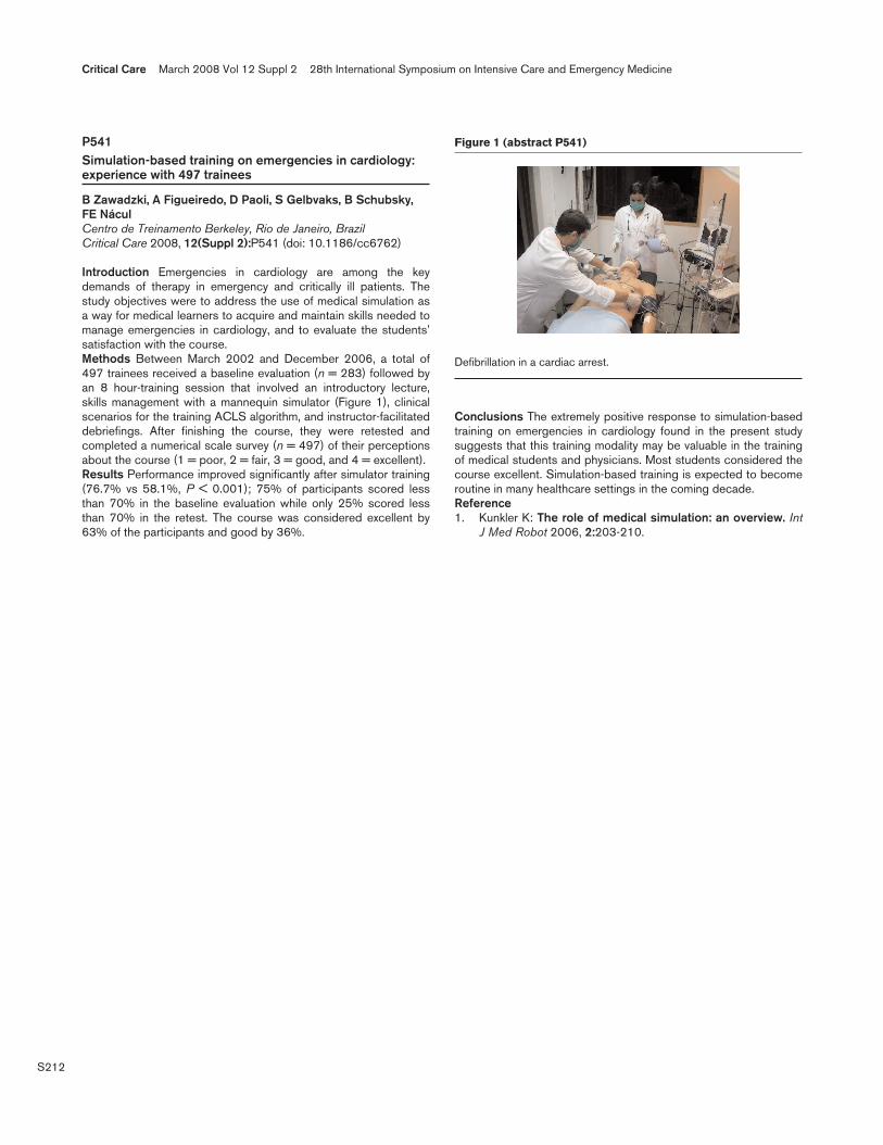
S212
P541Simulation-based training on emergencies in cardiology:experience with 497 trainees
B Zawadzki, A Figueiredo, D Paoli, S Gelbvaks, B Schubsky,FE NáculCentro de Treinamento Berkeley, Rio de Janeiro, BrazilCritical Care 2008, 12(Suppl 2):P541 (doi: 10.1186/cc6762)
Introduction Emergencies in cardiology are among the keydemands of therapy in emergency and critically ill patients. Thestudy objectives were to address the use of medical simulation asa way for medical learners to acquire and maintain skills needed tomanage emergencies in cardiology, and to evaluate the students’satisfaction with the course.Methods Between March 2002 and December 2006, a total of497 trainees received a baseline evaluation (n = 283) followed byan 8 hour-training session that involved an introductory lecture,skills management with a mannequin simulator (Figure 1), clinicalscenarios for the training ACLS algorithm, and instructor-facilitateddebriefings. After finishing the course, they were retested andcompleted a numerical scale survey (n = 497) of their perceptionsabout the course (1 = poor, 2 = fair, 3 = good, and 4 = excellent).Results Performance improved significantly after simulator training(76.7% vs 58.1%, P < 0.001); 75% of participants scored lessthan 70% in the baseline evaluation while only 25% scored lessthan 70% in the retest. The course was considered excellent by63% of the participants and good by 36%.
Conclusions The extremely positive response to simulation-basedtraining on emergencies in cardiology found in the present studysuggests that this training modality may be valuable in the trainingof medical students and physicians. Most students considered thecourse excellent. Simulation-based training is expected to becomeroutine in many healthcare settings in the coming decade.Reference1. Kunkler K: The role of medical simulation: an overview. Int
J Med Robot 2006, 2:203-210.
Critical Care March 2008 Vol 12 Suppl 2 28th International Symposium on Intensive Care and Emergency Medicine
Figure 1 (abstract P541)
Defibrillation in a cardiac arrest.
Related Documents