Effects of the endocrine disrupter chemicals chlordane and lindane on the male green neon shrimp (Neocaridina denticulata) Da-Ji Huang a , Shu-Yin Wang b , Hon-Cheng Chen a,c, * a Institute of Zoology, National Taiwan University, No. 1, Sec. 4, Roosevelt Road, Taipei 106, Taiwan, Province of China b Department of Animal Science, Chinese Culture University, Yang Ming Shan, Taiwan 111, Province of China c Institute of Fisheries Sciences, National Taiwan University, Taipei 106, Taiwan, Province of China Received 14 October 2003; received in revised form 11 August 2004; accepted 23 August 2004 Abstract The purpose of this study was to investigate the effect of chlordane and lindane on morphological changes and endo- crine disruption in male Green neon shrimp (Neocaridina denticulata). In this experiment, individuals of N. denticulata, a common inhabitant of freshwater systems in Taiwan, were exposed to chlordane (1 and 10 ng/l) and lindane (0.1 and 1 lg/l). Morphological changes and reproductive hormone levels were observed following four weeks exposure. Accord- ing to our findings, an increase in estrogen, a reduction in testosterone, and morphological alternations of the masculine appendage were observed in both chlordane- and lindane-treated shrimp, while induction of a vitellogenin-like protein appeared only in shrimp treated with 10 ng/l chlordane. An endocrine disruption effect on N. denticulata was demon- strated, and may apply to other organochlorine pesticides or endocrine disruption chemicals. Ó 2004 Elsevier Ltd. All rights reserved. Keywords: Endocrine disruption chemicals; Chlordane; Lindane; Neocaridina denticulata 1. Introduction Chlordane and lindane are both organochlorine pes- ticides (OCPs), are widely distributed contaminants in environments such as aquatic systems, and act as endo- crine disruption chemicals (EDCs) to many organisms. OCPs are some of the most dangerous pesticides because of their toxicity, stability, high liposolubility, and long biological half-life. OCPs can exhibit a high degree of bioaccumulation and biomagnification within food chains, and are known to have carcinogenic, teratogenic, and endocrine-disruptive effects in humans and wildlife. Previous studies showed that chlordane may perform complex and variable biological functions such as estro- gen-like functions, while lindane has estrogenic-like ef- fects on some organisms, but anti-estrogenic effects on others. Chlordane and lindane have been shown to have similar symptoms to EDCs. In animal studies, these compounds were shown to exhibit an estrogenic-like effect, and to cause detrimental effects to reproductive 0045-6535/$ - see front matter Ó 2004 Elsevier Ltd. All rights reserved. doi:10.1016/j.chemosphere.2004.08.063 * Corresponding author. Address: Institute of Zoology, National Taiwan University, No. 1, Sec. 4, Roosevelt Road, Taipei 106, Taiwan, Province of China. Tel.: +886 2 2363 0231x3324; fax: +886 2 2363 6837. E-mail addresses: [email protected] (D.-J. Huang), [email protected] (S.-Y. Wang), honcheng@ccms. ntu.edu.tw (H.-C. Chen). Chemosphere 57 (2004) 1621–1627 www.elsevier.com/locate/chemosphere

Welcome message from author
This document is posted to help you gain knowledge. Please leave a comment to let me know what you think about it! Share it to your friends and learn new things together.
Transcript

Chemosphere 57 (2004) 1621–1627
www.elsevier.com/locate/chemosphere
Effects of the endocrine disrupter chemicals chlordaneand lindane on the male green neon shrimp
(Neocaridina denticulata)
Da-Ji Huang a, Shu-Yin Wang b, Hon-Cheng Chen a,c,*
a Institute of Zoology, National Taiwan University, No. 1, Sec. 4, Roosevelt Road, Taipei 106, Taiwan, Province of Chinab Department of Animal Science, Chinese Culture University, Yang Ming Shan, Taiwan 111, Province of China
c Institute of Fisheries Sciences, National Taiwan University, Taipei 106, Taiwan, Province of China
Received 14 October 2003; received in revised form 11 August 2004; accepted 23 August 2004
Abstract
The purpose of this study was to investigate the effect of chlordane and lindane on morphological changes and endo-
crine disruption in male Green neon shrimp (Neocaridina denticulata). In this experiment, individuals of N. denticulata,
a common inhabitant of freshwater systems in Taiwan, were exposed to chlordane (1 and 10ng/l) and lindane (0.1 and
1lg/l). Morphological changes and reproductive hormone levels were observed following four weeks exposure. Accord-
ing to our findings, an increase in estrogen, a reduction in testosterone, and morphological alternations of the masculine
appendage were observed in both chlordane- and lindane-treated shrimp, while induction of a vitellogenin-like protein
appeared only in shrimp treated with 10ng/l chlordane. An endocrine disruption effect on N. denticulata was demon-
strated, and may apply to other organochlorine pesticides or endocrine disruption chemicals.
� 2004 Elsevier Ltd. All rights reserved.
Keywords: Endocrine disruption chemicals; Chlordane; Lindane; Neocaridina denticulata
1. Introduction
Chlordane and lindane are both organochlorine pes-
ticides (OCPs), are widely distributed contaminants in
environments such as aquatic systems, and act as endo-
crine disruption chemicals (EDCs) to many organisms.
0045-6535/$ - see front matter � 2004 Elsevier Ltd. All rights reserv
doi:10.1016/j.chemosphere.2004.08.063
* Corresponding author. Address: Institute of Zoology,
National Taiwan University, No. 1, Sec. 4, Roosevelt Road,
Taipei 106, Taiwan, Province of China. Tel.: +886 2 2363
0231x3324; fax: +886 2 2363 6837.
E-mail addresses: [email protected] (D.-J. Huang),
[email protected] (S.-Y. Wang), honcheng@ccms.
ntu.edu.tw (H.-C. Chen).
OCPs are some of the most dangerous pesticides because
of their toxicity, stability, high liposolubility, and long
biological half-life. OCPs can exhibit a high degree of
bioaccumulation and biomagnification within food
chains, and are known to have carcinogenic, teratogenic,
and endocrine-disruptive effects in humans and wildlife.
Previous studies showed that chlordane may perform
complex and variable biological functions such as estro-
gen-like functions, while lindane has estrogenic-like ef-
fects on some organisms, but anti-estrogenic effects on
others. Chlordane and lindane have been shown to have
similar symptoms to EDCs. In animal studies, these
compounds were shown to exhibit an estrogenic-like
effect, and to cause detrimental effects to reproductive
ed.

1622 D.-J. Huang et al. / Chemosphere 57 (2004) 1621–1627
systems (Colborn et al., 1993). Although many develop-
ing and developed countries have prohibited the use of
these compounds for many years, OCPs are consistently
detected in ecosystems (Chen et al., 1999). Therefore, the
influence of OCPs residues on the development of wild-
life has become a major concern.
Several reports have indicated that changes in the
reproductive system and morphology of wildlife take
place after exposure to xenoestrogen (synthetic, indus-
try-derived estrogenic compounds) (Colborn et al.,
1993; Sharara et al., 1998). Crustaceans are frequently
used as a target animal in aquatic systems to examine
endocrine disruptor chemicals (Depledge and Billing-
hurst, 1999; Hutchinson, 2002). The physiological role
of the ‘‘vertebrate type’’ steroid hormones, testosterone,
and estrogen, has been identified in the shrimp (Quinitio
et al., 1991; Cardoso et al., 1997). Treatment with natural
vertebrate estrogen was reported to induce maturation in
crustaceans (Rinderhagen et al., 2000). Several reports
showed that the natural hormone and xenoestrogen
could effect sexual development and sexual hormone reg-
ulation (Colborn et al., 1993; Sharara et al., 1998). Huang
and Chen (2004) demonstrated that chlordane and lin-
dane cause an alternation in testosterone and vitellogenin
(Vg) levels in juvenile shrimp (N. denticulata).
Atyid shrimp are common inhabitants of freshwater
systems. In Taiwan, 13 species of atyid shrimp belonging
to three genera have been described, among which, Neo-
caridina denticulata is the only one belonging to the
genus Neocaridina (Hung et al., 1993). This species is
commonly and extensively distributed in many streams,
ponds, swamps, and rivers in East Asia and the Hawai-
ian islands (Hung et al., 1993; Englund and Cai, 1999).
Thus, N. denticulata is one of the most commonly used
aquatic organisms for environmentally related studies
of freshwater systems (Chen et al., 1999). However, in
Taiwan, the quantity of N. denticulata in natural habi-
tats is on the decline due to human activities, including
pollution, destruction of natural habitats, and overfish-
ing (Shy and Yu, 1998).
Despite having a common distribution, only a few at-
tempts have been made to determine the impacts of
chlordane and lindane in freshwater on N. denticulata.
The purpose of this study was to use the length of the
masculine appendage, and levels of vitellogenin, estra-
diol, and testosterone as indicators to reflect alterations
in morphology and hormone regulation in male N. den-
ticulata after chlordane and lindane exposure.
2. Materials and methods
2.1. Shrimp collection and maintenance
Green neon shrimp (N. denticulata) were taken from
rivers in Taipei County, northern Taiwan for laboratory
testing. They were transferred to a 50-l glass aquarium
after being identified. This aquarium was equipped with
a water-cycling device, pH was maintained at 7.4–7.8,
dissolved oxygen concentration exceeded 7.3mg/l, and
hardness was 38–45mgCaCO3/l. Temperature was
maintained at 25 ± 1 �C, and a 12-h light–dark photope-
riod was used. Under these conditions, shrimp were fed
twice a day and were allowed to acclimate for two weeks
before testing.
2.2. Exposure of shrimp to 17b-estradiol, chlordane, andlindane
Ninety-six hour LC50 levels for chlordane and lin-
dane for N. denticulata are 127.03ng/l and 9.36lg/l,respectively (Huang and Chen, 2004). Sublethal concen-
trations of chlordane and lindane were 1, 10ng/l, and
0.1, 1lg/l, respectively. Experiments with positive con-
trols (10 and 100lg/l 17b-estradiol in alcohol) with no
pesticide added were also carried out, as well as those
using a vehicle control group in which only acetone
was added. In total, there were 150 male shrimp (body
length, 13 � 16mm) of the same size in each group (in
a 10-l glass beaker, run in triplicate for each). Males
were early identified as the endopod of the second pleo-
pods has a masculine appendage (Fig. 1a) that is oval-
shaped and is surrounded by cilia (Hung et al., 1993;
Shy and Yu, 1998; Englund and Cai, 1999). Samples
were taken at the end of days 1, 3, 7, 14, and 28.
2.3. Morphological study
In the morphological study, we observed the mor-
phology of the masculine appendage after 3, 7, 14, and
28 days exposure. We also measured the length of the
masculine appendage on the second pleopods (Fig. 1b)
and the length of the cephalothorax. Data shown in-
clude the ratio of the lengths of the masculine appendage
and cephalothorax.
2.4. Testing preparation
Shrimp samples were homogenized with a Teflon pes-
tle (Kontes, Vineland, NJ, USA) in ice-cold 25mM Tris–
HCl with EDTA. The homogenate was centrifuged at
10000·g (for 20min at 4 �C), and the supernatant was
collected and stored at �20 �C until analysis.
2.5. Determination of hemolymph estradiol
Levels of estradiol were determined using an EIA kit
(DSLabs, Webster, TX, USA). The assay uses the
competitive binding enzyme immunoassay format
(Maxey et al., 1992). In the assay, standards, controls,
and unknowns containing estradiol are incubated with
biotin-labeled estradiol and rabbit anti-estradiol anti-

Fig. 1. The major characteristic of male Neocaridina denticulata in the control and treated group (100lg/l 17b-estradiol). (a)
Appearance of an oval-like endopod (O) on the first pleopod and masculine appendage. (A) Endopod of second pleopods with cilia
(bar = 1mm). (b) ‘‘d’’ indicates the length of the masculine appendage on the second pleopods. (c) Second pleopods of the control
groups (d = 0.9mm, cephalothorax = 5.38mm), and (d) second pleopods of the treated group (100lg/l 17b-estradiol) on day 7
(d = 0.65mm, cephalothorax = 5.4mm) (bar = 1mm).
D.-J. Huang et al. / Chemosphere 57 (2004) 1621–1627 1623
serum in microtitration wells where the unlabeled and
biotin-labeled antigens compete for a limited number
of anti-estradiol binding sites. After incubation and
washing, the wells are incubated with streptavidin-
HRPO, which binds to the biotinylated estradiol. The
unbound streptavidin-HRPO is washed, followed by
incubation with the substrate tetramethylbenzidine
(TMB). An acidic stopping solution is then added, and
the degree of enzymatic turnover of the substrate is
determined by dual wavelength absorbance measure-
ments at 450 and 620nm.
2.6. Determination of hemolymph testosterone
Levels of testosterone were determined using an EIA
kit (Cayman, Ann Arbor, MI, USA). This assay is based
on the competition between testosterone and a testoster-
one-acetylcholinesterase (AChE) conjugate (testosterone
tracer) for a limited number of testosterone-specific rab-
bit anti-serum binding sites (Pradelles et al., 1985;
Maxey et al., 1992). The product of this enzymatic reac-
tion has a distinct yellow color and a strong peak
absorbance at 405nm by spectrophotometry.
2.7. Determination of hemolymph vitellogenin
The level of vitellogenin-like protein in the hemo-
lymph was mediately determined by using an alkali-la-
bile phosphate (ALP) measurement assay, and the
quantity of ALP in the hemolymph was obtained similar
to the method of Gange and Blaise (2000). Shrimp
hemolymph (in 5 and 45ll of Tris–HCl buffer, 25mM,

1624 D.-J. Huang et al. / Chemosphere 57 (2004) 1621–1627
pH 7.5) was mixed with 50ll of ice-cold 20% trichloro-
acetic acid (TCA) and incubated at room temperature
for 15min. The mixture was centrifuged at 10000·gfor 10min at 4 �C. The protein pellet was resuspended
in 200ll 1M NaOH and then heated to 75 �C for
60min before determination. The level of free phos-
phates was determined according to the phosphomolyb-
denum method, and the optical absorbance was read at
600nm (Ellman et al., 1961; Gange and Blaise, 2000).
2.8. Statistical analysis
Statistical analysis used MicrocalTM origin 6.0.
(Northampton, MA, USA, 1999). Experimental and
control values were compared using Student�s t-test
(paired assay, p < 0.05).
3. Results
3.1. Levels of estradiol and testosterone in hemolymph
In our present study, high levels of estradiol were
detected after males of N. denticulata treated with 17b-estradiol, chlordane, and lindane (p < 0.01) (Fig. 2).
0 5 10 15 20 25 300
1
2
3
ng/g
Day
Lindane 0.1µg/l Lindane 1µg/l
0 5 10 15 20 25 300
1
2
3
ng/g
Chlordane 1ng/l Chlordane 10ng/l
0 5 10 15 20 25 300
36
9
12
ng/g
Control 17β-Estradiol 10µg/l 17β-Estradiol 100µg/l
Fig. 2. Hemolymph estradiol response levels in male Neocar-
idina denticulata exposed to 17b-estradiol, chlordane, and
lindane for 1, 3, 7, 14, and 28 days (mean ± SD, n = 15).
It was also interesting that the levels of testosterone
in the hemolymph of 17b-estradiol-, chlordane-, and
lindane-treated N. denticulata showed lower values
compared with those of control shrimp, especially after
14 and 28 days of exposure (p < 0.05) (Fig. 3). The
R2 of standard curves in estradiol and testosterone as-
says were higher than 0.99; standard check and relative
percent difference were less than 10% and 5%,
respectively.
3.2. Vitellogenin levels in hemolymph
Treatment with 17b-estradiol, chlordane, and lindane
changed the levels of vitellogenin (Vg) in the hemo-
lymph, as determined by ALP assays. Vg levels in male
N. denticulata hemolymph were induced by treatment
with 100lg/l 17b-estradiol on days 14 and 28,
(p < 0.05), while 10lg/l 17b-estradiol caused a signifi-
cant induction of Vg levels only after 28 days of expo-
sure (p < 0.05). Vg was induced in male N. denticulata
treated with 10ng/l chlordane for 14 and 28 days. There
were no significant differences between treatment groups
(0.1 and 1lg/l lindane, and 1ng/l chlordane) and the
control group (Fig. 4). The R2 of standard curve in
vitellogenin assay was 0.9965 (>0.99); standard check
0 5 10 15 20 25 300
250
500
750
1000
*
**
**
**
pg/g
Day
Control Lindane 0.1µg/l Lindane 1µg/l
0 5 10 15 20 25 300
250
500
750
1000
*
**
**
*
pg/g
Control Chlordane 1ng/l Chlordane 10ng/l
0 5 10 15 20 25 300
250
500
750
1000
* ***
**
pg/g
Control 17β-Estradiol 10µg/l 17β-Estradiol 100µg/l
Fig. 3. Hemolymph testosterone response levels in male Neo-
caridina denticulata exposed to 17b-estradiol, chlordane, andlindane for 1, 3, 7, 14, and 28 days (mean ± SD, n = 15). An
asterisk ( ) indicates that the difference between the experi-
mental and control group was significant at p < 0.05.
0 5 10 15 20 25 30
0
2
4
Day
Lindane 0.1 µ g/l Lindane 1 µ g/l
0
2
4
*
*
Alk
ali-
labi
le p
hosp
hate
rat
io
(tre
ated
/con
trol
) Chlordane 1ng/l Chlordane 10ng /l
0
2
4
*
** 17β-Estradiol 10µg/l
17β-Estradiol 100µg/l
Fig. 4. Hemolymph APL response levels in male Neocaridina
denticulata exposed to 17b-estradiol, chlordane, and lindane for
1, 3, 7, 14, and 28 days (mean ± SD, n = 15). An asterisk ( )
indicates that a difference between the experimental and control
group was significant at p < 0.05.
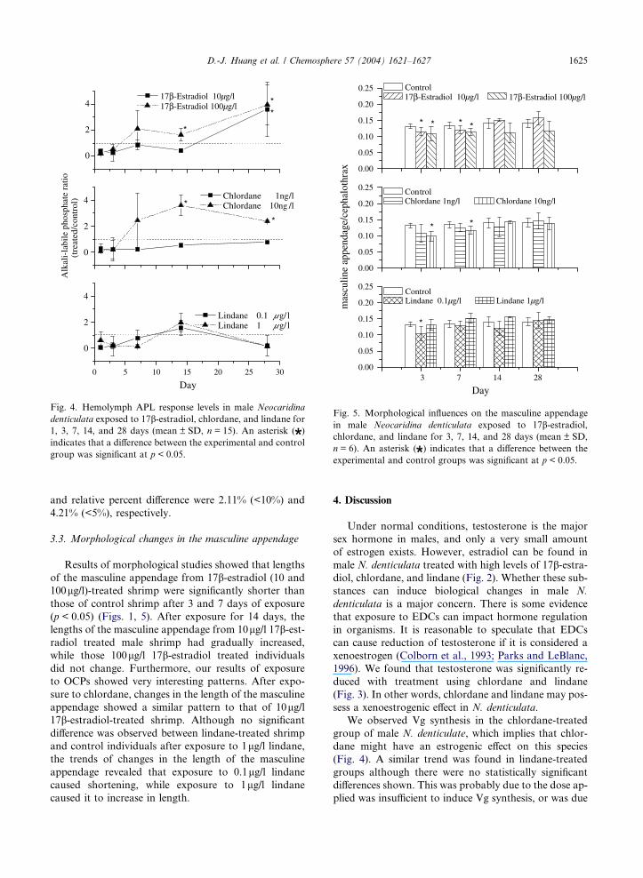
0.00
0.05
0.10
0.15
0.20
0.25
*
3 7 14 28
Day
Control Lindane 0.1µg/l Lindane 1µg/l
0.00
0.05
0.10
0.15
0.20
0.25
**
mas
culin
e ap
pend
age/
ceph
alot
hrax
Control Chlordane 1ng/l Chlordane 10ng/l
0.00
0.05
0.10
0.15
0.20
0.25
****
Control 17β-Estradiol 10µg/l 17β-Estradiol 100µg/l
Fig. 5. Morphological influences on the masculine appendage
in male Neocaridina denticulata exposed to 17b-estradiol,chlordane, and lindane for 3, 7, 14, and 28 days (mean ± SD,
n = 6). An asterisk ( ) indicates that a difference between the
experimental and control groups was significant at p < 0.05.
D.-J. Huang et al. / Chemosphere 57 (2004) 1621–1627 1625
and relative percent difference were 2.11% (<10%) and
4.21% (<5%), respectively.
3.3. Morphological changes in the masculine appendage
Results of morphological studies showed that lengths
of the masculine appendage from 17b-estradiol (10 and
100lg/l)-treated shrimp were significantly shorter than
those of control shrimp after 3 and 7 days of exposure
(p < 0.05) (Figs. 1, 5). After exposure for 14 days, the
lengths of the masculine appendage from 10lg/l 17b-est-radiol treated male shrimp had gradually increased,
while those 100lg/l 17b-estradiol treated individuals
did not change. Furthermore, our results of exposure
to OCPs showed very interesting patterns. After expo-
sure to chlordane, changes in the length of the masculine
appendage showed a similar pattern to that of 10lg/l17b-estradiol-treated shrimp. Although no significant
difference was observed between lindane-treated shrimp
and control individuals after exposure to 1lg/l lindane,the trends of changes in the length of the masculine
appendage revealed that exposure to 0.1lg/l lindane
caused shortening, while exposure to 1lg/l lindane
caused it to increase in length.
4. Discussion
Under normal conditions, testosterone is the major
sex hormone in males, and only a very small amount
of estrogen exists. However, estradiol can be found in
male N. denticulata treated with high levels of 17b-estra-diol, chlordane, and lindane (Fig. 2). Whether these sub-
stances can induce biological changes in male N.
denticulata is a major concern. There is some evidence
that exposure to EDCs can impact hormone regulation
in organisms. It is reasonable to speculate that EDCs
can cause reduction of testosterone if it is considered a
xenoestrogen (Colborn et al., 1993; Parks and LeBlanc,
1996). We found that testosterone was significantly re-
duced with treatment using chlordane and lindane
(Fig. 3). In other words, chlordane and lindane may pos-
sess a xenoestrogenic effect in N. denticulata.
We observed Vg synthesis in the chlordane-treated
group of male N. denticulate, which implies that chlor-
dane might have an estrogenic effect on this species
(Fig. 4). A similar trend was found in lindane-treated
groups although there were no statistically significant
differences shown. This was probably due to the dose ap-
plied was insufficient to induce Vg synthesis, or was due

1626 D.-J. Huang et al. / Chemosphere 57 (2004) 1621–1627
to the lack of an effect on the estrogen receptor of N.
denticulata (Fig. 5). It has been shown that some Vg-like
proteins found in crustaceans are used as precursors of
vitellin (Vazquez Boucard et al., 2002). However, unlike
fish, whose Vg synthesis occurs in the liver, synthesis has
been observed in the hepatopancreas and ovaries in
crustaceans (Tseng et al., 2001; Tsang et al., 2003). Un-
der normal circumstances, a mature female organism
usually shows a high level of estrogen, which can induce
Vg synthesis, while a male or immature individual shows
a much lower level of estrogen, which cannot trigger Vg
synthesis. However, the circumstance changes when the
male or immature organisms are exposed to a xenoestro-
gen. Vg synthesis can possibly be induced via the bind-
ing of the xenoestrogen to estrogen receptors (Kime
et al., 1999).
Chlordane, lindane, and OCPs can cause estrogenic,
anti-estrogenic, and anti-androgenic effects (Danzo,
1998). These effects may be caused by direct binding to
estrogen and androgen receptors and the effect on the
activities of sex hormone-metabolizing enzymes, such
as DDT and DDE (Colborn et al., 1993; Danzo,
1998). Chlordane may reveal complex and variable bio-
logical functions as does estrogen (Cranmer et al., 1984;
Cassidy et al., 1994). These include the findings of Vg
synthesis, an increase in estrogen, and a reduction in tes-
tosterone on N. denticulata. Lindane has been shown to
inhibit the cholesterol side-chain cleavage in mice, and
may have both estrogenic (Lahiri et al., 1985) and
anti-estrogenic effects (Chadwick et al., 1988; Cooper
et al., 1989). Although lindane does not appear to di-
rectly alter the number and affinity of estrogen receptors
(Laws et al., 1994), it might be able to compete with or
affect the binding of estrogen to the receptors. Although
Vg synthesis was not found in lindane-treated male N.
denticulata in this study, the observed increased level
of estrogen and decreased level of testosterone indicate
that lindane may produce hormonal disorders in male
N. denticulata.
Alteration in steroid hormone metabolism by EDCs
can significantly affect steroid hormone-dependent proc-
esses, such as growth, reproduction, the sex ratio, mor-
phology, and in some cases, a drop in the production
of viable offspring (Colborn et al., 1993). Chlordane
and lindane caused changes in hormone levels in male
N. denticulata. Although we cannot definitely be certain
whether such changes would handicap the reproduction
of N. denticulate, the masculine appendage, a male sex-
ual characteristic, was affected (Fig. 4). In some aquatic
organisms, sexual characteristics are changed after expo-
sure to EDCs (Colborn et al., 1993; Taylor and Harri-
son, 1999). For example, a reduction in the length of
the phallus of male alligators was documented after
exposure to OCPs (DDT, DDE, and DDD) in the Lake
Apopka, FL, USA (Crain and Guillette, 1998; Taylor
and Harrison, 1999). A similar intersexual result was
observed in fish exposed to sewage effluent containing
estrogenic activity (Denton et al., 1985; Jobling et al.,
1996; Harshbarger et al., 2000). Nucella lapillus showed
imposex with the development of a penis and vas defe-
rens in females after exposure to tributyltin (TBT)
(Gibbs et al., 1988). Studies on the biological effects of
EDCs have made important contributions to elucidating
some of the basic events in pathophysiology, which is a
key element for risk assessment of EDCs (Lebel et al.,
1998; Mantovani et al., 1999; van Wezel et al., 2000).
In this study, it was obvious that the masculine append-
age had changed after 3 and 7 days of exposure to 17b-estradiol, chlordane, and lindane. Furthermore, chlor-
dane and lindane caused alterations in the structure of
male characteristics of N. denticulata within a shorter
time than in the other organisms mentioned above.
Using the structure of the masculine appendage as an
element in risk assessment is a simple and time-saving
task. However, its reference requires further evaluation.
References
Cardoso, A.M., Barros, C.M.F., Correia, A.J.F., 1997. Iden-
tification of vertebrate type steroid hormones in the shrimp
Penaeus japonicus by tandem mass spectrometry and
sequential product ion scanning. J. Am. Soc. Mass Spec-
trom. 8, 365–370.
Cassidy, R.A., Vorhees, C.V., Minnema, D.J., 1994. The effects
of chlordane exposure during pre- and postnatal periods at
environmentally relevant levels on sex steroid-mediated
behaviors and functions in the rat. Toxicol. Appl. Pharma-
col. 126, 326–337.
Chadwick, R.W., Cooper, R.L., Chang, J., Rehnberg,
G.L., McElory, W.K., 1988. Possible antiestrogenic
activity of lindane in female rats. J. Biochem. Toxicol.
3, 147–158.
Chen, H.C., Wang, Y.S., Yaun, J.H., 1999. Standard methods
to study and detect bioaccumulation in fish and shellfish
(EPA-88-1502-03-01). Environmental Protection Adminis-
tration of Taiwan, Taiwan (in Chinese).
Colborn, T., Vom-Saal, F.S., Soto, A.M., 1993. Developmental
effects of endocrine disrupting chemicals in wildlife and
humans. Environ. Health Perspect. 101, 378–384.
Cooper, R.L., Chadwick, R.W., Rehnberg, G.L., Goldman,
J.M., Booth, K.C., Hein, J.F., McElroy, W.K., 1989. Effect
of lindane on hormonal control of reproductive function in
the female rat. Toxicol. Appl. Pharmacol. 99, 384–394.
Crain, D.A., Guillette Jr., L.J., 1998. Reptiles as models of
contaminant-induced endocrine disruption. Animal
Reprod. Sci. 53, 77–86.
Cranmer, J.M., Cranmer, M.F., Goad, P.T., 1984. Prenatal
chlordane exposure: effects on plasma corticosterone con-
centrations over the lifespan of mice. Environ. Res. 35, 204–
210.
Danzo, B.J., 1998. The effects of environmental hormones on
reproduction. Cell. Mol. Life Sci. 54, 1249–1264.
Denton, T.E., Howell, W.M., Allison, J.J., McCollum, J.,
Marks, B., 1985. Masculinization of female mosquitofish by

D.-J. Huang et al. / Chemosphere 57 (2004) 1621–1627 1627
exposure to plant sterols and Mycobacterium smegmatis.
Bull. Environ. Contam. Toxicol. 35, 627–632.
Depledge, M.H., Billinghurst, Z., 1999. Ecological significance
of endocrine disruption in marine invertebrates. Mar.
Pollut. Bull. 39, 32–38.
Ellman, G.L., Courtney, K.D., Andres, V., Featherstone,
R.M., 1961. A new and rapid colorimetric determination
of acetylcholinesterase activity. Biochem. Pharamcol. 7, 88–
95.
Englund, R.A., Cai, Y., 1999. The occurrence and description
of Neocaridina denticulate sinensis (Kemp, 1918) (Crustacea:
Decapoda: Atyidae), a new introduction to Hawaiian
Island. Bishop. Mus. Occas. Pap. 58, 58–65.
Gange, F., Blaise, C., 2000. Organic alkali-labile phosphates in
biological materials: a generic assay to detect vitellogenin in
biological tissues. Environ. Toxicol. 15, 243–247.
Gibbs, P.E., Pascoe, P.L., Burt, G.R., 1988. Sex change in the
female dog-whelk, Nucella lapillus, induced by tributyltin
from antifouling paints. J. Mar. Biol. Assoc. 68, 715–731.
Harshbarger, J.C., Coffey, M.J., Young, M.Y., 2000. Intersexes
in Mississippi River shovelnose sturgeon sampled below St.
Louis, Missouri, USA. Mar. Environ. Res. 50, 247–250.
Huang, D.-J., Chen, H.-C., 2004. Effects of Chlordane and
lindane on testosterone and vitellogenin levels in green neon
shrimp (Neocaridina denticulata). Int. J. Toxicol. 232, 91–96.
Hung, M.S., Chan, T.Y., Yu, H.P., 1993. Atyid shrimps
(Decapoda: Caridea) of Taiwan, with descriptions of three
new species. J. Crustac. Biol. 13, 481–503.
Hutchinson, T.H., 2002. Reproductive and developmental
effects of endocrine disrupters in invertebrates: in vitro
and in vivo approaches. Toxicol. Lett. 131, 75–81.
Jobling, S., Sheahan, D., Osborne, J.A., Matthiessen, P.,
Sumpter, J.P., 1996. Inhibition of testicular growth in
rainbow trout (Oncorhynchus mykiss) exposed to estrogenic
alkylphenolic chemicals. Environ. Toxicol. Chem. 15, 194–
202.
Kime, D.E., Nash, J.P., Scott, A.P., 1999. Vitellogenesis as a
biomarker of reproductive disruption by xenobiotics. Aqua-
culture 177, 345–352.
Lahiri, P., Chakravarty, S., Mondal, A., Sircar, S., 1985. Effect
of lindane on cytology and cyto-chemistry of exfoliated
vaginal cells. Exp. Clin. Endocrinol. 85, 303–308.
Laws, S.C., Carey, S.A., Hart, D.W., Cooper, R.L., 1994.
Lindane does not alter the estrogen receptor or the estrogen-
dependent induction of progesterone receptors in sexually
immature or ovariectomized adult rats. Toxicology 6 (92),
127–142.
Lebel, G., Dodin, S., Ayotte, P., Marcoux, S., Ferron, L.A.,
Dewailly, E., 1998. Organochlorine exposure and the risk of
endometriosis. Fertil. Steril. 69, 221–228.
Mantovani, A., Stazi, A.V., Macrı, C., Maranghi, F., Ricciardi,
C., 1999. Problems in testing and risk assessment of
endocrine disrupting chemicals with regard to developmen-
tal toxicology. Chemosphere 39, 1293–1300.
Maxey, K.M., Maddipati, K.R., Birkmeter, J., 1992. Interfer-
ence in enzyme immunoassays. J. Clin. Immunoassay 15,
120–166.
Parks, L.G., LeBlanc, G.A., 1996. Reductions in steroid
hormone biotransformation/elimination as a biomarker of
pentachlorophenol chronic toxicity. Aquat. Toxicol. 34,
291–303.
Pradelles, P., Grassi, J., Maclouf, J.A., 1985. Enzyme immu-
noassays of eicosanoids using acetylcholine esterase as label:
an alternative to radioimmunoassay. Anal. Chem. 57, 1170–
1173.
Quinitio, E.T., Yamauchi, K., Hara, A., Fuji, A., 1991. Profiles
of progesterone- and estradiol-like substances in the hemo-
lymph of female Pandalus kessleri during an annual
reproductive cycle. Gen. Comp. Endocrinol. 81, 343–348.
Rinderhagen, M., Ritterhoff, J., Zauke, G.P., 2000. Crustaceans
as bioindicators. Environ. Res. 9, 161–194.
Sharara, F.I., Seifer, D.B., Flaws, J.A., 1998. Environmental
toxicants and female reproduction. Fertil. Steril. 70, 613–
622.
Shy, J.Y., Yu, H.P., 1998. Freshwater shrimps of Taiwan.
National Museum of Marine Biology and Aquarium,
Taiwan (in Chinese).
Taylor, M.R., Harrison, P.T.C., 1999. Ecological effects of
endocrine disruption: current evidence and research prior-
ities. Chemosphere 39, 1237–1248.
Tsang, W.S., Quackenbush, L.S., Chow, B.K.C., Tiu, S.H.K.,
He, J.G., Chan, S.M., 2003. Organization of the shrimp
vitellogenin gene: evidence of multiple genes and tissue
specific expression by the ovary and hepatopancreas. Gene
303, 99–109.
Tseng, D.Y., Chen, Y.N., Kou, G.H., Lo, C.F., Kuo, C.M.,
2001. Hepatopancreas is the extraovarian site of vitellogenin
synthesis in black tiger shrimp, Penaeus monodon. Comp.
Biochem. Physiol. A 129, 909–917.
van Wezel, A.P., van Vlaardingen, P., Posthumus, R., Crom-
mentuijn, G.H., Sijm, D.T.H.M., 2000. Environmental risk
limits for two phthalates, with special emphasis on endo-
crine disruptive properties. Ecotoxicol. Environ. Saf. 46,
305–321.
Vazquez Boucard, C.G., Levy, P., Ceccaldi, H.J., Brogen, C.H.,
2002. Developmental changes in concentrations of vitellin,
vitellogenin, and lipids in hemolymph, hepatopancreas
and ovaries from different ovarian stages of Indian white
prawn Fenneropenaeus indicus. J. Exp. Mar. Biol. Ecol. 281,
63–75.
Related Documents


![ContinuingOurCommitment · Benzo(a)pyrene[PAHs] Carbofuran Chlordane Dalapon Di(2-ethylhexyl)adipate Di(2-ethylhexyl)phthalate Dinoseb Diquat Dioxin[2,3,7,8-TCDD] Chloramines Chlorite](https://static.cupdf.com/doc/110x72/5e671debe9979b0ba7521704/continuingourcommitment-benzoapyrenepahs-carbofuran-chlordane-dalapon-di2-ethylhexyladipate.jpg)