METHODOLOGY ARTICLE Open Access Effects of some anesthetic agents on skin microcirculation evaluated by laser Doppler perfusion imaging in mice Sara Gargiulo 1,2,3*† , Matteo Gramanzini 1,2,3† , Raffaele Liuzzi 1,2 , Adelaide Greco 1,2,3 , Arturo Brunetti 1,2,3 and Giancarlo Vesce 4 Abstract Background: Anesthetic agents alter microcirculation, influencing tissue oxygenation and delivery of vital substrates. Laser Doppler perfusion imaging is a widespread technique in the field of microvascular research that can evaluate noninvasively and in real time the effects of environmental conditions, physical manipulations, diseases and treatments on peripheral perfusion. This study aims to evaluate laser Doppler perfusion imaging as a means to detect changes in skin microcirculation induced by some popular anesthetic agents in a murine model. Twenty-four age- and gender-matched healthy CD1 mice were examined by laser Doppler perfusion imaging. The skin microcirculatory response was measured at the level of plantar surfaces during isoflurane anesthesia with or without subsequent dexmedetomidine or acepromazine. At the end of the procedure, dexmedetomidine was reversed by atipamezole administration. Results: In all mice, skin blood flow under isoflurane anesthesia did not show significant differences over time (P = 0.1). The serial perfusion pattern and values following acepromazine or dexmedetomidine administration differed significantly (P < 0.05). Conclusions: We standardized a reliable laser Doppler perfusion imaging protocol to non-invasively assess changes in skin microcirculation induced by anesthesia in mice, considering the advantages and drawbacks of this technique and its translational value. Keywords: Microvascular perfusion, Anesthesia, Murine model, Laser Doppler perfusion imaging Background Microcirculation is the final link between the cardiovas- cular system and cellular interfaces and, ultimately, molecular processes. Many studies have investigated the effect of anesthetics on peripheral and systemic microcir- culation in humans [1-5], especially their effects on microvascular perfusion, aiming to ensure adequate tissue oxygenation and nutritional supply. Mice are an ideal model to study anesthetic action due to their easy manipulation, well-established behavioral and homeostatic responses to anesthesia, and well-known genetic background. Outbred mouse strains are widely used in toxicology and pharmacology, and CD1 mice have been employed in anesthesia research [6-8] on the as- sumption that most characteristics of interest have a poly- genic inheritance and are related to phenotypic variation in a genetically heterogeneous population [9,10]. More- over, anesthesia is required for most in vivo studies using mouse microcirculatory models, and the use of diverse anesthetic agents in translational research can interfere with experimental results [11,12]. As an example, pento- barbital [13,14], midazolam-medetomidine [15] and isoflurane [16] have been used in preclinical studies on peripheral arterial disease to evaluate the effects of new angiogenetic therapies. Microcirculatory responses to the most popular inhalation (halothane, isoflurane) or * Correspondence: [email protected] † Equal contributors 1 Institute of Biostructures and Bioimages of the National Council of Research, Via T. De Amicis 95, Naples 80145, Italy 2 Department of Advanced Biomedical Sciences, University of Naples Federico II, Via Pansini 5, Naples 80145, Italy Full list of author information is available at the end of the article © 2013 Gargiulo et al.; licensee BioMed Central Ltd. This is an Open Access article distributed under the terms of the Creative Commons Attribution License (http://creativecommons.org/licenses/by/2.0), which permits unrestricted use, distribution, and reproduction in any medium, provided the original work is properly cited. The Creative Commons Public Domain Dedication waiver (http://creativecommons.org/publicdomain/zero/1.0/) applies to the data made available in this article, unless otherwise stated. Gargiulo et al. BMC Veterinary Research 2013, 9:255 http://www.biomedcentral.com/1746-6148/9/255

Welcome message from author
This document is posted to help you gain knowledge. Please leave a comment to let me know what you think about it! Share it to your friends and learn new things together.
Transcript
METHODOLOGY ARTICLE Open Access
Effects of some anesthetic agents on skinmicrocirculation evaluated by laser Dopplerperfusion imaging in miceSara Gargiulo1,2,3*†, Matteo Gramanzini1,2,3†, Raffaele Liuzzi1,2, Adelaide Greco1,2,3, Arturo Brunetti1,2,3
and Giancarlo Vesce4
Abstract
Background: Anesthetic agents alter microcirculation, influencing tissue oxygenation and delivery of vitalsubstrates. Laser Doppler perfusion imaging is a widespread technique in the field of microvascular research thatcan evaluate noninvasively and in real time the effects of environmental conditions, physical manipulations,diseases and treatments on peripheral perfusion. This study aims to evaluate laser Doppler perfusion imaging as ameans to detect changes in skin microcirculation induced by some popular anesthetic agents in a murine model.Twenty-four age- and gender-matched healthy CD1 mice were examined by laser Doppler perfusion imaging. Theskin microcirculatory response was measured at the level of plantar surfaces during isoflurane anesthesia with orwithout subsequent dexmedetomidine or acepromazine. At the end of the procedure, dexmedetomidine wasreversed by atipamezole administration.
Results: In all mice, skin blood flow under isoflurane anesthesia did not show significant differences over time(P = 0.1). The serial perfusion pattern and values following acepromazine or dexmedetomidine administrationdiffered significantly (P < 0.05).
Conclusions: We standardized a reliable laser Doppler perfusion imaging protocol to non-invasively assess changesin skin microcirculation induced by anesthesia in mice, considering the advantages and drawbacks of this techniqueand its translational value.
Keywords: Microvascular perfusion, Anesthesia, Murine model, Laser Doppler perfusion imaging
BackgroundMicrocirculation is the final link between the cardiovas-cular system and cellular interfaces and, ultimately,molecular processes. Many studies have investigated theeffect of anesthetics on peripheral and systemic microcir-culation in humans [1-5], especially their effects onmicrovascular perfusion, aiming to ensure adequatetissue oxygenation and nutritional supply. Mice are anideal model to study anesthetic action due to theireasy manipulation, well-established behavioral and
homeostatic responses to anesthesia, and well-knowngenetic background. Outbred mouse strains are widelyused in toxicology and pharmacology, and CD1 mice havebeen employed in anesthesia research [6-8] on the as-sumption that most characteristics of interest have a poly-genic inheritance and are related to phenotypic variationin a genetically heterogeneous population [9,10]. More-over, anesthesia is required for most in vivo studies usingmouse microcirculatory models, and the use of diverseanesthetic agents in translational research can interferewith experimental results [11,12]. As an example, pento-barbital [13,14], midazolam-medetomidine [15] andisoflurane [16] have been used in preclinical studies onperipheral arterial disease to evaluate the effects of newangiogenetic therapies. Microcirculatory responses tothe most popular inhalation (halothane, isoflurane) or
* Correspondence: [email protected]†Equal contributors1Institute of Biostructures and Bioimages of the National Council of Research,Via T. De Amicis 95, Naples 80145, Italy2Department of Advanced Biomedical Sciences, University of Naples FedericoII, Via Pansini 5, Naples 80145, ItalyFull list of author information is available at the end of the article
© 2013 Gargiulo et al.; licensee BioMed Central Ltd. This is an Open Access article distributed under the terms of the CreativeCommons Attribution License (http://creativecommons.org/licenses/by/2.0), which permits unrestricted use, distribution, andreproduction in any medium, provided the original work is properly cited. The Creative Commons Public Domain Dedicationwaiver (http://creativecommons.org/publicdomain/zero/1.0/) applies to the data made available in this article, unless otherwisestated.
Gargiulo et al. BMC Veterinary Research 2013, 9:255http://www.biomedcentral.com/1746-6148/9/255
injectable anesthetics (propofol-fentanyl, barbiturates andketamine) have been investigated in rats at the level of in-testinal [17], cremaster or dorsal muscle microcirculation[18-20] using invasive dorsal microcirculatory chambersor intravital microscopy. So far, few data have been re-ported regarding the microvascular effects of the popularlaboratory-animal anesthetic agents acepromazine anddexmedetomidine. Acetylpromazine maleate is an α-adrenergic receptor antagonist broadly used for sedationand balanced anesthesia in animals [21]. Concurrent ad-ministration of acepromazine reduces the required dose ofisoflurane while potentiating peripheral vasodilation andlowering blood pressure in dogs [22]. The combinationof acepromazine with ketamine and xylazine is recom-mended for a safe and reliable surgical anesthesia inmice, although it is associated with marked hypotension[23,24]. Dexmedetomidine hydrochloride is a selectiveα2-adrenoceptor agonist with preferential affinity forα2A and α2B receptors [21]. Perioperative administrationof dexmedetomidine hydrochloride reduces the requireddoses of isoflurane, thiopental and propofol in humansand animals, and it reduces the activation of the sympa-thetic nervous system during surgery, preventing harmfulhemodynamic events such as acute kidney injury [25].Reliable techniques for measuring perfusion in accessibletissues such as skin may have significant potential to im-prove our understanding of microvasculature regulationunder anesthesia. Laser Doppler perfusion imaging (LDPI)is a noninvasive technique allowing real-time quantifica-tion of skin perfusion in two-dimensional color-codedimages. Enhancement of the measured area provides abetter evaluation of blood flow heterogeneity, allowingfor the identification of subtle changes in skin perfu-sion induced by anesthesia and indicating circulatorystatus in other areas [26,27]. Although LDPI offers asimple and accurate estimate of peripheral perfusion,a standard method for the study of microcirculatorychanges related to anesthesia in mice is lacking. Inthe present study, we reviewed several biological vari-ables, such as gender, environmental variables and oper-ational variables, such as body temperature, skin district andrecording conditions, to develop a LDPI protocol to evaluatethe effects of some anesthetic agents on microcirculationin mice. Our LDPI protocol is a potentially valuable re-search tool to detect in vivo real-time microcirculatorychanges in preclinical experiments in mice.
ResultsStandardized protocol for animal positioning and LDPIimage post-processing and measurement are describedin Figure 1. Sequential perfusion units (PU, volts) valuesfor each group are reported in Table 1 as median, mini-mum and maximum values. No significant differenceswere seen between males’ and females’ peripheral blood
flow (PBF) at any time point (P > 0.05). The effects ofdifferent anesthetics on peripheral perfusion for eachgroup are presented in Figure 2. In all mice, mean perfu-sion under isoflurane anesthesia showed an increasingtrend at 10 and 20 minutes after maintenance (4.25 to4.55 volts), reaching a steady perfusion value withoutsignificant differences among groups and in later times(P = 0.1). In contrast, the mean LDPI values followingacepromazine (group 1) and dexmedetomidine adminis-tration (group 2) differed significantly. Between 10 and20 minutes after acepromazine administration, a signifi-cant perfusion increase (P = 0.005) was observed, from4.55 to 4.85 volts. Dexmedetomidine administration pro-duced a clear biphasic effect, leading to a significantlyreduced (P = 0.0001) blood perfusion (2.47 volts) after5 minutes, followed by an increase to 4.32 volts (P = 0.008)
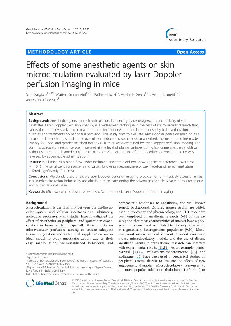
Figure 1 LDPI scan technique. (A) Animal positioning in sternalrecumbency on a light-absorbing pad, with the hind plantar surfacessymmetrical and perpendicular to the laser beam. (B) LDPI imagepost-processing and measurement standardized protocol: the meanintensity of the Doppler signal was registered in ROI encompassingthe hind paws and expressed as numerical value normalized for theirarea (perfusion color scale 0–5 volts).
Gargiulo et al. BMC Veterinary Research 2013, 9:255 Page 2 of 7http://www.biomedcentral.com/1746-6148/9/255
after 15 minutes. The latter perfusion value, close to thatunder isoflurane anesthesia (P = 0.6), was quite retained(4.34 volts) even following dexmedetomidine reversal byatipamezole (the antidote to the α2-receptor agonist)(P = 0.9). No significant peripheral perfusion changeswere observed in control mice after up to 30 minutes(4.57 volts) of 1.5% isoflurane anesthesia (P = 0.11).
DiscussionAnesthetics modulate microcirculation mainly via auto-nomic sympathetic and parasympathetic nerves on vas-cular smooth muscle. Phenothiazine tranquilizers as wellas α2-agonists exert their hemodynamic effects mainlyby interacting with α-adrenergic-receptors. Phenothia-zines cause vasodilation predominantly by blocking α1
receptors but are also dopamine receptor antagonists[28]. While D1-like dopamine receptors induce relaxationof resistance arteries [29,30] D2-like dopamine receptorsare typically present on postganglionic sympathetic neu-rons, where their excitation leads to a reduction of theneural release of norepinephrine, inducing a passive fall invascular resistance and heart rate [31]. Dexmedetomidineis a selective α2-adrenoceptor agonist that shows a dose-dependent, preferential affinity for α2A and α2B receptors[21], evoking a biphasic blood pressure response: a shorthypertensive phase mediated by the α2B receptors,followed by hypotension mediated by the α2A receptors[32,33]. The peripheral hemodynamic effects of phenothi-azines and of α2-agonists thus differ: while acepromazinecauses significant hypotension in isoflurane-anesthetized
Table 1 Microvascular perfusion valuesGroups Measurement time
10 min 20 min 25 min 30 min 35 min 40 min
1 4.37/3.73-4.90 4.57/3.67-5.52 4.53/3.98-5.53 4.84/4.29-5.76
2 4.32/3.69-4.75 4.52/3.62-5.33 2.37/2.02-3.13 4.32/3.73-4.81 4.41/3.72-4.72
3 4.34/3.71-4.83 4.52/3.66-5.50 4.57/3.85-5.78
Mouse peripheral PU (volts) in the isoflurane + acepromazine (1), isoflurane + dexmedetomidine (2) and isoflurane-alone (3) experimental groups at significativetime points (median/min-max).
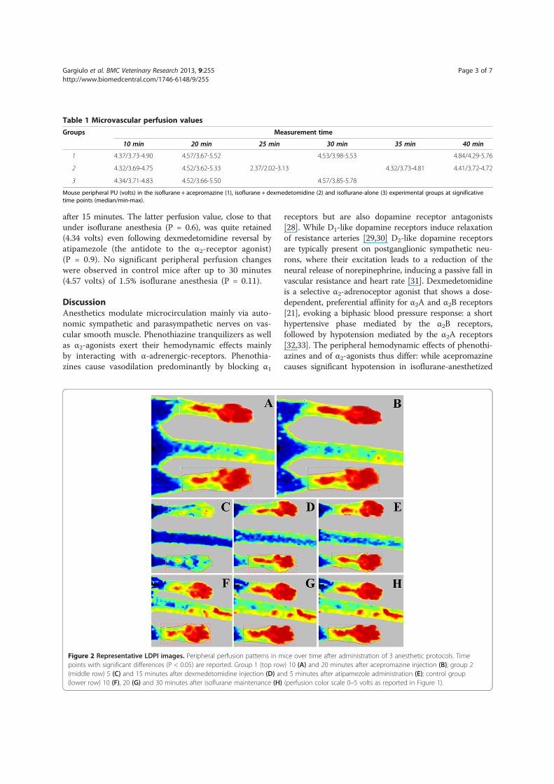
Figure 2 Representative LDPI images. Peripheral perfusion patterns in mice over time after administration of 3 anesthetic protocols. Timepoints with significant differences (P < 0.05) are reported. Group 1 (top row) 10 (A) and 20 minutes after acepromazine injection (B); group 2(middle row) 5 (C) and 15 minutes after dexmedetomidine injection (D) and 5 minutes after atipamezole administration (E); control group(lower row) 10 (F), 20 (G) and 30 minutes after isoflurane maintenance (H) (perfusion color scale 0–5 volts as reported in Figure 1).
Gargiulo et al. BMC Veterinary Research 2013, 9:255 Page 3 of 7http://www.biomedcentral.com/1746-6148/9/255
animals [34], dexmedetomidine [35,36] increases periph-eral vascular tone, counteracting the isoflurane-inducedvasodilation and reduction in arterial blood pressure [22].LDPI permits a noninvasive, real-time measurement ofmicrovascular blood flow using two-dimensional color-coded images of skin perfusion. The use of laser dopplerflowmetry technique to detect the sympathetic toneduring general anesthesia in humans has been reported[3,37,38], and translational approaches using LDPI inmicrovascular perfusion mouse models offer the advan-tages of being easy and fast [39]. Moreover, various anes-thetics alter blood flow in rodents [40-43], so anestheticregimens used in mouse microcirculatory models shouldbe taken into account, and they should not adversely affectthe vascular bed to be examined. The main finding in thisstudy is that LDPI is able to evaluate in real time theanesthesia-induced changes in mouse peripheral microcir-culation. The hemodynamic effects recorded during thedifferent anesthetic protocols were as expected based onprevious clinical and animal studies, although the analyz-ing techniques were different [2-5,22]. Because LDPI scansare disrupted by motion [44] and single or combined sed-atives have lacked restraining effects in mice, we chose toperform our study under 1.5% isoflurane anesthesia torecord reference perfusion values. Isoflurane producesonly minor effects on murine hemodynamic status [6,7].Costantinides et al. (2011) [45] reported that 1.5% iso-flurane produces stable body temperature, mean arter-ial pressure (MAP) and heart rate (HR) values in mice,comparable to those observed in awake animals, so theyrecommended it for physiological and pharmacologicalstudies of cardiac function and to facilitate translationalresearch in non-invasive imaging platforms. In the presentstudy, isoflurane anesthesia yielded a reproducible andstable effect on peripheral blood perfusion over time(P = 0.1). Acepromazine increased isoflurane plantar per-fusion, as reported by Lemke et al. [22], and reduced vas-cular tone and arterial pressure due to its α-blockingaction. After dexmedetomidine administration, a rapidand intense decrease in plantar perfusion was followed bya longer phase of increased perfusion, in agreement withthe typical biphasic hemodynamic effect of this classof sedatives. In all of the mice, the increased perfu-sion recorded 15 minutes after dexmedetomidine ad-ministration did not surpass the perfusion brought aboutby isoflurane (P = 0.6), and it was noticeably lower thanthe perfusion recorded following acepromazine adminis-tration (P = 0.01), which did not increase significantlyeven after atipamezole injection (P = 0.9). Special care wastaken to avoid methodological bias. To date, the skin hasbeen used as a model of microcirculation to investigatevascular mechanisms in cardiovascular [46-48] or kidneydiseases [49] and diabetes [50,51]. Autonomic innervationof microvessels in the region of interest [3,28,52-54],
somatic stimulation of cutaneous arterial sympatheticnerve activity [55], positioning and body temperature [56]are all crucial factors affecting skin blood flow measuredby LDPI. Glabrous skin areas are highly innervated bynoradrenergic sympathetic vasoconstrictor nerves [3,28],which are regulated by α-adrenoceptors [57] in severalspecies [52-54]. For these reasons, we chose plantar regionto investigate blood flow changes brought about byanesthetic drugs, also avoiding hair clipping, which mightaffect cutaneous arterial sympathetic nerve activity andalter LDPI measurements [55]. In our setting, precise hindplantar surface positioning was achieved to keep the siteof interest highly symmetrical and precisely perpendicularto the laser beam (Figure 1) [58-60]. Moreover, bodytemperature was monitored by a rectal probe and adjustedbetween 35.5-36.5°C by an infrared lamp. Our experi-ments were performed in a temperature-controlled room[29], and we started LDPI recordings after each animalhad acclimatized. The effects of sex hormones on vasculartone continue to be a matter of debate [61,62]. Stuckeret al. (2001) [63] reported in an LDPI study only a ten-dency toward higher perfusion values in men than inwomen, stating that moderate gender differences in skinperfusion between study groups should be tolerated. Simi-larly, Kunkel et al. (2007) [64] found that foot skin perfu-sion in normal human subjects was independent ofgender. In our experience, no significant differences be-tween males and females were found in the peripheralblood flow in either control or treated animals. In accord-ance with the manufacturers’ technical instructions, theroom lighting should be kept to a minimum brightness.We set our lighting discrimination between backgroundand the site of measurement to the default threshold levelof 6.2 volts, adjusting the backscattered laser light inten-sity in the range of 7–9 volts, obtaining an optimum qual-ity of data. To compare perfusion in different images, auser-defined color scale was adopted during the acquisi-tion process, ranging from 0 to 5 volts (perfusion outputvalue of 0 volts was calibrated to 0% perfusion, whereas 5volts was calibrated to 100%). The average perfusion ineach region of interest (ROI) was normalized to the wallplantar surface area to reduce bias related to unavoidableanatomical and position variance. To further minimizeany data divergence, the hind paw perfusion value for eachanimal at each time point was calculated as the averagevalue of both hind paw ROIs.
ConclusionsLDPI is able to evaluate noninvasively and in real timethe skin microcirculation changes induced by generalanesthesia in mouse models. LDPI could be useful forstudying the effects of anesthetics on peripheral microcircu-lation and to avoid the inconsistent use of anesthetic agentsin cardiovascular translational research. Standardization of
Gargiulo et al. BMC Veterinary Research 2013, 9:255 Page 4 of 7http://www.biomedcentral.com/1746-6148/9/255
an appropriate LDPI procedure is needed in preclinicalstudies to avoid bias in experimental results.
MethodsEthical permissionThis study was approved by the animal welfare regula-tion committee (CESA) of the University “Federico II”of Naples and by the Italian Ministry of Health. It com-plied with the Guide for the Care and Use of LaboratoryAnimals published by the US National Institutes of Health(NIH Publication No. 85–23, revised 1996).
Study subjects and designTwenty-four CD1 mice (15 females and 9 males), 8 to10 weeks old, were randomly assigned to one of threeexperimental groups (5 females and 3 males) and se-quentially examined in identical ambient conditions.Skin perfusion was recorded by LDPI under isofluraneanesthesia combined or not with acepromazine or dex-medetomidine, as well as after the administration of ati-pamezole to antagonize dexmedetomidine’s effects.
Experimental protocolAnimals were acclimated for 15 min at a room temperatureof 27 ± 3°C before anesthetic induction. During LDPI re-cording, the ambient lighting was kept at a minimum.Body temperature was monitored by a rectal temperatureprobe (Harvard Apparatus®, MLT1404) and closely ad-justed to 35.5 ± 0.5°C by an infrared lamp kept 60 cm awayfrom the body surface. On the basis of a critical revisionof the existing literature, peripheral perfusion was mea-sured at the level of the hairless, highly sympathetic inner-vated plantar surfaces [3,28]. Animals were placed insternal recumbency on the light-absorbing pad providedby the apparatus company, positioning the hind plantarsurfaces symmetrically and perpendicularly to the laserbeam (Figure 1). Isoflurane induction and maintenancewere identical for all mice: each animal was weighedon a precision scale and transferred from a holdingcage to a small rodent anesthetic chamber (isoflurane4% in 2 L/min oxygen) (ISOFLURANE-VET®, MERIALITALIA S.p.A.®). When deeply anesthetized, animals wereplaced in sternal recumbency on the recording bedand fitted with a facial mask delivering isoflurane 1.5%in 1 L/min oxygen. LDPI scans were recorded 10 and20 minutes after isoflurane maintenance. Subsequentgroup treatments were carried out according to the sched-ule below, with precise time intervals between the LDPIrecordings based on the pharmacodynamics of thedifferent anesthetic agents:Group 1 (8 subjects): Acepromazine (PREQUILLAN®,
FATRO S.p.A.®) 5 mg/kg (= 0.99 mg/kcal) was adminis-tered intraperitoneally (IP), followed by two LDPI scansat an interval of ten minutes.
Group 2 (8 subjects): Dexmedetomidine (DEXDOMITOR®,Pfizer Italia Srl®) 1 mg/kg (= 0.19 mg/kcal) was adminis-tered IP, followed by two LDPI scans after 5 and 15 mi-nutes. Finally, dexmedetomidine was reversed by injectingthe α2-adrenoceptor antagonist atipamezole (ATIPAM,Fatro®) 2.5 mg/kg (= 0.49 mg/kcal) IP, and a further LDPIscan was performed after 5 minutes.Group 3 “control” (8 subjects): an additional LDPI scan
was recorded 30 minutes after isoflurane maintenance.
Laser Doppler imaging systemThe Periscan® apparatus displayed the blood perfusionsignal both as a numerical PU (volts) and as a color-coded image ranging from dark blue (low perfusion) tobright red (high perfusion). The settings used in thepresent study were laser beam power = 1 mV; wave-length = 670 nm; pixel size = 0.25 × 0.25 mm2; scannerhead distance =15 cm; scanning area = 3 × 2 cm2; scan-ning time = 2 minutes.
Data processingThe mean intensity of the Doppler signal was quantifiedusing proprietary software in a fixed ROI, encompassingthe corresponding hind paw regions, normalized for theareas of the hind paws and expressed as numericalvalues (volts) to reduce the bias related to unavoidableanatomical and position variance. To further minimizeany data divergence, the hind paw perfusion value foreach animal at each time point was calculated as theaverage value of both hind paw ROIs.
Data analysisStatistical analysis was carried out using the softwareSPSS 18.0.2. (SPSS, Chicago, IL). To compare inter-group differences, one way Friedman ANOVA was used.A post hoc analysis with Dunn’s test was performedwhen appropriate. A linear generalized model (LGM) forrepeated measurements (two-way ANOVA) was used toassess perfusion patterns at different times within groups.A P value <0.05 was considered statistically significant.
AbbreviationsHR: Heart rate; IP: Intraperitoneally; LDPI: Laser Doppler perfusion imaging;MAP: Mean arterial pressure; PU: Perfusion units; ROI: Region of interest.
Competing interestsThe authors declare that they have no competing interests.
Authors’ contributionsSG, MG, AB and GV conceived and designed this study, as well ascontribuited to data interpretation and drafted the manuscript. SG, MG, andGV carried out the experiments. AG took part in the data collection. SG andMG analysed and arranged data for statistical analysis. RL performed thestatistical analyses. SG and MG made equal contribution to this study andshould be considered first authors. All authors read and approved the finalmanuscript.
Gargiulo et al. BMC Veterinary Research 2013, 9:255 Page 5 of 7http://www.biomedcentral.com/1746-6148/9/255
Author details1Institute of Biostructures and Bioimages of the National Council of Research,Via T. De Amicis 95, Naples 80145, Italy. 2Department of AdvancedBiomedical Sciences, University of Naples Federico II, Via Pansini 5, Naples80145, Italy. 3CEINGE scarl, Via G. Salvatore 486, Naples 80145, Italy.4Department of Veterinary Medicine and Animal Productions, University ofNaples Federico II, Via Delpino 1, Naples 80137, Italy.
Received: 9 September 2013 Accepted: 2 December 2013Published: 17 December 2013
References1. Longnecker DE: Effects of general anesthetics on the microcirculation.
Microcirc Endothelium Lymphatics 1984, 2:129–150.2. Lamblin V, Favory R, Boulo M, Mathieu D: Microcirculatory alterations
induced by sedation in intensive care patients: effects of midazolamalone and in association with sufentanil. Crit Care 2006, 6:176–185.
3. Landsverk SA, Kvandal P, Bernjak A, Stefanovska A, Kirkeboe KA: The effectsof general anesthesia on human skin microcirculation evaluated bywavelet transform. Anesth Analg 2007, 4:1012–1019.
4. Turek Z, Sykora R, Matejovic M, Cerni V: Anesthesia and microcirculation.Semin Cardiothorac Vasc Anesth 2009, 4:249–258.
5. Klamt JC, de Andrade Vincente WV, Vicente Garcia L: Effects ofdexmedetomidine-fentanyl infusion on blood pressure and heart rateduring cardiac surgery in children. Anesthesiol Res Pract 2010. doi:10.1155/2010/869049.
6. Zuurbier CJ, Emons VM, Ince C: Hemodynamics of anesthetized ventilatedmouse models: aspects of anesthetics, fluid support and strain. Am JPhysiol 2002, 6:2099–2105.
7. Janssen BJA, De Celle T, Debets JJM, Brouns AE, Callahan MF, Smith TL:Effects of anesthetics on systemic hemodynamics in mice. Am J PhysiolHeart Circ Physiol 2004, 4:1618–1624.
8. Wang Q, Zheng Y, Lu J, Chen L, Wang GN, Zhou JX: Isoflurane potency inmice from the first and second parity. JALAAS 2009, 6:714–717.
9. Yalcin B, Nicod J, Bhomra A, Davidson S, Cleak J, Farinelli L, Osteras M,Whitley A, Yuan W, Gan X, Goodson M, Klenerman P, Satpathy A, Mathis D,Benoist C, Adams DJ, Mott R, Flint J: Commercially available outbred micefor genome-wide association studies. PLoS Genet 2010, 9. doi:10.1371/journal.pgen.1001085.
10. Aldinger KA, Sokoloff G, Rosenberg DM, Palmer AA, Millen KJ: Geneticvariation and population substructure in outbred CD-1 mice: implicationsfor genome-wide association studies. PLoS One 2009, 3:e4729. doi:10.1371/journal.pone.0004729.
11. Gargiulo S, Greco A, Gramanzini M, Esposito S, Affuso A, Brunetti A, Vesce G:Mice anesthesia, analgesia, and care, part I: anesthetic considerations inpreclinical research. ILAR J 2012, 53:55–69.
12. Gargiulo S, Greco A, Gramanzini M, Esposito S, Affuso A, Brunetti A, Vesce G:Mice anesthesia, analgesia, and care, part II: special considerations forpreclinical imaging studies. ILAR J 2012, 53:70–81.
13. Couffinhal T, Silver M, Zheng LP, Kearney M, Witzenbichler B, Isner JM:Mouse model of angiogenesis. Am J Pathol 1998, 6:1667–1679.
14. Li Y, Zhang D, Zhang Y, He G, Zhang F: Augmentation ofneovascularization in murine hindlimb ischemia by combined therapywith simvastatin and bone marrow-derived mesenchymal stem cellstransplantation. J Biomed Sci 2010, 17. doi: 10.1186/1423-0127-17-75.
15. Hellingman AA, Bastiaansen AJNM, de Vries MR, Seghers L, Lijkwan MA,Löwik CW, Hamming JF, Quax PHA: Variations in surgical procedures forhind limb ischaemia mouse models result in differences in collateralformation. Eur J Vasc Endovasc Surg 2010, 6:796–803.
16. Silvestre JS, Mallat Z, Duriez M, Tamarat R, Bureau MF, Scherman D,Duverger N, Branellec D, Tedgui A, Levy BI: Antiangiogenic effect ofinterleukin-10 in ischemia-induced angiogenesis in mice hindlimb.Circ Res 2000, 6:448–452.
17. Lehmann CH, Feyerherd F, Feyerherd TH, Fogliata M, Grundling M,Usichenko TI, Meisser K, Wendt M, Pavlovic D: Ketamine does not affectintestinal microcirculation in pentobarbital-anaesthetized rats duringexperimental endotoxaemia. Lab Anim 2007, 1:55–62.
18. Kusza K, Siemionow M, Nalbantoglu U, Hayes J, Wong KC: Microcirculatoryresponse to halothane and isoflurane anesthesia. Ann Plast Surg 1999,1:57–66.
19. Brookes ZLS, Brown NJ, Reilly CS: Intravenous anaesthesia and the ratmicrocirculation: microcirculatory chamber. Brit J Anaesth 2000,6:901–903.
20. Brookes ZL, Brown NJ, Reilly CS: Response of the rat cremaster microcirculationto hemorrhage in vivo: differential effects of intravenous anesthetic agents.Shock 2002, 6:542–548.
21. Posner LP, Burns P: Sedative agents: tranquilizers α-2agonists and relatedagents. In Veterinary pharmacology and therapeutics. Edited by Riviere JE,Papich MG. Ames, Iowa: Wiley Blackwell; 2009:337–380.
22. Lemke KA: Perioperative use of selective α-2 agonists and antagonists insmall animals. Can Vet J 2004, 6:475–480.
23. Arras M, Autenried P, Rettich A, Spaeni D, Tülicke T: Optimization ofintraperitoneal injection anesthesia in mice: drugs, dosages, adverseeffects and anesthesia depth. Comp Med 2001, 5:443–456.
24. Buitrago S, Martin TE, Tetens-Woodring J, Belicha-Villaneueva A, Wilding GE:Safety and efficacy of various combinations of injectable anesthetics inBalb/C mice. JAALAS 2008, 1:11–17.
25. Leino K, Hynynen M, Jalonen J, Salmenperä M, Scheinin H, Aanta R: Renaleffects of dexmedetomidine during coronary artery bypass surgery:a randomized control-controlled study. BMC Anesthesiol 2011, 23:9–19.
26. Holowatz LA, Thompson-Torgerson CS, Kenney WL: The human cutaneouscirculation as a model of generalized microvascular function. J ApplPhysiol 2008, 1:370–372.
27. Samuelsson A: Effects of burns and vasoactive drugs on human skin.In Clinical and Experimental studies using microdialysis. Linköping, Sweden:Linköping University Medical Dissertations No. 1195 LIU-tryck; 2010.
28. Pascoe PJ, Taylor MA: Effects of dopamine antagonists on alfentanil-induced locomotor activity in horses. Vet Anaesth Analg 2003, 3:165–171.
29. Zeng C, Eisner GM, Felder RA, Jose PA: Dopamine receptor andhypertension. Curr Med Chem Cardiovasc Hematol Agents 2005, 1:69–77.
30. Jose PA, Eisner GM, Felder RA: Role of dopamine receptors in the kidneyin the regulation of blood pressure. Curr Opin Nephrol Hypertens 2002,1:87–92.
31. Cavero I, Massingham R, Borg FL: Peripheral dopamine receptors,potential targets for a new class of antihypertensive agents: Part I:subclassification and functional description. Life Sci 1982, 10:939–948.
32. Kaur M, Singh PM: Current role of dexmedetomidine in clinical anesthesiaand intensive care. Anesth Essays Res 2011, 2:128–133.
33. Papadakos PJ, Compolo F: Sedation in the ICU: shifts and strategies.Anesthesiology News 2011:80–91.
34. Bostrom I, Nyman G, Kampa N, Haggstrom J, Lord P: Effects of acepromazineon renal function in anesthetized dogs. Am J Vet Res 2003, 5:590–598.
35. Cullen LK: Medetomidine sedation in dogs and cats: a review of itspharmacology, antagonism and dose. Br Vet J 1996, 5:519–535.
36. Baker NJ, Schofield JC, Caswell MD: Effects of early atipamezole reversal ofmedetomidine-ketamine anesthesia in mice. J Am Assoc Lab Anim Sci2011, 6:916–920.
37. Humeau A, Steenbergen W, Nilsson H, Strümberg T: Laser dopplerperfusion monitoring and imaging: novel approaches. Med Biol EngComput 2007, 5:421–435.
38. Takashi M, Ping Z, Takahiko K, Yoshimi I, Akitoshi O, Atsushi Y, Ikuto Y: Laserdoppler skin blood flow and sympathetic nervous responses to surgicalincision during halothane and isoflurane anesthesia. Anesth Analg 1997,2:291–298.
39. Al-Mubarak HA, Alamri TM, Aljabab SA, Atteya M, Quan A, Teoh H,Shukla PC, Verma S, Aldahmash A, Aljabri B, Napoli C, Al-Omran M: Effects onduration of post-operative ischemia and patterns of blood flow recoveryin different conditions of mouse hind limb ischemia. Vasc Cell 2011,3. doi:10.1186/2045-824X-3-14.
40. Dalkara T, Irikura K, Huang Z, Panahian N, Moskowitz MA: Cerebrovascularresponses under controlled and monitored physiological conditions inthe anesthetized mouse. J Cereb Blood Flow Metab 1995, 4:631–638.
41. Koorn R, Kahn RA, Brannan TS, Martinez-Tica J, Weinberger J, Reich DL:Effect of isoflurane and halothane on in vivo ischemia-induced dopaminerelease in the corpus striatum of the rat: a study using cerebralmicrodialysis. Anesthesiology 1993, 1:827–835.
42. Lindauer U, Villringer A, Dirnagl U: Characterization of CBF response tosomatosensory stimulation: model and influence of anesthetics. Am JPhysiol 1993, 4:1223–1228.
43. Greco A, Ragucci M, Liuzzi R, Gargiulo S, Gramanzini M, Coda ARD, AlbaneseS, Mancini M, Salvatore M, Brunetti A: Reproducibility and standardization
Gargiulo et al. BMC Veterinary Research 2013, 9:255 Page 6 of 7http://www.biomedcentral.com/1746-6148/9/255

of laser doppler Imaging technique for the evaluation of normal micehindlimbs perfusion. Sensor 2012, 13:500–515.
44. Dirnagl U, Kaplan B, Jacewicz M, Pulsinelli W: Continuous measurement ofcerebral cortical blood flow by laser-Doppler flowmetry in a rat strokemodel. J Cereb Blood Flow Metab 1989, 5:589–596.
45. Constantinides C, Mean R, Janssen BJ: Effects of isoflurane anesthesia onthe cardiovascular function of the C57BL/6 mouse. ILAR J 2011, 52:21–31.
46. Antonios TF, Singer DR, Markandu ND, Mortimer PS, MacGregor GA:Structural skin capillary rarefaction in essential hypertension.Hypertension 1999, 33:998–1001.
47. Feihl F, Liaudet L, Waeber B, Levy BI: Hypertension: a disease of themicrocirculation? Hypertension 2006, 6:1012–1017.
48. Levy BI, Schiffrin EL, Mourad JJ, Agostani D, Vicaut E, Safar ME, Struijker-Boudier HA: Impaired tissue perfusion: a pathology common tohypertension, obesity, and diabetes mellitus. Circulation 2008, 9:968–976.
49. Kruger A, Stewart J, Sahityani R, O’Riordan E, Thompson C, Adler S, Garrick R,Vallance P, Goligorsky MS: Laser Doppler flowmetry detection ofendothelial dysfunction in end-stage renal disease patients: correlationwith cardiovascular risk. Kidney Int 2006, 1:157–164.
50. Chang CH, Tsai RK, Wu WC, Kuo SL, Yu HS: Use of dynamic capillaroscopyfor studying cutaneous microcirculation in patients with diabetesmellitus. Microvasc Res 1997, 2:121–127.
51. Yamamoto-Suganuma R, Aso Y: Relationship between post-occlusiveforearm skin reactive hyperaemia and vascular disease in patients withType 2 diabetes – a novel index for detecting micro- and macrovasculardysfunction using laser Doppler flowmetry. Diabet Med 2009, 1:83–88.
52. Koganezawa T, Ishikawa T, Fujita Y, Yamashita T, Tajima T, Honda M,Nakayama K: Local regulation of skin blood flow during cooling involvingpresynaptic P2 purinoceptors in rats. Br J Pharmacol 2006, 5:579–586.
53. Honda M, Suzuki M, Nakayama K, Ishikawa T: Role of a2C-adrenoceptors inthe reduction of skin blood flow induced by local cooling in mice. Br JPharmacol 2007, 1:91–100.
54. Mayrovitz HN, Carta SG: Laser Doppler Imaging Assessment of skinHyperemia indicator of trauma after adhesive strip removal. Adv WoundCare 1996, 4:38–42.
55. Horii Y, Tanida M, Shen J, Fujisaki Y, Fuyuki R, Hashimoto K, Niijima A,Nakashima T, Katsuya N: Skin application of urea-containing creamaffected cutaneous arterial sympathetic nerve activity, blood flow,and water evaporation. Skin Res Technol 2011, 1:75–81.
56. Kuluz JW, Prado R, Chang J, Ginsberg MD, Schleien CL, Busto R: Selectivebrain cooling increases cortical cerebral blood flow in rats. Am J Physiol1993, 3:824–827.
57. Lawrence CJ, Prinzen FW, de Lange S: The effect of dexmedetomidine onnutrient organ blood flow. Anesth Analg 1996, 6:1160–1165.
58. Belin de Chantemèle EJ, Ali MI, Mitz J, Stepp DW: Obesity induced insulinresistance causes endothelial dysfunction without reducing the vascularresponse to hindlimb ischemia. Basic Res Cardiol 2009, 6:707–717.
59. Anderson RR, Parrish JA: The optics of human skin. J Invest Dermatol 1981,1:13–19.
60. Fredriksson I, Larsson M, Strömberg T: Measurement depth and volume inlaser Doppler flowmetry. Microvasc Res 2009, 1:4–13.
61. Cankar K, Finderle Z, Strucl M: Role of alpha-adrenoceptors in the cutaneouspostocclusive reactive hyperaemia. Pflugers Arch 2000, 5:121–122.
62. Cankar K, Finderle Z, Strucl M: The role of alpha1- and alpha2- adrenoceptorsin gender differences in cutaneous LD flux response to local cooling.Microvasc Res 2004, 2:126–131.
63. Stücker M, Steinberg J, Memmel U, Avermaete A, Hoffmann K, Altmeyer P:Differences in the two-dimensionally measured laser Doppler flow atdifferent skin localisations. Skin Pharmacol Appl Skin Physiol 2001, 1:44–51.
64. Kunkel CF, Figoni SF, Baumgarten JM: Scanning laser-Doppler imaging ofleg- and foot-skin perfusion in normal subjects: analysis of age, gender,site, and laser-type effects. Am J Phys Med Rehabil 2007, 4:262–271.
doi:10.1186/1746-6148-9-255Cite this article as: Gargiulo et al.: Effects of some anesthetic agents onskin microcirculation evaluated by laser Doppler perfusion imaging inmice. BMC Veterinary Research 2013 9:255.
Submit your next manuscript to BioMed Centraland take full advantage of:
• Convenient online submission
• Thorough peer review
• No space constraints or color figure charges
• Immediate publication on acceptance
• Inclusion in PubMed, CAS, Scopus and Google Scholar
• Research which is freely available for redistribution
Submit your manuscript at www.biomedcentral.com/submit
Gargiulo et al. BMC Veterinary Research 2013, 9:255 Page 7 of 7http://www.biomedcentral.com/1746-6148/9/255
Related Documents