applied sciences Article Effects of Simulated Gastric Acid Exposure on Surface Topography, Mechanical and Optical Features of Commercial CAD/CAM Ceramic Blocks Ioana Ligia Pîrvulescu 1 , Daniel Pop 1,2 , Elena-Alina Moacă 3,4, * , Ciprian-Valentin Mihali 5,6 , Codrut , a Ille 1 and Anca Jivănescu 1,2 Citation: Pîrvulescu, I.L.; Pop, D.; Moac˘ a, E.-A.; Mihali, C.-V.; Ille, C.; Jiv˘ anescu, A. Effects of Simulated Gastric Acid Exposure on Surface Topography, Mechanical and Optical Features of Commercial CAD/CAM Ceramic Blocks. Appl. Sci. 2021, 11, 8703. https://doi.org/10.3390/ app11188703 Academic Editor: Yong-Deok Kim Received: 31 July 2021 Accepted: 14 September 2021 Published: 18 September 2021 Publisher’s Note: MDPI stays neutral with regard to jurisdictional claims in published maps and institutional affil- iations. Copyright: © 2021 by the authors. Licensee MDPI, Basel, Switzerland. This article is an open access article distributed under the terms and conditions of the Creative Commons Attribution (CC BY) license (https:// creativecommons.org/licenses/by/ 4.0/). 1 Department of Prosthodontics, Faculty of Dental Medicine, “Victor Babes” University of Medicine and Pharmacy, Revolutiei Ave. 1989, No. 9, 300580 Timis , oara, Romania; [email protected] (I.L.P.); [email protected] (D.P.); [email protected] (C.I.); [email protected] (A.J.) 2 TADERP Reseach Center—Advanced and Digital Techniques for Endodontic, Restorative and Prosthetic Treatment, “Victor Babes , ” University of Medicine and Pharmacy, Revolutiei Ave. 1989, No. 9, 300041 Timi¸ soara, Romania 3 Department of Toxicology and Drug Industry, Faculty of Pharmacy, “Victor Babes , ” University of Medicine and Pharmacy Timisoara, 2nd Eftimie Murgu Square, 300041 Timisoara, Romania 4 Research Centre for Pharmaco-Toxicological Evaluation, “Victor Babes , ” University of Medicine and Pharmacy, 2nd Eftimie Murgu Square, 300041 Timis , oara, Romania 5 The Institute of Life Sciences, “Vasile Goldis” Western University of Arad, Liviu Rebreanu Street, No. 86, 310414 Arad, Romania; [email protected] 6 The Molecular Research Department, Research and Development Station for Bovine Arad, Bodrogului Street, No. 32, 310059 Arad, Romania * Correspondence: [email protected]; Tel.: +40-745-762-600 Featured Application: Degradation of the material used for prosthetic rehabilitation leads to more or less serious problems, namely impairment of aesthetics, craniofacial disorders, discom- fort especially during chewing food, which will be increasingly reduced, leading in the end to fatigue of the masticatory muscles. The most harmful for the dental structure is considered gas- tric juice. This acidic substance produce degradation both enamel, as well as some restorative materials. In this context, the present work consists in evaluating the surface topography and mechanical properties of some CAD/CAM monolithic materials (already existing on the market), used for prosthetic restoration, thus helping dentists to choose those ceramic materials that are resistant to the effects of gastric juice. Abstract: Gastric acid exposure produces tooth structure demineralization and dental ceramic degradation. The most affected patients are those who suffer from gastroesophageal reflux disease, bulimia nervosa, and pregnant women with prolonged severe nausea. In order to protect this kind of patient, the purpose of this study was to determine whether simulated gastric acid exposure leads to microstructural changes in surface topography, hardness, color changes, and translucency of some ceramic materials, which are already on the market. Forty disks (Triluxe Forte, Cerasmart, Enamic and Empress CAD) were analyzed before and after immersion in simulated gastric acid juice, in terms of microhardness, surface roughness, translucency, and surface morphology using scanning electron microscopy. Color change was assessed by using a spectrophotometer based on CIELab parameters and the results showed that, after exposure, ΔE ab remained under the threshold of acceptability and perceptibility. In terms of microhardness, Cerasmart is the only material that did not undergo changes after immersion. SEM images illustrated observable changes surface topography after acid exposure for all the tested materials. In conclusion, Triluxe Forte suffered the most important changes after simulated gastric acid juice exposure, and Cerasmart proved to be the least affected material. Keywords: CAD/CAM; ceramic materials; gastric acid; surface roughness; microhardness; sur- face morphology Appl. Sci. 2021, 11, 8703. https://doi.org/10.3390/app11188703 https://www.mdpi.com/journal/applsci

Welcome message from author
This document is posted to help you gain knowledge. Please leave a comment to let me know what you think about it! Share it to your friends and learn new things together.
Transcript
applied sciences
Article
Effects of Simulated Gastric Acid Exposure on SurfaceTopography, Mechanical and Optical Features of CommercialCAD/CAM Ceramic Blocks
Ioana Ligia Pîrvulescu 1, Daniel Pop 1,2, Elena-Alina Moacă 3,4,* , Ciprian-Valentin Mihali 5,6, Codrut,a Ille 1
and Anca Jivănescu 1,2
�����������������
Citation: Pîrvulescu, I.L.; Pop, D.;
Moaca, E.-A.; Mihali, C.-V.; Ille, C.;
Jivanescu, A. Effects of Simulated
Gastric Acid Exposure on Surface
Topography, Mechanical and Optical
Features of Commercial CAD/CAM
Ceramic Blocks. Appl. Sci. 2021, 11,
8703. https://doi.org/10.3390/
app11188703
Academic Editor: Yong-Deok Kim
Received: 31 July 2021
Accepted: 14 September 2021
Published: 18 September 2021
Publisher’s Note: MDPI stays neutral
with regard to jurisdictional claims in
published maps and institutional affil-
iations.
Copyright: © 2021 by the authors.
Licensee MDPI, Basel, Switzerland.
This article is an open access article
distributed under the terms and
conditions of the Creative Commons
Attribution (CC BY) license (https://
creativecommons.org/licenses/by/
4.0/).
1 Department of Prosthodontics, Faculty of Dental Medicine, “Victor Babes” University of Medicine and Pharmacy,Revolutiei Ave. 1989, No. 9, 300580 Timis, oara, Romania; [email protected] (I.L.P.);[email protected] (D.P.); [email protected] (C.I.); [email protected] (A.J.)
2 TADERP Reseach Center—Advanced and Digital Techniques for Endodontic, Restorative and Prosthetic Treatment,“Victor Babes, ” University of Medicine and Pharmacy, Revolutiei Ave. 1989, No. 9, 300041 Timisoara, Romania
3 Department of Toxicology and Drug Industry, Faculty of Pharmacy, “Victor Babes, ” University of Medicineand Pharmacy Timisoara, 2nd Eftimie Murgu Square, 300041 Timisoara, Romania
4 Research Centre for Pharmaco-Toxicological Evaluation, “Victor Babes, ” University of Medicine and Pharmacy,2nd Eftimie Murgu Square, 300041 Timis, oara, Romania
5 The Institute of Life Sciences, “Vasile Goldis” Western University of Arad, Liviu Rebreanu Street, No. 86,310414 Arad, Romania; [email protected]
6 The Molecular Research Department, Research and Development Station for Bovine Arad, Bodrogului Street,No. 32, 310059 Arad, Romania
* Correspondence: [email protected]; Tel.: +40-745-762-600
Featured Application: Degradation of the material used for prosthetic rehabilitation leads tomore or less serious problems, namely impairment of aesthetics, craniofacial disorders, discom-fort especially during chewing food, which will be increasingly reduced, leading in the end tofatigue of the masticatory muscles. The most harmful for the dental structure is considered gas-tric juice. This acidic substance produce degradation both enamel, as well as some restorativematerials. In this context, the present work consists in evaluating the surface topography andmechanical properties of some CAD/CAM monolithic materials (already existing on the market),used for prosthetic restoration, thus helping dentists to choose those ceramic materials that areresistant to the effects of gastric juice.
Abstract: Gastric acid exposure produces tooth structure demineralization and dental ceramicdegradation. The most affected patients are those who suffer from gastroesophageal reflux disease,bulimia nervosa, and pregnant women with prolonged severe nausea. In order to protect this kind ofpatient, the purpose of this study was to determine whether simulated gastric acid exposure leads tomicrostructural changes in surface topography, hardness, color changes, and translucency of someceramic materials, which are already on the market. Forty disks (Triluxe Forte, Cerasmart, Enamicand Empress CAD) were analyzed before and after immersion in simulated gastric acid juice, in termsof microhardness, surface roughness, translucency, and surface morphology using scanning electronmicroscopy. Color change was assessed by using a spectrophotometer based on CIELab parametersand the results showed that, after exposure, ∆Eab remained under the threshold of acceptabilityand perceptibility. In terms of microhardness, Cerasmart is the only material that did not undergochanges after immersion. SEM images illustrated observable changes surface topography after acidexposure for all the tested materials. In conclusion, Triluxe Forte suffered the most important changesafter simulated gastric acid juice exposure, and Cerasmart proved to be the least affected material.
Keywords: CAD/CAM; ceramic materials; gastric acid; surface roughness; microhardness; sur-face morphology
Appl. Sci. 2021, 11, 8703. https://doi.org/10.3390/app11188703 https://www.mdpi.com/journal/applsci
Appl. Sci. 2021, 11, 8703 2 of 14
1. Introduction
The evolution of dentistry has advanced and will continue to advance through theappearance of intelligent materials on the market, designed to contribute to oral health careand, thus, to the improvement of quality of life. To date, many types of dental materialshave been generated and are available for clinical applications. Resin based-compositesand materials conjugated in cements and metals have been widely produced to replacemissing tooth structures and mimic the aesthetic characteristics of tooth structures [1–7].Resin based-composites with different inorganic reinforcement particles are commonlycommercialized due to their excellent mechanical and physicochemical properties, such astheir high strength, bright surface, low surface roughness, resistance to wear/fracture, andadherence to dental tissues [8–10]. Therapeutic restorative materials are used for minimallyinvasive and a traumatic restorative dentistry and, in this context, the development ofnew restorative materials that can mimic the mechanical, physicochemical and aestheticsfeatures of dentin and enamel are necessary [11,12].
In the recent years, computer-aided design and computer-aided manufacturing (CAD-CAM) systems have allowed monolithic restorations to be made in a single session.CAD-CAM restorative materials have been widely used in dentistry due to their sta-ble quality of materials and lower costs. CAD-CAM technology satisfies aesthetic demands,such as color and translucency reproduction, and even chromatic stability under clinicalconditions [13–15]. While most studies have reported extensive details on the positive char-acteristics of these materials [16–22], few studies have investigated the surface behavior ofthese materials in contact with endogenous (salivary enzymes, gastric juice) or exogenous(acidic beverages) substances. These acidic elements have been correlated with severedental structure degradation and restoration, most probably due to their acidic pH [23,24].Regardless of the origin, these acidic substances attack the surface of the tooth, as well asthe surface of CAD-CAM restorative materials, changing their structure over time [25–27].It was established that gastric juice produce more severe degradation of dental structuresthan acidic beverages [28–30]. Gastric juice comes into contact with the oral cavity as aresult of gastroesophageal reflux disease or bulimia nervosa [31–33]. Even the case ofpregnant women with prolonged severe nausea episodes during pregnancy can face theissue gastric juices [23].
Dental erosion, defined as a progressive loss of hard dental tissues due to chemicalprocesses, without the action of bacteria, can be correlated with the intrinsic and/orextrinsic origin of acid. The results of chemical erosion consist in hard, polished, andsmooth depression of a tooth’s surface [34]. Extrinsic tooth erosion is mainly due to theconsumption of low-pH beverages and foods. It may also be due to the use of medications,such as aspirin, which is a low pH drug [35].
All-ceramic restorations that offer biocompatibility and aesthetics may be affectedby gastric acid, although less intensely than enamel [31]. Chemical degradation can leadto microstructural changes in surface topography, which can affect light reflection, colorperception, and stability [36]. The knowledge of how ceramic materials react to gastric acidcan help a dentist in selecting suitable materials for prosthetic restorations for the specialpatients mentioned above, though not limited to them. Although the literature reports onthe effects of acidic elements on the surface of different restorative materials [25,31,37–41],to the best of authors’ knowledge, no study has yet to compare, under the same conditions,these four monolithic CAD-CAM materials: feldspathic ceramic, nanoceramic resin, hybridceramic, and leucite-reinforced glass ceramic.
In this regard, the purpose of the present in vitro study was to evaluate the effects ofsurface topography and the mechanical features of different restorative computer-aideddesigns and computer-aided manufacturing (CAD-CAM) dental materials after exposureto simulated gastric juices. We assumed that with these materials, already available onthe market, the exposure to simulated gastric juice for 18 h would not change the surfacetopography nor the mechanical properties of the monolithic materials under consideration.
Appl. Sci. 2021, 11, 8703 3 of 14
2. Materials and Methods2.1. Specimens Preparation
The monolithic materials tested in this in vitro study along with their compositionsare listed in Table 1. Forty disks (n = 10/subgroup) of feldspathic ceramic (Triluxe Forte,VITA, Zahnfabrik), nanoceramic resin (Cerasmart, GC Europe), hybrid ceramic (Enamic,VITA, Zahnfabrik), and leucite-reinforced glass ceramic (Empress CAD, Ivoclar, Vivadent)were used in this study.
Table 1. The composition, manufacturer, and classification of the tested monolithic materials.
Material Classification Composition Manufacturer
Triluxe Forte feldspathic ceramics SiO2 56–64%; Al2O3 20–23%; Na2O 6–9%; K2O6–8%; CaO 0.3–0.6%; TiO2 0.0–0.1%
VITA Zahnfabrik (Bad Säckingen,Germany)
Cerasmart nanoceramic resin 71% Silica and barium glass nanoparticles GC Europe (Tokyo, Japan)
Enamic hybrid ceramic with adual network structure
SiO2 5–63%; Al2O3 20–23%; Na2O 6–11%; K2O4–6%; B2O3 0.5–2%; CaO < 1%; TiO2 < 1%
VITA Zahnfabrik (Bad Säckingen,Germany)
Empress CAD leucite-reinforcedglass-ceramic
SiO2 60–65%; Al2O3 16–20%; K2O 10–14%;Na2O 3.5–6.5%
Ivoclar Vivadent (Zurich,Schwitzerland)
CAD-CAM blocks were milled into disks (1 mm thick, 14 mm long, and 12 mm wide),with a precision saw (IsoMet 1000-Buehler, Lake Bluff, IL, USA) and a special disk usedfor cutting structured ceramics (IsoMet Diamond Wafering Blade, 15LC-Buehler, LakeBluff, IL, USA), at a speed of 100 rotations per minute. The resulting ceramic disks werefinished with abrasive paper (Klingspor, Haiger, Germany) using different granulations(P240, P400, P800, P1000, and P1200). All analyses were carried out before and after gastricacid exposure.
2.2. Immersion of Ceramic Disks in Simulated Gastric Acid Solution
Specimens were individually immersed for 18 h in 5 mL of simulated gastric acidsolution and stored in an incubator (Cultura, Ivoclar, Zurich, Schwitzerland) at 37 ◦C. Thesimulated gastric acid solution was prepared with 0.113% (0.06 M) hydrochloric acid (HCl)solution in deionized water and adjusted to pH 1.2 [29,42]. Considering that a patientsuffering from bulimia has, on average, vomits 3 times daily, and the contact time betweenvomit and restoration material is under 1 min [40], it can be estimated that the immersiontime used corresponds with 2 years of gastric juice exposure.
2.3. Investigation of Optical Parameters
Color parameters were analyzed using a spectrophotometer (VITA Easyshade V, BadSäckingen, Germany). The CIELab metric values were recorded and stored for color differ-ence measurements after the discs were immersed in the simulated gastric acid solution.Calibration of the spectrophotometer was done before each specimen’s measurement.
After immersion, the disks were rinsed with water and the CIELab metric values wereagain determined. Mean values of L*, a*, b* were compared with the data gathered prior tothe immersion. The total color change (∆E) was calculated for each monolithic materialusing the following equation:
∆Eab =
√[(∆L∗)2 + (∆a∗)2 + (∆b∗)2
](1)
where L*—the lightness coefficient, ranging from 0 (black) to 100 (white); a*—the shade ofredness (positive values) and greenness (negative values); b*—denotes yellowness (positivevalues) and blueness (negative values).
Relative values of ∆L∗, ∆a∗, ∆b∗ corresponded to differences before and after immer-sion. Color differences were classified as follows: ∆E > 1 the color change can be detected
Appl. Sci. 2021, 11, 8703 4 of 14
visually, but is acceptable; ∆E > 3.3 the color changes are clinically unacceptable [43] and∆E < 1 are considered undetectable color changes to the human eye [44].
2.4. Evaluation of Translucency Parameters
Translucency is the ability of a material to transmit light and it can be calculated bythe translucency parameter (TP), applying the following equation [45]:
TP =
√(L∗
B − L∗W)2
+(a∗B − a∗W
)2+(b∗B − b∗W
)2 (2)
where the B and W subscripts, refer to color coordinates over black and white backgrounds.
2.5. Microhardness Measurements
Microhardness was measured using a Digital Display Vickers (Novotest-TB-MCV-1A, Novotest, Meschede, Germany) micro-hardness tester. A load of 1000 g was appliedwithout impact. The diamond indenter was held in place for 15 s. The two indentationdiagonals were measured. Four indentations were made in each specimen, before and afterimmersion (HVi and HVf, respectively). Indentation dimensions were measured using theeyepiece of a microscope and the hardness values were obtained from standard tables.
2.6. Surface Roughness Measurements
Surface roughness (Ra) was assessed using a profilometer (Perthometer M2, Mahr,Gottingen, Germany) at baseline (Ra1) and after the immersion time (Ra2). In order tomeasure the roughness, a diamond stylus with a 5-µm tip radius was used. The styluswas moved across the ceramic disc surface under a constant load of 3.9 nM at a speedof 0.12 mm/s. The readings made before and after simulated acid exposure (18 h) werecarried out on the same location and the procedure was repeated 4 times for each specimen.
2.7. Scanning Electron Microscopy (SEM) and Energy Dispersive X-ray Spectroscopy(EDX) Analysis
A scanning electron microscope was used to examine the surface microstructure andwas used to determine the degree of damage on the ceramic surface after exposure to gastricacid juice. SEM analysis was carried out on an FEI Quanta 250 microscope (Eindhoven, TheNetherlands). SEM analysis parameters were HV mode, 30 kV, ETD (Everhart–Thornleydetector for secondary electrons), with two magnification orders, one for a general overview(100×) of the image/measurements and another for a higher surface topography (5000×)for regions of interest. The observations were made before and after immersion. EDXanalysis (energy dispersive X-ray spectroscopy) was carried out with an EDAX system(Apollo X detector, Ametek, Mahwah, NJ, USA). For better conductivity and to acquirehigh resolution SEM imaging, each specimen was covered with gold: 4 nm/deposition,3 times, using an AutoAgar Sputter Coater (Agar Scientific Ltd., Essex, UK). The identifiedchemical species were expressed as atomic relative percent (At%) and weight relativepercent (Wt%).
2.8. Statistical Analysis
The statistical programs applied in the present study were one-way analysis of vari-ance (ANOVA) followed by Tukey’s test, employed to evaluate the statistically significantdifferences, before and after immersion in gastric acid solution (*** p < 0.001).
3. Results3.1. Optical Parameters
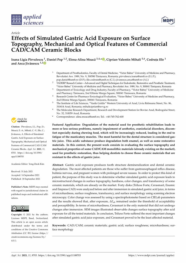
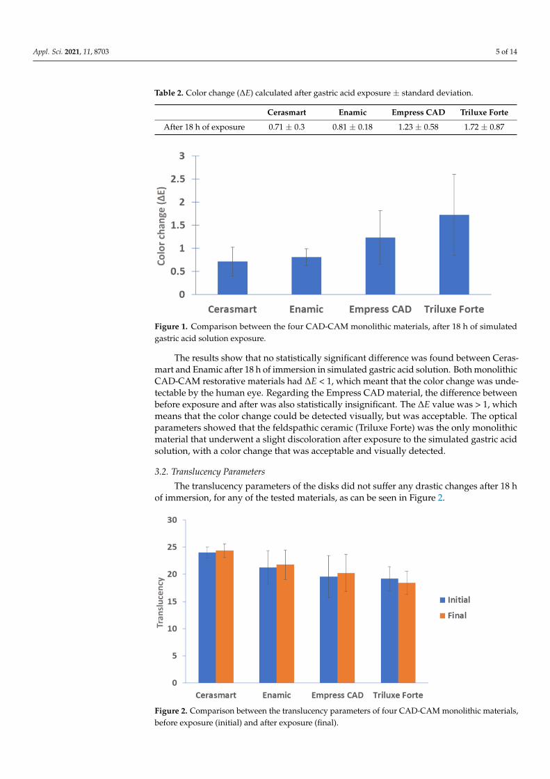
Table 2 shows the values regarding the color change for each material and the com-parisons between the four monolithic materials were taken into account, as depicted inFigure 1.
Appl. Sci. 2021, 11, 8703 5 of 14
Table 2. Color change (∆E) calculated after gastric acid exposure ± standard deviation.
Cerasmart Enamic Empress CAD Triluxe Forte
After 18 h of exposure 0.71 ± 0.3 0.81 ± 0.18 1.23 ± 0.58 1.72 ± 0.87
Appl. Sci. 2021, 11, x FOR PEER REVIEW 5 of 16
2.8. Statistical Analysis
The statistical programs applied in the present study were one-way analysis of vari-
ance (ANOVA) followed by Tukey’s test, employed to evaluate the statistically significant
differences, before and after immersion in gastric acid solution (*** p < 0.001).
3. Results
3.1. Optical Parameters
Table 2 shows the values regarding the color change for each material and the com-
parisons between the four monolithic materials were taken into account, as depicted in
Figure 1.
Table 2. Color change (ΔE) calculated after gastric acid exposure ± standard deviation.
Cerasmart Enamic Empress CAD Triluxe Forte
After 18 h of exposure 0.71 ± 0.3 0.81 ± 0.18 1.23 ± 0.58 1.72 ± 0.87
Figure 1. Comparison between the four CAD-CAM monolithic materials, after 18 h of simulated gastric acid solution
exposure.
The results show that no statistically significant difference was found between
Cerasmart and Enamic after 18 h of immersion in simulated gastric acid solution. Both
monolithic CAD-CAM restorative materials had ΔE < 1, which meant that the color
change was undetectable by the human eye. Regarding the Empress CAD material, the
difference between before exposure and after was also statistically insignificant. The ΔE
value was ˃ 1, which means that the color change could be detected visually, but was
acceptable. The optical parameters showed that the feldspathic ceramic (Triluxe Forte)
was the only monolithic material that underwent a slight discoloration after exposure to
the simulated gastric acid solution, with a color change that was acceptable and visually
detected.
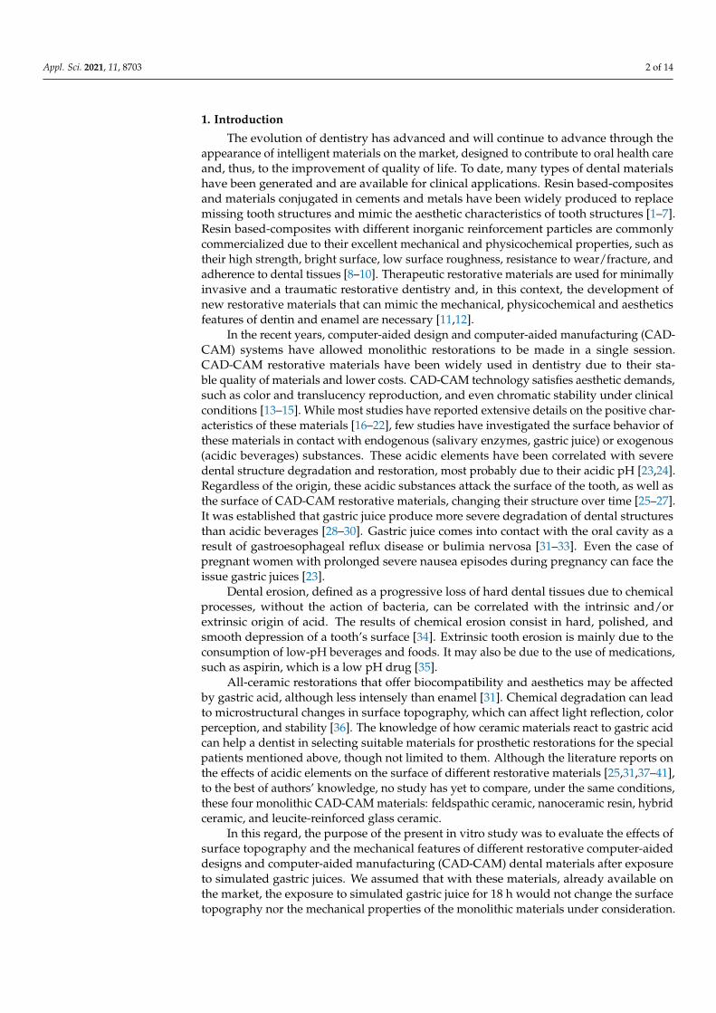
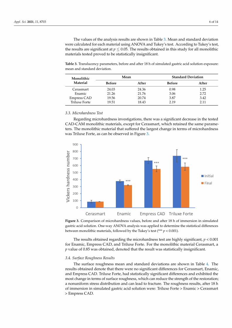
3.2. Translucency Parameters
The translucency parameters of the disks did not suffer any drastic changes after 18
h of immersion, for any of the tested materials, as can be seen in Figure 2.
Figure 1. Comparison between the four CAD-CAM monolithic materials, after 18 h of simulatedgastric acid solution exposure.
The results show that no statistically significant difference was found between Ceras-mart and Enamic after 18 h of immersion in simulated gastric acid solution. Both monolithicCAD-CAM restorative materials had ∆E < 1, which meant that the color change was unde-tectable by the human eye. Regarding the Empress CAD material, the difference betweenbefore exposure and after was also statistically insignificant. The ∆E value was > 1, whichmeans that the color change could be detected visually, but was acceptable. The opticalparameters showed that the feldspathic ceramic (Triluxe Forte) was the only monolithicmaterial that underwent a slight discoloration after exposure to the simulated gastric acidsolution, with a color change that was acceptable and visually detected.
3.2. Translucency Parameters
The translucency parameters of the disks did not suffer any drastic changes after 18 hof immersion, for any of the tested materials, as can be seen in Figure 2.
Appl. Sci. 2021, 11, x FOR PEER REVIEW 6 of 16
Figure 2. Comparison between the translucency parameters of four CAD-CAM monolithic materi-
als, before exposure (initial) and after exposure (final).
The values of the analysis results are shown in Table 3. Mean and standard deviation
were calculated for each material using ANOVA and Tukey’s test. According to Tukey’s
test, the results are significant at p ≤ 0.05. The results obtained in this study for all mono-
lithic materials tested proved to be statistically insignificant.
Table 3. Translucency parameters, before and after 18 h of simulated gastric acid solution exposure: mean and standard
deviation.
Monolithic
Material
Mean Standard Deviation
Before After Before After
Cerasmart 24.03 24.36 0.98 1.25
Enamic 21.26 21.76 3.06 2.72
Empress CAD 19.56 20.74 3.87 3.42
Triluxe Forte 19.51 18.43 2.19 2.11
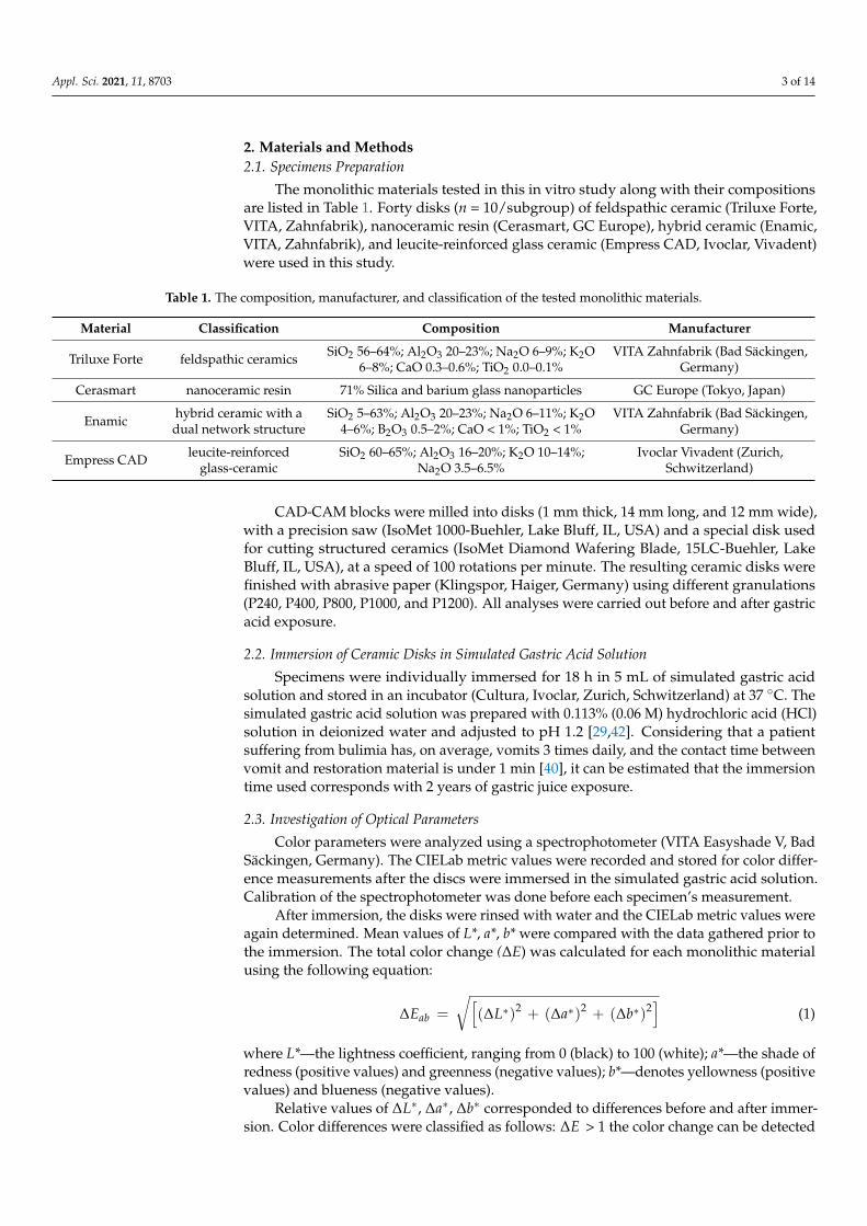
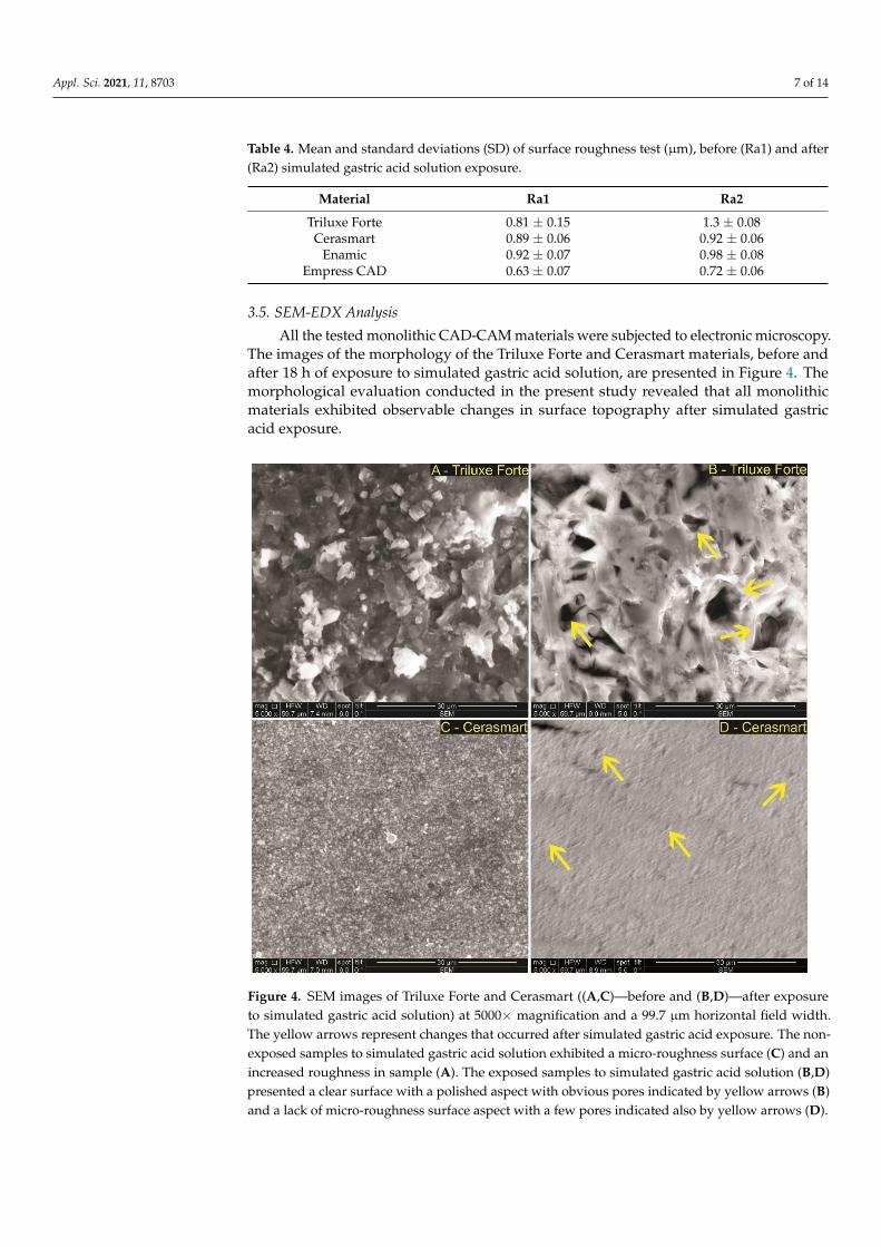
3.3. Microhardness Test
Regarding microhardness investigations, there was a significant decrease in the
tested CAD-CAM monolithic materials, except for Cerasmart, which retained the same
parameters. The monolithic material that suffered the largest change in terms of micro-
hardness was Triluxe Forte, as can be observed in Figure 3.
Figure 2. Comparison between the translucency parameters of four CAD-CAM monolithic materials,before exposure (initial) and after exposure (final).
Appl. Sci. 2021, 11, 8703 6 of 14
The values of the analysis results are shown in Table 3. Mean and standard deviationwere calculated for each material using ANOVA and Tukey’s test. According to Tukey’s test,the results are significant at p ≤ 0.05. The results obtained in this study for all monolithicmaterials tested proved to be statistically insignificant.
Table 3. Translucency parameters, before and after 18 h of simulated gastric acid solution exposure:mean and standard deviation.
MonolithicMaterial
Mean Standard Deviation
Before After Before After
Cerasmart 24.03 24.36 0.98 1.25Enamic 21.26 21.76 3.06 2.72
Empress CAD 19.56 20.74 3.87 3.42Triluxe Forte 19.51 18.43 2.19 2.11
3.3. Microhardness Test
Regarding microhardness investigations, there was a significant decrease in the testedCAD-CAM monolithic materials, except for Cerasmart, which retained the same parame-ters. The monolithic material that suffered the largest change in terms of microhardnesswas Triluxe Forte, as can be observed in Figure 3.
Appl. Sci. 2021, 11, x FOR PEER REVIEW 7 of 16
Figure 3. Comparison of microhardness values, before and after 18 h of immersion in simulated
gastric acid solution. One-way ANOVA analysis was applied to determine the statistical differ-
ences between monolithic materials, followed by the Tukey’s test (*** p < 0.001).
The results obtained regarding the microhardness test are highly significant, p < 0.001
for Enamic, Empress CAD, and Triluxe Forte. For the monolithic material Cerasmart, a p
value of 0.85 was obtained, denoted that the result was statistically insignificant.
3.4. Surface Roughness Results
The surface roughness mean and standard deviations are shown in Table 4. The re-
sults obtained denote that there were no significant differences for Cerasmart, Enamic,
and Empress CAD. Triluxe Forte, had statistically significant differences and exhibited
the most change in terms of surface roughness, which can reduce the strength of the res-
toration; a nonuniform stress distribution and can lead to fracture. The roughness results,
after 18 h of immersion in simulated gastric acid solution were: Triluxe Forte ˃ Enamic ˃
Cerasmart ˃ Empress CAD.
Table 4. Mean and standard deviations (SD) of surface roughness test (µm), before (Ra1) and after (Ra2) simulated gastric
acid solution exposure.
Material. Ra1 Ra2
Triluxe Forte 0.81 ± 0.15 1.3 ± 0.08
Cerasmart 0.89 ± 0.06 0.92 ± 0.06
Enamic 0.92 ± 0.07 0.98 ± 0.08
Empress CAD 0.63 ± 0.07 0.72 ± 0.06
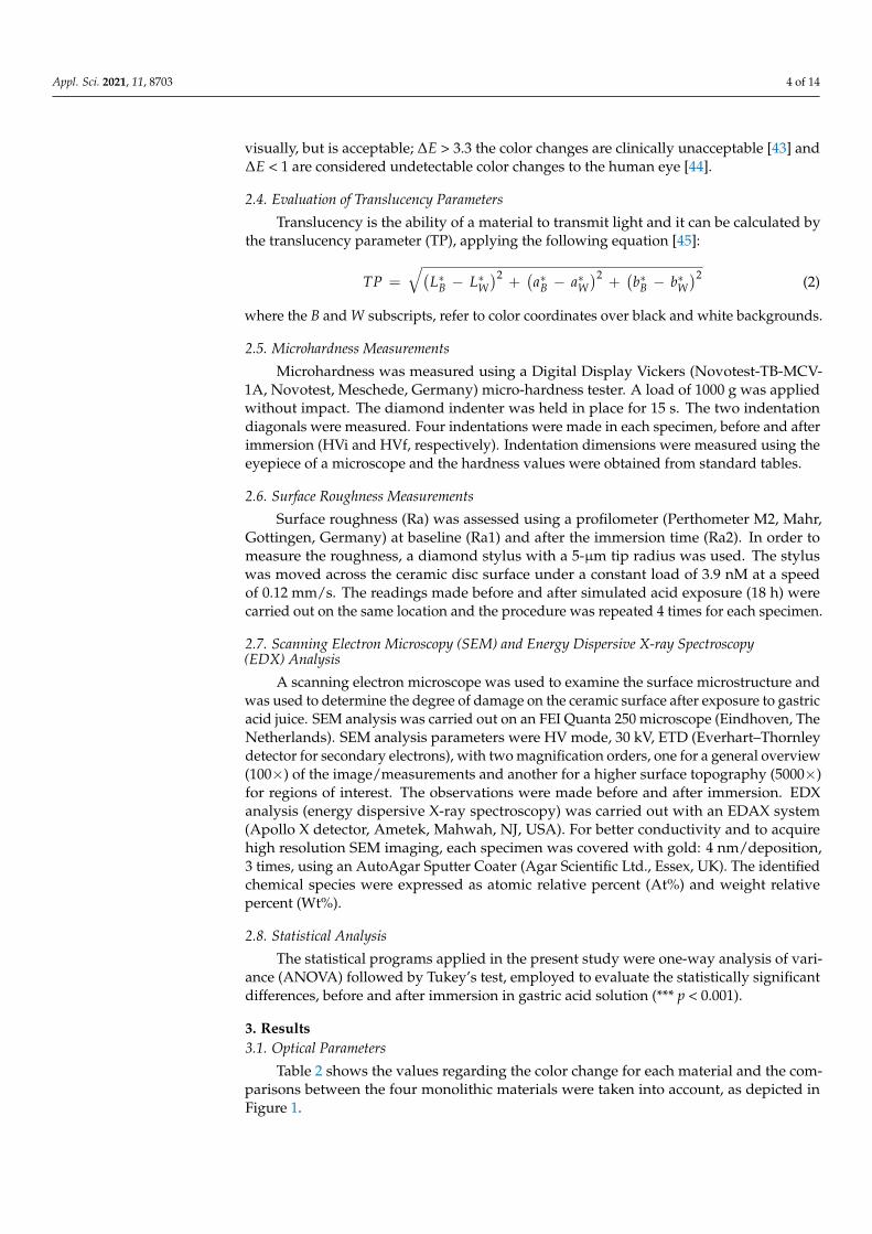
3.5. SEM-EDX Analysis
All the tested monolithic CAD-CAM materials were subjected to electronic micros-
copy. The images of the morphology of the Triluxe Forte and Cerasmart materials, before
and after 18 h of exposure to simulated gastric acid solution, are presented in Figure 4.
The morphological evaluation conducted in the present study revealed that all monolithic
materials exhibited observable changes in surface topography after simulated gastric acid
exposure.
Figure 3. Comparison of microhardness values, before and after 18 h of immersion in simulatedgastric acid solution. One-way ANOVA analysis was applied to determine the statistical differencesbetween monolithic materials, followed by the Tukey’s test (*** p < 0.001).
The results obtained regarding the microhardness test are highly significant, p < 0.001for Enamic, Empress CAD, and Triluxe Forte. For the monolithic material Cerasmart, ap value of 0.85 was obtained, denoted that the result was statistically insignificant.
3.4. Surface Roughness Results
The surface roughness mean and standard deviations are shown in Table 4. Theresults obtained denote that there were no significant differences for Cerasmart, Enamic,and Empress CAD. Triluxe Forte, had statistically significant differences and exhibited themost change in terms of surface roughness, which can reduce the strength of the restoration;a nonuniform stress distribution and can lead to fracture. The roughness results, after 18 hof immersion in simulated gastric acid solution were: Triluxe Forte > Enamic > Cerasmart> Empress CAD.
Appl. Sci. 2021, 11, 8703 7 of 14
Table 4. Mean and standard deviations (SD) of surface roughness test (µm), before (Ra1) and after(Ra2) simulated gastric acid solution exposure.
Material Ra1 Ra2
Triluxe Forte 0.81 ± 0.15 1.3 ± 0.08Cerasmart 0.89 ± 0.06 0.92 ± 0.06
Enamic 0.92 ± 0.07 0.98 ± 0.08Empress CAD 0.63 ± 0.07 0.72 ± 0.06
3.5. SEM-EDX Analysis
All the tested monolithic CAD-CAM materials were subjected to electronic microscopy.The images of the morphology of the Triluxe Forte and Cerasmart materials, before andafter 18 h of exposure to simulated gastric acid solution, are presented in Figure 4. Themorphological evaluation conducted in the present study revealed that all monolithicmaterials exhibited observable changes in surface topography after simulated gastricacid exposure.
Appl. Sci. 2021, 11, x FOR PEER REVIEW 8 of 16
Figure 4. SEM images of Triluxe Forte and Cerasmart ((A,C)—before and (B,D)—after exposure to
simulated gastric acid solution) at 5000× magnification and a 99.7 µm horizontal field width. The
yellow arrows represent changes that occurred after simulated gastric acid exposure. The non-ex-
posed samples to simulated gastric acid solution exhibited a micro-roughness surface (C) and an
increased roughness in sample (A). The exposed samples to simulated gastric acid solution (B,D)
presented a clear surface with a polished aspect with obvious pores indicated by yellow arrows (B)
and a lack of micro-roughness surface aspect with a few pores indicated also by yellow arrows (D).
An irregular surface, pores, and grooves of different sizes were observed in the feld-
spathic ceramic (Figure 4A—Triluxe Forte) before immersion. After immersion, the mate-
rial surface showed larger grooves and less irregularities (Figure 4B).
Cerasmart is a hybrid ceramic, composed of a resin matrix with silica and barium
glass nanoparticles. The glassy phase might be attacked less by gastric acid than the resin
phase, which leads to an increase in surface roughness.
In Figure 5, SEM images of Enamic and Empress monolithic materials are shown.
The non-exposed samples to simulated gastric acid solution exhibited a rough surface
lacking in sharpness (Figure 5A and the presence of a slight surface matrix appearance
(Figure 5C). The exposed samples to simulated gastric acid solution (Figure 5B,D) pre-
sented, in both samples, surfaces that were rough and obvious, the surface structures were
sharp compared to the control samples (non-exposed), and are highlighted by yellow ar-
rows.
Figure 4. SEM images of Triluxe Forte and Cerasmart ((A,C)—before and (B,D)—after exposureto simulated gastric acid solution) at 5000× magnification and a 99.7 µm horizontal field width.The yellow arrows represent changes that occurred after simulated gastric acid exposure. The non-exposed samples to simulated gastric acid solution exhibited a micro-roughness surface (C) and anincreased roughness in sample (A). The exposed samples to simulated gastric acid solution (B,D)presented a clear surface with a polished aspect with obvious pores indicated by yellow arrows (B)and a lack of micro-roughness surface aspect with a few pores indicated also by yellow arrows (D).
Appl. Sci. 2021, 11, 8703 8 of 14
An irregular surface, pores, and grooves of different sizes were observed in thefeldspathic ceramic (Figure 4A—Triluxe Forte) before immersion. After immersion, thematerial surface showed larger grooves and less irregularities (Figure 4B).
Cerasmart is a hybrid ceramic, composed of a resin matrix with silica and bariumglass nanoparticles. The glassy phase might be attacked less by gastric acid than the resinphase, which leads to an increase in surface roughness.
In Figure 5, SEM images of Enamic and Empress monolithic materials are shown. Thenon-exposed samples to simulated gastric acid solution exhibited a rough surface lackingin sharpness (Figure 5A and the presence of a slight surface matrix appearance (Figure 5C).The exposed samples to simulated gastric acid solution (Figure 5B,D) presented, in bothsamples, surfaces that were rough and obvious, the surface structures were sharp comparedto the control samples (non-exposed), and are highlighted by yellow arrows.
Appl. Sci. 2021, 11, x FOR PEER REVIEW 9 of 16
Figure 5. SEM images of Enamic and Empress CAD (A,C)—before and (B,D)—after exposure to
simulated gastric acid Scheme 5000× magnification and a 59.7–74.6 µm horizontal field width. The
yellow arrows represent the changes that occurred after simulated gastric acid exposure.
Enamic is a ceramic polymer interpenetrating network material. The Enamic material
consists of 75% feldspathic ceramic matrix, into which an organic phase of dimethacrylate
resin containing urethane dimethacrylate and triethylene glycol dimethacrylate is infil-
trated [46]. The microstructure has a hybrid structure with interpenetrating networks of
ceramic and polymer that mimic the interlocking of prisms in natural teeth. After simu-
lated gastric acid exposure, a porous microstructure with irregularities can be observed.
The microstructure of IPS Empress CAD consisted in a glassy matrix and leucite crys-
tals. The SEM image before exposure shows an interlocking microstructure that remained
after exposure, but with the appearance of irregularities and porosities.
The chemical composition of the elements contained in each material was determined
using EDX analysis. Figures 6 and 7 reveal the presence of peak elements recorded at a
specific value of amplitude. Table 5 shows the atomic percentage and weight percentage
of the recorded elements. According to both Figures 6 and 7 as well as Table 5, after 18 h
of simulated gastric acid solution exposure, the used restorative materials have approxi-
mately the same elements as before exposure (data not shown).
Figure 5. SEM images of Enamic and Empress CAD (A,C)—before and (B,D)—after exposure tosimulated gastric acid Scheme 5000× magnification and a 59.7–74.6 µm horizontal field width. Theyellow arrows represent the changes that occurred after simulated gastric acid exposure.
Enamic is a ceramic polymer interpenetrating network material. The Enamic materialconsists of 75% feldspathic ceramic matrix, into which an organic phase of dimethacrylateresin containing urethane dimethacrylate and triethylene glycol dimethacrylate is infil-trated [46]. The microstructure has a hybrid structure with interpenetrating networks ofceramic and polymer that mimic the interlocking of prisms in natural teeth. After simulatedgastric acid exposure, a porous microstructure with irregularities can be observed.
The microstructure of IPS Empress CAD consisted in a glassy matrix and leucitecrystals. The SEM image before exposure shows an interlocking microstructure thatremained after exposure, but with the appearance of irregularities and porosities.
Appl. Sci. 2021, 11, 8703 9 of 14
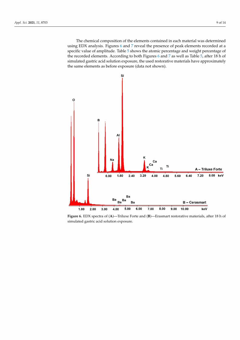
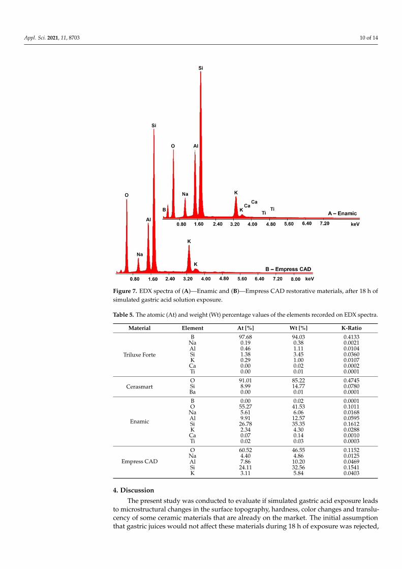
The chemical composition of the elements contained in each material was determinedusing EDX analysis. Figures 6 and 7 reveal the presence of peak elements recorded at aspecific value of amplitude. Table 5 shows the atomic percentage and weight percentage ofthe recorded elements. According to both Figures 6 and 7 as well as Table 5, after 18 h ofsimulated gastric acid solution exposure, the used restorative materials have approximatelythe same elements as before exposure (data not shown).
Appl. Sci. 2021, 11, x FOR PEER REVIEW 11 of 16
Figure 6. EDX spectra of (A)—Triluxe Forte and (B)—Erasmart restorative materials, after 18 h of
simulated gastric acid solution exposure.
Figure 6. EDX spectra of (A)—Triluxe Forte and (B)—Erasmart restorative materials, after 18 h ofsimulated gastric acid solution exposure.
Appl. Sci. 2021, 11, 8703 10 of 14Appl. Sci. 2021, 11, x FOR PEER REVIEW 12 of 16
Figure 7. EDX spectra of (A)—Enamic and (B)—Empress CAD restorative materials, after 18 h of
simulated gastric acid solution exposure.
4. Discussion
The present study was conducted to evaluate if simulated gastric acid exposure leads
to microstructural changes in the surface topography, hardness, color changes and trans-
lucency of some ceramic materials that are already on the market. The initial assumption
that gastric juices would not affect these materials during 18 h of exposure was rejected,
due to the fact that some features of the investigated monolithic materials were affected
by the simulated gastric acid solution.
The main objective of restorative dentistry is to replace a lost tooth structure with a
material that has optical and mechanical properties that are as close as possible to those
of natural tooth. According to Choi and co-workers [47], the mechanical behavior of re-
storative materials under multiaxial masticatory loading in the oral cavity depends on
flexure strengths under different loading conditions. The authors investigated the flexural
strengths and elastic properties of resin-composite block materials for CAD/CAM and
demonstrated that the strength reliability and the elastic modulus values of the tested ma-
terials were significantly different, depending on the testing method, despite the null hy-
pothesis from which they started, namely that there are no differences regarding the flex-
ural and elastic properties of resin-composite CAD/CAM block materials determined by
uniaxial and biaxial tests.
It must be also taken into account that oral restorations are exposed to various com-
plex oral conditions during their lifetimes. Dentists review medical histories and medica-
tions that identify patients with a diagnosis of acid reflux and they can clinically observe
the dental manifestations of gastroesophageal reflux [48]. Gastroesophageal reflux disease
is characterized by regular regurgitations of gastric juice from the stomach into the oral
Figure 7. EDX spectra of (A)—Enamic and (B)—Empress CAD restorative materials, after 18 h ofsimulated gastric acid solution exposure.
Table 5. The atomic (At) and weight (Wt) percentage values of the elements recorded on EDX spectra.
Material Element At [%] Wt [%] K-Ratio
Triluxe Forte
B 97.68 94.03 0.4133Na 0.19 0.38 0.0021Al 0.46 1.11 0.0104Si 1.38 3.45 0.0360K 0.29 1.00 0.0107Ca 0.00 0.02 0.0002Ti 0.00 0.01 0.0001
CerasmartO 91.01 85.22 0.4745Si 8.99 14.77 0.0780Ba 0.00 0.01 0.0001
Enamic
B 0.00 0.02 0.0001O 55.27 41.53 0.1011
Na 5.61 6.06 0.0168Al 9.91 12.57 0.0595Si 26.78 35.35 0.1612K 2.34 4.30 0.0288Ca 0.07 0.14 0.0010Ti 0.02 0.03 0.0003
Empress CAD
O 60.52 46.55 0.1152Na 4.40 4.86 0.0125Al 7.86 10.20 0.0469Si 24.11 32.56 0.1541K 3.11 5.84 0.0403
4. Discussion
The present study was conducted to evaluate if simulated gastric acid exposure leadsto microstructural changes in the surface topography, hardness, color changes and translu-cency of some ceramic materials that are already on the market. The initial assumptionthat gastric juices would not affect these materials during 18 h of exposure was rejected,
Appl. Sci. 2021, 11, 8703 11 of 14
due to the fact that some features of the investigated monolithic materials were affected bythe simulated gastric acid solution.
The main objective of restorative dentistry is to replace a lost tooth structure with amaterial that has optical and mechanical properties that are as close as possible to thoseof natural tooth. According to Choi and co-workers [47], the mechanical behavior ofrestorative materials under multiaxial masticatory loading in the oral cavity depends onflexure strengths under different loading conditions. The authors investigated the flexuralstrengths and elastic properties of resin-composite block materials for CAD/CAM anddemonstrated that the strength reliability and the elastic modulus values of the testedmaterials were significantly different, depending on the testing method, despite the nullhypothesis from which they started, namely that there are no differences regarding theflexural and elastic properties of resin-composite CAD/CAM block materials determinedby uniaxial and biaxial tests.
It must be also taken into account that oral restorations are exposed to various complexoral conditions during their lifetimes. Dentists review medical histories and medicationsthat identify patients with a diagnosis of acid reflux and they can clinically observe thedental manifestations of gastroesophageal reflux [48]. Gastroesophageal reflux diseaseis characterized by regular regurgitations of gastric juice from the stomach into the oralcavity [49]. If a patient suffering from gastroesophageal reflux needs a prosthetic restoration,a clinician must take the effect of gastric acid on the restorative material that will beused into account. Resistance to chemical degradation of dental materials is a principalrequirement for intra-oral use and represents a decisive factor when choosing the type ofthe restoration.
The in vitro simulation of the acid on the surface of dental ceramics depends on theconcentration of the acid, the time of immersion, and the temperature [48]. In this study, theworking pH was 1.2 and the immersion time was 18 h at 37 ◦C. In the literature, there arestudies that considered that the simulated gastric acid exposure of CAD-CAM materials for7.5 h, represent one month of gastric acid exposure, 45 h—represent six months of exposureand 91 h—represent one year of exposure. Backer et al. used CAD-CAM materials exposedto simulated gastric acid for 6 and 18 h, and they calculated that these times represent 2 and8 years of exposure of dental structure to vomiting [27]. Sulaiman et al. exposed monolithiczirconia to acid solution for 96 h, thus simulating over 10 years of dental structure exposureto vomiting [29]. Evaluating these studies, we can say that, in the literature, there is noclear consensus regarding the method of gastric acid simulation and the equivalent timefor replication for an in vivo model. According to ISO standard 6872, which refers to thesolubility test for dental materials, the use of 4% acetic acid and an exposure time of 16 h at80 ◦C is the equivalent with 2 years of clinical exposure [50].
It is well known that gastric juice has a demineralization effect on enamel, dentin,and cement [28]. Due to its very low pH, it can dissolve the glassy matrix of ceramicmaterials [31]. In the present study, it was demonstrated that, in the case of feldspathicceramic, nanoceramic resin, hybrid ceramic, and leucite-reinforced glass ceramic, scanningelectron microscopy revealed observable changes in the surface topography after simulatedgastric acid exposure for all tested monolithic materials. In the case of Triluxe Forte,the pores and grooves present before immersion were larger after, but there were lessirregularities on the material surface. Regarding the Cerasmart material, exposure tosimulated gastric acid solution led to increased surface roughness. This phenomena couldbe explained by the continued leaching of the particles in the presence of the simulatedgastric acid solution. The Enamic and Empress CAD materials became more porous witha lot of irregularities. This could be due to the dissolution of the ceramic part containedin the monolithic material or other components [30]. Following analyses on the surfacemorphology after exposure to simulated gastric acid solution, we can say that Triluxe Fortedisplayed areas of possible degradation, due to the formed crater-like grooves (Figure 4B).In addition, the porosity of the surfaces leads to an accumulation of bacterial plaque and
Appl. Sci. 2021, 11, 8703 12 of 14
the wear of antagonistic natural teeth. The appearance of pores denotes that exposure toan acidic element will influence the surface of the exposed material [51].
The impact of the simulated gastric acid solution on the optical properties of allfour evaluated monolithic materials showed that there were no differences among thespecimens, except for Triluxe Forte, which underwent a slight discoloration after exposureto simulated gastric acid. Passing light through a translucent material is reduced by thescattering of small-sized particles, such as filler particles and porosity voids, which caninfluence color perception and appearance of dental ceramics [52]. In the present studythe translucency of the tested materials increased insignificantly after simulated gastricacid exposure and the color values had undergone minor changes, which do not affectthe perceptibility and the acceptability thresholds. Cruz et al. obtained the same results,stating that all the ceramic materials tested promoted a color change of ∆E < 1, classified asclinically undetectable [30].
The color of a restoration can be affected by a rough surface that reflects an irregularand diffuse pattern of light [53]. According to the results obtained (Table 4), at baseline,leucite-reinforced glass-ceramic showed the lowest surface roughness, while hybrid ceramicshowed the highest surface roughness. After 18 h of immersion in simulated gastric acidsolution, the surface characteristics of the hybrid ceramics and leucite glass ceramics didnot suffer any significant change.
The simulated gastric acid solution seriously affected the hardness of three of fourCAD-CAM monolithic restorative materials, namely, Enamic, Empress CAD, and TriluxeForte. Among the three restorative materials, once again, Triluxe Forte was most affected,presenting a significant decrease in microhardness after simulated gastric acid exposure.
Our findings, regarding all the investigations are in agreement with the literaturedata [28–30,37,54,55].
Limitations of the present study include the fact that the materials were not alsoexposed to saliva to fully mimic the oral environment. Exposing ceramic materials to saliva,as well as gastric acid, might have more closely replicated clinical situations. Another limi-tation of the current study is that the ceramic discs were not glazed, only polished, whichfor some materials (i.e., Cerasmart and Enamic) is considered a better finishing method.
5. Conclusions
Based on the results of this in vitro study, it can be concluded that there is an increasingneed in manufacturing of improve restorative materials, which also comply with the specialneeds of patients with medical problems, such as gastroesophageal reflux, bulimia, orprolonged severe nausea episodes during pregnancy. For these kinds of patients, thedevelopment of materials with good mechanical, optical, physicochemical, and surfacetopography, will improve their quality of life. The results obtained in this study will helpdentists in choosing the best material for patients with other medical problems. In thisregard, the results of the present study clearly showed that Triluxe Forte was the CAD-CAMmonolithic restorative material that suffered the most important changes after exposureto simulated gastric acid solution (i.e., decrease of hardness, increasing roughness, colorchange, appearance of pores and irregularities and visualization of crater-like grooves,which means degradation of ceramic part or other components embedded into the material).On the other hand, the Cerasmart monolithic restorative material was proven to be theleast affected after simulated gastric acid exposure.
Author Contributions: Conceptualization, I.L.P. and A.J.; methodology, I.L.P., D.P., E.-A.M., C.-V.M.and C.I.; software, D.P., E.-A.M. and C.-V.M.; validation, E.-A.M. and A.J.; formal analysis, I.L.P., D.P.,C.-V.M. and C.I..; investigation, I.L.P., D.P., E.-A.M., C.-V.M. and C.I.; resources, A.J.; data curation,I.L.P., D.P., E.-A.M., C.-V.M. and C.I.; writing—original draft preparation, I.L.P.; writing—review andediting, E.-A.M.; visualization, A.J.; supervision, A.J.; project administration, A.J. All authors haveread and agreed to the published version of the manuscript.
Funding: This research received no external funding.
Appl. Sci. 2021, 11, 8703 13 of 14
Data Availability Statement: Data presented in this study are available on request from the first author.
Conflicts of Interest: The authors declare no conflict of interest.
References1. Mörmann, W.H.; Stawarczyk, B.; Ender, A.; Sener, B.; Attin, T.; Mehl, A. Wear characteristics of current aesthetic dental restorative
CAD/CAM materials: Two-body wear, gloss retention, roughness and Martens hardness. J. Mech. Behav. Biomed. Mater. 2013, 20,113–125. [CrossRef] [PubMed]
2. Gracis, S.; Thompson, V.P.; Ferencz, J.L.; Silva, N.R.; Bonfante, E.A. A new classification system for all ceramic and ceramic-likerestorative materials. Int. J. Prosthodont. 2015, 28, 227–235. [CrossRef]
3. Traini, T.; Sinjari, B.; Pascetta, R.; Serafini, N.; Perfetti, G.; Trisi, P.; Caputi, S. The zirconia-reinforced lithium silicate ceramic:Lights and shadows of a new material. Dent. Mater. J. 2016, 35, 748–755. [CrossRef]
4. Sato, T.P.; Anami, L.C.; Melo, R.M.; Valandro, L.F.; Bottino, M.A. Effects of Surface Treatments on the Bond Strength BetweenResin Cement and a New Zirconia-reinforced Lithium Silicate Ceramic. Oper. Dent. 2016, 41, 284–292. [CrossRef] [PubMed]
5. Elsaka, S.E.; Elnaghy, A.M. Mechanical properties of zirconia reinforced lithium silicate glass-ceramic. Dent. Mater. 2016, 32,908–914. [CrossRef] [PubMed]
6. Alves, L.M.M.; Contreras, L.P.C.; Campos, T.M.B.; Bottino, M.A.; Valandro, L.F.; Melo, R.M. In vitro wear of a zirconium-reinforcedlithium silicate ceramic against different restorative materials. J. Mech. Behav. Biomed. Mater. 2019, 100, 103403. [CrossRef][PubMed]
7. Maenosono, R.M.; Brianezzi, L.F.F.; Ishikiriama, S.K.; Furuse, A.Y. Clinical perceptions of zirconia-reinforced lithium silicateceramic: A case report and 6-month follow-up. Gen. Dent. 2019, 67, 47–50. [PubMed]
8. Curtis, A.R.; Shortall, A.C.; Marquis, P.M.; Palin, W.M. Water uptake and strength characteristics of a nanofilled resin-basedcomposite. J. Dent. 2008, 36, 186–193. [CrossRef]
9. Kim, Y.S.; Kwon, H.K.; Kim, B.I. Effect of nano-carbonate apatite to prevent restain after dental bleaching in vitro. J. Dent. 2011,39, 636–642. [CrossRef]
10. Ernst, C.P.; Brandenbusch, M.; Meyer, G.; Canbek, K.; Gottschalk, F.; Willershausen, B. Two-year clinical performance of ananofiller vs. a fine-particle hybrid resin composite. Clin. Oral Investig. 2006, 10, 119–125. [CrossRef]
11. Sauro, S.; Osorio, R.; Watson, T.F.; Toledano, M. Therapeutic effects of novel resin bonding systems containing bioactive glasseson mineral-depleted areas within the bonded-dentine interface. J. Mater. Sci. Mater. Med. 2012, 23, 1521–1532. [CrossRef]
12. De Fúcio, S.B.; Bolzan de Paula, A.; Galbiatti de Carvalho, F.; Pinheiro Feitosa, V.; Ambrosano, G.M.B.; Puppin-Rontani, R.M.Biomechanical degradation of the nano-filled resin-modified glass-ionomer surface. Am. J. Dent. 2012, 25, 315–320.
13. Chen, C.; Trindade, F.Z.; de Jager, N.; Kleverlaan, C.J.; Feilzer, A.J. The fracture resistance of a CAD/CAM Resin Nano Ceramic(RNC) and a CAD ceramic at different thicknesses. Dent. Mater. 2014, 30, 954–962. [CrossRef] [PubMed]
14. Alves, P.B.; Brandt, W.C.; Neves, A.C.; Gonçalves Cunha, L.; Silva-Concilio, L.R. Mechanical properties of direct and indirectcomposites after storage for 24 h and 10 months. Eur J. Dent. 2013, 7, 117–122. [PubMed]
15. Belli, R.; Geinzer, E.; Muschweck, A.; Petschelt, A.; Lohbauer, U. Mechanical fatigue degradation of ceramics versus resincomposites for dental restorations. Dent. Mater. 2014, 30, 424–432. [CrossRef] [PubMed]
16. Sen, N.; Us, Y.O. Mechanical and optical properties of monolithic CAD-CAM restorative materials. J. Prosthet Dent. 2018, 119,593–599. [CrossRef] [PubMed]
17. Belli, R.; Wendler, M.; de Ligny, D.; Cicconi, M.R.; Petschelt, A.; Peterlik, H.; Lohbauer, U. Chairside CAD/CAM materials. Part 1:Measurement of elastic constants and microstructural characterization. Dent. Mater. 2017, 33, 84–98. [CrossRef] [PubMed]
18. Lawson, N.C.; Bansal, R.; Burgess, J.O. Wear, strength, modulus and hardness of CAD/CAM restorative materials. Dent. Mater.2016, 32, 275–283. [CrossRef]
19. Albero, A.; Pascual, A.; Camps, I.; Grau-Benitez, M. Comparative characterization of a novel cad-cam polymer-infiltrated-ceramic-network. J. Clin. Exp. Dent. 2015, 7, 495–500. [CrossRef]
20. Acar, O.; Yilmaz, B.; Altintas, S.H.; Chandrasekaran, I.; Johnston, W.M. Color stainability of CAD/CAM and nanocomposite resinmaterials. J. Prosthet. Dent. 2016, 115, 71–75. [CrossRef]
21. Stawarczyk, B.; Liebermann, A.; Eichberger, M.; Güth, J.F. Evaluation of mechanical and optical behavior of current estheticdental restorative CAD/CAM composites. J. Mech. Behav. Biomed. Mater. 2016, 55, 1–11. [CrossRef]
22. Karakaya, I.; Cengiz, E. Effect of 2 bleaching agents with a content of high concentrated hydrogen peroxide on stained 2CAD/CAM blocks and a nanohybrid composite resin: An AFM evaluation. Biomed. Res. Int. 2017, 2017, 6347145. [CrossRef]
23. Jones, L.; Lekkas, D.; Hunt, D.; McIntyre, J.; Rafir, W. Studies on dental erosion: An in vivo-in vitro model of endogenous dentalerosion—Its application to testing protection by fluoride gel application. Aust. Dent. J. 2002, 47, 304–308. [CrossRef] [PubMed]
24. Turssi, C.P.; Hara, A.T.; de Magalhaes, C.S.; Campos Serra, M.; Rodrigues Jr, A.L. Influence of storage regime prior to abrasion onsurface topography of restorative materials. J. Biomed. Mater. Res. B Appl. Biomater. 2003, 65, 227–232. [CrossRef] [PubMed]
25. Yu, H.; Wegehaupt, F.J.; Wiegand, A.; Roos, M.; Attin, T.; Buchalla, W. Erosion and abrasion of tooth-colored restorative materialsand human enamel. J. Dent. 2009, 37, 913–922. [CrossRef] [PubMed]
26. Matsou, E.; Vouroutzis, N.; Kontonasaki, E.; Paraskevopoulos, K.M.; Koidis, P. Investigation of the influence of gastric acid onthe surface roughness of ceramic materials of metal-ceramic restorations. An in vitro study. Int. J. Prosthodont. 2011, 24, 26–29.[PubMed]
Appl. Sci. 2021, 11, 8703 14 of 14
27. Backer, A.D.; Münchow, E.A.; Eckert, G.J.; Hara, A.T.; Platt, J.A.; Bottino, M.C. Effects of simulated gastric juice on CAD/CAMresin composites-morphological and mechanical evaluations. J. Prosthodont. 2017, 26, 424–443. [CrossRef]
28. Bartlett, D.W.; Coward, P.Y. Comparison of the erosive potential of gastric juice and a carbonated drink in vitro. J. Oral Rehabil.2001, 28, 1045–1047. [CrossRef]
29. Sulaiman, T.A.; Abdulmajeed, A.A.; Shahramian, K.; Hupa, L.; Donovan, T.E.; Vallittu, P.; Närhi, T.O. Impact of gastric acidicchallenge on surface topography and optical properties of monolithic zirconia. Dent. Mater. 2015, 31, 1445–1452. [CrossRef]
30. Cruz, M.E.M.; Simoes, R.; Martins, S.B.; Trinidade, F.Z.; Dovigo, L.N.; Fonseca, R.G. Influence of simulated gastric juice on surfacecharacteristics of CAD-CAM monolithic materials. J. Prosthet Dent. 2020, 123, 483–490. [CrossRef]
31. Harryparsad, A.; Dullabh, H.; Sykes, L.; Herbst, D. The effects of hydrochloric acid on all-ceramic restorative materials: Anin-vitro study. SADJ 2014, 69, 106–111.
32. Castillo, M.; Weiselberg, E. Bulimia nervosa/purging disorder. Curr. Probl. Pediatr. Adolesc. Health Care 2017, 47, 85–94. [CrossRef]33. Sengupta, A. Dental Erosion: Etiology, Diagnosis and Management. Acta Sci. Dent. Sci. 2018, 2, 43–48.34. Haq, M.W.; Batool, M.; Ahsan, S.H.; Lone, M.A.; Islam, T. Dental erosion: Influencing factors & pH analysis. Can. J. Appl. Sci.
2012, 2, 222–232.35. Zhang, J.; Du, Y.; Wei, Z.; Tai, B.; Jiang, H.; Du, M. The prevalence and risk indicators of tooth wear in 12- and 15-year-old
adolescents in Central China. BMC Oral Health. 2015, 15, 120. [CrossRef] [PubMed]36. Theocharidou, A.; Kontonasaki, E.; Koukousaki, I.; Koumpouli, A.; Betsani, I.; Koidis, P. Effect of in vitro aging and acidic storage
on color, translucency, and contrast ratio of monolithic zirconia and lithium disilicate ceramics. J. Prosthet. Dent. 2021, 3913,30805–30815.
37. Kulkarni, A.; Rothrock, J.; Thompson, J. Impact of Gastric Acid Induced Surface Changes on Mechanical Behavior and OpticalCharacteristics of Dental Ceramics. J. Prosthodont. 2020, 29, 207–218. [CrossRef] [PubMed]
38. Egilmez, F.; Ergun, G.; Cekic-Nagas, I.; Vallittu, P.K.; Lassila, L.V.J. Does artificial aging affect mechanical properties of CAD/CAMcomposite materials. J. Prosthodont. Res. 2018, 62, 65–74. [CrossRef]
39. Kaur, S.; Makkar, S.; Kumar, R.; Pasricha, S.; Gupta, P. Comparative evaluation of surface properties of enamel and differentesthetic restorative materials under erosive and abrasive challenges: An in vitro study. Indian J. Dent. 2015, 6, 172–180.
40. Zaki, D.Y.I.; Hamzawy, E.M.A.; El Halim, S.A.; Amer, M.A. Effect of simulated gastric juice on surface characteristics of directesthetic restorations. Aust. J. Basic Appl. Sci. 2012, 6, 686–694.
41. Cengiz, S.; Sarac, S.; Özcan, M. Effects of simulated gastric juice on color stability, surface roughness and microhardness oflaboratory-processed composites. Dent. Mater. J. 2014, 33, 343–348. [CrossRef] [PubMed]
42. Bakar, W.Z.W.; McIntyre, J. Susceptibility of selected tooth-coloured dental materials to damage by common erosive acids. Aust.Dent. J. 2008, 53, 226–234. [CrossRef] [PubMed]
43. de Carvalho Panzeri Pires-de-Souza, F.; Assirati Casemiro, L.; da Fonseca Roberti Garcia, L.; Rodrigues Cruvinel, D. Colorstability of dental ceramics submitted to artificial accelerated aging after repeated firings. J. Prosthet. Dent. 2009, 101, 13–18.[CrossRef]
44. Pires, L.A.; Novais, P.M.; Araújo, V.D.; Pegoraro, L.F. Effects of the type and thickness of ceramic, substrate, and cement on theoptical color of a lithium disilicate ceramic. J. Prosthet Dent. 2017, 117, 144–149. [CrossRef] [PubMed]
45. Salas, M.; Lucena, C.; Javier Herrera, L.; Yebra, A.; Della Bona, A.; Pérez, M.M. Translucency thresholds for dental materials. Dent.Mater. 2018, 34, 1168–1174. [CrossRef] [PubMed]
46. Zhang, Y.; Kelly, J.R. Dental Ceramics for Restoration and Metal Veneering. Dent. Clin. N. Am. 2017, 61, 797–819. [CrossRef]47. Choi, B.J.; Yoon, S.; Im, I.W.; Lee, J.H.; Jung, H.J.; Lee, H.H. Uniaxial/biaxial flexure strengths and elastic properties of resin-
composite block materials for CAD/CAM. Dent. Mater. 2019, 35, 389–401. [CrossRef]48. Aldamaty, M.F.; Haggag, K.M.; Othman, H.I. Effect of simulated gastric acid on surface roughness of different monolithic ceramics.
Al-Azhar J. Dent. Sci. 2020, 23, 327–334. [CrossRef]49. El-Serag, H.B.; Sweet, S.; Winchester, C.C.; Dent, J. Update on the epidemiology of gastro-oesophageal reflux disease: A systematic
review. Gut 2014, 63, 871–880. [CrossRef]50. International Organization for Standardization. International Standards for Dental Ceramics; ISO 6872; International Organization
for Standardization: Geneva, Switzerland, 1995.51. Carvalho Ramos, N.; Campos, T.M.; Paz, I.S.; Machado, J.P.; Bottino, M.A.; Cesar, P.F.; de Melo, R.M. Microstructure characteriza-
tion and SCG of newly engineered dental ceramics. Dent. Mater. 2016, 32, 870–878. [CrossRef]52. Della Bona, A.; Nogueira, A.D.; Pecho, O.E. Optical properties of CAD-CAM ceramic systems. J. Dent. 2014, 42, 1202–1209.
[CrossRef] [PubMed]53. Sarac, D.; Sarac, Y.S.; Yuzbasioglu, E.; Bal, S. The effects of porcelain polishing systems on the color and surface texture of
feldspathic porcelain. J. Prosthet. Dent. 2006, 96, 122–128. [CrossRef] [PubMed]54. Kukiattrakoon, B.; Hengtrakool, C.; Kedjarune-Leggat, U. Chemical durability and microhardness of dental ceramics immersed
in acidic agents. Acta Odontol. Scand. 2010, 68, 1–10. [CrossRef] [PubMed]55. Zakir, T.; Dandekeri, S.; Suhaim, K.S.; Shetty, N.H.G.; Ragher, M.; Shetty, S.K. Influence of aerated drink, mouthwash, and
simulated gastric acid on the surface roughness of dental ceramics: A comparative in Vitro study. J. Pharm. Bioallied. Sci. 2020, 12,S480–S4871.
Related Documents


