HAL Id: tel-01881106 https://tel.archives-ouvertes.fr/tel-01881106 Submitted on 25 Sep 2018 HAL is a multi-disciplinary open access archive for the deposit and dissemination of sci- entific research documents, whether they are pub- lished or not. The documents may come from teaching and research institutions in France or abroad, or from public or private research centers. L’archive ouverte pluridisciplinaire HAL, est destinée au dépôt et à la diffusion de documents scientifiques de niveau recherche, publiés ou non, émanant des établissements d’enseignement et de recherche français ou étrangers, des laboratoires publics ou privés. Effects of prenatal stress on sepia offcinalis Caitlin O Brien To cite this version: Caitlin O Brien. Effects of prenatal stress on sepia offcinalis. Animal biology. Normandie Université, 2017. English. NNT : 2017NORMC249. tel-01881106

Welcome message from author
This document is posted to help you gain knowledge. Please leave a comment to let me know what you think about it! Share it to your friends and learn new things together.
Transcript

HAL Id: tel-01881106https://tel.archives-ouvertes.fr/tel-01881106
Submitted on 25 Sep 2018
HAL is a multi-disciplinary open accessarchive for the deposit and dissemination of sci-entific research documents, whether they are pub-lished or not. The documents may come fromteaching and research institutions in France orabroad, or from public or private research centers.
L’archive ouverte pluridisciplinaire HAL, estdestinée au dépôt et à la diffusion de documentsscientifiques de niveau recherche, publiés ou non,émanant des établissements d’enseignement et derecherche français ou étrangers, des laboratoirespublics ou privés.
Effects of prenatal stress on sepia officinalisCaitlin O Brien
To cite this version:Caitlin O Brien. Effects of prenatal stress on sepia officinalis. Animal biology. Normandie Université,2017. English. �NNT : 2017NORMC249�. �tel-01881106�
THESE
Pour obtenir le diplôme de doctorat
Spécialité Physiologie et Biologie des Organismes, Populations, Interactions
Préparée au sein de Université de Caen-Normandie
Effects of Prenatal Stress on Sepia officinalis
Présentée et soutenue par
Caitlin Elizabeth O’BRIEN
Thèse dirigée par Ludovic DICKEL, UMR 6552 EthoS
Thèse soutenue publiquement le 08/12/2017
devant le jury composé de
Mr Olivier BASUYAUX Docteur, SMEL, Blainville sur Mer Examinateur
Mme Aline BERTIN Chargée de Recherches INRA, HDR, Université de Tours Rapporteur
Mr Paco BUSTAMANTE Professeur des Universités, Université de LaRochelle Rapporteur
Mme Anne-Sophie DARMAILLACQ Maître de Conférences, HDR, Université de Caen-Normandie Examinateur
Mme Patrizia D’ETORRE Professeur des Universités, Université de Paris XIII Examinateur
Mr Ludovic DICKEL Professeur des Universités, Université de Caen-Normandie Directeur de thèse
Mr Jean-Paul ROBIN Professeur des Universités, Université de Caen-Normandie Examinateur

Publications and Submitted Manuscripts 1. O'Brien, C. E., Bowie, M., Billard, P., Darmaillacq, A. S., Jozet-Alves, C., Behaïm, D., Basuyaux, O., & Dickel, L. "The effect of an artificial incubation environment on hatchling size and behavior in the cuttlefish, Sepia officinalis." Vie et Milieu—Life and Environment 66.1 (2016): 97–105. Article #5, Pages 130-148. 2. O'Brien, C. E., Mezrai, N., Darmaillacq, A. S., & Dickel, L. Behavioral development in embryonic and early juvenile cuttlefish (Sepia officinalis). Developmental Psychobiology 9999 (2016): 1–16. Article #2, Pages 36-66. 3. Darmaillacq, A. S., Mezrai, N., O'Brien, C. E., & Dickel, L. Visual ecology and the development of visually guided behavior in the cuttlefish. Frontiers in Physiology, 8 (2017): 1–8. Article #3, Pages 67-81. 4. O’Brien, C. E., Bellanger, C., Jozet-Alves, C., Mezrai, N., Darmaillacq, A. S., & Dickel, L. Effects of Maternal and Embryonic Stress on Egg Production and Offspring in the Cuttlefish, Sepia officinalis. Journal of Experimental Marine Biology and Ecology (Under Revision). Article #1, Pages 11-28. 5. O’Brien, C. E., Jozet-Alves, C., Mezrai, N., Bellanger, C., Darmaillacq, A. S., & Dickel, L. Maternal and Embryonic Stress Influence Offspring Behavior in the Cuttlefish Sepia officinalis. Frontiers in Physiology (Under Revision). Article #4, Pages 82-102. 6. O’Brien, C.E., Roumbedakis, K., Winkelmann, I. The Future of Cephalopod Science: Perspectives from Three Early-Career Researchers. Frontiers in Physiology (In Preparation). Article #6, Pages 159-168.

International Congresses
Oral Presentations
O’Brien, C.E. “Cephalopod Research: Visions of the Future; Behavior, Cognition and Neurobiology.” 2017 COST Action CephsInAction and CIAC meeting (“Cephalopod Science; from Biology to Welfare”) in Heraklion, Crete (Greece), March 28-30, 2017.* *Keynote lecture O’Brien, C.E., Jozet-Alves, C., Darmaillacq A-S., Mezrai, N., Bellanger, C., Dickel L. “Effects of Reproductive Stress on Offspring Behavior in the cuttlefish, Sepia officinalis.” 2017 COST Action CephsInAction and CIAC meeting (“Cephalopod Science; from Biology to Welfare”) in Heraklion, Crete (Greece), March 28-30, 2017.
Posters
O’Brien, C.E., Bellanger, C., Jozet-Alves, C., Mezrai, N., Darmaillacq A-S., Dickel L. “Reproductive Effects of Maternal and Prenatal Stress in the Cuttlefish, Sepia officinalis.” COST Action CephsInAction and CIAC meeting (“Cephalopod Science; from Biology to Welfare”) in Heraklion, Crete (Greece), March 28-30, 2017. O’Brien, C.E., Bellanger, C., Jozet-Alves, C., Mezrai, N., Darmaillacq A-S., Dickel L. “Reproductive Effects of Maternal and Prenatal Stress in the Cuttlefish, Sepia officinalis.” 2017 L'Ecole doctorale Normande de Biologie Intégrative, Santé et Environnement (EdNBISE) conference in Le Havre, France, March 16-17, 2017.
National Congresses
Oral Presentations
O’Brien, C.E., Jozet-Alves, C., Darmaillacq A-S., Mezrai, N., Bellanger, C., Dickel L. “Effects of maternal stress on behavior in juvenile cuttlefish (Sepia officinalis).” 2016 Société Française pour l’Étude du Comportement Animal (SFECA) conference in Caen, France, March 22-24, 2016.*
*recipient of the Castor Prize for best oral presentation O’Brien, C.E., Jozet-Alves, C., Darmaillacq A-S., Mezrai, N., Bellanger, C., Dickel L. “Effects of artificial incubation and prenatal predator exposure on hatchling behavior in Sepia officinalis.” 2015 Société Française pour l’Étude du Comportement Animal (SFECA) conference in Strasbourg, France, April 20-23, 2015.
Posters
O’Brien, C.E., Bowie, M., Billard, P., Darmaillacq, A.S., Jozet-Alves, C., Benhaïm, D., Basuyaux, O., Dickel. L. “Embryonic predator-exposure affects visual acuity during the first prey encounter of Sepia officinalis.” 2015 L'Ecole doctorale Normande de Biologie Intégrative, Santé et Environnement (EdNBISE) conference in Le Havre, France, March 16-17, 2017.

Acknowledgments
First off, I would like to thank my adviser and mentor, Dr. Ludovic Dickel, for reaching out to me
four years ago and giving me this life-changing opportunity to pursue my dream of cephalopod research.
Your patience and guidance have been immeasurably helpful to this research and my professional
development, and I feel incredibly honored and grateful to have had you as a supervisor. Merci.
I am also grateful for the guidance of Anne-Sophie Darmaillacq, who generously shared her vast
knowledge of ethology and prenatal development with me.
Without the wisdom of Christelle Jozet-Alves, I could never have found my way around the
marine station or completed a statistical analysis. Thank you for tolerating my constant unscheduled
demands on your knowledge!
In addition to collaboration and guidance at work, Dr. Cécile Bellanger invited me into her home
and family. Dinners and outings with the Bellanger family were a highlight of my week, and made me
feel welcome in an unfamiliar country. Merci Cecile, Laurent, Alice, Hugo and Praline.
I am also incredibly thankful to have had the aid of Isabelle Chevalier, Nadège Villain-Naud and
Céline Thomasse. Merci beaucoup ladies! Je pouvais toujours compter sur vous!
At the Centre de Recherches en Environnement Côtier (CREC), Jean Paul Lehodey, David
Liegard, Frédéric Guyon and David Lemeille provided logistical support. The Synergie Mer et Littoral
(SMEL) was kind enough to host me during several experiments. These experiments benefitted from the
assistance of several WONDERFUL technicians, including Sébastien Pien, Vincent Lefebvre, Suzy Moal
and Jean-Louis Lesoif.
Moreover, Dr. Olivier Basuyaux, as well as Cécile, Yaël Yuna and Yeti Basuyaux generously
invited me to stay with them in their home in Blainville while I conducted these experiments. The time I
spent in this bucolic heaven-on-earth with la petit famille is definitely the highlight of my three years in
France. Merci!
Several students of mine became good friends in the course of our time together. Kevin Bairos-
Novak, you were the best summer companion I could have asked for! Pauline Billiard, thank you for
being such a good sport with a mentor who was new to France and clueless! Camille Auger, I was super
impressed with your motivation to learn something outside your chosen field. And Estelle Paupy, you
did a tremendous job analyzing neurobiological samples and learning experiments. Other students who
provided invaluable assistance with experiments and analysis included Chloe Jane Way, Héloïse Duretz,
Alexis Perret, Hannah Cockerton, Hannah Amor and Tim Pakyar. Thank you!
My officemates at the university were a source of guidance, laughter and comfort during the
three years of my thesis, especially when it came to French to English translation! Flavie Bidel, you were
a constant source of support, advice and inspiration! Anne Quiédeville what little French I know is
thanks to you and your patience. Greg Beaudet, I miss your random nerf intrusions during slow
afternoons. Thanks to the three of you for adopting this wayward American. Katia Hamidouche, thanks
for making me laugh whenever I needed it. Apolline Chabenat, you’re a great housemate. Nawel
Mezrai, it’s the sun, stupid! Rachel Asselot, thanks for distracting me daily with your dreamy stares.
Pierre Lecouflet, thanks for playing the crossword with me. I would also like to thank Alex Schnell, who
was a source of professional inspiration and guidance, as well as a weekly bowl of pho. And many thanks
as well to Sophie Corvaisier and Marc Pignon for all of the delightful outings and laughter in Bretagne!
To all the other wonderful folks at the GMPc, thank you for welcoming me to your lab and
tolerating my terrible French!
Thanks also to Anna Mazaleyrat and Julie "Pangolin" Fleitz for introducing me to all the
wonderful things to see in Normandie during my first summer in France.
I would also like to thank the country of France and the European Union for being bastions of
rationality and scientific advancement at a time when my own country has apparently decided to
sacrifice science on the altar of populism. Also, France, you make REALLY good bread and pastries. Yum!
Finally, I would like to thank my outstanding parents, Barbara and Michael O’Brien, without
whom I would not be the person I am today, and who provided me with the love, opportunity and
support to pursue my interests and career.
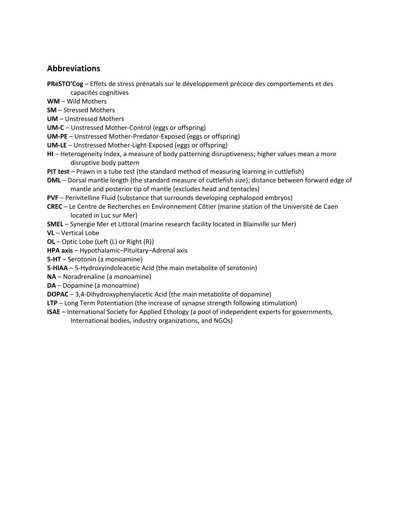
Abbreviations
PReSTO’Cog – Effets de stress prénatals sur le développement précoce des comportements et des capacités cognitives
WM – Wild Mothers SM – Stressed Mothers UM – Unstressed Mothers UM-C – Unstressed Mother-Control (eggs or offspring) UM-PE – Unstressed Mother-Predator-Exposed (eggs or offspring) UM-LE – Unstressed Mother-Light-Exposed (eggs or offspring) HI – Heterogeneity Index, a measure of body patterning disruptiveness; higher values mean a more
disruptive body pattern PIT test – Prawn in a tube test (the standard method of measuring learning in cuttlefish) DML – Dorsal mantle length (the standard measure of cuttlefish size); distance between forward edge of
mantle and posterior tip of mantle (excludes head and tentacles) PVF – Perivitelline Fluid (substance that surrounds developing cephalopod embryos) CREC – Le Centre de Recherches en Environnement Côtier (marine station of the Université de Caen
located in Luc sur Mer) SMEL – Synergie Mer et Littoral (marine research facility located in Blainville sur Mer) VL – Vertical Lobe OL – Optic Lobe (Left (L) or Right (R)) HPA axis – Hypothalamic–Pituitary–Adrenal axis 5-HT – Serotonin (a monoamine) 5-HiAA – 5-Hydroxyindoleacetic Acid (the main metabolite of serotonin) NA – Noradrenaline (a monoamine) DA – Dopamine (a monoamine) DOPAC – 3,4-Dihydroxyphenylacetic Acid (the main metabolite of dopamine) LTP – Long Term Potentiation (the increase of synapse strength following stimulation) ISAE – International Society for Applied Ethology (a pool of independent experts for governments,
International bodies, industry organizations, and NGOs)
Table of Contents Publications and Submitted Manuscripts
International Congresses
Oral Presentations
Posters
National Congresses
Oral Presentations
Posters
Acknowledgments
Abbreviations
General Introduction
I. General Introduction:............................................................................................................................. 1
Ethology and Stress ............................................................................................................................... 1
Prenatal stress ....................................................................................................................................... 2
PrestoCog, a comparative study of prenatal stress effects in oviparous species ................................. 3
Presentation of the study animal .......................................................................................................... 4
Thesis overview ..................................................................................................................................... 7
Chapter 1: Stress and Reproduction
I. Article #1: “Effects of Maternal and Embryonic Stress on Egg Production and Offspring in the
Cuttlefish, Sepia officinalis” .................................................................................................................... 11
Introduction ........................................................................................................................................ 12
Methods .............................................................................................................................................. 15
Results ................................................................................................................................................. 20
Discussion ............................................................................................................................................ 24
Conclusion ........................................................................................................................................... 28
II. Supplementary data ............................................................................................................................ 29
Laying Site Choice Experiment ............................................................................................................ 29
IV. Chapter Summary .............................................................................................................................. 32
Chapter 2: Prenatal Stress Effects on Offspring
I. Article #2: “Behavioral development in embryonic and early juvenile cuttlefish (Sepia officinalis)” . 36
Introduction ........................................................................................................................................ 37
Embryonic Development .................................................................................................................... 37

Non-Associative Learning .................................................................................................................... 43
Associative Learning and Memory ...................................................................................................... 58
Conclusion ........................................................................................................................................... 61
II. Article #3: “Visual ecology and the development of visually guided behavior in the cuttlefish”....... 67
1. Introduction .................................................................................................................................... 68
2. Embryonic development of the visual system and embryos’ responses to visual stimuli ............. 70
3. Development of PS, contrast sensitivity, visual acuity and visual lateralization ............................ 72
4. Defensive behavior ......................................................................................................................... 75
5. Conclusion: embryonic ecology ...................................................................................................... 80
III. Article #4: “Maternal and Embryonic Stress Influence Offspring Behavior in the Cuttlefish Sepia
officinalis” ............................................................................................................................................... 82
Introduction ........................................................................................................................................ 83
Methods .............................................................................................................................................. 86
Results ................................................................................................................................................. 92
Discussion ............................................................................................................................................ 97
IV. Cognitive effects of prenatal stress. ................................................................................................ 103
Materials and Methods ..................................................................................................................... 103
Results ............................................................................................................................................... 106
Discussion .......................................................................................................................................... 113
V. Neurobiological effects of prenatal stress ........................................................................................ 115
Monoamines ..................................................................................................................................... 115
Volumetry and Growth ..................................................................................................................... 119
Discussion .......................................................................................................................................... 124
VI. Chapter summary ............................................................................................................................ 126
Chapter 3: Related Experiments
I. Article #5: “The effect of an artificial incubation environment on hatchling size and behavior in the
cuttlefish, Sepia officinalis” ................................................................................................................... 130
Introduction ...................................................................................................................................... 131
Methods ............................................................................................................................................ 134
Results ............................................................................................................................................... 139
Discussion .......................................................................................................................................... 144
Conclusion ......................................................................................................................................... 148
II. Chapter Summary ............................................................................................................................. 149
General Discussion
I. Prenatal Stress and Cuttlefish ............................................................................................................ 152
II. An Ethological Model of Prenatal Stress ........................................................................................... 154
Conclusion
I. Thesis Summary ................................................................................................................................. 156
II. Article #6: “The Future of Cephalopod Research; Perspectives of Three Early-Career Researchers.”
.............................................................................................................................................................. 159
References ................................................................................................................................................ 171
List of Figures and Tables .......................................................................................................................... 193
Synthèse en français ................................................................................................................................. 200

1

2
General
Introduction

1
1
I. General Introduction:
Ethology and Stress
In 1963, Niko Tinbergen outlined an investigative framework for behavioral analysis, identifying
four guiding analytical perspectives: mechanistic (i.e. the physiological and molecular processes that
cause a behavior), ontogenetic (i.e. the events during development that affect behavior), adaptive (i.e.
the ways in which a behavior augments survival or reproduction) and phylogenetic (i.e. the degree to
which behavior is shaped by ancestry). These four perspectives form the foundation of ethology, the
study of animal behavior (Tinbergen 1963). Originally, ethologists were mainly interested in basic
research documenting animal behavior. Since the 1970s, however, with the establishment of the
International Society for Applied Ethology (ISAE), ethologists have become more and more interested in
the overarching processes which can explain general trends in animal and human behavior. At the same
time, one of the primary goals of ISAE and the ethological community is to improve the welfare of
captive species in zoos, aquariums, laboratories and agricultural facilities by our increasing our ability to
balance human needs with the needs of animals, whether physiological or behavioral. For this reason, a
great deal of attention has lately been focused on the study of stress. Though the exact definition of
stress is sometimes controversial, it here refers to a suite of physiological, morphological and behavioral
changes that occur in the face of external challenges in an attempt to re-establish homeostasis or to
lessen the impact of the offending stressor.
Stress can have both “positive” and “negative” effects on organisms. When the stressor is short-
term and one that has been encountered during the evolutionary history of the species, the stress
response should be able to mitigate its negative effects and increase overall fitness. However, when the
stressor is chronic or novel, the organism’s own stress response may actually have more of a detrimental
impact on health and fitness than the stressor itself. This is well-illustrated in our own species: when
facing immediate danger, such as a predator or an oncoming automobile, the hypothalamic–pituitary–
adrenal (HPA) axis will initiate a suite of automatic physiological changes (“fight or flight response”) that
enable one to escape the situation as quickly as possible (Cannon 1939). Over the long term however,
the continuous activation of the same HPA axis can damage various body systems (e.g. the immune
system), degrade health (e.g. impaired sleep) and reduce quality of life (e.g. anxiety). Likewise, many
health and societal ills result from a mismatch between our evolved stress responses and modern
challenges. Such health issues have broad societal implications, resulting in huge expenditures on
healthcare and social services, as well as lost productivity and lower workplace performance (Greenberg
2
et al. 1999). Growing awareness of these negative effects of stress has spawned a large body of work
concerned with better-understanding these effects in ourselves and on the evolution of species (Seyle,
1976).
Prenatal stress
In the study of stress, the period of reproduction, spawning and embryonic development is
particularly interesting due to its importance in establishing patterns of future physiology, morphology
and behavior (Gottlieb and Wagner 1991; Bremner, Lewkowicz, and Spence 2012; Houdelier et al. 2013).
Indeed, stress during this time (referred to as “prenatal stress”) can have profound effects not seen
when the same stimulus occurs elsewhere in the lifecycle. While prenatal stress often enables organisms
to predict and adapt to challenges present in the postnatal environment (Gluckman and Hanson 2004),
it can also result in a lifetime of problems. Prenatal stress in humans has been linked to disorders in
behavior, cognition and emotion, such as attention deficit hyperactivity disorder (ADHD), post-traumatic
stress disorder (PTSD), depression, anxiety and schizophrenia (Charil et al. 2010).
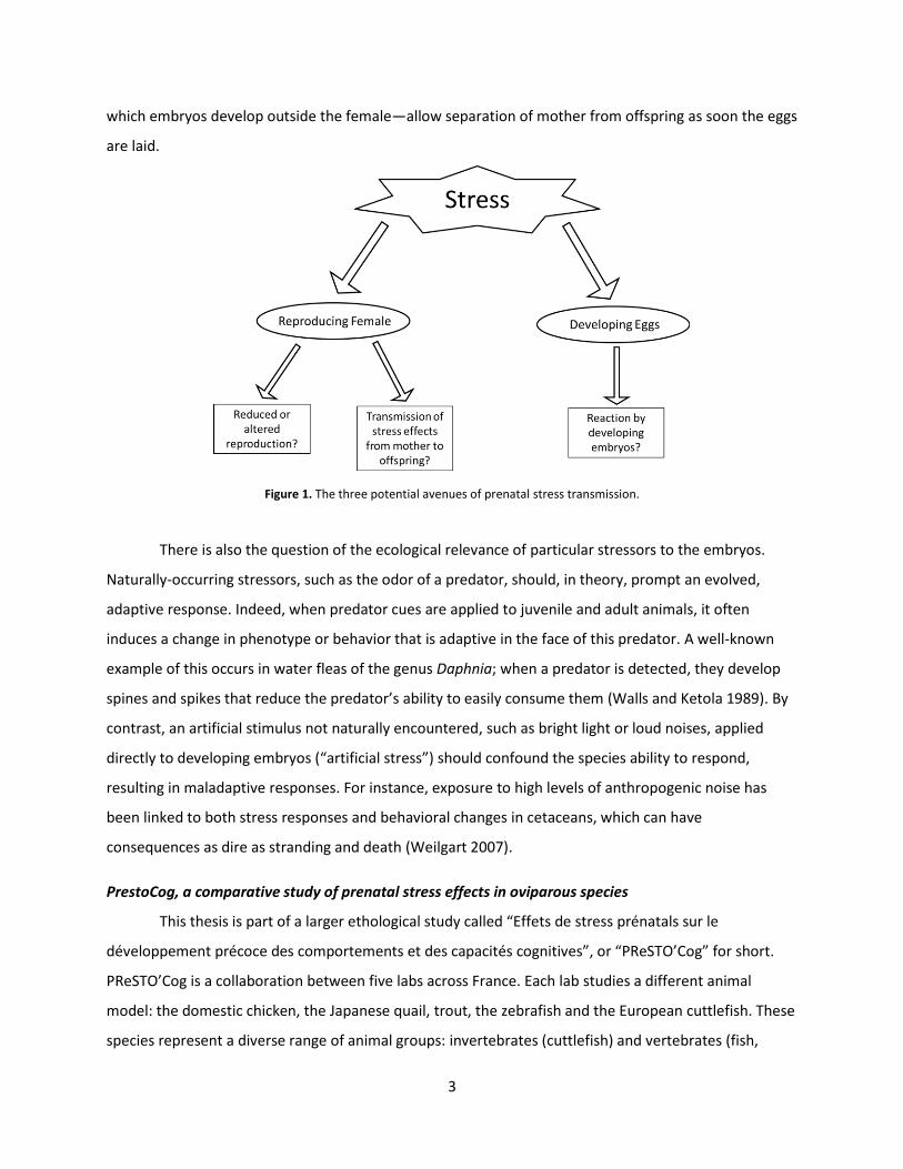
There are three potential avenues by which prenatal stress can exert its effects: 1) on the
mother herself (by affecting fecundity, mating behavior or egg-laying), 2) through the mother to the
offspring (e.g. via hormone transmission or perhaps sperm selection) or 3) direct perception of and
reaction to the stressor by the embryo (Fig. 1). Understanding the relative contribution of these three
potential paths of stress and their interactions is necessary to comprehending the ways that stress can
impact health, society, behavior and the evolution of organisms. For instance, stressors applied to
females during the reproductive period (“maternal stress”) have been shown to affect offspring survival,
behavior, learning and anxiety in diverse groups such as primates, rodents, birds and fish (reviewed in
Braastad 1998; Schreck, Contreras-Sanchez, and Fitzpatrick 2001; Henriksen, Rettenbacher, and
Groothuis 2011). These effects could either result from a direct stress reaction by the mother herself
(affecting the number of or the genetic composition of her embryos) or the result of transfer from
mother to offspring via provisioning or hormones in the placenta or egg yolk (Groothuis et al. 2005;
Hayward and Wingfield 2004; Lemaire et al. 2000; Weinstock 2008). Embryos themselves may also
perceive and react to stressors (“embryonic stress”) applied to spawning or brooding females. In order
to distinguish between maternally-mediated effects and direct perception, embryos must be isolated
from the female and have the stressors applied directly to them. However, it is currently difficult or
impossible to separate a mother from her developing offspring in species with internal development
(but see Roberts 2017) or maternal care of offspring after birth. However, oviparous species—those in
3
which embryos develop outside the female—allow separation of mother from offspring as soon the eggs
are laid.
Figure 1. The three potential avenues of prenatal stress transmission.
There is also the question of the ecological relevance of particular stressors to the embryos.
Naturally-occurring stressors, such as the odor of a predator, should, in theory, prompt an evolved,
adaptive response. Indeed, when predator cues are applied to juvenile and adult animals, it often
induces a change in phenotype or behavior that is adaptive in the face of this predator. A well-known
example of this occurs in water fleas of the genus Daphnia; when a predator is detected, they develop
spines and spikes that reduce the predator’s ability to easily consume them (Walls and Ketola 1989). By
contrast, an artificial stimulus not naturally encountered, such as bright light or loud noises, applied
directly to developing embryos (“artificial stress”) should confound the species ability to respond,
resulting in maladaptive responses. For instance, exposure to high levels of anthropogenic noise has
been linked to both stress responses and behavioral changes in cetaceans, which can have
consequences as dire as stranding and death (Weilgart 2007).
PrestoCog, a comparative study of prenatal stress effects in oviparous species
This thesis is part of a larger ethological study called “Effets de stress prénatals sur le
développement précoce des comportements et des capacités cognitives”, or “PReSTO’Cog” for short.
PReSTO’Cog is a collaboration between five labs across France. Each lab studies a different animal
model: the domestic chicken, the Japanese quail, trout, the zebrafish and the European cuttlefish. These
species represent a diverse range of animal groups: invertebrates (cuttlefish) and vertebrates (fish,
4
birds) as well as wild (cuttlefish), and domesticated species (chicken) and both poïkilotherms (cuttlefish,
fish) and homeotherms (birds). All are oviparous and precocial, allowing offspring to be experimentally
isolated from the female during embryonic development and free of the post-natal influence of
maternal interaction. Finally, they are also relatively self-sufficient at birth, permitting immediate
behavioral testing of the offspring. By comparing such phylogenetically-distant species, we address the
topic of prenatal stress from Tinbergen’s third and fourth perspectives—the adaptive and
phylogenetic—uncovering clues to the evolutionary pressures and family history that led to the behavior
we see in these species today. Ultimately, such insights could be applicable to other animal groups,
including mammals, leading to improvements in human and animal welfare.
The unifying theme of this project is to determine if prenatal stress induces changes in offspring,
and whether the type of prenatal stressor experienced affects the manner in which the offspring reacts.
Do the effects of maternally-applied stressors on offspring differ from those of stressors applied directly
to the embryos themselves? Does the response to an artificial stressor differ from that occurs in
response to a naturally-occurring one? The effects of prenatal stress are assessed through a range of
physiological, behavioral and learning tests of young offspring. We also search for clues as to the
mechanisms of such effects, especially endocrinological evidence for the transfer of stress hormones
from mother to offspring and changes in brain growth and morphology. These questions probe behavior
from Tinbergen’s first two perspectives—the mechanistic and ontogenetic—parsing the innate biological
processes and external influences which interact to produce a particular behavioral repertoire.
Some definitions used in this thesis should be clarified. “Chronic stress” refers to stress induced
by a stressors experienced continuously or repeatedly over an extended period of time. By contrast,
“acute stress” is experienced after a single occurrence of a stressor. This thesis is predominantly focused
on chronic stress, since it is generally associated with stronger and more long-term effects with greater
implications for fitness. Note also that in the literature, many studies do not make the distinction
between maternally-applied and direct embryonic stress, and refer simply to prenatal stress regardless
of whether it was applied to the mother, to her offspring or to both.
Presentation of the study animal
Many invertebrates are both oviparous and precocial, making them potential candidates to
study the ways that stress can affect a species as discussed above. Moreover, invertebrates represent
97% of the species on earth, live in nearly every part of the planet and demonstrate an awesome
diversity in modes of life and behavior. Next to arthropods, the invertebrate molluscs are the second
most populous phylum, and like arthropods, have successfully colonized sea, freshwater and terrestrial
5
habitats. Certain molluscs have also evolved in many ways that are convergent with vertebrates (e.g. the
“lung” of terrestrial gastropods), making them good comparative models. The cephalopoda is a group of
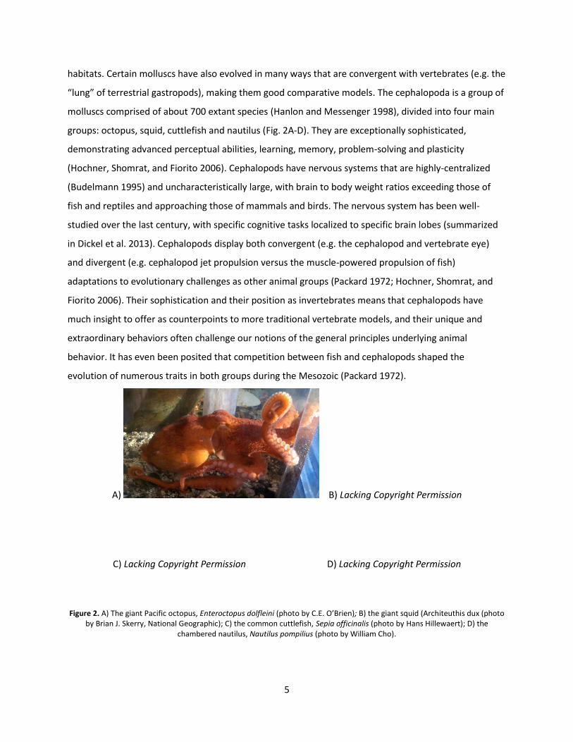
molluscs comprised of about 700 extant species (Hanlon and Messenger 1998), divided into four main
groups: octopus, squid, cuttlefish and nautilus (Fig. 2A-D). They are exceptionally sophisticated,
demonstrating advanced perceptual abilities, learning, memory, problem-solving and plasticity
(Hochner, Shomrat, and Fiorito 2006). Cephalopods have nervous systems that are highly-centralized
(Budelmann 1995) and uncharacteristically large, with brain to body weight ratios exceeding those of
fish and reptiles and approaching those of mammals and birds. The nervous system has been well-
studied over the last century, with specific cognitive tasks localized to specific brain lobes (summarized
in Dickel et al. 2013). Cephalopods display both convergent (e.g. the cephalopod and vertebrate eye)
and divergent (e.g. cephalopod jet propulsion versus the muscle-powered propulsion of fish)
adaptations to evolutionary challenges as other animal groups (Packard 1972; Hochner, Shomrat, and
Fiorito 2006). Their sophistication and their position as invertebrates means that cephalopods have
much insight to offer as counterpoints to more traditional vertebrate models, and their unique and
extraordinary behaviors often challenge our notions of the general principles underlying animal
behavior. It has even been posited that competition between fish and cephalopods shaped the
evolution of numerous traits in both groups during the Mesozoic (Packard 1972).
A) B) Lacking Copyright Permission
C) Lacking Copyright Permission D) Lacking Copyright Permission
Figure 2. A) The giant Pacific octopus, Enteroctopus dolfleini (photo by C.E. O’Brien); B) the giant squid (Architeuthis dux (photo by Brian J. Skerry, National Geographic); C) the common cuttlefish, Sepia officinalis (photo by Hans Hillewaert); D) the
chambered nautilus, Nautilus pompilius (photo by William Cho).
6
Like the other PReSTO’Cog models, the common cuttlefish Sepia officinalis (Linnaeus 1758) (Fig.
2C), is an excellent model for studying the effects of prenatal stress because it is oviparous and
precocial. S. officinalis is also a species that is important both commercially and scientifically: fisheries
exist in both the Atlantic and Mediterranean (Dunn 1999) and it is cultured in several laboratories and
aquaculture facilities (Pascual 1978; Forsythe, DeRusha, and Hanlon 1994; Domingues, Sykes, and
Andrade 2002). Indeed, along with Octopus vulgaris, it is one of the most commonly-studied species of
cephalopod. Most importantly, cuttlefish and other cephalopods have the advantage of being
phylogenetically-distant from more typical animal models like rats and monkeys: they are an
invertebrate group separated from vertebrates by hundreds of millions of years of distinct evolution,
allowing them to serve as a reference point to determine whether the stress effects that we observe in
different species are products of shared ancestry or separate evolutionary developments (Tinbergen’s
third and fourth questions). Ultimately, a better understanding of the effects of prenatal stress in S.
officinalis will yield general insight into the processes and strategies by which organisms survive.
Additionally, this research will help fill gaps in knowledge about the specific biological needs of
cuttlefish, particularly those regarding housing, reproduction and behavioral markers of welfare.
Hopefully, insight from this work will improve the ability of aquaculturists and researchers to set
standards of care and standard practice. This is particularly necessary due to the recent inclusion of
cuttlefish and other cephalopods in European animal welfare legislation (Directive 2010/63/EU)
governing the use of animals in experimental procedures. It may also help with future captive-rearing
and release efforts, which sadly may become increasingly necessary with growing food demands and
climate change. For instance, Sepia apama, the giant Australian cuttlefish, was recently designated as
“near threatened” due to intensive fishing of breeding aggregations in specific locations and is facing a
projected 20% decrease in population levels if current catch rates continue (IUCN 2017). And alarmingly,
local British newspapers report that fishermen are taking advantage of lax regulations and are
harvesting cuttlefish in the south west of the United Kingdom at unprecedented rates in response to
demand in Asia (Jones 2017; Payne 2017). If this trend continues, we may soon have greater ecological
and economic incentives to augment natural stocks artificially. One particularly economic strategy would
be to recover and culture the eggs laid on cuttlefish traps and normally lost when the gear is cleaned at
the end of the harvest season (Blanc and Daguzan 1998). Already, pilot efforts to culture cuttlefish eggs
and hatchlings in large, outdoor ponds have been undertaken with moderate success (Roussel and
Basuyaux, 2016). The work presented here will hopefully inform these efforts.
7
Thesis overview
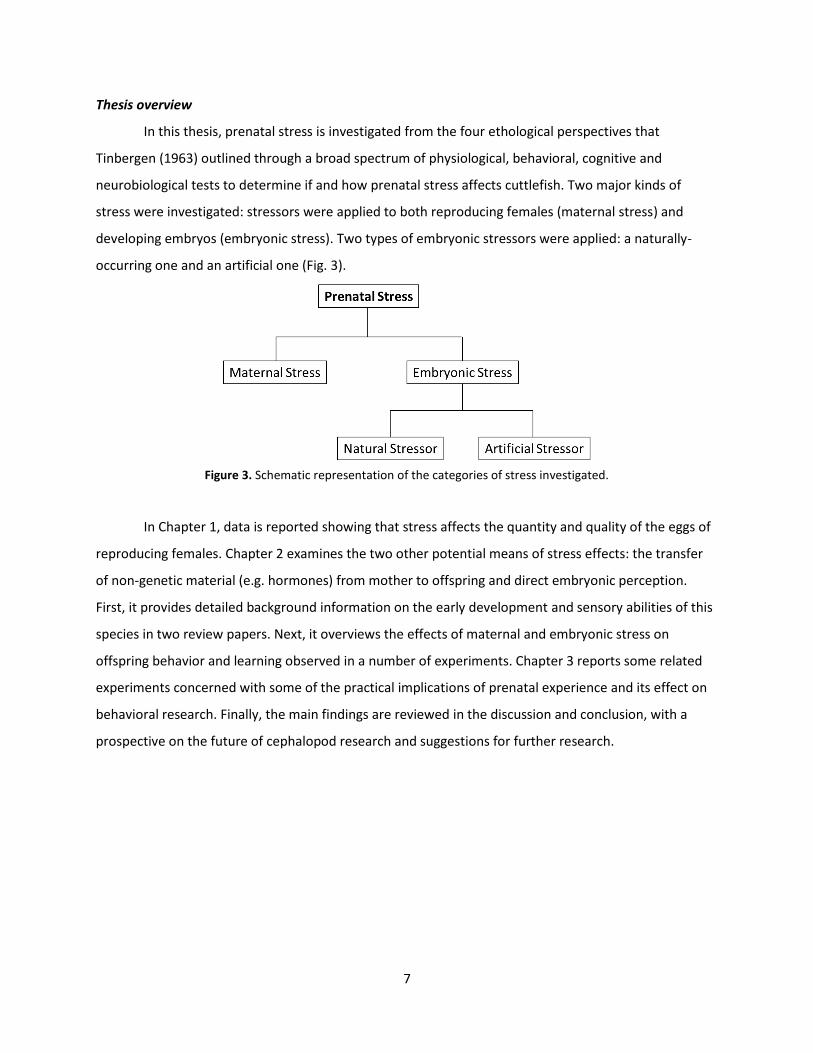
In this thesis, prenatal stress is investigated from the four ethological perspectives that
Tinbergen (1963) outlined through a broad spectrum of physiological, behavioral, cognitive and
neurobiological tests to determine if and how prenatal stress affects cuttlefish. Two major kinds of
stress were investigated: stressors were applied to both reproducing females (maternal stress) and
developing embryos (embryonic stress). Two types of embryonic stressors were applied: a naturally-
occurring one and an artificial one (Fig. 3).
Figure 3. Schematic representation of the categories of stress investigated.
In Chapter 1, data is reported showing that stress affects the quantity and quality of the eggs of
reproducing females. Chapter 2 examines the two other potential means of stress effects: the transfer
of non-genetic material (e.g. hormones) from mother to offspring and direct embryonic perception.
First, it provides detailed background information on the early development and sensory abilities of this
species in two review papers. Next, it overviews the effects of maternal and embryonic stress on
offspring behavior and learning observed in a number of experiments. Chapter 3 reports some related
experiments concerned with some of the practical implications of prenatal experience and its effect on
behavioral research. Finally, the main findings are reviewed in the discussion and conclusion, with a
prospective on the future of cephalopod research and suggestions for further research.

8
Chapter 1:
Stress and
Reproduction
in Cuttlefish

10
Chapter 1: Stress and Reproduction in Cuttlefish
This chapter presents a brief overview of reproduction, followed by an article outlining the
effects of stress on cuttlefish reproduction and finally, an account of a “mesocosm” experiment which
provides anecdotal support for the conclusions of the article.
11
I. Article #1: “Effects of Maternal and Embryonic Stress on Egg Production and Offspring in the Cuttlefish, Sepia officinalis”
In the English Channel, Sepia officinalis has a one to two year life cycle (Gauvrit, Goff, and
Daguzan 1997) spent mostly living in deeper offshore waters. Towards the end of their life, they migrate
to shallower coastal waters to mate, lay eggs and die soon thereafter (Boletzky 1987). Each female can
lay dozens to thousands of eggs fertilized by stored sperm from one or more males (Hanlon, Ament, and
Gabr 1999). Depending on the temperature, these eggs hatch 2-3 months later as autonomous benthic
hatchlings about 1cm in total length (O’Brien, Mezrai, et al. 2016). While we were primarily interested in
quantifying the effects of prenatal stress on cuttlefish offspring, the data collected in the course of
testing the effects of maternal stress on hatchling cuttlefish also demonstrated the effects of stress on
cuttlefish egg production. These results are reported in the following manuscript.
Effects of Maternal and Embryonic Stress on Egg Production and Offspring in the Cuttlefish, Sepia officinalis
Caitlin E. O’Briena, Cécile Bellangera, Christelle Jozet-Alvesa, Nawel Mezraia, Anne-Sophie Darmaillacqa
and Ludovic Dickela*
aNormandie Univ, UNICAEN, CNRS, NECC, UMR EthoS 6552, 14032 Caen cedex, France *Corresponding author ([email protected])
Key words: handling, egg-laying, hatching rate, fecal corticosterone, predator cues, LED light
Abstract
Stress has been shown to have profound effects on animals, particularly if it occurs during reproduction or embryonic development. Invertebrate mollusc cephalopods offer unique points of comparison to typical vertebrate models in the study of stress. We investigated the effects of stressors applied to reproducing and developing cuttlefish, Sepia officinalis, by comparing the number of eggs produced by females in a typical captive setting with females subjected to moderate stress during egg-laying (confined space and repeated removal from the water). We also subjected their eggs to naturally-occurring (predator cues) and artificial (random bouts of bright LED light) stressors during development in order to gauge the impact of direct stress on embryos. We found that stressed females produced fewer eggs and that fewer of those eggs hatched. Simultaneous attempts to identify a simple proxy for stress levels (fecal corticosterone or unused reproductive material) in reproducing females were unsuccessful, although we found that a few stressed mothers laid mostly transparent eggs lacking the dark pigment typical of this species. In contrast to maternal stress, stressors applied directly to developing embryos had no effect on hatching rate. Neither stress type was associated with differences in hatching size. Our results suggest that reducing stress during egg-laying may increase aquacultural egg yields in S. officinalis and that developing embryos are less affected by stress applied directly to them than to their mother.
12
Introduction Organisms have evolved to react to unexpected phenomena in their environment (e.g.
predators, changes in environmental parameters, unfamiliar sensory stimuli) or to anticipated pain or
suffering with a physiological or behavioral response. Such responses are referred to as “stress”
(occurring in response to “stressors”), and involve a variety of physiological changes with which the
organism attempts to avoid death (Schreck, Contreras-Sanchez, and Fitzpatrick 2001). For example, in
numerous species of the freshwater crustacean genus Daphnia, early exposure to odor cues from
predators induces the development of defensive features—including defensive spines and changes in
body size and shape—that deter consumption (e.g. Krueger and Dodson 1981; Tollrian 1995). However,
while these responses have evolved to enhance fitness in the face of predators, they divert resources
away from other life functions. In this way, stress can reduce overall fitness, especially if exposure to the
stressor is recurring, continuous, or if the response is poorly-adapted to the situation (McEwen and
Wingfield 2003). In D. pulex, a species that develops spines in response to predator cues for instance,
predator exposure usually delayed sexual maturity by one full growth stage (instar), reducing the
likelihood of survival until reproduction (Walls and Ketola 1989). In this case, it appears that the
resources diverted to the predator stress response detract from growth and maturation.
When stress is experienced during reproduction or embryonic development, its effects may be
especially profound. In some cases, stress may increase reproductive output or offspring success by
inducing adaptive responses on the part of the mother or her offspring. For instance, when spawning
female sticklebacks sense predators, their offspring exhibit stronger anti-predator behavior (tighter
shoaling) than offspring from unstressed females (Giesing et al. 2010). But the preponderance of
literature (primarily regarding fish) suggests that stress reduces reproductive output (reviewed in
Braastad 1998; Schreck 2010). In catfish for instance, the stress induced by human handling of females
was associated with reduced egg production (Soso et al. 2008).
In fish, birds and mammals, “stress hormones,” including adrenal glucocorticoids, are thought to
be the mediator of the stress response (Moberg 1991). When exposed to stressors, animals secrete
these hormones, which induce changes in behavior, metabolism and physiology. There are numerous
studies associating maternal stress with changes in offspring, and it seems likely that stress hormones
are being transferred from mother to her offspring via the placenta or egg yolk and mediating some of
these changes (Braastad 1998; Welberg and Seckl 2001; Henriksen, Rettenbacher, and Groothuis 2011).
However, the embryos of many of these species are capable of sensing the environment outside the egg
or placenta to some degree (Gottlieb 1976) and could potentially experience stress directly from
13
environmental stimuli during development (we refer to this as “embryonic stress”). Thus, it is sometimes
unclear if stress reactions observed in offspring are due to the transfer of maternal hormones or to a
stress response by the embryos themselves, especially in animals in which fetuses or eggs develop
within the female. Comparing the effects of maternal and embryonic stressors is necessary, but since
this separation is not possible in viviparous and ovoviviparous animals, inferences must be drawn from
animals with different reproductive habits.
Along with fish, reptiles and amphibians, many invertebrate species are good candidates for this
kind of investigation since they are oviparous (the embryos typically develop separately from the
mother), allowing them to be experimentally isolated from the female for testing the effects of
embryonic stress. Testing both maternal and embryonic stress in a complementary and concurrent way
allows general comparisons to be made between the stress types. Among invertebrates, cephalopod
molluscs are particularly good comparative models, since their physiology and behavior converge with
that of vertebrates in many instances (Grasso and Basil 2009). Comparisons of phylogenetically distant
species that have features demonstrating convergent evolution allow us to infer whether the
adaptations to ecological challenges that we observe represent novel evolutionary “solutions” or are
dictated by ancestry. Stress responses have scarcely been investigated in cephalopods, but there are
preliminary indications that there is at least some similarity to the stress responses of vertebrates. In
one study, elevated levels of the stress hormone corticosterone were measured in the feces of the giant
Pacific octopus after injection of adrenal hormone (ACTH) or saline solution (Larson and Anderson
2010). Similarly, an acute instance of air exposure has been shown to elevate noradrenaline in the
hemolymph of the octopus Eledone cirrhosa (Malham et al. 2002). As in vertebrates, these stress
hormones are thought to influence various aspects of cephalopod biology, including the secretion of
reproductive hormones (Di Cosmo and Polese 2016).
The cuttlefish Sepia officinalis is a cephalopod mollusc inhabiting the Mediterranean and the
Atlantic coasts of Europe and North Africa. A well-established fishery and aquaculture industry make it
economically important and it is a model species in biological research (Bloor et al. 2013). S. officinalis
reproduces only once at the end of life and dies very soon after, though egg-laying may last for several
days or weeks (Boletzky 1987). In captivity, females typically lay up to 1500 eggs each (Domingues,
Sykes, and Andrade 2002, 2001; Correia et al. 2005; Sykes et al. 2009; Sykes et al. 2013). Embryonic
development lasts between 40 and 90 days, with higher temperatures accelerating embryogenesis
(Bouchaud 1991; Bouchaud and Galois 1990; Bouchaud and Daguzan 1990) but usually yielding smaller
14
hatchlings (Gauvrit, Goff, and Daguzan 1997). Eggs develop outside the mother and there is no direct
parental care during embryonic development or after (Bloor et al. 2013).
Despite interest in culturing cuttlefish, there are few studies on the effects of stress on female
reproduction or embryonic development in this species to date. Moreover, there are very few studies
that allow for a comparison between the effects of maternal and embryonic stress in any animal. We
designed an experiment to test for effects of maternal stress on egg output, embryonic survival and
growth, as well for effects of embryonic stress on embryonic survival and growth in S. officinalis. Since
confined tank space and occasional brief removal from the water are often unavoidable aspects of
cuttlefish capture and aquaculture, we used these factors as chronic and repeated acute stressors to
investigate maternal stress. We expected that such treatment would reduce egg laying, since females
would likely be forced to expend resources in reacting to the stressors. In a second experiment, we
examined the effects of embryonic stress. Since stress responses of embryos are poorly understood, we
tested both a naturally-occurring and an artificial stressor in order to represent a range of potential
sources of stress present in the natural environment and in captive settings. As a natural stressor, we
selected a fish common in the English Channel that preys on small cuttlefish. As an artificial stressor we
selected bouts of bright artificial (LED) light timed randomly throughout the day over the course of
embryonic development. Importantly, previous experiments have shown that late-stage cuttlefish
embryos react to both predator odor and bright light with changes in mantle contraction rate (Romagny
et al. 2012), indicating a definite ability to perceive these stimuli. In comparing the two kinds of stress,
we predicted that embryonic stressors would have a stronger negative impact on hatching rate and
hatching size than maternal stressors since the limited resources of embryos would need to be diverted
away from growth in order to mount a stress response.
Finally, we also attempted to find a simple way to measure stress in cuttlefish, a tool that would
allow us to assess the suitability of captive conditions for spawning females. Since measurement of fecal
glucocorticoids is used in many species as a non-invasive way to quantify stress (e.g. Tempel and
Gutiérrez 2003; Metrione and Harder 2011), we tested whether or not fecal corticosterone could be
used to assess stress levels in S. officinalis. (Attempts were also made to test corticosterone levels in
embryos directly, but the values obtained fell below the quantification limit of the detection kit.) We
also examined the amount of unused reproductive material (oocytes) remaining in the females at death,
reasoning that stress could reduce the utilization of reproductive capacity. We predicted higher levels of
corticosterone and more unused reproductive material in stressed females than in control females.
15
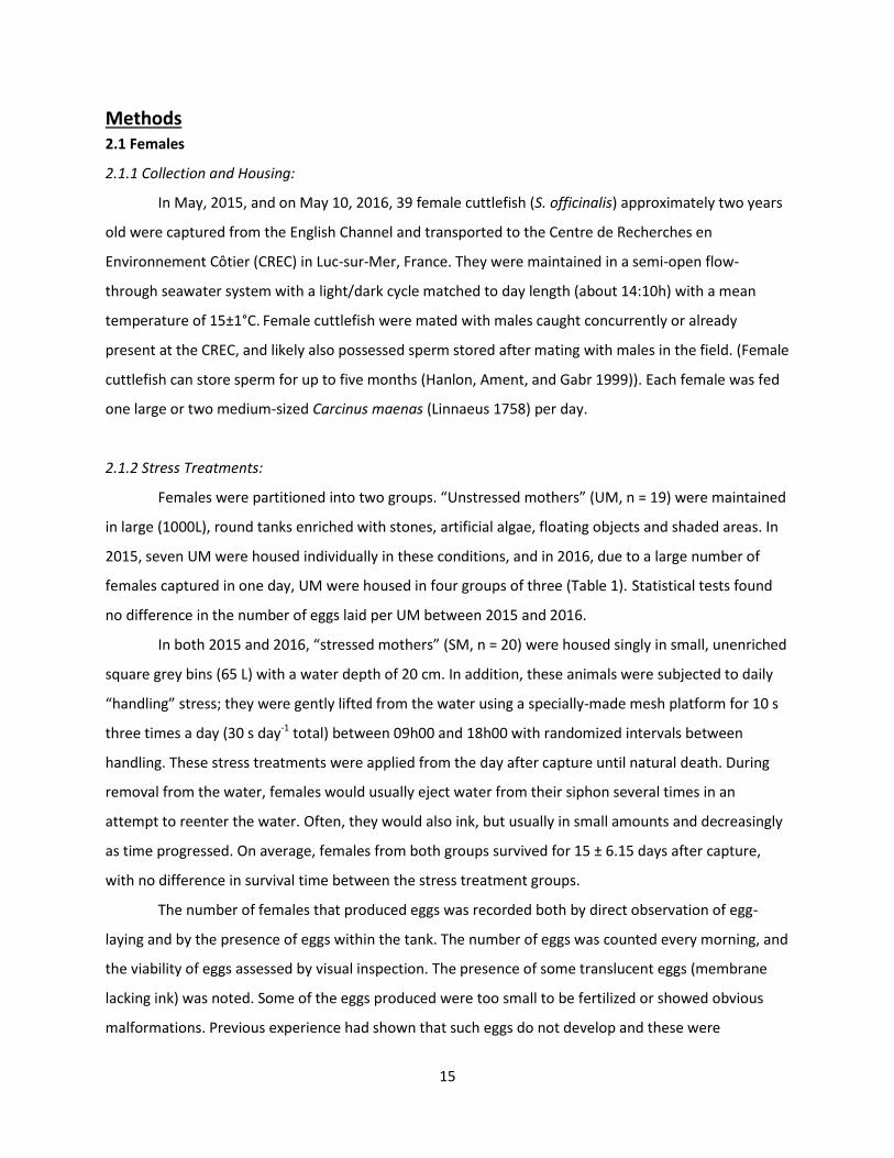
Methods 2.1 Females
2.1.1 Collection and Housing:
In May, 2015, and on May 10, 2016, 39 female cuttlefish (S. officinalis) approximately two years
old were captured from the English Channel and transported to the Centre de Recherches en
Environnement Côtier (CREC) in Luc-sur-Mer, France. They were maintained in a semi-open flow-
through seawater system with a light/dark cycle matched to day length (about 14:10h) with a mean
temperature of 15±1°C. Female cuttlefish were mated with males caught concurrently or already
present at the CREC, and likely also possessed sperm stored after mating with males in the field. (Female
cuttlefish can store sperm for up to five months (Hanlon, Ament, and Gabr 1999)). Each female was fed
one large or two medium-sized Carcinus maenas (Linnaeus 1758) per day.
2.1.2 Stress Treatments:
Females were partitioned into two groups. “Unstressed mothers” (UM, n = 19) were maintained
in large (1000L), round tanks enriched with stones, artificial algae, floating objects and shaded areas. In
2015, seven UM were housed individually in these conditions, and in 2016, due to a large number of
females captured in one day, UM were housed in four groups of three (Table 1). Statistical tests found
no difference in the number of eggs laid per UM between 2015 and 2016.
In both 2015 and 2016, “stressed mothers” (SM, n = 20) were housed singly in small, unenriched
square grey bins (65 L) with a water depth of 20 cm. In addition, these animals were subjected to daily
“handling” stress; they were gently lifted from the water using a specially-made mesh platform for 10 s
three times a day (30 s day-1 total) between 09h00 and 18h00 with randomized intervals between
handling. These stress treatments were applied from the day after capture until natural death. During
removal from the water, females would usually eject water from their siphon several times in an
attempt to reenter the water. Often, they would also ink, but usually in small amounts and decreasingly
as time progressed. On average, females from both groups survived for 15 ± 6.15 days after capture,
with no difference in survival time between the stress treatment groups.
The number of females that produced eggs was recorded both by direct observation of egg-
laying and by the presence of eggs within the tank. The number of eggs was counted every morning, and
the viability of eggs assessed by visual inspection. The presence of some translucent eggs (membrane
lacking ink) was noted. Some of the eggs produced were too small to be fertilized or showed obvious
malformations. Previous experience had shown that such eggs do not develop and these were
16
discounted. We incubated the eggs (see next section) and measured the hatching rate and hatching size
of offspring. Only eggs that were part of a cohort of at least 50 eggs laid after at least one week of
treatment were used in assessments of hatching success and size in order to ensure sufficient
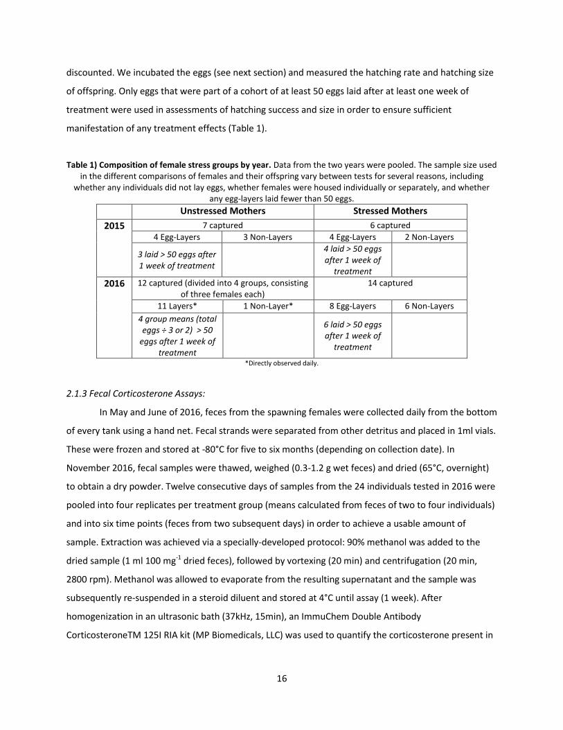
manifestation of any treatment effects (Table 1).
Table 1) Composition of female stress groups by year. Data from the two years were pooled. The sample size used in the different comparisons of females and their offspring vary between tests for several reasons, including
whether any individuals did not lay eggs, whether females were housed individually or separately, and whether any egg-layers laid fewer than 50 eggs.
Unstressed Mothers Stressed Mothers
2015 7 captured 6 captured
4 Egg-Layers 3 Non-Layers 4 Egg-Layers 2 Non-Layers
3 laid > 50 eggs after 1 week of treatment
4 laid > 50 eggs after 1 week of
treatment
2016 12 captured (divided into 4 groups, consisting of three females each)
14 captured
11 Layers* 1 Non-Layer* 8 Egg-Layers 6 Non-Layers
4 group means (total eggs ÷ 3 or 2) > 50
eggs after 1 week of treatment
6 laid > 50 eggs after 1 week of
treatment
*Directly observed daily.
2.1.3 Fecal Corticosterone Assays:
In May and June of 2016, feces from the spawning females were collected daily from the bottom
of every tank using a hand net. Fecal strands were separated from other detritus and placed in 1ml vials.
These were frozen and stored at -80°C for five to six months (depending on collection date). In
November 2016, fecal samples were thawed, weighed (0.3-1.2 g wet feces) and dried (65°C, overnight)
to obtain a dry powder. Twelve consecutive days of samples from the 24 individuals tested in 2016 were
pooled into four replicates per treatment group (means calculated from feces of two to four individuals)
and into six time points (feces from two subsequent days) in order to achieve a usable amount of
sample. Extraction was achieved via a specially-developed protocol: 90% methanol was added to the
dried sample (1 ml 100 mg-1 dried feces), followed by vortexing (20 min) and centrifugation (20 min,
2800 rpm). Methanol was allowed to evaporate from the resulting supernatant and the sample was
subsequently re-suspended in a steroid diluent and stored at 4°C until assay (1 week). After
homogenization in an ultrasonic bath (37kHz, 15min), an ImmuChem Double Antibody
CorticosteroneTM 125I RIA kit (MP Biomedicals, LLC) was used to quantify the corticosterone present in
17
the feces of each treatment group. A gamma counter measured relative radioactivity of the samples and
corticosterone concentrations were calculated via comparison to a standardized curve.
2.1.4 Ovary Dissections:
“Lifespan after capture” is the number of days between capture and natural death in the facility.
At death, we measured female Dorsal Mantle Length (DML, cm) and weight (kg) after water was drained
from the body cavity and the outer surface gently dried. The bodies were then frozen in a -20°C freezer.
In August, 2016, the bodies were thawed and dissected in order to count the number of oocytes
remaining in the pallial cavity.
2.2 Eggs
2.2.1 Egg Collection:
For the first three weeks of incubation, eggs were maintained in floating baskets in the maternal
treatment tank in which they were laid (up to 250 eggs per basket). After a suitable number was
collected (about three weeks after the first eggs were laid), eggs were moved from these conditions and
acclimatized over the course of a day to a mean seawater temperature between 17 and 19°C. In order to
ensure that any potential stress effects had time to manifest and that they were represented by
adequate sample sizes, only eggs from mothers that had laid at least 50 eggs after one week of
treatment were used to calculate hatching success and hatching size (see Table 1). Eggs were
maintained until hatching in floating trays in 65L tanks constantly renewed by seawater from a flow-
through system, with aeration from an air stone and exposure to the natural light cycle.
2.2.2 Stress Treatments:
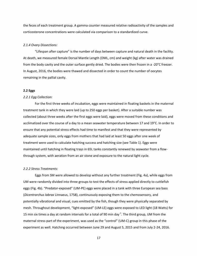
Eggs from SM were allowed to develop without any further treatment (Fig. 4a), while eggs from
UM were randomly divided into three groups to test the effects of stress applied directly to cuttlefish
eggs (Fig. 4b). “Predator-exposed” (UM-PE) eggs were placed in a tank with three European sea bass
(Dicentrarchus labrax Linnaeus, 1758), continuously exposing them to the chemosensory, and
potentially vibrational and visual, cues emitted by the fish, though they were physically separated by
mesh. Throughout development, “light-exposed” (UM-LE) eggs were exposed to LED light (18 Watts) for
15 min six times a day at random intervals for a total of 90 min day-1. The third group, UM from the
maternal stress part of the experiment, was used as the “control” (UM-C) group in this phase of the
experiment as well. Hatching occurred between June 29 and August 5, 2015 and from July 2-24, 2016.
18
Fig. 4) Schematic representation of experimental design. A) Maternal Stress Effects, B) Embryonic Stress Effects.
The arrows between panels A and B indicate that the eggs of UM were subdivided to create the embryonic treatment groups and that the same control group was used in both phases of the experiment.
2.3 Hatchlings
Hatchlings were counted at 08h00 each morning and used to calculate hatching rate. Each
hatchling was then gently moved from the hatching tank to a shallow, uniform grey container and
photographed with a Panasonic HDC-SD60 camera. Using ImageJ, DML (the tip of the mantle to the edge
just behind the eyes) was measured in two photos and averaged. If these two values differed by more
than 5%, a third photo was measured and the mean DML calculated from these three measurements.
Sex discrimination is not possible at this age.
2.4 Ethical Note
This research was conducted in accordance with Directive 2010/63/EU, under the approval of
the Comité d’Éthique NOmandie en Matière d’EXperimentation Animale (CENOMEXA) #54 (agreement
number A14384001).
2.5 Statistics
All statistical analyses were conducted in StatXact®7 (Cytel Inc.) and R. Because the trends from
2015 and 2016 only differed in a single instance (hatching size following maternal stress), samples were
pooled in order to achieve a usable sample size for statistical analysis. All values are reported as mean ±
standard deviation.
Unstressed Mother (UM-C)
19
To compare the number of females that laid eggs, as well as the number of females that laid
translucent eggs with those that laid normal eggs, chi-squared exact tests were used.
The number of eggs per female was calculated from laying females only. In 2016, UM were
housed in groups of three, and thus individual counts per female were not possible. Instead, the total
number of eggs produced by each tank was divided by three (or two in one case) to yield a mean value
for each tank (11 of 12 UM were directly observed by experimenters laying viable eggs in 2016). These
were combined with the individual UM egg counts from 2015, and compared to the eggs per female of
SM using exact permutation tests for independent samples. We also compared the eggs per female of
UM between years with an exact permutation test to test for any effect of housing singly or in groups.
The data for female size (weight and DML), lifespan after capture and the number of remaining
oocytes (2016 only) were not normally distributed, so means were compared using exact Pearson
permutation tests for independent samples. We also tested for a correlation between the number of
eggs laid and lifespan after capture of UM and SM with canonical correlation analysis. The sample sizes
used to calculate these values varied since some measurements were not possible in certain individuals.
Fecal corticosterone measurements were logit transformed and fitted with logit-log linear
regression (log10(corticosterone concentration) ~ treatment + (1 | testing.days)) using the “lme4”
package in R.
The hatching rate for SM was calculated as the number of live hatchlings divided by the total
number of eggs laid. Due to the large number of eggs laid, not all the eggs from UM were measured for
this experiment. Instead, a large subset of the eggs was partitioned into three embryonic stress groups
(UM-C, UM-PE and UM-LE eggs). Females that produced fewer than 50 viable eggs after one week of
treatment (one UM in 2015 and two SM in 2016) were excluded. 2x2 chi-squared tests were used to
compare UM and SM, and a Cochran-Mantel-Haenszel chi-squared test was conducted to compare UM-
C, UM-PE and UM-LE eggs.
Hatching DMLs were normally distributed and there was equal variance between treatment
groups, enabling parametric analysis. UM and SM were compared using an independent T-test, while C,
PE and LE eggs were compared using a two-way ANOVA with stressor type and mother as main factors.

20
Results 3.1 Females
3.1.1 Egg-laying:
The proportion of SM (60%) that produced eggs did not differ from UM (78.95%) (two-tailed chi-
squared exact test: X2 = 1.64, UM n = 19, SM n = 20, p = 0.3; Table 2). 15 UM laid a total of 6567 eggs
while 12 SM laid a total of 1831 eggs.
No significant difference existed in DML, weight at death, lifespan after capture or the amount
of remaining reproductive material at death (remaining oocytes) between UM and SM (Table 2). For
UM, there was a strong correlation between lifespan after capture and eggs per female (canonical
correlation test: R = 0.90, n =12, p < 0.0001; Table 2), and only a weak correlation for SM (canonical
correlation test: R = 0.34, n = 20, p = 0.14).
Table 2) Proportion of egg layers, size (DML and weight), lifespan after capture (days), the correlation between lifespan after capture and number of eggs laid and remaining oocytes (mean ± s.d.) of female cuttlefish. UM: n = 19 females housed individually or in four groups of three; SM: n = 20 females housed individually. The proportion
of egg layers was tested with a Fisher exact test, all others with exact permutation tests (these calculations include both egg-layers and non-layers).
Unstressed Mothers Stressed Mothers Comparison
Proportion of Egg Layers 78.95% n = 19
60.0% n = 20
p = 0.3 X2 = 1.642
DML (cm) 23.29 ± 3.25,
n = 17* 23.03 ± 1.89,
n = 17* p = 0.81 t ≥ 396
Weight at death (kg) 1.29 ± 0.30,
n = 17* 1.31 ± 0.25,
n = 16* p = 0.84 t ≥ 21.85
Lifespan after Capture (days)
15.63 ± 7.21, n = 19
14 ± 5.0, n = 20
p = 0.38 t ≥ 297
Correlation: Number of Eggs Laid and Lifespan after
Capture
p < 0.0001 n = 12
p = 0.14 n = 20
Remaining Oocytes (2016 only)
108.33 ± 33.26, n = 12**
117.5 ± 48.64, n = 12**
p = 0.65 t ≥ 1300
*Accurate body measurements were not possible for some specimens. **For technical reasons, dissection was not possible for some specimens.
3.1.2 Fecal Corticosterone:
The mean corticosterone concentration over six days in UM was 0.70 ± 0.36 ng mg-1 dry feces
and 0.79 ± 0.56 ng cort mg-1 dry feces in SM. No significant difference existed between treatment groups
(GLMM: X2 ≥ 0.07, n = 4 (means calculated from the pooled feces of one to four individuals over two
days), p = 0.79; Fig. 5).
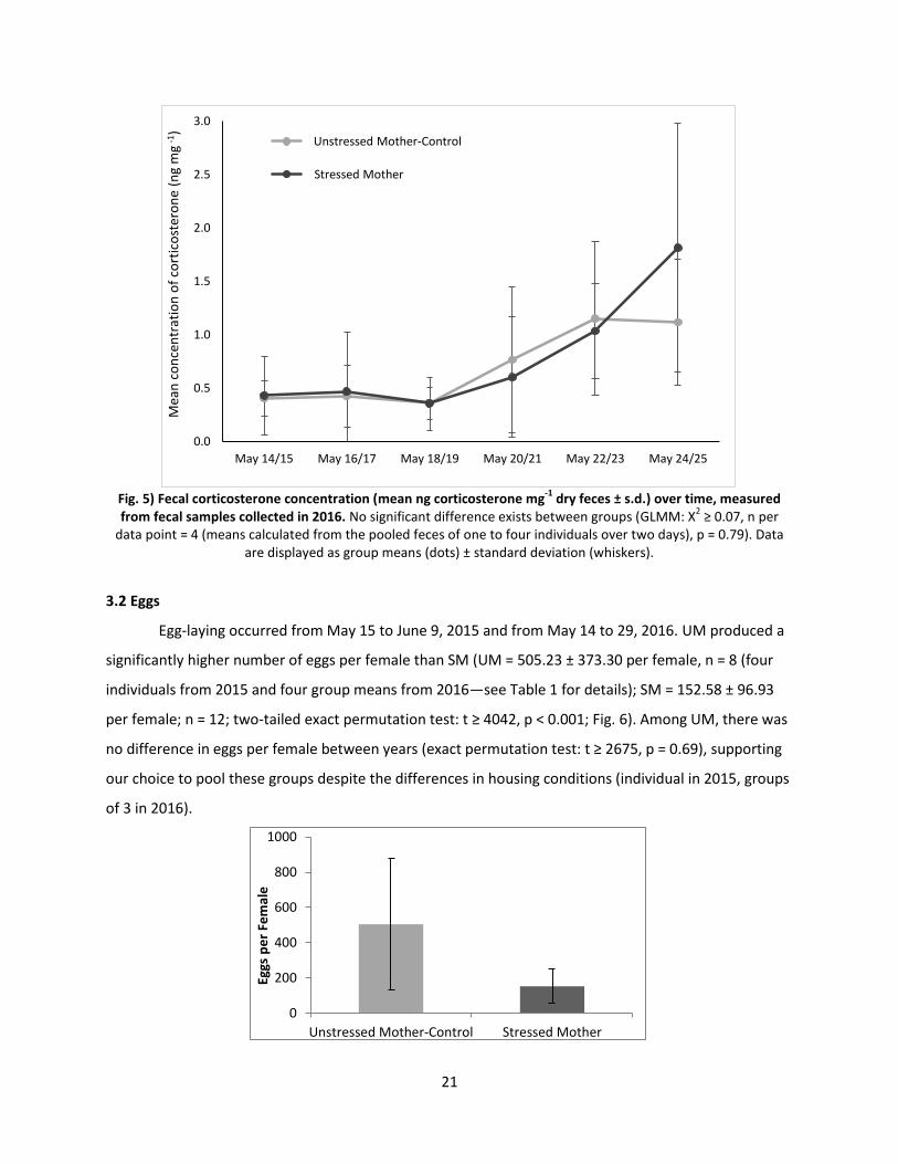
21
Fig. 5) Fecal corticosterone concentration (mean ng corticosterone mg
-1 dry feces ± s.d.) over time, measured
from fecal samples collected in 2016. No significant difference exists between groups (GLMM: X2 ≥ 0.07, n per
data point = 4 (means calculated from the pooled feces of one to four individuals over two days), p = 0.79). Data are displayed as group means (dots) ± standard deviation (whiskers).
3.2 Eggs
Egg-laying occurred from May 15 to June 9, 2015 and from May 14 to 29, 2016. UM produced a
significantly higher number of eggs per female than SM (UM = 505.23 ± 373.30 per female, n = 8 (four
individuals from 2015 and four group means from 2016—see Table 1 for details); SM = 152.58 ± 96.93
per female; n = 12; two-tailed exact permutation test: t ≥ 4042, p < 0.001; Fig. 6). Among UM, there was
no difference in eggs per female between years (exact permutation test: t ≥ 2675, p = 0.69), supporting
our choice to pool these groups despite the differences in housing conditions (individual in 2015, groups
of 3 in 2016).
0.0
0.5
1.0
1.5
2.0
2.5
3.0
May 14/15 May 16/17 May 18/19 May 20/21 May 22/23 May 24/25
Mea
n c
on
cen
trat
ion
of
cort
ico
ster
on
e (n
g m
g -1)
USM
SM
Unstressed Mother-Control
Stressed Mother
0
200
400
600
800
1000
Unstressed Mother-Control Stressed Mother
Eggs
pe
r Fe
mal
e
22
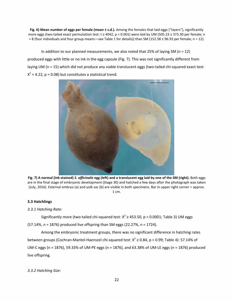
Fig. 6) Mean number of eggs per female (mean ± s.d.). Among the females that laid eggs (“layers”), significantly more eggs (two-tailed exact permutation test: t ≥ 4042, p < 0.001) were laid by UM (505.23 ± 373.30 per female; n = 8 (four individuals and four group means—see Table 1 for details)) than SM (152.58 ± 96.93 per female; n = 12).
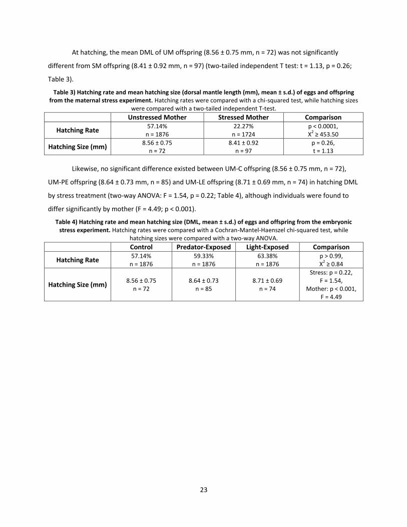
In addition to our planned measurements, we also noted that 25% of laying SM (n = 12)
produced eggs with little or no ink in the egg capsule (Fig. 7). This was not significantly different from
laying UM (n = 15) which did not produce any viable translucent eggs (two-tailed chi-squared exact test:
X2 = 4.22, p = 0.08) but constitutes a statistical trend.
Fig. 7) A normal (ink-stained) S. officinalis egg (left) and a translucent egg laid by one of the SM (right). Both eggs are in the final stage of embryonic development (Stage 30) and hatched a few days after the photograph was taken (July, 2016). External embryo (a) and yolk sac (b) are visible in both specimens. Bar in upper right corner = approx.
1 cm.
3.3 Hatchlings
3.3.1 Hatching Rate:
Significantly more (two-tailed chi-squared test: X2 ≥ 453.50, p < 0.0001; Table 3) UM eggs
(57.14%, n = 1876) produced live offspring than SM eggs (22.27%, n = 1724).
Among the embryonic treatment groups, there was no significant difference in hatching rates
between groups (Cochran-Mantel-Haenszel chi-squared test: X2 ≥ 0.84, p > 0.99; Table 4): 57.14% of
UM-C eggs (n = 1876), 59.33% of UM-PE eggs (n = 1876), and 63.38% of UM-LE eggs (n = 1876) produced
live offspring.
3.3.2 Hatching Size:
23
At hatching, the mean DML of UM offspring (8.56 ± 0.75 mm, n = 72) was not significantly
different from SM offspring (8.41 ± 0.92 mm, n = 97) (two-tailed independent T test: t = 1.13, p = 0.26;
Table 3).
Table 3) Hatching rate and mean hatching size (dorsal mantle length (mm), mean ± s.d.) of eggs and offspring from the maternal stress experiment. Hatching rates were compared with a chi-squared test, while hatching sizes
were compared with a two-tailed independent T-test. Unstressed Mother Stressed Mother Comparison
Hatching Rate 57.14%
n = 1876 22.27%
n = 1724 p < 0.0001, X
2 ≥ 453.50
Hatching Size (mm) 8.56 ± 0.75
n = 72 8.41 ± 0.92
n = 97 p = 0.26, t = 1.13
Likewise, no significant difference existed between UM-C offspring (8.56 ± 0.75 mm, n = 72),
UM-PE offspring (8.64 ± 0.73 mm, n = 85) and UM-LE offspring (8.71 ± 0.69 mm, n = 74) in hatching DML
by stress treatment (two-way ANOVA: F = 1.54, p = 0.22; Table 4), although individuals were found to
differ significantly by mother (F = 4.49; p < 0.001).
Table 4) Hatching rate and mean hatching size (DML, mean ± s.d.) of eggs and offspring from the embryonic stress experiment. Hatching rates were compared with a Cochran-Mantel-Haenszel chi-squared test, while
hatching sizes were compared with a two-way ANOVA. Control Predator-Exposed Light-Exposed Comparison
Hatching Rate 57.14%
n = 1876 59.33%
n = 1876 63.38%
n = 1876 p > 0.99, X
2 ≥ 0.84
Hatching Size (mm) 8.56 ± 0.75
n = 72 8.64 ± 0.73
n = 85 8.71 ± 0.69
n = 74
Stress: p = 0.22, F = 1.54,
Mother: p < 0.001, F = 4.49

24
Discussion The purpose of this study was to determine whether maternal and embryonic stressors affect
cuttlefish reproduction, hatching rate and hatching size. Using an oviparous species also allowed us to
compare the relative impact of maternal versus embryonic stressors. In parallel, we attempted to
determine whether fecal corticosterone levels (a commonly-used and noninvasive measurement of
stress levels in many animals) or the number of unused eggs remaining at death (a proxy for unused
reproductive potential), would correspond to any reproductive effects of maternal stress observed in
cuttlefish.
Maternal stress clearly reduced egg-laying in cuttlefish. This difference could not be explained
by female size or survival time: There was no difference in mean weight or DML between the stress
groups, and females from both groups survived for a little over two weeks after capture before they
died naturally, with no difference in lifespan between groups (Table 2). One potential explanation for
the reduction in the number of eggs laid by SM might be that stress responses depleted energy reserves
necessary to sustain egg laying activity. Since cuttlefish generally do not eat during this time (Boletzky
1986), the energy for egg laying and basic life processes is derived from the set amount of body reserves
remaining to the female. Thousands of immature oocytes are stored in the ovary, and released into the
genital tract in batches of a couple hundred to be fertilized, encapsulated and laid (Bernay 2005). Once a
batch of eggs has been laid (usually over a relatively short period of hours or days), another batch can be
recruited from the ovary. Thus, egg-laying appears not to be limited by the total number of oocytes
available, but by time and energy stores. Reacting to stressors may accelerate energy consumption, and
could therefore deplete the resources that females would otherwise use to lay eggs, whereas favorable
conditions may permit multiple bouts of oocyte maturation from the ovary, resulting in the intermittent
laying over the course of weeks or even months that is sometimes observed (Boletzky 1987; Boletzky
1988). This hypothesis is supported by the positive correlation between the lifespan after capture and
eggs per female for UM (Table 2), suggesting that for this group, the number of eggs produced was
largely a function of how long a female survived. By contrast, the weaker correlation between these
factors for SM suggests that another factor is responsible for the reduced egg output.
Interestingly, 25% of SM and none of the UM that produced viable eggs laid partially or entirely
translucent ones (Fig. 7). In most cases, the egg membrane of S. officinalis is impregnated with dark ink
from the mother, although translucent eggs are occasionally seen in both aquaculture and in the wild
(Fig. 8). In our experiment, the ratio of SM displaying this trait did not differ significantly from UM, but it
constitutes a statistical trend and we believe that it may be related to the stress treatment. It is possible
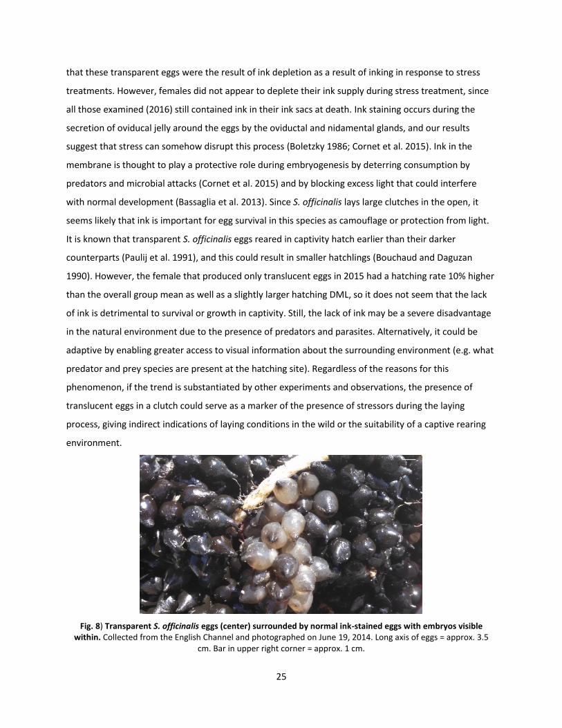
25
that these transparent eggs were the result of ink depletion as a result of inking in response to stress
treatments. However, females did not appear to deplete their ink supply during stress treatment, since
all those examined (2016) still contained ink in their ink sacs at death. Ink staining occurs during the
secretion of oviducal jelly around the eggs by the oviductal and nidamental glands, and our results
suggest that stress can somehow disrupt this process (Boletzky 1986; Cornet et al. 2015). Ink in the
membrane is thought to play a protective role during embryogenesis by deterring consumption by
predators and microbial attacks (Cornet et al. 2015) and by blocking excess light that could interfere
with normal development (Bassaglia et al. 2013). Since S. officinalis lays large clutches in the open, it
seems likely that ink is important for egg survival in this species as camouflage or protection from light.
It is known that transparent S. officinalis eggs reared in captivity hatch earlier than their darker
counterparts (Paulij et al. 1991), and this could result in smaller hatchlings (Bouchaud and Daguzan
1990). However, the female that produced only translucent eggs in 2015 had a hatching rate 10% higher
than the overall group mean as well as a slightly larger hatching DML, so it does not seem that the lack
of ink is detrimental to survival or growth in captivity. Still, the lack of ink may be a severe disadvantage
in the natural environment due to the presence of predators and parasites. Alternatively, it could be
adaptive by enabling greater access to visual information about the surrounding environment (e.g. what
predator and prey species are present at the hatching site). Regardless of the reasons for this
phenomenon, if the trend is substantiated by other experiments and observations, the presence of
translucent eggs in a clutch could serve as a marker of the presence of stressors during the laying
process, giving indirect indications of laying conditions in the wild or the suitability of a captive rearing
environment.
Fig. 8) Transparent S. officinalis eggs (center) surrounded by normal ink-stained eggs with embryos visible within. Collected from the English Channel and photographed on June 19, 2014. Long axis of eggs = approx. 3.5
cm. Bar in upper right corner = approx. 1 cm.
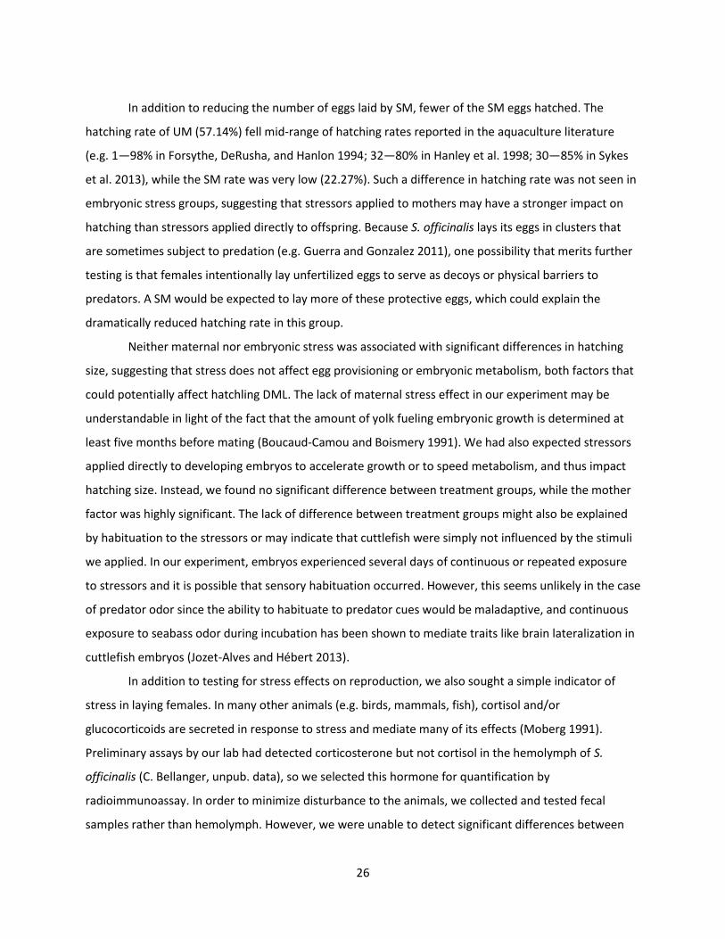
26
In addition to reducing the number of eggs laid by SM, fewer of the SM eggs hatched. The
hatching rate of UM (57.14%) fell mid-range of hatching rates reported in the aquaculture literature
(e.g. 1—98% in Forsythe, DeRusha, and Hanlon 1994; 32—80% in Hanley et al. 1998; 30—85% in Sykes
et al. 2013), while the SM rate was very low (22.27%). Such a difference in hatching rate was not seen in
embryonic stress groups, suggesting that stressors applied to mothers may have a stronger impact on
hatching than stressors applied directly to offspring. Because S. officinalis lays its eggs in clusters that
are sometimes subject to predation (e.g. Guerra and Gonzalez 2011), one possibility that merits further
testing is that females intentionally lay unfertilized eggs to serve as decoys or physical barriers to
predators. A SM would be expected to lay more of these protective eggs, which could explain the
dramatically reduced hatching rate in this group.
Neither maternal nor embryonic stress was associated with significant differences in hatching
size, suggesting that stress does not affect egg provisioning or embryonic metabolism, both factors that
could potentially affect hatchling DML. The lack of maternal stress effect in our experiment may be
understandable in light of the fact that the amount of yolk fueling embryonic growth is determined at
least five months before mating (Boucaud-Camou and Boismery 1991). We had also expected stressors
applied directly to developing embryos to accelerate growth or to speed metabolism, and thus impact
hatching size. Instead, we found no significant difference between treatment groups, while the mother
factor was highly significant. The lack of difference between treatment groups might also be explained
by habituation to the stressors or may indicate that cuttlefish were simply not influenced by the stimuli
we applied. In our experiment, embryos experienced several days of continuous or repeated exposure
to stressors and it is possible that sensory habituation occurred. However, this seems unlikely in the case
of predator odor since the ability to habituate to predator cues would be maladaptive, and continuous
exposure to seabass odor during incubation has been shown to mediate traits like brain lateralization in
cuttlefish embryos (Jozet-Alves and Hébert 2013).
In addition to testing for stress effects on reproduction, we also sought a simple indicator of
stress in laying females. In many other animals (e.g. birds, mammals, fish), cortisol and/or
glucocorticoids are secreted in response to stress and mediate many of its effects (Moberg 1991).
Preliminary assays by our lab had detected corticosterone but not cortisol in the hemolymph of S.
officinalis (C. Bellanger, unpub. data), so we selected this hormone for quantification by
radioimmunoassay. In order to minimize disturbance to the animals, we collected and tested fecal
samples rather than hemolymph. However, we were unable to detect significant differences between
27
groups, despite the strong effect on egg laying and hatching rate. The values detected were also larger
and more variable than those observed in giant Pacific octopus: 0.146 to 3.28 ng mg-1 vs. 0.010 to 0.022
ng mg -1 dry feces (Larson and Anderson 2010). Thus, it appears that fecal corticosterone levels were not
a particularly good indicator of stress in this species (at least during reproduction), while the number of
eggs per female and hatching rate are simpler and more reliable. A similar conclusion was reached in an
experiment examining corticosterone levels in mice after surgery; while plasma corticosterone
concentration was correlated with higher stress levels, fecal levels did not (Sundbom et al. 2011).
Indeed, in rats, weak group differences in circulating corticosterone that can be detected in blood
plasma sometimes cannot be identified in fecal assays (Chaby et al. 2015). Alternatively, the
endocrinology of the stress response in cephalopods may be more complex than a simple increase in
corticosterone. Existing studies of the endocrine system in cephalopods suggest complex interactions
with both the nervous and immune systems (Di Cosmo and Polese 2016).
We also attempted to relate the unused reproductive material of deceased females with stress
levels. Eggs ready for fertilization, encapsulation and laying are stored in the pallial cavity, which at
death, may contain up to 500 oocytes (Boletzky 1988; Laptikhovsky et al. 2003). Since unused oocytes in
the pallial cavity of deceased females indicates unused reproductive potential, we examined the number
remaining at death, hypothesizing that there would be more remaining in SM at death. However, there
was no difference between the two groups (Table 2, “remaining oocytes”), and so this trait cannot be
used as a proxy for stress levels.
28
Conclusion It appears that stress has a very strong impact on reproducing female cuttlefish (demonstrated
by the total number of eggs and hatchlings produced). By contrast, there was no change in hatching rate
when stressors were applied directly to eggs. Though maternal stress treatment clearly affected
reproduction, it did not result in a measurable difference in fecal corticosterone concentration (a
method used commonly in other species) or unused reproductive material, nor did stress have any
discernable effect on the hatching size of offspring. Unfortunately, this leaves us without any simple
marker of maternal stress in cuttlefish besides a reduction in the number of eggs and the occasional
absence of ink.
As global demand for protein increases with human population, understanding the effects of
rearing conditions and external factors on reproduction in cuttlefish and other farmed species is critical
to optimizing yields and animal welfare (Villanueva et al. 2014; Xavier et al. 2015). Our results suggest
that providing adequate tank space and minimizing handling of female cuttlefish may increase the
number of eggs laid and the hatching rate in aquaculture. In particular, catching females well before
copulation in order to allow them time to recover from the stress of capture and acclimate to an
artificial setting may result in higher offspring yields. (On the other hand, it may interfere with other
processes such as yolk reserve formation (which occurs months before) or even the proclivity to
copulate.) While we found that maternal stress had mostly negative effects in this experiment, the
possibility remains that stress conveys an advantage to offspring through some unknown means. Stress
has been hypothesized as a potential inducer of phenotypic plasticity that enables offspring to tailor a
response targeted to a particular threat (e.g. the presence of predators elicits defensive morphology)
present in the environment through physiology or behavior (Sheriff and Love 2013). This possibility was
tested in concurrent studies and will be discussed in forthcoming work.
Acknowledgments This work was completed as part of a Ph.D. thesis at the Université de Caen, Normandie, France. It is part of PReSTO’Cog, a research project supported by the Agence Nationale de la Recherche (ANR) and the Région Basse-Normandie (ANR-13-BSV7-0002). It would not have been possible without the generous assistance of Olivier Basuyaux, Sébastien Pien, the Synergie Mer et Littoral (SMEL), Jean Paul Lehodey, Nadège Villain-Naud, Céline Thomasse, the Centre de Recherches en Environnement Côtier (CREC, Université de Caen), Anais Meurie, Alex Schnell, Carole Di Poi, Camille Auger, Chloe Jane Way, Pauline Billiard, Kevin Bairos-Novak, Sarah Lefevre Barrière, Barbara O’Brien, Justine Lequesne, David Liegard, Frédéric Guyon and David Lemeille.
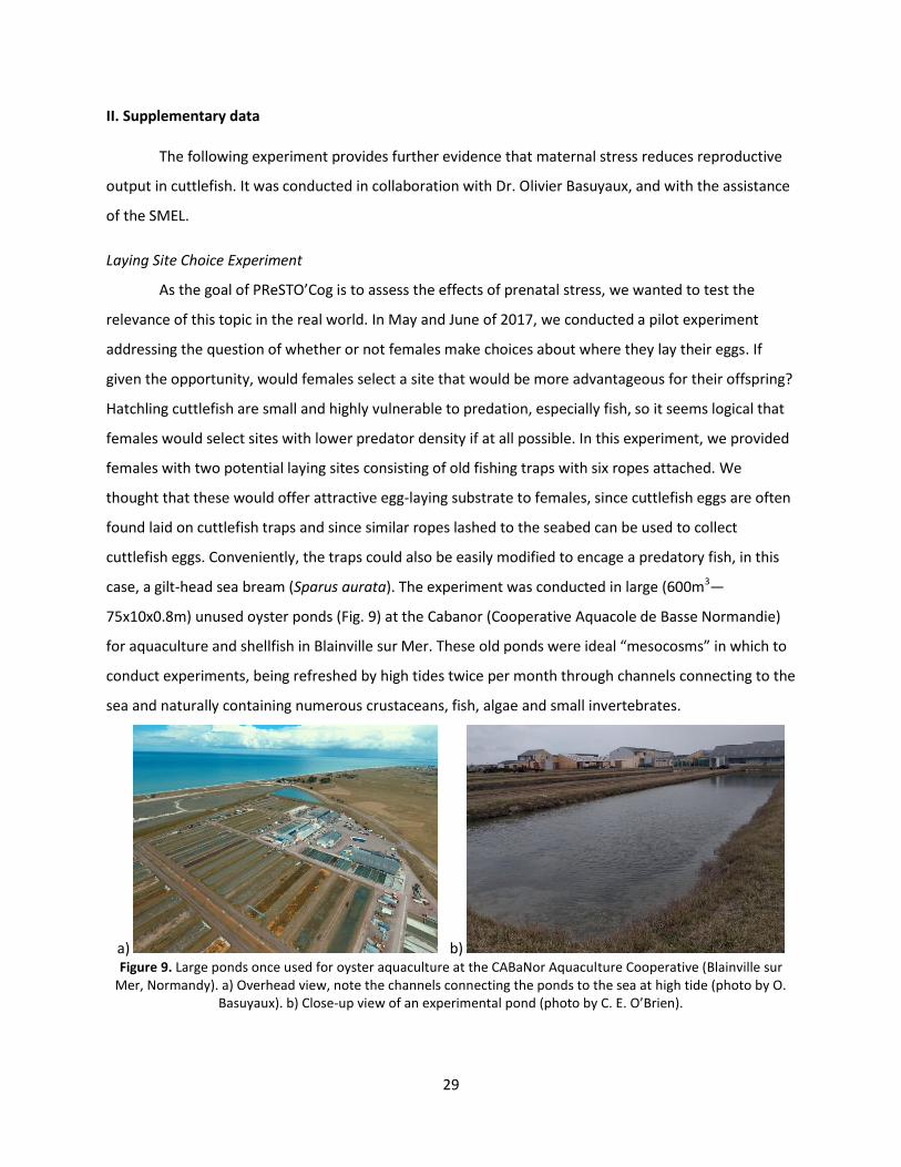
29
II. Supplementary data
The following experiment provides further evidence that maternal stress reduces reproductive
output in cuttlefish. It was conducted in collaboration with Dr. Olivier Basuyaux, and with the assistance
of the SMEL.
Laying Site Choice Experiment
As the goal of PReSTO’Cog is to assess the effects of prenatal stress, we wanted to test the
relevance of this topic in the real world. In May and June of 2017, we conducted a pilot experiment
addressing the question of whether or not females make choices about where they lay their eggs. If
given the opportunity, would females select a site that would be more advantageous for their offspring?
Hatchling cuttlefish are small and highly vulnerable to predation, especially fish, so it seems logical that
females would select sites with lower predator density if at all possible. In this experiment, we provided
females with two potential laying sites consisting of old fishing traps with six ropes attached. We
thought that these would offer attractive egg-laying substrate to females, since cuttlefish eggs are often
found laid on cuttlefish traps and since similar ropes lashed to the seabed can be used to collect
cuttlefish eggs. Conveniently, the traps could also be easily modified to encage a predatory fish, in this
case, a gilt-head sea bream (Sparus aurata). The experiment was conducted in large (600m3—
75x10x0.8m) unused oyster ponds (Fig. 9) at the Cabanor (Cooperative Aquacole de Basse Normandie)
for aquaculture and shellfish in Blainville sur Mer. These old ponds were ideal “mesocosms” in which to
conduct experiments, being refreshed by high tides twice per month through channels connecting to the
sea and naturally containing numerous crustaceans, fish, algae and small invertebrates.
a) b) Figure 9. Large ponds once used for oyster aquaculture at the CABaNor Aquaculture Cooperative (Blainville sur
Mer, Normandy). a) Overhead view, note the channels connecting the ponds to the sea at high tide (photo by O. Basuyaux). b) Close-up view of an experimental pond (photo by C. E. O’Brien).
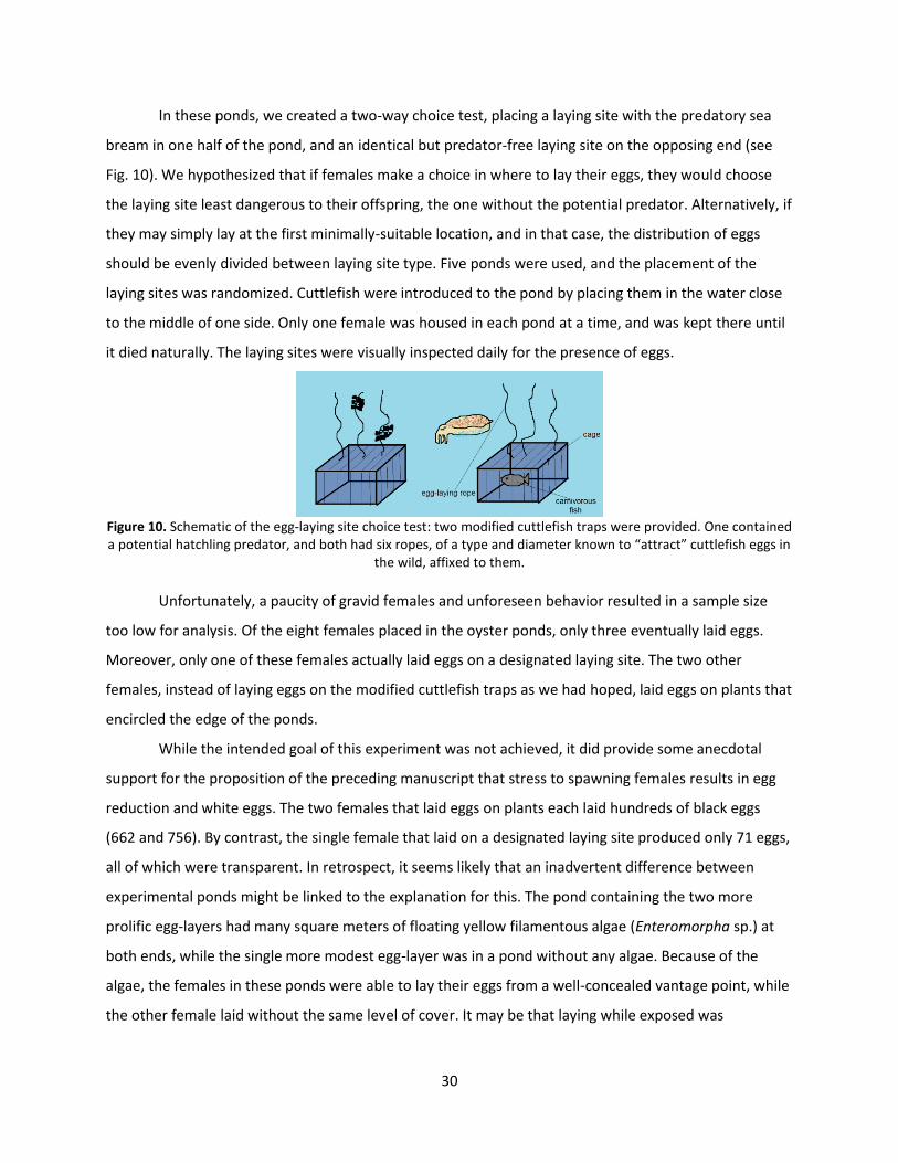
30
In these ponds, we created a two-way choice test, placing a laying site with the predatory sea
bream in one half of the pond, and an identical but predator-free laying site on the opposing end (see
Fig. 10). We hypothesized that if females make a choice in where to lay their eggs, they would choose
the laying site least dangerous to their offspring, the one without the potential predator. Alternatively, if
they may simply lay at the first minimally-suitable location, and in that case, the distribution of eggs
should be evenly divided between laying site type. Five ponds were used, and the placement of the
laying sites was randomized. Cuttlefish were introduced to the pond by placing them in the water close
to the middle of one side. Only one female was housed in each pond at a time, and was kept there until
it died naturally. The laying sites were visually inspected daily for the presence of eggs.
Figure 10. Schematic of the egg-laying site choice test: two modified cuttlefish traps were provided. One contained a potential hatchling predator, and both had six ropes, of a type and diameter known to “attract” cuttlefish eggs in
the wild, affixed to them.
Unfortunately, a paucity of gravid females and unforeseen behavior resulted in a sample size
too low for analysis. Of the eight females placed in the oyster ponds, only three eventually laid eggs.
Moreover, only one of these females actually laid eggs on a designated laying site. The two other
females, instead of laying eggs on the modified cuttlefish traps as we had hoped, laid eggs on plants that
encircled the edge of the ponds.
While the intended goal of this experiment was not achieved, it did provide some anecdotal
support for the proposition of the preceding manuscript that stress to spawning females results in egg
reduction and white eggs. The two females that laid eggs on plants each laid hundreds of black eggs
(662 and 756). By contrast, the single female that laid on a designated laying site produced only 71 eggs,
all of which were transparent. In retrospect, it seems likely that an inadvertent difference between
experimental ponds might be linked to the explanation for this. The pond containing the two more
prolific egg-layers had many square meters of floating yellow filamentous algae (Enteromorpha sp.) at
both ends, while the single more modest egg-layer was in a pond without any algae. Because of the
algae, the females in these ponds were able to lay their eggs from a well-concealed vantage point, while
the other female laid without the same level of cover. It may be that laying while exposed was

31
“stressful”, resulting in the lower number of eggs and their transparent membranes. Methodologically, if
this particular experiment is repeated, it should be conducted in ponds with the algae removed.
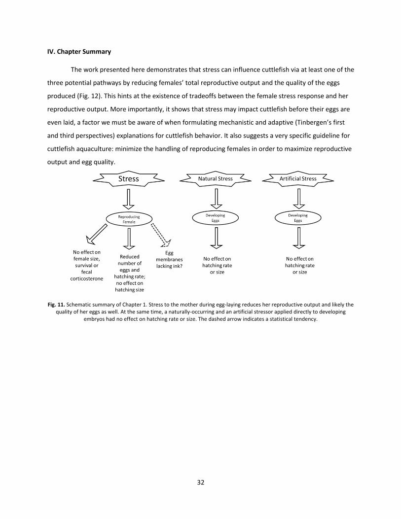
32
IV. Chapter Summary
The work presented here demonstrates that stress can influence cuttlefish via at least one of the
three potential pathways by reducing females’ total reproductive output and the quality of the eggs
produced (Fig. 12). This hints at the existence of tradeoffs between the female stress response and her
reproductive output. More importantly, it shows that stress may impact cuttlefish before their eggs are
even laid, a factor we must be aware of when formulating mechanistic and adaptive (Tinbergen’s first
and third perspectives) explanations for cuttlefish behavior. It also suggests a very specific guideline for
cuttlefish aquaculture: minimize the handling of reproducing females in order to maximize reproductive
output and egg quality.
Fig. 11. Schematic summary of Chapter 1. Stress to the mother during egg-laying reduces her reproductive output and likely the quality of her eggs as well. At the same time, a naturally-occurring and an artificial stressor applied directly to developing
embryos had no effect on hatching rate or size. The dashed arrow indicates a statistical tendency.


34
Chapter 2:
Prenatal Stress
Effects on
Offspring
35
Chapter 2: Prenatal Stress Effects on Offspring
This chapter explores two potential avenues of stress influence: from mother to offspring and
direct perception by the embryo itself. It is composed of two extensive reviews of cuttlefish
development and sensory abilities, followed by reports from numerous behavioral and learning tests
and neurobiological assays.
36
I. Article #2: “Behavioral development in embryonic and early juvenile cuttlefish (Sepia officinalis)”
This manuscript offers an in-depth overview of cuttlefish development, providing context to the
experiments that follow.
Behavioral Development in Embryonic and Early
Juvenile Cuttlefish (Sepia officinalis)
Caitlin E. O'Brien, Nawel Mezrai, Anne-Sophie Darmaillacq, Ludovic Dickel
1
1
Groupe Mémoire et Plasticité Comportementale (GMPc EA 4259), Université de Caen-Normandie, Caen, France
Dev Psychobiol 2016; 9999: 1–16, DOI 10.1002/dev.21476
Though a marine mollusc, the cuttlefish Sepia officinalis possesses a sophisticated brain,
advanced sensory systems and a large behavioral repertoire. Cuttlefish provide a unique
perspective on animal behavior due to their phylogenic distance from more traditional
(vertebrate) models. S. officinalis is well-suited to addressing questions of behavioral ontogeny.
As embryos, they can perceive and learn from their environment and experience no direct
parental care. A marked progression in learning and behavior is observed during late embryonic
and early juvenile development. This improvement is concomitant with expansion and
maturation of the vertical lobe, the cephalopod analog of the mammalian hippocampus. This
review synthesizes existing knowledge regarding embryonic and juvenile development in this
species in an effort to better understand cuttlefish behavior and animal behavior in general. It
will serve as a guide to future researchers and encourage greater awareness of the utility of this
species to behavioral science.
Keywords: Common cuttlefish, defense, predation, plasticity, learning, memory, perception,
habituation, recognition learning, associative learning, welfare
37
Introduction
The common cuttlefish, Sepia officinalis (Linnaeus 1758), along with other cephalopods,
possesses a centralized nervous system capable of learning and memory, advanced sensory
systems and a highly sophisticated behavioral repertoire that is comparable to that of vertebrates
(Packard 1972; Hanlon and Messenger 1998). The life history and habits of cuttlefish enables
such behavioral research to be pursued through both field and laboratory study. With this
invertebrate model, we can address questions about complex behavior and learning in a marine
mollusc, a group genetically very distant from more traditional models, such as birds and
rodents. This phylogenic distance provides an alternative perspective that is critical to
understanding the ways that natural selection, ancestral history and non-hereditary processes
interact to shape animal behavior.
S. officinalis is particularly well-suited for the study of behavioral ontogeny. Like many
fish, this species has gelatinous, semi-permeable eggs and rapidly-developing sensory abilities
that allow them exceptional sensory access to the surrounding environment during the final
stages of embryonic development (Romagny et al. 2012). Coupled with their oviparity and the
lack of direct maternal care, these characteristics allow stimuli to be applied directly to embryos
rather than being transmitted and mediated by the mother, as in rodents and birds. At hatching,
this species possesses one of the richest behavioral repertoires in the animal kingdom. This
review provides an overview of existing knowledge regarding the sensory experience, habitat,
learning and behavior of embryonic, hatchling (<1week after eclosion) and juvenile (up to 17
weeks) cuttlefish to serve as a starting point for further inquiry. In order to properly contextualize
the behavior of this less-familiar species, this review also draws insight from and parallels to
other species of cuttlefish, as well as some of their closest coleoid cephalopod relatives—
octopuses and squids.
Embryonic Development
Embryogenesis and Yolk Reserves
Spawning of S. officinalis eggs occurs in the English Channel in shallow (5-60m), well-lit
coastal waters throughout the spring and summer (Nixon and Mangold 1998; Basuyaux &
Legrand, 2013). Eggs are usually attached in clusters to objects on the seabed, such as algae,
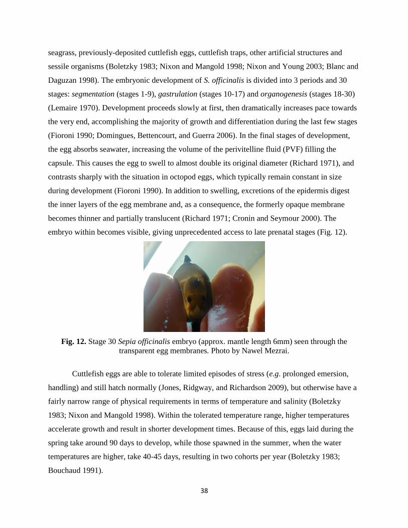
38
seagrass, previously-deposited cuttlefish eggs, cuttlefish traps, other artificial structures and
sessile organisms (Boletzky 1983; Nixon and Mangold 1998; Nixon and Young 2003; Blanc and
Daguzan 1998). The embryonic development of S. officinalis is divided into 3 periods and 30
stages: segmentation (stages 1-9), gastrulation (stages 10-17) and organogenesis (stages 18-30)
(Lemaire 1970). Development proceeds slowly at first, then dramatically increases pace towards
the very end, accomplishing the majority of growth and differentiation during the last few stages
(Fioroni 1990; Domingues, Bettencourt, and Guerra 2006). In the final stages of development,
the egg absorbs seawater, increasing the volume of the perivitelline fluid (PVF) filling the
capsule. This causes the egg to swell to almost double its original diameter (Richard 1971), and
contrasts sharply with the situation in octopod eggs, which typically remain constant in size
during development (Fioroni 1990). In addition to swelling, excretions of the epidermis digest
the inner layers of the egg membrane and, as a consequence, the formerly opaque membrane
becomes thinner and partially translucent (Richard 1971; Cronin and Seymour 2000). The
embryo within becomes visible, giving unprecedented access to late prenatal stages (Fig. 12).
Fig. 12. Stage 30 Sepia officinalis embryo (approx. mantle length 6mm) seen through the
transparent egg membranes. Photo by Nawel Mezrai.
Cuttlefish eggs are able to tolerate limited episodes of stress (e.g. prolonged emersion,
handling) and still hatch normally (Jones, Ridgway, and Richardson 2009), but otherwise have a
fairly narrow range of physical requirements in terms of temperature and salinity (Boletzky
1983; Nixon and Mangold 1998). Within the tolerated temperature range, higher temperatures
accelerate growth and result in shorter development times. Because of this, eggs laid during the
spring take around 90 days to develop, while those spawned in the summer, when the water
temperatures are higher, take 40-45 days, resulting in two cohorts per year (Boletzky 1983;
Bouchaud 1991).
39
Throughout prenatal and early post-natal development, embryos are sustained by internal
and external yolk reserves (Lemaire 1970). Those that develop more quickly because of higher
temperatures hatch with a larger yolk reserve remaining (S. v Boletzky 1975; O. Bouchaud and
Daguzan 1990; Olivier Bouchaud 1991; Dickel, Chichery, and Chichery 1997a). However, this
more rapid development results in smaller hatchlings (Roger T. Hanlon and Messenger 1988; O.
Bouchaud and Daguzan 1990), and this has implications for survival and the onset of predatory
behavior ( Boletzky 1994; Bouchaud 1991). Water temperature varies with location, depth,
season, currents, tide and weather, so the timing and position of an egg can strongly affect its
later prospects (Bloor et al. 2013).
Sensory Systems
Embryos are buffered and protected from the external environment by their egg capsule,
but they are not entirely isolated from it. At the beginning of development, the layer of ink in the
egg membrane absorbs light and prevents most visual information from penetrating in either
direction (Paulij et al. 1991). In addition, eggs can be laid down to a depth at which only 10% of
surface light intensity remains (Bloor et al. 2013). Despite these limitations, the membrane
becomes translucent due to egg expansion, the optic lobes and lens mature during stages 20 and
21(Lemaire 1970; Lemaire and Richard 1978), and as a result, reaction to visual stimulation in
the external environment is observed by stage 25 (Romagny et al. 2012).
Likewise, S. officinalis embryos are able to perceive waterborne chemical cues that
diffuse through the egg membrane and tactile stimuli from movement in the external
environment by stage 23 (Romagny et al. 2012). Thus, the organogenesis period of embryonic
development (stages 18-30) is characterized by a progressive build-up in the amount of sensory
information penetrating the egg membrane in conjunction with a gradually-increasingly ability to
perceive this information. (It is possible that sensory perception is possible even earlier than
stage 23, but the ability to respond is not.)
Maternal Influence and the Prenatal Environment
Cuttlefish eggs are abandoned by the mother soon after laying, and thus receive no direct
parental care or protection (Darmaillacq et al. 2006). Nevertheless, offspring can be affected by
maternal influence. For instance, embryo provisioning in cephalopods affects the size, molecular

40
composition, and ultimately the survival and success of eggs (“maternal effect”, Bloor et al.
2013). Nutritional stress in another cephalopod, the dumpling squid (Euprymna tasmanica), has
been shown to cause females to lay fewer eggs with less yolk that result in lower levels of
survival and success (Steer et al. 2004). Among cephalopods, S. officinalis have some of the
largest eggs, reflecting a high amount of maternal provisioning (Fioroni 1990).
More importantly, since attachment renders eggs sessile, a female’s timing and choice of
egg-laying site dictates the environmental conditions experienced throughout egg development
(Bloor et al. 2013). Ultimately, these environmental conditions can influence survival, growth
rate, hatching time and post-natal behavior. For example, extreme salinities or contamination by
pollutants (e.g. heavy metals or pharmaceutical residues), which can slow or interfere with
development, may result from being located close to shore (Paulij et al. 1990; Paulij, Bogaards,
and Denucé 1990; Bloor et al. 2013; Di Poi et al. 2014; Bidel, Di Poi, et al. 2016). As a mostly
semelparous species with only a single spawning period at the end of life (Boletzky 1987), there
is presumably strong selective pressure on females to lay eggs in an appropriate location, at a
suitable time and in appropriate densities (Bloor et al. 2013).
The revelation that perception is possible from within the egg has important implications
for the understanding of behavioral development in juveniles. It suggests that S. officinalis
embryos are developmentally and behaviorally plastic, and demonstrates that they begin adapting
to their environment long before hatching. The particular suite of prenatal stimuli experienced by
embryos will depend on their location. Field observations concerning the environment around
spawning sites are scarce. However, since S. officinalis lay their eggs on submerged objects
(Boletzky 1983), and because such objects tend to attract other marine life to the area, it is likely
that developing embryos will sense predators, future prey, as well as other species during the
course of development (Fig. 13). They can then use this information to prepare for the post-
hatching environment. Indeed, several experiments have found that predator and prey stimuli
strongly influence juvenile behavior (Darmaillacq, Lesimple, and Dickel 2008a; Guibé, Boal,
and Dickel 2010; Guibé et al. 2012; Jozet-Alves and Hebert 2012; Jozet-Alves and Hébert 2013).
Careful field observations documenting the identity and prevalence of other species at spawning
sites would be extremely useful in piecing together a picture of the sensory experience of
embryos during development.
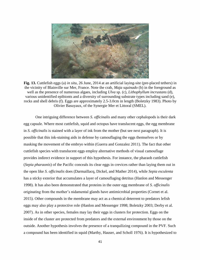
41
Fig. 13. Cuttlefish eggs (a) in situ, 26 June, 2014 at an artificial laying-site (pre-placed tethers) in
the vicinity of Blainville sur Mer, France. Note the crab, Maja squinado (b) in the foreground as
well as the presence of numerous algaes, including Ulva sp. (c), Lithophyllum incrustans (d),
various unidentified epibionts and a diversity of surrounding substrate types including sand (e),
rocks and shell debris (f). Eggs are approximately 2.5-3.0cm in length (Boletzky 1983). Photo by
Olivier Basuyaux, of the Synergie Mer et Littoral (SMEL).
One intriguing difference between S. officinalis and many other cephalopods is their dark
egg capsule. Where most cuttlefish, squid and octopus have translucent eggs, the egg membrane
in S. officinalis is stained with a layer of ink from the mother (but see next paragraph). It is
possible that this ink-staining aids in defense by camouflaging the eggs themselves or by
masking the movement of the embryo within (Guerra and Gonzalez 2011). The fact that other
cuttlefish species with translucent eggs employ alternative methods of visual camouflage
provides indirect evidence in support of this hypothesis. For instance, the pharaoh cuttlefish
(Sepia pharaonis) of the Pacific conceals its clear eggs in crevices rather than laying them out in
the open like S. officinalis does (Darmaillacq, Dickel, and Mather 2014), while Sepia esculenta
has a sticky exterior that accumulates a layer of camouflaging detritus (Hanlon and Messenger
1998). It has also been demonstrated that proteins in the outer egg membrane of S. officinalis
originating from the mother’s nidamental glands have antimicrobial properties (Cornet et al.
2015). Other compounds in the membrane may act as a chemical deterrent to predators lefish
eggs may also play a protective role (Hanlon and Messenger 1998; Boletzky 2003; Derby et al.
2007). As in other species, females may lay their eggs in clusters for protection. Eggs on the
inside of the cluster are protected from predators and the external environment by those on the
outside. Another hypothesis involves the presence of a tranquilizing compound in the PVF. Such
a compound has been identified in squid (Marthy, Hauser, and Scholl 1976). It is hypothesized to
42
reduce the likelihood of detection and predation by reducing embryo movement and preventing
premature hatching (Marthy, Hauser, and Scholl 1976; Weischer and Marthy 1983). While this
agent has not yet been identified in cuttlefish, its presence is probable and the tranquilizing
compound from squid has been shown to reduce activity in cuttlefish and other cephalopods
(Weischer and Marthy 1983).
Embryonic Movement and Sleep:
Embryos exhibit a variety of movements and behaviors. Respiration is visible from
outside of the egg. In addition, mantle contractions—pumping motions involving the whole
mantle and its musculature—are also visible (Corner 1977). While the function of this behavior
is unclear, novel visual and odor stimuli will cause a more or less immediate change in the rate
of mantle contractions (Romagny et al. 2012). Finally, embryos also display periods of
unprovoked activity including movements of the arms, tentacles, fins and funnel, including
twitching, exercise of the muscles controlling the chromatophores, mantle contractions and
apparent stretching of the arms and tentacles (Corner 2013a). These recurring episodes are
analogous to REM sleep in vertebrates and are referred to as “motorically active sleep” (MAS)
(Corner 2013a). This behavior starts sometime before stage 29, and continues into postnatal life
(Corner 2013a). Other cephalopods (squid and octopus) and invertebrates (e.g. nematodes,
annelids, cnidarians, and insects) also exhibit similar prenatal or larval behavior (Corner 2013b).
43
Non-Associative Learning
Habituation
Habituation is a simple form of learning in which an organism ceases responding to a
stimulus after repeated or extended exposure to it (Bouton, 2007). The prenatal occurrence of
habituation is difficult to ascertain due to the relative inaccessibility of most developing
embryos. In species for which this type of prenatal learning has been demonstrated (e.g. superb
fairy wrens, rats, humans), inquiry has been largely restricted to chemosensory and vibroacoustic
stimuli (e.g. Goldkrand & Litvack, 1991; Smotherman & Robinson, 1992; van Heteren,
Boekkooi, Schiphorst, Jongsma, & Nijhuis, 2001; Colombelli-Négrel, Hauber, & Kleindorfer,
2014). In S. officinalis however, the partial translucence of late stage eggs enables observation of
the mantle movements of the embryo within and expands the range of stimuli that can be tested
to the visual. In response to novel visual, tactile or chemosensory stimulus, embryos will reduce
respiration and mantle movements. The subsequent resumption of an increased mantle
movement after repeated or chronic exposure to the stimulus indicates habituation. This has been
demonstrated in final-stage (30) embryos with repeated exposures to bright light (Romagny et
al., 2012). Likewise, stage 30 embryos can be habituated to the sight of Carcinus maenas (green
crabs) (N. Mezrai, unpub. data). Habituation conserves energy by allowing animals to eliminate
unnecessary behavioral responses (Rankin, Abrams, Barry, Bhatnagar, Clayton, Colombo,
Coppolag, Geyerh, Glanzmani, Marslandj, McSweeney, Wilsonl, Wum, & Thompsonn 2009).
Prenatal Imprinting and Exposure Learning
Imprinting is another form of learning characterized by the establishment of an
irreversible preference for something during a limited sensitive period, usually early in
development. This preference is expressed later in life and is considered “indelible” in that it
cannot be reversed after the sensitive period (Lorenz, 1937). Often, this preference will be
generalized to other objects sharing similar characteristics (Sluckin, 2007). The classic example
comes from filial imprinting in precocial birds that imprint on their mother during a particular
window after hatching and generalize this preference to sexual partners later in life (Bolhuis,
1991). Other forms include imprinting for prey, habitat, host or a sexual partner (Bouton, 2007).
A similar form of recognition learning is perceptual learning. Like imprinting, perceptual
learning may occur early in life and involves a learned preference for something after exposure,
44
but otherwise does not meet the criteria for imprinting, such as indelibility and generalization
(Shettleworth, 2009).
Imprinting and perceptual learning for prey preference have been demonstrated in S.
officinalis. As reported by Wells (1958) and others (e.g. Darmaillacq, Chichery, Poirier, &
Dickel, 2004; Darmaillacq et al., 2006a; Darmaillacq , Chichery, Shashar, & Dickel 2006; Guibé
et al., 2012) hatchlings have an “innate” preference for shrimp or shrimp-shaped objects (but see
below). Darmaillacq (et al., 2004a; 2006a; 2006b) demonstrated that this preference could be
overridden by chemical and/or visual exposure to crabs shortly after hatching. This induced
preference fit the criteria for imprinting: it lasted for at least three days, persisted after the
cuttlefish had consumed a shrimp and was only induced during a short sensitive period early in
the life of the cuttlefish (Darmaillacq et al., 2004a; 2006a; 2006b; Healy, 2006). Interestingly,
this sensitive period for prey preference induction seems to begin before hatching: hatchlings
from embryos visually exposed to crabs for a week or more prior to hatching (and not after)
preferred crabs in a choice test seven days later (Darmaillacq et al., 2008). Other experiments
demonstrated that this sensitive period closes about 6 hours after sunrise on the day of hatching
(Darmaillacq et al., 2006a).
Imprinting and perceptual learning allow cuttlefish to learn the characteristics of
available prey in their environment during the perinatal period (Healy, 2006; Darmaillacq et al.,
2014). Shrimp abundance may vary between egg laying sites and perinatal exposure to co-
occuring species could transmit information about the relative abundance of predators and prey
in the environment. Such information seems especially advantageous to S. officinalis, which uses
different strategies in response to different species, both predator and prey (described later).
Information about species abundance would allow cuttlefish to prioritize the development of one
strategy over another. Similar and analogous instances of food imprinting and perceptual
learning exist in other phyla (e.g. amphibians, Hepper & Waldman, 1992; birds, Bertin,
Calandreau, Arnould, Nowak, Levy, Noirot, Bouvarel, & Leterrier, 2010; insects, Quesada &
Schausberger, 2012). This suggests that prenatal learning may be a common method of energy
conservation and risk reduction during the vulnerable time following hatching or birth
throughout the animal kingdom.
The discovery of imprinting in S. officinalis may also completely overturn the notion that
cuttlefish “innately” prefer shrimp (Wells, 1958). Instead, it may be that the sight of other
45
developing cuttlefish—elongate objects that move along their horizontal axis—actually induces a
preference for shrimp through generalization. Indeed, cuttlefish from eggs incubated in isolation
tend to not prefer crab or shrimps (L. Dickel pers. obs.). Unfortunately, Wells (1958), the first to
record the shrimp preference, did not report whether the cuttlefish in his experiments were reared
socially or in isolation (Darmaillacq et al., 2014). Regardless of whether this preference is pre-
programmed, it does appear that cuttlefish have the innate ability to distinguish between species.
In addition to the capacity to distinguish between different kinds of decapod crustaceans,
prey preference could also be induced on the basis of brightness contrast: Where naïve cuttlefish
preferred dark to white crabs as their initial meal, embryos and hatchlings exposed to white crabs
later preferred these over dark crabs (Guibé et al., 2012). This demonstrates that S. officinalis is
able to learn about multiple characteristics of prey (shape and/or contrast). Moreover, cuttlefish
pre- or postnatally exposed to white crabs preferred black crabs over shrimp, indicating that S.
officinalis will generalize the characteristics of a learned preference to the closest alternative if
the preferred item is not available (Guibé et al., 2012).
One experiment in S. officinalis also investigated the interaction between sensory
modalities in the induction of prey choice. Hatchlings from eggs exposed to waterborne
chemosensory cues from shrimp, crab and two control cues were tested for visual preference.
Cuttlefish that had been incubated with crab or control cues showed either no preference or the
“typical” preference for shrimp (Guibé et al., 2010). Cuttlefish that were exposed to
chemosensory cues from crabs later showed a visual preference for shrimp (Guibé et al., 2010).
These puzzling results suggest that cross-modal effects (VanderSal & Hebets, 2007) are
operating between the chemosensory and visual systems that merit further investigation. In
cuttlefish, sensory integration occurs in the superior frontal lobes of the brain (Nixon & Young,
2003) and is thus the likely site of these putative interactions (Guibé et al., 2010).
Finally, in addition to the demonstration of prenatal food preference learning, it has been
shown that exposure to other ecologically-salient objects in the incubation environment can
affect future behavior. Naïve hatchlings were found to spontaneously prefer dark shelters
(bivalve shells). Prenatal exposure to white shelters eliminated this preference, resulting in
cuttlefish that were equally likely to hide under a black or white shelter (Guibé & Dickel, 2011).
Like recognition learning, knowledge about the characteristics of objects in the surrounding
46
environment could be adaptive. In this case, a cuttlefish may be learning that white objects are
plentiful in the area, stationary, and may be a source of future refuge.
Lateralization
Lateralization is the tendency to process information through one side of the brain in a
particular type of situation. Potentially, lateralization yields advantages in speed and efficiency
of information processing via the specialization of each side of the brain (Jozet-Alves & Hébert,
2013). Lateralization is seen in numerous vertebrates, including primates, birds, fishes and
amphibians. In these animals, left brain lateralization is often coupled with rapid responses,
especially escape reflexes (Jozet-Alves et al., 2012).
In cuttlefish, brain lateralization seems to occur in cuttlefish after prenatal exposure to
predator odor (Jozet-Alves et al., 2012; Jozet-Alves & Hébert, 2013). Over the course of the first
month of life, juveniles that had been exposed prenatally to predator odor develop a bias for
turning towards a shelter on the left, rather than on the right side (Jozet-Alves et al., 2012). This
behavior may reflect a preference for using the left eye and left side of the brain (visual input is
processed by the same side from which it is perceived in cuttlefish). The tendency to develop a
turning bias is another instance of embryos perceiving information from the prenatal
environment and adapting accordingly. Octopus vulgaris, another well-studied model species in
the study of cephalopod behavior, also appears to show brain lateralization in adult individuals’
tendency to favor one eye over the other (Byrne, Kuba, & Griebel, 2002). Further study of this
phenomenon in both cephalopods and vertebrates could help reveal the selective pressures that
promote the evolution of brain lateralization in complex, centralized nervous systems.
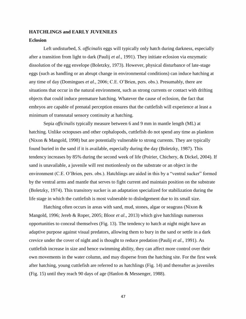
47
HATCHLINGS and EARLY JUVENILES
Eclosion
Left undisturbed, S. officinalis eggs will typically only hatch during darkness, especially
after a transition from light to dark (Paulij et al., 1991). They initiate eclosion via enzymatic
dissolution of the egg envelope (Boletzky, 1973). However, physical disturbance of late-stage
eggs (such as handling or an abrupt change in environmental conditions) can induce hatching at
any time of day (Domingues et al., 2006; C.E. O’Brien, pers. obs.). Presumably, there are
situations that occur in the natural environment, such as strong currents or contact with drifting
objects that could induce premature hatching. Whatever the cause of eclosion, the fact that
embryos are capable of prenatal perception ensures that the cuttlefish will experience at least a
minimum of transnatal sensory continuity at hatching.
Sepia officinalis typically measure between 6 and 9 mm in mantle length (ML) at
hatching. Unlike octopuses and other cephalopods, cuttlefish do not spend any time as plankton
(Nixon & Mangold, 1998) but are potentially vulnerable to strong currents. They are typically
found buried in the sand if it is available, especially during the day (Boletzky, 1987). This
tendency increases by 85% during the second week of life (Poirier, Chichery, & Dickel, 2004). If
sand is unavailable, a juvenile will rest motionlessly on the substrate or an object in the
environment (C.E. O’Brien, pers. obs.). Hatchlings are aided in this by a “ventral sucker” formed
by the ventral arms and mantle that serves to fight current and maintain position on the substrate
(Boletzky, 1974). This transitory sucker is an adaptation specialized for stabilization during the
life stage in which the cuttlefish is most vulnerable to dislodgement due to its small size.
Hatching often occurs in areas with sand, mud, stones, algae or seagrass (Nixon &
Mangold, 1996; Jereb & Roper, 2005; Bloor et al., 2013) which give hatchlings numerous
opportunities to conceal themselves (Fig. 13). The tendency to hatch at night might have an
adaptive purpose against visual predators, allowing them to bury in the sand or settle in a dark
crevice under the cover of night and is thought to reduce predation (Paulij et al., 1991). As
cuttlefish increase in size and hence swimming ability, they can affect more control over their
own movements in the water column, and may disperse from the hatching site. For the first week
after hatching, young cuttlefish are referred to as hatchlings (Fig. 14) and thereafter as juveniles
(Fig. 15) until they reach 90 days of age (Hanlon & Messenger, 1988).
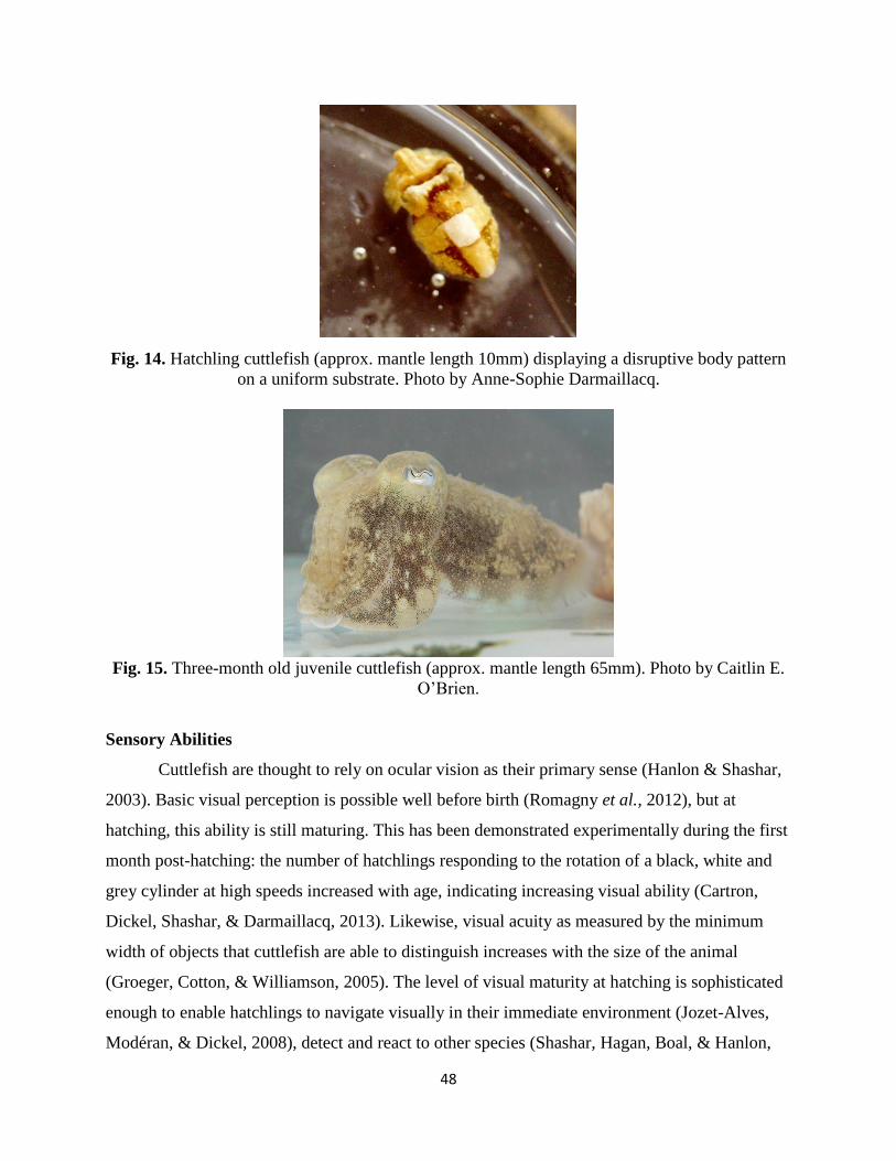
48
Fig. 14. Hatchling cuttlefish (approx. mantle length 10mm) displaying a disruptive body pattern
on a uniform substrate. Photo by Anne-Sophie Darmaillacq.
Fig. 15. Three-month old juvenile cuttlefish (approx. mantle length 65mm). Photo by Caitlin E.
O’Brien.
Sensory Abilities
Cuttlefish are thought to rely on ocular vision as their primary sense (Hanlon & Shashar,
2003). Basic visual perception is possible well before birth (Romagny et al., 2012), but at
hatching, this ability is still maturing. This has been demonstrated experimentally during the first
month post-hatching: the number of hatchlings responding to the rotation of a black, white and
grey cylinder at high speeds increased with age, indicating increasing visual ability (Cartron,
Dickel, Shashar, & Darmaillacq, 2013). Likewise, visual acuity as measured by the minimum
width of objects that cuttlefish are able to distinguish increases with the size of the animal
(Groeger, Cotton, & Williamson, 2005). The level of visual maturity at hatching is sophisticated
enough to enable hatchlings to navigate visually in their immediate environment (Jozet-Alves,
Modéran, & Dickel, 2008), detect and react to other species (Shashar, Hagan, Boal, & Hanlon,
49
2000) and to gauge characteristics of the visual environment for body patterning (Chiao &
Hanlon, 2001).
Polarization sensitivity (PS) is the ability to distinguish between different linear
polarizations of light. If a cuttlefish is placed inside a rotating cylinder with alternating bars of
oppositely polarized information, it will usually display an optomotor response, following the
motion of the cylinder with eyes and body (Cartron et al., 2013b). This apparatus has been used
to demonstrate that PS appears in cuttlefish around seven days post-hatching. This ability
matured in a manner similar to that of contrast vision, with the number of individuals reacting to
the rotating polarized cylinder increasing with age, although at a slower pace (Cartron et al.,
2013b). In squid, polarization has been shown to improve the ability to visually detect prey at a
distance (Shashar, Hanlon, & deM Petz, 1998). Likewise, S. officinalis detect prey faster and
preferentially attack them when they reflect polarized light rather than only luminance
information (Shashar, Hagan, Boal, & Hanlon, 2000). PS has been hypothesized to aid with
capture detection of silvery fish, transparent prey like shrimp and in low-luminance contrast
situations (Shashar et al., 1998; 2000; Cartron, Josef, Lerner, McCusker, Darmaillacq, Dickel, &
Shashar, 2013). Indeed, it has been suggested that the rapid increase in prey detection observed
in cuttlefish during the first week after hatching may be concomitant with the maturation of this
system (Dickel et al., 1997).
While they are thought to rely primarily on vision, evidence increasingly demonstrates
the role of chemoreception for cuttlefish and other cephalopods (Hanlon & Shashar, 2003). We
know that this ability is functional before hatching (Romagny et al., 2012) and chemoreceptor
cells are present at hatching in both the suckers of the arms and tentacles, as well as the region
surrounding the mouth (Sundermann, 1983; Nixon & Mangold, 1998). However, the relative
maturity of this system at hatching is unknown, and more experiments are needed.
Statocysts are the organs responsible for perception of gravity, acceleration and low
frequency vibrations. In addition, cuttlefish possess an analog of the lateral line system in fish,
consisting of thousands of sensory hair cells (Budelmann & Bleckmann, 1988). Both of these
mechanoreceptive organs are present at hatching. It has been demonstrated that month-old
juveniles are able to detect a range of vibrations with one or both of these systems. Low
frequency vibrations (20–600 Hz) were shown to induce defensive behaviors such as burrowing,
changes in body pattern and moving, although not at every frequency in that range (Komak,
50
Boal, Dickel, & Budelmann, 2005). Interestingly, juvenile cuttlefish (one and three months old)
could not be habituated to vibrational stimuli, even after five consecutive presentations. This is
consistent with the hypothesis that mechanoreception plays a role in predator detection, since
habituation would be highly inappropriate in this context. Between the ages of one and three
months, juveniles show a decrease in responsiveness to vibrational stimuli. At three months of
age they cease burrowing in response, despite the fact that the sensory acuity of their statocysts
and lateral-line analog is thought to increase with size (Budelmann, 1995). This contrast implies
that defensive behavior, especially burrowing, may become less relevant as the cuttlefish
increases in size (see later discussion), although mechanoreception continues to play a defensive
role (Komak et al., 2005).
Body Patterning and Defense
Juveniles’ primary ecological challenge is avoiding predators. Defensive tactics fall into
two categories: primary defenses to prevent detection and secondary defenses to affect escape if
primary defenses fail (Cott, 1941; Hanlon & Messenger, 1996). Primary defenses are mainly
cryptic, including countershading, deceptive resemblance and camouflage. Secondary defenses
include inking, jetting, and threat displays (Hanlon & Messenger, 1988; Ferguson & Messenger,
1991; Ferguson, Messenger, & Budelmann, 1994; Hanlon & Messenger, 1996). Most of the
known predators of juveniles are visual hunters (Hanlon & Messenger, 1996), making body
patterning a critical aspect of defense.
Body patterns in cuttlefish are created with numerous patches of pigmented cells
(chromatophores). When contracted, the pigment of the chromatophore is obscured, creating a
light patch. When expanded, the pigment becomes visible and creates a dark patch. Expansion
and contraction of these cells are controlled via direct innervation from the brain (Florey, 1969).
Different groups of chromatophores are expanded or contracted in unison to create 33 chromatic
components. The most prominent of these components is a large white square in the center of the
dorsal mantle (see illustrations in Hanlon & Messenger, 1988). These components combine to
form a continuum of 13 formally-defined stereotypical body patterns. Among these, there are
three main categories of body patterns used to achieve primary crypsis: uniform (entirely dark or
light), mottle (a mixture of small dark and light patches) and disruptive (a mixture of large dark
and light patches) (Hanlon & Messenger, 1988). Chromatophores begin to appear in stage 25
51
embryos (Andouche, 2013) and the brain structures controlling body patterning (optic lobes,
lateral basal lobes and chromatophore lobes) are well-developed but not fully mature at hatching
(Dickel et al., 1997). As a result, hatchling cuttlefish are capable of almost the entire repertoire
of adult body patterns (10 out of 13, Hanlon & Messenger, 1988). This situation differs from that
of many Loliginid squid and octopuses, which have few chromatophores at hatching (Fioroni,
1990) and can’t produce full body patterns (Hanlon and Messenger 1988).
After leaving the egg clutch, a hatchling in the English Channel may settle on a uniform
dark background such as mud or a uniform light background such as sand (Blanc et al., 1998).
Alternatively, it may come to reside on a heterogeneous background consisting of combinations
of algae, rock, shell debris, sand and mud (Hanlon & Messenger, 1996). In the laboratory, the
uniform pattern can usually be prompted by a solid colored artificial background or sand. The
mottle pattern is elicited by gravel and artificial checkerboards with squares 4-12% of the
juvenile’s own white square, while the disruptive pattern is induced by squares 40 to 120% of the
cuttlefish’s own white square and small rocks (Barbosa, Mäthger, Chubb, Florio, Chiao, &
Hanlon, 2007; Mäthger & Hanlon, 2007). S. officinalis appears to be employing a rule based on
the size of nearby objects and its own increasing size. Notably, this is accomplished without
color vision (Messenger, 1977; Mäthger, Barbosa, Miner, & Hanlon, 2006) and without any
visual feedback from cuttlefish’s own body pattern (Barbosa et al., 2007).
Despite their ability to display almost the entire range of body patterns, newly-hatched
cuttlefish often show the disruptive pattern on uniform backgrounds (Fig. 3; Hanlon &
Messenger, 1988; Poirier, Chichery, & Dickel, 2005; Dickel, Darmaillacq, Poirier, Agin,
Bellanger, & Chichery, 2006). Young S. pharaonis will also display the disruptive pattern on
uniform background (but see discussion below) (Lee, Yan, & Chiao, 2010). This seemingly
“inappropriate” behavior may be explained by the fact that cuttlefish have other strategies for
crypsis besides simple background matching, including disruptive coloration and deceptive
resemblance (Cott, 1941; Hanlon & Messenger, 1988). A hatchling displaying a disruptive body
pattern on a uniform background may be unable to produce a uniform pattern or it may be
attempting deceptive resemblance of a stone or shell fragment (Hanlon & Messenger, 1988;
O’Brien, Bowie, Billard, Darmaillacq, Jozet-Alves, Benhaïm, Basuyaux, & Dickel, 2016). It is
difficult to interpret a cuttlefish’s strategy since any particular body pattern may be employed in
multiple strategies and several strategies may serve equally well in a given situation.
52
Additionally, the strategy employed by a cuttlefish changes with body size (Hanlon &
Messenger, 1988; Lee et al., 2010), the distance of a perceived threat (Shohet, Baddeley,
Anderson, & Osorio, 2007) and the type of threat detected (Adamo, Ehgoetz, Sangster, &
Whitehorne, 2006; Langridge, Broom, & Osorio, 2007; Langridge, 2009; Staudinger, Buresch,
Mäthger, Fry, McAnulty, Ulmer, & Hanlon, 2013).
Any potential deficiency in crypsis may be partially compensated for by the tendency of
S. officinalis and S. pharaonis to rest on contrasted and black backgrounds when given a choice
(Poirier et al., 2004; Lee, Yan, & Chiao, 2012). In particular, S. officinalis hatchlings have been
observed to settle on the egg clutch from which they recently hatched. On the dark membrane,
their tendency to produce disruptive body patterns is sufficient to achieve partial camouflage to
the human eye (Dickel et al., 2006). Indeed, human observers releasing hatchlings and juveniles
into the field very find them difficult to locate once they have settled on the substrate (Hanlon &
Messenger, 1988). In any case, the “inability” to display uniform body patterns and the
preference for dark and contrasted substrates disappears after a few months (Hanlon &
Messenger, 1988; Poirier et al., 2005; Allen, Mäthger, Barbosa, & Hanlon, 2009). It is possible
that this delay in camouflage ability reflects further brain maturation, particularly of the optic
lobes (Dickel et al., 1997), and that the preference for dark/contrasted substrates is an adaptation
to compensate in the meantime. Further bolstering the idea that this improvement is a reflection
of brain maturation is the fact that both S. officinalis and S. pharaonis from socially- and/or
environmentally-enriched backgrounds show different camouflage efficiency than individuals
raised in comparatively impoverished conditions (Dickel, Boal, & Budelmann, 2000; Poirier et
al., 2004; Poirier et al., 2005; Lee et al., 2010).
Juveniles face a diverse set of potential predators with varying sensory acuity and attack
strategies, especially teleosts (Le Mao, 1985; Hanlon & Messenger, 1988; Blanc & Daguzan,
1999; Langridge et al., 2007). Naïve cuttlefish are able to distinguish between these predators
and other, non-predatory, fish the first time they encountered them in the field (Hanlon &
Messenger, 1988; Staudinger, et al., 2013), only displaying body patterns to visual predators and
chemosensory ones (Langridge et al., 2007). The tactics and body patterns used for primary and
secondary defense change as cuttlefish age and grow. For instance, the deimatic display,
consisting of paling, freezing and flattening of the body and the sudden appearance of dark spots
on the dorsal mantle, undergoes a metamorphosis during growth: Hatchlings and young juveniles
53
incorporate four pairs of spots into this display, but when the cuttlefish grows to about 3.5cm
ML, the display changes to just one pair of distinct “eyespots” (Hanlon & Messenger, 1996).
This pattern is thought to act as a startle or intimidation display, and hatchlings and young
juveniles will use it when they encounter a non-predatory fish species (Hanlon & Messenger,
1996; Langridge, 2009). The two-spot manifestation of the pattern is very similar to the deimatic
display observed in other cephalopods (e.g. Octopus bimaculoides, Sepioteuthis sepioidea).Other
body patterns expand and take on social meaning in late juvenile and adult cuttlefish (Hanlon &
Messenger, 1988; Hanlon & Messenger, 1996). For instance, the “zebra pattern”, used both as a
social signal and a potential form of crypsis, only appears in sexually-mature cuttlefish (Hanlon
& Messenger, 1996). The disruptive pattern also changes with age: The number of chromatic
components expressed by S. officinalis increases over time (Poirier et al., 2005), although the
overall strength of expression of the disruptive pattern weakens and the combinations of
chromatic components displayed change (Barbosa et al., 2007). Interestingly, the case is the
reverse in S. pharaonis: like S. officinalis, it tends to display the disruptive pattern even on
uniform substrates, but unlike S. officinalis, shows an increase in the number of disruptive
components expressed with age and growth (Lee et al., 2010). Clearly, much remains to be
resolved in our interpretation of cuttlefish body patterning.
Other Defensive Behavior
In addition to their camouflage abilities, hatchlings and juveniles possess several
defensive behaviors that do not involve body-patterning. From the moment of hatching, they are
capable of burying themselves using their funnel and fins to dig a shallow depression and cover
themselves with sand (Boletzky, 1974; Mather, 1986), although not all hatchlings do this
immediately. Burrowing entails a three-step sequence that lasts about five seconds, and appears
to be prompted by exposure to light and contact with a sandy substrate (Mather, 1986). The
behavior can seem highly fixed, with one act highly likely to be followed by the next in the
sequence, but in reality a number of external factors are known to modify the pattern (Mather,
1986). For instance, early experience with a sandy bottom improves later burrowing abilities, and
the propensity to attempt burrowing increases with age during the first two weeks of life (Poirier
et al., 2004).
54
Like many other benthic cephalopods, S. officinalis is innately shelter-seeking and
photophobic (Nixon & Mangold, 1998). They are not known to establish a den as many species
of octopus do, but they will take advantage of objects in the environment for concealment.
Unlike fishes and some of their more gregarious decapod relatives (squid), S. officinalis has no
propensity to group or school (Hanlon & Messenger, 1988), though the limited swimming and
dispersal abilities of juveniles sometimes results in the formation of small clusters (C.E. O’Brien.
pers. obs.). Instead, they tend to spend the day buried in the sand or motionless on the surface of
the substrate.
Hatchlings also possess a number of secondary defensive tactics that do not involve body
patterning. For instance, recent evidence suggests that a “freeze” response employed by
cuttlefish may be able to counter the electrical detection by sharks and other non-visual predators
(Bedore, Kajiura, & Johnsen, 2015). Whether this occurs in hatchlings and juveniles has yet to be
determined. From hatching, they are also capable of inking and rapid escape via jet propulsion
(Bather, 1895). Ink can be used in two ways: as a “smoke screen” to disappear behind or as a
pseudomorph, a decoy to misdirect a predator (Hanlon & Messenger, 1996). In conjunction with
ink ejection, the forceful expulsion of water from the siphon permits rapid movement away from
predators. Often, juveniles will escape via a path that is highly erratic, thus making it difficult for
the predator to predict the cuttlefish’s location (Hanlon & Messenger, 1988). In confined
situations, after ejecting several globules of ink and jetting to another location, a young cuttlefish
will sometimes return to a globule of expelled ink and cling to its underside, effectively
camouflaging itself as its own ink (C.E. O’Brien, pers. obs.). This behavior is also seen in at least
one species of octopus (Moynihan, 1985). Overall, juvenile defensive behaviors are equivalent to
those of adults except in scale. As they grow, cuttlefish achieve size refuge from certain
predators (Sogard, 1997), while becoming a more attractive meal for others (Bloor et al., 2013).
Overall, the defensive tactics of cuttlefish appear to serve them well. In one of the few
existing field experiments with S. officinalis, primary crypsis was sufficient to prevent detection
by fishes that came into proximity in 40 observed instances (Hanlon & Messenger, 1988). In
several dozen instances in which a juvenile was detected by the comber (Serranus cabrilla), only
17.1% of attacks were successful (Hanlon & Messenger, 1988). More field experiments and
observations of this kind are needed in order to better understand the types and extent of
predation pressure experienced by cuttlefish during the first few months of life.
55
Predation
In the late stages of embryonic development, yolk is transported from the outer sac to the
inner via the yolk collar (Boucher-Rodoni, Boucaud-Camou, & Mangold, 1987; Boletzky, 1983).
Hatchlings are born with internal and sometimes external yolk remaining (Wells, 1958). If
present at hatching, the external yolk sac is quickly shed, usually within minutes of eclosion
(C.E. O’Brien, pers. obs.), but the internal sac remains for several days (Bouchaud, 1991; Dickel
et al., 1997) and intracellular digestion of the yolk continues. The amount of yolk at hatching
depends on prenatal temperature and the rate of embryonic development (Dickel et al., 1997).
Those that develop at higher temperatures are smaller at hatching (Boletzky 1994; Bouchaud
1991; Dickel et al., 1997). For two to five days after hatching, hatchlings do not hunt, subsist on
internal reserves and grow relatively slowly (Wells, 1958; Richard, 1971; Messenger, 1973;
Boucaud-Camou, Yim, & Tresgot, 1985; Nixon, 1985).
Hatchlings usually begin feeding on prey before their yolk is entirely exhausted (Wells,
1958; Boletzky, 1975; Boletzky, 1987; Dickel et al., 1997). If juveniles have not been able to
feed by the fifth day, their cuttlebone becomes positively buoyant, rendering them unable to hunt
and they quickly perish (Boucher-Rodoni et al., 1987). At hatching, the digestive gland is not yet
fully mature, and it is the initial consumption of food prompts maturation (Yim, 1979; Boucher-
Rodoni et al., 1987). For several days, prey consumption and yolk absorption continue
concurrently (Blanc et al., 1998) and growth proceeds rapidly (Boucaud-Camou et al., 1985). It
is unclear why cuttlefish begin consuming food before total yolk absorption. One hypothesis is
that initial prey captures are a form of “practice” for hatchlings, in which they refine their
predatory abilities during a period in which food consumption is not essential. This possibility is
backed by the fact that hatchlings exhibit a very high rate of aborted captures (Dickel et al.
1997).
Most encounters with prey follow a stereotypical sequence initiated by the sight of a
prey-shaped stimulus (Wells, 1958): detection, orientation (with or without pursuit) and capture
(Messenger, 1968). Detection is marked by the focusing of the eyes, and followed by orientation
of the head and whole body towards a potential prey item. The cuttlefish then swims (usually by
gently undulating its fins) to within about body length of the prey. At this point, the cuttlefish
can employ one of two capture strategies: a “tentacle strike” or a “jumping” attack (Messenger,
56
1977). In the former, the tentacles are rapidly extended from a pouch below the eyes towards the
prey. The suckers on the tentacle clubs adhere to the prey and bring it to the mouth when the
tentacles are retracted. In the jumping attack, the cuttlefish positions itself behind the crab (away
from the claws) and pounces on it with all eight arms. It then rotates the crab into a position
which allows it to bite the junction between the periopods and the main carapace (Chichery &
Chichery, 1988). Their saliva contains a toxin which quickly paralyzes the crab, enabling easy
consumption.
Cuttlefish employ the tentacle ejection strategy for shrimp and small crabs, and the
jumping strategy for large crabs. The “jumping” attack necessitates handling of the crab for
proper positioning and to avoid damage from the claws (Chichery & Chichery, 1988). This
strategy is thus more time consuming (50s or more) than a tentacle attack (<300ms), leaving
cuttlefish more vulnerable to their own predators (Chichery, 1992). In addition to personal risk,
prey size and form, the choice between these two strategies may also be influenced by the speed
with which a prey item can potentially escape: regardless of size, fish (which have a rapid escape
response) were always captured with the tentacles (Chichery, 1992). While both strategies appear
pre-programmed, they can also be modified through experience. After the majority of cuttlefish
first attacked crabs from the front, leaving them susceptible to pinches from the crabs’ claws, the
majority later used a dorsal attack (Dickel, 1997; Boal, Wittenberg, & Hanlon, 2000).
Juvenile cuttlefish capture and digest prey items rapidly (Yim, 1979), consuming about
40% of their body weight daily (Choe, 1966). Rapid consumption, in turn, fuels rapid growth. As
in many other marine species, rapid growth can promote survival by minimizing the time to
achieve size refuge from certain predators (Sogard, 1997). Like embryos, growth in juveniles can
be strongly affected by environmental factors: cooler water temperatures can slow the rate of
metabolism (Forsythe, 1994) while the use of dark rearing tanks has been shown to increase
growth in hatchlings and juveniles (Sykes, Domingues, Márquez, & Andrade, 2010). As early
juveniles, cuttlefish typically consume only shrimp, but between the first and second month of
life, will expand their diet to include crabs and small fish (Le Mao, 1985; Blanc et al., 1998).
Notably, this is concurrent with the maturation of the digestive gland (Yim, 1979), but may also
reflect neural maturation and the refinement of attack strategies.
Sleep
57
Juvenile cuttlefish display a nocturnal pattern of activity, with movement peaking during
the night (Frank, Waldrop, Dumoulin, Aton, & Boal, 2012), and the day mostly spent buried or
camouflaged (Hanlon & Messenger, 1988). Two potential forms of sleep are present:
homeostatically-regulated periods of quiescence as well as a quiescent state associated with rapid
eye movement, expansion and contraction of the chromatophores and arm movements that
resembles the REM sleep of vertebrates (Frank et al., 2012). The latter is a continuation of the
MAS behavior seen in embryos and occasionally escalates to “acting out” of waking behaviors
(Corner, 2013b). Evidence for the importance of this behavior comes from experiments showing
that when deprived of the ability to rest for 48 hours, the cuttlefish spends more time resting in
the subsequent 24 hours, presumably to compensate for the deprivation (Frank et al., 2012).
Hatching marks the advent of wake-like behavior, and with age, the incidence of sleep decreases,
while wake-like behavior increases (Corner, 2013b).
In contrast to cuttlefish, O. vulgaris does not develop sleep-like behavior until well after
hatching. Still, the presence of sleep-like behavior in cuttlefish and other invertebrates is
interesting from a phylogenic perspective, since it has established that sleep is a feature universal
to all animals (Corner, 2013a), and thus probably of early evolutionary origin. The subject of
invertebrate sleep is just beginning, and S. officinalis is an ideal model with which to study it.
58
Associative Learning and Memory
Associative learning is defined as a learned link between two events or between a
behavior and its consequences (Bouton 2007). There is a growing body of literature documenting
this sophisticated ability in cuttlefish and other invertebrates including octopuses (M. J. Wells
1968; Young 1961), gastropods (e.g. Sahley, Rudy, and Gelperin 1981; Walters, Carew, and
Kandel 1981), bees (e.g. Couvillon and Bitterman 1980), insects (e.g. Dukas 1999) and worms
(e.g. Rankin, Beck, and Chiba 1990; Avarguès-Weber et al. 2010).
In cuttlefish, this phenomenon was first demonstrated in adultsand subadults by
(Darmaillacq, Dickel, et al. 2004) using a taste aversion paradigm: 81% of cuttlefish preferred
crabs 1-3 days after attacking a shrimp coated with an unpleasant chemical (quinine). It seems
that they had learned to associate their normally-preferred prey (shrimp) with a negative
consequence (unpleasant flavor). Associative learning has also been demonstrated in juvenile
cuttlefish using a paradigm known as the “prawn in a tube” (PIT) test: a shrimp is placed in a
clear tube (glass or plastic) and offered to the cuttlefish. Because of the tube, cuttlefish are able
to see the shrimp but unable to capture it despite vigorous attempts to do so. Experiments in
which a dishabituatory stimulus failed to reverse PIT learning indicate that this task is learned
through association not habituation (Agin 2006a; Purdy et al. 2006). Other experiments used
animals with tentacles surgically removed (Messenger 1973) and another involved the crab
“jumping” strategy instead of tentacle ejection (Cartron, Darmaillacq, and Dickel 2013). This
research confirmed that the associative learning in this task results from an association between
the presence of the tube and the lack or food reward, rather from any pain that might be
experienced during a failed capture. Cuttlefish are able to detect differences in the polarization of
light (polarization vision) and this enables them to detect the presence of the tube (Cartron et al.
2013; Dickel et al. 2013).
After several unsuccessful attacks on the inaccessible prawn in the tube, adult cuttlefish
are able to remember the association for several minutes (Wells 1958, 1962; J. B. Messenger
1973). If presented a shrimp in a tube between 20min and 60min after learning, they attack again
as if never having learned the task, but if presented a shrimp an hour or more after learning, they
again remember not to attack (Messenger 1973, 1971). This pattern is thought to result from
separate short-term and long-term memory (STM and LTM) processes (Dickel, Chichery, and
Chichery 1998a).
59
In contrast to adults, cuttlefish less than 8 days old will continue to attack an inaccessible
shrimp in a tube for hours, showing that they have no ability to acquire an association between
the presence of the tube and a lack of reward (Dickel, Chichery, and Chichery 1998; Agin
2006b). After this age, cuttlefish display fully-operational STM (retention for <5min) with either
a 5 or 20min training session (Agin et al. 1998; Dickel, Chichery, and Chichery 1998). By
contrast, a separate LTM (retention for 1hr or more) emerges about 15 days after hatching and
continues to improve over the next several weeks: the ability to retain learning for1hr retention
reaches maximum levels around 60 days (Dickel et al., 1998), while 24hr retention matures
around 90 days of age (Dickel, Chichery, and Chichery 2001a). Likewise, experiments
manipulating environmental enrichment (discussed in the next section) early in life indicated that
the period between the first and second month after birth is particularly influential in the
development of memory (Dickel, Boal, and Budelmann 2000a). That STM and LTM are two
distinct processes is supported by the fact that the regulation of cholinergic enzymes occurs via
different mechanisms in STM and LTM (Bellanger et al. 2003).
One question that naturally arises when comparing associative learning and memory with
non-associative learning in cuttlefish is the difference in emergence times: imprinting and
habituation are possible as early as the prenatal period, while associative learning and memory
do not appear for two weeks or more. Physiologically, we can point to isometric differences in
the development and maturation of the brain lobes associated with certain tasks as potential
explanations for these emergence times. Imprinting and habituation are thought to involve brain
structures functional before hatching: the optic, basal and peduncle lobes (Dickel 1997;
Darmaillacq 2005; N. Mezrai, unpub. data). By contrast, associative learning and memory
require the vertical complex (the VL, SFL, inferior frontal lobe and vertical–subvertical lobe
tracts). The vertical lobe complex continues to mature after hatching, increasing 1.7 times in size,
much more than the rest of the brain, although the growth is not as great as that of O. vulgaris,
which shows a 2.5 increase in VL volume (Grant et al. 1995; Dickel, Chichery, and Chichery
1997a, 2001b; Dickel et al. 2006; Nixon and Mangold 1998; Véronique Agin et al. 2006); Agin
et al., 2006b). Additionally, staining with phosphorylated neurofilament of high molecular
weight (NF-H), a marker of neural stability, shows that the VL is still undergoing maturation:
none is present in embryos, while adults show a high concentration of NF-H and newly-hatched
cuttlefish show only a little (Dickel 1997; N. Mezrai, unpub. data).
60
Latencies associated with predation behavior also appear to be explained by isometric
differences in brain development. The initial emergence of predatory behavior between 3 and 5
days appears to be correlated with the appearance of the fiber tract between the VL and sub-VL
(Dickel, Chichery, and Chichery 1997a). Likewise, hatchlings at first also show long latencies to
attack when prey is introduced, a delay that lessens with each subsequent attack (Wells 1958).
This is also probably a reflection of VL maturation. It is worth noting that in this case,
“maturation” is only associated with a decrease in attack latency (the time between the detection
of the shrimp and capture) and not an increase in accuracy or speed of capture (Wells 1958).
Thus, this delay does not seem to reflect a deficiency in perceptual abilities but rather a lag in
behavioral reaction. Finally, although cuttlefish are able to detect and capture prey by day 3, their
ability to pursue prey if it leaves its visual field only develops later (Sanders and Young 1940).
The neural substrates thought to be needed for basic predation behavior (detection, orientation
and capture) are the peduncle, basal and optic lobes, all of which are mature at hatching (Dickel,
Boal, and Budelmann 2000a; Dickel, Chichery, and Chichery 1997a). Prey pursuit requires
STM, which occurs around 8 days (Dickel, Chichery, and Chichery 1998a) and is correlated with
the advent of the VL/sub-VL tract (Dickel, Chichery, and Chichery 1997a).
The immaturity of the VL complex and delay in learning and remembering the negative
consequences of a behavior may be ecologically adaptive to the cuttlefish (Darmaillacq, Dickel,
and Mather 2014). Early predation experiences probably have a strong effect on developing
cuttlefish (primacy effect—Burghardt and Hess 1966). As they are small and inexperienced,
these early predation experiences may be unsuccessful and even involve injury to the cuttlefish.
Thus, there may be a danger that prey will become associated with a lack of reward or aversive
stimuli, which could permanently deter them from pursuing prey. The lack of associative
memory before 8 days eliminates this potential. Additionally, the lack of STM which prevents
them from pursuing prey that leave the visual field limits them to a “lie in wait” (rather than
actively-searching ) predatory strategy (Dickel et al. 2006) that renders them less likely to attract
the attention of predators.
61
Conclusion
Behavioral Plasticity
Traditionally, molluscs were thought to have highly rigid, innate and pre-programmed
behavioral regimes. However, for cuttlefish (and other cephalopods), it seems that most
behaviors are partially innate and partially plastic. For instance, the actions of burying are highly
stereotyped but the latencies to burrow and the durations of particular steps vary with grain size
(Mather 1986). Likewise, assessments of covering abilities reveal that sand burrowing abilities
are partially experience-dependent (Poirier, Chichery, and Dickel 2004). Similarly, after an
initial encounter with crabs, most hatchlings learned not to use a frontal attack and instead attack
from behind, avoiding the claws (Dickel 1997). Other behaviors, such as body patterning and
predation, also appear to have pre-programmed and plastic facets: different camouflage patterns,
postures, and strategies (e.g. inking), while highly stereotyped in their expression, are used in
predator-specific contexts (Adamo et al. 2006; Langridge 2006; Langridge 2009; Staudinger et
al. 2013). In the related S. pharaonis, exposure to a contrasted substrate as a young juvenile
changes later body patterning expression (Lee, Yan, and Chiao 2010) and substrate preference
(Lee, Yan, and Chiao 2012). It seems that initially, juveniles display very stereotyped behavior
(e.g. chronic disruptive pattern, no associative learning, no retention). However, with prenatal
and early postnatal modifications of prey preference (Darmaillacq et al. 2004, 2006;
Darmaillacq, Chichery, and Dickel 2006), the expansion of the diet around one month (M. J.
Wells 1962), the increasing range of body patterns (Hanlon and Messenger 1988), increasing
learning and memory abilities (Dickel, Chichery, and Chichery 1998b, 2001b) and use of
multiple predation strategies (Dickel, Chichery, and Chichery 1997a), behavioral plasticity
increases dramatically between the first and second month of life. Experiences prior to this
period of plasticity (late prenatal stages and first month of life) are probably critical to the
development of these behavioral responses. The goal going forward is to further quantify the role
of plasticity and learning in the development of cuttlefish behavior.
Implications of Artificial Rearing
It is judicious to exercise caution when interpreting results from lab-reared animals. First,
laboratory conditions may induce behaviors that are not at all adaptive to the natural environment
(e.g. reduced reactivity to stressful stimuli). Second, we must consider the fact that cuttlefish
62
hatched in the lab do not undergo natural selection. Cuttlefish in the wild are subject to strong
ecological challenges (such as predation and starvation) that quickly eliminate numerous “unfit”
hatchlings from the population and leave only a few “fit” individuals. It would be better to
conduct behavioral experiments on individuals that survive the gamut of natural selection.
Unfortunately, it is very difficult to do this when studying cuttlefish, since the surest means of
obtaining a large number of juvenile subjects is to collect eggs. This does not negate the utility of
cuttlefish as models, since it applies to many of the animals currently used in research, but must
be considered when applying conclusions from laboratory experiments to the natural world.
Thirdly, the natural environment provides numerous sources of stimulation not present in
an artificial setting (e.g. epibionts, currents, predators and prey), and the plasticity of this species
manifests in response to the individual experience of each cuttlefish. However, a recent
experiment did not find any effects of a standard artificial incubation environment on some basic
measurements of growth and behavior in hatchlings (O’Brien, Bowie, et al. 2016). The
conclusions of this study were constrained by the fact that embryos had to be removed from the
stimulation of the natural environment during their most sensitive period (the last two weeks of
embryonic development). Thus, if it is the case that the effects of stimulation by a natural
incubation environment occur in the last few days of embryonic development, our experiment
would have missed them. It is also possible that differences due to prenatal enrichment would
have manifested later or in different behaviors than were investigated (O’Brien, Bowie, et al.
2016).
Enrichment and Welfare
One way to counteract any potential deficits associated with rearing in captivity and
improve the quality of experimental data is environmental enrichment. This is defined as
providing stimuli (e.g. environmental complexity, novel objects, other organisms, cognitive
challenges) that promote the psychological and physiological health of an animal in captivity by
allowing it to express behaviors natural to its species (Shepherdson, Mellen, and Hutchins 2012).
It is generally recognized that enrichment and other such measures aimed at reducing animal
distress enhance not only animal welfare, but the quality of scientific data as well (Andrews et al.
2013). Enrichment may also be a way to promote behavioral plasticity, one of the features that
make cuttlefish such interesting subjects of study.
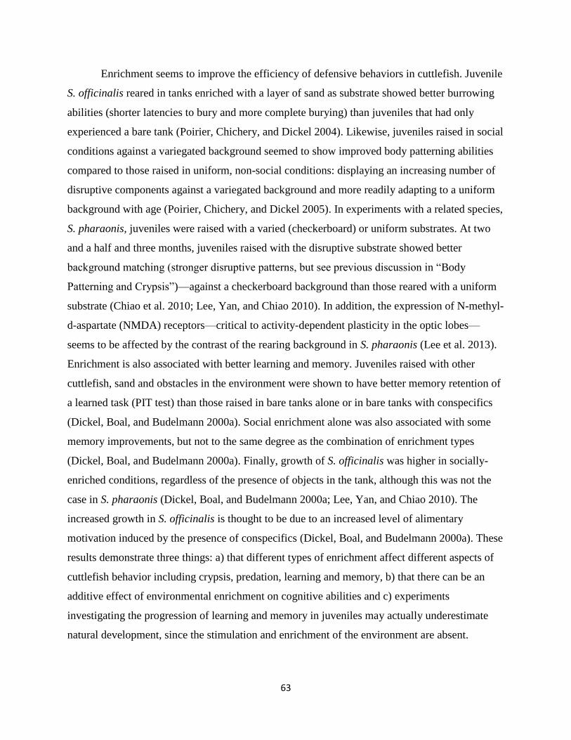
63
Enrichment seems to improve the efficiency of defensive behaviors in cuttlefish. Juvenile
S. officinalis reared in tanks enriched with a layer of sand as substrate showed better burrowing
abilities (shorter latencies to bury and more complete burying) than juveniles that had only
experienced a bare tank (Poirier, Chichery, and Dickel 2004). Likewise, juveniles raised in social
conditions against a variegated background seemed to show improved body patterning abilities
compared to those raised in uniform, non-social conditions: displaying an increasing number of
disruptive components against a variegated background and more readily adapting to a uniform
background with age (Poirier, Chichery, and Dickel 2005). In experiments with a related species,
S. pharaonis, juveniles were raised with a varied (checkerboard) or uniform substrates. At two
and a half and three months, juveniles raised with the disruptive substrate showed better
background matching (stronger disruptive patterns, but see previous discussion in “Body
Patterning and Crypsis”)—against a checkerboard background than those reared with a uniform
substrate (Chiao et al. 2010; Lee, Yan, and Chiao 2010). In addition, the expression of N-methyl-
d-aspartate (NMDA) receptors—critical to activity-dependent plasticity in the optic lobes—
seems to be affected by the contrast of the rearing background in S. pharaonis (Lee et al. 2013).
Enrichment is also associated with better learning and memory. Juveniles raised with other
cuttlefish, sand and obstacles in the environment were shown to have better memory retention of
a learned task (PIT test) than those raised in bare tanks alone or in bare tanks with conspecifics
(Dickel, Boal, and Budelmann 2000a). Social enrichment alone was also associated with some
memory improvements, but not to the same degree as the combination of enrichment types
(Dickel, Boal, and Budelmann 2000a). Finally, growth of S. officinalis was higher in socially-
enriched conditions, regardless of the presence of objects in the tank, although this was not the
case in S. pharaonis (Dickel, Boal, and Budelmann 2000a; Lee, Yan, and Chiao 2010). The
increased growth in S. officinalis is thought to be due to an increased level of alimentary
motivation induced by the presence of conspecifics (Dickel, Boal, and Budelmann 2000a). These
results demonstrate three things: a) that different types of enrichment affect different aspects of
cuttlefish behavior including crypsis, predation, learning and memory, b) that there can be an
additive effect of environmental enrichment on cognitive abilities and c) experiments
investigating the progression of learning and memory in juveniles may actually underestimate
natural development, since the stimulation and enrichment of the environment are absent.
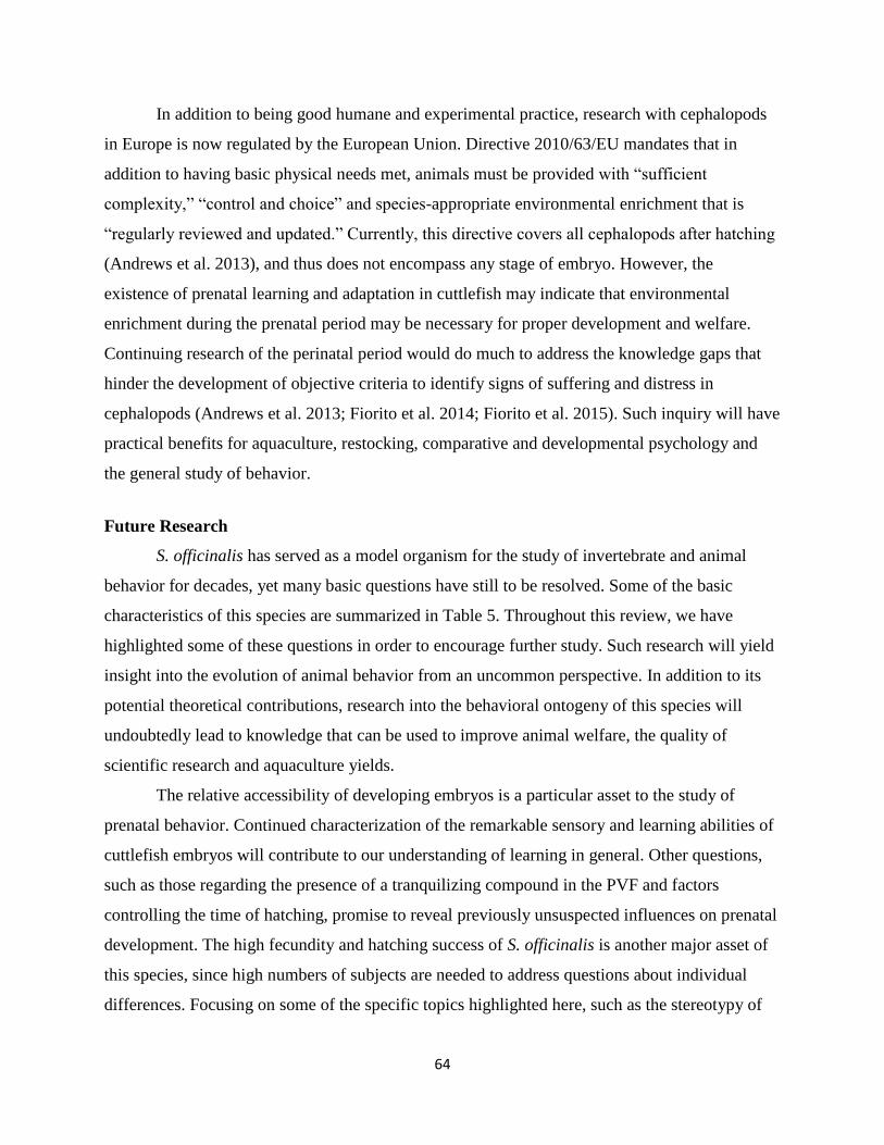
64
In addition to being good humane and experimental practice, research with cephalopods
in Europe is now regulated by the European Union. Directive 2010/63/EU mandates that in
addition to having basic physical needs met, animals must be provided with “sufficient
complexity,” “control and choice” and species-appropriate environmental enrichment that is
“regularly reviewed and updated.” Currently, this directive covers all cephalopods after hatching
(Andrews et al. 2013), and thus does not encompass any stage of embryo. However, the
existence of prenatal learning and adaptation in cuttlefish may indicate that environmental
enrichment during the prenatal period may be necessary for proper development and welfare.
Continuing research of the perinatal period would do much to address the knowledge gaps that
hinder the development of objective criteria to identify signs of suffering and distress in
cephalopods (Andrews et al. 2013; Fiorito et al. 2014; Fiorito et al. 2015). Such inquiry will have
practical benefits for aquaculture, restocking, comparative and developmental psychology and
the general study of behavior.
Future Research
S. officinalis has served as a model organism for the study of invertebrate and animal
behavior for decades, yet many basic questions have still to be resolved. Some of the basic
characteristics of this species are summarized in Table 5. Throughout this review, we have
highlighted some of these questions in order to encourage further study. Such research will yield
insight into the evolution of animal behavior from an uncommon perspective. In addition to its
potential theoretical contributions, research into the behavioral ontogeny of this species will
undoubtedly lead to knowledge that can be used to improve animal welfare, the quality of
scientific research and aquaculture yields.
The relative accessibility of developing embryos is a particular asset to the study of
prenatal behavior. Continued characterization of the remarkable sensory and learning abilities of
cuttlefish embryos will contribute to our understanding of learning in general. Other questions,
such as those regarding the presence of a tranquilizing compound in the PVF and factors
controlling the time of hatching, promise to reveal previously unsuspected influences on prenatal
development. The high fecundity and hatching success of S. officinalis is another major asset of
this species, since high numbers of subjects are needed to address questions about individual
differences. Focusing on some of the specific topics highlighted here, such as the stereotypy of
65
body patterning and the extent of associative learning, could help reveal the interaction between
phenotypic/behavioral plasticity and genetically pre-programmed reactions in shaping individual
behavior.
Table 5. A summary of development in Sepia officinalis. Modified from Dickel et al. 2006.
Sources are cited throughout the text.
Late Embryo (Stages 23-30)
Hatchling (0-7 days post hatching)
Early Juvenile (1-9 weeks)
Late Juvenile (10-17 weeks)
Living
Conditions
(From inside
egg) perceives
chemosensory,
visual and
tactile stimuli
from
immediate
environment
Usually on or
near the egg,
using ventral
sucker to adhere
to substrate
Some
dispersion from
egg mass, on
the benthos,
using ventral
sucker to adhere
to substrate
Benthos.
Substrate may
consist of
gravel, sand,
algae or a
mixture thereof
Sensory
Capabilities
Stage 23:
chemosensory
and tactile
Stage 25:
ocular
perception of
visual stimuli
Contrast and
polarization
vision
functioning but
not fully mature,
state of
chemoreception,
statocysts and
mechano-
receptors
unknown
Sensory acuity
continues to
increase with
size
Mature
Feeding Inner and outer
yolk reserves
Inner yolk
reserves
Usually shrimp,
sometimes
crabs
Shrimp, crabs,
fishes
Defense
Egg membrane
and low
activity
PRIMARY:
Semi-successful
camouflage,
SECONDARY:
inking, escape
jetting
PRIMARY:
Camouflage,
burying,
shelter-seeking
SECONDARY:
8-spot deimatic
display, inking,
escape jetting
PRIMARY:
Camouflage,
burying,
shelter-seeking
SECONDARY:
2-spot deimatic
display, inking,
escape jetting
Learning
and
Memory
Prey
imprinting and
habituation
possible
Prey imprinting
and habituation
possible
Increasing
STM, LTM and
associative
learning
Fully mature
STM, LTM and
associative
learning

66
ACKNOWLEDGMENTS This work was financed as part of PReSTO’Cog (ANR-13-BSV7-0002), a research project funded by the Agence Nationale de la Recherche (ANR) and the Région Basse-Normandie. The manuscript benefited greatly from the contributions of Olivier Basuyaux, Christelle Jozet-Alves, Cécile Bellanger, Nadav Shashar, Flavie Bidel, Alexandra Schnell, Isabelle Chevalier, Nadège Villain-Naud, Céline Thomasse, Anne-Marie Rusig, Barbara O’Brien and three anonymous reviewers.
67
II. Article #3: “Visual ecology and the development of visually guided behavior in the cuttlefish”
Among all of the cuttlefish’s sensory systems, the visual system is thought to be of primary
importance. The following is an in-depth review of the development of this system during the early life
stages involved in this project.
Visual ecology and the development of visually guided
behavior in the cuttlefish
Anne-Sophie Darmaillacq, Nawel Mezrai, Caitlin E. O’Brien, Ludovic Dickel
Normandie Univ, UNICAEN, UMR 6552 CNRS-Université de Caen-Université de Rennes 1,
team NECC, 14000 Caen, France
Correspondence : Anne-Sophie Darmaillacq [email protected]
Abstract
Cuttlefish are highly visual animals, a fact reflected in the large size of their eyes and
visual-processing centers of their brain. Adults detect their prey visually, navigate using visual
cues such as landmarks or the e-vector of polarized light and display intense visual patterns
during mating and agonistic encounters. Although much is known about the visual system in
adult cuttlefish, few studies have investigated its development and that of visually-guided
behavior in juveniles. This review summarizes the results of studies of visual development in
embryos and young juveniles. The visual system is the last to develop, as in vertebrates, and is
functional before hatching. Indeed, embryonic exposure to prey, shelters or complex background
alters postembryonic behavior. Visual acuity and lateralization, and polarization sensitivity
improve throughout the first months after hatching. The production of body patterning in
juveniles is not the simple stimulus-response process commonly presented in the literature.
Rather, it likely requires the complex integration of visual information, and is subject to inter-
individual differences. Though the focus of this review is vision in cuttlefish, it is important to
note that other senses, particularly sensitivity to vibration and to waterborne chemical signals,
also play a role in behavior. Considering the multimodal sensory dimensions of natural stimuli
and their integration and processing by individuals offer new exciting avenues of future inquiry.
Keywords: cephalopod, vision, embryo, brain, polarization, camouflage, behavioral plasticity
This study was carried out in accordance with the recommendations of 'name of guidelines,
name of committee'. The protocol was approved by the 'name of committee'.
Word count: 3855 / number of figures: 4
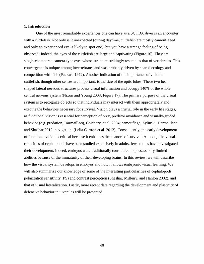
68
1. Introduction
One of the most remarkable experiences one can have as a SCUBA diver is an encounter
with a cuttlefish. Not only is it unexpected (during daytime, cuttlefish are mostly camouflaged
and only an experienced eye is likely to spot one), but you have a strange feeling of being
observed! Indeed, the eyes of the cuttlefish are large and captivating (Figure 16). They are
single-chambered camera-type eyes whose structure strikingly resembles that of vertebrates. This
convergence is unique among invertebrates and was probably driven by shared ecology and
competition with fish (Packard 1972). Another indication of the importance of vision to
cuttlefish, though other senses are important, is the size of the optic lobes. These two bean-
shaped lateral nervous structures process visual information and occupy 140% of the whole
central nervous system (Nixon and Young 2003; Figure 17). The primary purpose of the visual
system is to recognize objects so that individuals may interact with them appropriately and
execute the behaviors necessary for survival. Vision plays a crucial role in the early life stages,
as functional vision is essential for perception of prey, predator avoidance and visually-guided
behavior (e.g. predation, Darmaillacq, Chichery, et al. 2004; camouflage, Zylinski, Darmaillacq,
and Shashar 2012; navigation, (Lelia Cartron et al. 2012). Consequently, the early development
of functional vision is critical because it enhances the chances of survival. Although the visual
capacities of cephalopods have been studied extensively in adults, few studies have investigated
their development. Indeed, embryos were traditionally considered to possess only limited
abilities because of the immaturity of their developing brains. In this review, we will describe
how the visual system develops in embryos and how it allows embryonic visual learning. We
will also summarize our knowledge of some of the interesting particularities of cephalopods:
polarization sensitivity (PS) and contrast perception (Shashar, Milbury, and Hanlon 2002), and
that of visual lateralization. Lastly, more recent data regarding the development and plasticity of
defensive behavior in juveniles will be presented.
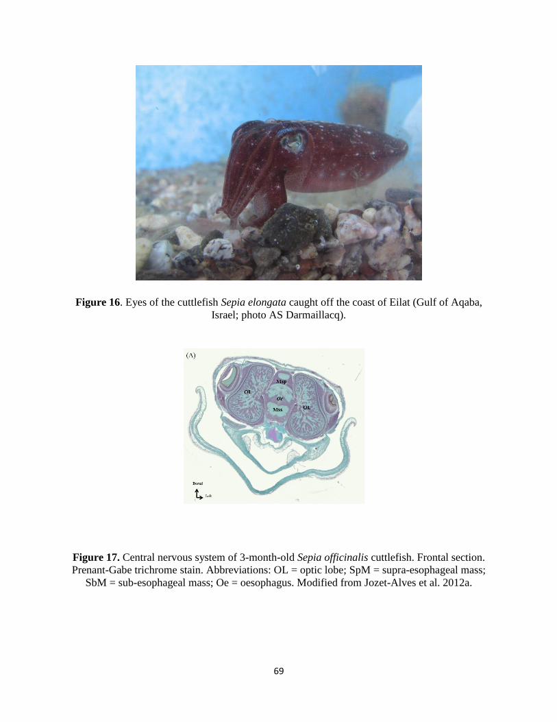
69
Figure 16. Eyes of the cuttlefish Sepia elongata caught off the coast of Eilat (Gulf of Aqaba,
Israel; photo AS Darmaillacq).
Figure 17. Central nervous system of 3-month-old Sepia officinalis cuttlefish. Frontal section.
Prenant-Gabe trichrome stain. Abbreviations: OL = optic lobe; SpM = supra-esophageal mass;
SbM = sub-esophageal mass; Oe = oesophagus. Modified from Jozet-Alves et al. 2012a.
70
2. Embryonic development of the visual system and embryos’ responses to visual stimuli
2.1 Development of sensory systems
Sepia officinalis eggs are laid in clusters on various kinds of rigid support such as algae,
tubeworms, ropes or nets. Unlike other species of Sepia, the eggs are usually darkened with
maternal ink but become more translucent due to the expansion of the capsule during embryonic
development (Boletzky 2003). S. pharaonis eggs are completely translucent.
During the final phase of embryonic development (stages 23 to 30; Boletzky, Andouche,
and Bonnaud-Ponticelli 2016), rhythmic mantle contractions are visible through the egg capsule
after removal of the outer darker envelopes. These can be measured to assess embryonic
responses to various external stimuli. Like this, Romagny et al. (2012) showed that in cuttlefish
embryos, the order of the onset of function of chemosensitivity, touch and vision follows the
same sequence as that of birds and mammals, with the visual system being the last to develop.
Neurobiological data illustrating the early development of sensory neurons in embryos support
these behavioural observations (Baratte and Bonnaud 2009). This is another evidence of
convergent evolution between cephalopods and vertebrates, perhaps instigated by similar
environmental pressures and direct competition (Packard 1972). Because embryonic
development takes place outside of the mother and in the absence of direct parental care, there is
strong evolutionary pressure for the rapid development of functional sensory systems, so that
predators can be avoided and feeding can begin. Unlike some vertebrate species, in which the
visual system is still immature at birth (Bremner, Lewkowicz, and Spence 2012), indirect
evidence suggests that cuttlefish embryos can discriminate objects outside the egg. However, to
date, no systematic study has been conducted on the development of retina morphology and
physiology in the embryo (but see Imarazene et al. 2017 this volume).
2.2 Embryonic visual responses
There is increasing empirical evidence that prenatal experience influences postnatal
perception, cognitive performance and behavior. Embryonic perceptual learning, (tested in
neonates) has been demonstrated across many taxa, including insects (Caubet, Jaisson, and
Lenoir 1992), amphibians (Mathis et al. 2008), rats (Hepper 1988), dogs (Wells and Hepper
2006), precocial birds (Sneddon, Hadden, and Hepper 1998), altricial birds (Colombelli-Négrel
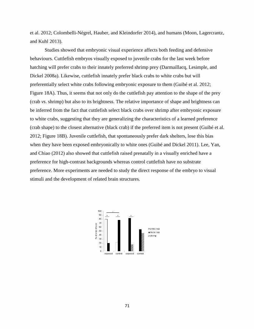
71
et al. 2012; Colombelli-Négrel, Hauber, and Kleindorfer 2014), and humans (Moon, Lagercrantz,
and Kuhl 2013).
Studies showed that embryonic visual experience affects both feeding and defensive
behaviours. Cuttlefish embryos visually exposed to juvenile crabs for the last week before
hatching will prefer crabs to their innately preferred shrimp prey (Darmaillacq, Lesimple, and
Dickel 2008a). Likewise, cuttlefish innately prefer black crabs to white crabs but will
preferentially select white crabs following embryonic exposure to them (Guibé et al. 2012;
Figure 18A). Thus, it seems that not only do the cuttlefish pay attention to the shape of the prey
(crab vs. shrimp) but also to its brightness. The relative importance of shape and brightness can
be inferred from the fact that cuttlefish select black crabs over shrimp after embryonic exposure
to white crabs, suggesting that they are generalizing the characteristics of a learned preference
(crab shape) to the closest alternative (black crab) if the preferred item is not present (Guibé et al.
2012; Figure 18B). Juvenile cuttlefish, that spontaneously prefer dark shelters, lose this bias
when they have been exposed embryonically to white ones (Guibé and Dickel 2011). Lee, Yan,
and Chiao (2012) also showed that cuttlefish raised prenatally in a visually enriched have a
preference for high-contrast backgrounds whereas control cuttlefish have no substrate
preference. More experiments are needed to study the direct response of the embryo to visual
stimuli and the development of related brain structures.
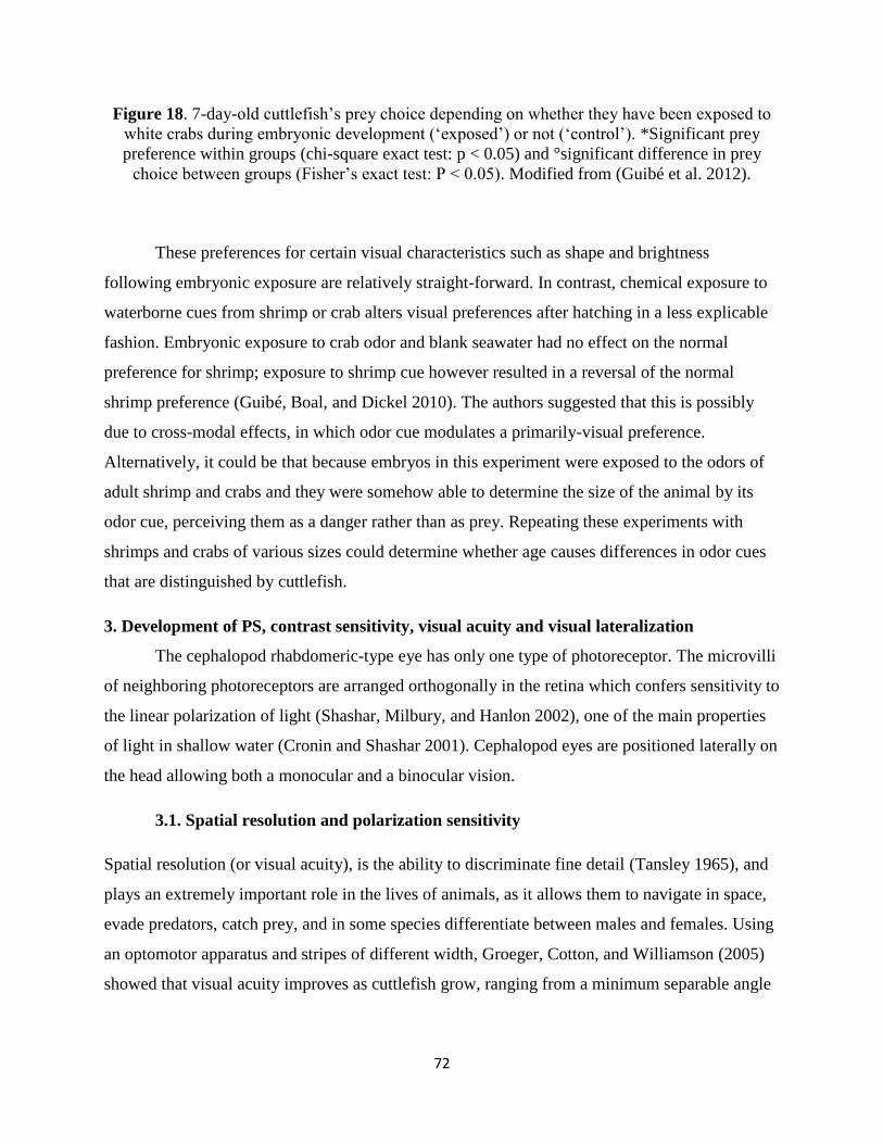
72
Figure 18. 7-day-old cuttlefish’s prey choice depending on whether they have been exposed to
white crabs during embryonic development (‘exposed’) or not (‘control’). *Significant prey
preference within groups (chi-square exact test: p < 0.05) and °significant difference in prey
choice between groups (Fisher’s exact test: P < 0.05). Modified from (Guibé et al. 2012).
These preferences for certain visual characteristics such as shape and brightness
following embryonic exposure are relatively straight-forward. In contrast, chemical exposure to
waterborne cues from shrimp or crab alters visual preferences after hatching in a less explicable
fashion. Embryonic exposure to crab odor and blank seawater had no effect on the normal
preference for shrimp; exposure to shrimp cue however resulted in a reversal of the normal
shrimp preference (Guibé, Boal, and Dickel 2010). The authors suggested that this is possibly
due to cross-modal effects, in which odor cue modulates a primarily-visual preference.
Alternatively, it could be that because embryos in this experiment were exposed to the odors of
adult shrimp and crabs and they were somehow able to determine the size of the animal by its
odor cue, perceiving them as a danger rather than as prey. Repeating these experiments with
shrimps and crabs of various sizes could determine whether age causes differences in odor cues
that are distinguished by cuttlefish.
3. Development of PS, contrast sensitivity, visual acuity and visual lateralization
The cephalopod rhabdomeric-type eye has only one type of photoreceptor. The microvilli
of neighboring photoreceptors are arranged orthogonally in the retina which confers sensitivity to
the linear polarization of light (Shashar, Milbury, and Hanlon 2002), one of the main properties
of light in shallow water (Cronin and Shashar 2001). Cephalopod eyes are positioned laterally on
the head allowing both a monocular and a binocular vision.
3.1. Spatial resolution and polarization sensitivity
Spatial resolution (or visual acuity), is the ability to discriminate fine detail (Tansley 1965), and
plays an extremely important role in the lives of animals, as it allows them to navigate in space,
evade predators, catch prey, and in some species differentiate between males and females. Using
an optomotor apparatus and stripes of different width, Groeger, Cotton, and Williamson (2005)
showed that visual acuity improves as cuttlefish grow, ranging from a minimum separable angle
73
of 2.5° to 0.57° (a decrease in this angle value means a better spatial resolution). A decrease in
light intensity affects visual acuity whatever the age of the individual.
Polarization sensitivity (PS) improves the visibility of objects by enhancing the contrast
between them and the background. In cephalopods, PS increases the success of predation on
transparent prey or silvery fish (Shashar, Hanlon, and deM Petz 1998; Shashar et al. 2000); in
cuttlefish, it may also play a role in communication between adults (Shashar, Rutledge, and
Cronin 1996; Boal et al. 2004a) and in navigation (Cartron et al. 2012). PS matures gradually
after hatching. Cartron et al. (2013) found that only 20% of cuttlefish hatchlings showed an
OMR to a polarized striped pattern when it was rotated slowly. The proportion of cuttlefish
responding increased throughout the first month of life (100% by the age of 30 days; Figure 19).
However, a choice test with fully polarized or depolarized mysids (transparent shrimps) showed
that one week-old cuttlefish detect polarized shrimp faster than non-polarized, suggesting an
earlier maturation of PS (Cartron et al. 2013). These apparently contradictory results could be
explained by the motion of the rotating pattern in the OMR apparatus compared with the more
stationary prey. It is possible that polarization contrast is more useful in assessing the shape of
prey and that motion can interfere somewhat with this ability. This deficiency could be mitigated
by the fact that polarization is not the only quality of light to which cuttlefish are sensitive.
Though colorblind (Mäthger et al. 2006; but see Stubbs and Stubbs 2016), cuttlefish are sensitive
to contrast. Indeed, most hatchling cuttlefish (75%) showed an OMR to the black, white and grey
striped pattern rotating at the lowest velocity, with the proportion reaching 100% by the age of
one month. Thus, it can be hypothesized that polarization and luminance signals are processed
separately and may play different roles in vision as observed in insects (Pfeiffer, Kinoshita, and
Homberg 2005). In the desert locust Schistocerca gregaria for instance, a group of neurons in
the central complex (a neuropil in the center of the brain), has been found to be sensitive to
polarized light while neighboring neurons are not (although all neurons responded to unpolarized
light). More experiments, notably electrophysiological and immunochemistry investigations, are
needed in order to determine the neural pathways for polarization and luminance information
processing in cuttlefish.
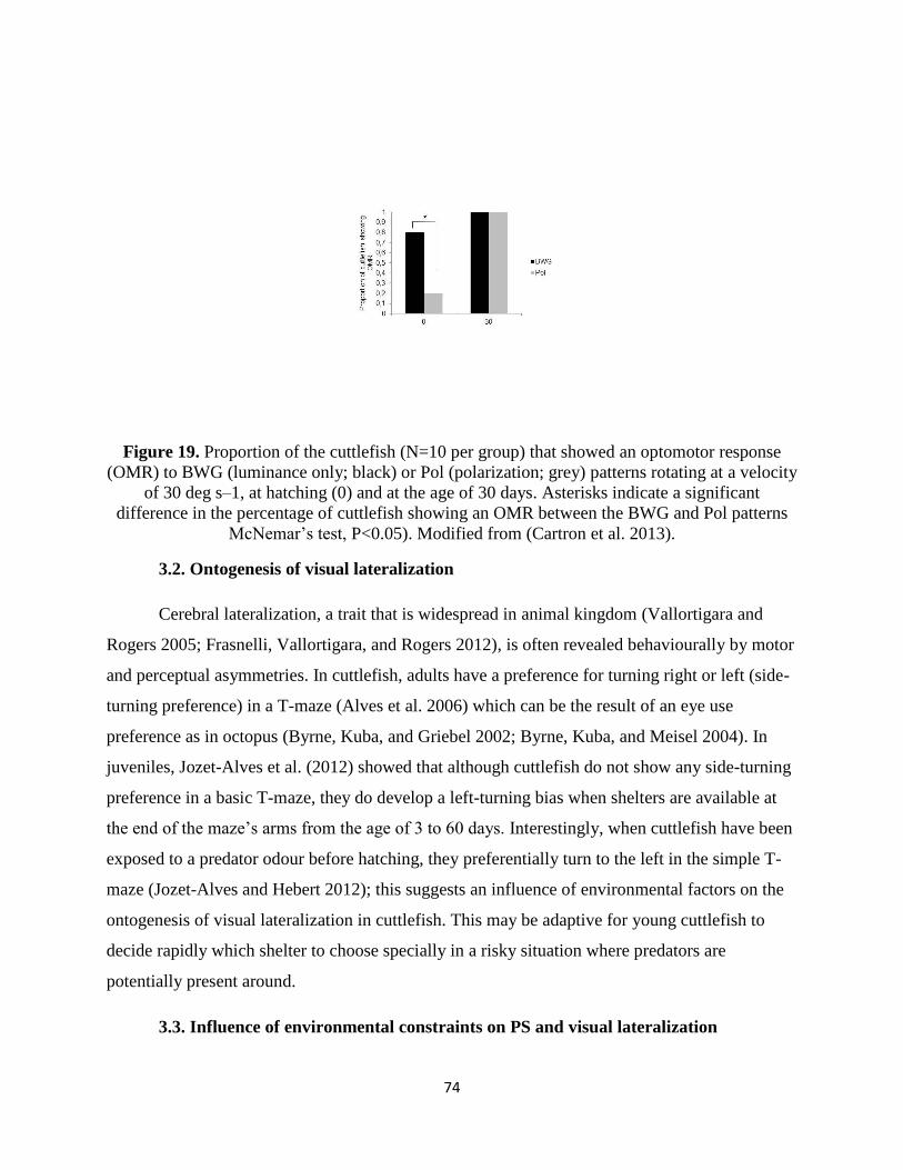
74
Figure 19. Proportion of the cuttlefish (N=10 per group) that showed an optomotor response
(OMR) to BWG (luminance only; black) or Pol (polarization; grey) patterns rotating at a velocity
of 30 deg s–1, at hatching (0) and at the age of 30 days. Asterisks indicate a significant
difference in the percentage of cuttlefish showing an OMR between the BWG and Pol patterns
McNemar’s test, P<0.05). Modified from (Cartron et al. 2013).
3.2. Ontogenesis of visual lateralization
Cerebral lateralization, a trait that is widespread in animal kingdom (Vallortigara and
Rogers 2005; Frasnelli, Vallortigara, and Rogers 2012), is often revealed behaviourally by motor
and perceptual asymmetries. In cuttlefish, adults have a preference for turning right or left (side-
turning preference) in a T-maze (Alves et al. 2006) which can be the result of an eye use
preference as in octopus (Byrne, Kuba, and Griebel 2002; Byrne, Kuba, and Meisel 2004). In
juveniles, Jozet-Alves et al. (2012) showed that although cuttlefish do not show any side-turning
preference in a basic T-maze, they do develop a left-turning bias when shelters are available at
the end of the maze’s arms from the age of 3 to 60 days. Interestingly, when cuttlefish have been
exposed to a predator odour before hatching, they preferentially turn to the left in the simple T-
maze (Jozet-Alves and Hebert 2012); this suggests an influence of environmental factors on the
ontogenesis of visual lateralization in cuttlefish. This may be adaptive for young cuttlefish to
decide rapidly which shelter to choose specially in a risky situation where predators are
potentially present around.
3.3. Influence of environmental constraints on PS and visual lateralization
75
S. officinalis, the European cuttlefish, is widespread in the English Channel, the Atlantic
Ocean and the Mediterranean Sea where the turbidity can be high. On the other hand, S.
pharaonis and S. prashadi are found in the Red Sea, on coral reefs, where the water is clearer.
All these species are able to detect a polarized stimulus at higher turbidity levels than an
unpolarized one (Cartron et al. 2013; Cartron et al. 2013), indicating that PS can improve the
capacity for object detection through turbid waters when intensity information alone is
insufficient. S. officinalis can detect objects, whether polarized or unpolarized, at higher turbidity
levels than the other two (Cartron et al. 2013). It is thus likely that PS, which is present in most
cuttlefish species (but see Darmaillacq and Shashar 2008), is a product of natural selection
driven by visual features of the species’ environment. This hypothesis is supported by the fact
that the S. officinalis used in this experiment were lab-reared individuals that had never
encountered turbidity, yet were still better-equipped to discriminate objects under these
conditions.
4. Defensive behavior
Cephalopods are known for their skills in quickly changing skin patterns in response to
environmental change, a property referred to as “dynamic camouflage” (Hanlon and Messenger
1998; Hanlon 2007). This dramatic behavior is made possible by their unique skin structure that
comprises three layers of cells: the chromatophores (containing dark-brown, reddish-orange or
yellow pigments), within the most superficial dermis of the dorsal part of the mantle and arms,
under the direct control of the brain; the iridophores, underneath, that reflect environmental light
to create iridescence (particularly prominent on the ventral part); and the leucophores, the
deepest, that reflect mainly white. Together with textural, postural and locomotor components,
these chromatic elements constitute the “body pattern” of cuttlefish (Hanlon and Messenger
1988). Body patterns displayed in a chronic fashion are mainly used for crypsis in juveniles as a
primary defense strategy to avoid detection. Cuttlefish adopt a brightness similar to the substrate
(general color resemblance), or a display disruptive colorations that breaks up the outline of the
body so that the overall form of the animal is lost (Hanlon et al. 2009). The disruptive pattern has
been the most studied. In the lab, it has been shown that artificial backgrounds such as 2d
checkerboards can elicit this pattern (Chiao and Hanlon 2001; Chiao, Chubb, and Hanlon 2007).
More, several authors (Chiao and Hanlon 2001; Barbosa et al. 2008; Barbosa et al. 2007) showed
that both check size and achromatic contrast affected the body patterns. Other characteristics of
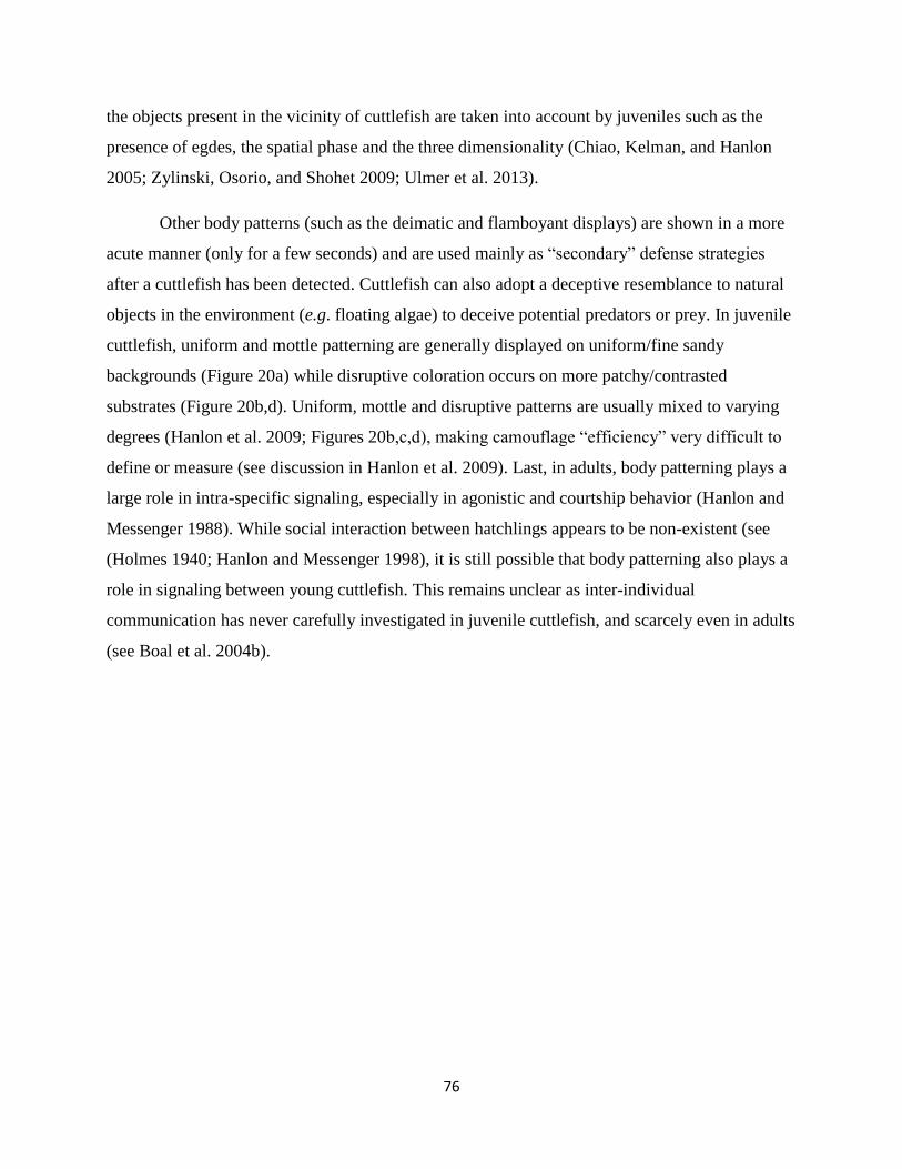
76
the objects present in the vicinity of cuttlefish are taken into account by juveniles such as the
presence of egdes, the spatial phase and the three dimensionality (Chiao, Kelman, and Hanlon
2005; Zylinski, Osorio, and Shohet 2009; Ulmer et al. 2013).
Other body patterns (such as the deimatic and flamboyant displays) are shown in a more
acute manner (only for a few seconds) and are used mainly as “secondary” defense strategies
after a cuttlefish has been detected. Cuttlefish can also adopt a deceptive resemblance to natural
objects in the environment (e.g. floating algae) to deceive potential predators or prey. In juvenile
cuttlefish, uniform and mottle patterning are generally displayed on uniform/fine sandy
backgrounds (Figure 20a) while disruptive coloration occurs on more patchy/contrasted
substrates (Figure 20b,d). Uniform, mottle and disruptive patterns are usually mixed to varying
degrees (Hanlon et al. 2009; Figures 20b,c,d), making camouflage “efficiency” very difficult to
define or measure (see discussion in Hanlon et al. 2009). Last, in adults, body patterning plays a
large role in intra-specific signaling, especially in agonistic and courtship behavior (Hanlon and
Messenger 1988). While social interaction between hatchlings appears to be non-existent (see
(Holmes 1940; Hanlon and Messenger 1998), it is still possible that body patterning also plays a
role in signaling between young cuttlefish. This remains unclear as inter-individual
communication has never carefully investigated in juvenile cuttlefish, and scarcely even in adults
(see Boal et al. 2004b).
77
Figure 20. The diversity of body patterns displayed by 2-month-old cuttlefish (ca. 3-4 cm dorsal
mantle length). a) stipple-uniform pattern elicited on uniform blue gravel; b) disruptive pattern
elicited on a black and white checkerboard combined with mottle pattern; c) deimatic pattern
following exposure to a “threat” d) mottle coloration with some components of the disruptive
pattern (i.e. white square, white head bar and paired black dots). Note that patterns are not
always fully expressed but exist in combination with others and may or may not directly reflect
the visual background.
Functional chromatophores first appear in ovo during stage 25 of embryonic
development, when the dorsal mantle length of the animal is about 2 mm (Bonnaud-Ponticelli
and Boletzky 2016). While the total number of chromatophores increases with age, their density
progressively decreases from 400-500/mm² at hatching to 35-50/mm² in adults (Hanlon and
Messenger 1988). Nevertheless, both juveniles and adults possess a high density of cells that
allow them to express an infinite range of gradations of various components of their body
patterns, depending on background and lighting (Hanlon and Messenger 1988). Thirteen
78
“typical” body patterns have been identified in adults, but since the body patterning related to
sexual behavior is absent in juveniles, the number of color, postural-kinetic, and structural
components is lower - only nine distinct patterns (Hanlon and Messenger 1988). Qualitative
changes in body patterning also occur in juveniles. For example, when a late juvenile (about >6
weeks) or adult is threatened by a small predator, it often displays a “deimatic pattern” in an
attempt at intimidation: it flattens its body and flashes two big spots against a white dorsal
mantle in a manner resembling eyes (Figure 20c). In younger animals, this pattern appears very
rarely (Thorpe 1956; Hanlon and Messenger 1988), and though the postural components are the
same as in adults they flash not two but six dark spots (Hanlon and Messenger 1988; Mangold
1989) until about 2 weeks of age. While this version of the deimatic display is used sometimes,
newly-hatched cuttlefish are more likely to respond to potential danger with a general darkening
or blanching of its body or a cryptic flamboyant display (Hanlon and Messenger 1988). One
wonders whether body patterning development in juvenile cuttlefish is rigidly fixed or is more
influenced by prior individual experience. Simple observations of body patterning in early
juveniles speak to this question: when placed on the same background different individuals
display different body patterns, suggesting that the response is partially determined by previous
experience. Other anecdotal and experimental evidence has the opposite implication however.
Hanlon and Messenger (1988) released young cuttlefish (from <1 to 17 weeks of age) previously
reared in captivity into the field and observed that they concealed themselves effectively against
every substrate encountered and were extremely difficult to see by human observers.
Unfortunately, the personal histories of individuals were not described (i.e., whether they were
reared in groups or in isolation, the amount of time spent in the wild before the behavioral
observations, etc.), so we cannot make any definitive conclusions. Still, this observation suggests
that body patterning development could be hard-wired since the impoverished artificial
conditions of rearing do not seem to have any deleterious effects on the concealment skills in
juveniles.
More controlled experiments also support an innate origin. Cuttlefish were reared in
either “impoverished” conditions (housed individual tanks on a dark uniform background) or in
“enriched” conditions (housed in groups in a variegated environment with sand, stones, shells
and artificial seaweeds) for two months (Poirier, Chichery, and Dickel 2005). Later, individuals
from each group were tested on either a uniform grey substrate or checkered black and white
79
background. In juveniles, a uniform background should elicit a uniform or slightly mottled body
pattern (but see discussion in Hanlon et al. 2009), while a disruptive color pattern seems most
adaptive against a contrasted background. The authors then assessed camouflage efficiency of by
measuring the hue and intensity of various components of body patterning, on both uniform and
contrasted substrates. At hatching, many cuttlefish display disruptive patterning regardless of
background type. But starting at 15 days of age, cuttlefish previously reared in enriched
conditions were better able to match both background types. Cuttlefish raised in enriched
conditions also had greater cell proliferation in the optic lobes than those of cuttlefish from
impoverished conditions. This makes sense, as the optic lobes are key structures controlling body
patterning in cephalopods (Nixon and Young 2003). Further evidence for greater innate or “hard-
wired” control of body patterning comes from experiments with potential predators, in which S.
officinalis was found to show the deimatic pattern towards small, low-threat teleost fish but not
toward larger more dangerous predators such as sea bass or small sharks (Langridge, Broom, and
Osorio 2007; Langridge 2009). Moreover, these reactions occur the first time such threats are
encountered, suggesting innate recognition of threat type.
While the preponderance of evidence suggests that body patterning is preprogrammed the
fact that different individuals may use a different concealment strategies when placed in the same
environment (Darmaillacq, Chichery, et al. 2004), suggest some amount of experience-
dependence, potentially through learning and phenotypic plasticity, although we cannot rule out
the possibility that these inter-individual differences are the result of genetic history or parental
experience. These data lead us to conclude that body patterning in cuttlefish is definitely not a
simple stimulus-response process, as it is commonly presented in the literature. It probably
involves a complex integration of visual information, genetic history and individual experience
(West-Eberhard 1989), possibly even before hatching (Figure 21). Thus, further investigation of
body pattern development could lead to insight not only about camouflage and defense, but also
to a better understanding of learning, plasticity, decision making and higher-order cognitive
processes in cephalopods (Vitti 2013; Skelhorn and Rowe 2016).
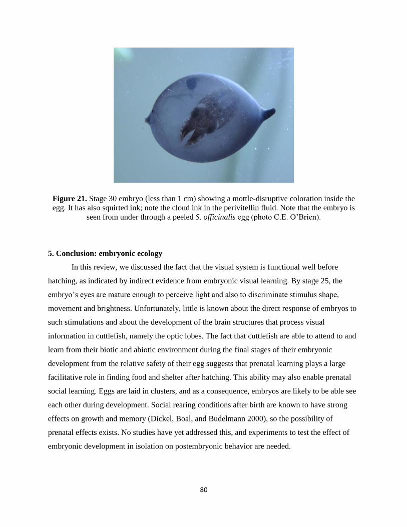
80
Figure 21. Stage 30 embryo (less than 1 cm) showing a mottle-disruptive coloration inside the
egg. It has also squirted ink; note the cloud ink in the perivitellin fluid. Note that the embryo is
seen from under through a peeled S. officinalis egg (photo C.E. O’Brien).
5. Conclusion: embryonic ecology
In this review, we discussed the fact that the visual system is functional well before
hatching, as indicated by indirect evidence from embryonic visual learning. By stage 25, the
embryo’s eyes are mature enough to perceive light and also to discriminate stimulus shape,
movement and brightness. Unfortunately, little is known about the direct response of embryos to
such stimulations and about the development of the brain structures that process visual
information in cuttlefish, namely the optic lobes. The fact that cuttlefish are able to attend to and
learn from their biotic and abiotic environment during the final stages of their embryonic
development from the relative safety of their egg suggests that prenatal learning plays a large
facilitative role in finding food and shelter after hatching. This ability may also enable prenatal
social learning. Eggs are laid in clusters, and as a consequence, embryos are likely to be able see
each other during development. Social rearing conditions after birth are known to have strong
effects on growth and memory (Dickel, Boal, and Budelmann 2000), so the possibility of
prenatal effects exists. No studies have yet addressed this, and experiments to test the effect of
embryonic development in isolation on postembryonic behavior are needed.
81
Many questions about the development of vision in cuttlefish remain to be explored. For
instance, do females actively choose their egg-laying site in order to increase offspring learning
and survival (i.e. non genetic maternal effects)? Cuttlefish reproduce only once in their lifetime
and hence, have only a single opportunity to produce offspring. This, combined with the
potential for juvenile behavior to be shaped by embryonic learning, implies that strong selection
pressure (based on the presence of predators, shelters or prey for juveniles) is exerted on
females’ decision. Since it has long been assumed that invertebrate behaviors are mostly
genetically programmed, attention should be paid to such previously-neglected effects.
This synthesis highlights the importance of vision in embryo and juvenile cuttlefish
behaviors. However, like other animals, cuttlefish live in a multisensory world, and even if
vision appears predominant, their behaviors may be influenced by other senses. In most animals,
the senses are not equal in their ability to provide accurate information about the environment
(Bremner, Lewkowicz, and Spence 2012). For example, in a turbid environment, relying only on
vision may be risky, and other senses may play a greater role. Komak et al. (2005) have
demonstrated that young cuttlefish are sensitive to local water movements thanks to specialized
cells on the arms and the head that are analogous to the lateral lines of fish. Water movement
detected by these cells could alert cuttlefish to the presence of prey or predators before it is
possible to see them. The importance of particular senses may also vary throughout the life of an
individual. In cuttlefish, given the opacity of the egg capsule, the sensory world of embryos is
probably dominated by chemosensory information. This likely changes as soon as the cuttlefish
leaves the egg. Assessing the relative importance of vision and its interactions with the other
senses through multimodal perception in different situations and at different ages offers exciting
new tracks of research such as prey and predator recognition through visual and/or chemical
information.
82
III. Article #4: “Maternal and Embryonic Stress Influence Offspring Behavior in the Cuttlefish Sepia officinalis”
This manuscript reports data from a number of behavioral experiments conducted with
cuttlefish hatchlings after maternal or embryonic stress treatments.
Maternal and Embryonic Stress Influence Offspring Behavior in the Cuttlefish
Sepia officinalis
Prenatal Stress Affects Cuttlefish Behavior
By Caitlin E. O’Brien, Christelle Jozet-Alves, Nawel Mezrai, Cécile Bellanger, Anne-Sophie Darmaillacq, Ludovic Dickel*
Normandie Univ, UNICAEN, CNRS, NECC, UMR EthoS 6552, 14032 Caen cedex, France
*Corresponding author ([email protected])
Abstract
Stress experienced during prenatal development—either applied to reproducing females
(maternal stress), directly to developing offspring (embryonic stress) or in combination—is
associated with a range of post-natal behavioral effects in numerous organisms. We conducted an
experiment to discern if maternal and embryonic stressors affect the behavior of hatchlings of the
cuttlefish, Sepia officinalis, a species with features that allow for the examination of these stress
types in isolation. We also compared the effect of a naturally-occurring (predator cue) and an
“artificial” (bright, randomly-occurring LED light) embryonic stressor. We found that the
maternal stressor was associated with significant differences in body patterning and activity
patterns. By contrast, embryonic exposure to stressors increased the proportion of individuals
that pursued prey. From these results, it appears that in cuttlefish, maternal and embryonic
stressors affect different post-natal behavior in offspring. In addition, the effect of the artificial
stressor suggests that organisms can sometimes react adaptively to a stressor even if it is not one
that has been encountered during the evolutionary history of the species.
Keywords Body patterning, predation, visual lateralization, activity, threat response
Summary statement The effects of several chronic prenatal stressors (maternal stress, embryonic exposure to
predator odor or bright light) on hatchling cuttlefish are compared in five tests.
83
Introduction Stress responses occur in reaction to any external or anticipated threat. In response to a predator,
for instance, an animal may increase its metabolism and divert resources to its muscles and away from
less critical functions like digestion and foraging behavior—the “fight or flight” stress response (Cannon
1939). Other kinds of stressors will induce different reactions. In response to food scarcity, for instance,
an animal may have the opposite reaction, prioritizing digestive processes to extract the maximum
amount of energy from food items and even undertaking risky foraging behavior (Wang, Hung, &
Randall, 2006). While stress responses have presumably evolved to increase survival in the face of an
immediate stressor, there is an increasing awareness that stress responses come with a host of negative
fitness consequences. This often depends on whether the stressor causing the response is acute or chronic:
A short, single experience of a stressor (e.g. a single encounter with a predator) often produces a short-
term, adaptive response while long-term or repeated exposure to stressors (e.g. prolonged food shortage)
can have lasting negative impacts on fitness (Jones, 1996; Miller, Chen, & Zhou, 2007). These costs come
from the energetic tradeoffs involved in maintaining the response or in the form of missed opportunities
(e.g. lost foraging time, mating opportunities). Chronic and repeated stressors are often associated with
reductions in immune function, the advent of various diseases, negative impacts on psychological health
and disruptions to normal biological functions (e.g. Favreau-Peigné et al. 2014; Katz, Roth, and Carroll
1981; Miller, Chen, and Zhou 2007). Thus, understanding the underlying causes and effects of stress
responses has implications for medicine, psychology and developmental biology, and is studied in a
number of animal models.
The long-term effects of stress that occurs during the embryonic development of an organism are
known to be especially significant. Research in a number of vertebrate taxa demonstrates that stress
responses in reproducing females can have a strong impact on the behavior of her offspring. In some
cases, such stress may serve as an indicator of prospective environment, prompting adaptive changes to
the offspring phenotype that help it cope with future challenges. Stress responses can also be associated
with reduced offspring fitness; normal developmental processes can be disrupted and the animal may be
more susceptible to disease (Gluckman and Hanson 2004). While the effects of prenatal stress have been
relatively well-documented in a number of taxa, it is often unclear if effects observed are the direct result
of a stress response in the offspring or a maternally–transmitted effect. One potential mechanism for
prenatal stress effects in offspring is the transfer of “stress hormones” (e.g. glucocorticoids,
catecholamines) from mother to developing embryo. Such hormones are secreted by animals in response
to stressors and affect physiology, behavior and metabolism. Their transfer to offspring via the placenta or
egg yolk could explain many of the alterations to offspring phenotype that are sometimes observed
(Groothuis et al. 2005; Hayward and Wingfield 2004; Weinstock 2008).
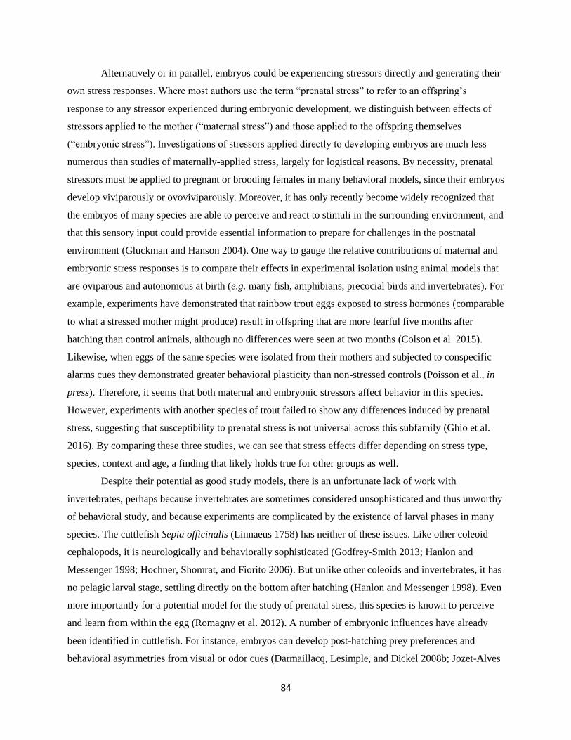
84
Alternatively or in parallel, embryos could be experiencing stressors directly and generating their
own stress responses. Where most authors use the term “prenatal stress” to refer to an offspring’s
response to any stressor experienced during embryonic development, we distinguish between effects of
stressors applied to the mother (“maternal stress”) and those applied to the offspring themselves
(“embryonic stress”). Investigations of stressors applied directly to developing embryos are much less
numerous than studies of maternally-applied stress, largely for logistical reasons. By necessity, prenatal
stressors must be applied to pregnant or brooding females in many behavioral models, since their embryos
develop viviparously or ovoviviparously. Moreover, it has only recently become widely recognized that
the embryos of many species are able to perceive and react to stimuli in the surrounding environment, and
that this sensory input could provide essential information to prepare for challenges in the postnatal
environment (Gluckman and Hanson 2004). One way to gauge the relative contributions of maternal and
embryonic stress responses is to compare their effects in experimental isolation using animal models that
are oviparous and autonomous at birth (e.g. many fish, amphibians, precocial birds and invertebrates). For
example, experiments have demonstrated that rainbow trout eggs exposed to stress hormones (comparable
to what a stressed mother might produce) result in offspring that are more fearful five months after
hatching than control animals, although no differences were seen at two months (Colson et al. 2015).
Likewise, when eggs of the same species were isolated from their mothers and subjected to conspecific
alarms cues they demonstrated greater behavioral plasticity than non-stressed controls (Poisson et al., in
press). Therefore, it seems that both maternal and embryonic stressors affect behavior in this species.
However, experiments with another species of trout failed to show any differences induced by prenatal
stress, suggesting that susceptibility to prenatal stress is not universal across this subfamily (Ghio et al.
2016). By comparing these three studies, we can see that stress effects differ depending on stress type,
species, context and age, a finding that likely holds true for other groups as well.
Despite their potential as good study models, there is an unfortunate lack of work with
invertebrates, perhaps because invertebrates are sometimes considered unsophisticated and thus unworthy
of behavioral study, and because experiments are complicated by the existence of larval phases in many
species. The cuttlefish Sepia officinalis (Linnaeus 1758) has neither of these issues. Like other coleoid
cephalopods, it is neurologically and behaviorally sophisticated (Godfrey-Smith 2013; Hanlon and
Messenger 1998; Hochner, Shomrat, and Fiorito 2006). But unlike other coleoids and invertebrates, it has
no pelagic larval stage, settling directly on the bottom after hatching (Hanlon and Messenger 1998). Even
more importantly for a potential model for the study of prenatal stress, this species is known to perceive
and learn from within the egg (Romagny et al. 2012). A number of embryonic influences have already
been identified in cuttlefish. For instance, embryos can develop post-hatching prey preferences and
behavioral asymmetries from visual or odor cues (Darmaillacq, Lesimple, and Dickel 2008b; Jozet-Alves
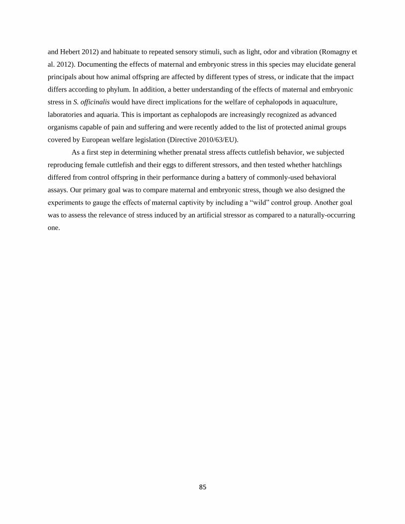
85
and Hebert 2012) and habituate to repeated sensory stimuli, such as light, odor and vibration (Romagny et
al. 2012). Documenting the effects of maternal and embryonic stress in this species may elucidate general
principals about how animal offspring are affected by different types of stress, or indicate that the impact
differs according to phylum. In addition, a better understanding of the effects of maternal and embryonic
stress in S. officinalis would have direct implications for the welfare of cephalopods in aquaculture,
laboratories and aquaria. This is important as cephalopods are increasingly recognized as advanced
organisms capable of pain and suffering and were recently added to the list of protected animal groups
covered by European welfare legislation (Directive 2010/63/EU).
As a first step in determining whether prenatal stress affects cuttlefish behavior, we subjected
reproducing female cuttlefish and their eggs to different stressors, and then tested whether hatchlings
differed from control offspring in their performance during a battery of commonly-used behavioral
assays. Our primary goal was to compare maternal and embryonic stress, though we also designed the
experiments to gauge the effects of maternal captivity by including a “wild” control group. Another goal
was to assess the relevance of stress induced by an artificial stressor as compared to a naturally-occurring
one.
86
Methods Two different experiments were conducted, one testing for the potential transfer of the effects of
captivity or stress from reproducing females to their offspring, and a second exploring the impact of
stressors applied directly to developing embryos. In the first experiment, we exposed spawning female
cuttlefish to daily removal from the water. We then compared the behavior of their offspring (SM) to that
of offspring of a group of captive but unstressed mother controls (UM-C). We also compared both of
these groups to offspring from naturally-spawned eggs collected from the wild (WM). While the maternal
experience of these eggs was unknown and uncontrolled, their inclusion gives a sense of the effects of
maternal capture and captivity (Fig. 1).
In the second experiment testing embryonic stress, we subdivided eggs from the unstressed
control mothers into three groups in order to investigate the effects of stimuli applied directly to embryos.
We applied two kinds of stressors: a naturally-occurring stressor consisting of odor cues from common
predatory fish (UM-PE) and an artificial stressor consisting of high intensity LED light timed randomly
and unpredictably throughout the day and night (UM-LE). These two groups were compared to the
unstressed mother control (UM-C) group used in the maternal stress comparisons (Fig. 22).
Fig 22.) Schematic representation of the experimental design.
After hatching, the effects of prenatal stress treatments on offspring were assessed with a battery
of tests covering various aspects of the cuttlefish behavioral repertoire, including body patterning, visual
lateralization, predation, activity patterns and fear response. These tests allowed us to make a broad
assessment as to whether stressors affect offspring behavior and to make general comparisons between
embryonic and maternal stress and between a natural and an artificial stressor. We predicted that the
direct experience of an embryonic stressor would have a stronger effect on offspring behavior than
maternal stress, which consists of information that must be transmitted indirectly to offspring through the
mother. We also expected that cuttlefish would have evolved adaptive responses to the natural embryonic

87
stressor (predator odor), but would demonstrate inappropriate and likely maladaptive responses to the
artificial stressor since its response to this stimulus could not have been shaped by natural selection.
Animal Collection and Housing
Adult Females
Cuttlefish traps were set off the coast of France in the English Channel. Thirty seven adult female
cuttlefish (Sepia officinalis) were captured between May and June, 2015 and 28 were captured in May,
2016 and transported to the Centre de Recherches en Environnement Côtier (CREC, marine station of the
University of Caen, Luc-sur-Mer, France). These females were mated with males and then placed in
treatment tanks in a semi-open flow-through seawater system (15±1°C) under a 16:8h light/dark cycle.
Captured females were split randomly into two groups, and eggs collected from them were
designated as “unstressed mother—control” (UM-C) and “stressed mother” (SM). The unstressed captive
females designated as unstressed mothers (six in 2015 and 12 in 2016) were maintained in large (1000 L),
round tanks enriched with stones, plastic algae, floating objects and plenty of shaded area. In 2015, these
females were housed in these tanks singly, but in 2016, the capture of two dozen cuttlefish on a single day
necessitated housing in groups of three. Those females assigned to be in the stressed mothers group were
isolated in bare tanks (65 L) with a water depth of 19cm and subjected to randomized 10-second removals
from the water three times a day using a specially-made mesh platform. Eggs spawned by these stressed
captive females after at least one week in these conditions were collected. Four unstressed mothers and
four stressed mothers spawned between May 15 and June 9, 2015 and 11 unstressed mothers and eight
stressed mothers spawned between May 14 and 29, 2016.
Eggs and Hatchlings
Wild mother eggs (WM) were collected by SCUBA divers from pre-placed tethers in the English
Channel (49°19.667N-0°18.767W) in June, 2015 from a depth of 13.7 m. These, along with eggs
collected from unstressed and stressed mothers in captivity, were moved to floating trays in 65L tanks
after eight hours of steady temperature habituation (from 15°C to 20°C). These were housed in a darkened
room with exposure to the natural light cycle and supplied with seawater from a gently flowing open
system and aerated by an airstone. A randomly-selected third of the control mother eggs, designated as
controls (UM-C), along with WM and stressed mother SM eggs, were not treated any further. The other
two thirds of the control mother eggs were divided randomly into predator-exposed (UM-PE) and light-
exposed (UM-LE) groups. Three sea bass (Dicentrarchus labrax; Linnaeus, 1758; total length = 25-30
cm) were housed with UM-PE eggs, separated by a mesh barrier that allowed the eggs chemosensory and
visual exposure to the predatory fish. Light-exposed eggs experienced strong LED illumination (20.7klux,
88
approximately 10cm from surface of water) for 90 min a day (six randomly-timed periods of 15 min). All
eggs were gently agitated once a day to remove detritus and discourage parasite growth.
Hatchlings were recorded and collected at 08:00 each morning between June 29 and August 5,
2015, and July 2-24, 2016, and then transferred to a new tank to remove them from any further exposure
to the stress treatments. Between experiments, hatchlings and juveniles were maintained in individually-
labeled compartments to preserve identity. These compartments were situated in an aerated open seawater
system (19-23°C) with a water depth of 7cm. Sex determination was not possible at this age. All
hatchlings born on a single day comprised a daily cohort. A total of 22 cohorts (numbering up to 12
individuals each) were hatched and tested daily between July and August. In 2015, after the predation
experiment on Day 4, individuals were fed a single shrimp (Crangon crangon; Linnaeus, 1758) per day.
In 2016, hatchlings were fed ad libitum starting on Day 4.
Behavioral Experiments
The resulting offspring were then subjected to a battery of tests aimed at determining whether the
stress treatments had affected any aspects of the behavioral phenotype (Fig. 23). The data resulting from
these tests were analyzed in R, GraphPad (Prism®) and StatXact®7 (Cytel Inc.). All p-values are two-
tailed.
Fig. 23) Timeline of stress treatments and behavioral tests. All tests except for the threat response activity analysis
occurred in 2015.
Body Patterning
In 2015, on the day of hatching (Day 1), between 9:00-10:30, up to 12 cuttlefish at a time were
placed in randomized order in small uniform grey (“uniform background”) circular compartments with
slanted sides to minimize shadows (radius = 2.9cm bottom, 3.35cm top, length of sides = 2.5cm; mean
grey value = 101 +/-3.9) under white LED light (0.63 to 0.88 klux) and photographed at 0, 5, 15 and
30min after placement on the background with a Panasonic HDC-SD60 camera. On Day 2, between
10:30-12:00, cuttlefish order was re-randomized and each was photographed four times (0, 5, 15 and 30
min after placement) against a checkered pattern (“disruptive background”). The check size of the
disruptive background was selected to be approximately the size of a hatchling’s main body-patterning
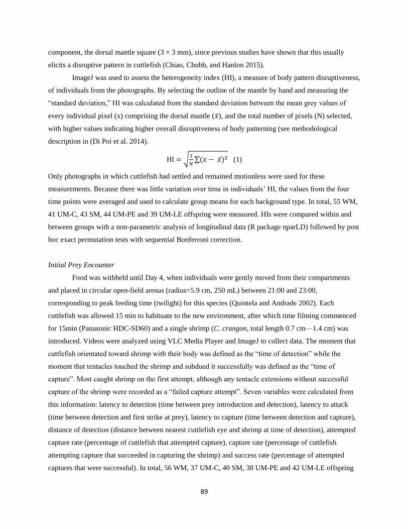
89
component, the dorsal mantle square (3 × 3 mm), since previous studies have shown that this usually
elicits a disruptive pattern in cuttlefish (Chiao, Chubb, and Hanlon 2015).
ImageJ was used to assess the heterogeneity index (HI), a measure of body pattern disruptiveness,
of individuals from the photographs. By selecting the outline of the mantle by hand and measuring the
“standard deviation,” HI was calculated from the standard deviation between the mean grey values of
every individual pixel (x) comprising the dorsal mantle (�̅�), and the total number of pixels (N) selected,
with higher values indicating higher overall disruptiveness of body patterning (see methodological
description in (Di Poi et al. 2014).
HI = √1
𝑁∑(𝑥 − �̅�)2 (1)
Only photographs in which cuttlefish had settled and remained motionless were used for these
measurements. Because there was little variation over time in individuals’ HI, the values from the four
time points were averaged and used to calculate group means for each background type. In total, 55 WM,
41 UM-C, 43 SM, 44 UM-PE and 39 UM-LE offspring were measured. HIs were compared within and
between groups with a non-parametric analysis of longitudinal data (R package nparLD) followed by post
hoc exact permutation tests with sequential Bonferroni correction.
Initial Prey Encounter
Food was withheld until Day 4, when individuals were gently moved from their compartments
and placed in circular open-field arenas (radius=5.9 cm, 250 mL) between 21:00 and 23:00,
corresponding to peak feeding time (twilight) for this species (Quintela and Andrade 2002). Each
cuttlefish was allowed 15 min to habituate to the new environment, after which time filming commenced
for 15min (Panasonic HDC-SD60) and a single shrimp (C. crangon, total length 0.7 cm—1.4 cm) was
introduced. Videos were analyzed using VLC Media Player and ImageJ to collect data. The moment that
cuttlefish orientated toward shrimp with their body was defined as the “time of detection” while the
moment that tentacles touched the shrimp and subdued it successfully was defined as the “time of
capture”. Most caught shrimp on the first attempt, although any tentacle extensions without successful
capture of the shrimp were recorded as a “failed capture attempt”. Seven variables were calculated from
this information: latency to detection (time between prey introduction and detection), latency to attack
(time between detection and first strike at prey), latency to capture (time between detection and capture),
distance of detection (distance between nearest cuttlefish eye and shrimp at time of detection), attempted
capture rate (percentage of cuttlefish that attempted capture), capture rate (percentage of cuttlefish
attempting capture that succeeded in capturing the shrimp) and success rate (percentage of attempted
captures that were successful). In total, 56 WM, 37 UM-C, 40 SM, 38 UM-PE and 42 UM-LE offspring
90
were tested. Latencies and distance of detection were compared with exact Kruskal-Wallis tests by Monte
Carlo sampling followed by post hoc exact permutation tests (sequential Bonferroni correction). The
variables attempted capture, capture and success rates were compared with chi square exact tests.
Visual Laterality Test
These tests were conducted between 10:00 and 22:00 five days after hatching. The testing
apparatus consisted of a start box (3.5 cm x 5 cm), a movable transparent barrier and two darkened
shelters (3.5 cm x 4 cm) located 15cm apart (see Jozet-Alves et al. 2012). Each shelter contained blue
aquarium gravel and was shaded with a plastic cover. The apparatus was filled with seawater (renewed
between trials) and placed under a bright fluorescent lamp (5.5 lux at the surface of the arena). In order to
determine if stress induced a population-level eye-use preference, individuals were tested for shelter
choice (in randomized order) by gently positioning them in the start box in such a way that it could view
both shelters. Once the cuttlefish was in position, the transparent barrier was removed and the cuttlefish
was allowed free access to the entire arena. Bright light is unpleasant to cuttlefish, and thus they were
highly motivated to exit the start box and seek one of the darkened shelters. In total, 43 WM, 40 UM-C,
43 SM, 42 UM-PE and 41 UM-LE offspring were tested. Within-group comparisons (the proportion
turning right versus left) were made with binomial tests and between-group comparisons (whether the
proportion of those turning left differed between maternal or embryonic stress groups) were analyzed with
chi square exact tests.
Overnight Activity Analysis
At midnight of Day 9, four cuttlefish from each daily cohort were randomly selected and placed
in a circular open-field arena (radius = 5.9 cm, depth = 2.3 cm, 250 mL) made of opaque white plastic
(sides) and a glass base. Illuminated from below by infrared light (which is not visible to the cuttlefish but
is recorded by the camera), each individual was filmed from overhead for 6hrs with a software-specific
camera in a darkened room. This period corresponds with the times at which cuttlefish have been found to
be most active (Denton and Gilpin-Brown 1961; Frank et al. 2012; Jäckel et al. 2007; Oliveira et al.
2017). Videos were analyzed with Ethovision (Noldus®), a software package for behavioral tracking. The
total distance traveled, time spent moving, and mean meander were recorded for each individual. Some
individuals were unusable due to poor lighting and were excluded. In total, 20 WM, 10 UM-C, 15 SM, 8
UM-PE and eight UM-LE offspring were analyzed. These data were analyzed with exact Kruskal-Wallis
tests followed by post hoc exact permutation tests (sequential Bonferroni correction).
Threat Response Activity Analysis
91
At noon on Day 7, two pairs of treatment- and age-matched cuttlefish were randomly selected
from the daily cohort. They were placed in the open-field arena described in the previous paragraph and
recorded and tracked in the same manner. After one hour of filming, 50 ml of “blank” water from the
UM-C egg tank was added to the arena of one member of each pair and 50 ml of “predator odor” water
from the UM-PE egg tank was added to their counterparts’ arenas. This was accomplished using tubes
already present beneath the waterline of each arena in order to minimize the disturbance of the addition of
water. The total distance traveled and time spent moving were recorded for each individual in the same
manner as described above. To control for individual differences, post treatment values are expressed as a
percentage of the initial hour for each individual (baseline). In total, groups of 10 UM-C, SM, UM-PE
and UM-LE offspring were divided into “blank” (n = 5 per stress group) and “predator odor” treatments
(n = 5 per stress group). These data were analyzed with a non-parametric analysis of longitudinal data (R
package “nparLD”) followed by post hoc exact permutation tests (sequential Bonferroni correction).
Ethical Note:
This research followed the guidance given by Directive 2010/63/EU, and French regulations
regarding the use of animals for experimental procedures, and was approved by the Regional Ethical
Committee Cenomexa (Committee agreement number: 54; project agreement number: A14384001). The
experiment was designed to decrease animal distress by minimizing the number of animals. Enrichment
was provided to unstressed captive adult cuttlefish. After spawning, adult females died naturally
following senescence (June/July). After the completion of behavioral experiments, juvenile cuttlefish
were anaesthetized in 17.5g/L MgCl2 and euthanized with an overdose of ethanol (2%) for
neurobiological testing (section VI).
92
Results
Body Patterning
In the maternal stress groups, the non-parametric analysis for longitudinal data revealed a
significant effect of the background type (i.e. uniform versus disruptive: p = 0.0002; F = 13.46), and of
the treatment groups (i.e. WM vs. UM-C vs. SM: p < 0.0001; F = 14.74). As no interaction was found (p
= 0.956), this analysis showed that mean HI are higher on the disruptive background whatever the group
considered (Fig. 24). Pairwise post hoc comparisons showed that mean HI values are lower in UM-C eggs
than in WM eggs (p < 0.001) and SM eggs (p = 0.005). Mean HI values were also significantly higher in
WM than in SM eggs (p = 0.021).
Wild Mother (WM)
Unstressed Mother-Control (U
M-C)
Stressed Mother (SM)
0
10
20
30Uniform Background
Disruptive Background
***
*
***
Mean
Hete
rog
en
eit
y I
nd
ex (
HI)
Fig. 24) Heterogeneity Index (HI) ± s.d. of maternal stress groups on uniform and disruptive backgrounds. Between groups, WM
offspring had significantly higher HI than the other maternal stress treatment groups on both backgrounds (p < 0.05), and SM (n
= 43) had significantly higher HI on the disruptive background than UM-C (n = 41; p = 0.005). Significant differences between
groups are indicated by connecting brackets. * indicates p < 0.05; *** indicates p< 0.001.
In the embryonic stress groups, the non-parametric analysis for longitudinal data revealed a
significant effect of the background type (i.e. uniform versus disruptive: p = 0.002; F = 10.03), and of the
treatment groups (i.e. UM-C vs. UM-PE vs. UM-LE: p = 0.046; F = 3.1). As no interaction was found (p
= 0.7143), this analysis shows that mean HIs are higher on the disruptive background whatever the group
considered (Fig. 4). Pairwise post hoc comparisons showed that UM-LE have significantly higher mean
HI than in UM-PE (p = 0.017) and statistical tendency for higher HIs than UM-C (p = 0.069).
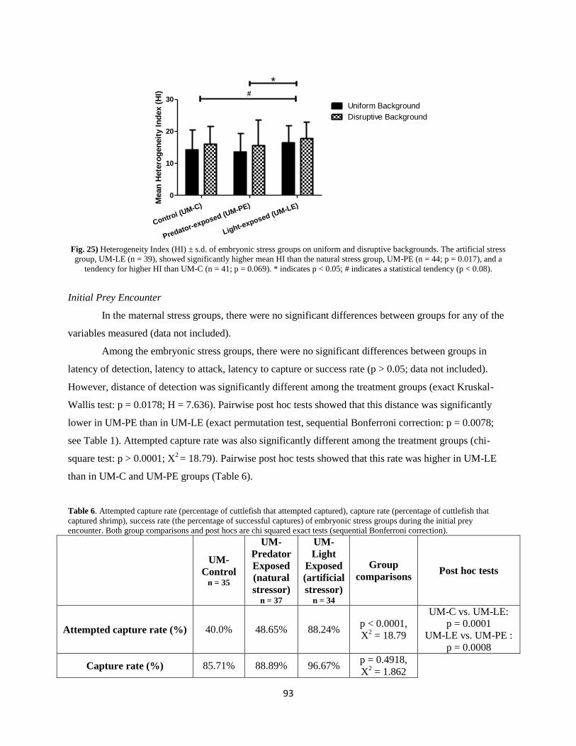
93
Control (UM-C)
Predator-exposed (UM-PE)
Light-exposed (UM-LE)
0
10
20
30#
*
Uniform Background
Disruptive Background
Mean
Hete
rog
en
eit
y I
nd
ex (
HI)
Fig. 25) Heterogeneity Index (HI) ± s.d. of embryonic stress groups on uniform and disruptive backgrounds. The artificial stress
group, UM-LE (n = 39), showed significantly higher mean HI than the natural stress group, UM-PE (n = 44; p = 0.017), and a
tendency for higher HI than UM-C (n = 41; p = 0.069). * indicates p < 0.05; # indicates a statistical tendency (p < 0.08).
Initial Prey Encounter
In the maternal stress groups, there were no significant differences between groups for any of the
variables measured (data not included).
Among the embryonic stress groups, there were no significant differences between groups in
latency of detection, latency to attack, latency to capture or success rate (p > 0.05; data not included).
However, distance of detection was significantly different among the treatment groups (exact Kruskal-
Wallis test: p = 0.0178; H = 7.636). Pairwise post hoc tests showed that this distance was significantly
lower in UM-PE than in UM-LE (exact permutation test, sequential Bonferroni correction: p = 0.0078;
see Table 1). Attempted capture rate was also significantly different among the treatment groups (chi-
square test: p > 0.0001; X2 = 18.79). Pairwise post hoc tests showed that this rate was higher in UM-LE
than in UM-C and UM-PE groups (Table 6).
Table 6. Attempted capture rate (percentage of cuttlefish that attempted captured), capture rate (percentage of cuttlefish that
captured shrimp), success rate (the percentage of successful captures) of embryonic stress groups during the initial prey
encounter. Both group comparisons and post hocs are chi squared exact tests (sequential Bonferroni correction).
UM-
Control n = 35
UM-
Predator
Exposed
(natural
stressor) n = 37
UM-
Light
Exposed
(artificial
stressor) n = 34
Group
comparisons Post hoc tests
Attempted capture rate (%) 40.0% 48.65% 88.24% p < 0.0001,
X2 = 18.79
UM-C vs. UM-LE:
p = 0.0001
UM-LE vs. UM-PE :
p = 0.0008
Capture rate (%) 85.71% 88.89% 96.67% p = 0.4918,
X2 = 1.862
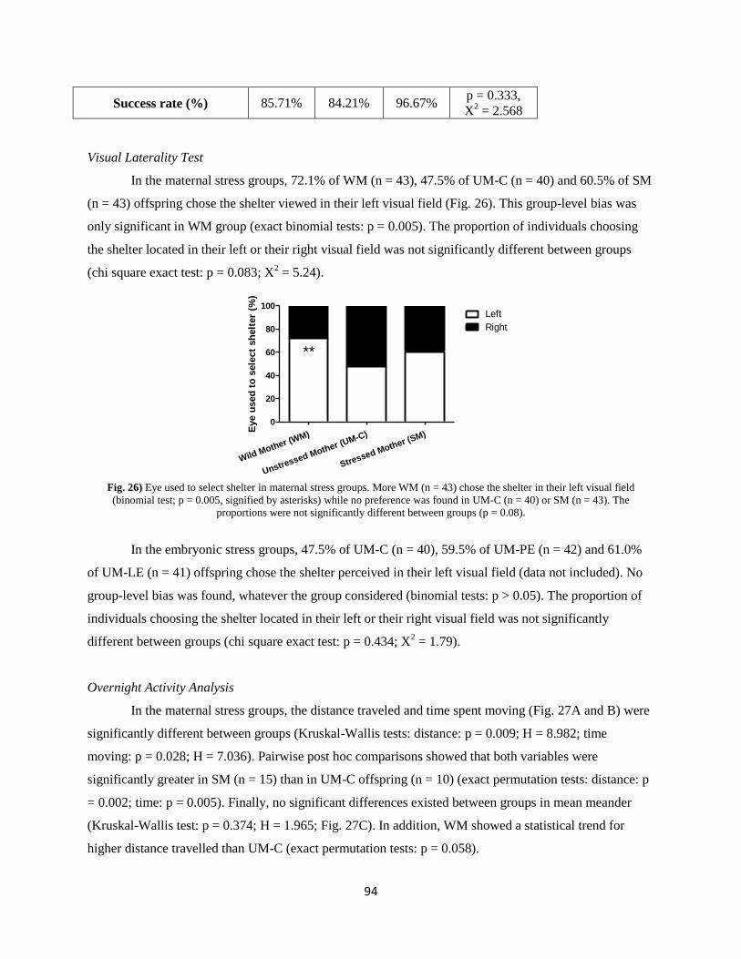
94
Success rate (%) 85.71% 84.21% 96.67% p = 0.333,
X2 = 2.568
Visual Laterality Test
In the maternal stress groups, 72.1% of WM (n = 43), 47.5% of UM-C (n = 40) and 60.5% of SM
(n = 43) offspring chose the shelter viewed in their left visual field (Fig. 26). This group-level bias was
only significant in WM group (exact binomial tests: p = 0.005). The proportion of individuals choosing
the shelter located in their left or their right visual field was not significantly different between groups
(chi square exact test: p = 0.083; X2 = 5.24).
Wild Mother (WM)
Unstressed Mother (UM-C)
Stressed Mother (SM)
0
20
40
60
80
100Left
Right
**
Eye u
sed
to
sele
ct
sh
elt
er
(%)
Fig. 26) Eye used to select shelter in maternal stress groups. More WM (n = 43) chose the shelter in their left visual field
(binomial test; p = 0.005, signified by asterisks) while no preference was found in UM-C (n = 40) or SM (n = 43). The
proportions were not significantly different between groups (p = 0.08).
In the embryonic stress groups, 47.5% of UM-C (n = 40), 59.5% of UM-PE (n = 42) and 61.0%
of UM-LE (n = 41) offspring chose the shelter perceived in their left visual field (data not included). No
group-level bias was found, whatever the group considered (binomial tests: p > 0.05). The proportion of
individuals choosing the shelter located in their left or their right visual field was not significantly
different between groups (chi square exact test: p = 0.434; X2 = 1.79).
Overnight Activity Analysis
In the maternal stress groups, the distance traveled and time spent moving (Fig. 27A and B) were
significantly different between groups (Kruskal-Wallis tests: distance: p = 0.009; H = 8.982; time
moving: p = 0.028; H = 7.036). Pairwise post hoc comparisons showed that both variables were
significantly greater in SM (n = 15) than in UM-C offspring (n = 10) (exact permutation tests: distance: p
= 0.002; time: p = 0.005). Finally, no significant differences existed between groups in mean meander
(Kruskal-Wallis test: p = 0.374; H = 1.965; Fig. 27C). In addition, WM showed a statistical trend for
higher distance travelled than UM-C (exact permutation tests: p = 0.058).
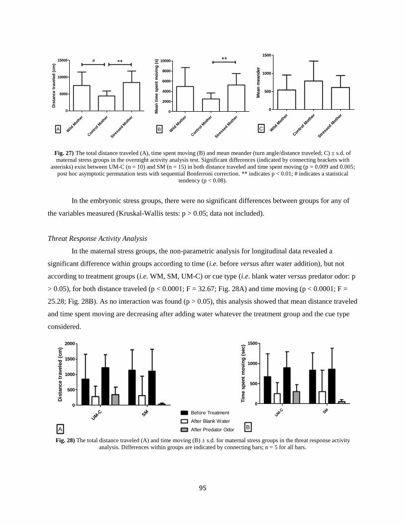
95
Wild
Moth
er
Contr
ol Moth
er
Stres
sed M
other
0
5000
10000
15000 **
A
#
Dis
tan
ce t
ravele
d (
cm
)
Wild
Moth
er
Contr
ol Moth
er
Stres
sed M
other
0
2000
4000
6000
8000
10000 **
B
Mean
tim
e s
pen
t m
ovin
g (
s)
Wild
Moth
er
Contr
ol Moth
er
Stres
sed M
other
0
500
1000
1500
C
Mean
mean
der
Fig. 27) The total distance traveled (A), time spent moving (B) and mean meander (turn angle/distance traveled; C) ± s.d. of
maternal stress groups in the overnight activity analysis test. Significant differences (indicated by connecting brackets with
asterisks) exist between UM-C (n = 10) and SM (n = 15) in both distance traveled and time spent moving (p = 0.009 and 0.005;
post hoc asymptotic permutation tests with sequential Bonferroni correction. ** indicates p < 0.01; # indicates a statistical
tendency (p < 0.08).
In the embryonic stress groups, there were no significant differences between groups for any of
the variables measured (Kruskal-Wallis tests: p > 0.05; data not included).
Threat Response Activity Analysis
In the maternal stress groups, the non-parametric analysis for longitudinal data revealed a
significant difference within groups according to time (i.e. before versus after water addition), but not
according to treatment groups (i.e. WM, SM, UM-C) or cue type (i.e. blank water versus predator odor: p
> 0.05), for both distance traveled (p < 0.0001; F = 32.67; Fig. 28A) and time moving (p < 0.0001; F =
25.28; Fig. 28B). As no interaction was found (p > 0.05), this analysis showed that mean distance traveled
and time spent moving are decreasing after adding water whatever the treatment group and the cue type
considered.
UM
-C SM
0
500
1000
1500
2000
A
Dis
tan
ce t
ravele
d (
cm
)
UM
-C SM
0
500
1000
1500
Before Treatment
After Blank Water
After Predator Odor B
Tim
e s
pen
t m
ovin
g (
sec)
Fig. 28) The total distance traveled (A) and time moving (B) ± s.d. for maternal stress groups in the threat response activity
analysis. Differences within groups are indicated by connecting bars; n = 5 for all bars.
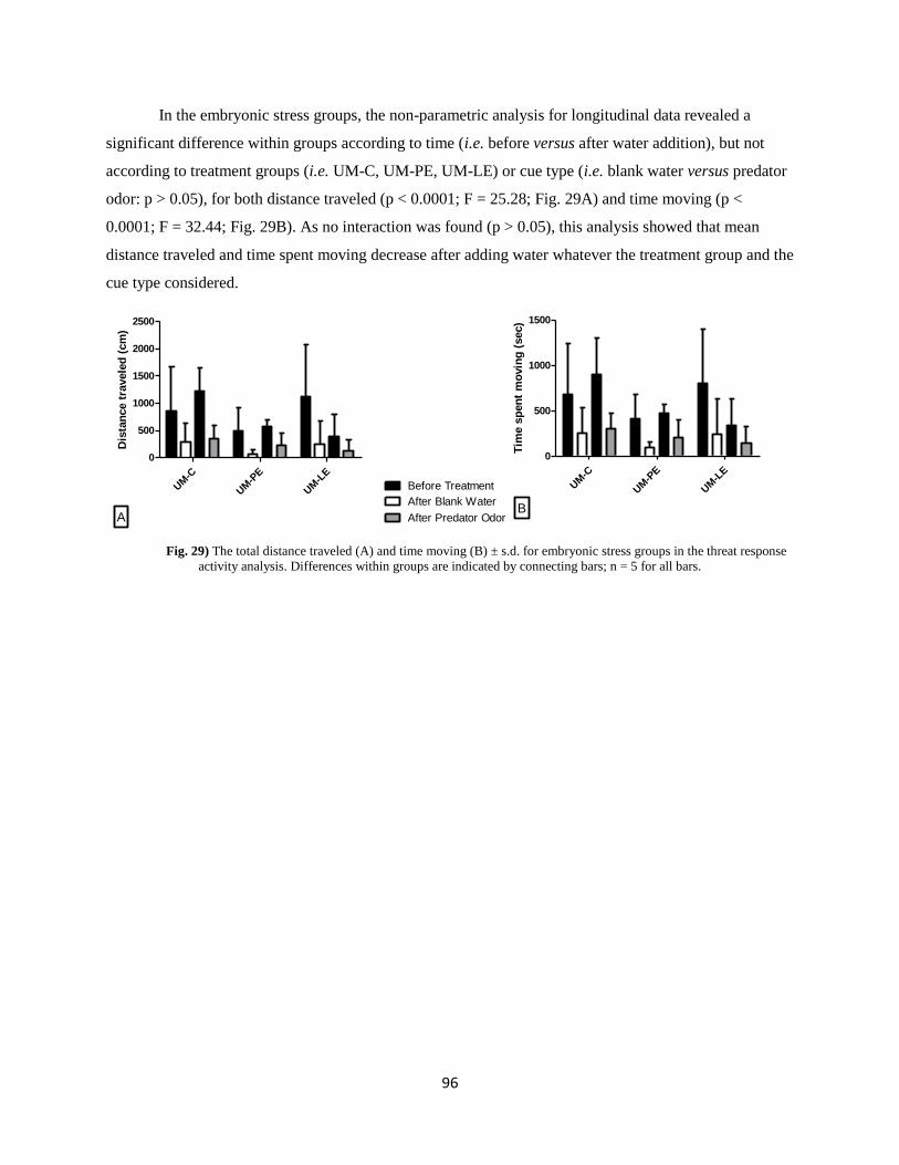
96
In the embryonic stress groups, the non-parametric analysis for longitudinal data revealed a
significant difference within groups according to time (i.e. before versus after water addition), but not
according to treatment groups (i.e. UM-C, UM-PE, UM-LE) or cue type (i.e. blank water versus predator
odor: p > 0.05), for both distance traveled (p < 0.0001; F = 25.28; Fig. 29A) and time moving (p <
0.0001; F = 32.44; Fig. 29B). As no interaction was found (p > 0.05), this analysis showed that mean
distance traveled and time spent moving decrease after adding water whatever the treatment group and the
cue type considered.
UM
-C
UM
-PE
UM
-LE
0
500
1000
1500
2000
2500
A
Dis
tan
ce t
ravele
d (
cm
)
UM
-C
UM
-PE
UM
-LE
0
500
1000
1500
Before Treatment
After Blank Water
After Predator OdorB
Tim
e s
pen
t m
ovin
g (
sec)
Fig. 29) The total distance traveled (A) and time moving (B) ± s.d. for embryonic stress groups in the threat response
activity analysis. Differences within groups are indicated by connecting bars; n = 5 for all bars.
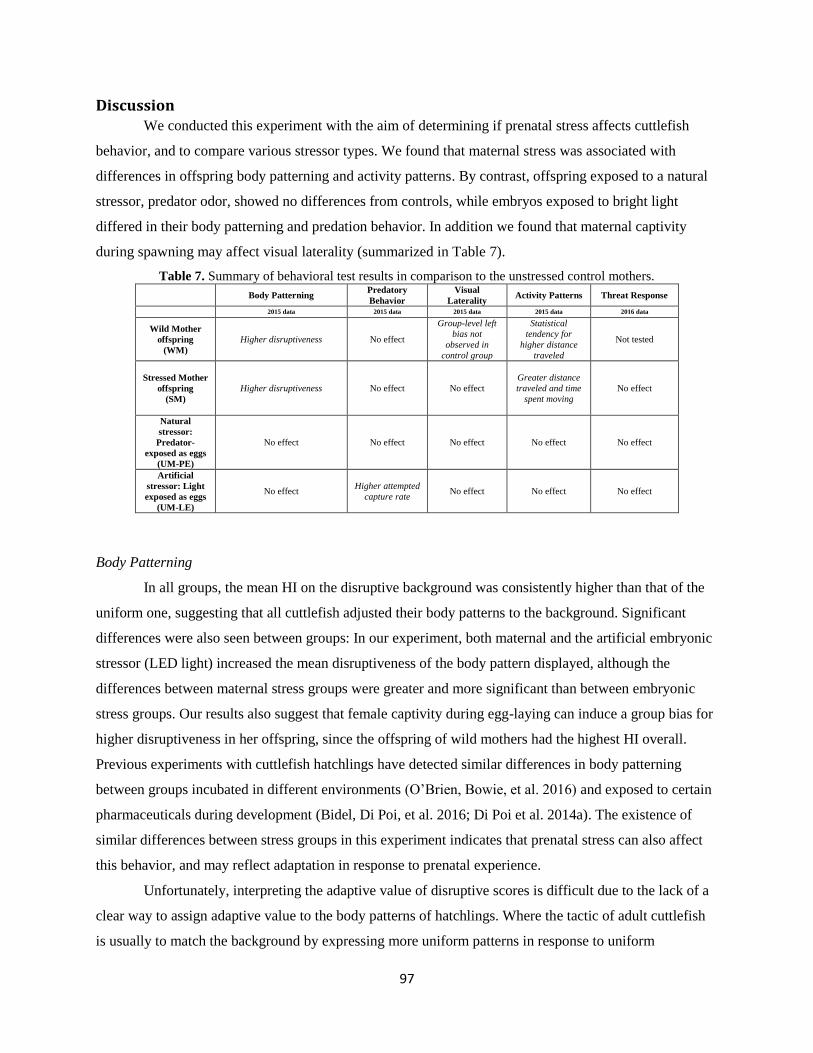
97
Discussion We conducted this experiment with the aim of determining if prenatal stress affects cuttlefish
behavior, and to compare various stressor types. We found that maternal stress was associated with
differences in offspring body patterning and activity patterns. By contrast, offspring exposed to a natural
stressor, predator odor, showed no differences from controls, while embryos exposed to bright light
differed in their body patterning and predation behavior. In addition we found that maternal captivity
during spawning may affect visual laterality (summarized in Table 7).
Table 7. Summary of behavioral test results in comparison to the unstressed control mothers.
Body Patterning Predatory
Behavior
Visual
Laterality Activity Patterns Threat Response
2015 data 2015 data 2015 data 2015 data 2016 data
Wild Mother
offspring
(WM)
Higher disruptiveness No effect
Group-level left
bias not
observed in
control group
Statistical
tendency for
higher distance
traveled
Not tested
Stressed Mother
offspring
(SM)
Higher disruptiveness No effect No effect
Greater distance
traveled and time
spent moving
No effect
Natural
stressor:
Predator-
exposed as eggs
(UM-PE)
No effect No effect No effect
No effect
No effect
Artificial
stressor: Light
exposed as eggs
(UM-LE)
No effect Higher attempted
capture rate No effect No effect No effect
Body Patterning
In all groups, the mean HI on the disruptive background was consistently higher than that of the
uniform one, suggesting that all cuttlefish adjusted their body patterns to the background. Significant
differences were also seen between groups: In our experiment, both maternal and the artificial embryonic
stressor (LED light) increased the mean disruptiveness of the body pattern displayed, although the
differences between maternal stress groups were greater and more significant than between embryonic
stress groups. Our results also suggest that female captivity during egg-laying can induce a group bias for
higher disruptiveness in her offspring, since the offspring of wild mothers had the highest HI overall.
Previous experiments with cuttlefish hatchlings have detected similar differences in body patterning
between groups incubated in different environments (O’Brien, Bowie, et al. 2016) and exposed to certain
pharmaceuticals during development (Bidel, Di Poi, et al. 2016; Di Poi et al. 2014a). The existence of
similar differences between stress groups in this experiment indicates that prenatal stress can also affect
this behavior, and may reflect adaptation in response to prenatal experience.
Unfortunately, interpreting the adaptive value of disruptive scores is difficult due to the lack of a
clear way to assign adaptive value to the body patterns of hatchlings. Where the tactic of adult cuttlefish
is usually to match the background by expressing more uniform patterns in response to uniform
98
backgrounds and more disruptive patterns in response to disruptive ones (Barbosa et al. 2008; Mathger et
al. 2007), young cuttlefish usually display a fairly chronic body pattern that often clashes with the
background (Hanlon and Messenger 1988; O’Brien, Mezrai, et al. 2016; Poirier, Chichery, and Dickel
2005).
Predation
Almost twice as many UM-LE offspring attempted capture than UM-C or UM-PE. Light is
known to influence the timing of hatching (Paulij et al. 1991), and it is possible that these offspring were
hungrier at the same age than other hatchlings because of increased energetic needs due to accelerated
embryonic development. Faster development could also have accelerated visual maturation, leading UM-
LE hatchlings to be better than their siblings at detecting prey. Indeed, UM-LE were able to detect prey at
a significantly greater distance than UM-PE, though the fact that they were statistically identical to the
control group in this respect makes this difficult to interpret. More experiments are necessary to explore
this hypothesis. It is also worth noting that although a greater proportion of UM-LE captured shrimp, they
were not better predators than the other groups, since the capture and success didn’t differ significantly
between groups (close to 100%). This is in accordance with early experiments suggesting that prey
capture operates using a highly-stereotyped program that improves little with age or experience (Wells
1958).
Visual Laterality
In our experiment, no group-level bias was found in the control group. This is in accordance with
previous experiments showing that a left eye-use preference for shelter seeking isn’t fully developed until
a month after hatching (Jozet-Alves et al. 2012). Among all other groups, only WM group displayed a
group-level preference towards the left side on Day 5. These results do conflict somewhat with the
findings of Jozet-Alves & Hebert (2012); In that study, the authors showed that prenatal exposure to
predator odor induced a left preference three days after hatching. However, this preference was slight, and
it was necessary to test each cuttlefish more than once to detect it. Thus, it is possible that in our
experiment, running only one trial with each individual did not allow us to detect the presence of the
fledgling eye-use preference seen in the other groups.
The fact that eye-use preference did exist in the WM group suggests that when egg-laying and
early development occur in the wild, the maturation of the left eye use preference is faster. Being
lateralized from hatching may have an adaptive advantage by rendering WM offspring able to dual task
(Vallortigara and Rogers 2005). For example, while using their right eye for hunting (Schnell et al. 2016)
99
they can simultaneously “keep an eye out” for shelter with their left should the need for a rapid escape
arise.
Overnight Activity
Based on the fact that cuttlefish rely heavily on crypsis to avoid predators (Hanlon and Messenger
1998), our prediction was that all stress types would cause a reduction in the activity of hatchling
cuttlefish. In our experiment however, we found no differences between embryonic stress groups, while
stressed mother offspring were associated with greater activity than control mother offspring: the overall
distance traveled and time spent moving were higher on average in SM than UM-C offspring, though
similar to that of WM. We also observed a statistical tendency for WM hatchlings to travel a greater
distance than UM-C.
These results indicate that the offspring of stressed mothers were more active, spending more
time in motion and consequently traveling a greater distance than the offspring of UM-C. This greater
activity level may reflect a search for shelter or food or an urge to escape. Another, not mutually
exclusive hypothesis is that maternal stress retards the development of the vertical lobe, responsible for
behavioral inhibition, resulting in less mature SM offspring and leading to an increased level of basal
activity. More experiments are needed in order to establish the motivations underlying this behavior in
cuttlefish.
Activity levels and open field behavior have been used in behavioral research as a means of
quantifying the impacts of various prenatal stressors in a variety of animals. Some species, including
rhesus monkeys and salmon, demonstrate decreases in overall activity after maternal or embryonic stress
(Clarke et al. 1996; Espmark et al. 2008; Schneider 1992), while others, including blue foxes and
Japanese quail, show increases in activity and steps taken in open field tests (Braastad 1998; Guibert et al.
2011). The effects of prenatal stress on activity have been studied most extensively in rodents, especially
rats, and results are mixed. Some authors (Hilakivi, Ota, and Lister 1989; Masterpasqua, Chapman, and
Lore 1976; Peters 1986; Sandi, Venero, and Guaza 1996; Wilson, Vazdarjanova, and Terry 2013) report
increases in exploration and open field activity. Others report no or little effect of stress (Chapman and
Stern 1979; Van den Hove et al. 2005), or even opposite effects according to sex (Alonso et al. 1991).
The majority of studies however, find decreases in movement and “exploration” in the offspring of
females subjected to a variety of stressors during pregnancy (Fride et al. 1986; Fujioka et al. 2001;
Hockman 1961; Patin et al. 2004; Poltyrev et al. 1996; Suchecki and Neto 1991; Vallee et al. 1997). Thus
it seems that cuttlefish may differ in this respect from most vertebrate models and could therefore serve as
a means to explore the factors driving the evolution of this response in different animal groups.
100
Threat Response
No group showed a response to predator odor different from that of their response to blank water.
A reduction in activity was observed in all groups after the addition of either stimulus in continuation of a
pattern of progressively decreasing activity over time. This is odd considering that many other animals
strongly alter their activity patterns in response to the introduction of predator odor (reviewed in
(Apfelbach et al. 2005). Moreover, previous experiments have shown that cuttlefish respond to predator
odor by inducing a turning bias in hatchlings (Jozet-Alves and Hebert 2012). Likewise, the presence of an
actual predator induces an increase in swimming activity in adult cuttlefish (Staudinger et al. 2013). We
can only speculate as to the reason for the apparent lack of effect in our experiment: either hatchling
cuttlefish do not alter their activity patterns in response to predator odor, or the amount of predator odor
was too small to elicit an effect.
Maternal versus Embryonic Stress
When examining the results of these behavioral experiments as a whole, it seems that maternal
stress and embryonic stress both affect body patterning, while activity patterns were affected by maternal
stress, and predatory behavior was affected by embryonic stress. Additionally, the fact that eggs from
captive control mothers differed from eggs collected from the wild in camouflage, activity and turning
bias behavior suggests that the environment in which eggs are laid can affect offspring behavior. This
should be taken into account when interpreting laboratory experiments—the behaviors observed may
differ depending on how subjects were obtained (i.e. bred in captivity or collected from the wild), and
experimenters should carefully consider their experimental priorities (i.e. whether they are trying to assess
natural behavior) before they source cuttlefish eggs for experiments.
While it is unclear if the maternal stress effects on body patterning and overnight activity increase
offspring fitness, the left turning bias of WM certainly has the potential to be adaptive in the face of a
threat. Maternal experience is known to “program” offspring in many other species: in numerous species,
the offspring of mothers exposed to a particular predator showed adaptive responses when encountering
that predator itself (reviewed in Agrawal, Laforsch, & Tollrian, 1999; Storm & Lima, 2010). These
effects are likely mediated by the transfer of stress hormones in the egg or placenta (Groothuis et al. 2005;
Hayward and Wingfield 2004; Weinstock 2008) and it is possible that a similar phenomenon is at play in
cuttlefish.
Artificial versus Natural Embryonic Stressor
We were surprised to find that the naturally-occurring predator cues had no discernable
behavioral effect. By contrast, the bright LED light, which was artificial not only in its source but also in
101
brightness and timing, affected body patterning and predation. We had expected the opposite trend: a
strong, adaptive response to a natural stressor, and no response to a stressor not phylogenetically
encountered. The predator odor would seem to be a particularly-relevant stressor to cuttlefish—seabass in
the natal environment signal a direct post-natal threat, and strong predator-induced responses are
widespread throughout the animal (reviewed in Agrawal, Laforsch, and Tollrian 1999; Apfelbach et al.
2005; Chivers and Smith 1998; Lass and Spaak 2003), so presumably selective pressure on this trait is
strong. Indeed, embryonic exposure to seabass odor is associated with increased lateralization in cuttlefish
hatchlings (Jozet-Alves and Hebert 2012). Thus, it is rather surprising that, in an embryo with the sensory
abilities to obtain information about its hatching environment (Romagny et al. 2012), as well as the ability
to develop food preferences based on this information (Darmaillacq, Lesimple, and Dickel 2008), little or
no embryonic plasticity occurs. Any preparation that could be made to prepare for post-hatching life
should be promoted by natural selection. We have two hypotheses regarding these unexpected results.
The first concerns a tranquilizing compound thought to be present in the perivitelline fluid (PVF)
surrounding developing cephalopod embryos (Marthy, Hauser, and Scholl 1976). This compound reduces
the activity of embryos (Weischer and Marthy 1983) and may also dampen embryos’ reaction to some
potential stressors. Further experimentation is necessary to test this idea.
Our second hypothesis involves habituation. In our experiment, predator odor exposure was
experienced continuously for the majority of development. Post-natal studies in other animals, including
fish, rats and lizards, have shown that while acute stress exposure can result in adaptive changes (e.g.
increased predator avoidance behavior or HPA-axis sensitivity), long-term or repeated exposure can
actually reduce or eliminate the adaptive response (Dielenberg and McGregor 1999; Weinberg et al.
2009). On the other hand, some studies show a lack of habituation to predator odor applied long-term
(e.g. Epple et al. 1993). If habituation to predator odor is indeed occurring in cuttlefish, the evolutionary
reason for this merits further scrutiny. One possibility is that the cognitive complexity of cuttlefish allows
for complex discrimination between dangerous and harmless stimuli. Since the predator odor was not
paired with alarm cues from injured conspecifics in our experiment, it is possible that the cuttlefish
embryos learned to regard it as benign. Such a phenomenon occurs in harbor seals, which learn to
distinguish between the calls of fish-eating and seal-eating orca populations and behave accordingly
(Deecke, Slater, and Ford 2002).
Conclusion
S. officinalis is a behavioral model poised to yield important insights in the study of prenatal
stress in animals. In the future, experiments should be conducted to explore why the effects of maternal
stress and embryonic stimulation differ. An experiments examining why an artificial stimulus has strong
102
effects while a natural stressor has none might reveal previously-unknown prenatal pressures driving
offspring behavior. At the same time, greater effort should be made to account for the effects of spawning
environment in interpreting laboratory experiments. These experiments represent an important first step in
this investigation.
Acknowledgments
This work was supported by the Agence Nationale de la Recherche (ANR) and the Région Basse-
Normandie as part of the research project PReSTO’Cog (ANR-13-BSV7-0002). We are grateful for the
assistance of Olivier Basuyaux and Sébastien Pien of the Synergie Mer et Littoral (SMEL), Nadège
Villain-Naud and Céline Thomasse of NECC Ethos (Université de Caen), Jean Paul Lehodey, David
Liegard, Frédéric Guyon and David Lemeille of the Centre de Recherches en Environnement Côtier
(CREC, Université de Caen), as well as the numerous students who worked with us, including Kevin
Bairos-Novak, Héloise Duretz, Camille Auger, Chloe Jane Way, Pauline Billiard and Justine Lequesne.
The manuscript was proofread by Barbara O’Brien.
103
IV. Cognitive effects of prenatal stress.
For several decades, the “prawn in a tube” test, or PIT test, has been used to assess learning and
memory in cuttlefish (e.g. Messenger 1977; Dickel, Chichery, and Chichery 1998b; Cartron et al. 2013).
Materials and Methods
Stress Treatments:
On May 10, 2016, 26 adult female cuttlefish (approximately two years old) were captured from
the English Channel by trap and transported to the Centre Régional d'Etudes Côtières (CREC, Luc sur
Mer, France), which is equipped with a semi-open flow-through seawater system with a mean
temperature of 15±1°C and a 16:8h light/dark cycle. Here, the females were randomly divided into an
unstressed group and a “stressed” group. The unstressed mothers (UM) were housed in groups of three
in 1000L tanks enriched with natural and artificial objects. The stressed mothers (SM) were housed
individually in bare 65L tanks and subjected to daily stress treatments. These treatments consisted of
removing cuttlefish from the water on a soft mesh platform for 10 sec, three times a day. These
treatments occurred between 9h and 17h with the timing randomized each day to avoid habituation.
The combination of confined space and daily stress treatments were designed to induce chronic stress.
On arrival, females were mated with males captured concurrently and they began to lay eggs
several days later. To ensure that any potential effects of the stress treatment had manifested, and to
allow the UM to recover from the stress of capture, only eggs that were laid at least one week after
arrival at the CREC were used in the experiments described below. In addition, only eggs from SM that
had laid at least 150 eggs were used in order to ensure similar representation between mothers and
across stress groups. In total, 11 UM (grouped into four groups since eggs laid in the same tank could
not be attributed to individual females) and five SM laid eggs that met these criteria between May 14
and 29. After a sufficient number of eggs had been collected, they were habituated to a temperature of
20±2°C over the course of eight hours and moved to shallow trays floating in 65L aerated tanks for
incubation.
The eggs of UM were divided into three treatment groups: Control (UM-C), predator-exposed
(UM-PE) and light-exposed (UM-LE). The eggs of UM and SM were allowed to develop without further
manipulation. UM-PE eggs were incubated with three sea bass (Dicentrarchus labrax) for the remainder
of development (three to five weeks). Bright LED light was shined on the UM-LE group for 15m six times
a day (90m total) at random intervals. Eggs hatched between July 2 and 24. On the day of hatching,
cuttlefish were placed in an individually-labelled compartment (6.8 x 12.1cm, depth 5.5cm) at a mean
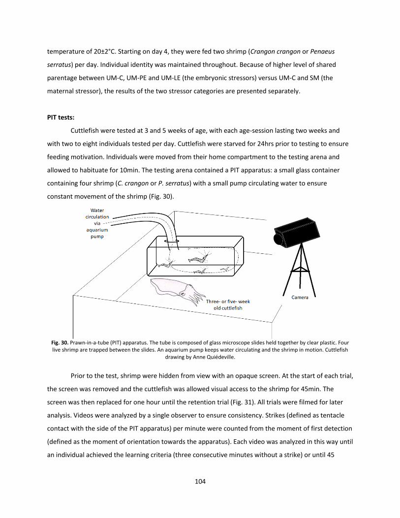
104
temperature of 20±2°C. Starting on day 4, they were fed two shrimp (Crangon crangon or Penaeus
serratus) per day. Individual identity was maintained throughout. Because of higher level of shared
parentage between UM-C, UM-PE and UM-LE (the embryonic stressors) versus UM-C and SM (the
maternal stressor), the results of the two stressor categories are presented separately.
PIT tests:
Cuttlefish were tested at 3 and 5 weeks of age, with each age-session lasting two weeks and
with two to eight individuals tested per day. Cuttlefish were starved for 24hrs prior to testing to ensure
feeding motivation. Individuals were moved from their home compartment to the testing arena and
allowed to habituate for 10min. The testing arena contained a PIT apparatus: a small glass container
containing four shrimp (C. crangon or P. serratus) with a small pump circulating water to ensure
constant movement of the shrimp (Fig. 30).
Fig. 30. Prawn-in-a-tube (PIT) apparatus. The tube is composed of glass microscope slides held together by clear plastic. Four live shrimp are trapped between the slides. An aquarium pump keeps water circulating and the shrimp in motion. Cuttlefish
drawing by Anne Quiédeville.
Prior to the test, shrimp were hidden from view with an opaque screen. At the start of each trial,
the screen was removed and the cuttlefish was allowed visual access to the shrimp for 45min. The
screen was then replaced for one hour until the retention trial (Fig. 31). All trials were filmed for later
analysis. Videos were analyzed by a single observer to ensure consistency. Strikes (defined as tentacle
contact with the side of the PIT apparatus) per minute were counted from the moment of first detection
(defined as the moment of orientation towards the apparatus). Each video was analyzed in this way until
an individual achieved the learning criteria (three consecutive minutes without a strike) or until 45

105
minutes of tube presentation had elapsed, in order to avoid unnecessary stress to the cuttlefish. The
mean strikes per minute after detection was plotted for each group until the number of individuals
striking fell below n = 5. Six variables were measured: the proportion of each group that reached
criterion, the latency to detection (time between removal of the occlusive screen and orientation
toward the apparatus), latency to attack (time between detection and the first strike), the initial strike
level (number of strikes made in the first 3 min), the mean time to criterion (time between detection
and reaching criterion), and the mean strikes to criterion (number of strikes made before three minutes
without striking). Differences between groups in these variables could suggest differences in learning
abilities, memory capacity, visual abilities, feeding motivation, predation strategy and/ or fatigue.
Fig. 31. Timeline of PIT sessions. Juveniles were tested at three and five weeks of age in two 45min sessions separated by an
hour-long rest interval.
Group success rates were compared by stress group with a Cochran-Mantel-Haenszel chi-
squared test for count data using the mantelhaen.test function in R. The remaining variables were
compared by stress group and within groups by performances during the initial session versus the
retention session. Latencies and initial strike levels were calculated from all cuttlefish that responded to
the PIT apparatus during either or both sessions, while the time and number of strikes to criterion were
calculated only from cuttlefish that responded in both the initial and retention sessions in order to
ensure a balanced data set (the majority of individuals in all groups responded during both sessions).
Latency to detect, latency to attack, strike level in first three minutes, minutes to criterion, strikes to
criterion were failed to adhere to parametric assumptions, so a non-parametric repeated measures
ANOVA was used to compared between and within groups (ANOVA.test and pair.comparison functions
in R package nparLD).
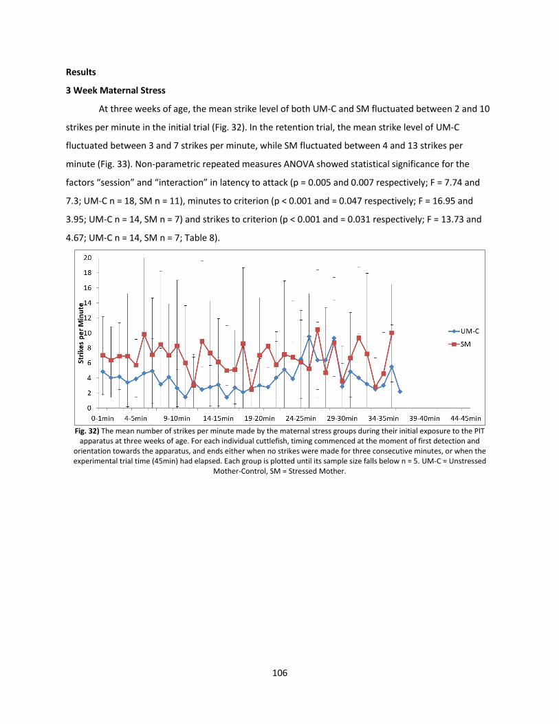
106
Results
3 Week Maternal Stress
At three weeks of age, the mean strike level of both UM-C and SM fluctuated between 2 and 10
strikes per minute in the initial trial (Fig. 32). In the retention trial, the mean strike level of UM-C
fluctuated between 3 and 7 strikes per minute, while SM fluctuated between 4 and 13 strikes per
minute (Fig. 33). Non-parametric repeated measures ANOVA showed statistical significance for the
factors “session” and “interaction” in latency to attack (p = 0.005 and 0.007 respectively; F = 7.74 and
7.3; UM-C n = 18, SM n = 11), minutes to criterion (p < 0.001 and = 0.047 respectively; F = 16.95 and
3.95; UM-C n = 14, SM n = 7) and strikes to criterion (p < 0.001 and = 0.031 respectively; F = 13.73 and
4.67; UM-C n = 14, SM n = 7; Table 8).
Fig. 32) The mean number of strikes per minute made by the maternal stress groups during their initial exposure to the PIT
apparatus at three weeks of age. For each individual cuttlefish, timing commenced at the moment of first detection and orientation towards the apparatus, and ends either when no strikes were made for three consecutive minutes, or when the experimental trial time (45min) had elapsed. Each group is plotted until its sample size falls below n = 5. UM-C = Unstressed
Mother-Control, SM = Stressed Mother.
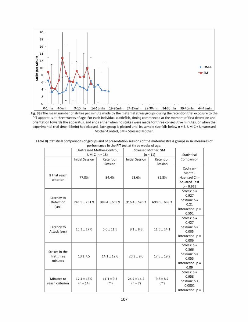
107
Fig. 33) The mean number of strikes per minute made by the maternal stress groups during the retention trial exposure to the
PIT apparatus at three weeks of age. For each individual cuttlefish, timing commenced at the moment of first detection and orientation towards the apparatus, and ends either when no strikes were made for three consecutive minutes, or when the experimental trial time (45min) had elapsed. Each group is plotted until its sample size falls below n = 5. UM-C = Unstressed
Mother-Control, SM = Stressed Mother.
Table 8) Statistical comparisons of groups and of presentation sessions of the maternal stress groups in six measures of
performance in the PIT test at three weeks of age.
Unstressed Mother-Control, UM-C (n = 18)
Stressed Mother, SM (n = 11) Statistical
Comparison Initial Session Retention Session
Initial Session Retention Session
% that reach criterion
77.8% 94.4% 63.6% 81.8%
Cochran-Mantel-
Haenszel Chi-Squared Test
p = 0.965
Latency to Detection
(sec) 245.5 ± 251.9 388.4 ± 605.9 316.4 ± 520.2 600.0 ± 638.3
Stress: p = 0.927
Session: p = 0.21
Interaction: p = 0.551
Latency to Attack (sec)
15.3 ± 17.0 5.6 ± 11.5 9.1 ± 8.8 11.5 ± 14.1
Stress: p = 0.427
Session: p = 0.005
Interaction: p = 0.006
Strikes in the first three minutes
13 ± 7.5 14.1 ± 12.6 20.3 ± 9.0 17.5 ± 19.9
Stress: p = 0.366
Session: p = 0.055
Interaction: p = 0.09
Minutes to reach criterion
17.4 ± 13.0 (n = 14)
11.1 ± 9.3 (“”)
24.7 ± 14.2 (n = 7)
9.8 ± 8.7 (“”)
Stress: p = 0.958
Session: p < 0.0001
Interaction: p =
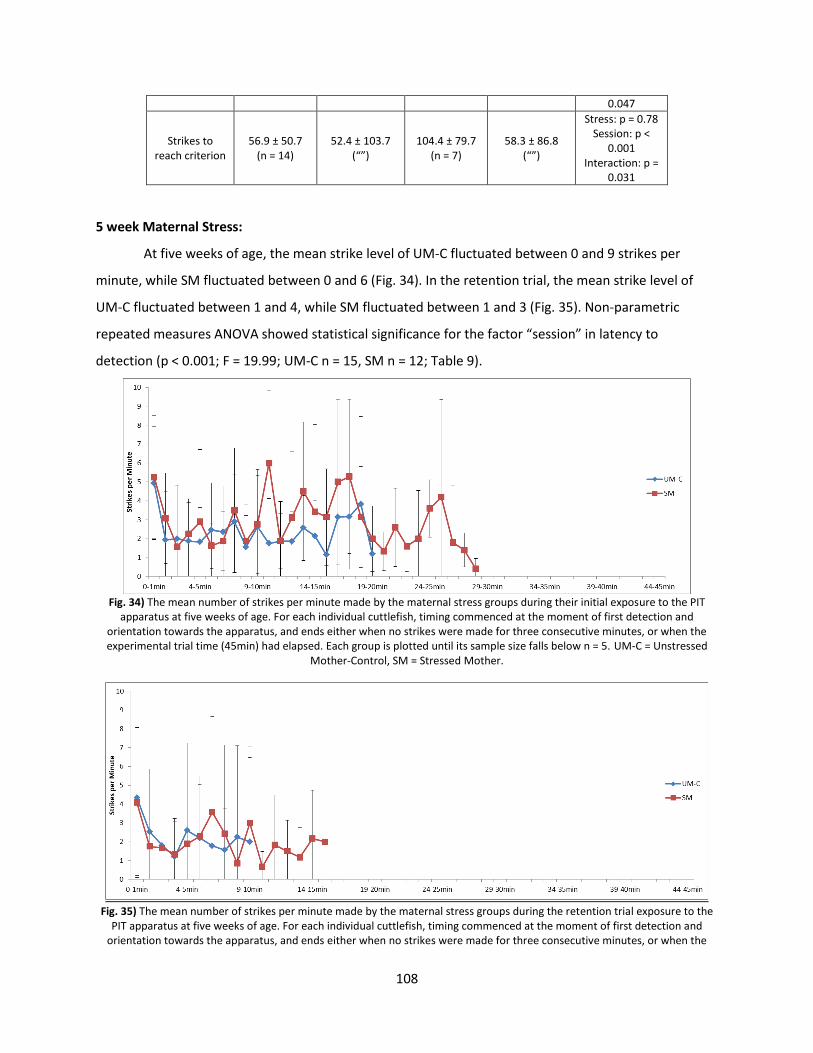
108
0.047
Strikes to reach criterion
56.9 ± 50.7 (n = 14)
52.4 ± 103.7 (“”)
104.4 ± 79.7 (n = 7)
58.3 ± 86.8 (“”)
Stress: p = 0.78 Session: p <
0.001 Interaction: p =
0.031
5 week Maternal Stress:
At five weeks of age, the mean strike level of UM-C fluctuated between 0 and 9 strikes per
minute, while SM fluctuated between 0 and 6 (Fig. 34). In the retention trial, the mean strike level of
UM-C fluctuated between 1 and 4, while SM fluctuated between 1 and 3 (Fig. 35). Non-parametric
repeated measures ANOVA showed statistical significance for the factor “session” in latency to
detection (p < 0.001; F = 19.99; UM-C n = 15, SM n = 12; Table 9).
Fig. 34) The mean number of strikes per minute made by the maternal stress groups during their initial exposure to the PIT
apparatus at five weeks of age. For each individual cuttlefish, timing commenced at the moment of first detection and orientation towards the apparatus, and ends either when no strikes were made for three consecutive minutes, or when the experimental trial time (45min) had elapsed. Each group is plotted until its sample size falls below n = 5. UM-C = Unstressed
Mother-Control, SM = Stressed Mother.
Fig. 35) The mean number of strikes per minute made by the maternal stress groups during the retention trial exposure to the
PIT apparatus at five weeks of age. For each individual cuttlefish, timing commenced at the moment of first detection and orientation towards the apparatus, and ends either when no strikes were made for three consecutive minutes, or when the
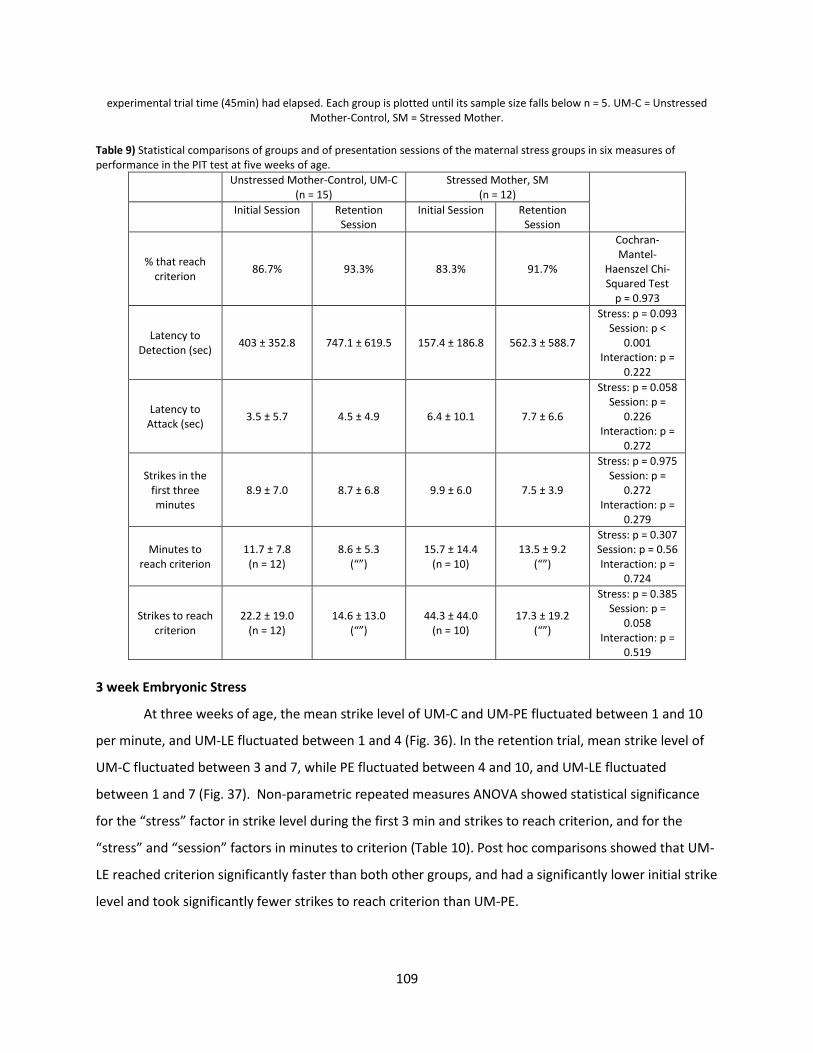
109
experimental trial time (45min) had elapsed. Each group is plotted until its sample size falls below n = 5. UM-C = Unstressed Mother-Control, SM = Stressed Mother.
Table 9) Statistical comparisons of groups and of presentation sessions of the maternal stress groups in six measures of performance in the PIT test at five weeks of age.
Unstressed Mother-Control, UM-C (n = 15)
Stressed Mother, SM (n = 12)
Initial Session Retention Session
Initial Session Retention Session
% that reach criterion
86.7% 93.3% 83.3% 91.7%
Cochran-Mantel-
Haenszel Chi-Squared Test
p = 0.973
Latency to Detection (sec)
403 ± 352.8 747.1 ± 619.5 157.4 ± 186.8 562.3 ± 588.7
Stress: p = 0.093 Session: p <
0.001 Interaction: p =
0.222
Latency to Attack (sec)
3.5 ± 5.7 4.5 ± 4.9 6.4 ± 10.1 7.7 ± 6.6
Stress: p = 0.058 Session: p =
0.226 Interaction: p =
0.272
Strikes in the first three minutes
8.9 ± 7.0 8.7 ± 6.8 9.9 ± 6.0 7.5 ± 3.9
Stress: p = 0.975 Session: p =
0.272 Interaction: p =
0.279
Minutes to reach criterion
11.7 ± 7.8 (n = 12)
8.6 ± 5.3 (“”)
15.7 ± 14.4 (n = 10)
13.5 ± 9.2 (“”)
Stress: p = 0.307 Session: p = 0.56 Interaction: p =
0.724
Strikes to reach criterion
22.2 ± 19.0 (n = 12)
14.6 ± 13.0 (“”)
44.3 ± 44.0 (n = 10)
17.3 ± 19.2 (“”)
Stress: p = 0.385 Session: p =
0.058 Interaction: p =
0.519
3 week Embryonic Stress
At three weeks of age, the mean strike level of UM-C and UM-PE fluctuated between 1 and 10
per minute, and UM-LE fluctuated between 1 and 4 (Fig. 36). In the retention trial, mean strike level of
UM-C fluctuated between 3 and 7, while PE fluctuated between 4 and 10, and UM-LE fluctuated
between 1 and 7 (Fig. 37). Non-parametric repeated measures ANOVA showed statistical significance
for the “stress” factor in strike level during the first 3 min and strikes to reach criterion, and for the
“stress” and “session” factors in minutes to criterion (Table 10). Post hoc comparisons showed that UM-
LE reached criterion significantly faster than both other groups, and had a significantly lower initial strike
level and took significantly fewer strikes to reach criterion than UM-PE.
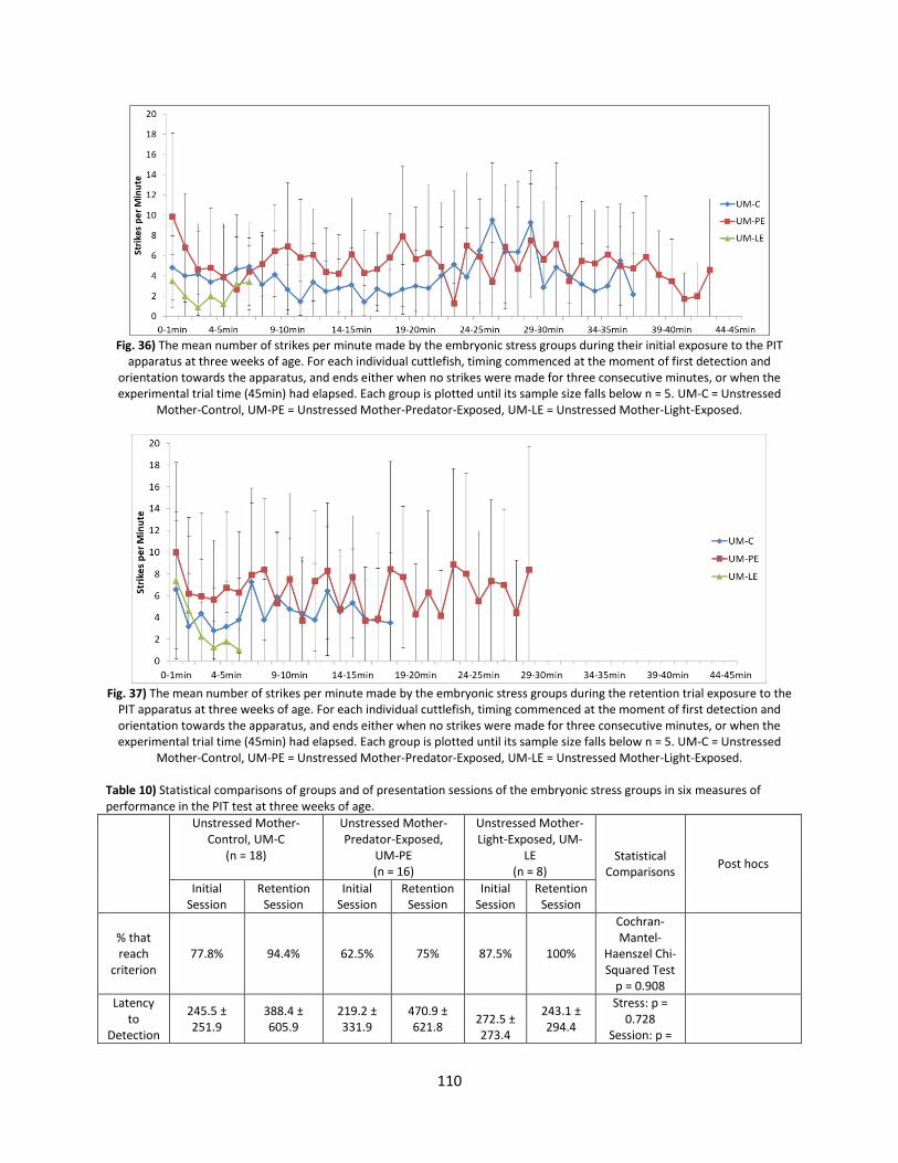
110
Fig. 36) The mean number of strikes per minute made by the embryonic stress groups during their initial exposure to the PIT
apparatus at three weeks of age. For each individual cuttlefish, timing commenced at the moment of first detection and orientation towards the apparatus, and ends either when no strikes were made for three consecutive minutes, or when the experimental trial time (45min) had elapsed. Each group is plotted until its sample size falls below n = 5. UM-C = Unstressed
Mother-Control, UM-PE = Unstressed Mother-Predator-Exposed, UM-LE = Unstressed Mother-Light-Exposed.
Fig. 37) The mean number of strikes per minute made by the embryonic stress groups during the retention trial exposure to the
PIT apparatus at three weeks of age. For each individual cuttlefish, timing commenced at the moment of first detection and orientation towards the apparatus, and ends either when no strikes were made for three consecutive minutes, or when the experimental trial time (45min) had elapsed. Each group is plotted until its sample size falls below n = 5. UM-C = Unstressed
Mother-Control, UM-PE = Unstressed Mother-Predator-Exposed, UM-LE = Unstressed Mother-Light-Exposed. Table 10) Statistical comparisons of groups and of presentation sessions of the embryonic stress groups in six measures of performance in the PIT test at three weeks of age.
Unstressed Mother-Control, UM-C
(n = 18)
Unstressed Mother-Predator-Exposed,
UM-PE (n = 16)
Unstressed Mother-Light-Exposed, UM-
LE (n = 8)
Statistical Comparisons
Post hocs
Initial Session
Retention Session
Initial Session
Retention Session
Initial Session
Retention Session
% that reach
criterion 77.8% 94.4% 62.5% 75% 87.5% 100%
Cochran-Mantel-
Haenszel Chi-Squared Test
p = 0.908
Latency to
Detection
245.5 ± 251.9
388.4 ± 605.9
219.2 ± 331.9
470.9 ± 621.8
272.5 ± 273.4
243.1 ± 294.4
Stress: p = 0.728
Session: p =
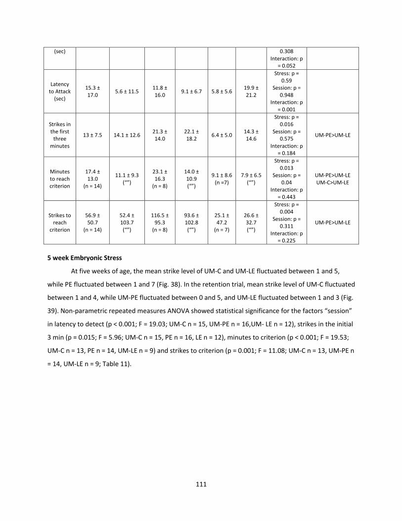
111
(sec) 0.308 Interaction: p
= 0.052
Latency to Attack
(sec)
15.3 ± 17.0
5.6 ± 11.5 11.8 ± 16.0
9.1 ± 6.7 5.8 ± 5.6 19.9 ± 21.2
Stress: p = 0.59
Session: p = 0.948
Interaction: p = 0.001
Strikes in the first
three minutes
13 ± 7.5 14.1 ± 12.6 21.3 ± 14.0
22.1 ± 18.2
6.4 ± 5.0 14.3 ± 14.6
Stress: p = 0.016
Session: p = 0.575
Interaction: p = 0.184
UM-PE>UM-LE
Minutes to reach criterion
17.4 ± 13.0
(n = 14)
11.1 ± 9.3 (“”)
23.1 ± 16.3
(n = 8)
14.0 ± 10.9 (“”)
9.1 ± 8.6 (n =7)
7.9 ± 6.5 (“”)
Stress: p = 0.013
Session: p = 0.04
Interaction: p = 0.443
UM-PE>UM-LE UM-C>UM-LE
Strikes to reach
criterion
56.9 ± 50.7
(n = 14)
52.4 ± 103.7 (“”)
116.5 ± 95.3
(n = 8)
93.6 ± 102.8 (“”)
25.1 ± 47.2
(n = 7)
26.6 ± 32.7 (“”)
Stress: p = 0.004
Session: p = 0.311
Interaction: p = 0.225
UM-PE>UM-LE
5 week Embryonic Stress
At five weeks of age, the mean strike level of UM-C and UM-LE fluctuated between 1 and 5,
while PE fluctuated between 1 and 7 (Fig. 38). In the retention trial, mean strike level of UM-C fluctuated
between 1 and 4, while UM-PE fluctuated between 0 and 5, and UM-LE fluctuated between 1 and 3 (Fig.
39). Non-parametric repeated measures ANOVA showed statistical significance for the factors “session”
in latency to detect (p < 0.001; F = 19.03; UM-C n = 15, UM-PE n = 16,UM- LE n = 12), strikes in the initial
3 min (p = 0.015; F = 5.96; UM-C n = 15, PE n = 16, LE n = 12), minutes to criterion (p < 0.001; F = 19.53;
UM-C n = 13, PE n = 14, UM-LE n = 9) and strikes to criterion (p = 0.001; F = 11.08; UM-C n = 13, UM-PE n
= 14, UM-LE n = 9; Table 11).
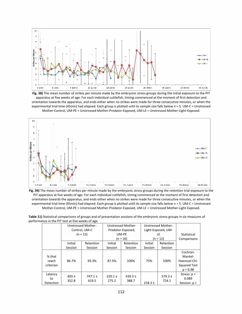
112
Fig. 38) The mean number of strikes per minute made by the embryonic stress groups during the initial exposure to the PIT
apparatus at five weeks of age. For each individual cuttlefish, timing commenced at the moment of first detection and orientation towards the apparatus, and ends either when no strikes were made for three consecutive minutes, or when the experimental trial time (45min) had elapsed. Each group is plotted until its sample size falls below n = 5. UM-C = Unstressed
Mother-Control, UM-PE = Unstressed Mother-Predator-Exposed, UM-LE = Unstressed Mother-Light-Exposed.
Fig. 39) The mean number of strikes per minute made by the embryonic stress groups during the retention trial exposure to the
PIT apparatus at five weeks of age. For each individual cuttlefish, timing commenced at the moment of first detection and orientation towards the apparatus, and ends either when no strikes were made for three consecutive minutes, or when the experimental trial time (45min) had elapsed. Each group is plotted until its sample size falls below n = 5. UM-C = Unstressed
Mother-Control, UM-PE = Unstressed Mother-Predator-Exposed, UM-LE = Unstressed Mother-Light-Exposed. Table 11) Statistical comparisons of groups and of presentation sessions of the embryonic stress groups in six measures of performance in the PIT test at five weeks of age.
Unstressed Mother-Control, UM-C
(n = 15)
Unstressed Mother-Predator-Exposed,
UM-PE (n = 16)
Unstressed Mother-Light-Exposed, UM-
LE (n = 12)
Statistical Comparisons
Initial Session
Retention Session
Initial Session
Retention Session
Initial Session
Retention Session
% that reach
criterion 86.7% 93.3% 87.5% 100% 75% 100%
Cochran-Mantel-
Haenszel Chi-Squared Test
p = 0.98
Latency to
Detection
403 ± 352.8
747.1 ± 619.5
220.1 ± 275.2
439.3 ± 388.7
218.3 ±
579.3 ± 724.1
Stress: p = 0.089
Session: p <
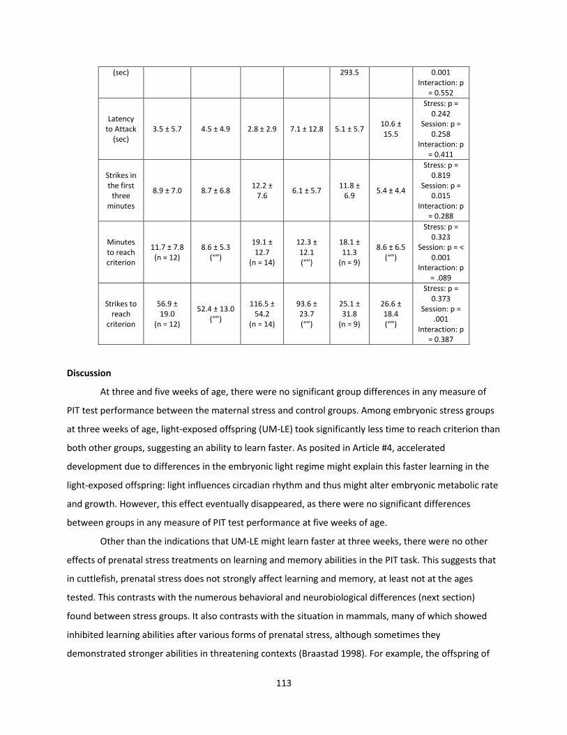
113
(sec) 293.5 0.001 Interaction: p
= 0.552
Latency to Attack
(sec) 3.5 ± 5.7 4.5 ± 4.9 2.8 ± 2.9 7.1 ± 12.8 5.1 ± 5.7
10.6 ± 15.5
Stress: p = 0.242
Session: p = 0.258
Interaction: p = 0.411
Strikes in the first
three minutes
8.9 ± 7.0 8.7 ± 6.8 12.2 ±
7.6 6.1 ± 5.7
11.8 ± 6.9
5.4 ± 4.4
Stress: p = 0.819
Session: p = 0.015
Interaction: p = 0.288
Minutes to reach criterion
11.7 ± 7.8 (n = 12)
8.6 ± 5.3 (“”)
19.1 ± 12.7
(n = 14)
12.3 ± 12.1 (“”)
18.1 ± 11.3
(n = 9)
8.6 ± 6.5 (“”)
Stress: p = 0.323
Session: p = < 0.001
Interaction: p = .089
Strikes to reach
criterion
56.9 ± 19.0
(n = 12)
52.4 ± 13.0 (“”)
116.5 ± 54.2
(n = 14)
93.6 ± 23.7 (“”)
25.1 ± 31.8
(n = 9)
26.6 ± 18.4 (“”)
Stress: p = 0.373
Session: p = .001
Interaction: p = 0.387
Discussion
At three and five weeks of age, there were no significant group differences in any measure of
PIT test performance between the maternal stress and control groups. Among embryonic stress groups
at three weeks of age, light-exposed offspring (UM-LE) took significantly less time to reach criterion than
both other groups, suggesting an ability to learn faster. As posited in Article #4, accelerated
development due to differences in the embryonic light regime might explain this faster learning in the
light-exposed offspring: light influences circadian rhythm and thus might alter embryonic metabolic rate
and growth. However, this effect eventually disappeared, as there were no significant differences
between groups in any measure of PIT test performance at five weeks of age.
Other than the indications that UM-LE might learn faster at three weeks, there were no other
effects of prenatal stress treatments on learning and memory abilities in the PIT task. This suggests that
in cuttlefish, prenatal stress does not strongly affect learning and memory, at least not at the ages
tested. This contrasts with the numerous behavioral and neurobiological differences (next section)
found between stress groups. It also contrasts with the situation in mammals, many of which showed
inhibited learning abilities after various forms of prenatal stress, although sometimes they
demonstrated stronger abilities in threatening contexts (Braastad 1998). For example, the offspring of
114
female rats that were shocked during pregnancy showed deficits compared to control animals in four
different learning tests (Smith, Wills, and Naylor 1981). Likewise, prenatally-stressed rhesus monkeys
showed cognitive impairments in object-permanence tests (Schneider 1992). The lack of stress effect in
cuttlefish could indicate that, unlike the mammalian nervous system, the cephalopod cognitive system is
largely immune to the effects of prenatal stress. Alternatively, the existence of stress effects in some of
the other tests reported in this thesis but not the PIT test might encourage us to formulate new learning
and memory tests to use in addition to the PIT.
115
V. Neurobiological effects of prenatal stress
In order to test for stress-related differences in the cuttlefish brain growth and composition,
several neurobiological assays were conducted on representative samples of each stress treatment
group from 2015 and 2016. The results and conclusions presented here (section V) and in section IV are
preliminary, as we are still working to examine and interpret the potential connections and correlations
between these data sets.
Monoamines
The concentrations of various monoamines in neural tissue can be used to gauge the status of
various neurotransmitter systems in the brains of animals. We quantified seven monoamines: serotonin
(5-HT), its main metabolite 5-hydroxyindolacetic acid (5-HiAA), dopamine and its main metabolite 3,4-
dihydroxyphenyl acetic acid (DOPAC), glutamate (Glu), noradrenaline (NA) and octopamine. These
monoamines were selected since each has been found to occur in the cephalopod central nervous
system (CNS) (Andrews, Messenger, and Tansey 1983; Kime and Messenger 1990; Juorio and Molinoff
1974). Most importantly for this research, serotonin, noradrenaline and dopamine are known to be
involved in instigating the stress reactions of vertebrates and mammals—noradrenaline initiates the
“fight or flight” hormonal cascade (Cannon 1939), while serotonin and dopamine modulate the response
as neurotransmitters (reviewed in Thierry et al. 1968; Roth et al. 1988; Joca, Ferreira, and Guimarães
2007).
In cephalopods, serotonin is found throughout the CNS, where at least three types of receptors
exist. It has been implicated in synaptic transmission and long-term potentiation (LTP), meaning it plays
an important role in “higher” cognitive processes (Messenger 1996; Shomrat et al. 2010). The
catecholamines—dopamine and noradrenaline—function as neurotransmitters and possibly guide
axonal growth in cephalopods (Baratte and Bonnaud 2009). Glutamate also functions as a
neurotransmitter in cephalopods, as well as playing a role in LTP (Andrews, Messenger, and Tansey
1983; Hochner et al. 2003). Finally, octopamine, first identified in the cephalopods, is related to
noradrenaline and functions as a neurotransmitter and modulator (Kime and Messenger 1990). In
addition to raw concentrations, the ratio of serotonin or dopamine to their main metabolites (5-HiAA
and DOPAC respectively) also give an indication of turnover rate, with a higher ratio meaning greater
turnover of that monoamine (Bidel 2015). Though the neurochemistry of cephalopods is still a nascent
field, we hope to correlate behavioral differences between stress groups with monoamine
concentrations. These measurements were accomplished in collaboration with Dr. Raphael Delepee at

116
the Université de Caen’s Plate-forme de Recherche et d’Innovation en Spectrométroie de Masse et
Métabolomique, SF ICORE 4206 (PRISMM, SF ICORE 4206).
Material and Methods
Representatives from each stress group were selected for monoamine quantification. Samples
were collected in 2016 on the day of hatching. Hatchlings were euthanized with an overdose of 2%
ethanol following anesthesia with MgCl (17.5g/L) and then stored at -80°C before treatment.
Quantification was achieved by ultra-high-pressure liquid chromatography coupled with
tandem-mass spectrometry (UHPLC-MS/MS). Glutamate was also added to the quantification as a
precursor of monoamines. Two internal standards were used. The first one was 5-hydroxy-N--
methyltryptamine (5-HMT) used for the quantification serotonin and 5-hydroxyindolacetic acid. The
second one was 3,4-dihydroxybenzylamine (DHBA) used for the quantification of dopamine,
noradrenaline, octopamine and 3,4-dihydroxyphenyl acetic acid.
Sample preparation consisted of separating head from the body, weighing this structure
(between 3 and 20 mg) and adding 0.5 µL of a solution containing internal standards (5 µg/mL and 50
ng/mL of DHBA and 5-HMT, respectively) per mg of sample. The mixture was stored for 30 min in ice in
darkness. Then the volume was augmented with a solution of 2% (w/v) formic acid in water (10 times
the volume of internal standards solution). The samples were then crushed for 10 min at 20 Hz in
tissuelyser Qiagen (Retsch, Haan, Germany) and centrifuged for 5 min at 10 000 x g at +4°C. A volume of
7 µL of the supernatant was injected in the UHPLC-MS/MS system.
Analyses were carried out on an electrospray triple quadrupole mass spectrometer (LCMS 8030
Plus; Shimadzu, Kyoto, Japan) interfaced to an UHPLC Nexera X2 system consisting of two binary pumps
with an auto-sampler and a column oven (Shimadzu). The mobile phase, composed of 0.1% (v/v)
aqueous acetic acid (solvent A) and acetonitrile (solvent B), was delivered at a flow rate of 0.6 mL/min
under the following gradient conditions: starting from 5% B to 1.5 min and then increasing to 27.5% B in
1.5 min. The column was then equilibrated by holding the initial conditions for 2 min. UHPLC was carried
out on a Kinetex XB C18 column (100 mm x 2.1 mm i.d., 1.7 µm particle diameter) from Phenomenex
(Shandon, UK). The temperature was fixed at 30°C. The column was equilibrated by maintaining the
initial conditions for 2 min.
117
Mass spectrometry was used for detection of compounds. The UHPLC system was interfaced
with an electrospray triple quadrupole mass spectrometer (LCMS 8030 Plus; Shimadzu). The mass
spectrometer was used in the Multiple Reaction Monitoring (MRM) acquisition mode after positive ESI
except for 3,4-dihydroxy-phenyl acetic acid using negative ESI. LabSolutions 5.86 SP1 software was used
to process the data. The measurements were performed at 250°C desolvation temperature, 300°C
source temperature, 2.5 L/min cone gas (N2) and 15 L/min desolvation gas (N2). The capillary voltage
was +4.5 kV. The mass spectrometer was programmed to allow the [M+H]+ or [M-H]- ions of
monoamines and their metabolites to pass through the first quadrupole (Q1) into the collision cell (Q2).
The product ions were monitored through the third quadrupole (Q3) for each compound, two masses
were selected, the first one for quantification and the second one for confirmation of the compound
according to European Commission Decision 2002/657/EC.
Calibrators were prepared in water at the following concentrations: 0.1, 0.5, 1, 2, 5, 10, 20, 50,
100, 500, 750 and 1000 ng/mL; A weighting of 1/X2 was used for all the calibration-curve fittings
between 0.1 and 1000 ng/mL for dopamine, serotonin and glutamate, between 0.5 and 1000 ng/mL for
all other compounds. The limits of quantification were considered as the lowest concentrations of the
calibration curve.
Data were analyzed using R and StatXact (Cytel Studio). Extreme outliers which differed from
the first and third interquartiles by 1.5 interquartile ranges or more were excluded from the analysis.
Exact permutation tests were used to compare concentrations of UM-C and SM, while exact Kruskal
Wallis tests followed by post hoc permutation test were used to compare embryonic stress groups.
Results
For all but two monoamines, no significant differences existed between groups. Thus, the
amount of 5-HiAA, dopamine and its main metabolite DOPAC, glutamate and octopamine in the
hatchling brain do not appear to be affected by the stress treatments.
All prenatally stressed groups showed lower serotonin (5-HT) concentrations in comparison to
controls (Fig. 40A and B). The control group, UM-C had significantly higher serotonin than SM offspring
(exact permutation test; p = .0032; test stat = 5.14). Likewise, significant differences existed between
embryonic stress groups (Kruskal Wallis; p = 0.0002; test stat = 13.92): UM-C had significantly higher
serotonin than UM-PE and UM-LE (post hoc exact permutation tests; p < 0.0001 and = 0.0014). UM-LE
had significantly higher serotonin levels than UM-PE (post hoc exact permutation test; p < 0.0001).
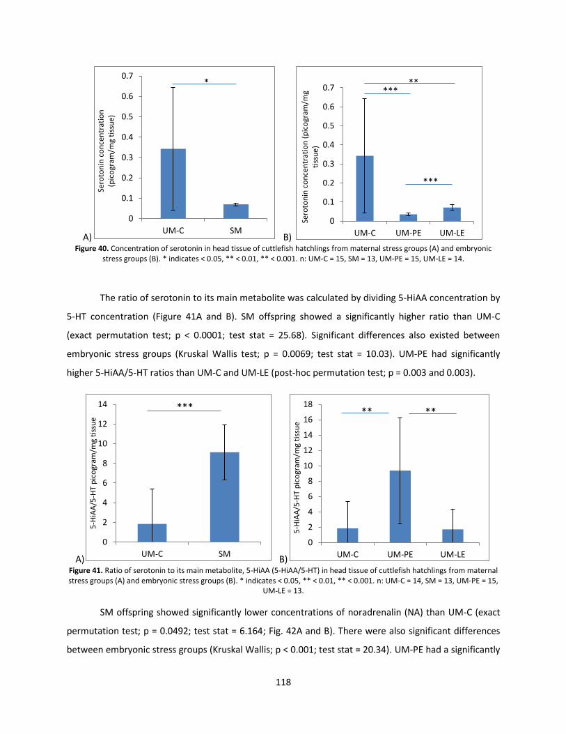
118
A) B) Figure 40. Concentration of serotonin in head tissue of cuttlefish hatchlings from maternal stress groups (A) and embryonic
stress groups (B). * indicates < 0.05, ** < 0.01, ** < 0.001. n: UM-C = 15, SM = 13, UM-PE = 15, UM-LE = 14.
The ratio of serotonin to its main metabolite was calculated by dividing 5-HiAA concentration by
5-HT concentration (Figure 41A and B). SM offspring showed a significantly higher ratio than UM-C
(exact permutation test; p < 0.0001; test stat = 25.68). Significant differences also existed between
embryonic stress groups (Kruskal Wallis test; p = 0.0069; test stat = 10.03). UM-PE had significantly
higher 5-HiAA/5-HT ratios than UM-C and UM-LE (post-hoc permutation test; p = 0.003 and 0.003).
A) B)
Figure 41. Ratio of serotonin to its main metabolite, 5-HiAA (5-HiAA/5-HT) in head tissue of cuttlefish hatchlings from maternal stress groups (A) and embryonic stress groups (B). * indicates < 0.05, ** < 0.01, ** < 0.001. n: UM-C = 14, SM = 13, UM-PE = 15,
UM-LE = 13.
SM offspring showed significantly lower concentrations of noradrenalin (NA) than UM-C (exact
permutation test; p = 0.0492; test stat = 6.164; Fig. 42A and B). There were also significant differences
between embryonic stress groups (Kruskal Wallis; p < 0.001; test stat = 20.34). UM-PE had a significantly
0
0.1
0.2
0.3
0.4
0.5
0.6
0.7
UM-C SM
Sero
ton
in c
on
cen
trat
ion
(p
ico
gram
/mg
tiss
ue)
*
0
0.1
0.2
0.3
0.4
0.5
0.6
0.7
UM-C UM-PE UM-LE
Sero
ton
in c
on
cen
trat
ion
(p
ico
gram
/mg
tiss
ue)
***
***
**
0
2
4
6
8
10
12
14
UM-C SM
5-H
iAA
/5-H
T p
ico
gram
/mg
tiss
ue
***
0
2
4
6
8
10
12
14
16
18
UM-C UM-PE UM-LE
5-H
iAA
/5-H
T p
ico
gram
/mg
tiss
ue
** **
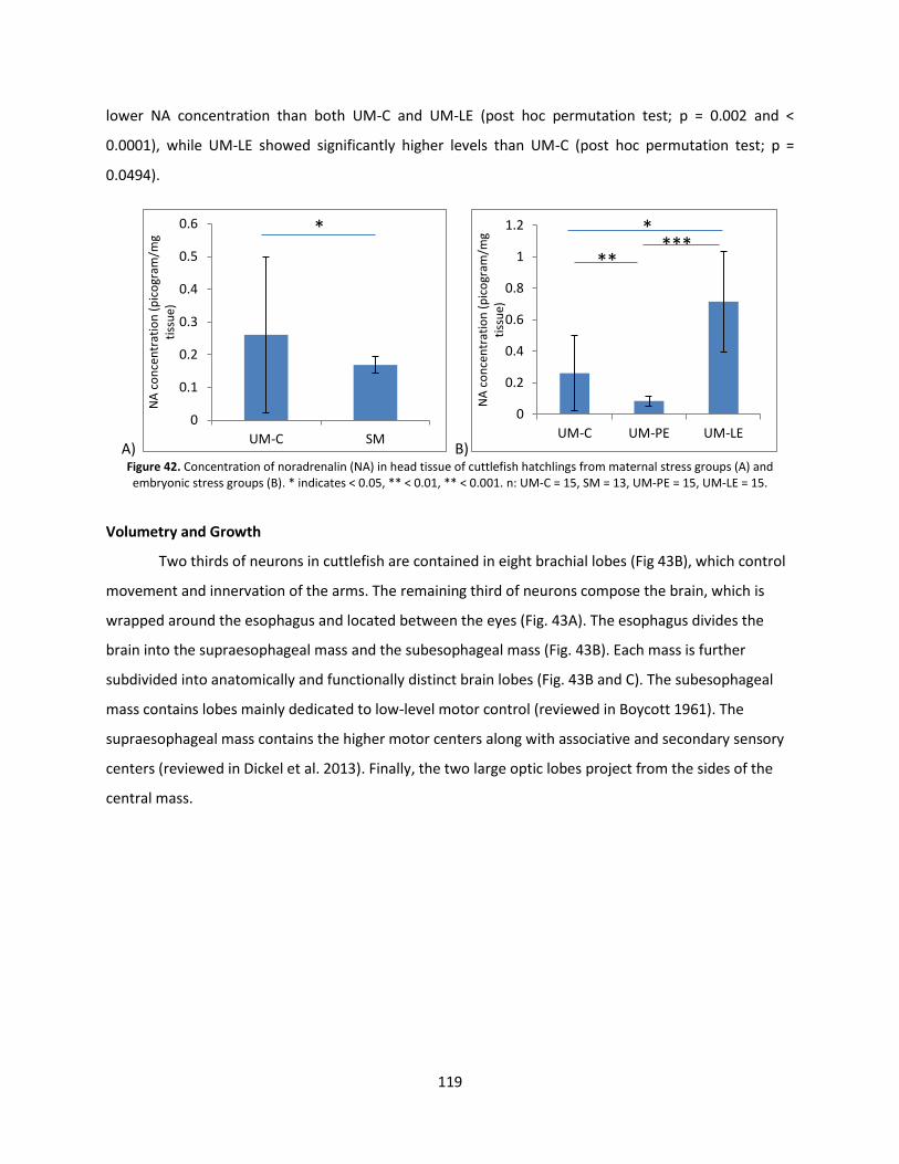
119
lower NA concentration than both UM-C and UM-LE (post hoc permutation test; p = 0.002 and <
0.0001), while UM-LE showed significantly higher levels than UM-C (post hoc permutation test; p =
0.0494).
A) B) Figure 42. Concentration of noradrenalin (NA) in head tissue of cuttlefish hatchlings from maternal stress groups (A) and embryonic stress groups (B). * indicates < 0.05, ** < 0.01, ** < 0.001. n: UM-C = 15, SM = 13, UM-PE = 15, UM-LE = 15.
Volumetry and Growth
Two thirds of neurons in cuttlefish are contained in eight brachial lobes (Fig 43B), which control
movement and innervation of the arms. The remaining third of neurons compose the brain, which is
wrapped around the esophagus and located between the eyes (Fig. 43A). The esophagus divides the
brain into the supraesophageal mass and the subesophageal mass (Fig. 43B). Each mass is further
subdivided into anatomically and functionally distinct brain lobes (Fig. 43B and C). The subesophageal
mass contains lobes mainly dedicated to low-level motor control (reviewed in Boycott 1961). The
supraesophageal mass contains the higher motor centers along with associative and secondary sensory
centers (reviewed in Dickel et al. 2013). Finally, the two large optic lobes project from the sides of the
central mass.
0
0.1
0.2
0.3
0.4
0.5
0.6
UM-C SM
NA
co
nce
ntr
atio
n (
pic
ogr
am/m
g ti
ssu
e)
*
0
0.2
0.4
0.6
0.8
1
1.2
UM-C UM-PE UM-LE
NA
co
nce
ntr
atio
n (
pic
ogr
am/m
g ti
ssu
e)
*
** ***
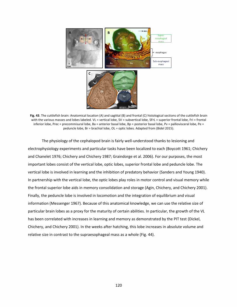
120
Fig. 43. The cuttlefish brain: Anatomical location (A) and sagittal (B) and frontal (C) histological sections of the cuttlefish brain with the various masses and lobes labeled. VL = vertical lobe, SV = subvertical lobe, SFrL = superior frontal lobe, Fri = frontal
inferior lobe, Prec = precommisural lobe, Ba = anterior basal lobe, Bp = posterior basal lobe, Pv = palliovisceral lobe, Pe = peduncle lobe, Br = brachial lobe, OL = optic lobes. Adapted from (Bidel 2015).
The physiology of the cephalopod brain is fairly well-understood thanks to lesioning and
electrophysiology experiments and particular tasks have been localized to each (Boycott 1961; Chichery
and Chanelet 1976; Chichery and Chichery 1987; Graindorge et al. 2006). For our purposes, the most
important lobes consist of the vertical lobe, optic lobes, superior frontal lobe and peduncle lobe. The
vertical lobe is involved in learning and the inhibition of predatory behavior (Sanders and Young 1940).
In partnership with the vertical lobe, the optic lobes play roles in motor control and visual memory while
the frontal superior lobe aids in memory consolidation and storage (Agin, Chichery, and Chichery 2001).
Finally, the peduncle lobe is involved in locomotion and the integration of equilibrium and visual
information (Messenger 1967). Because of this anatomical knowledge, we can use the relative size of
particular brain lobes as a proxy for the maturity of certain abilities. In particular, the growth of the VL
has been correlated with increases in learning and memory as demonstrated by the PIT test (Dickel,
Chichery, and Chichery 2001). In the weeks after hatching, this lobe increases in absolute volume and
relative size in contrast to the supraesophageal mass as a whole (Fig. 44).
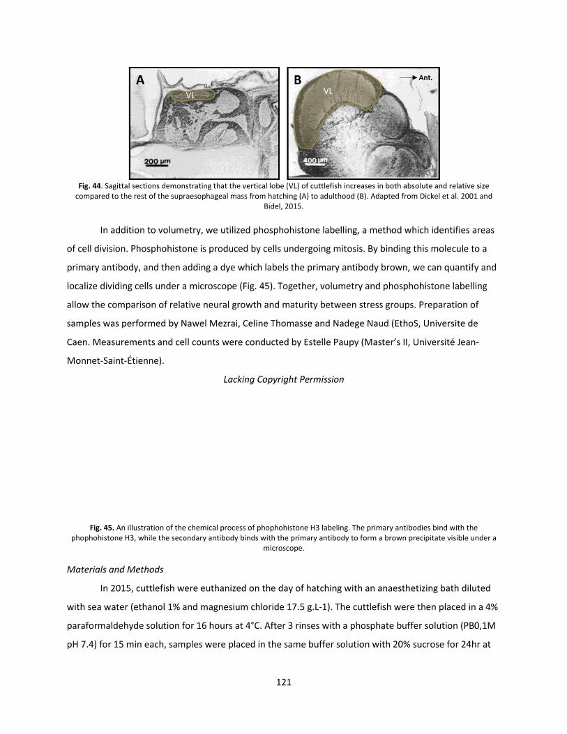
121
Fig. 44. Sagittal sections demonstrating that the vertical lobe (VL) of cuttlefish increases in both absolute and relative size
compared to the rest of the supraesophageal mass from hatching (A) to adulthood (B). Adapted from Dickel et al. 2001 and Bidel, 2015.
In addition to volumetry, we utilized phosphohistone labelling, a method which identifies areas
of cell division. Phosphohistone is produced by cells undergoing mitosis. By binding this molecule to a
primary antibody, and then adding a dye which labels the primary antibody brown, we can quantify and
localize dividing cells under a microscope (Fig. 45). Together, volumetry and phosphohistone labelling
allow the comparison of relative neural growth and maturity between stress groups. Preparation of
samples was performed by Nawel Mezrai, Celine Thomasse and Nadege Naud (EthoS, Universite de
Caen. Measurements and cell counts were conducted by Estelle Paupy (Master’s II, Université Jean-
Monnet-Saint-Étienne).
Lacking Copyright Permission
Fig. 45. An illustration of the chemical process of phophohistone H3 labeling. The primary antibodies bind with the phophohistone H3, while the secondary antibody binds with the primary antibody to form a brown precipitate visible under a
microscope.
Materials and Methods
In 2015, cuttlefish were euthanized on the day of hatching with an anaesthetizing bath diluted
with sea water (ethanol 1% and magnesium chloride 17.5 g.L-1). The cuttlefish were then placed in a 4%
paraformaldehyde solution for 16 hours at 4°C. After 3 rinses with a phosphate buffer solution (PB0,1M
pH 7.4) for 15 min each, samples were placed in the same buffer solution with 20% sucrose for 24hr at
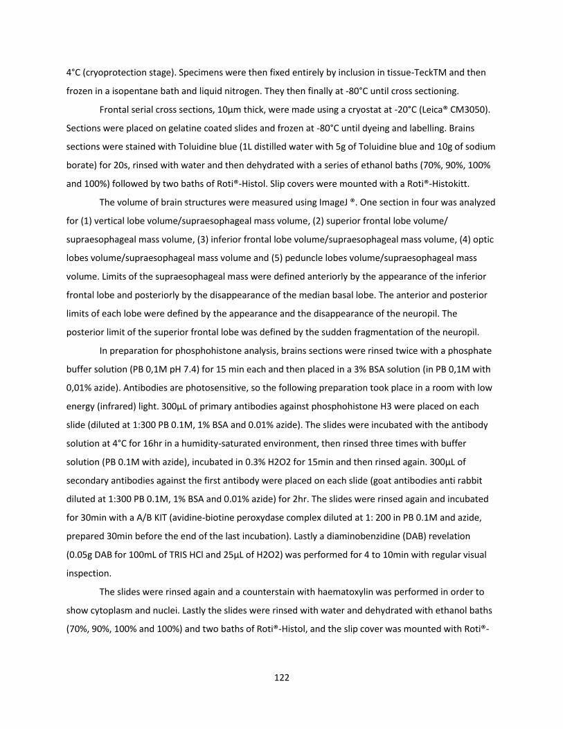
122
4°C (cryoprotection stage). Specimens were then fixed entirely by inclusion in tissue-TeckTM and then
frozen in a isopentane bath and liquid nitrogen. They then finally at -80°C until cross sectioning.
Frontal serial cross sections, 10μm thick, were made using a cryostat at -20°C (Leica® CM3050).
Sections were placed on gelatine coated slides and frozen at -80°C until dyeing and labelling. Brains
sections were stained with Toluidine blue (1L distilled water with 5g of Toluidine blue and 10g of sodium
borate) for 20s, rinsed with water and then dehydrated with a series of ethanol baths (70%, 90%, 100%
and 100%) followed by two baths of Roti®-Histol. Slip covers were mounted with a Roti®-Histokitt.
The volume of brain structures were measured using ImageJ ®. One section in four was analyzed
for (1) vertical lobe volume/supraesophageal mass volume, (2) superior frontal lobe volume/
supraesophageal mass volume, (3) inferior frontal lobe volume/supraesophageal mass volume, (4) optic
lobes volume/supraesophageal mass volume and (5) peduncle lobes volume/supraesophageal mass
volume. Limits of the supraesophageal mass were defined anteriorly by the appearance of the inferior
frontal lobe and posteriorly by the disappearance of the median basal lobe. The anterior and posterior
limits of each lobe were defined by the appearance and the disappearance of the neuropil. The
posterior limit of the superior frontal lobe was defined by the sudden fragmentation of the neuropil.
In preparation for phosphohistone analysis, brains sections were rinsed twice with a phosphate
buffer solution (PB 0,1M pH 7.4) for 15 min each and then placed in a 3% BSA solution (in PB 0,1M with
0,01% azide). Antibodies are photosensitive, so the following preparation took place in a room with low
energy (infrared) light. 300μL of primary antibodies against phosphohistone H3 were placed on each
slide (diluted at 1:300 PB 0.1M, 1% BSA and 0.01% azide). The slides were incubated with the antibody
solution at 4°C for 16hr in a humidity-saturated environment, then rinsed three times with buffer
solution (PB 0.1M with azide), incubated in 0.3% H2O2 for 15min and then rinsed again. 300μL of
secondary antibodies against the first antibody were placed on each slide (goat antibodies anti rabbit
diluted at 1:300 PB 0.1M, 1% BSA and 0.01% azide) for 2hr. The slides were rinsed again and incubated
for 30min with a A/B KIT (avidine-biotine peroxydase complex diluted at 1: 200 in PB 0.1M and azide,
prepared 30min before the end of the last incubation). Lastly a diaminobenzidine (DAB) revelation
(0.05g DAB for 100mL of TRIS HCl and 25μL of H2O2) was performed for 4 to 10min with regular visual
inspection.
The slides were rinsed again and a counterstain with haematoxylin was performed in order to
show cytoplasm and nuclei. Lastly the slides were rinsed with water and dehydrated with ethanol baths
(70%, 90%, 100% and 100%) and two baths of Roti®-Histol, and the slip cover was mounted with Roti®-
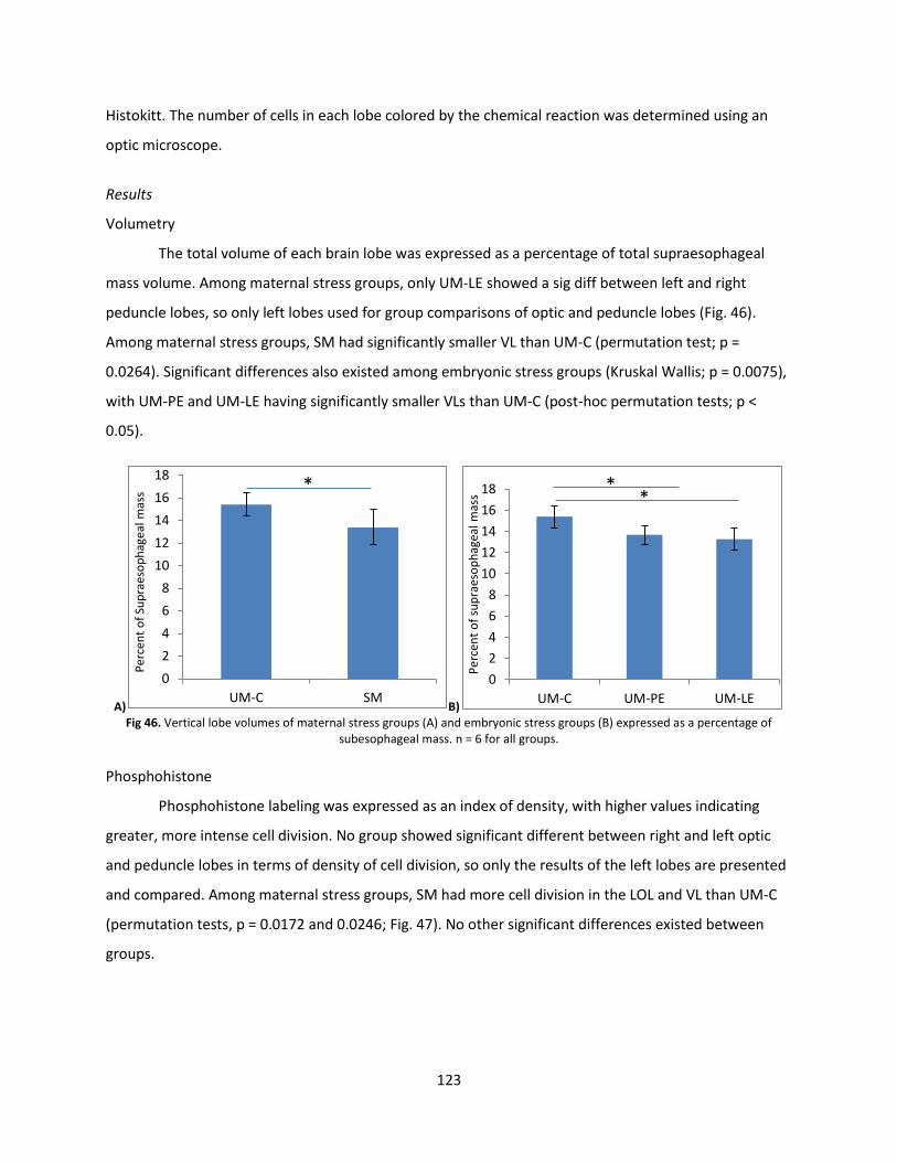
123
Histokitt. The number of cells in each lobe colored by the chemical reaction was determined using an
optic microscope.
Results
Volumetry
The total volume of each brain lobe was expressed as a percentage of total supraesophageal
mass volume. Among maternal stress groups, only UM-LE showed a sig diff between left and right
peduncle lobes, so only left lobes used for group comparisons of optic and peduncle lobes (Fig. 46).
Among maternal stress groups, SM had significantly smaller VL than UM-C (permutation test; p =
0.0264). Significant differences also existed among embryonic stress groups (Kruskal Wallis; p = 0.0075),
with UM-PE and UM-LE having significantly smaller VLs than UM-C (post-hoc permutation tests; p <
0.05).
A) B) Fig 46. Vertical lobe volumes of maternal stress groups (A) and embryonic stress groups (B) expressed as a percentage of
subesophageal mass. n = 6 for all groups.
Phosphohistone
Phosphohistone labeling was expressed as an index of density, with higher values indicating
greater, more intense cell division. No group showed significant different between right and left optic
and peduncle lobes in terms of density of cell division, so only the results of the left lobes are presented
and compared. Among maternal stress groups, SM had more cell division in the LOL and VL than UM-C
(permutation tests, p = 0.0172 and 0.0246; Fig. 47). No other significant differences existed between
groups.
0
2
4
6
8
10
12
14
16
18
UM-C SM
Per
cen
t o
f Su
pra
eso
ph
agea
l mas
s *
0
2
4
6
8
10
12
14
16
18
UM-C UM-PE UM-LE
Per
cen
t o
f su
pra
eso
ph
agea
l mas
s *
*
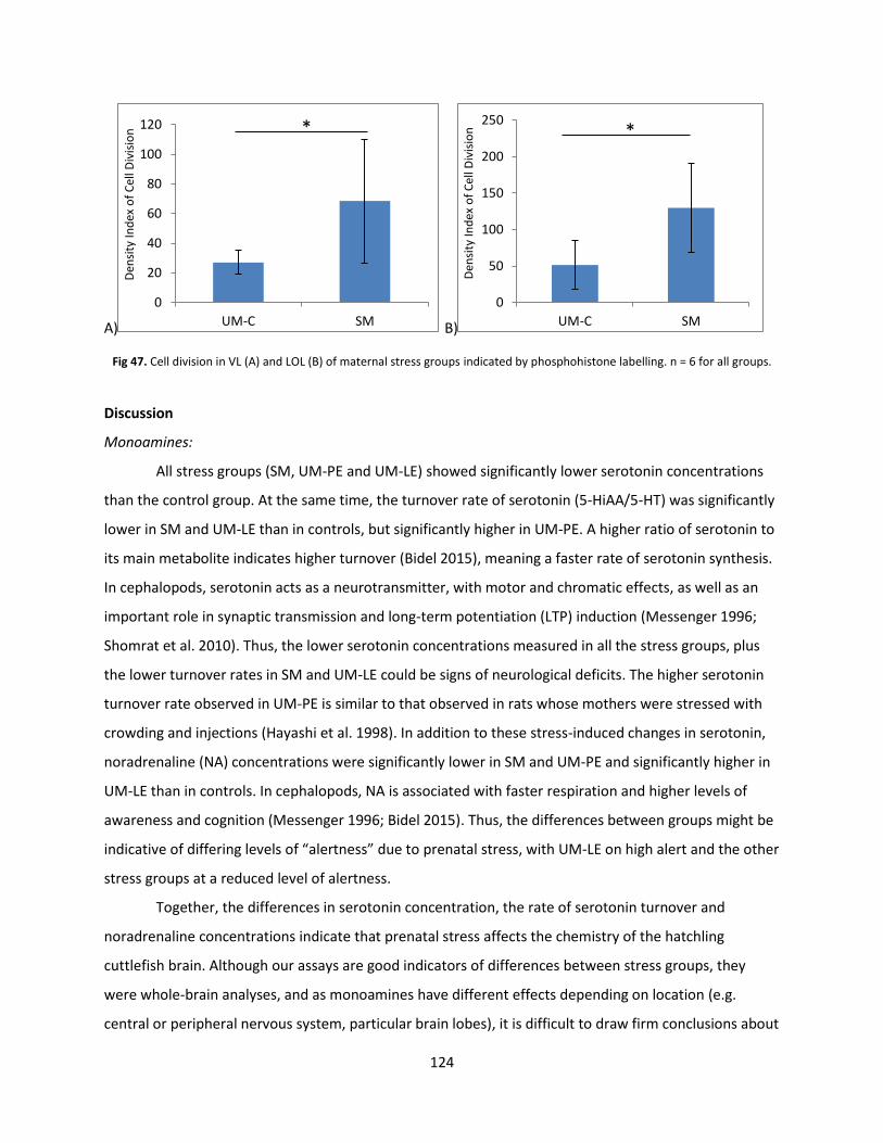
124
A) B)
Fig 47. Cell division in VL (A) and LOL (B) of maternal stress groups indicated by phosphohistone labelling. n = 6 for all groups.
Discussion
Monoamines:
All stress groups (SM, UM-PE and UM-LE) showed significantly lower serotonin concentrations
than the control group. At the same time, the turnover rate of serotonin (5-HiAA/5-HT) was significantly
lower in SM and UM-LE than in controls, but significantly higher in UM-PE. A higher ratio of serotonin to
its main metabolite indicates higher turnover (Bidel 2015), meaning a faster rate of serotonin synthesis.
In cephalopods, serotonin acts as a neurotransmitter, with motor and chromatic effects, as well as an
important role in synaptic transmission and long-term potentiation (LTP) induction (Messenger 1996;
Shomrat et al. 2010). Thus, the lower serotonin concentrations measured in all the stress groups, plus
the lower turnover rates in SM and UM-LE could be signs of neurological deficits. The higher serotonin
turnover rate observed in UM-PE is similar to that observed in rats whose mothers were stressed with
crowding and injections (Hayashi et al. 1998). In addition to these stress-induced changes in serotonin,
noradrenaline (NA) concentrations were significantly lower in SM and UM-PE and significantly higher in
UM-LE than in controls. In cephalopods, NA is associated with faster respiration and higher levels of
awareness and cognition (Messenger 1996; Bidel 2015). Thus, the differences between groups might be
indicative of differing levels of “alertness” due to prenatal stress, with UM-LE on high alert and the other
stress groups at a reduced level of alertness.
Together, the differences in serotonin concentration, the rate of serotonin turnover and
noradrenaline concentrations indicate that prenatal stress affects the chemistry of the hatchling
cuttlefish brain. Although our assays are good indicators of differences between stress groups, they
were whole-brain analyses, and as monoamines have different effects depending on location (e.g.
central or peripheral nervous system, particular brain lobes), it is difficult to draw firm conclusions about
0
20
40
60
80
100
120
UM-C SM
Den
sity
Ind
ex o
f C
ell D
ivis
ion
*
0
50
100
150
200
250
UM-C SM
Den
sity
Ind
ex o
f C
ell D
ivis
ion
*
125
how the differences observed here affect the cuttlefish nervous system. However, in the future, more
precise techniques, such as HPLC-ECD (Bidel, Corvaisier, et al. 2016) may allow us to localize monoamine
levels in specific regions of the brain.
Volumetry and Neurogenesis:
Measurement of the various brain lobes in relation to overall brain size revealed significantly
smaller vertical lobes (VL) in all of the stress groups in comparison to the control group. At the same
time, phosphohistone staining showed higher neurogenesis (i.e. growth) in the optic lobes (OL) and VL
of SM. The VL and the OL are seats of learning and memory in cephalopods, and their relative size and
growth is correlated with learning and memory (Dickel, Chichery, and Chichery 2001). The smaller size of
VLs in all stress groups suggests that they are cognitively-compromised at hatching in comparison to the
control group. In addition, the reduction in neurogenesis in the VL and OL of SM suggests that this group
was further neurodevelopmentally-delayed. This would be consistent with numerous studies in rats
which find maternal stress associated with reductions in neurogenesis (e.g. Weinstock et al. 1992;
Lemaire et al. 2000; Van den Hove et al. 2006). It would also be consistent with the reductions in
serotonin and noradrenaline seen in certain stress groups, both of which are associated with mitosis
(Bidel 2015). Oddly though, these results contrast with the overall lack of learning differences in the PIT
test, where we would expect lower performance in animals with smaller VLs. Indeed, the growth of the
VL has been directly correlated with the emergence of predatory pursuit behavior as well as learning
and memory performance in the PIT test (Dickel, Chichery, and Chichery 2001a, 1997a). One potential
explanation for this is that the brain samples were taken at eclosion, and the differences seen then may
have disappeared after several weeks. Whatever the case, the higher neurogenesis in the VL and OL of
SM suggests that stress passed from mother to offspring has a stronger effect on VL size and growth
than embryonic stress.
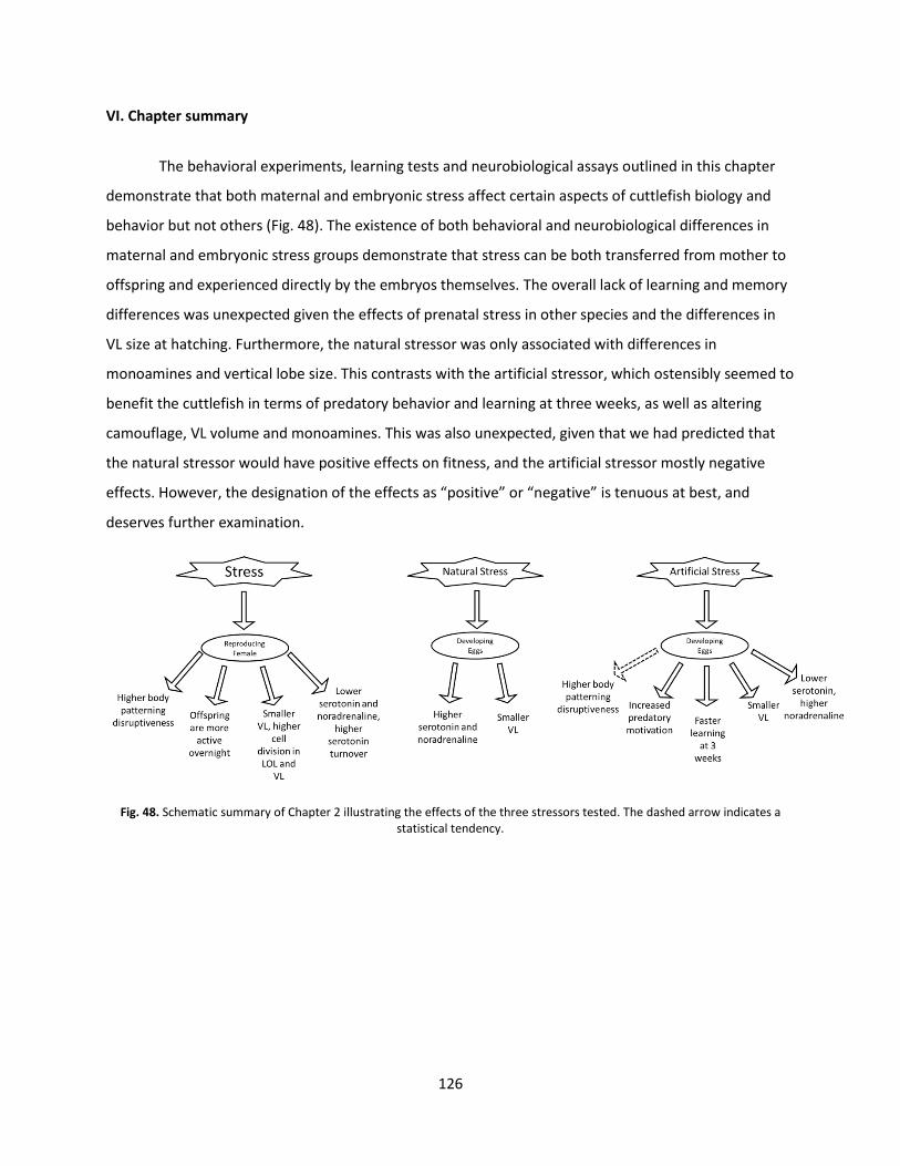
126
VI. Chapter summary
The behavioral experiments, learning tests and neurobiological assays outlined in this chapter
demonstrate that both maternal and embryonic stress affect certain aspects of cuttlefish biology and
behavior but not others (Fig. 48). The existence of both behavioral and neurobiological differences in
maternal and embryonic stress groups demonstrate that stress can be both transferred from mother to
offspring and experienced directly by the embryos themselves. The overall lack of learning and memory
differences was unexpected given the effects of prenatal stress in other species and the differences in
VL size at hatching. Furthermore, the natural stressor was only associated with differences in
monoamines and vertical lobe size. This contrasts with the artificial stressor, which ostensibly seemed to
benefit the cuttlefish in terms of predatory behavior and learning at three weeks, as well as altering
camouflage, VL volume and monoamines. This was also unexpected, given that we had predicted that
the natural stressor would have positive effects on fitness, and the artificial stressor mostly negative
effects. However, the designation of the effects as “positive” or “negative” is tenuous at best, and
deserves further examination.
Fig. 48. Schematic summary of Chapter 2 illustrating the effects of the three stressors tested. The dashed arrow indicates a statistical tendency.

127

128
Chapter 3:
Related
Experiments
129
Chapter 3: Related Experiments
Captivity and/or the lack of environmental enrichment could also be considered as a form of
stress. Dickel showed that hatchling rearing environment affects learning and growth (Dickel, Boal, and
Budelmann 2000). Likewise, prenatal rearing environment might have similar effects.
130
I. Article #5: “The effect of an artificial incubation environment on hatchling size and behavior in the cuttlefish, Sepia officinalis”
This manuscript outlines the results of an experiment which investigated whether embryonic
development in the lab results in offspring that differ from those that develop in the wild.
THE EFFECT OF AN ARTIFICIAL INCUBATION ENVIRONMENT ON HATCHLING SIZE AND BEHAVIOR IN THE CUTTLEFISH, SEPIA OFFICINALIS
CAITLIN E. O’BRIEN1, MARIANNE BOWIE2, PAULINE BILLARD1, ANNE-SOPHIE DARMAILLACQ1, CHRISTELLE JOZET-ALVES1, DAVID BENHAÏM3, OLIVIER BASUYAUX4, LUDOVIC DICKEL*1
1Groupe Mémoire et Plasticité comportementale (GMPc) - EA 4259, Université de Caen Normandie
2UFR LSHS, Université Paris 13
3Institut National des Sciences et Techniques de la Mer (Cnam-Intechmer)
4Synergie Mer et Littoral, Conseil Départemental de la Manche
Vie et milieu - Life and environment, 2016, 66 (1): 97-105
A great deal is known about development in Sepia officinalis; however, much of this knowledge comes from animals incubated in laboratory conditions. Since cuttlefish are behaviorally plastic and known as embryos to perceive environmental stimuli from within the egg, we wondered if they are affected by incubation environment and thus whether laboratory-incubated cuttlefish exhibit natural behavior. We investigated the effects of incubation environment on hatchling size, defense and predation behavior in Sepia officinalis by comparing artificially-incubated hatchlings to naturally-incubated ones. Contrary to our expectations, no significant differences were apparent in hatchling size, disruptive body patterning and predation. A significant difference did exist between groups in one type of body patterning: Artificially-incubated hatchlings appeared to be better at producing a uniform body pattern than naturally-incubated individuals, possibly due to their prenatal experience with a homogeneous artificial substrate. This difference suggests some caution when interpreting experiments utilizing laboratory-incubated hatchlings, but overall, the limited effect of artificial incubation conditions demonstrated in this experiment bolsters confidence in previous behavioral results. These results are also promising for cuttlefish culture; eggs that would otherwise be lost as bycatch could instead be cultured artificially. Keywords: Embryonic experience, behavioral plasticity, body patterning, predation, cuttlefish culture
131
Introduction:
Sepia officinalis (Linnaeus 1758) rely heavily on both body patterning and predation skills for
survival and growth. Cuttlefish skin has both chromatic and textural components that allow it to change
appearance and effect crypsis. Cryptic strategies include background matching (general color
resemblance or variable color resemblance), disruptive coloration and deceptive resemblance (Cott
1940; Hanlon and Messenger 1998). Though a wide range of body patterns are possible, most work has
focused on the two extremes of the spectrum: the uniform pattern, in which homogeneous color and
brightness are present over the entire body, and the disruptive pattern, in which the cuttlefish projects
many small, irregular, disparate pieces in a way that breaks up the outline of the body (Cott 1940;
Boycott 1965). On uniform substrates such as sand or mud, the uniform pattern is generally thought to
be employed in background matching, while the disruptive pattern can be used for both background
matching (variable color resemblance) on a variegated substrate or as a way to occlude body shape.
(However, these patterns may also function in deceptive resemblance, a possibility which will be
explored in the discussion.) Cuttlefish are also voracious hunters of shrimp, using vision to target their
prey (Wells 1958). They have two methods of attack: a rapid tentacular strike for shrimp, fish and small
crabs and a pouncing maneuver for large crabs (Messenger 1968; Duval, Chichery, and Chichery 1984;
Chichery and Chichery 1988). While effective prey capture is essential for obtaining food, foraging and
prey capture can potentially put a cuttlefish at risk of detection by predators. Thus, both body
patterning and predation may be subject to behavioral plasticity.
Of the 30 stages of development currently recognized in cuttlefish embryos (Lemaire 1970),
response to touch and odors is possible by the 23rd (Romagny et al. 2012). Response to visual
information is made possible by the 25th stage via early maturity of the visual system and increased
transparency of the egg membrane due to osmotic swelling (Paulij, Bogaards, and Denucé 1990;
Darmaillacq, Lesimple, and Dickel 2008; Romagny et al. 2012). Thus, during the last weeks of embryonic
development, cuttlefish embryos are able to perceive the outside world, and may learn and modify their
behavior in response. As one example, stage 30 embryos exposed to bright light six times for three
minutes at a time over the course of a few hours decreased their behavioral response (measured by
mantle contraction) over time, demonstrating habituation to the stimulus (Romagny et al. 2012).
Similarly, cuttlefish exposed to a non-preferred prey species (crabs) during the last week of
development showed a preference for this prey item after hatching (Darmaillacq, Lesimple, and Dickel
2008; Mathieu Guibé, Boal, and Dickel 2010). Finally, prenatal cues from potential predators influence
the strength of brain lateralization in cuttlefish hatchlings (Jozet-Alves and Hebert 2012), a trait
132
associated with more efficient information-processing in vertebrates (see Jozet-Alves et al. 2012 for
discussion). These learning abilities and behavioral plasticity have tremendous potential advantages for
young cuttlefish by allowing embryos to adapt to their post-hatching environment before eclosion. For
instance, knowledge of the presence of a predator with a particular predation strategy could allow
cuttlefish to prioritize the development of suitable defense strategies. Indeed, older cuttlefish
discriminate between predators and employ targeted defensive strategies in response to different
species (Langridge 2009).
Young cuttlefish are behaviorally plastic. Previous experiments with hatchlings (up to one week
old) and juveniles (up to 17 weeks) (Hanlon and Messenger 1988) have demonstrated behavioral
plasticity in response to limited enrichment of a laboratory setting with sensory stimuli during the post-
embryonic period. For instance, sensory enrichment of a bare tank with a sandy substrate, rocks, shells,
artificial algae and the presence of conspecifics is associated with faster growth and memory maturation
during the first three months of life (Dickel, Boal, and Budelmann 2000). Likewise, experience with a
sandy substrate soon after hatching results in young that are better at burying in the following days
(Poirier, Chichery, and Dickel 2004). It is also worth noting that the brightness of artificial rearing tanks
has been shown to affect growth, with a dark (black) background associated with the highest growth
(Sykes et al. 2011). Extending the logic of this work to the pre-hatching period, we hypothesized that
experiencing an artificial environment during the prenatal period could affect hatchling behavior as well.
With a few exceptions, knowledge about early behavior in cuttlefish comes from experiments
on animals hatched in the laboratory (e.g. Wells 1958; Messenger 1968; Dickel, Boal, and Budelmann
2000b; Poirier, Chichery, and Dickel 2004; Chiao, Kelman, and Hanlon 2005; Poirier, Chichery, and Dickel
2005; Jozet-Alves and Hebert 2012), a fact not always indicated by the authors. However, little research
attempting to quantify the effects of artificial incubation on behavior in S. officinalis exists. This lack of
knowledge regarding the effects of artificial incubation is problematic for our ability to generalize
conclusions based on the results from artificially-incubated individuals to “natural” behavior. Research
with other species and with Sepia suggest that a (post-embryonic) laboratory situation can profoundly
affect behavioral traits and that behavioral results often diverge between different laboratories due to
variance in environmental parameters and experimenters (Dickel, Boal, and Budelmann 2000b; Chesler
et al. 2002; Poirier, Chichery, and Dickel 2004; Lewejohann et al. 2006; Sykes et al. 2011). This can result
directly from differences in experimental protocol or genetics, but also via an interaction between
genotype and laboratory environment (Chesler et al. 2002).
133
Given the highly-developed brain and behavioral plasticity of cuttlefish (e.g. Dickel, Boal, and
Budelmann 2000b; Poirier, Chichery, and Dickel 2004; Agin et al. 2006), coupled with the embryo’s
ability to perceive, and be influenced by, the world outside the egg (Romagny et al. 2012), it seems likely
that artificial conditions during embryonic development may affect the behavior of hatchling S.
officinalis. If so, it could cause their behavior to differ from animals developed under natural conditions.
In that case, experiments on cuttlefish reared in an artificial environment might yield unrealistic
behavioral results. The natural environment provides numerous sources of sensory stimulation not
present in most laboratory settings. For cuttlefish, these could include natural light, a natural light cycle,
water-borne odors from numerous other organisms, water movement (currents, tidal rhythm, etc.) and
fluctuations in temperature. In contrast, cuttlefish eggs in most artificial situations are exposed to more
stable physical parameters in their environment such as temperature and light, a lack of certain natural
stimuli (including currents and organisms such as epibionts and plankton), but a surfeit of unnatural
ones (including unnatural color schemes and anthropic handling). No study has directly addressed
whether incubation in a standard laboratory setting would have a detectable impact on basic hatchling
behavior.
In addition to corroborating or casting doubt on existing experimental results, this question has
implications for cuttlefish culture. Female S. officinalis lay their eggs on vertical objects on the seabed
including seagrasses and algae (Basuyaux and Legrand 2013), as well as basket traps set by commercial
fishermen to capture adult cuttlefish. Each year, thousands of cuttlefish eggs are laid on such traps in
the English Channel, and when these traps are retrieved, the eggs are discarded, damaged or destroyed
(Blanc and Daguzan 1998). Potentially, these eggs could be saved and cultured artificially. Indeed,
several authors (e.g. Pascual 1978; Forsythe, DeRusha, and Hanlon 1994; Domingues, Sykes, and
Andrade 2002) have already managed to culture multiple generations in the laboratory. This raises the
possibility of redirecting a normally squandered resource for later release, harvest or experimentation.
An assessment of the effects of an artificial incubation environment on some basic behaviors necessary
for growth and survival is a critical first step in the pursuit of this possibility.
Our experiments aimed to evaluate the reliability of existing results obtained with cuttlefish
incubated ex situ and their chances of survival in the first week by quantifying the effect of laboratory
incubation on the subsequent hatchling behavior of this species. In order to assess the effect of an
artificial incubation environment, we tested hatchling size (one measure), body patterning behavior
(three measures) and predatory behavior (five measures) of hatchlings incubated in the sea and in the
lab. These nine measures represent only a small fraction of the behavioral tests possible (other potential
134
assays include prey preference trials, reaction to predator odor and the Prawn-in-a-Tube procedure) but
our goal was to assess behavior directly relevant to hatchling survival. These data will help us to assess
the validity of prior experimental results obtained from hatchlings incubated in the laboratory and add
to our knowledge about cuttlefish culture.
Methods:
Egg Collection and Treatment:
Sepia officinalis eggs (135) were recovered on May 16, 2014 by a professional diver (OB) from a
pre-placed tether at a depth of 3m in the area of Pointe d’Agon (48° 59.547 N - 1° 38.671 W, English
Channel) and taken to the Synergie Mer et Littoral (SMEL), a marine research facility in Blainville-sur-
Mer, France. These eggs were in the very early stages of embryonic development (<15 days old) and
were designated as the artificially-incubated treatment group (“Lab”). At the same time, 150 eggs from
the same location were left in situ and designated as the naturally-incubated treatment group (“Sea”).
“Lab” eggs were placed in a polyethylene basket (29cm x 19cm x 14.5cm) and immersed in an aerated
200L tank matched to local sea surface temperature (14.5°C-18.9°C). Temperature affects the rate of
embryonic growth and yolk consumption, as well as the ultimate size of the hatchling (Boletzky 1983,
Bouchaud 1991). (Though regional surface sea temperature was monitored throughout the experiment,
no local temperature data are available for the eggs left in-situ.) Eggs were exposed to artificial
illumination from 08h00 to 18h00.
As the estimated date of hatching approached (based on the technique of accumulated degree
days from Basuyaux (2011)), “Sea” eggs were monitored by a professional diver before being collected
June 26, 2014 and transported to the laboratory, 7-12 days before the dates of hatching. “Sea” embryos
were estimated (Romagny et al. 2012) to be between stages 26 and 30 when collected. After “Sea” egg
collection, both “Lab” and “Sea” eggs were placed in separate hatching tanks (90liters, 720 x 470 x
360mm, opaque white) with 100% daily water renewal (18.9°C-19.2°C).
Hatching occurs mostly at night (Paulij et al. 1991), and hatchlings were collected at 08h00 each
morning. (The morning after hatching was designated as “Day 1”.) Hatchlings were placed in
individually-labeled vials (diameter=4.5cm, height=6.4cm) with numerous perforations for water flow.
Cuttlefish remained in these floating tubes in their natal treatment tank until testing. Individual identity
was maintained throughout the study. Experiments did not begin on a treatment group until at least
three hatchings had occurred in a single night. All experiments were conducted using water (19.2°C)
135
from hatchlings’ natal tank. In total, 47 individuals from the “Sea” group (hatched July 4-7) and 51 from
the “Lab” group (hatched July 7-10) were tested.
Mantle Length:
Immediately prior to their first encounter with prey, mantle length (ML) was estimated in order
to compare growth between the two incubation conditions. Day 4 was chosen rather than Day 1 as it
reflects the consumption of most or all of the embryonic yolk reserve prior to any prey ingestion
(Boletzky 1983). To estimate cuttlefish ML, two photographs were taken in which the cuttlefish was lying
flat on the bottom and not moving. Using the image analysis software ImageJ, the distance between the
tip of the mantle and a point midway between the eyes was measured. Eyes were used instead of the
mantle edge since they were easily-identifiable in photographs. The two measurements were averaged,
unless they deviated by more than 0.2 cm from each other, in which case a third photograph and
measurement were made. The most disparate of the three values was eliminated and the resulting pair
of values was averaged.
Behavioral Tests:
Uniform Background Test:
Testing:
On the morning after hatching (Day 1), hatchlings were selected in a randomized order for
testing. Between 9h00-10h30, six cuttlefish were placed individually in small (diameter=100cm,
height=1cm) uniform medium gray (Mean Gray Value (MGV)=101+/-3.9) arenas and filmed concurrently
for 22min. This was repeated until all of the day’s hatchlings had been tested. The arenas were lit
indirectly with two Zenitech 0221A 500W lights (540lux) mounted on tripods and filmed with a
Panasonic HDC-SEM60 camera. After filming, cuttlefish were returned to their individually-labeled tubes
in their natal tanks.
Video Analysis:
Using VLC Media Player, two snapshots were captured of each individual at 11 and 21min,
allowing the cuttlefish time to habituate to the experimental arena. Snapshots were only taken during
moments after cuttlefish had settled and were motionless. Following the method developed by (Di Poi
et al. 2014a), we used ImageJ to select the outline of the cuttlefish mantle and measure the
Heterogeneity Index (HI). This value was calculated in ImageJ by an equation using the deviation of the
136
MGV of each individual pixel (x) from the MGV of the whole cuttlefish (�̅�), and the total number of pixels
selected (N): HI = √1
𝑁∑(𝑥 − �̅�)2 (Di Poi et al. 2014a). This value measures body pattern uniformity
(higher HI=less uniform). The two values were averaged for each individual.
We also used ImageJ to compare the MGV of the cuttlefish to the MGV of the substrate. After
selecting the outline of and measuring the MGV of the cuttlefish mantle, we then measured an
equivalently-sized portion of the adjacent substrate. By dividing the mantle MGV by the substrate MGV,
we were able to calculate a ratio from 0 to 1 expressing the degree of match between the mantle and
the substrate (0=no match, 1=perfect match). The two values were averaged for each individual and
group medians calculated. A low HI and high MGV match indicate a good match between mantle
appearance and the surrounding substrate.
Disruptive Background Test:
Testing:
Using the same set-up and procedure as for the Uniform Background test, cuttlefish were filmed
individually on Day 2 for 25min against a “checkered” background (squares=3x3mm) between 10h30
and 12h00. After filming, cuttlefish were returned to their individually-labeled tubes in their natal tanks.
Video Analysis:
Using VLC Media Player, two snapshots were captured of each individual at 15 and 22min. In order to
assess disruptiveness, we employed the procedure developed by Barbosa et al. (2007), and graded 11
components of body pattern on a scale of 0-3 based on relative strength of expression. This resulted in a
“Disruptive Score” that ranged from 0 to 33 (higher score=more disruptive). The two values were
averaged for each individual.
Initial Prey Encounter:
Shrimp Collection:
Shrimp (Crangon crangon and Palaemonetes sp.) were collected as needed in the vicinity of
Blainville-sur-Mer via hand nets in small pools during low tide. They were maintained in aquaria with
well-oxygenated water for one to five days prior to testing. Only shrimp between 0.7 cm and 1.4 cm
were selected for predation experiments. One to four hours before testing, shrimp were collected from
the larger aquaria and placed in smaller individual containers until the moment of testing.
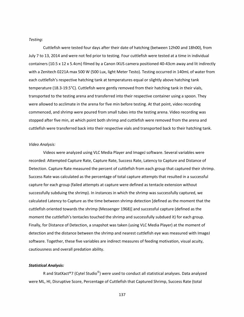
137
Testing:
Cuttlefish were tested four days after their date of hatching (between 12h00 and 18h00), from
July 7 to 13, 2014 and were not fed prior to testing. Four cuttlefish were tested at a time in individual
containers (10.5 x 12 x 5.4cm) filmed by a Canon IXUS camera positioned 40-43cm away and lit indirectly
with a Zenitech 0221A max 500 W (500 Lux, light Meter Testo). Testing occurred in 140mL of water from
each cuttlefish’s respective hatching tank at temperatures equal or slightly above hatching tank
temperature (18.3-19.5°C). Cuttlefish were gently removed from their hatching tank in their vials,
transported to the testing arena and transferred into their respective container using a spoon. They
were allowed to acclimate in the arena for five min before testing. At that point, video recording
commenced, and shrimp were poured from small tubes into the testing arena. Video recording was
stopped after five min, at which point both shrimp and cuttlefish were removed from the arena and
cuttlefish were transferred back into their respective vials and transported back to their hatching tank.
Video Analysis:
Videos were analyzed using VLC Media Player and ImageJ software. Several variables were
recorded: Attempted Capture Rate, Capture Rate, Success Rate, Latency to Capture and Distance of
Detection. Capture Rate measured the percent of cuttlefish from each group that captured their shrimp.
Success Rate was calculated as the percentage of total capture attempts that resulted in a successful
capture for each group (failed attempts at capture were defined as tentacle extension without
successfully subduing the shrimp). In instances in which the shrimp was successfully captured, we
calculated Latency to Capture as the time between shrimp detection [defined as the moment that the
cuttlefish oriented towards the shrimp (Messenger 1968)] and successful capture (defined as the
moment the cuttlefish’s tentacles touched the shrimp and successfully subdued it) for each group.
Finally, for Distance of Detection, a snapshot was taken (using VLC Media Player) at the moment of
detection and the distance between the shrimp and nearest cuttlefish eye was measured with ImageJ
software. Together, these five variables are indirect measures of feeding motivation, visual acuity,
cautiousness and overall predation ability.
Statistical Analysis:
R and StatXact®7 (Cytel Studio©) were used to conduct all statistical analyses. Data analyzed
were ML, HI, Disruptive Score, Percentage of Cuttlefish that Captured Shrimp, Success Rate (total

138
number successful captures/total number of attempted captures), Latency to Capture and Distance of
Detection. The ML met parametric assumptions (Shapiro Wilk, α=0.05, p>0.05), so data are reported as
means +/- SEM and a T-test was used to compare the two groups. All other data failed to meet
parametric assumptions (Shapiro Wilk, α=0.05, p<.0001), so data are reported as medians +/- SEM and
non-parametric Fisher Exact tests and permutation tests were employed.
Ethical Considerations:
All procedures were carried out in accordance with the Directive of the European Parliament
and of the Council of the European Union (2010/63/UE) regarding the care and use of animals for
experimental procedures, and approved by the regional ethical committee (Comité d'Ethique NOrmandie
en Matière d'EXpérimentation Animale, CENOMEXA; agreement number 54) (cuttlefish: project
authorization on September 25, 2014). Experiments were supervised by several individuals (ASD, CJA, LD
and OB) certified to work with cephalopods.
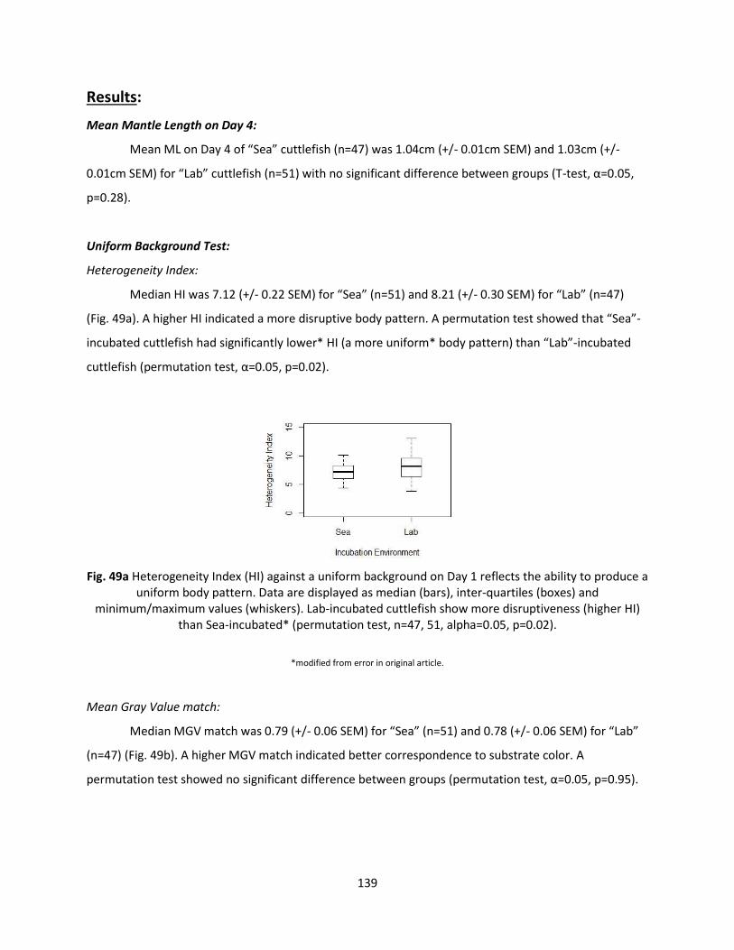
139
Results:
Mean Mantle Length on Day 4:
Mean ML on Day 4 of “Sea” cuttlefish (n=47) was 1.04cm (+/- 0.01cm SEM) and 1.03cm (+/-
0.01cm SEM) for “Lab” cuttlefish (n=51) with no significant difference between groups (T-test, α=0.05,
p=0.28).
Uniform Background Test:
Heterogeneity Index:
Median HI was 7.12 (+/- 0.22 SEM) for “Sea” (n=51) and 8.21 (+/- 0.30 SEM) for “Lab” (n=47)
(Fig. 49a). A higher HI indicated a more disruptive body pattern. A permutation test showed that “Sea”-
incubated cuttlefish had significantly lower* HI (a more uniform* body pattern) than “Lab”-incubated
cuttlefish (permutation test, α=0.05, p=0.02).
Fig. 49a Heterogeneity Index (HI) against a uniform background on Day 1 reflects the ability to produce a
uniform body pattern. Data are displayed as median (bars), inter-quartiles (boxes) and minimum/maximum values (whiskers). Lab-incubated cuttlefish show more disruptiveness (higher HI)
than Sea-incubated* (permutation test, n=47, 51, alpha=0.05, p=0.02).
*modified from error in original article.
Mean Gray Value match:
Median MGV match was 0.79 (+/- 0.06 SEM) for “Sea” (n=51) and 0.78 (+/- 0.06 SEM) for “Lab”
(n=47) (Fig. 49b). A higher MGV match indicated better correspondence to substrate color. A
permutation test showed no significant difference between groups (permutation test, α=0.05, p=0.95).
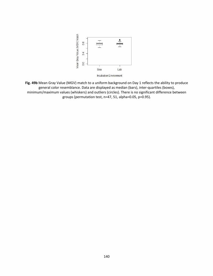
140
Fig. 49b Mean Gray Value (MGV) match to a uniform background on Day 1 reflects the ability to produce
general color resemblance. Data are displayed as median (bars), inter-quartiles (boxes), minimum/maximum values (whiskers) and outliers (circles). There is no significant difference between
groups (permutation test, n=47, 51, alpha=0.05, p=0.95).
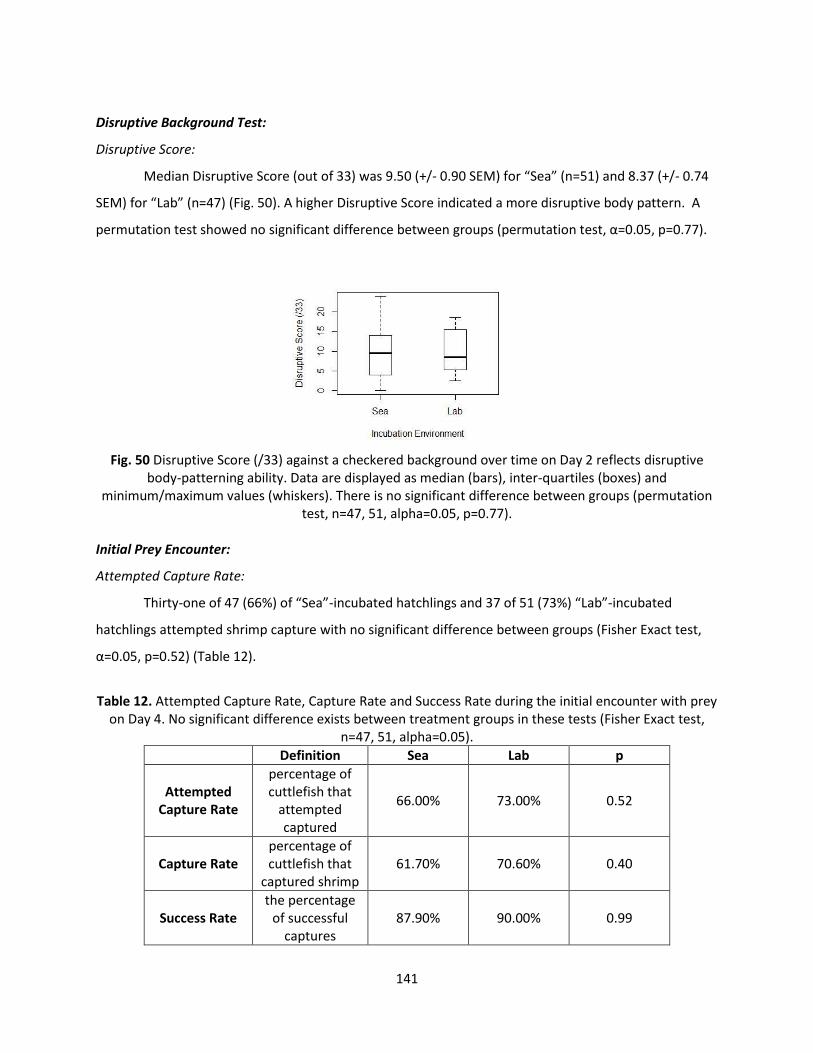
141
Disruptive Background Test:
Disruptive Score:
Median Disruptive Score (out of 33) was 9.50 (+/- 0.90 SEM) for “Sea” (n=51) and 8.37 (+/- 0.74
SEM) for “Lab” (n=47) (Fig. 50). A higher Disruptive Score indicated a more disruptive body pattern. A
permutation test showed no significant difference between groups (permutation test, α=0.05, p=0.77).
Fig. 50 Disruptive Score (/33) against a checkered background over time on Day 2 reflects disruptive
body-patterning ability. Data are displayed as median (bars), inter-quartiles (boxes) and minimum/maximum values (whiskers). There is no significant difference between groups (permutation
test, n=47, 51, alpha=0.05, p=0.77).
Initial Prey Encounter:
Attempted Capture Rate:
Thirty-one of 47 (66%) of “Sea”-incubated hatchlings and 37 of 51 (73%) “Lab”-incubated
hatchlings attempted shrimp capture with no significant difference between groups (Fisher Exact test,
α=0.05, p=0.52) (Table 12).
Table 12. Attempted Capture Rate, Capture Rate and Success Rate during the initial encounter with prey
on Day 4. No significant difference exists between treatment groups in these tests (Fisher Exact test, n=47, 51, alpha=0.05).
Definition Sea Lab p
Attempted Capture Rate
percentage of cuttlefish that
attempted captured
66.00% 73.00% 0.52
Capture Rate percentage of cuttlefish that
captured shrimp 61.70% 70.60% 0.40
Success Rate the percentage
of successful captures
87.90% 90.00% 0.99
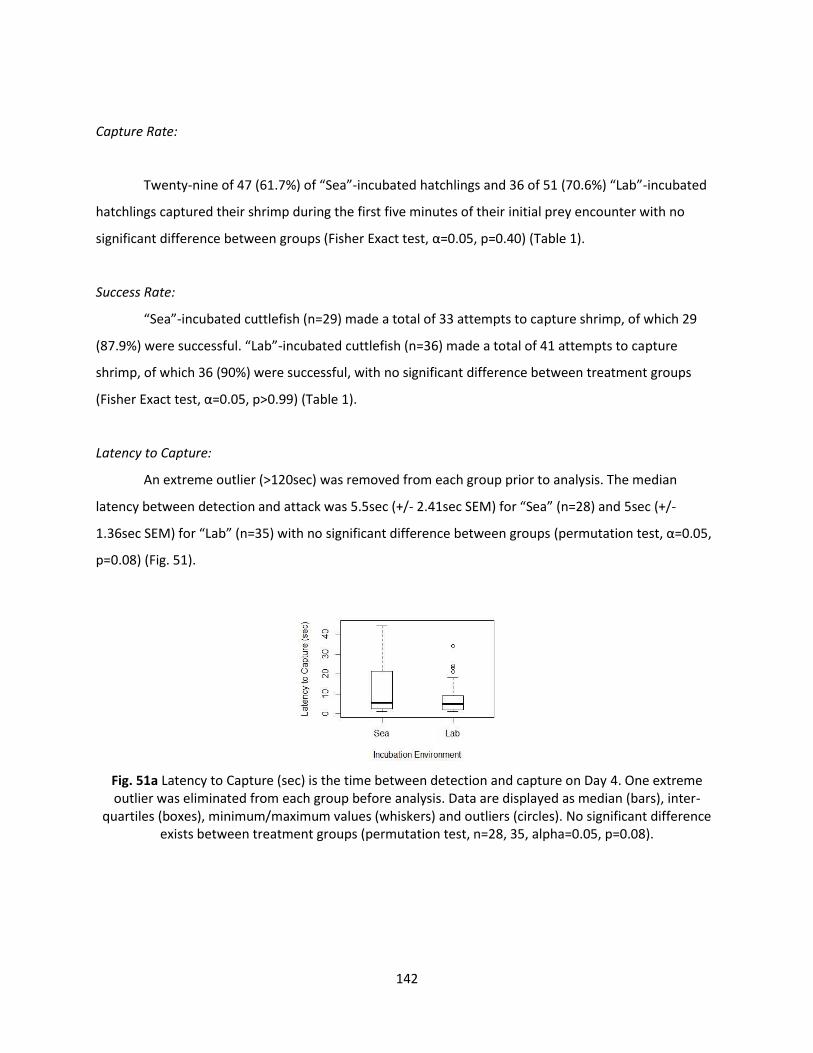
142
Capture Rate:
Twenty-nine of 47 (61.7%) of “Sea”-incubated hatchlings and 36 of 51 (70.6%) “Lab”-incubated
hatchlings captured their shrimp during the first five minutes of their initial prey encounter with no
significant difference between groups (Fisher Exact test, α=0.05, p=0.40) (Table 1).
Success Rate:
“Sea”-incubated cuttlefish (n=29) made a total of 33 attempts to capture shrimp, of which 29
(87.9%) were successful. “Lab”-incubated cuttlefish (n=36) made a total of 41 attempts to capture
shrimp, of which 36 (90%) were successful, with no significant difference between treatment groups
(Fisher Exact test, α=0.05, p>0.99) (Table 1).
Latency to Capture:
An extreme outlier (>120sec) was removed from each group prior to analysis. The median
latency between detection and attack was 5.5sec (+/- 2.41sec SEM) for “Sea” (n=28) and 5sec (+/-
1.36sec SEM) for “Lab” (n=35) with no significant difference between groups (permutation test, α=0.05,
p=0.08) (Fig. 51).
Fig. 51a Latency to Capture (sec) is the time between detection and capture on Day 4. One extreme outlier was eliminated from each group before analysis. Data are displayed as median (bars), inter-
quartiles (boxes), minimum/maximum values (whiskers) and outliers (circles). No significant difference exists between treatment groups (permutation test, n=28, 35, alpha=0.05, p=0.08).
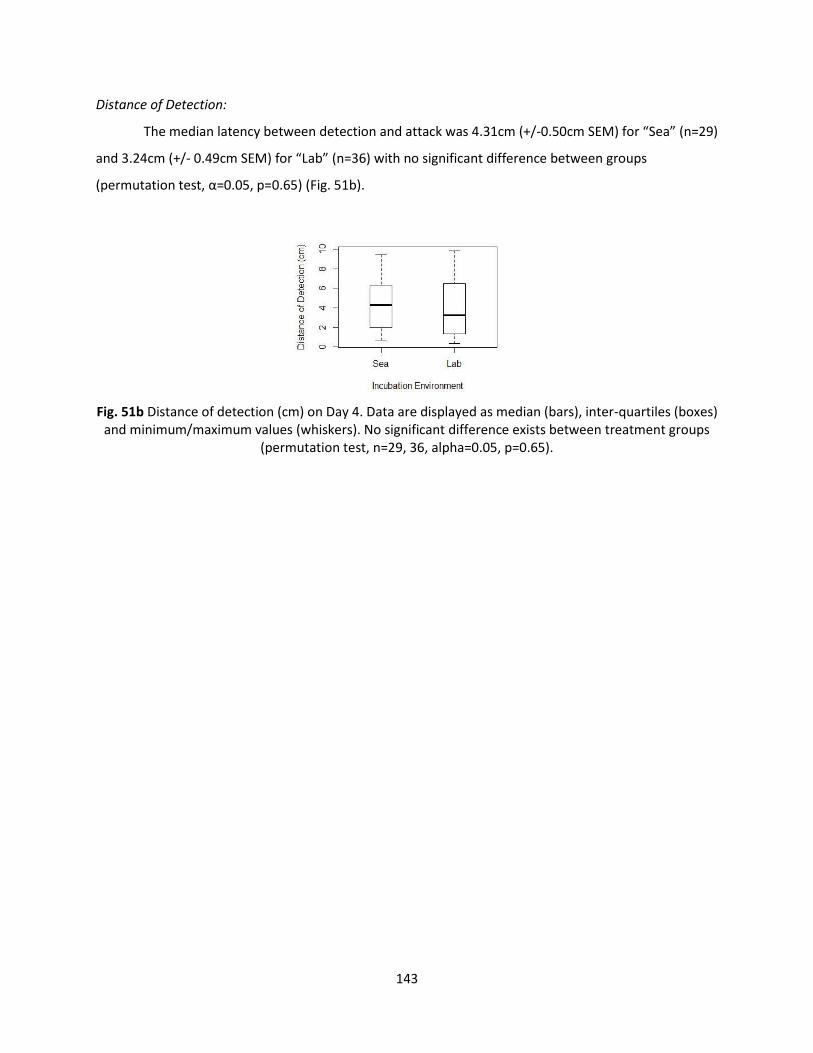
143
Distance of Detection:
The median latency between detection and attack was 4.31cm (+/-0.50cm SEM) for “Sea” (n=29)
and 3.24cm (+/- 0.49cm SEM) for “Lab” (n=36) with no significant difference between groups
(permutation test, α=0.05, p=0.65) (Fig. 51b).
Fig. 51b Distance of detection (cm) on Day 4. Data are displayed as median (bars), inter-quartiles (boxes)
and minimum/maximum values (whiskers). No significant difference exists between treatment groups (permutation test, n=29, 36, alpha=0.05, p=0.65).
144
Discussion:
We investigated the effects that an artificial incubation environment has on subsequent
hatchling size and behavior. Contrary to our expectations, we saw only one difference between “Sea”
and “Lab” groups in nine measures of size, body patterning and predation, suggesting that artificial
incubation conditions had little effect on rates of development and subsequent hatchling behavior.
Mantle length estimated on Day 4 represents embryonic and hatchling growth determined
exclusively by yolk reserves. Water temperature can have dramatic effects on the pace and duration of
embryonic growth, which in turn affects the rate of yolk consumption, feeding motivation and predatory
behavior. Higher temperatures accelerate growth and yolk absorption, resulting in shorter development
time (which can vary as much as 40 to 90 days) but also in smaller hatchlings (Boletzky 1983; Bouchaud
1991). Temperature is also associated with differences in the emergence of hatchling pursuit behavior
(Dickel, Chichery, and Chichery 1997). While the “Lab” cuttlefish in our experiment (and in typical
artificial environments) experienced steady water temperatures, it is likely that the thermal regimes
experienced by the “Sea” group included frequent fluctuations due to currents and tides. Because of this
more complex temperature regime, we had expected that “Sea” hatchlings would differ in size from
“Lab” cuttlefish maintained at the same mean temperature. In contrast, ML was similar between the
two treatment groups, suggesting that the overall thermal mean is more relevant to embryonic growth
than any thermal fluctuations experienced. This possibility deserves further exploration, as no in situ
temperature measurements were made during this experiment, and prior investigations of temperature
and embryo growth have involved steady thermal regimes (e.g. Boletzky 1983; Bouchaud 1991)).
In addition to size, we measured two body patterns. These tests have been used extensively in
the literature (e.g.Chiao, Kelman, and Hanlon 2005; Barbosa et al. 2007; Barbosa et al. 2008; Di Poi et al.
2014a) to measure defensive ability, since body patterning is the primary means of defense in cuttlefish
(Hanlon and Messenger 1998). Heterogeneity Index (HI) measures the overall uniformity of pattern and
a hatchling with low HI is considered to be well-camouflaged on a uniform background. Hatchlings are
notoriously poor at producing uniform body patterns (Hanlon and Messenger 1988; Dickel et al. 2006),
and we hypothesized that animals enriched by the natural environment would be better at this task.
Results show some evidence that this is indeed the case. Heterogeneity Index against a uniform
background ranged between 3.87 and 13.06 and “Lab” hatchlings showed a significantly higher HI (more
disruptive) than “Sea” hatchlings. The fact that “Sea” incubated cuttlefish are slightly better at
producing uniform body patterns is strange given that they likely experienced a heterogeous
145
background during development, while “Lab” cuttlefish experienced an artificially homogeneous
substrate (PVC plastic) during development. This disparity between our expectations and experimental
results deserves further inquiry. Moreover, the potential for incubation environment to influence body
patterning in cuttlefish must be considered when interpreting past and future behavioral results.*
*modified from error in original text.
In contrast, while “Lab” cuttlefish showed stronger uniform pattern-matching in this test, the
same was not true of their ability to match the color of the background. Mean Gray Value match
assessed the correspondence between the overall color of the mantle to that of the immediately
adjacent substrate, and values closer to 1 indicate a high degree of correspondence. Mean Gray Value
match ranged between 0.64 and 0.95, and there was no significant difference between the MGV match
of “Sea” and “Lab”, suggesting that unlike HI, the color-matching abilities are not affected by incubation
environment.
Body patterning against a checkered background was also tested and the overall
“disruptiveness” evaluated. Disruptive Score allowed us to obtain an overall sense of difference in the
disruptive pattern between incubation groups. Disruptive Scores ranged from 8 to 10 (out of 33) and did
not significantly differ between groups. We had expected “Sea” cuttlefish to be more efficient in their
cryptic abilities due to their prenatal exposure to a variegated natural background. In contrast, the lack
of difference between the “Sea”- and “Lab”-incubated hatchlings in Disruptive Score suggests that this
feature of body patterning behavior is not affected by incubation environment.
While we have followed traditional body pattern interpretations and concluded that a less
disruptive (lower HI) and better color match (higher MGV match) on a uniform substrate and that a
higher Disruptive Score on a checkered substrate are most adaptive, it is not entirely clear that this is
the case. Cuttlefish have multiple strategies for creating crypsis (background matching, disruptive
coloration and deceptive resemblance). Any particular body pattern may function in multiple cryptic
strategies and two different patterns may be equally effective in certain circumstances (Hanlon and
Messenger 1988). For instance, a cuttlefish displaying a disruptive body pattern on a uniform
background may not be attempting background matching, but rather deceptive resemblance of stone or
shell fragments. This possibility is supported by the fact that hatchling cuttlefish often display seemingly
“inappropriate” body patterns (i.e. a disruptive body pattern on a uniform substrate) for the first two to
three days (Hanlon and Messenger 1988). Perhaps we have misinterpreted the “ideal” cryptic strategy.
It seems instead that the proper strategy is determined by size and thus changes throughout the lifetime
of an individual (Hanlon and Messenger 1988). Further investigation is necessary to resolve this
146
question, and the interpretation of our camouflage results may change as we learn more about
strategies for different size classes on a variety of substrates.
Finally, we measured five aspects of predatory behavior during hatchlings’ first encounter with
their preferred prey, shrimp (Wells 1958): Attempted Capture Rate, Capture Rate, Success Rate, Latency
to Capture and Distance of Detection. Successful predation is critical at this time, since hatchlings’ yolk
reserves are nearly depleted and they are at their most vulnerable (Wells 1958). While many factors
influence these variables, making it impossible to impute a single cause to each measure, together they
give us an overall sense of motivation and predation efficiency in hatchling cuttlefish and allow us to
identify differences due to incubation environment. We hypothesized that the enrichment present in
the natural incubation environment would stimulate development and result in more adept hatchlings
with higher Capture Rate and visual skills. We also reasoned that hatchlings incubated in the natural
environment would show more cautiousness due to their experience with visual and odor cues from
other organisms and differences in feeding motivation due to their exposure to a more variable
temperature regime. In contrast, none of these variables differed significantly between groups and
Capture Rate was very high (between 85% and 90% in accordance with Messenger 1968). This suggests
that lab-raised hatchlings would be equally capable of feeding themselves during this critical period of
growth.
The literature suggests a high degree of behavioral plasticity in hatchling and juvenile cuttlefish.
In terms of growth, enrichment and environmental factors such as dark tank color promote growth
(Dickel, Boal, and Budelmann 2000; Sykes et al. 2011), while experience with external stimuli such as a
natural substrate or conspecifics alter behavior (Poirier, Chichery, and Dickel 2004; Poirier, Chichery, and
Dickel 2005). Given the cuttlefish’s high plasticity in other areas, the strong evolutionary pressure on the
tested behaviors and the embryo’s ability to perceive beyond the egg, it seemed logical that prenatal
environment would have dramatic effects on size and behavior. This was not supported by our data.
One potential reason for this unexpected outcome is that our experiment focused on hatchlings fewer
than five days old, and it is possible that differences due to incubation environment would manifest
later in development or in some unmeasured aspect of post-predatory growth or behavior. Behavioral
plasticity probably requires a fair amount of brain development, especially of such crucial structures in
the supraesophageal mass as the vertical lobe. These structures are still developing during the first few
months (Dickel, Chichery, and Chichery 1997, 1998; Dickel et al. 2013), so perhaps behavioral plasticity
only develops later. Alternatively, it may simply be that these particular aspects of behavior are highly
pre-programmed and not subject to plasticity.
147
There were some significant limitations to the data we were able to collect during this
experiment. Logistical constraints necessitated that “Sea” eggs be removed from the wild 7 to 12 days
prior to the majority of eclosions (stages 26-30) when embryos were most sensitive to external
stimulation. It is also possible that the laying site from which the eggs were collected was atypically
barren of stimulation and enrichment, which would limit the effect of natural incubation. This could be
addressed in the future by collecting eggs from multiple laying sites and via censuses of the marine life
that typically occurs at these sites. Most importantly, we were only able to investigate a limited range of
behaviors in our experiment. It is possible that plasticity is not manifest in hatchling growth, body
patterning or predation, but is present in other aspects of behavior, such as learning, memory or
activity.
Despite the limitations in experimental design which constrain our conclusions from this
experiment, the lack of strong differences between groups is manifest. The overall lack of difference
between incubation groups bolsters confidence in existing behavioral findings and has practical
implications for cuttlefish conservation. These results, added to the success of several authors (e.g.
Pascual 1978; Forsythe, DeRusha, and Hanlon 1994; Domingues, Sykes, and Andrade 2002) in culturing
multiple generations in the laboratory, suggest that artificial incubation does not alter natural hatchling
behavior. This implies, but in no way guarantees, that the survival, at least of hatchlings, will not be
compromised, although survival was not tested directly in our experiment. In the future, cuttlefish eggs
laid on basket traps normally lost during the harvest of adults could instead be incubated and
repurposed for conservation, research or as an additional source of product. Indeed, larger-scale trials
examining the feasibility of egg and hatchling incubation in artificial tanks are already in progress.
The next obvious step in the characterization of the effects of artificial incubation is to repeat
these experiments with cuttlefish collected from the wild at hatching and compare them to eggs
spawned by captive females. This may allow us to gauge more fully the entire course of embryonic
development as well as the contribution that a laboratory setting can have on the embryo indirectly
through any maternal transmission of hormones (as seen in birds—Groothuis and Schwabl 2008). In
addition, direct observation of eggs and hatchlings in the field would give a clearer picture of the stimuli
experienced by embryos in the wild and the natural behavior of hatchlings. Despite the difficulties, field
observations (via SCUBA, remote sensing or photography) are certainly possible with today’s technology
and would do much to elucidate the effect of prenatal stimulation on subsequent behavior. For
example, measurements of physiological parameters such as turbidity would allow us to estimate the
visual field of cuttlefish embryos and censuses of marine fauna at laying sites would provide an idea of
148
what odor cues might be sensed. Finally, behavioral experiments conducted in the natural environment
would be the ultimate assessment of the effect of incubation environment.
Conclusion:
This experiment provides evidence that incubation in a laboratory environment does not
strongly affect three fundamental aspects of behavior and survival in cuttlefish hatchlings: embryonic
growth, body patterning and predation ability. The one difference identified in uniform body patterning
urges further investigation as well as caution when interpreting results from body pattern experiments
with artificially-incubated hatchlings. (We may also need to reevaluate “conventional” interpretations of
what constitutes the best cryptic strategy in a particular situation.) The overall lack of differences
between naturally- and artificially-incubated hatchlings bolsters confidence in existing experimental
data. Our results are also encouraging from a conservation perspective; artificially-incubated cuttlefish
could augment fishery stocks or replace wild-caught cuttlefish in certain situations. Future work on this
question should strive to test cuttlefish spawned from captive females and collected directly from the
wild at the moment of hatching and to test a broader range of behaviors for the entire juvenile period.
Acknowledgments: This experiment was made possible through funding provided by the Conseil Régional de Basse-Normandie. The authors benefited greatly from the assistance and expertise of Drs. Nadav Shashar, Cécile Bellanger, Flavie Bidel and the employees of the SMEL in conducting this research, and are grateful to the anonymous reviewers and Barbara O’Brien for editorial assistance.

149
II. Chapter Summary
Up until the final two weeks of development, egg incubation environment only affected one
aspect of body patterning behavior and not predation. This was similar to the effect of spawning
environment, except in the direction of body patterning disruptiveness (Fig. 52). This demonstrates that
both the environment in which females lay their eggs and the environment in which those eggs develop
are potential sources of future behavioral variation. Like stress, these effects may be transmitted both
indirectly through the mother or directly experienced by offspring.
Fig. 52. Comparison of the effects of spawning in the natural environment to spawning + incubation in the natural environment.

150

151
General
Discussion
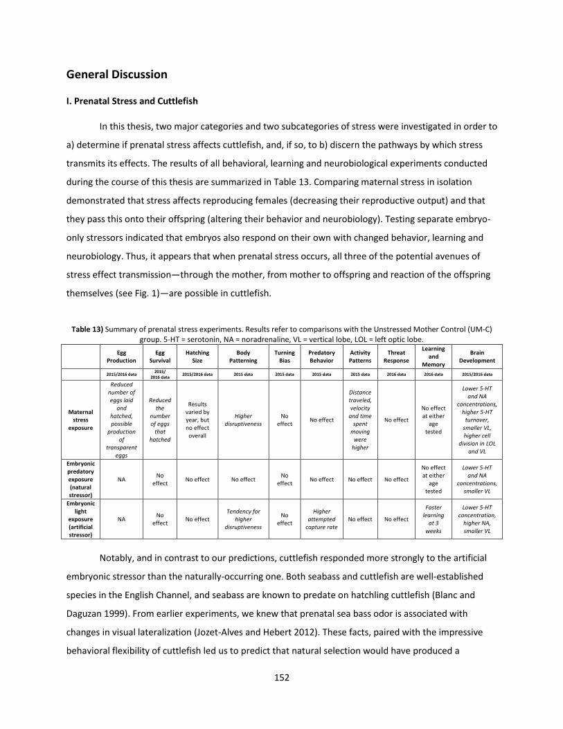
152
General Discussion
I. Prenatal Stress and Cuttlefish
In this thesis, two major categories and two subcategories of stress were investigated in order to
a) determine if prenatal stress affects cuttlefish, and, if so, to b) discern the pathways by which stress
transmits its effects. The results of all behavioral, learning and neurobiological experiments conducted
during the course of this thesis are summarized in Table 13. Comparing maternal stress in isolation
demonstrated that stress affects reproducing females (decreasing their reproductive output) and that
they pass this onto their offspring (altering their behavior and neurobiology). Testing separate embryo-
only stressors indicated that embryos also respond on their own with changed behavior, learning and
neurobiology. Thus, it appears that when prenatal stress occurs, all three of the potential avenues of
stress effect transmission—through the mother, from mother to offspring and reaction of the offspring
themselves (see Fig. 1)—are possible in cuttlefish.
Table 13) Summary of prenatal stress experiments. Results refer to comparisons with the Unstressed Mother Control (UM-C) group. 5-HT = serotonin, NA = noradrenaline, VL = vertical lobe, LOL = left optic lobe.
Egg
Production Egg
Survival Hatching
Size Body
Patterning Turning
Bias Predatory Behavior
Activity Patterns
Threat Response
Learning and
Memory
Brain Development
2015/2016 data 2015/
2016 data 2015/2016 data 2015 data 2015 data 2015 data 2015 data 2016 data 2016 data 2015/2016 data
Maternal
stress exposure
Reduced number of eggs laid
and hatched, possible
production of
transparent eggs
Reduced the
number of eggs
that hatched
Results varied by year, but no effect overall
Higher disruptiveness
No effect
No effect
Distance traveled, velocity
and time spent
moving were
higher
No effect
No effect at either
age tested
Lower 5-HT and NA
concentrations, higher 5-HT
turnover, smaller VL, higher cell
division in LOL and VL
Embryonic predatory exposure (natural stressor)
NA No
effect No effect No effect
No effect
No effect
No effect
No effect
No effect at either
age tested
Lower 5-HT and NA
concentrations, smaller VL
Embryonic light
exposure (artificial stressor)
NA No
effect No effect
Tendency for higher
disruptiveness
No effect
Higher attempted
capture rate No effect No effect
Faster learning
at 3 weeks
Lower 5-HT concentration,
higher NA, smaller VL
Notably, and in contrast to our predictions, cuttlefish responded more strongly to the artificial
embryonic stressor than the naturally-occurring one. Both seabass and cuttlefish are well-established
species in the English Channel, and seabass are known to predate on hatchling cuttlefish (Blanc and
Daguzan 1999). From earlier experiments, we knew that prenatal sea bass odor is associated with
changes in visual lateralization (Jozet-Alves and Hebert 2012). These facts, paired with the impressive
behavioral flexibility of cuttlefish led us to predict that natural selection would have produced a
153
behavioral reaction to this predator. Instead, predator-exposed cuttlefish showed no differences from
the control group in any of our behavioral tests, although they did show changes in brain chemistry and
morphology. The presence of neurobiological changes in offspring from predator-exposed eggs
demonstrates that the cuttlefish embryos are physically affected by prenatal stress, while the lack of any
behavioral reaction suggests habituation to the prenatal predator cues as discussed in Article #4.
Indeed, other species can tailor their innate anti-predator reactions according to the level of threat.
Many gastropods, a sister group to cephalopods, show reactions to predator odors, and these reactions
are stronger when they are paired with alarm cues from conspecifics (e.g. McCarthy and Fisher 2000;
Jacobsen and Stabell 2004; Dalesman et al. 2006). At the same time, we know that cuttlefish habituate
to certain prenatal stimuli (Romagny et al. 2012), so it seems plausible that the predator-odor, paired
with the lack of alarm cue from the numerous other cuttlefish embryos present, did not result in
behavioral changes because the cuttlefish learned to perceive it as non-threatening.
In contrast to the natural stressor, the artificial stressor showed both behavioral and learning
differences, as well as differences in brain chemistry and morphology. Since a mismatch between a
stress response and the stressor itself often leads to negative side effects, we had predicted that this the
artificial stressor (which was not something the species could have ever experienced historically in order
to evolve a reaction via natural selection) would have a “negative” effect on offspring. Arguably though,
the light-exposed group showed “positive” effects in the behaviors tested—better learning abilities and
greater feeding motivation leading to a higher predation rate. One hypothesis to explain this is that
prenatal exposure to an unprecedented stressor actually induced a generalized alarm response that
resulted in acceleration in development. Alternatively, prenatal light exposure is known to affect
circadian rhythm in a range of animals, including cuttlefish, which can hatch earlier or later depending
on the light regime (Paulij et al. 1991). In this case, the light may have accelerated development.
Finally, we were also able to make some indirect comparisons between artificial incubation and
artificial spawning and found similar impacts on the behaviors tested (no effect on predatory behavior,
minor effects on body patterning). This shows that the environment, as well as specific stressors, can
potentially impact the post-natal behavior of offspring, a fact which must be taken into account when
designing and interpreting experiments with young cuttlefish.
154
II. An Ethological Model of Prenatal Stress
The results presented here allow us to begin formulating a rough model of prenatal stress in
cuttlefish from Tinbergen’s four perspectives. In terms of the mechanisms of prenatal stress
transmission (Tinbergen’s first perspective), we hypothesized that female cuttlefish might transfer
corticosterone to offspring in the egg yolk. This has been the subject of some interest among
vertebrates, particularly birds, which have been found to impregnate their egg yolk with stress
hormones that affect the hypothalamic–pituitary–adrenal (HPA) axis of their offspring (e.g. Charil et al.
2010). We failed to find definitive evidence for or against this hypothesis in cuttlefish, but our work does
suggest that other possible avenues of stress transmission from mother to offspring are possible, since
offspring of stressed mother differed from other stress groups nonetheless. Hopefully, as molecular and
neurological analytic techniques become more refined and widely-available, we will be able to pin down
the exact mechanisms that lead to behavioral changes after prenatal stress.
Our experiments had a great deal of insight to shed on Tinbergen’s second and third
perspectives, the ontogenic and the adaptive. From the differences in behavior of the artificial stress
group, we see that, ontogenically, the prenatal period cannot be ignored as a source of behavioral
influence, since stressors experienced by the embryo appear to alter future behavior. Previous
generations of researchers had largely overlooked this time, since it was presumed that embryos could
not perceive the outside world, although this view has been overthrown in more recent decades. Our
results provide further evidence (along with Darmaillacq, Lesimple, and Dickel 2008; Romagny et al.
2012; Jozet-Alves and Hebert 2012) that cuttlefish can perceive the world outside the egg membrane
and alter their future behavior in response. From an adaptive perspective, we found indications that
maternal stress may be a better indicator of future prospects for embryonic cuttlefish than direct
embryonic experience: many more behavioral differences were apparent in association with maternal
stress than with embryonic. On the other hand, one could argue that the responses to the embryonic
stressor were more clearly “adaptive” (better learning and predation), suggesting that direct embryonic
experience is more informative to cuttlefish after all. Clearly, the responses of cuttlefish are complex,
and further experiments are required to understand the interplay of selective forces driving stress-
induced post-natal behavior.
Finally, we were able to address prenatal stress from Tinbergen’s fourth perspective, the
phylogenic, by comparing our results in cuttlefish to the other PReSTO’Cog models. All four models
(chickens, quail, zebrafish and trout) showed effects of maternal stress as well as artificial and natural
embryonic stressors. These results imply that existence of stress responses is highly conserved across

155
phylogenetic groups. Also, it seems that like cuttlefish, the effects of prenatal stress in animals manifest
on a multitude of levels, and are complicated by numerous interactions.
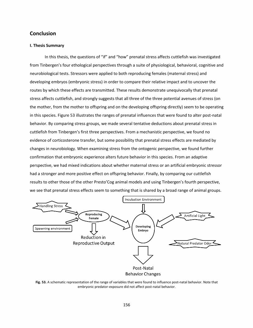
156
Conclusion
I. Thesis Summary
In this thesis, the questions of “if” and “how” prenatal stress affects cuttlefish was investigated
from Tinbergen’s four ethological perspectives through a suite of physiological, behavioral, cognitive and
neurobiological tests. Stressors were applied to both reproducing females (maternal stress) and
developing embryos (embryonic stress) in order to compare their relative impact and to uncover the
routes by which these effects are transmitted. These results demonstrate unequivocally that prenatal
stress affects cuttlefish, and strongly suggests that all three of the three potential avenues of stress (on
the mother, from the mother to offspring and on the developing offspring directly) seem to be operating
in this species. Figure 53 illustrates the ranges of prenatal influences that were found to alter post-natal
behavior. By comparing stress groups, we made several tentative deductions about prenatal stress in
cuttlefish from Tinbergen’s first three perspectives. From a mechanistic perspective, we found no
evidence of corticosterone transfer, but some possibility that prenatal stress effects are mediated by
changes in neurobiology. When examining stress from the ontogenic perspective, we found further
confirmation that embryonic experience alters future behavior in this species. From an adaptive
perspective, we had mixed indications about whether maternal stress or an artificial embryonic stressor
had a stronger and more positive effect on offspring behavior. Finally, by comparing our cuttlefish
results to other those of the other Presto’Cog animal models and using Tinbergen’s fourth perspective,
we see that prenatal stress effects seem to something that is shared by a broad range of animal groups.
Fig. 53. A schematic representation of the range of variables that were found to influence post-natal behavior. Note that
embryonic predator-exposure did not affect post-natal behavior.
157
Further work is necessary to confirm and extend these conclusions. Replicating these
experiments in cuttlefish with other types of prenatal stressors will give further insight into the factors
that are relevant indicators of future conditions to cuttlefish embryos. One particularly intriguing
possibility would be to compare the effects of subjecting reproducing females to the same randomized
artificial light regime as the embryos were exposed to, so that we may directly compare the effects of
maternal transmission versus direct experience. Our comparisons of the effects of spawning
environment to incubation environment (Chapter 3) give some preliminary clues to this question by
showing that both have a similar effect on offspring predatory behavior, but opposing effects on body
patterning disruptiveness. Unfortunately, we did not test the full range of behaviors in the incubation
experiment. Nevertheless, it is apparent that maternal or embryonic environment can exert and
influence on post-natal behavior.
In order to better-understand the mechanisms of stress effect, our lab is currently testing the
responses of embryos directly, while they are still in the egg, by observing changes in mantle
contractions or food preferences (Mezrai, in preparation). This is made possible by certain
experimentally-conducive characteristics of cuttlefish eggs, including the lack of parental care and a
semi-transparent membrane. Identifying and testing other species with similar traits would reveal
whether the trends seen here widespread throughout the animal kingdom or another example of
cephalopod distinctiveness. In particular, it would be especially enlightening to see if maternal stress
transmission and embryonic stress exist in “social” invertebrates like ants and bees, which have a
completely different social structure than most other animal groups.
By studying prenatal stress in animals, ethologists can gain insight that can be used to improve
animal welfare as well as human health and society. A better understanding of prenatal stress across a
broad range of species might lead to improvements in the way we treat pregnant female animals in
agriculture and laboratories. For instance, the existence of prenatal stress effects from handled mother
to offspring in this species and a wide range of others (e.g. salmon, Sigholt et al. 1997; farmed blue
foxes, Braastad 1998; cattle, Grandin, Oldfield, and Boyd 1998) adds further incentive for the
agricultural industry to avoid things like gestation crates for pigs, which may not only decrease animal
welfare but the amount and quality of meat product produced. Likewise, a better understanding of
prenatal stress may lead to ways to mitigate its negative impacts on children and adults, and perhaps to
prevent it from even occurring in the first place. For instance, the children of women who have been
known to have experienced a stressor during pregnancy could be targeted for extra maternal attention
or early-childhood therapy, since parental care has been shown to mitigate some of the negative effects
158
of prenatal stress in rodents (reviewed in Francis and Meaney 1999). This research would also help us
predict what sorts of things may actually be stressful to a fetus. Here, we saw possible habituation in
response to a predator odor that we expected to be very stressful. This suggests that we should be
aware of the possibility that human embryos could habituate to stressors as well. Indeed, human fetuses
are known to habituate to repeated vibriotactile stimuli perceived from within the womb (Leader et al.
1982).
159
II. Article #6: “The Future of Cephalopod Research; Perspectives of Three Early-Career Researchers.”
Given the insight a cephalopod mollusc has provided here, it seems logical that cephalopods
should continue to be used as a model with which to study animal behavior and search for broad
commonalities across phylogenetic distance. The following manuscript outlines my views on the future
of research of this group.
The future of cephalopod science: Perspectives from three early-career researchers
Behavior, Cognition and Neuroscience By Caitlin E. O’Brien; Excerpt from a collaboration with Drs. Katina Roumbedakis and Inger Winkelmann
Introduction:
The charismatic behavior, sophisticated cognition and advanced neurophysiology of
cephalopods is intriguing to scientists and the general public alike. This is reflected in both their
popularity as aquarium specimens (despite the challenges associated with keeping them) and their
prevalence on social media. They, or creatures strongly resembling them, are also often depicted in
works of fiction (e.g. Finding Dory (2016), The Arrival (2016), Life (2017)). Some of their most unique
characteristics have also served as inspiration for many recent developments in technology, such as
adaptive camouflage based on cephalopod skin (Wang et al. 2014; Yu et al. 2014), suction cups for
wound repair (Choi et al. 2016), propulsion and buoyancy systems for Autonomous Underwater Vehicles
(Song et al. 2016) and as models for soft robotics (Renda et al. 2012; Laschi et al. 2012). A number of
other cephalopod features, such as sophisticated “camera” eyes and advanced cognition, have
experienced convergent evolution with vertebrates, allowing cephalopods to serve as a
phylogenetically-distant reference point from which to examine our own evolution. Unfortunately,
despite their great popularity and relevance, only about 8% (60 species) of the 800 or so species known
have been thoroughly described. Here, potential developments in the study of cephalopod behavior,
cognition and neurobiology are discussed, and some recent examples of progress are highlighted.
Behavior:
Our understanding of cephalopod behavior is limited mainly to laboratory observations.
Without the ecological context of the natural environment, we often cannot perceive the true
160
“purpose” of particular behaviors, leading to misinterpretations. In order to improve our understanding
of cephalopod behavior in particular, more field observations and field experiments are necessary. Like
many animals, cephalopods are influenced by and adapt to their surroundings (e.g. Dickel, Boal, and
Budelmann 2000b; Anderson and Wood 2001), constraining or altering the behavioral phenotype. While
there are obvious difficulties to field work, the insight gained will be well-worth the effort. A recent
study by Schnell et al. (2015) is a good illustration of this: via controlled laboratory experiments, the
authors found that the white lateral stripe displayed by female Sepia apama signals non-receptivity for
mating (they are less likely to mate when showing it). However, observations of natural behavior in the
field showed that males largely ignored this and mated anyway. This combination of laboratory tests
with natural observations allowed observers to deduce the intended meaning of an intraspecific signal
but also provide contextual evidence about its relevance and efficacy in actual mating situations.
Fieldwork can not only validate and contextualize laboratory experiments, but will improve overall
animal welfare in experiments by minimizing disturbance and manipulations of animals.
The effort to increase our canon of field data will likely be aided by the general trends of
technological development and decreasing costs of data acquisition tools. Various types of tagging have
been utilized successfully in recent years to answer questions about geographic range, migration and
diving habits (e.g. Bazzino et al. 2010; Liu et al. 2016; Sykes et al. 2017). Remote monitoring through
videography and photography is another increasingly accessible option thanks to the profusion of low-
cost cameras that have come on the market in recent years. Remotely-operated underwater vehicles
(ROVs), AUVs and submersibles have also become more affordable, and they have greatly expanded our
knowledge of deep-sea cephalopod behavior (e.g. Trueblood et al. 2015; Thomas, Robison, and Johnsen
2017), even capturing footage of the elusive giant squid (Robey, 2012). In addition to embracing the
benefits of evolving technology, the community of cephalopod researchers should consider creating a
shared, open access data repository of video footage and data sets. Such a repository would allow
students and researchers lacking funds, facilities or animals to conduct their own experiments and
contribute to the body of knowledge. Other authors (Xavier et al. 2015) have urged a community-wide
shift in focus from data acquisition to data analysis, and open access to shared data would accelerate
this process. Finally, the reuse of raw data and footage would improve welfare by reducing the total
number of animals manipulated (Fiorito et al. 2014).
When we do conduct laboratory work, the cephalopod research community would be wise to
make greater efforts at standardization across experiments. Due to the sensitivity and advanced
perceptive abilities of cephalopods, even minor methodological differences can skew results and lead to
161
inaccurate conclusions. For example, the standard method of measuring learning and memory in
cuttlefish is the “Prawn-in-the-Tube” (PIT) procedure (see Messenger 1973 for methodological
description) which has been used for decades by a number of research groups. While this standardized
method theoretically allows direct comparisons to be made between experiments conducted at
different times, the more recent discovery that cuttlefish and other cephalopods are able to perceive
differences in the polarization of light has led to the realization that the seemingly irrelevant choice of
tube material (e.g. glass versus plastic, each of which alters the properties of light in different ways)
could potentially affect results (Cartron, Darmaillacq, and Dickel 2013). One technique to increase
standardization across experiments and research groups is video playback of standardized behavioral
stimuli (e.g. approach of a predator, prey item or conspecific). Such a method has already been used by
Pronk, Wilson, and Harcourt (2010) to study the reactions of octopus over time and between individuals,
and other labs have developed cephalopod-specific video playback systems (G. Fiorito pers. comm.). If
such video clips were shared to a common video library, experiments could be replicated by different
labs in a standardized fashion using identical audiovisual playback equipment.
In addition to standardizing and replicating within the same species, the cephalopod research
community should also strive to duplicate the same observations and experiments across multiple
species. Having corresponding data on closely related animals allows us to make comparisons and draw
conclusions about the entire lineage by giving us a sense of what behaviors are evolutionary conserved
from earlier shared ancestors and which represent novel adaptations to the particular environment of
that species. In the family Hominidae for example, social differences between such congeners as apes,
chimps and bonobos allow us to assess the factors driving behavioral evolution (e.g. Stanford 1998;
Malone, Fuentes, and White 2012). Similar comparisons between such commonly-studied cephalopod
species as Octopus vulgaris, Loligo vulgaris and S. officinalis would be a good place to start, although the
eventual goal should be to assess behavior across a wide variety of species, including the non-coleoid
cephalopod Nautilus spp., which can serve as an ancestral reference point (R. Crook and Basil 2008).
We would also benefit greatly from the formal investigation of inter-individual differences and
behavioral plasticity in this group. Anecdotal observations by aquarists and researchers give the distinct
impression that individual animals have distinct “personalities.” Indeed, Carere et al. (2015) found that
in S. officinalis, certain behaviors were expressed predictably and consistently over time, although the
expression of other behaviors differed between testing situations. Further research into this subject may
indicate that we need to apply different tactics and interpretations at the population level, such as
distinguishing between “personality types” when calculating group means. Ultimately, we may find that
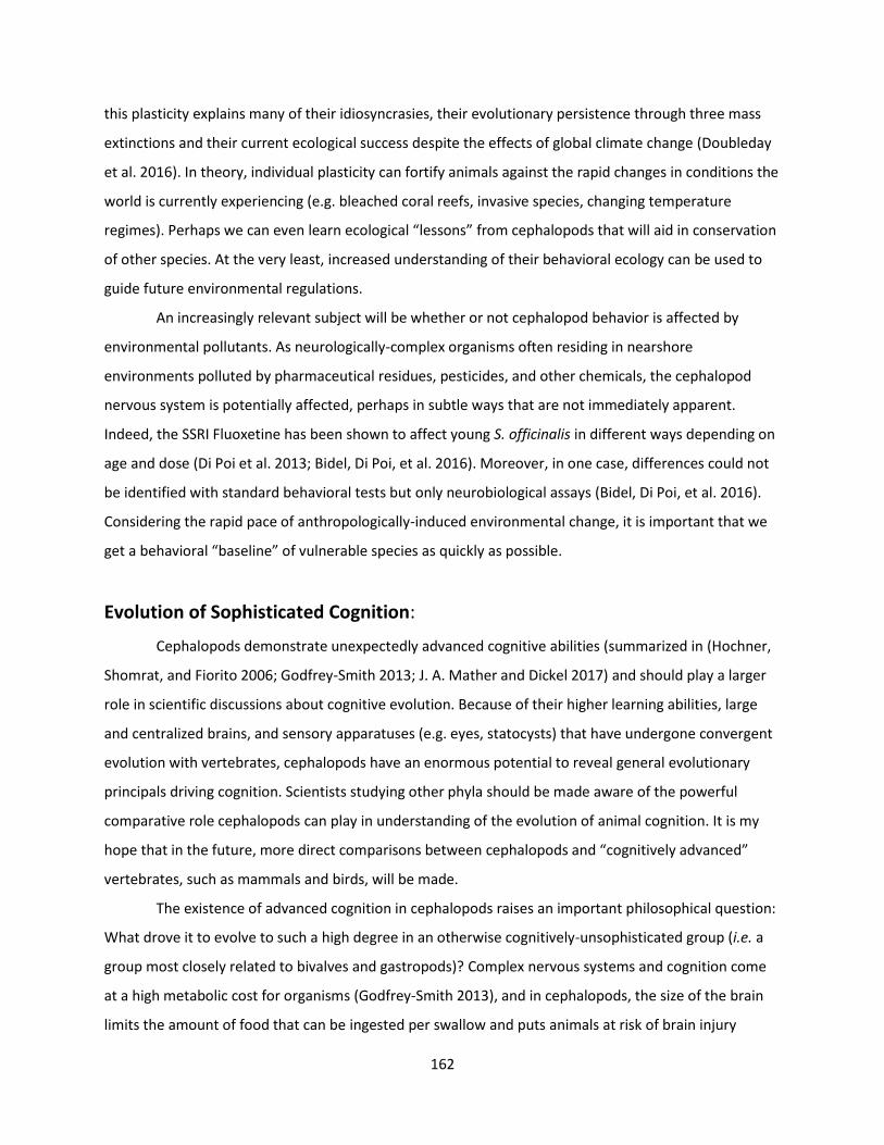
162
this plasticity explains many of their idiosyncrasies, their evolutionary persistence through three mass
extinctions and their current ecological success despite the effects of global climate change (Doubleday
et al. 2016). In theory, individual plasticity can fortify animals against the rapid changes in conditions the
world is currently experiencing (e.g. bleached coral reefs, invasive species, changing temperature
regimes). Perhaps we can even learn ecological “lessons” from cephalopods that will aid in conservation
of other species. At the very least, increased understanding of their behavioral ecology can be used to
guide future environmental regulations.
An increasingly relevant subject will be whether or not cephalopod behavior is affected by
environmental pollutants. As neurologically-complex organisms often residing in nearshore
environments polluted by pharmaceutical residues, pesticides, and other chemicals, the cephalopod
nervous system is potentially affected, perhaps in subtle ways that are not immediately apparent.
Indeed, the SSRI Fluoxetine has been shown to affect young S. officinalis in different ways depending on
age and dose (Di Poi et al. 2013; Bidel, Di Poi, et al. 2016). Moreover, in one case, differences could not
be identified with standard behavioral tests but only neurobiological assays (Bidel, Di Poi, et al. 2016).
Considering the rapid pace of anthropologically-induced environmental change, it is important that we
get a behavioral “baseline” of vulnerable species as quickly as possible.
Evolution of Sophisticated Cognition:
Cephalopods demonstrate unexpectedly advanced cognitive abilities (summarized in (Hochner,
Shomrat, and Fiorito 2006; Godfrey-Smith 2013; J. A. Mather and Dickel 2017) and should play a larger
role in scientific discussions about cognitive evolution. Because of their higher learning abilities, large
and centralized brains, and sensory apparatuses (e.g. eyes, statocysts) that have undergone convergent
evolution with vertebrates, cephalopods have an enormous potential to reveal general evolutionary
principals driving cognition. Scientists studying other phyla should be made aware of the powerful
comparative role cephalopods can play in understanding of the evolution of animal cognition. It is my
hope that in the future, more direct comparisons between cephalopods and “cognitively advanced”
vertebrates, such as mammals and birds, will be made.
The existence of advanced cognition in cephalopods raises an important philosophical question:
What drove it to evolve to such a high degree in an otherwise cognitively-unsophisticated group (i.e. a
group most closely related to bivalves and gastropods)? Complex nervous systems and cognition come
at a high metabolic cost for organisms (Godfrey-Smith 2013), and in cephalopods, the size of the brain
limits the amount of food that can be ingested per swallow and puts animals at risk of brain injury
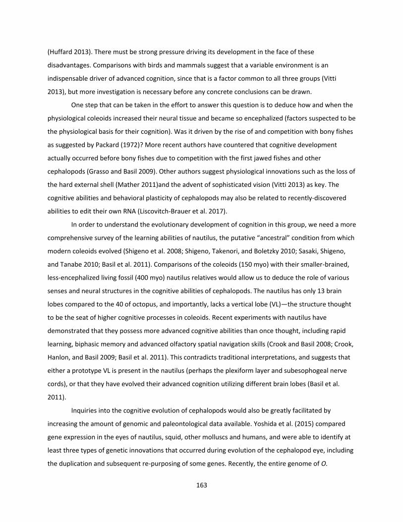
163
(Huffard 2013). There must be strong pressure driving its development in the face of these
disadvantages. Comparisons with birds and mammals suggest that a variable environment is an
indispensable driver of advanced cognition, since that is a factor common to all three groups (Vitti
2013), but more investigation is necessary before any concrete conclusions can be drawn.
One step that can be taken in the effort to answer this question is to deduce how and when the
physiological coleoids increased their neural tissue and became so encephalized (factors suspected to be
the physiological basis for their cognition). Was it driven by the rise of and competition with bony fishes
as suggested by Packard (1972)? More recent authors have countered that cognitive development
actually occurred before bony fishes due to competition with the first jawed fishes and other
cephalopods (Grasso and Basil 2009). Other authors suggest physiological innovations such as the loss of
the hard external shell (Mather 2011)and the advent of sophisticated vision (Vitti 2013) as key. The
cognitive abilities and behavioral plasticity of cephalopods may also be related to recently-discovered
abilities to edit their own RNA (Liscovitch-Brauer et al. 2017).
In order to understand the evolutionary development of cognition in this group, we need a more
comprehensive survey of the learning abilities of nautilus, the putative “ancestral” condition from which
modern coleoids evolved (Shigeno et al. 2008; Shigeno, Takenori, and Boletzky 2010; Sasaki, Shigeno,
and Tanabe 2010; Basil et al. 2011). Comparisons of the coleoids (150 myo) with their smaller-brained,
less-encephalized living fossil (400 myo) nautilus relatives would allow us to deduce the role of various
senses and neural structures in the cognitive abilities of cephalopods. The nautilus has only 13 brain
lobes compared to the 40 of octopus, and importantly, lacks a vertical lobe (VL)—the structure thought
to be the seat of higher cognitive processes in coleoids. Recent experiments with nautilus have
demonstrated that they possess more advanced cognitive abilities than once thought, including rapid
learning, biphasic memory and advanced olfactory spatial navigation skills (Crook and Basil 2008; Crook,
Hanlon, and Basil 2009; Basil et al. 2011). This contradicts traditional interpretations, and suggests that
either a prototype VL is present in the nautilus (perhaps the plexiform layer and subesophogeal nerve
cords), or that they have evolved their advanced cognition utilizing different brain lobes (Basil et al.
2011).
Inquiries into the cognitive evolution of cephalopods would also be greatly facilitated by
increasing the amount of genomic and paleontological data available. Yoshida et al. (2015) compared
gene expression in the eyes of nautilus, squid, other molluscs and humans, and were able to identify at
least three types of genetic innovations that occurred during evolution of the cephalopod eye, including
the duplication and subsequent re-purposing of some genes. Recently, the entire genome of O.
164
bimaculoides was sequenced, and already, examination of these data has shown that unlike other
molluscs, this species has experienced expansion of some of the same gene families involved in
vertebrate neuronal development (Albertin et al. 2015). Another study used data from the same 180
genes across 26 species to test hypotheses about divergence times (Tanner et al. 2017). The complete
sequencing of other species along with open access to these genomes would certainly lead to further
revelations.
While the recent boom in genetic data has led to some neglect of more traditional
paleontological and morphological methods (Xavier et al. 2015), new techniques are being used to
extract more information from existing fossil specimens. For example, Klug et al. (2016) used UV light to
reveal structures not normally visible in a fossilized belemnite (Acanthoteuthis speciosus), including
cranial cartilage, vague imprints of the statocysts and the first-ever evidence of a belemnitid radula.
Though we lack a fossil record for most soft-bodied cephalopods, a few specimens do exist. Recently,
Kruta et al. (2016)were able to reconstruct soft body parts in three dimensions (including the eyes and
some suckers) from a fossilized octopus using synchrotron microtomography. The presence of suckers in
this specimen forces researchers to re-evaluate the advent of this structure, which was thought to be a
more recent development. Other possible tools include isotope analysis of fossil material and X-ray
tomography, a method which allows the internal investigation of fossils and which can reveal preserved
soft tissues. Synthesis of paleontology, traditional phylogenetic methods and “modern” biology
promises to be very fruitful. One recent example is a phylogenetic analysis conducted with a new
morphological dataset gleaned from both extinct and extant forms, which was able to confirm many of
the putative relationships between coleoid groups, but found a few to be para- or polyphyletic (Sutton,
Perales-Raya, and Gilbert 2016).
Study of cephalopods may ultimately demonstrate that cognition is an inevitable emergent
property past a certain level of neural size or centralization. Or it may demonstrate that certain kinds of
structures and organizational features are essential before advanced cognition can evolve. Either way, a
better understanding of cephalopod cognition would give us insight into the evolution of cognition in
animals. Integrating all the techniques at our disposal—genetic, paleontological, phylogenetic—will
greatly facilitate progress.
Neuroscience:
Neuroscience is an area of fast-paced change, with many promising new techniques that have
the potential to address the questions about cephalopod behavior and cognition discussed above. It is
165
impossible to survey them all here, but a few that have recently added to our knowledge on these fronts
will be highlighted.
Using anatomical and histological comparisons between the hatchlings of six different species of
coleiods, Wild et al. (2015) showed that the sizes and shapes of the visual and nervous systems
demonstrate plasticity according to each species’ respective ecological niche. This information could be
useful in situations where the origin of a specimen is unknown—measurement of the relative size of
various neural structures might yield clues about its ecological niche, much the same way as tooth shape
suggests diet in vertebrates. Another group Wollesen et al. (2014) compared the expression of four
genes encoding transcription factors important for nervous system development in squid to that of
other bilaterians. They found that the roles of these genes have been largely conserved across these
widely divergent groups, and thus represent a shared legacy. Bidel, Corvaisier, et al. (2016) recently
validated a method to quantify dopamine, serotonin, norepinephrine and their metabolites
simultaneously in brains of cuttlefish using high performance liquid chromatography (HPLC) electro-
chemical detection. Array tomography is another imaging method which might soon be possible with
cephalopods. With this technique, tissues are stabilizated by a glass substrate that allows samples to be
stained with multiple markers so that both brain structure and 20 or more neurotransmitters can be
viewed simultaneously and visualized in three dimensions (F. Bidel, pers. comm.). Another method on
the cusp of accessibility is primary neuronal cell culture, which may one day be used to reconstruct
cephalopod brain networks in vitro (F. Bidel, pers. comm.).
As we continue to make neurobiological progress, we should make every effort to avoid
unnecessary pain, suffering, death and or lasting harm (PSDLH). The first step will be to determine
whether or not cephalopods experience pain and suffering, and to validate our standards of anesthesia
for this group (Andrews et al. 2013). Such work is especially important given recent legislative changes
(Fiorito et al. 2015) and our growing knowledge of their sensory and intellectual sophistication. Luckily,
technological advances and cost-reductions have made some non-invasive methods available. Portable
ultrasound machines have recently been used to study brain size in octopus (Grimaldi, Agnisola, and
Fiorito 2007), while non-destructive X-ray microtomography has been used to map the brain of bobtail
squid (Kerbl et al. 2013). As we utilize these methods to glean new data, this and existing information
should be digitized and shared (Xavier et al. 2015). In particular, the development of a shared digital
brain atlas like those that exist for rodents (e.g. Allen Brain Atlas) based on the existing data from early
lesion and electrical stimulation data is currently within reach. This has been done already for two
166
species of squid (Shigeno et al. 2001; Yamamoto, Shimazaki, and Shigeno 2003), and should be repeated
with other commonly-studied cephalopod models, like Octopus vulgaris, Loligo vulgaris and S. officinalis.
Some other important research topics to be pursued in the near future include investigation of
the interplay between PNS and CNS, especially regarding how the 8 arms are controlled. For example,
are all arms represented equally, or is there a hierarchy among them, with a certain arm or arms
dominating like in human beings? This question could be pursued through both behavioral and
neurobiological inquiry; frequency of arm use could be compared to the relative size of the brachial
lobes. Another topic that deserves more inquiry is sleep; cephalopods appear to undergo periods of
behavioral and physiological quiescence that strongly resembles sleep in vertebrates (e.g. Mather 2008;
Meisel et al. 2011; Frank et al. 2012). A better understanding of this phenomenon in cephalopods would
give insight into the phylogenetic origins and biological reasons for sleep. Observations of neuroactivity
might soon be made non-invasively and could yield insight into more general question of why sleep is
physiologically necessary in animals. Finally, greater exploration of the nautilus nervous system would
provide a better idea of the presumably more basic nervous system from which the modern coleoid
brain evolved.
Final Thoughts:
People are naturally drawn to “charismatic” animals, and often feel more empathy towards
creatures that display human-like characteristics such as intelligence and apparent personality. These
traits probably account for a great deal of the growing popularity of cephalopods with the non-scientific
public. This popularity might be leveraged to promote conservation and to encourage research and sea
exploration. For instance, the cephalopods could serve as compelling representatives that draw
attention to the consequences of anthropological damage to vulnerable habitats, while exciting video
footage taken by submersibles could be used to highlight the benefits of deep sea exploration.
Researchers should also be on the lookout for new creative ways to disseminate knowledge and
augment public awareness and interest. Some recent novel outreach ideas include an interactive
museum exhibit that encourages visitors to participate in their own neuroscientific data analysis
(“Surprising Minds” at the Brighton Sea Life Centre, UK), a graphic novella illustrating the results of a
scientific study (“Cuttlefish Brawl” by Shanna Baker and illustrated by Mark Garrison) and a virtual
reality game allowing visitors to see through the eyes of a cuttlefish (“Eye Sea” by Darmaillacq et al.
2016). However, even while we encourage and harness their popularity for good causes, we must also
167
be aware of the pitfalls that come with fame and work to ensure that their popularity does not lead to
over-exploitation by fisheries or the aquarium trade.
Public interest in cephalopods might also be channeled into non-traditional uses, such as citizen
science via crowd-sourced data collection and analysis. Dozens to hundreds of photographs and videos
of cephalopods are shared to social media every year. There is no reason why such media cannot be put
to scientific use by posting them to an open access online repository as suggested by Xavier et al. (2015).
Aquarists, divers and fishermen should be encouraged to share observations, photographs, videos and
data with the cephalopod research community. We could also harness public aid in analyzing large data
sets. The National Aeronautics and Space Administration (NASA, USA) encourages public participation in
hunting for exoplanets (“Backyard Worlds: Planet 9”); we could use similar initiatives to crowd-source
analysis of things like the manual assessment of cuttlefish body pattern components per the method
first described Mathger et al. (2007) or for measuring the size of brain structures from digitized thin
sections.
The development of a shared, open-access platform (CephsInAction website) dedicated to
cephalopod research would facilitate progress. With a rapidly-changing climate and growing food
demands, it is ever more important that we continue to generate and disseminate data that can guide
fisheries and environmental practices in order to mitigate human impact. In the near future, we may
face some difficulty in funding basic research. Hopefully, rather than let this constrain progress, we can
use this as an opportunity to synthesize existing research and data. There are many researchers who
may not have access to animals or proper equipment to conduct their own experiments, but could make
use of shared data or media. Shared open-access tools and data should also help us pursue our research
in a way which minimizes pain, suffering and lasting harm, by reducing the total number of animals that
need to be manipulated and by promoting best-practices. In addition, researchers working with other
invertebrates that are not currently regulated but will likely be in the future (e.g. bees, decapods), could
refer to this platform in developing their own welfare practices. Finally, researchers could use this
platform to share information with each other regarding the health and maintenance of animals in their
care, and publicize their own research findings.
Acknowledgments: I would like to thank Drs. Alex Schnell, Flavie Bidel and Anne Quiedville for sharing their perspectives as fellow young researchers. I would also like to thank Drs. Graziano Fiorito, Lindy Holden-Dye, Jochen Pflueger, Ansgar Büschges, Paul Flecknell, Sylvia Anton and Ludovic Dickel for their mentorship and guidance.

168

169
References

170
171
References
Adamo, Shelley A., Kelly Ehgoetz, Cheryl Sangster, and Ivy Whitehorne. 2006. “Signaling to the Enemy? Body Pattern Expression and Its Response to External Cues during Hunting in the Cuttlefish Sepia Officinalis (Cephalopoda).” The Biological Bulletin 210 (3):192–200.
Adams, A.; Renner, J.; Whitaker, F. 2017. Arrival. Directed by Roger Villaneuve. Los Angeles, CA: Paramount Pictures.
Agin, V. 2006a. “The ‘Prawn-in-the-Tube’ Procedure in the Cuttlefish: Habituation or Passive Avoidance Learning?” Learning & Memory 13 (1):97–101. https://doi.org/10.1101/lm.90106.
Agin, Véronique, Raymond Chichery, and Marie-Paule Chichery. 2001. “Effects of Learning on Cytochrome Oxidase Activity in Cuttlefish Brain.” Neuroreport 12 (1):113–116.
Agin, Véronique, Raymond Chichery, Marie-Paule Chichery, Ludovic Dickel, Anne Sophie Darmaillacq, and Cécile Bellanger. 2006. “Behavioural Plasticity and Neural Correlates in Adult Cuttlefish.” Vie et Milieu 56 (2):81–88.
Agin, Véronique, Ludovic Dickel, Raymond Chichery, and Marie-Paule Chichery. 1998. “Evidence for a Specific Short-Term Memory in the Cuttlefish, Sepia.” Behavioural Processes 43 (3):329–334.
Agrawal, Anurag A., Christian Laforsch, and Ralph Tollrian. 1999. “Transgenerational Induction of Defences in Animals and Plants.” Nature 401 (6748):60.
Albertin, Caroline B., Oleg Simakov, Therese Mitros, Z. Yan Wang, Judit R. Pungor, Eric Edsinger-Gonzales, Sydney Brenner, Clifton W. Ragsdale, and Daniel S. Rokhsar. 2015. “The Octopus Genome and the Evolution of Cephalopod Neural and Morphological Novelties.” Nature 524 (7564):220–224.
Alonso, S. J., R. Arevalo, D. Afonso, and M. Rodriguez. 1991. “Effects of Maternal Stress during Pregnancy on Forced Swimming Test Behavior of the Offspring.” Physiology & Behavior 50 (3):511–517.
Alves, Christelle, Raymond Chichery, Jean Geary Boal, and Ludovic Dickel. 2006. “Orientation in the Cuttlefish Sepia Officinalis: Response versus Place Learning.” Animal Cognition 10 (1):29–36. https://doi.org/10.1007/s10071-006-0027-6.
Anderson, Roland C., and James B. Wood. 2001. “Enrichment for Giant Pacific Octopuses: Happy as a Clam?” Journal of Applied Animal Welfare Science 4 (2):157–168.
Andrews, P. L. R., J. B. Messenger, and E. M. Tansey. 1983. “The Chromatic and Motor Effects of Neurotransmitter Injection in Intact and Brain-Lesioned Octopus.” Journal of the Marine Biological Association of the United Kingdom 63 (2):355–370.
Andrews, Paul L.R., Anne-Sophie Darmaillacq, Ngaire Dennison, Ian G. Gleadall, Penny Hawkins, John B. Messenger, Daniel Osorio, Valerie J. Smith, and Jane A. Smith. 2013. “The Identification and Management of Pain, Suffering and Distress in Cephalopods, Including Anaesthesia, Analgesia and Humane Killing.” Journal of Experimental Marine Biology and Ecology 447 (September):46–64. https://doi.org/10.1016/j.jembe.2013.02.010.
Apfelbach, Raimund, Caroline D. Blanchard, Robert J. Blanchard, R. Andrew Hayes, and Iain S. McGregor. 2005. “The Effects of Predator Odors in Mammalian Prey Species: A Review of Field and Laboratory Studies.” Neuroscience & Biobehavioral Reviews 29 (8):1123–1144.
Avarguès-Weber, Aurore, Maria G. de Brito Sanchez, Martin Giurfa, and Adrian G. Dyer. 2010. “Aversive Reinforcement Improves Visual Discrimination Learning in Free-Flying Honeybees.” PLoS One 5 (10):e15370.
Backyard Worlds: Planet 9 NASA. https://www.zooniverse.org/projects/marckuchner/backyard-worlds-planet-9
Baker, S. “Cuttlefish Brawl,” Hakai Magazine, March 6, 2017. https://www.hakaimagazine.com/comic/cuttlefish-brawl
Baratte, Sébastien, and Laure Bonnaud. 2009. “Evidence of Early Nervous Differentiation and Early Catecholaminergic Sensory System during Sepia Officinalis Embryogenesis.” Journal of Comparative Neurology 517 (4):539–549.
172
Barbosa, A., L. M. Mathger, C. Chubb, C. Florio, C.-C. Chiao, and R. T. Hanlon. 2007. “Disruptive Coloration in Cuttlefish: A Visual Perception Mechanism That Regulates Ontogenetic Adjustment of Skin Patterning.” Journal of Experimental Biology 210 (7):1139–47. https://doi.org/10.1242/jeb.02741.
Barbosa, Alexandra, Lydia M. Mäthger, Kendra C. Buresch, Jennifer Kelly, Charles Chubb, Chuan-Chin Chiao, and Roger T. Hanlon. 2008. “Cuttlefish Camouflage: The Effects of Substrate Contrast and Size in Evoking Uniform, Mottle or Disruptive Body Patterns.” Vision Research 48 (10):1242–53. https://doi.org/10.1016/j.visres.2008.02.011.
Basil, Jennifer, G. Barord, Robyn Crook, R. Derman, C. Hui Ju, L. Travis, and T. Vargas. 2011. “A Synthetic Approach to the Study of Learning and Memory in Chambered Nautilus L. (Cephalopoda, Nautiloidea).” Vie et Milieu-Life and Environment 61 (4):231–242.
Bassaglia, Yann, Auxane Buresi, Delphine Franko, Aude Andouche, Sébastien Baratte, and Laure Bonnaud. 2013. “Sepia Officinalis: A New Biological Model for Eco-Evo-Devo Studies.” Journal of Experimental Marine Biology and Ecology 447:4–13.
Basuyaux, O. 2011. “Sepia Officinalis. Influence de La Température et de l’hydrodynamisme Sur Le Développement Des ø Eufs.” Technical report for the Conseil général of La Manche. www. smel. fr.
Basuyaux, O., & Legrand, V. 2013. La seiche sur la cote ouest du Cotentin – De la ponte a la capture. Rapport d’etude. www.smel.fr.
Bazzino, Gastón, William F. Gilly, Unai Markaida, César A. Salinas-Zavala, and Jorge Ramos-Castillejos. 2010. “Horizontal Movements, Vertical-Habitat Utilization and Diet of the Jumbo Squid (Dosidicus Gigas) in the Pacific Ocean off Baja California Sur, Mexico.” Progress in Oceanography 86 (1):59–71.
Bellanger, C, F Dauphin, M Chichery, and R Chichery. 2003. “Changes in Cholinergic Enzyme Activities in the Cuttlefish Brain during Memory Formation.” Physiology & Behavior 79 (4–5):749–56. https://doi.org/10.1016/S0031-9384(03)00188-4.
Bernay, Benoît. 2005. “Contrôle de La Ponte Ovulaire Chez l’huître Creuse Crassostrea Gigas: Utilisation Du Modèle Sepia Officinalis Pour La Caractérisation de Peptides Régulateurs.” Caen.
Bidel, Flavie. 2015. “Impact d’une Pollution Marine Par Des Résidus Psychotropes Sur La Maturation Précoce Du Cerveau et Des Comportements Chez La Seiche.” Caen.
Bidel, Flavie, Sophie Corvaisier, Christelle Jozet-Alves, Ivannah Pottier, François Dauphin, Nadège Naud, and Cécile Bellanger. 2016. “An HPLC-ECD Method for Monoamines and Metabolites Quantification in Cuttlefish (Cephalopod) Brain Tissue.” Biomedical Chromatography 30 (8):1175–1183.
Bidel, Flavie, Carole Di Poi, Boudjema Imarazene, Noussithé Koueta, Hélène Budzinski, Pierre Van Delft, Cécile Bellanger, and Christelle Jozet-Alves. 2016. “Pre-Hatching Fluoxetine-Induced Neurochemical, Neurodevelopmental, and Immunological Changes in Newly Hatched Cuttlefish.” Environmental Science and Pollution Research 23 (6):5030–5045.
Blanc, A., and J. Daguzan. 1998. “Artificial Surfaces for Cuttlefish Eggs (Sepia Officinalis L.) in Morbihan Bay, France.” Fisheries Research 38 (3):225–231.
———. 1999. “Young Cuttlefish Sepia Officinalis (Mollusca: Sepiidae) in the Morbihan Bay (South Brittany, France): Accessory Prey of Predators.” Journal of the Marine Biological Association of the United Kingdom 79 (6):1133–1134.
Bloor, Isobel S.M., Victoria J. Wearmouth, Stephen P. Cotterell, Matthew J. McHugh, Nicolas E. Humphries, Emma L. Jackson, Martin J. Attrill, and David W. Sims. 2013. “Movements and Behaviour of European Common Cuttlefish Sepia Officinalis in English Channel Inshore Waters: First Results from Acoustic Telemetry.” Journal of Experimental Marine Biology and Ecology 448 (October):19–27. https://doi.org/10.1016/j.jembe.2013.06.013.
Boal, Jean G., Nadav Shashar, Melissa M. Grable, Katrina H. Vaughan, Ellis R. Loew, and Roger T. Hanlon. 2004a. “Behavioral Evidence for Intraspecific Signaling with Achromatic and Polarized Light by Cuttlefish (Mollusca: Cephalopoda).” Behaviour 141 (7):837–861.
173
———. 2004b. “Behavioral Evidence for Intraspecific Signaling with Achromatic and Polarized Light by Cuttlefish (Mollusca: Cephalopoda).” Behaviour 141 (7):837–861.
Boletzky, S. v. 1975. “A Contribution to the Study of Yolk Absorption in the Cephalopoda.” Zoomorphology 80 (3):229–246.
Boletzky, S. V. 1983. “Sepia Officinalis.” Cephalopod Life Cycles 1.
Boletzky, S. v. 1986. “Encapsulation of Cephalopod Embryos: A Search for Functional Correlations.” Amer. Malac. Bull. 4:217–227.
Boletzky, S. V., A. Andouche, and L. Bonnaud-Ponticelli. 2016. “A Developmental Table of Embryogenesis in Sepia Officinalis.” Vie et Milieu-Life and Environment 66 (1):11–23.
Boletzky, S. von. 1988. “A New Record of Long-Continued Spawning in Sepia Officinalis (Mollusca, Cephalopoda).” Rapp Comm Int Mer Médit 31 (2):257.
———. 1994. “Embryonic Development of Cephalopods at Low Temperatures.” Antarctic Science 6 (2):139–142.
———. 2003. “Biology of Early Life Stages in Cephalopod Molluscs.” Advances in Marine Biology 44:144–204.
Boletzky, Sigurd v. 1987. “Fecundity Variation in Relation to Intermittent or Chronic Spawning in the Cuttlefish, Sepia Officinalis L.(Mollusca, Cephalopoda).” Bulletin of Marine Science 40 (2):382–388.
Bonnaud-Ponticelli, L., and S. V. Boletzky. 2016. “Cephalopod Development: From Descriptive Embryology to Evolutionary Developmental Biology (Evo-Devo).” Vie et Milieu-Life and Environment 66 (1):3–9.
Boucaud-Camou, E., and J. Boismery. 1991. “The Migrations of the Cuttlefish (Sepia Officinalis L.) in the English Channel.” In Acta of the 1st International Symposium on the Cuttlefish Sepia, 179–189.
Bouchaud, O., and J. Daguzan. 1990. “Etude Expérimentale de l’influence de La Température Sur Le Déroulement Embryonnaire de La Seiche Sepia Officinalis L.(Céphalopode, Sepioidae).” Cahiers de Biologie Marine 31 (1):131–145.
Bouchaud, O., and R. Galois. 1990. “Utilization of Egg-Yolk Lipids during the Embryonic Development of Sepia Officinalis L. in Relation to Temperature of the Water.” Comparative Biochemistry and Physiology Part B: Comparative Biochemistry 97 (3):611–615.
Bouchaud, Olivier. 1991. “Energy Consumption of the Cuttlefish Sepia Officinalis L.(Mollusca: Cephalopoda) during Embryonic Development, Preliminary Results.” Bulletin of Marine Science 49 (1–2):333–340.
Bouton, Mark E. 2007. Learning and Behavior: A Contemporary Synthesis. Sinauer Associates.
Boycott, B. B. 1961. “The Functional Organization of the Brain of the Cuttlefish Sepia Officinalis.” Proceedings of the Royal Society of London B: Biological Sciences 153 (953):503–534.
———. 1965. “A Comparison of Living Sepioteuthis Sepioidea and Doryteuthis Plei with Other Squids, and with Sepia Officinalis.” Journal of Zoology 147 (3):344–351.
Braastad, Bjarne O. 1998. “Effects of Prenatal Stress on Behaviour of Offspring of Laboratory and Farmed Mammals.” Applied Animal Behaviour Science 61 (2):159–180.
Bremner, Andrew J., David J. Lewkowicz, and Charles Spence. 2012. Multisensory Development. Oxford University Press.
Budelmann, B. U. 1995. “Cephalopod Sense Organs, Nerves and the Brain: Adaptations for High Performance and Life Style.” Marine and Freshwater Behaviour and Physiology 25 (1–3):13–33.
Burghardt, Gordon M., and Eckhard H. Hess. 1966. “Food Imprinting in the Snapping Turtle, Chelydra Serpentina.” Science 151 (3706):108–109.
174
Byrne, Ruth A., Michael Kuba, and Ulrike Griebel. 2002. “Lateral Asymmetry of Eye Use in Octopus Vulgaris.” Animal Behaviour 64 (3):461–468.
Byrne, Ruth A., Michael J. Kuba, and Daniela V. Meisel. 2004. “Lateralized Eye Use in Octopus Vulgaris Shows Antisymmetrical Distribution.” Animal Behaviour 68 (5):1107–1114.
Cannon, Walter B. 1939. “The Wisdom of the Body. 1932.” NY Norton.
Cannon, Walter Bradford. 1932. “The Wisdom of the Body.”
Carere, Claudio, Giacomo Grignani, Roberto Bonanni, Marco Della Gala, Alessandro Carlini, Dario Angeletti, Roberta Cimmaruta, Giuseppe Nascetti, and Jennifer A. Mather. 2015. “Consistent Individual Differences in the Behavioural Responsiveness of Adult Male Cuttlefish (Sepia Officinalis).” Applied Animal Behaviour Science 167 (June):89–95. https://doi.org/10.1016/j.applanim.2015.03.005.
Cartron, L., L. Dickel, N. Shashar, and A.-S. Darmaillacq. 2013. “Maturation of Polarization and Luminance Contrast Sensitivities in Cuttlefish (Sepia Officinalis).” Journal of Experimental Biology 216 (11):2039–45. https://doi.org/10.1242/jeb.080390.
Cartron, Lelia, Anne-Sophie Darmaillacq, and Ludovic Dickel. 2013. “The ‘Prawn-in-the-Tube’ Procedure: What Do Cuttlefish Learn and Memorize?” Behavioural Brain Research 240 (March):29–32. https://doi.org/10.1016/j.bbr.2012.11.010.
Cartron, Lelia, Anne-Sophie Darmaillacq, Christelle Jozet-Alves, Nadav Shashar, and Ludovic Dickel. 2012. “Cuttlefish Rely on Both Polarized Light and Landmarks for Orientation.” Animal Cognition 15 (4):591–96. https://doi.org/10.1007/s10071-012-0487-9.
Cartron, Lelia, Noam Josef, Amit Lerner, Steven D. McCusker, Anne-Sophie Darmaillacq, Ludovic Dickel, and Nadav Shashar. 2013. “Polarization Vision Can Improve Object Detection in Turbid Waters by Cuttlefish.” Journal of Experimental Marine Biology and Ecology 447 (September):80–85. https://doi.org/10.1016/j.jembe.2013.02.013.
Caubet, Yves, Pierre Jaisson, and Alain Lenoir. 1992. “Preimaginal Induction of Adult Behaviour in Insects.” Quarterly Journal of Experimental Psychology: Section B 44 (3–4):165–178.
Chaby, L.E., S.A. Cavigelli, A.M. Hirrlinger, M.J. Caruso, and V.A. Braithwaite. 2015. “Chronic Unpredictable Stress during Adolescence Causes Long-Term Anxiety.” Behavioural Brain Research 278 (February):492–95. https://doi.org/10.1016/j.bbr.2014.09.003.
Chapman, Robert H., and Judith M. Stern. 1979. “Failure of Severe Maternal Stress or ACTH during Pregnancy to Affect Emotionality of Male Rat Offspring: Implications of Litter Effects for Prenatal Studies.” Developmental Psychobiology 12 (3):255–267.
Charil, Arnaud, David P. Laplante, Cathy Vaillancourt, and Suzanne King. 2010. “Prenatal Stress and Brain Development.” Brain Research Reviews 65 (1):56–79.
Chesler, Elissa J., Sonya G. Wilson, William R. Lariviere, Sandra L. Rodriguez-Zas, and Jeffrey S. Mogil. 2002. “Identification and Ranking of Genetic and Laboratory Environment Factors Influencing a Behavioral Trait, Thermal Nociception, via Computational Analysis of a Large Data Archive.” Neuroscience & Biobehavioral Reviews 26 (8):907–923.
Chiao, Chuan-Chin, Charles Chubb, Kendra C. Buresch, Alexandra Barbosa, Justine J. Allen, Lydia M. Mäthger, and Roger T. Hanlon. 2010. “Mottle Camouflage Patterns in Cuttlefish: Quantitative Characterization and Visual Background Stimuli That Evoke Them.” Journal of Experimental Biology 213 (2):187–199.
Chiao, Chuan-Chin, Charles Chubb, and Roger T. Hanlon. 2007. “Interactive Effects of Size, Contrast, Intensity and Configuration of Background Objects in Evoking Disruptive Camouflage in Cuttlefish.” Vision Research 47 (16):2223–35. https://doi.org/10.1016/j.visres.2007.05.001.
———. 2015. “A Review of Visual Perception Mechanisms That Regulate Rapid Adaptive Camouflage in Cuttlefish.” Journal of Comparative Physiology A 201 (9):933–945.
175
Chiao, Chuan-Chin, and Roger T. Hanlon. 2001. “Cuttlefish Camouflage: Visual Perception of Size, Contrast and Number of White Squares on Artificial Checkerboard Substrata Initiates Disruptive Coloration.” Journal of Experimental Biology 204 (12):2119–2125.
Chiao, Chuan-Chin, Emma J. Kelman, and Roger T. Hanlon. 2005. “Disruptive Body Patterning of Cuttlefish (Sepia Officinalis) Requires Visual Information Regarding Edges and Contrast of Objects in Natural Substrate Backgrounds.” The Biological Bulletin 208 (1):7–11.
Chichery, M. P., and R. Chichery. 1987. “The Anterior Basal Lobe and Control of Prey-Capture in the Cuttlefish (Sepia Officinalis).” Physiology & Behavior 40 (3):329–336.
———. 1988. “Manipulative Motor Activity of the Cuttlefish Sepia Officinalis during Prey-Capture.” Behavioural Processes 17 (1):45–56.
Chichery, R., and J. Chanelet. 1976. “Motor and Behavioural Responses Obtained by Stimulation with Chronic Electrodes of the Optic Lobe OfSepia Officinalis.” Brain Research 105 (3):525–532.
Chivers, Douglas P., and R. Jan F. Smith. 1998. “Chemical Alarm Signalling in Aquatic Predator-Prey Systems: A Review and Prospectus.” Ecoscience 5 (3):338–352.
Choi, Moon Kee, Ok Kyu Park, Changsoon Choi, Shutao Qiao, Roozbeh Ghaffari, Jaemin Kim, Dong Jun Lee, Myungbin Kim, Wonji Hyun, and Seok Joo Kim. 2016. “Cephalopod-Inspired Miniaturized Suction Cups for Smart Medical Skin.” Advanced Healthcare Materials 5 (1):80–87.
Clarke, A. Susan, Angel Soto, Teresa Bergholz, and Mary L. Schneider. 1996. “Maternal Gestational Stress Alters Adaptive and Social Behavior in Adolescent Rhesus Monkey Offspring.” Infant Behavior and Development 19 (4):451–461.
Colombelli-Négrel, Diane, Mark E. Hauber, and Sonia Kleindorfer. 2014. “Prenatal Learning in an Australian Songbird: Habituation and Individual Discrimination in Superb Fairy-Wren Embryos.” Proceedings of the Royal Society of London B: Biological Sciences 281 (1797):20141154.
Colombelli-Négrel, Diane, Mark E. Hauber, Jeremy Robertson, Frank J. Sulloway, Herbert Hoi, Matteo Griggio, and Sonia Kleindorfer. 2012. “Embryonic Learning of Vocal Passwords in Superb Fairy-Wrens Reveals Intruder Cuckoo Nestlings.” Current Biology 22 (22):2155–2160.
Colson, Violaine, Claudiane Valotaire, Benjamin Geffroy, and Pia Kiilerich. 2015. “Egg Cortisol Exposure Enhances Fearfulness in Larvae and Juvenile Rainbow Trout.” Ethology 121 (12):1191–1201.
Corner, Michael A. 1977. “Sleep and the Beginnings of Behavior in the Animal Kingdom—Studies of Ultradian Motility Cycles in Early Life.” Progress in Neurobiology 8:279–295.
———. 2013a. “Postnatal Persistence of Episodic Spontaneous Rapid-Body-Movement Bursts and Twitches in the Cuttlefish, Sepia Officinalis.” Behaviour, January, 1–12. https://doi.org/10.1163/1568539X-00003091.
———. 2013b. “Call It Sleep — What Animals without Backbones Can Tell Us about the Phylogeny of Intrinsically Generated Neuromotor Rhythms during Early Development.” Neuroscience Bulletin 29 (3):373–80. https://doi.org/10.1007/s12264-013-1313-3.
Cornet, Valérie, Joël Henry, Didier Goux, Emilie Duval, Benoit Bernay, Gildas Le Corguillé, Erwan Corre, and Céline Zatylny-Gaudin. 2015. “How Egg Case Proteins Can Protect Cuttlefish Offspring?” PloS One 10 (7):e0132836.
Correia, Miguel, Pedro M. Domingues, António Sykes, and José P. Andrade. 2005. “Effects of Culture Density on Growth and Broodstock Management of the Cuttlefish, Sepia Officinalis (Linnaeus, 1758).” Aquaculture 245 (1):163–173.
Cott, Hugh B. 1940. Adaptive Coloration in Animals. Methuen; London.
176
Couvillon, P. A., and M. E. Bitterman. 1980. “Some Phenomena of Associative Learning in Honeybees.” Journal of Comparative and Physiological Psychology 94 (5):878.
Cronin, E. R., and R. S. Seymour. 2000. “Respiration of the Eggs of the Giant Cuttlefish Sepia Apama.” Marine Biology 136 (5):863–870.
Cronin, Thomas W., and Nadav Shashar. 2001. “The Linearly Polarized Light Field in Clear, Tropical Marine Waters: Spatial and Temporal Variation of Light Intensity, Degree of Polarization and e-Vector Angle.” Journal of Experimental Biology 204 (14):2461–2467.
Crook, Robyn, and Jennifer Basil. 2008. “A Biphasic Memory Curve in the Chambered Nautilus, Nautilus Pompilius L.(Cephalopoda: Nautiloidea).” Journal of Experimental Biology 211 (12):1992–1998.
Crook, Robyn J., Roger T. Hanlon, and Jennifer A. Basil. 2009. “Memory of Visual and Topographical Features Suggests Spatial Learning in Nautilus (Nautilus Pompilius L.).” Journal of Comparative Psychology 123 (3):264.
Dalesman, Sarah, Simon D. Rundle, Ross A. Coleman, and Pete A. Cotton. 2006. “Cue Association and Antipredator Behaviour in a Pulmonate Snail, Lymnaea Stagnalis.” Animal Behaviour 71 (4):789–797.
Darmaillacq, A.S. & Bellanger, C. (2016). “Eye Sea”. Virtual reality program for the Oculus Rift headset (Oculus VR). Winner of the 2016 Prix Musée Schlumberger.
Darmaillacq, Anne-Sophie. 2005. “Plasticité Des Préférences Alimentaires Chez La Seiche Sepia Officinalis: Approches Ontogénétique et Neuro-Ethologique.” Paris 13.
Darmaillacq, Anne-Sophie, Raymond Chichery, Roseline Poirier, and Ludovic Dickel. 2004. “Effect of Early Feeding Experience on Subsequent Prey Preference by Cuttlefish,Sepia Officinalis.” Developmental Psychobiology 45 (4):239–44. https://doi.org/10.1002/dev.20034.
Darmaillacq, Anne-Sophie, Raymond Chichery, Nadav Shashar, and Ludovic Dickel. 2006. “Early Familiarization Overrides Innate Prey Preference in Newly Hatched Sepia Officinalis Cuttlefish.” Animal Behaviour 71 (3):511–14. https://doi.org/10.1016/j.anbehav.2005.04.019.
Darmaillacq, Anne-Sophie, Ludovic Dickel, Marie-Paule Chichery, Véronique Agin, and Raymond Chichery. 2004. “Rapid Taste Aversion Learning in Adult Cuttlefish, Sepia Officinalis.” Animal Behaviour 68 (6):1291–98. https://doi.org/10.1016/j.anbehav.2004.01.015.
Darmaillacq, Anne-Sophie, Ludovic Dickel, and Jennifer Mather. 2014. Cephalopod Cognition. Cambridge University Press.
Darmaillacq, Anne-Sophie, Clémence Lesimple, and Ludovic Dickel. 2008a. “Embryonic Visual Learning in the Cuttlefish, Sepia Officinalis.” Animal Behaviour 76 (1):131–34. https://doi.org/10.1016/j.anbehav.2008.02.006.
———. 2008b. “Embryonic Visual Learning in the Cuttlefish, Sepia Officinalis.” Animal Behaviour 76 (1):131–34. https://doi.org/10.1016/j.anbehav.2008.02.006.
———. 2008c. “Embryonic Visual Learning in the Cuttlefish, Sepia Officinalis.” Animal Behaviour 76 (1):131–34. https://doi.org/10.1016/j.anbehav.2008.02.006.
Darmaillacq, Anne-Sophie, and Nadav Shashar. 2008. “Lack of Polarization Optomotor Response in the Cuttlefish Sepia Elongata (d’Orbigny, 1845).” Physiology & Behavior 94 (4):616–20. https://doi.org/10.1016/j.physbeh.2008.01.018.
Darmaillacq, A.-S., R. Chichery, and L. Dickel. 2006. “Food Imprinting, New Evidence from the Cuttlefish Sepia Officinalis.” Biology Letters 2 (3):345–47. https://doi.org/10.1098/rsbl.2006.0477.
Deecke, Volker B., Peter JB Slater, and John KB Ford. 2002. “Selective Habituation Shapes Acoustic Predator Recognition in Harbour Seals.” Nature 420 (6912):171.
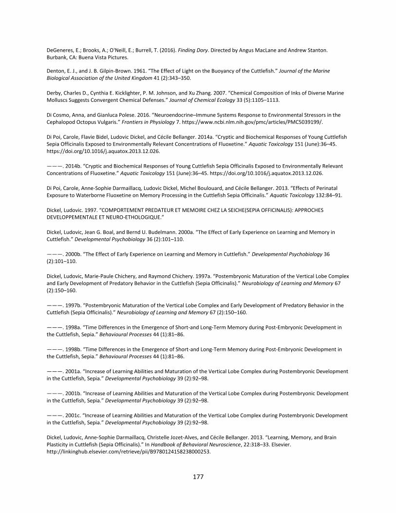
177
DeGeneres, E.; Brooks, A.; O'Neill, E.; Burrell, T. (2016). Finding Dory. Directed by Angus MacLane and Andrew Stanton. Burbank, CA: Buena Vista Pictures.
Denton, E. J., and J. B. Gilpin-Brown. 1961. “The Effect of Light on the Buoyancy of the Cuttlefish.” Journal of the Marine Biological Association of the United Kingdom 41 (2):343–350.
Derby, Charles D., Cynthia E. Kicklighter, P. M. Johnson, and Xu Zhang. 2007. “Chemical Composition of Inks of Diverse Marine Molluscs Suggests Convergent Chemical Defenses.” Journal of Chemical Ecology 33 (5):1105–1113.
Di Cosmo, Anna, and Gianluca Polese. 2016. “Neuroendocrine–Immune Systems Response to Environmental Stressors in the Cephalopod Octopus Vulgaris.” Frontiers in Physiology 7. https://www.ncbi.nlm.nih.gov/pmc/articles/PMC5039199/.
Di Poi, Carole, Flavie Bidel, Ludovic Dickel, and Cécile Bellanger. 2014a. “Cryptic and Biochemical Responses of Young Cuttlefish Sepia Officinalis Exposed to Environmentally Relevant Concentrations of Fluoxetine.” Aquatic Toxicology 151 (June):36–45. https://doi.org/10.1016/j.aquatox.2013.12.026.
———. 2014b. “Cryptic and Biochemical Responses of Young Cuttlefish Sepia Officinalis Exposed to Environmentally Relevant Concentrations of Fluoxetine.” Aquatic Toxicology 151 (June):36–45. https://doi.org/10.1016/j.aquatox.2013.12.026.
Di Poi, Carole, Anne-Sophie Darmaillacq, Ludovic Dickel, Michel Boulouard, and Cécile Bellanger. 2013. “Effects of Perinatal Exposure to Waterborne Fluoxetine on Memory Processing in the Cuttlefish Sepia Officinalis.” Aquatic Toxicology 132:84–91.
Dickel, Ludovic. 1997. “COMPORTEMENT PREDATEUR ET MEMOIRE CHEZ LA SEICHE(SEPIA OFFICINALIS): APPROCHES DEVELOPPEMENTALE ET NEURO-ETHOLOGIQUE.”
Dickel, Ludovic, Jean G. Boal, and Bernd U. Budelmann. 2000a. “The Effect of Early Experience on Learning and Memory in Cuttlefish.” Developmental Psychobiology 36 (2):101–110.
———. 2000b. “The Effect of Early Experience on Learning and Memory in Cuttlefish.” Developmental Psychobiology 36 (2):101–110.
Dickel, Ludovic, Marie-Paule Chichery, and Raymond Chichery. 1997a. “Postembryonic Maturation of the Vertical Lobe Complex and Early Development of Predatory Behavior in the Cuttlefish (Sepia Officinalis).” Neurobiology of Learning and Memory 67 (2):150–160.
———. 1997b. “Postembryonic Maturation of the Vertical Lobe Complex and Early Development of Predatory Behavior in the Cuttlefish (Sepia Officinalis).” Neurobiology of Learning and Memory 67 (2):150–160.
———. 1998a. “Time Differences in the Emergence of Short-and Long-Term Memory during Post-Embryonic Development in the Cuttlefish, Sepia.” Behavioural Processes 44 (1):81–86.
———. 1998b. “Time Differences in the Emergence of Short-and Long-Term Memory during Post-Embryonic Development in the Cuttlefish, Sepia.” Behavioural Processes 44 (1):81–86.
———. 2001a. “Increase of Learning Abilities and Maturation of the Vertical Lobe Complex during Postembryonic Development in the Cuttlefish, Sepia.” Developmental Psychobiology 39 (2):92–98.
———. 2001b. “Increase of Learning Abilities and Maturation of the Vertical Lobe Complex during Postembryonic Development in the Cuttlefish, Sepia.” Developmental Psychobiology 39 (2):92–98.
———. 2001c. “Increase of Learning Abilities and Maturation of the Vertical Lobe Complex during Postembryonic Development in the Cuttlefish, Sepia.” Developmental Psychobiology 39 (2):92–98.
Dickel, Ludovic, Anne-Sophie Darmaillacq, Christelle Jozet-Alves, and Cécile Bellanger. 2013. “Learning, Memory, and Brain Plasticity in Cuttlefish (Sepia Officinalis).” In Handbook of Behavioral Neuroscience, 22:318–33. Elsevier. http://linkinghub.elsevier.com/retrieve/pii/B9780124158238000253.
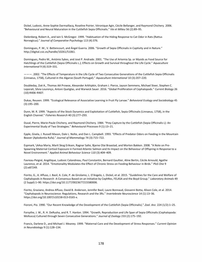
178
Dickel, Ludovic, Anne-Sophie Darmaillacq, Roseline Poirier, Véronique Agin, Cécile Bellanger, and Raymond Chichery. 2006. “Behavioural and Neural Maturation in the Cuttlefish Sepia Officinalis.” Vie et Milieu 56 (2):89–95.
Dielenberg, Robert A., and Iain S. McGregor. 1999. “Habituation of the Hiding Response to Cat Odor in Rats (Rattus Norvegicus).” Journal of Comparative Psychology 113 (4):376.
Domingues, P. M., V. Bettencourt, and Ángel Guerra. 2006. “Growth of Sepia Officinalis in Captivity and in Nature.” http://digital.csic.es/handle/10261/51061.
Domingues, Pedro M., António Sykes, and José P. Andrade. 2001. “The Use of Artemia Sp. or Mysids as Food Source for Hatchlings of the Cuttlefish (Sepia Officinalis L.); Effects on Growth and Survival throughout the Life Cycle.” Aquaculture International 9 (4):319–331.
———. 2002. “The Effects of Temperature in the Life Cycle of Two Consecutive Generations of the Cuttlefish Sepia Officinalis (Linnaeus, 1758), Cultured in the Algarve (South Portugal).” Aquaculture International 10 (3):207–220.
Doubleday, Zoë A., Thomas AA Prowse, Alexander Arkhipkin, Graham J. Pierce, Jayson Semmens, Michael Steer, Stephen C. Leporati, Sílvia Lourenço, Antoni Quetglas, and Warwick Sauer. 2016. “Global Proliferation of Cephalopods.” Current Biology 26 (10):R406–R407.
Dukas, Reuven. 1999. “Ecological Relevance of Associative Learning in Fruit Fly Larvae.” Behavioral Ecology and Sociobiology 45 (3):195–200.
Dunn, M. R. 1999. “Aspects of the Stock Dynamics and Exploitation of Cuttlefish, Sepia Officinalis (Linnaeus, 1758), in the English Channel.” Fisheries Research 40 (3):277–293.
Duval, Pierre, Marie-Paule Chichery, and Raymond Chichery. 1984. “Prey Capture by the Cuttlefish (Sepia Officinalis L): An Experimental Study of Two Strategies.” Behavioural Processes 9 (1):13–21.
Epple, Gisela, J. Russell Mason, Dale L. Nolte, and Dan L. Campbell. 1993. “Effects of Predator Odors on Feeding in the Mountain Beaver (Aplodontia Rufa).” Journal of Mammalogy 74 (3):715–722.
Espmark, \AAsa Maria, Marit Skog Eriksen, Ragnar Salte, Bjarne Olai Braastad, and Morten Bakken. 2008. “A Note on Pre-Spawning Maternal Cortisol Exposure in Farmed Atlantic Salmon and Its Impact on the Behaviour of Offspring in Response to a Novel Environment.” Applied Animal Behaviour Science 110 (3):404–409.
Favreau-Peigné, Angélique, Ludovic Calandreau, Paul Constantin, Bernard Gaultier, Aline Bertin, Cécile Arnould, Agathe Laurence, et al. 2014. “Emotionality Modulates the Effect of Chronic Stress on Feeding Behaviour in Birds.” PloS One 9 (2):e87249.
Fiorito, G., A. Affuso, J. Basil, A. Cole, P. de Girolamo, L. D’Angelo, L. Dickel, et al. 2015. “Guidelines for the Care and Welfare of Cephalopods in Research -A Consensus Based on an Initiative by CephRes, FELASA and the Boyd Group.” Laboratory Animals 49 (2 Suppl):1–90. https://doi.org/10.1177/0023677215580006.
Fiorito, Graziano, Andrea Affuso, David B. Anderson, Jennifer Basil, Laure Bonnaud, Giovanni Botta, Alison Cole, et al. 2014. “Cephalopods in Neuroscience: Regulations, Research and the 3Rs.” Invertebrate Neuroscience 14 (1):13–36. https://doi.org/10.1007/s10158-013-0165-x.
Fioroni, Pio. 1990. “Our Recent Knowledge of the Development of the Cuttlefish (Sepia Officinalis).” Zool. Anz. 224 (1/2):1–25.
Forsythe, J. W., R. H. DeRusha, and R. T. Hanlon. 1994. “Growth, Reproduction and Life Span of Sepia Officinalis (Cephalopoda: Mollusca) Cultured through Seven Consecutive Generations.” Journal of Zoology 233 (2):175–192.
Francis, Darlene D., and Michael J. Meaney. 1999. “Maternal Care and the Development of Stress Responses.” Current Opinion in Neurobiology 9 (1):128–134.
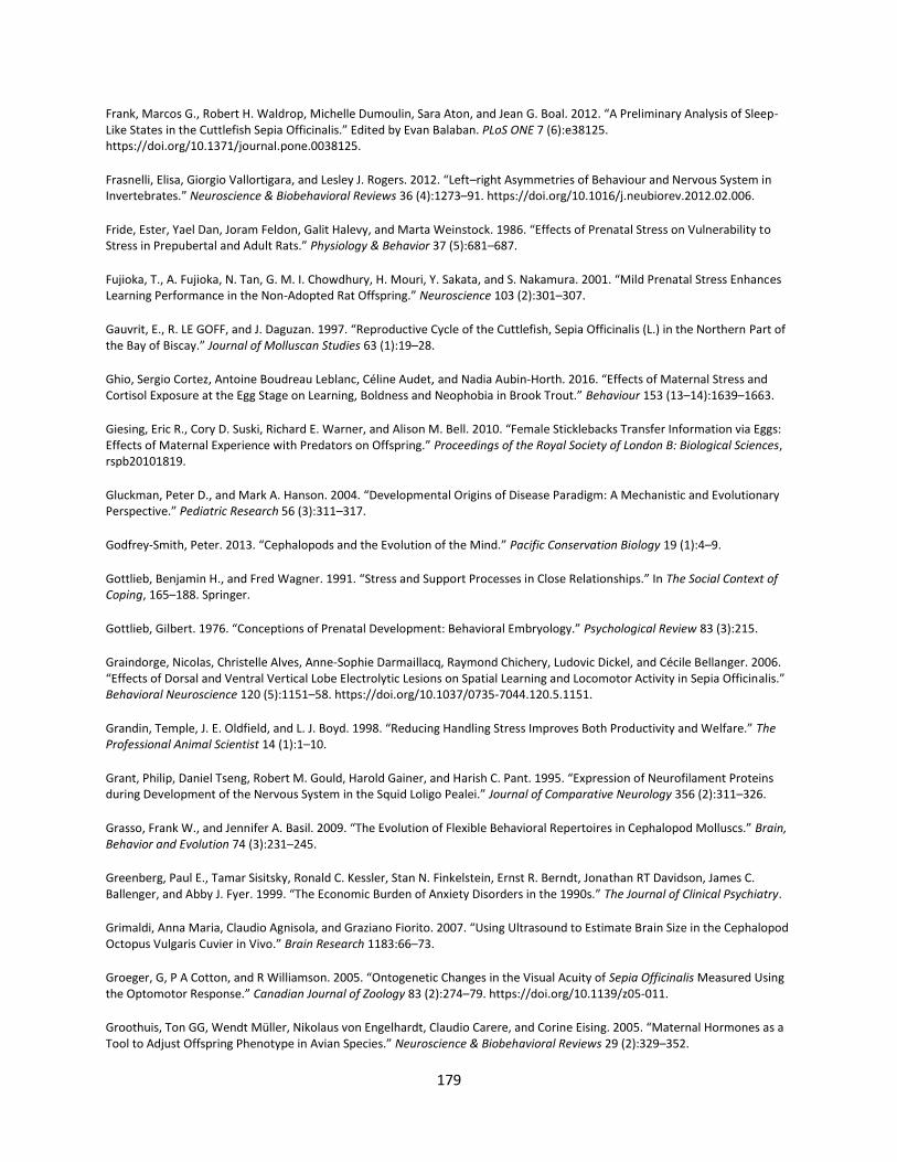
179
Frank, Marcos G., Robert H. Waldrop, Michelle Dumoulin, Sara Aton, and Jean G. Boal. 2012. “A Preliminary Analysis of Sleep-Like States in the Cuttlefish Sepia Officinalis.” Edited by Evan Balaban. PLoS ONE 7 (6):e38125. https://doi.org/10.1371/journal.pone.0038125.
Frasnelli, Elisa, Giorgio Vallortigara, and Lesley J. Rogers. 2012. “Left–right Asymmetries of Behaviour and Nervous System in Invertebrates.” Neuroscience & Biobehavioral Reviews 36 (4):1273–91. https://doi.org/10.1016/j.neubiorev.2012.02.006.
Fride, Ester, Yael Dan, Joram Feldon, Galit Halevy, and Marta Weinstock. 1986. “Effects of Prenatal Stress on Vulnerability to Stress in Prepubertal and Adult Rats.” Physiology & Behavior 37 (5):681–687.
Fujioka, T., A. Fujioka, N. Tan, G. M. I. Chowdhury, H. Mouri, Y. Sakata, and S. Nakamura. 2001. “Mild Prenatal Stress Enhances Learning Performance in the Non-Adopted Rat Offspring.” Neuroscience 103 (2):301–307.
Gauvrit, E., R. LE GOFF, and J. Daguzan. 1997. “Reproductive Cycle of the Cuttlefish, Sepia Officinalis (L.) in the Northern Part of the Bay of Biscay.” Journal of Molluscan Studies 63 (1):19–28.
Ghio, Sergio Cortez, Antoine Boudreau Leblanc, Céline Audet, and Nadia Aubin-Horth. 2016. “Effects of Maternal Stress and Cortisol Exposure at the Egg Stage on Learning, Boldness and Neophobia in Brook Trout.” Behaviour 153 (13–14):1639–1663.
Giesing, Eric R., Cory D. Suski, Richard E. Warner, and Alison M. Bell. 2010. “Female Sticklebacks Transfer Information via Eggs: Effects of Maternal Experience with Predators on Offspring.” Proceedings of the Royal Society of London B: Biological Sciences, rspb20101819.
Gluckman, Peter D., and Mark A. Hanson. 2004. “Developmental Origins of Disease Paradigm: A Mechanistic and Evolutionary Perspective.” Pediatric Research 56 (3):311–317.
Godfrey-Smith, Peter. 2013. “Cephalopods and the Evolution of the Mind.” Pacific Conservation Biology 19 (1):4–9.
Gottlieb, Benjamin H., and Fred Wagner. 1991. “Stress and Support Processes in Close Relationships.” In The Social Context of Coping, 165–188. Springer.
Gottlieb, Gilbert. 1976. “Conceptions of Prenatal Development: Behavioral Embryology.” Psychological Review 83 (3):215.
Graindorge, Nicolas, Christelle Alves, Anne-Sophie Darmaillacq, Raymond Chichery, Ludovic Dickel, and Cécile Bellanger. 2006. “Effects of Dorsal and Ventral Vertical Lobe Electrolytic Lesions on Spatial Learning and Locomotor Activity in Sepia Officinalis.” Behavioral Neuroscience 120 (5):1151–58. https://doi.org/10.1037/0735-7044.120.5.1151.
Grandin, Temple, J. E. Oldfield, and L. J. Boyd. 1998. “Reducing Handling Stress Improves Both Productivity and Welfare.” The Professional Animal Scientist 14 (1):1–10.
Grant, Philip, Daniel Tseng, Robert M. Gould, Harold Gainer, and Harish C. Pant. 1995. “Expression of Neurofilament Proteins during Development of the Nervous System in the Squid Loligo Pealei.” Journal of Comparative Neurology 356 (2):311–326.
Grasso, Frank W., and Jennifer A. Basil. 2009. “The Evolution of Flexible Behavioral Repertoires in Cephalopod Molluscs.” Brain, Behavior and Evolution 74 (3):231–245.
Greenberg, Paul E., Tamar Sisitsky, Ronald C. Kessler, Stan N. Finkelstein, Ernst R. Berndt, Jonathan RT Davidson, James C. Ballenger, and Abby J. Fyer. 1999. “The Economic Burden of Anxiety Disorders in the 1990s.” The Journal of Clinical Psychiatry.
Grimaldi, Anna Maria, Claudio Agnisola, and Graziano Fiorito. 2007. “Using Ultrasound to Estimate Brain Size in the Cephalopod Octopus Vulgaris Cuvier in Vivo.” Brain Research 1183:66–73.
Groeger, G, P A Cotton, and R Williamson. 2005. “Ontogenetic Changes in the Visual Acuity of Sepia Officinalis Measured Using the Optomotor Response.” Canadian Journal of Zoology 83 (2):274–79. https://doi.org/10.1139/z05-011.
Groothuis, Ton GG, Wendt Müller, Nikolaus von Engelhardt, Claudio Carere, and Corine Eising. 2005. “Maternal Hormones as a Tool to Adjust Offspring Phenotype in Avian Species.” Neuroscience & Biobehavioral Reviews 29 (2):329–352.
180
Groothuis, Ton GG, and Hubert Schwabl. 2008. “Hormone-Mediated Maternal Effects in Birds: Mechanisms Matter but What Do We Know of Them?” Philosophical Transactions of the Royal Society of London B: Biological Sciences 363 (1497):1647–1661.
Guerra, A., and J. L. Gonzalez. 2011. “First Record of Predation by a Tompot Blenny on the Common Cuttlefish Sepia Officinalis Eggs.” Vie et Milieu 61 (1):45–48.
Guibé, M., and L. Dickel. 2011. “Embryonic Visual Experience Influences Post-Hatching Shelter Preference in Cuttlefish.” Vie et Milieu 61 (4):243–246.
Guibé, Mathieu, Jean G. Boal, and Ludovic Dickel. 2010. “Early Exposure to Odors Changes Later Visual Prey Preferences in Cuttlefish.” Developmental Psychobiology 52 (8):833–37. https://doi.org/10.1002/dev.20470.
Guibé, Mathieu, Nicolas Poirel, Olivier Houdé, and Ludovic Dickel. 2012. “Food Imprinting and Visual Generalization in Embryos and Newly Hatched Cuttlefish, Sepia Officinalis.” Animal Behaviour 84 (1):213–17. https://doi.org/10.1016/j.anbehav.2012.04.035.
Guibert, Floriane, Marie-Annick Richard-Yris, Sophie Lumineau, Kurt Kotrschal, Aline Bertin, Christophe Petton, Erich Möstl, and Cécilia Houdelier. 2011. “Unpredictable Mild Stressors on Laying Females Influence the Composition of Japanese Quail Eggs and Offspring’s Phenotype.” Applied Animal Behaviour Science 132 (1):51–60.
Gyllenhaal, J., Ferguson, R., Reynolds, R., Sanada, H. Life. Directed by Daniel Espinosa. Culver City, CA: Columbia Pictures, 2017.
Hanley, Janice S., Nadav Shashar, Roxanna Smolowitz, Robert A. Bullis, William N. Mebane, Howaida R. Gabr, and Roger T. Hanlon. 1998. “Modified Laboratory Culture Techniques for the European Cuttlefish Sepia Officinalis.” Biological Bulletin 195 (2):223. https://doi.org/10.2307/1542850.
Hanlon, and John B. Messenger. 1988. “Adaptive Coloration in Young Cuttlefish (Sepia Officinalis L.): The Morphology and Development of Body Patterns and Their Relation to Behaviour.” Philosophical Transactions of the Royal Society B: Biological Sciences 320 (1200):437–487.
———. 1998. Cephalopod Behaviour. Cambridge University Press. https://books.google.fr/books?hl=en&lr=&id=Nxfv6xZZ6WYC&oi=fnd&pg=PR13&dq=hanlon+and+messenger&ots=sEB__SaOzc&sig=xQSd3OGBv5Ko2GoBm5qtJPDqf6I.
Hanlon, R. T., S. A. Ament, and H. Gabr. 1999. “Behavioral Aspects of Sperm Competition in Cuttlefish, Sepia Officinalis (Sepioidea: Cephalopoda).” Marine Biology 134 (4):719–728.
Hanlon, R. T., C.-C. Chiao, L. M. Mäthger, A. Barbosa, K. C. Buresch, and C. Chubb. 2009. “Cephalopod Dynamic Camouflage: Bridging the Continuum between Background Matching and Disruptive Coloration.” Philosophical Transactions of the Royal Society of London B: Biological Sciences 364 (1516):429–437.
Hanlon, Roger. 2007. “Cephalopod Dynamic Camouflage.” Current Biology 17 (11):R400–R404.
Hanlon, Roger T., and John B. Messenger. 1988. “Adaptive Coloration in Young Cuttlefish (Sepia Officinalis L.): The Morphology and Development of Body Patterns and Their Relation to Behaviour.” Philosophical Transactions of the Royal Society B: Biological Sciences 320 (1200):437–487.
———. 1998. Cephalopod Behaviour. Cambridge University Press.
Hayward, Lisa S., and John C. Wingfield. 2004. “Maternal Corticosterone Is Transferred to Avian Yolk and May Alter Offspring Growth and Adult Phenotype.” General and Comparative Endocrinology 135 (3):365–371.
Henriksen, Rie, Sophie Rettenbacher, and Ton G.G. Groothuis. 2011. “Prenatal Stress in Birds: Pathways, Effects, Function and Perspectives.” Neuroscience & Biobehavioral Reviews 35 (7):1484–1501. https://doi.org/10.1016/j.neubiorev.2011.04.010.
Hepper, Peter G. 1988. “Adaptive Fetal Learning: Prenatal Exposure to Garlic Affects Postnatal Preferences.” Animal Behaviour 36 (3):935–936.
181
Hilakivi, Leena A., M. Ota, and R. Lister. 1989. “Effect of Isolation on Brain Monoamines and the Behavior of Mice in Tests of Exploration, Locomotion, Anxiety and Behavioral ‘Despair.’” Pharmacology Biochemistry and Behavior 33 (2):371–374.
Hochner, Binyamin, Euan R. Brown, Marina Langella, Tal Shomrat, and Graziano Fiorito. 2003. “A Learning and Memory Area in the Octopus Brain Manifests a Vertebrate-like Long-Term Potentiation.” Journal of Neurophysiology 90 (5):3547–3554.
Hochner, Binyamin, Tal Shomrat, and Graziano Fiorito. 2006. “The Octopus: A Model for a Comparative Analysis of the Evolution of Learning and Memory Mechanisms.” The Biological Bulletin 210 (3):308–317.
Hockman, Charles H. 1961. “Prenatal Maternal Stress in the Rat: Its Effects on Emotional Behavior in the Offspring.” Journal of Comparative and Physiological Psychology 54 (6):679.
Holmes, William. 1940. “The Colour Changes and Colour Patterns of Sepia Officinalis L.” Journal of Zoology 110 (1–2):17–35.
Houdelier, Cécilia, Florent Pittet, Floriane Guibert, Emmanuel De Margerie, and Sophie Lumineau. 2013. “Non-Genetic Inheritance in Birds: Transmission of Behaviour from Mother to Offspring.” Non-Genet Inherit 1:62–68.
Huffard, Christine L. 2013. “Cephalopod Neurobiology: An Introduction for Biologists Working in Other Model Systems.” Invertebrate Neuroscience 13 (1):11–18.
Imarazene, Boudjema, Aude Andouche, Yann Bassaglia, Pascal-Jean Lopez, and Laure Bonnaud-Ponticelli. 2017. “Eye Development in Sepia Officinalis Embryo: What the Uncommon Gene Expression Profiles Tell Us about Eye Evolution.” Frontiers in Physiology 8:613.
IUCN 2017. The IUCN Red List of Threatened Species. Version 2017-2. <http://www.iucnredlist.org>.
Jäckel, Gilta, F. Mark, Magdalena Gutowska, M. Oellermann, Charlie Ellington, and H. Poertner. 2007. “Analysis of Diurnal Activity Patterns and Related Changes in Metabolism in the Cephalopod Sepia Officinalis.” Comparative Biochemistry and Physiology Part A: Molecular & Integrative Physiology 146 (4):S83.
Jacobsen, Helge P., and Ole B. Stabell. 2004. “Antipredator Behaviour Mediated by Chemical Cues: The Role of Conspecific Alarm Signalling and Predator Labelling in the Avoidance Response of a Marine Gastropod.” Oikos 104 (1):43–50.
Joca, Sâmia Regiane Lourenço, Frederico Rogério Ferreira, and Francisco Silveira Guimarães. 2007. “Modulation of Stress Consequences by Hippocampal Monoaminergic, Glutamatergic and Nitrergic Neurotransmitter Systems.” Stress 10 (3):227–249.
Jones, Daniel. September 25, 2017. “Fishing For Gold; Huge demand for cuttlefish is making a fortune for British fishermen.” The Sun.
Jones, N. J. E., I. D. Ridgway, and C. A. Richardson. 2009. “Transport of Cuttlefish, Sepia Officinalis, Eggs under Dry and Damp Conditions.” Journal of Molluscan Studies 75 (2):192–194.
Jones, R. Bryan. 1996. “Fear and Adaptability in Poultry: Insights, Implications and Imperatives.” World’s Poultry Science Journal 52 (2):131–174.
Jozet-Alves, C., and M. Hebert. 2012. “Embryonic Exposure to Predator Odour Modulates Visual Lateralization in Cuttlefish.” Proceedings of the Royal Society B: Biological Sciences 280 (1752):20122575–20122575. https://doi.org/10.1098/rspb.2012.2575.
Jozet-Alves, Christelle, and Marie Hébert. 2013. “Embryonic Exposure to Predator Odour Modulates Visual Lateralization in Cuttlefish.” Proceedings of the Royal Society of London B: Biological Sciences 280 (1752):20122575.
Jozet-Alves, Christelle, Vincent A. Viblanc, Sébastien Romagny, Matthieu Dacher, Susan D. Healy, and Ludovic Dickel. 2012. “Visual Lateralization Is Task and Age Dependent in Cuttlefish, Sepia Officinalis.” Animal Behaviour 83 (6):1313–18. https://doi.org/10.1016/j.anbehav.2012.02.023.
182
Juorio, A. V., and P. B. Molinoff. 1974. “The Normal Occurrence of Octopamine in Neural Tissues of the Octopus and Other Cephalopods.” Journal of Neurochemistry 22 (2):271–280.
Katz, Richard J., Kevin A. Roth, and Bernard J. Carroll. 1981. “Acute and Chronic Stress Effects on Open Field Activity in the Rat: Implications for a Model of Depression.” Neuroscience & Biobehavioral Reviews 5 (2):247–251.
Kerbl, Alexandra, Stephan Handschuh, Marie-Therese Nödl, Brian Metscher, Manfred Walzl, and Andreas Wanninger. 2013. “Micro-CT in Cephalopod Research: Investigating the Internal Anatomy of a Sepiolid Squid Using a Non-Destructive Technique with Special Focus on the Ganglionic System.” Journal of Experimental Marine Biology and Ecology 447:140–148.
Kime, David E., and John B. Messenger. 1990. “Monoamines in the Cephalopod CNS: An HPLC Analysis.” Comparative Biochemistry and Physiology Part C: Comparative Pharmacology 96 (1):49–57.
Klug, Christian, Günter Schweigert, Dirk Fuchs, Isabelle Kruta, and Helmut Tischlinger. 2016. “Adaptations to Squid-Style High-Speed Swimming in Jurassic Belemnitids.” Biology Letters 12 (1):20150877.
Komak, Spogmai, Jean G. Boal, Ludovic Dickel, and Bernd U. Budelmann. 2005. “Behavioural Responses of Juvenile Cuttlefish ( Sepia Officinalis ) to Local Water Movements.” Marine and Freshwater Behaviour and Physiology 38 (2):117–25. https://doi.org/10.1080/10236240500139206.
Krueger, Donald A., and Stanley I. Dodson. 1981. “Embryological Induction and Predation Ecology in Daphnia Pulex.” Limnology and Oceanography 26 (2):219–223.
Kruta, Isabelle, Isabelle Rouget, Sylvain Charbonnier, Jérémie Bardin, Vincent Fernandez, Damien Germain, Arnaud Brayard, and Neil Landman. 2016. “Proteroctopus Ribeti in Coleoid Evolution.” Palaeontology 59 (6):767–773.
Langridge, K. V. 2006. “Symmetrical Crypsis and Asymmetrical Signalling in the Cuttlefish Sepia Officinalis.” Proceedings of the Royal Society B: Biological Sciences 273 (1589):959–67. https://doi.org/10.1098/rspb.2005.3395.
Langridge, Keri V. 2009. “Cuttlefish Use Startle Displays, but Not against Large Predators.” Animal Behaviour 77 (4):847–56. https://doi.org/10.1016/j.anbehav.2008.11.023.
Langridge, Keri V., Mark Broom, and Daniel Osorio. 2007. “Selective Signalling by Cuttlefish to Predators.” Current Biology 17 (24):R1044–R1045.
Laptikhovsky, Vladimir, Alp Salman, Bahadir Önsoy, and Tuncer Katagan. 2003. “Fecundity of the Common Cuttlefish, Sepia Officinalis L.(Cephalopoda, Sepiida): A New Look at the Old Problem.” Scientia Marina 67 (3):279–284.
Larson, Shawn E., and Roland C. Anderson. 2010. “Fecal Hormones Measured within Giant Pacific Octopuses Enteroctopus Dofleini.” Journal of Aquatic Animal Health 22 (3):152–157.
Laschi, Cecilia, Matteo Cianchetti, Barbara Mazzolai, Laura Margheri, Maurizio Follador, and Paolo Dario. 2012. “Soft Robot Arm Inspired by the Octopus.” Advanced Robotics 26 (7):709–727.
Lass, Sandra, and Piet Spaak. 2003. “Chemically Induced Anti-Predator Defences in Plankton: A Review.” Hydrobiologia 491 (1–3):221–239.
Leader, Leo R., Peter Baillie, Bahia Martin, and Elsebeth Vermeulen. 1982. “The Assessment and Significance of Habituation to a Repeated Stimulus by the Human Fetus.” Early Human Development 7 (3):211–219.
Lee, Yi-Hsin, Yun-Chieh Chang, Hong Young Yan, and Chuan-Chin Chiao. 2013. “Early Visual Experience of Background Contrast Affects the Expression of NMDA-like Glutamate Receptors in the Optic Lobe of Cuttlefish, Sepia Pharaonis.” Journal of Experimental Marine Biology and Ecology 447:86–92.
Lee, Yi-Hsin, Hong Young Yan, and Chuan-Chin Chiao. 2010. “Visual Contrast Modulates Maturation of Camouflage Body Patterning in Cuttlefish (Sepia Pharaonis).” Journal of Comparative Psychology 124 (3):261–70. https://doi.org/10.1037/a0019461.
183
———. 2012. “Effects of Early Visual Experience on the Background Preference in Juvenile Cuttlefish Sepia Pharaonis.” Biology Letters 8 (5):740–743.
Lemaire, J. 1970. “Table de Développement Embryonnaire de Sepia Officinalis L.(Mollusque Céphalopode).” Bull Soc Zool Fr 95:773–782.
Lemaire, J., and A. Richard. 1978. “ORGANOGENESIS OF THE EYE OF THE CEPHALOPOD SEPIA-OFFICINALIS L.” Bulletin de La Societe Zoologique de France-Evolution et Zoologie 103 (3):373.
Lemaire, V., M. Koehl, M. Le Moal, and D. N. Abrous. 2000. “Prenatal Stress Produces Learning Deficits Associated with an Inhibition of Neurogenesis in the Hippocampus.” Proceedings of the National Academy of Sciences 97 (20):11032–11037.
Lewejohann, L., C. Reinhard, A. Schrewe, J. Brandewiede, A. Haemisch, N. Görtz, M. Schachner, and N. Sachser. 2006. “Environmental Bias? Effects of Housing Conditions, Laboratory Environment and Experimenter on Behavioral Tests.” Genes, Brain and Behavior 5 (1):64–72.
Linnaeus, C. von. 1758. “Systema Naturae, 10th Edn, Vol. 1.” Stockholm: L. Salvii.
Liscovitch-Brauer, Noa, Shahar Alon, Hagit T. Porath, Boaz Elstein, Ron Unger, Tamar Ziv, Arie Admon, Erez Y. Levanon, Joshua JC Rosenthal, and Eli Eisenberg. 2017. “Trade-off between Transcriptome Plasticity and Genome Evolution in Cephalopods.” Cell 169 (2):191–202.
Liu, Bi Lin, Jie Cao, Samuel B. Truesdell, Yong Chen, Xin Jun Chen, and Si Quan Tian. 2016. “Reconstructing Cephalopod Migration with Statolith Elemental Signatures: A Case Study Using Dosidicus Gigas.” Fisheries Science 82 (3):425–433.
Malham, Shelagh K., Arnaud Lacoste, Florence Gélébart, Anne Cueff, and Serge A. Poulet. 2002. “A First Insight into Stress-Induced Neuroendocrine and Immune Changes in the Octopus Eledone Cirrhosa.” Aquatic Living Resources 15 (3):187–192.
Malone, Nicholas, Agustín Fuentes, and Francis J. White. 2012. “Variation in the Social Systems of Extant Hominoids: Comparative Insight into the Social Behavior of Early Hominins.” International Journal of Primatology 33 (6):1251–1277.
Mangold, K. 1989. Traité de Zoologie-Cephalopodes (Tome V, Fascicule 4). Masson, Paris.
Marthy, H.-J., R. Hauser, and A. Scholl. 1976. “Natural Tranquilliser in Cephalopod Eggs.” Nature 261 (5560):496–497.
Masterpasqua, Frank, Robert H. Chapman, and Richard K. Lore. 1976. “The Effects of Prenatal Psychological Stress on the Sexual Behavior and Reactivity of Male Rats.” Developmental Psychobiology 9 (5):403–411.
Mather, Jennifer. 2011. “Consciousness in Cephalopods?” Journal of Cosmology 14.
Mather, Jennifer A. 1986. “Sand Digging in Sepia Officinalis: Assessment of a Cephalopod Mollusc’s" Fixed" Behavior Pattern.” Journal of Comparative Psychology 100 (3):315.
———. 2008. “Cephalopod Consciousness: Behavioural Evidence.” Consciousness and Cognition 17 (1):37–48. https://doi.org/10.1016/j.concog.2006.11.006.
Mather, Jennifer A., and Ludovic Dickel. 2017. “Cephalopod Complex Cognition.” Current Opinion in Behavioral Sciences 16:131–137.
Mathger, L. M., C.-C. Chiao, A. Barbosa, K. C. Buresch, S. Kaye, and R. T. Hanlon. 2007. “Disruptive Coloration Elicited on Controlled Natural Substrates in Cuttlefish, Sepia Officinalis.” Journal of Experimental Biology 210 (15):2657–66. https://doi.org/10.1242/jeb.004382.
Mäthger, Lydia M., Alexandra Barbosa, Simon Miner, and Roger T. Hanlon. 2006. “Color Blindness and Contrast Perception in Cuttlefish (Sepia Officinalis) Determined by a Visual Sensorimotor Assay.” Vision Research 46 (11):1746–53. https://doi.org/10.1016/j.visres.2005.09.035.
184
Mathis, Alicia, Maud CO Ferrari, Nathan Windel, François Messier, and Douglas P. Chivers. 2008. “Learning by Embryos and the Ghost of Predation Future.” Proceedings of the Royal Society of London B: Biological Sciences 275 (1651):2603–2607.
McCarthy, Thomas M., and Wendy A. Fisher. 2000. “Multiple Predator-Avoidance Behaviours of the Freshwater Snail Physella Heterostropha Pomila: Responses Vary with Risk.” Freshwater Biology 44 (3):387–397.
McEwen, Bruce S., and John C. Wingfield. 2003. “The Concept of Allostasis in Biology and Biomedicine.” Hormones and Behavior 43 (1):2–15.
Meisel, Daniela V., Ruth A. Byrne, Jennifer Mather, and Michael J. Kuba. 2011. “Behavioral Sleep in Octopus Vulgaris.” Vie et Milieu-Life and Environment 61 (4):185–190.
Messenger, J. B. 1967. “The Peduncle Lobe: A Visuo-Motor Centre in Octopus.” Proceedings of the Royal Society of London B: Biological Sciences 167 (1008):225–251.
———. 1971. “Two-Stage Recovery of a Response in Sepia.” Nature 232 (5307):202–203.
———. 1973. “Learning in the Cuttlefish, Sepia.” Animal Behaviour 21 (4):801–826.
———. 1977. “Prey-Capture and Learning in the Cuttlefish, Sepia.” In Symp. Zool. Soc. Lond, 38:347–376.
Messenger, John B. 1968. “The Visual Attack of the Cuttlefish, Sepia Officinalis.” Animal Behaviour 16 (2):342–357.
———. 1996. “Neurotransmitters of Cephalopods.” Invertebrate Neuroscience 2 (2):95–114.
Metrione, Lara C., and John D. Harder. 2011. “Fecal Corticosterone Concentrations and Reproductive Success in Captive Female Southern White Rhinoceros.” General and Comparative Endocrinology 171 (3):283–292.
Miller, Gregory E., Edith Chen, and Eric S. Zhou. 2007. If It Goes up, Must It Come down? Chronic Stress and the Hypothalamic-Pituitary-Adrenocortical Axis in Humans. American Psychological Association. http://psycnet.apa.org/journals/bul/133/1/25/.
Moberg, Gary P. 1991. “How Behavioral Stress Disrupts the Endocrine Control of Reproduction in Domestic Animals.” Journal of Dairy Science 74 (1):304–311.
Moon, Christine, Hugo Lagercrantz, and Patricia K. Kuhl. 2013. “Language Experienced in Utero Affects Vowel Perception after Birth: A Two-Country Study.” Acta Paediatrica 102 (2):156–160.
Nixon, Marion, and Katharina Mangold. 1998. “The Early Life of Sepia Officinalis, and the Contrast with That of Octopus Vulgaris (Cephalopoda).” Journal of Zoology 245 (4):407–421.
Nixon, Marion, and John Z. Young. 2003. The Brains and Lives of Cephalopods. Oxford University Press.
O’Brien, Caitlin E., Marianne Bowie, Pauline Billard, Anne-Sopie Darmaillacq, Christelle Jozet-Alves, David Behaim, Olivier Basuyaux, and Ludovic Dickel. 2016. “The Effect of an Artificial Incubation Environment on Hatchling Size and Behavior in the Cuttlefish, Sepia Officinalis.” Vie et Milieu-Life and Environment 66 (1):1–9.
O’Brien, Caitlin E., Nawel Mezrai, Anne-Sophie Darmaillacq, and Ludovic Dickel. 2016. “Behavioral Development in Embryonic and Early Juvenile Cuttlefish (Sepia Officinalis).” Developmental Psychobiology 59 (2):145–160.
Oliveira, Catarina CV, Mayra I. Grano-Maldonado, Rui A. Gonçalves, Paulo A. Frias, and António V. Sykes. 2017. “Preliminary Results on the Daily and Seasonal Rhythms of Cuttlefish Sepia Officinalis (Linnaeus, 1758) Locomotor Activity in Captivity.” Fishes 2 (3):9.
Packard, Andrew. 1972. “Cephalopods and Fish: The Limits of Convergence.” Biological Reviews 47 (2):241–307.
Pascual, E. 1978. “Crecimiento y Alimentacion de Tres Generaciones de Sepia Officinalis En Cultivo.” Investigacion Pesquera 42 (2):421–442.
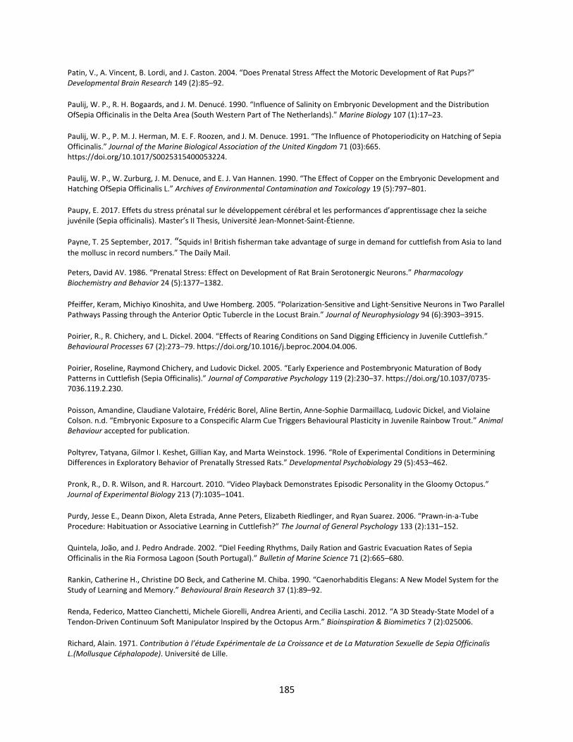
185
Patin, V., A. Vincent, B. Lordi, and J. Caston. 2004. “Does Prenatal Stress Affect the Motoric Development of Rat Pups?” Developmental Brain Research 149 (2):85–92.
Paulij, W. P., R. H. Bogaards, and J. M. Denucé. 1990. “Influence of Salinity on Embryonic Development and the Distribution OfSepia Officinalis in the Delta Area (South Western Part of The Netherlands).” Marine Biology 107 (1):17–23.
Paulij, W. P., P. M. J. Herman, M. E. F. Roozen, and J. M. Denuce. 1991. “The Influence of Photoperiodicity on Hatching of Sepia Officinalis.” Journal of the Marine Biological Association of the United Kingdom 71 (03):665. https://doi.org/10.1017/S0025315400053224.
Paulij, W. P., W. Zurburg, J. M. Denuce, and E. J. Van Hannen. 1990. “The Effect of Copper on the Embryonic Development and Hatching OfSepia Officinalis L.” Archives of Environmental Contamination and Toxicology 19 (5):797–801.
Paupy, E. 2017. Effets du stress prénatal sur le développement cérébral et les performances d’apprentissage chez la seiche juvénile (Sepia officinalis). Master’s II Thesis, Université Jean-Monnet-Saint-Étienne.
Payne, T. 25 September, 2017. “Squids in! British fisherman take advantage of surge in demand for cuttlefish from Asia to land
the mollusc in record numbers.” The Daily Mail.
Peters, David AV. 1986. “Prenatal Stress: Effect on Development of Rat Brain Serotonergic Neurons.” Pharmacology Biochemistry and Behavior 24 (5):1377–1382.
Pfeiffer, Keram, Michiyo Kinoshita, and Uwe Homberg. 2005. “Polarization-Sensitive and Light-Sensitive Neurons in Two Parallel Pathways Passing through the Anterior Optic Tubercle in the Locust Brain.” Journal of Neurophysiology 94 (6):3903–3915.
Poirier, R., R. Chichery, and L. Dickel. 2004. “Effects of Rearing Conditions on Sand Digging Efficiency in Juvenile Cuttlefish.” Behavioural Processes 67 (2):273–79. https://doi.org/10.1016/j.beproc.2004.04.006.
Poirier, Roseline, Raymond Chichery, and Ludovic Dickel. 2005. “Early Experience and Postembryonic Maturation of Body Patterns in Cuttlefish (Sepia Officinalis).” Journal of Comparative Psychology 119 (2):230–37. https://doi.org/10.1037/0735-7036.119.2.230.
Poisson, Amandine, Claudiane Valotaire, Frédéric Borel, Aline Bertin, Anne-Sophie Darmaillacq, Ludovic Dickel, and Violaine Colson. n.d. “Embryonic Exposure to a Conspecific Alarm Cue Triggers Behavioural Plasticity in Juvenile Rainbow Trout.” Animal Behaviour accepted for publication.
Poltyrev, Tatyana, Gilmor I. Keshet, Gillian Kay, and Marta Weinstock. 1996. “Role of Experimental Conditions in Determining Differences in Exploratory Behavior of Prenatally Stressed Rats.” Developmental Psychobiology 29 (5):453–462.
Pronk, R., D. R. Wilson, and R. Harcourt. 2010. “Video Playback Demonstrates Episodic Personality in the Gloomy Octopus.” Journal of Experimental Biology 213 (7):1035–1041.
Purdy, Jesse E., Deann Dixon, Aleta Estrada, Anne Peters, Elizabeth Riedlinger, and Ryan Suarez. 2006. “Prawn-in-a-Tube Procedure: Habituation or Associative Learning in Cuttlefish?” The Journal of General Psychology 133 (2):131–152.
Quintela, João, and J. Pedro Andrade. 2002. “Diel Feeding Rhythms, Daily Ration and Gastric Evacuation Rates of Sepia Officinalis in the Ria Formosa Lagoon (South Portugal).” Bulletin of Marine Science 71 (2):665–680.
Rankin, Catherine H., Christine DO Beck, and Catherine M. Chiba. 1990. “Caenorhabditis Elegans: A New Model System for the Study of Learning and Memory.” Behavioural Brain Research 37 (1):89–92.
Renda, Federico, Matteo Cianchetti, Michele Giorelli, Andrea Arienti, and Cecilia Laschi. 2012. “A 3D Steady-State Model of a Tendon-Driven Continuum Soft Manipulator Inspired by the Octopus Arm.” Bioinspiration & Biomimetics 7 (2):025006.
Richard, Alain. 1971. Contribution à l’étude Expérimentale de La Croissance et de La Maturation Sexuelle de Sepia Officinalis L.(Mollusque Céphalopode). Université de Lille.

186
Roberts, Claire T. 2017. “Biomedical Research: Premature Lambs Grown in a Bag.” Nature 546 (7656):45–46.
Robey, Jason (12 October 2012). "Giant Squid Captured on Video for First Time". Discovery. Archived from the original on 12 December 2012.
Romagny, S., A.-S. Darmaillacq, M. Guibe, C. Bellanger, and L. Dickel. 2012. “Feel, Smell and See in an Egg: Emergence of Perception and Learning in an Immature Invertebrate, the Cuttlefish Embryo.” Journal of Experimental Biology 215 (23):4125–30. https://doi.org/10.1242/jeb.078295.
Roth, Robert H., SEE-YING TAM, Yoshishige Ida, JING-XIA YANG, and Ariel Y. Deutch. 1988. “Stress and the Mesocorticolimbic Dopamine Systems.” Annals of the New York Academy of Sciences 537 (1):138–147.
Roussel, D. and Basuyaux, O. 2016. Valorisation des oeufs de seiches (Sepia officinalis) pondus sur les casiers: Développement d’une aquaculture de niche.
Sahley, Christie, Jerry W. Rudy, and Alan Gelperin. 1981. “An Analysis of Associative Learning in a Terrestrial Mollusc.” Journal of Comparative Physiology 144 (1):1–8.
Sanders, F. K., and J. Z. Young. 1940. “Learning and Other Functions of the Higher Nervous Centers of Sepia.” Journal of Neurophysiology.
Sandi, Carmen, César Venero, and Carmen Guaza. 1996. “Novelty-Related Rapid Locomotor Effects of Corticosterone in Rats.” European Journal of Neuroscience 8 (4):794–800.
Sasaki, TAKENORI, Shuichi Shigeno, and KAZUSHIGE Tanabe. 2010. “Anatomy of Living Nautilus: Reevaluation of Primitiveness and Comparison with Coleoidea.” Cephalopods—present and Past. Tokai University Press, Tokyo, 33–66.
Schneider, Mary L. 1992. “Prenatal Stress Exposure Alters Postnatal Behavioral Expression under Conditions of Novelty Challenge in Rhesus Monkey Infants.” Developmental Psychobiology 25 (7):529–540.
Schnell, Alexandra K., Roger T. Hanlon, Aïcha Benkada, and Christelle Jozet-Alves. 2016. “Lateralization of Eye Use in Cuttlefish: Opposite Direction for Anti-Predatory and Predatory Behaviors.” Frontiers in Physiology 7. https://www.ncbi.nlm.nih.gov/pmc/articles/PMC5149545/.
Schnell, Alexandra K., Carolynn L. Smith, Roger T. Hanlon, and Robert Harcourt. 2015. “Giant Australian Cuttlefish Use Mutual Assessment to Resolve Male-Male Contests.” Animal Behaviour 107 (September):31–40. https://doi.org/10.1016/j.anbehav.2015.05.026.
Schreck, Carl B. 2010. “Stress and Fish Reproduction: The Roles of Allostasis and Hormesis.” General and Comparative Endocrinology 165 (3):549–556.
Schreck, Carl B., Wilfrido Contreras-Sanchez, and Martin S. Fitzpatrick. 2001. “Effects of Stress on Fish Reproduction, Gamete Quality, and Progeny.” Aquaculture 197 (1):3–24.
Selye, Hans. 1976. “The Stress Concept.” Canadian Medical Association Journal 115 (8):718.
Shashar, Nadav, Roland Hagan, Jean G. Boal, and Roger T. Hanlon. 2000. “Cuttlefish Use Polarization Sensitivity in Predation on Silvery Fish.” Vision Research 40 (1):71–75.
Shashar, Nadav, Roger T. Hanlon, and Anne deM Petz. 1998. “Polarization Vision Helps Detect Transparent Prey.” Nature 393 (6682):222–223.
Shashar, Nadav, Coren Milbury, and Roger Hanlon. 2002. “Polarization Vision in Cephalopods: Neuroanatomical and Behavioral Features That Illustrate Aspects of Form and Function.” Marine and Freshwater Behaviour and Physiology 35 (1–2):57–68.
Shashar, Nadav, P. Rutledge, and T. Cronin. 1996. “Polarization Vision in Cuttlefish in a Concealed Communication Channel?” The Journal of Experimental Biology 199 (9):2077–2084.
187
Shepherdson, David J., Jill D. Mellen, and Michael Hutchins. 2012. Second Nature: Environmental Enrichment for Captive Animals. Smithsonian Institution.
Sheriff, M. J., and O. P. Love. 2013. “Determining the Adaptive Potential of Maternal Stress.” Edited by Jean Clobert. Ecology Letters 16 (2):271–80. https://doi.org/10.1111/ele.12042.
Shigeno, Shuichi, Hideaki Kidokoro, Kotaro Tsuchiya, Susumu Segawa, and Masamichi Yamamoto. 2001. “Development of the Brain in the Oegopsid Squid, Todarodes Pacificus: An Atlas up to the Hatching Stage.” Zoological Science 18 (4):527–541.
Shigeno, Shuichi, Takenori Sasaki, Takeya Moritaki, Takashi Kasugai, Michael Vecchione, and Kiyokazu Agata. 2008. “Evolution of the Cephalopod Head Complex by Assembly of Multiple Molluscan Body Parts: Evidence from Nautilus Embryonic Development.” Journal of Morphology 269 (1):1–17.
Shigeno, SHUICHI, SASAKI TAKENOrI, and S. von Boletzky. 2010. “The Origins of Cephalopod Body Plans: A Geometrical and Developmental Basis for the Evolution of Vertebrate-like Organ Systems.” Cephalopods-Present and Past 1:23–34.
Shomrat, T., N. Feinstein, M. Klein, and B. Hochner. 2010. “Serotonin Is a Facilitatory Neuromodulator of Synaptic Transmission and ‘Reinforces’ Long-Term Potentiation Induction in the Vertical Lobe of Octopus Vulgaris.” Neuroscience 169 (1):52–64.
Sigholt, T., U. Erikson, T. Rustad, S. Johansen, T. S. Nordtvedt, and A. Seland. 1997. “Handling Stress and Storage Temperature Affect Meat Quality of Farmed-Raised Atlantic Salmon (Salmo Salar).” Journal of Food Science 62 (4):898–905.
Skelhorn, John, and Candy Rowe. 2016. “Cognition and the Evolution of Camouflage.” In Proc. R. Soc. B, 283:20152890. The Royal Society.
Smith, Billy L., Georgia Wills, and David Naylor. 1981. “The Effects of Prenatal Stress on Rat Offsprings’ Learning Ability.” The Journal of Psychology 107 (1):45–51.
Sneddon, H., R. Hadden, and P. G. Hepper. 1998. “Chemosensory Learning in the Chicken Embryo.” Physiology & Behavior 64 (2):133–139.
Song, Zhuoyuan, Cameron Mazzola, Eric Schwartz, Ruirong Chen, Julian Finlaw, Mike Krieg, and Kamran Mohseni. 2016. “A Compact Autonomous Underwater Vehicle With Cephalopod-Inspired Propulsion.” Marine Technology Society Journal 50 (5):88–101.
Soso, Auren Benk, Leonardo José Gil Barcellos, Maria José Ranzani-Paiva, Luiz Carlos Kreutz, Rosmari Mezzalira Quevedo, Marina Lima, Leonardo Bolognesida Silva, Filipe Ritter, Alexandra Calliari Bedin, and Jovani Antônio Finco. 2008. “The Effects of Stressful Broodstock Handling on Hormonal Profiles and Reproductive Performance of Rhamdia Quelen (Quoy & Gaimard) Females.” Journal of the World Aquaculture Society 39 (6):835–841.
Stanford, Craig B. 1998. “The Social Behavior of Chimpanzees and Bonobos: Empirical Evidence and Shifting Assumptions.” Current Anthropology 39 (4):399–420.
Staudinger, Michelle D., Kendra C. Buresch, Lydia M. Mäthger, Charlie Fry, Sarah McAnulty, Kimberly M. Ulmer, and Roger T. Hanlon. 2013. “Defensive Responses of Cuttlefish to Different Teleost Predators.” The Biological Bulletin 225 (3):161–174.
Steer, M. A., N. A. Moltschaniwskyj, D. S. Nichols, and M. Miller. 2004. “The Role of Temperature and Maternal Ration in Embryo Survival: Using the Dumpling Squid Euprymna Tasmanica as a Model.” Journal of Experimental Marine Biology and Ecology 307 (1):73–89.
Storm, Jonathan J., and Steven L. Lima. 2010. “Mothers Forewarn Offspring about Predators: A Transgenerational Maternal Effect on Behavior.” The American Naturalist 175 (3):382–390.
Stubbs, Alexander L., and Christopher W. Stubbs. 2016. “Spectral Discrimination in Color Blind Animals via Chromatic Aberration and Pupil Shape.” Proceedings of the National Academy of Sciences 113 (29):8206–8211.
188
Suchecki, Deborah, and João Palermo Neto. 1991. “Prenatal Stress and Emotional Response of Adult Offspring.” Physiology & Behavior 49 (3):423–426.
Sundbom, Renée, Kirsten R. Jacobsen, Otto Kalliokoski, Jann Hau, and Klas SP Abelson. 2011. “Post-Operative Corticosterone Levels in Plasma and Feces of Mice Subjected to Permanent Catheterization and Automated Blood Sampling.” In Vivo 25 (3):335–342.
Sutton, Mark, Catalina Perales-Raya, and Isabel Gilbert. 2016. “A Phylogeny of Fossil and Living Neocoleoid Cephalopods.” Cladistics 32 (3):297–307.
Sykes, A. V., E. Almansa, A. Lorenzo, and J. P. Andrade. 2009. “Lipid Characterization of Both Wild and Cultured Eggs of Cuttlefish (Sepia Officinalis L.) throughout the Embryonic Development.” Aquaculture Nutrition 15 (1):38–53.
Sykes, António V., Alexandra Alves, Juan C. Capaz, Céline Madeira, Ana T. Couto, Rui A. Gonçalves, Paulo A. Frias, Irene Leal, and José P. Andrade. 2017. “Refining Tools for Studying Cuttlefish (Sepia Officinalis) Reproduction in Captivity: In Vivo Sexual Determination, Tagging and DNA Collection.” Aquaculture 479:13–16.
Sykes, António V., Pedro M. Domingues, Lorenzo Márquez, and José P. Andrade. 2011. “The Effects of Tank Colours on the Growth and Survival of Cuttlefish (Sepia Officinalis, Linnaeus 1758) Hatchlings and Juveniles.” Aquaculture Research 42 (3):441–449.
Sykes, António V., Diana Pereira, Covadonga Rodríguez, António Lorenzo, and José P. Andrade. 2013. “Effects of Increased Tank Bottom Areas on Cuttlefish (Sepia Officinalis, L.) Reproduction Performance.” Aquaculture Research 44 (7):1017–1028.
Tanner, Alastair R., Dirk Fuchs, Inger E. Winkelmann, M. Thomas P. Gilbert, M. Sabrina Pankey, Ângela M. Ribeiro, Kevin M. Kocot, Kenneth M. Halanych, Todd H. Oakley, and Rute R. da Fonseca. 2017. “Molecular Clocks Indicate Turnover and Diversification of Modern Coleoid Cephalopods during the Mesozoic Marine Revolution.” In Proc. R. Soc. B, 284:20162818. The Royal Society.
Tansley, Katharine. 1965. “Vision in Vertebrates.” Vision in Vertebrates.
Tempel, Douglas J., and R. J. Gutiérrez. 2003. “Fecal Corticosterone Levels in California Spotted Owls Exposed to Low-Intensity Chainsaw Sound.” Wildlife Society Bulletin, 698–702.
Thierry, Anne-Marie, FRANCE JAVOY, JACQUES GLOWINSKI, and SEYMOUR S. KETY. 1968. “Effects of Stress on the Metabolism of Norepinephrine, Dopamine and Serotonin in the Central Nervous System of the Rat. I. Modifications of Norepinephrine Turnover.” Journal of Pharmacology and Experimental Therapeutics 163 (1):163–171.
Thomas, Kate N., Bruce H. Robison, and Sönke Johnsen. 2017. “Two Eyes for Two Purposes: In Situ Evidence for Asymmetric Vision in the Cockeyed Squids Histioteuthis Heteropsis and Stigmatoteuthis Dofleini.” Phil. Trans. R. Soc. B 372 (1717):20160069.
Thorpe, William Homan. 1956. “Learning and Instinct in Animals.”
Tinbergen, Niko. 1963. “On Aims and Methods of Ethology.” Ethology 20 (4):410–433.
Tollrian, Ralpoh. 1995. “Predator-Induced Morphological Defenses: Costs, Life History Shifts, and Maternal Effects in Daphnia Pulex.” Ecology 76 (6):1691–1705.
Trueblood, Lloyd A., Sarah Zylinski, Bruce H. Robison, and Brad A. Seibel. 2015. “An Ethogram of the Humboldt Squid Dosidicus GigasOrbigny (1835) as Observed from Remotely Operated Vehicles.” Behaviour 152 (14):1911–1932.
Ulmer, Kevin M., Kendra C. Buresch, M. M. Kossodo, L. M. Mäthger, Liese A. Siemann, and Roger T. Hanlon. 2013. “Vertical Visual Features Have a Strong Influence on Cuttlefish Camouflage.” The Biological Bulletin 224 (2):110–118.
189
Vallee, Monique, Willy Mayo, Francoise Dellu, Michel Le Moal, Herve Simon, and Stefania Maccari. 1997. “Prenatal Stress Induces High Anxiety and Postnatal Handling Induces Low Anxiety in Adult Offspring: Correlation with Stress-Induced Corticosterone Secretion.” Journal of Neuroscience 17 (7):2626–2636.
Vallortigara, Giorgio, and Lesley J. Rogers. 2005. “Survival with an Asymmetrical Brain: Advantages and Disadvantages of Cerebral Lateralization.” Behavioral and Brain Sciences 28 (4):575–588.
Van den Hove, D. L. A., C. E. Blanco, B. Aendekerk, L. Desbonnet, Matteo Bruschettini, H. P. Steinbusch, JHHJ Prickaerts, and H. W. M. Steinbusch. 2005. “Prenatal Restraint Stress and Long-Term Affective Consequences.” Developmental Neuroscience 27 (5):313–320.
Van den Hove, D. L. A., H. W. M. Steinbusch, A. Scheepens, W. D. J. Van de Berg, L. A. M. Kooiman, B. J. G. Boosten, JHHJ Prickaerts, and C. E. Blanco. 2006. “Prenatal Stress and Neonatal Rat Brain Development.” Neuroscience 137 (1):145–155.
Villanueva, Roger, António V. Sykes, Erica AG Vidal, Carlos Rosas, Jaruwat Nabhitabhata, Lidia Fuentes, and José Iglesias. 2014. “Current Status and Future Challenges in Cephalopod Culture.” In Cephalopod Culture, 479–489. Springer.
Vitti, Joseph J. 2013. “Cephalopod Cognition in an Evolutionary Context: Implications for Ethology.” Biosemiotics 6 (3):393–401.
Walls, Mari, and Matti Ketola. 1989. “Effects of Predator-Induced Spines on Individual Fitness in Daphnia Pulex.” Limnology and Oceanography 34 (2):390–396.
Walters, Edgar T., Thomas J. Carew, and Eric R. Kandel. 1981. “Associative Learning in Aplysia: Evidence for Conditioned Fear in an Invertebrate.” Science, 504–506.
Wang, Qiming, Gregory R. Gossweiler, Stephen L. Craig, and Xuanhe Zhao. 2014. “Cephalopod-Inspired Design of Electro-Mechano-Chemically Responsive Elastomers for on-Demand Fluorescent Patterning.” Nature Communications 5.
Weilgart, Lindy S. 2007. “The Impacts of Anthropogenic Ocean Noise on Cetaceans and Implications for Management.” Canadian Journal of Zoology 85 (11):1091–1116.
Weinberg, Marc S., Aadra P. Bhatt, Milena Girotti, Cher V. Masini, Heidi EW Day, Serge Campeau, and Robert L. Spencer. 2009. “Repeated Ferret Odor Exposure Induces Different Temporal Patterns of Same-Stressor Habituation and Novel-Stressor Sensitization in Both Hypothalamic-Pituitary-Adrenal Axis Activity and Forebrain c-Fos Expression in the Rat.” Endocrinology 150 (2):749–761.
Weinstock, Marta. 2008. “The Long-Term Behavioural Consequences of Prenatal Stress.” Neuroscience & Biobehavioral Reviews 32 (6):1073–86. https://doi.org/10.1016/j.neubiorev.2008.03.002.
Weinstock, Marta, Erlena Matlina, Gilmor I. Maor, Haim Rosen, and Bruce S. McEwen. 1992. “Prenatal Stress Selectively Alters the Reactivity of the Hypothalamic-Pituitary Adrenal System in the Female Rat.” Brain Research 595 (2):195–200.
Weischer, M.-L., and H.-J. Marthy. 1983. “Chemical and Physiological Properties of the Natural Tranquilliser in the Cephalopod Eggs.” Marine & Freshwater Behaviour & Phy 9 (2):131–138.
Welberg, Leonie AM, and Jonathan R. Seckl. 2001. “Prenatal Stress, Glucocorticoids and the Programming of the Brain.” Journal of Neuroendocrinology 13 (2):113–128.
Wells, Deborah L., and Peter G. Hepper. 2006. “Prenatal Olfactory Learning in the Domestic Dog.” Animal Behaviour 72 (3):681–686.
Wells, M. J. 1958. “Factors Affecting Reactions to Mysis by Newly Hatched Sepia.” Behaviour 13 (1):96–111.
———. 1962. “Early Learning in Sepia.” In Symp. Zool. Soc. Lond, 8:149–169.
———. 1968. “Sensitization and the Evolution of Associative Learning.” In Neurobiology of Invertebrates, 391–411. Springer.
190
West-Eberhard, Mary Jane. 1989. “Phenotypic Plasticity and the Origins of Diversity.” Annual Review of Ecology and Systematics 20 (1):249–278.
Wild, Elvira, Tim Wollesen, Gerhard Haszprunar, and Martin He\s s. 2015. “Comparative 3D Microanatomy and Histology of the Eyes and Central Nervous Systems in Coleoid Cephalopod Hatchlings.” Organisms Diversity & Evolution 15 (1):37–64.
Wilson, Christina A., Almira Vazdarjanova, and Alvin V. Terry. 2013. “Exposure to Variable Prenatal Stress in Rats: Effects on Anxiety-Related Behaviors, Innate and Contextual Fear, and Fear Extinction.” Behavioural Brain Research 238:279–288.
Wollesen, Tim, Carmel McDougall, Bernard M. Degnan, and Andreas Wanninger. 2014. “POU Genes Are Expressed during the Formation of Individual Ganglia of the Cephalopod Central Nervous System.” EvoDevo 5 (1):41.
Xavier, José C., A. Louise Allcock, Yves Cherel, Marek R. Lipinski, Graham J. Pierce, Paul GK Rodhouse, Rui Rosa, Elizabeth K. Shea, Jan M. Strugnell, and Erica AG Vidal. 2015. “Future Challenges in Cephalopod Research.” Journal of the Marine Biological Association of the United Kingdom 95 (5):999–1015.
Yamamoto, Masamichi, Yumiko Shimazaki, and Shuichi Shigeno. 2003. “Atlas of the Embryonic Brain in the Pygmy Squid, Idiosepius Paradoxus.” Zoological Science 20 (2):163–179.
Yoshida, M. A., A. Ogura, K. Ikeo, S. Shigeno, T. Moritaki, G. C. Winters, A. B. Kohn, and L. L. Moroz. 2015. “Molecular Evidence for Convergence and Parallelism in Evolution of Complex Brains of Cephalopod Molluscs: Insights from Visual Systems.” Integrative and Comparative Biology 55 (6):1070–1083.
Young, J. Z. 1961. “Learning and Discrimination in the Octopus.” Biological Reviews 36 (1):32–95.
Yu, Cunjiang, Yuhang Li, Xun Zhang, Xian Huang, Viktor Malyarchuk, Shuodao Wang, Yan Shi, Li Gao, Yewang Su, and Yihui Zhang. 2014. “Adaptive Optoelectronic Camouflage Systems with Designs Inspired by Cephalopod Skins.” Proceedings of the National Academy of Sciences 111 (36):12998–13003.
Zylinski, S., A.-S. Darmaillacq, and N. Shashar. 2012. “Visual Interpolation for Contour Completion by the European Cuttlefish (Sepia Officinalis) and Its Use in Dynamic Camouflage.” Proceedings of the Royal Society B: Biological Sciences 279 (1737):2386–90. https://doi.org/10.1098/rspb.2012.0026.
Zylinski, S., D. Osorio, and A. J. Shohet. 2009. “Edge Detection and Texture Classification by Cuttlefish.” Journal of Vision 9 (13):13–13. https://doi.org/10.1167/9.13.13.

191
Figures and
Tables

192

193
List of Figures and Tables Figure 1) The three potential avenues of prenatal stress transmission. Page 3. Figure 2) A) The giant Pacific octopus, Enteroctopus dolfleini (photo by C.E. O’Brien); B) the giant squid (Architeuthis dux (photo by Brian J. Skerry, National Geographic); C) the common cuttlefish, Sepia officinalis (photo by Hans Hillewaert); D) the chambered nautilus, Nautilus pompilius (photo by William Cho). Page 5. Figure 3) Schematic representation of the categories of stress investigated. Page 7. Figure 4) Schematic representation of experimental design. A) Maternal Stress Effects, B) Embryonic Stress Effects. The arrows between panels A and B indicate that the eggs of UM were subdivided to create the embryonic treatment groups and that the same control group was used in both phases of the experiment. Page 18. Figure 5) Fecal corticosterone concentration (mean ng corticosterone mg-1 dry feces ± s.d.) over time, measured from fecal samples collected in 2016. No significant difference exists between groups (GLMM: X2 ≥ 0.07, n per data point = 4 (means calculated from the pooled feces of one to four individuals over two days), p = 0.79). Data are displayed as group means (dots) ± standard deviation (whiskers). Page 21. Figure 6) Mean number of eggs per female (mean ± s.d.). Among the females that laid eggs (“layers”), significantly more eggs (two-tailed exact permutation test: t ≥ 4042, p < 0.001) were laid by UM (505.23 ± 373.30 per female; n = 8 (four individuals and four group means—see Table 1 for details)) than SM (152.58 ± 96.93 per female; n = 12). Page 21. Figure 7) A normal (ink-stained) S. officinalis egg (left) and a translucent egg laid by one of the SM (right). Both eggs are in the final stage of embryonic development (Stage 30) and hatched a few days after the photograph was taken (July, 2016). External embryo (a) and yolk sac (b) are visible in both specimens. Bar in upper right corner = approx. 1 cm. Page 22. Figure 8) Transparent S. officinalis eggs (center) surrounded by normal ink-stained eggs with embryos visible within. Collected from the English Channel and photographed on June 19, 2014. Long axis of eggs = approx. 3.5 cm. Bar in upper right corner = approx. 1 cm. Page 25. Figure 9) Large ponds once used for oyster aquaculture at the CABaNor Aquaculture Cooperative (Blainville sur Mer, Normandy). a) Overhead view, note the channels connecting the ponds to the sea at high tide (photo by O. Basuyaux). b) Close-up view of an experimental pond (photo by C. E. O’Brien). Page 29. Figure 10) Schematic of the egg-laying site choice test: two modified cuttlefish traps were provided. One contained a potential hatchling predator, and both had six ropes, of a type and diameter known to “attract” cuttlefish eggs in the wild, affixed to them. Page 30. Figure 11) Schematic summary of Chapter 1. Stress to the mother during egg-laying reduces her reproductive output and likely the quality of her eggs as well. At the same time, a naturally-occurring
194
and an artificial stressor applied directly to developing embryos had no effect on hatching rate or size. The dashed arrow indicates a statistical tendency. Page 32. Figure 12) Stage 30 Sepia officinalis embryo (approx. mantle length 6mm) seen through the transparent egg membranes. Photo by Nawel Mezrai. Page 38. Figure 13) Cuttlefish eggs (a) in situ, 26 June, 2014 at an artificial laying-site (pre-placed tethers) in the vicinity of Blainville sur Mer, France. Note the crab, Maja squinado (b) in the foreground as well as the presence of numerous algaes, including Ulva sp. (c), Lithophyllum incrustans (d), various unidentified epibionts and a diversity of surrounding substrate types including sand (e), rocks and shell debris (f). Eggs are approximately 2.5-3.0cm in length (Boletzky, 1983). Photo by Olivier Basuyaux, of the Synergie Mer et Littoral (SMEL). Page 41. Figure 14) Hatchling cuttlefish (approx. mantle length 10mm) displaying a disruptive body pattern on a uniform substrate. Photo by Anne-Sophie Darmaillacq. Page 48. Figure 15) Three-month old juvenile cuttlefish (approx. mantle length 65mm). Photo by Caitlin E. O’Brien. Page 48. Figure 16) Eyes of the cuttlefish Sepia elongata caught off the coast of Eilat (Gulf of Aqaba, Israel; photo AS Darmaillacq). Page 69. Figure 17) Central nervous system of 3-month-old Sepia officinalis cuttlefish. Frontal section. Prenant-Gabe trichrome stain. Abbreviations: OL = optic lobe; SpM = supra-esophageal mass; SbM = sub-esophageal mass; Oe = oesophagus. Modified from Jozet-Alves et al. 2012a. Page 69. Figure 18) 7-day-old cuttlefish’s prey choice depending on whether they have been exposed to white crabs during embryonic development (‘exposed’) or not (‘control’). *Significant prey preference within groups (chi-square exact test: p < 0.05) and °significant difference in prey choice between groups (Fisher’s exact test: P < 0.05). Modified from Guibé et al. 2012. Page 71. Figure 19) Proportion of the cuttlefish (N=10 per group) that showed an optomotor response (OMR) to BWG (luminance only; black) or Pol (polarization; grey) patterns rotating at a velocity of 30 deg s–1, at hatching (0) and at the age of 30 days. Asterisks indicate a significant difference in the percentage of cuttlefish showing an OMR between the BWG and Pol patterns McNemar’s test, P<0.05). Modified from Cartron et al. 2013. Page 74. Figure 20) The diversity of body patterns displayed by 2-month-old cuttlefish (ca. 3-4 cm dorsal mantle length). a) stipple-uniform pattern elicited on uniform blue gravel; b) disruptive pattern elicited on a black and white checkerboard combined with mottle pattern; c) deimatic pattern following exposure to a “threat” d) mottle coloration with some components of the disruptive pattern (i.e. white square, white head bar and paired black dots). Note that patterns are not always fully expressed but exist in combination with others and may or may not directly reflect the visual background. Page 77. Figure 21) Stage 30 embryo (less than 1 cm) showing a mottle-disruptive coloration inside the egg. It has also squirted ink; note the cloud ink in the perivitellin fluid. Note that the embryo is seen from under through a peeled S. officinalis egg (photo C.E. O’Brien). Page 80.
195
Figure 22) Schematic representation of the experimental design. Page 86. Figure 23) Timeline of stress treatments and behavioral tests. All tests except for the threat response activity analysis occurred in 2015. Page 88. Figure 24) Heterogeneity Index (HI) ± s.d. of maternal stress groups on uniform and disruptive backgrounds. Between groups, WM offspring had significantly higher HI than the other maternal stress treatment groups on both backgrounds (p < 0.05), and SM (n = 43) had significantly higher HI on the disruptive background than UM-C (n = 41; p = 0.005). Significant differences between groups are indicated by connecting brackets. * indicates p < 0.05; *** indicates p< 0.001. Page 92. Figure 25) Heterogeneity Index (HI) ± s.d. of embryonic stress groups on uniform and disruptive backgrounds. The artificial stress group, UM-LE (n = 39), showed significantly higher mean HI than the natural stress group, UM-PE (n = 44; p = 0.017), and a tendency for higher HI than UM-C (n = 41; p = 0.069). * indicates p < 0.05; # indicates a statistical tendency (p < 0.08). Page 93. Figure 26) Eye used to select shelter in maternal stress groups. More WM (n = 43) chose the shelter in their left visual field (binomial test; p = 0.005, signified by asterisks) while no preference was found in UM-C (n = 40) or SM (n = 43). The proportions were not significantly different between groups (p = 0.08). Page 94. Figure 27) The total distance traveled (A), time spent moving (B) and mean meander (turn angle/distance traveled; C) ± s.d. of maternal stress groups in the overnight activity analysis test. Significant differences (indicated by connecting brackets with asterisks) exist between UM-C (n = 10) and SM (n = 15) in both distance traveled and time spent moving (p = 0.009 and 0.005; post hoc asymptotic permutation tests with sequential Bonferroni correction. ** indicates p < 0.01; # indicates a statistical tendency (p < 0.08). Page 95. Figure 28) The total distance traveled (A) and time moving (B) ± s.d. for maternal stress groups in the threat response activity analysis. Differences within groups are indicated by connecting bars; n = 5 for all bars. Page 95. Figure 29) The total distance traveled (A) and time moving (B) ± s.d. for embryonic stress groups in the threat response activity analysis. Differences within groups are indicated by connecting bars; n = 5 for all bars. Page 96. Figure 30) Prawn-in-a-tube (PIT) apparatus. The tube is composed of glass microscope slides held together by clear plastic. Four live shrimp are trapped between the slides. An aquarium pump keeps water circulating and the shrimp in motion. Cuttlefish drawing by Anne Quiédeville. Page 104. Figure 31) Timeline of PIT sessions. Juveniles were tested at three and five weeks of age in two 45min sessions separated by an hour-long rest interval. Page 105. Figure 32) The mean number of strikes per minute made by the maternal stress groups during their initial exposure to the PIT apparatus at three weeks of age. For each individual cuttlefish, timing commenced at the moment of first detection and orientation towards the apparatus, and ends either when no strikes were made for three consecutive minutes, or when the experimental trial time (45min)

196
had elapsed. Each group is plotted until its sample size falls below n = 5. UM-C = Unstressed Mother-Control, SM = Stressed Mother. Page 106. Figure 33) The mean number of strikes per minute made by the maternal stress groups during the retention trial exposure to the PIT apparatus at three weeks of age. For each individual cuttlefish, timing commenced at the moment of first detection and orientation towards the apparatus, and ends either when no strikes were made for three consecutive minutes, or when the experimental trial time (45min) had elapsed. Each group is plotted until its sample size falls below n = 5. UM-C = Unstressed Mother-Control, SM = Stressed Mother. Page 107. Figure 34) The mean number of strikes per minute made by the maternal stress groups during their initial exposure to the PIT apparatus at five weeks of age. For each individual cuttlefish, timing commenced at the moment of first detection and orientation towards the apparatus, and ends either when no strikes were made for three consecutive minutes, or when the experimental trial time (45min) had elapsed. Each group is plotted until its sample size falls below n = 5. UM-C = Unstressed Mother-Control, SM = Stressed Mother. Page 108. Figure 35) The mean number of strikes per minute made by the maternal stress groups during the retention trial exposure to the PIT apparatus at five weeks of age. For each individual cuttlefish, timing commenced at the moment of first detection and orientation towards the apparatus, and ends either when no strikes were made for three consecutive minutes, or when the experimental trial time (45min) had elapsed. Each group is plotted until its sample size falls below n = 5. UM-C = Unstressed Mother-Control, SM = Stressed Mother. Page 108. Figure 36) The mean number of strikes per minute made by the embryonic stress groups during their initial exposure to the PIT apparatus at three weeks of age. For each individual cuttlefish, timing commenced at the moment of first detection and orientation towards the apparatus, and ends either when no strikes were made for three consecutive minutes, or when the experimental trial time (45min) had elapsed. Each group is plotted until its sample size falls below n = 5. UM-C = Unstressed Mother-Control, UM-PE = Unstressed Mother-Predator-Exposed, UM-LE = Unstressed Mother-Light-Exposed. Page 110. Figure 37) The mean number of strikes per minute made by the embryonic stress groups during the retention trial exposure to the PIT apparatus at three weeks of age. For each individual cuttlefish, timing commenced at the moment of first detection and orientation towards the apparatus, and ends either when no strikes were made for three consecutive minutes, or when the experimental trial time (45min) had elapsed. Each group is plotted until its sample size falls below n = 5. UM-C = Unstressed Mother-Control, UM-PE = Unstressed Mother-Predator-Exposed, UM-LE = Unstressed Mother-Light-Exposed. Page 110. Figure 38) The mean number of strikes per minute made by the embryonic stress groups during the initial exposure to the PIT apparatus at five weeks of age. For each individual cuttlefish, timing commenced at the moment of first detection and orientation towards the apparatus, and ends either when no strikes were made for three consecutive minutes, or when the experimental trial time (45min) had elapsed. Each group is plotted until its sample size falls below n = 5. UM-C = Unstressed Mother-Control, UM-PE = Unstressed Mother-Predator-Exposed, UM-LE = Unstressed Mother-Light-Exposed. Page 112.
197
Figure 39) The mean number of strikes per minute made by the embryonic stress groups during the retention trial exposure to the PIT apparatus at five weeks of age. For each individual cuttlefish, timing commenced at the moment of first detection and orientation towards the apparatus, and ends either when no strikes were made for three consecutive minutes, or when the experimental trial time (45min) had elapsed. Each group is plotted until its sample size falls below n = 5. UM-C = Unstressed Mother-Control, UM-PE = Unstressed Mother-Predator-Exposed, UM-LE = Unstressed Mother-Light-Exposed. Page 112. Figure 40) Concentration of serotonin in head tissue of cuttlefish hatchlings from maternal stress groups (A) and embryonic stress groups (B). * indicates < 0.05, ** < 0.01, ** < 0.001. n: UM-C = 15, SM = 13, UM-PE = 15, UM-LE = 14. Page 118. Figure 41) Ratio of serotonin to its main metabolite, 5-HiAA (5-HiAA/5-HT) in head tissue of cuttlefish hatchlings from maternal stress groups (A) and embryonic stress groups (B). * indicates < 0.05, ** < 0.01, ** < 0.001. n: UM-C = 14, SM = 13, UM-PE = 15, UM-LE = 13. Page 118. Figure 42) Concentration of noradrenalin (NA) in head tissue of cuttlefish hatchlings from maternal stress groups (A) and embryonic stress groups (B). * indicates < 0.05, ** < 0.01, ** < 0.001. n: UM-C = 15, SM = 13, UM-PE = 15, UM-LE = 15. Page 119. Figure 43) The cuttlefish brain: Anatomical location (A) and sagittal (B) and frontal (C) histological sections of the cuttlefish brain with the various masses and lobes labeled. VL = vertical lobe, SV = subvertical lobe, SFrL = superior frontal lobe, Fri = frontal inferior lobe, Prec = precommisural lobe, Ba = anterior basal lobe, Bp = posterior basal lobe, Pv = palliovisceral lobe, Pe = peduncle lobe, Br = brachial lobe, OL = optic lobes. Adapted from Bidel, 2015. Page 120. Figure 44) Sagittal sections demonstrating that the vertical lobe (VL) of cuttlefish increases in both absolute and relative size compared to the rest of the supraesophageal mass from hatching (A) to adulthood (B). Adapted from Dickel et al. 2001 and Bidel, 2015. Page 121. Figure 45) An illustration of the chemical process of phophohistone H3 labeling. The primary antibodies bind with the phophohistone H3, while the secondary antibody binds with the primary antibody to form a brown precipitate visible under a microscope. Page 121. Figure 46) Vertical lobe volumes of maternal stress groups (A) and embryonic stress groups (B) expressed as a percentage of subesophageal mass. n = 6 for all groups. Page 123. Figure 47) Cell division in VL (A) and LOL (B) of maternal stress groups indicated by phosphohistone labelling. n = 6 for all groups. Page 124. Figure 48) Schematic summary of Chapter 2 illustrating the effects of the three stressors tested. The dashed arrow indicates a statistical tendency. Page 126. Figure 49a) Heterogeneity Index (HI) against a uniform background on Day 1 reflects the ability to produce a uniform body pattern. Data are displayed as median (bars), inter-quartiles (boxes) and minimum/maximum values (whiskers). Lab-incubated cuttlefish show more disruptiveness (higher HI) than Sea-incubated* (permutation test, n=47, 51, alpha=0.05, p=0.02). Page 139.
198
Figure 49b) Mean Gray Value (MGV) match to a uniform background on Day 1 reflects the ability to produce general color resemblance. Data are displayed as median (bars), inter-quartiles (boxes), minimum/maximum values (whiskers) and outliers (circles). There is no significant difference between groups (permutation test, n=47, 51, alpha=0.05, p=0.95). Page 140. Figure 50) Disruptive Score (/33) against a checkered background over time on Day 2 reflects disruptive body-patterning ability. Data are displayed as median (bars), inter-quartiles (boxes) and minimum/maximum values (whiskers). There is no significant difference between groups (permutation test, n=47, 51, alpha=0.05, p=0.77). Page 141. Figure 51a) Latency to Capture (sec) is the time between detection and capture on Day 4. One extreme outlier was eliminated from each group before analysis. Data are displayed as median (bars), inter-quartiles (boxes), minimum/maximum values (whiskers) and outliers (circles). No significant difference exists between treatment groups (permutation test, n=28, 35, alpha=0.05, p=0.08). Page 142. Figure 51b) Distance of detection (cm) on Day 4. Data are displayed as median (bars), inter-quartiles (boxes) and minimum/maximum values (whiskers). No significant difference exists between treatment groups (permutation test, n=29, 36, alpha=0.05, p=0.65). Page 143. Figure 52) Comparison of the effects of spawning in the natural environment to spawning + incubation in the natural environment. Page 149. Figure 53) A schematic representation of the range of variables that were found to influence post-natal behavior. Note that embryonic predator-exposure did not affect post-natal behavior. Page 156. Table 1) Composition of female stress groups by year. Data from the two years were pooled. The sample size used in the different comparisons of females and their offspring vary between tests for several reasons, including whether any individuals did not lay eggs, whether females were housed individually or separately, and whether any egg-layers laid fewer than 50 eggs. Page 16. Table 2) Proportion of egg layers, size (DML and weight), lifespan after capture (days), the correlation between lifespan after capture and number of eggs laid and remaining oocytes (mean ± s.d.) of female cuttlefish. UM: n = 19 females housed individually or in four groups of three; SM: n = 20 females housed individually. The proportion of egg layers was tested with a Fisher exact test, all others with exact permutation tests (these calculations include both egg-layers and non-layers). Page 20. Table 3) Hatching rate and mean hatching size (dorsal mantle length (mm), mean ± s.d.) of eggs and offspring from the maternal stress experiment. Hatching rates were compared with a chi-squared test, while hatching sizes were compared with a two-tailed independent T-test. Page 23. Table 4) Hatching rate and mean hatching size (DML, mean ± s.d.) of eggs and offspring from the embryonic stress experiment. Hatching rates were compared with a Cochran-Mantel-Haenszel chi-squared test, while hatching sizes were compared with a two-way ANOVA. Page 23. Table 5) A summary of development in Sepia officinalis. Modified from Dickel et al. 2006. Sources are cited throughout the text. Page 66.
199
Table 6) Attempted capture rate (percentage of cuttlefish that attempted captured), capture rate (percentage of cuttlefish that captured shrimp), success rate (the percentage of successful captures) of embryonic stress groups during the initial prey encounter. Both group comparisons and post hocs are chi squared exact tests (sequential Bonferroni correction). Pages 93-94. Table 7) Summary of behavioral test results in comparison to the unstressed control mothers. Page 97. Table 8) Statistical comparisons of groups and of presentation sessions of the maternal stress groups in six measures of performance in the PIT test at three weeks of age. Pages 107-108. Table 9) Statistical comparisons of groups and of presentation sessions of the maternal stress groups in six measures of performance in the PIT test at five weeks of age. Page 109. Table 10) Statistical comparisons of groups and of presentation sessions of the embryonic stress groups in six measures of performance in the PIT test at three weeks of age. Pages 110-111. Table 11) Statistical comparisons of groups and of presentation sessions of the embryonic stress groups in six measures of performance in the PIT test at five weeks of age. Pages 112-113. Table 12) Attempted Capture Rate, Capture Rate and Success Rate during the initial encounter with prey on Day 4. No significant difference exists between treatment groups in these tests (Fisher Exact test, n=47, 51, alpha=0.05). Pages 141-142. Table 13) Summary of prenatal stress experiments. Results refer to comparisons with the Unstressed Mother Control (UM-C) group. 5-HT = serotonin, NA = noradrenaline, VL = vertical lobe, LOL = left optic lobe. Page 152.
200
Synthèse de « Les effets du stress prénatal sur la seiche Sepia officinalis »
par Caitlin E. O’Brien Relecture par Nawel Mezrai
Introduction
En 1963, Niko Tinbergen a décrit une méthode d’investigation pour l'analyse comportementale,
identifiant quatre perspectives analytiques directrices: mécaniques (c'est-à-dire les processus
physiologiques et moléculaires qui causent un comportement), ontogénétiques (c'est-à-dire les
événements au cours du développement qui affectent le comportement), adaptatifs (c.-à-d. dans lequel
un comportement augmente la survie ou la reproduction) et phylogénétique (c'est-à-dire le degré
auquel le comportement est façonné par l'ascendance). Ces quatre perspectives forment le fondement
de l'éthologie, l'étude du comportement animal (Tinbergen 1963). À l'origine, les éthologues
s'intéressaient principalement à la recherche fondamentale documentant le comportement animal.
Cependant, depuis les années 1970, avec la création de la Société internationale pour l'éthologie
appliquée (ISAE), les éthologues sont de plus en plus intéressés par les processus généraux qui peuvent
expliquer les tendances générales du comportement animal et humain. Dans le même temps, l'un des
principaux objectifs de l'ISAE et de la communauté éthologique est d'améliorer le bien-être des espèces
captives dans les zoos, les aquariums, les laboratoires et les installations agricoles en augmentant notre
capacité à équilibrer les besoins humains avec les besoins des animaux, qu'ils soient physiologiques ou
comportementaux. Pour cette raison, une grande attention s'est récemment concentrée sur l'étude du
stress. Bien que la définition exacte du stress soit parfois controversée, elle se réfère à une suite de
changements physiologiques, morphologiques et comportementaux qui se posent face à des défis
externes dans une tentative de rétablir l'homéostasie ou pour atténuer l'impact du facteur stressant.
Le stress peut avoir des effets «positifs» et «négatifs» sur les organismes. Lorsque le facteur de
stress est à court terme et qui a été rencontré pendant l'histoire de l'évolution de l'espèce, la réponse
au stress devrait être en mesure d'atténuer ses effets négatifs et d'augmenter la forme physique
globale. Cependant, lorsque le facteur de stress est chronique ou nouveau, la réponse de stress de
l'organisme peut réellement avoir un impact néfaste sur la santé et la condition physique que le stress
lui-même. Ceci est bien illustré dans notre propre espèce: en cas de danger immédiat, comme un
prédateur ou une automobile à venir, l'axe hypothalamique-hypophysaire-surrénal (HPA) va initier une
série de changements physiologiques automatiques ("lutte ou fuite") qui permettent d'échapper à la
201
situation aussi rapidement que possible (Cannon 1932). Cependant, à plus long terme, l'activation
continue du même axe HPA peut endommager divers systèmes corporels (par exemple, le système
immunitaire), dégrader la santé (par exemple, troubles du sommeil) et réduire la qualité de vie (par
exemple, l'anxiété). De même, de nombreux problèmes de santé et de société résultent d'une
inadéquation entre nos réponses au stress évoluées et les défis modernes. Ces problèmes de santé ont
de vastes implications sociétales, ce qui entraîne d'énormes dépenses dans les soins sanitaires et les
services sociaux, ainsi que la perte de productivité et la baisse du rendement en milieu de travail
(Greenberg et al. 1999). La prise de conscience croissante de ces effets négatifs du stress a engendré un
grand nombre de travaux visant à mieux comprendre ces effets en nous-mêmes et sur l'évolution des
espèces (Selye 1976).
Dans l'étude du stress, la période de reproduction, de frai et de développement embryonnaire
est particulièrement intéressante en raison de son importance dans l'établissement de modèles de
physiologie, de morphologie et de comportement futurs (Gottlieb et Wagner 1991; Bremner,
Lewkowicz, et Spence 2012; Houdelier et al. 2013). En effet, le stress pendant ce temps (appelé «stress
prénatal») peut avoir des effets profonds non observés lorsque le même stimulus se produit ailleurs
dans le cycle de vie. Alors que le stress prénatal permet souvent aux organismes de prédire et de
s'adapter aux défis présents dans l'environnement postnatal (Gluckman et Hanson 2004), cela peut aussi
entraîner des problèmes de vie. Le stress prénatal chez l'homme a été lié à des troubles du
comportement, de la cognition et de l'émotion, tels que le trouble déficitaire de l'attention avec
hyperactivité (TDAH), le syndrome de stress post-traumatique (SSPT), la dépression, l'anxiété et la
schizophrénie (Charil et al. 2010).
Il existe trois pistes possibles par lesquelles le stress prénatal peut exercer ses effets: 1) sur la
mère elle-même (en affectant la fécondité, le comportement d'accouplement ou la ponte), 2) par la
mère à la progéniture (p. Ex. Par transmission hormonale ou peut-être par spermatozoïdes ) ou 3)
perception directe et réaction au stress par l'embryon (Fig. 1). La compréhension de la contribution
relative de ces trois voies potentielles de stress et de leurs interactions est nécessaire pour comprendre
comment le stress peut avoir une incidence sur la santé, la société, le comportement et l'évolution des
organismes. Par exemple, les facteurs de stress appliqués aux femelles au cours de la période de
reproduction («stress maternel») ont montré une incidence sur la survie, le comportement,
l'apprentissage et l'anxiété des descendants dans divers groupes tels que les primates, les rongeurs, les
oiseaux et les poissons (Braastad 1998 pour revue; Schreck, Contreras-Sanchez, et Fitzpatrick 2001;
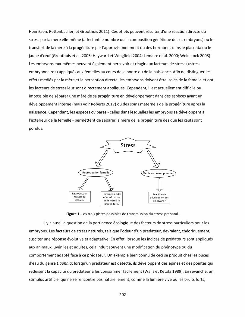
202
Henriksen, Rettenbacher, et Groothuis 2011). Ces effets peuvent résulter d'une réaction directe du
stress par la mère elle-même (affectant le nombre ou la composition génétique de ses embryons) ou le
transfert de la mère à la progéniture par l'approvisionnement ou des hormones dans le placenta ou le
jaune d'œuf (Groothuis et al. 2005; Hayward et Wingfield 2004; Lemaire et al. 2000; Weinstock 2008).
Les embryons eux-mêmes peuvent également percevoir et réagir aux facteurs de stress («stress
embryonnaire») appliqués aux femelles au cours de la ponte ou de la naissance. Afin de distinguer les
effets médiés par la mère et la perception directe, les embryons doivent être isolés de la femelle et ont
les facteurs de stress leur sont directement appliqués. Cependant, il est actuellement difficile ou
impossible de séparer une mère de sa progéniture en développement dans des espèces ayant un
développement interne (mais voir Roberts 2017) ou des soins maternels de la progéniture après la
naissance. Cependant, les espèces ovipares - celles dans lesquelles les embryons se développent à
l'extérieur de la femelle - permettent de séparer la mère de la progéniture dès que les œufs sont
pondus.
Figure 1. Les trois pistes possibles de transmission du stress prénatal.
Il y a aussi la question de la pertinence écologique des facteurs de stress particuliers pour les
embryons. Les facteurs de stress naturels, tels que l'odeur d'un prédateur, devraient, théoriquement,
susciter une réponse évolutive et adaptative. En effet, lorsque les indices de prédateurs sont appliqués
aux animaux juvéniles et adultes, cela induit souvent une modification du phénotype ou du
comportement adapté face à ce prédateur. Un exemple bien connu de ceci se produit chez les puces
d'eau du genre Daphnia; lorsqu'un prédateur est détecté, ils développent des épines et des pointes qui
réduisent la capacité du prédateur à les consommer facilement (Walls et Ketola 1989). En revanche, un
stimulus artificiel qui ne se rencontre pas naturellement, comme la lumière vive ou les bruits forts,
203
appliqué directement aux embryons en développement («stress artificiel») devrait confondre la capacité
de l'espèce à répondre, entraînant des réponses inadaptées. Par exemple, l'exposition à des niveaux
élevés de bruit anthropique a été liée à la fois aux réponses au stress et aux changements de
comportement chez les cétacés, ce qui peut avoir des conséquences aussi graves que l'échouage et la
mort (Weilgart 2007).
Cette thèse s'inscrit dans une étude éthologique plus large intitulée "Effets de stress prénatals
sur le développement précoce des comportements et des capacités cognitives", ou "PReSTO'Cog" pour
abréger. PReSTO'Cog est une collaboration entre cinq laboratoires à travers la France. Chaque
laboratoire étudie un modèle animal différent: la poule domestique, la caille japonaise, la truite, le
poisson zèbre et la seiche européenne. Ces espèces représentent une gamme variée de groupes
d'animaux: les invertébrés (seiches) et les vertébrés (poissons, oiseaux) ainsi que les espèces sauvages
(seiches) et les espèces domestiquées (poule) et les poïkilotherms (seiche, poisson) et les
homéothermies (oiseaux). Tous sont ovipares et nidifuges, ce qui permet à la progéniture d'être isolée
expérimentalement de la femelle pendant le développement embryonnaire et sans l'influence post-
natale de l'interaction maternelle. Enfin, ils sont également relativement autonomes à la naissance, ce
qui permet un test de comportement immédiat de la progéniture. En comparant ces espèces
phylogénétiquement éloignées, nous abordons le sujet du stress prénatal à partir des troisièmes et
quatrième perspectives de Tinbergen - les indices adaptatifs et phylogénétiques - découvrant les
pressions évolutives et les antécédents familiaux qui ont conduit au comportement que nous observons
aujourd'hui dans ces espèces. En fin de compte, de telles idées pourraient s'appliquer à d'autres groupes
d'animaux, y compris les mammifères, ce qui entraînerait des améliorations du bien-être humain et
animal.
Le thème unifiant de ce projet est de déterminer si le stress prénatal induit des changements
dans la progéniture et si le type de facteur de stress prénatal a eu une incidence sur la manière dont la
progéniture réagit. Les effets des facteurs de stress maternel sur les descendants diffèrent-ils de ceux
des facteurs de stress appliqués directement aux embryons eux-mêmes ? Est-ce que la réponse à un
facteur de stress artificiel diffère de ce qui se produit en réponse à un phénomène naturel ? Les effets
du stress prénatal sont évalués à travers une gamme de tests physiologiques, comportementaux et
d'apprentissage sur les jeunes descendants. Nous recherchons également des indices sur les
mécanismes de ces effets, en particulier les preuves endocrinologiques pour le transfert des hormones
du stress de la mère à la progéniture et des changements dans la croissance et la morphologie du
204
cerveau. Ces questions sondent le comportement des premières perspectives de Tinbergen - l'analyse
mécanique et ontogénétique - des processus biologiques innés et des influences externes qui
interagissent pour produire un répertoire comportemental particulier.
Modèle d'étude
Dans cette thèse, le stress prénatal est étudié à partir des quatre perspectives éthologiques
décrites par Tinbergen (1963) à travers un large spectre de tests physiologiques, comportementaux,
cognitifs et neurobiologiques pour déterminer si et comment le stress prénatal affecte la seiche. Deux
types principaux de stress ont été étudiés: les facteurs de stress ont été appliqués à la fois aux femelles
reproductrices (stress maternel) et aux embryons en développement (stress embryonnaire). Deux types
de facteurs de stress embryonnaires ont été appliqués: l'un naturel et l'autre artificiel (figure 2).
Beaucoup d'invertébrés sont à la fois ovipares et nidifuges, ce qui en fait des candidats
potentiels pour étudier les façons dont le stress peut affecter une espèce comme discuté ci-dessus. En
outre, les invertébrés représentent 97% des espèces sur terre, vivent dans presque toutes les parties de
la planète et démontrent une diversité impressionnante dans les modes de vie et de comportement. À
côté des arthropodes, les mollusques invertébrés sont le deuxième phylum le plus peuplé et, comme les
arthropodes, ont colonisé avec succès les habitats maritimes, d'eau douce et terrestre. Certains
mollusques ont également évolué de plusieurs façons convergentes avec les vertébrés (par exemple, le
«poumon» des gastéropodes terrestres), ce qui en fait de bons modèles comparatifs. Le céphalopode
est un groupe de mollusques composé d'environ 700 espèces existantes (Roger T. Hanlon and
Messenger 1998), divisé en quatre groupes principaux: poulpes, calmars, seiches et nautilus. Ils sont
exceptionnellement sophistiqués, démontrant des capacités perceptuelles avancées, l'apprentissage, la
mémoire, la résolution de problèmes et la plasticité (Hochner, Shomrat, et Fiorito 2006). Les
céphalopodes ont des systèmes nerveux fortement centralisés (Budelmann 1995) et peu importants,
avec des rapports cerveau / poids corporel supérieurs à ceux des poissons et des reptiles et approchant
ceux des mammifères et des oiseaux. Le système nerveux a été bien étudié au cours du siècle dernier,
avec des tâches cognitives spécifiques localisées dans des lobes spécifiques du cerveau (Dickel et al.
2013 pour revue). Les céphalopodes affichent des adaptations convergentes (par exemple, l'effet
céphalopode et les vertébrés) et divergentes (par exemple, propulsion par pont céphalopodique par
rapport à la propulsion musculaire des poissons) à des défis évolutifs comme autres groupes d'animaux
(Packard 1972; Hochner, Shomrat, et Fiorito 2006). Leur sophistication et leur position d'invertébrés
signifient que les céphalopodes ont beaucoup d'intuition pour offrir des contrepoints aux modèles de
205
vertébrés plus traditionnels, et leurs comportements uniques et extraordinaires remettent souvent en
question nos notions sur les principes généraux qui sous-tendent le comportement animal. Il a même
été posé que la concurrence entre les poissons et les céphalopodes a façonné l'évolution de nombreux
traits dans les deux groupes pendant le mésozoïque (Packard 1972).
Comme les autres modèles PReSTO'Cog, la seiche commune Sepia officinalis (Linnaeus 1758) est
un excellent modèle pour étudier les effets du stress prénatal parce qu'il est ovipare et nidifuge. S.
officinalis est également une espèce importante sur le plan commercial et scientifique: des pêches
existent à la fois dans l'Atlantique et la Méditerranée (Dunn 1999) et elle est cultivée dans plusieurs
laboratoires et aquacoles (Pascual 1978; Forsythe, DeRusha, et Hanlon 1994; Domingues, Sykes, and
Andrade 2002). En effet, avec Octopus vulgaris, c'est l'une des espèces de céphalopodes les plus
étudiées. Plus important encore, les seiches et autres céphalopodes ont l'avantage d'être
phylogénétiquement éloignés des modèles animaux plus typiques comme les rats et les singes: ils sont
un groupe d’invertébré séparé des vertébrés par des centaines de millions d'années d'évolution
distinctes, ce qui leur permet de servir de référence indiquez si les effets de stress que nous observons
dans différentes espèces sont des produits d'ascendance partagée ou des développements évolutifs
distincts (troisième et quatrième questions de Tinbergen). En fin de compte, une meilleure
compréhension des effets du stress prénatal chez S. officinalis donnera un aperçu général des processus
et des stratégies par lesquels les organismes survivent.
En outre, cette recherche aidera à combler les lacunes dans la connaissance des besoins
biologiques spécifiques de la seiche, en particulier ceux concernant le logement, la reproduction et les
marqueurs comportementaux du bien-être. L'aperçu de ce travail améliorera la capacité des
aquaculteurs et des chercheurs à établir des normes de soins et des pratiques courantes. Cela est
particulièrement nécessaire en raison de l'inclusion récente des seiches et d'autres céphalopodes dans
la législation européenne en matière de protection des animaux (directive 2010/63/UE) régissant
l'utilisation des animaux dans les procédures expérimentales. Cela pourrait aussi contribuer aux futurs
efforts d'élevage et de relance captifs, ce qui risque de devenir de plus en plus nécessaire avec la
demande alimentaire croissante et le changement climatique. Par exemple, Sepia apama, la seiche
australienne géante, a récemment été désignée comme «presque menacée» en raison de la pêche
intensive d'agrégats de reproduction dans des endroits spécifiques et fait face à une diminution prévue
de 20% des niveaux de population si les taux de capture actuels se poursuivent (UICN 2017). Et de façon
alarmante, les journaux britanniques locaux signalent que les pêcheurs profitent de la réglementation
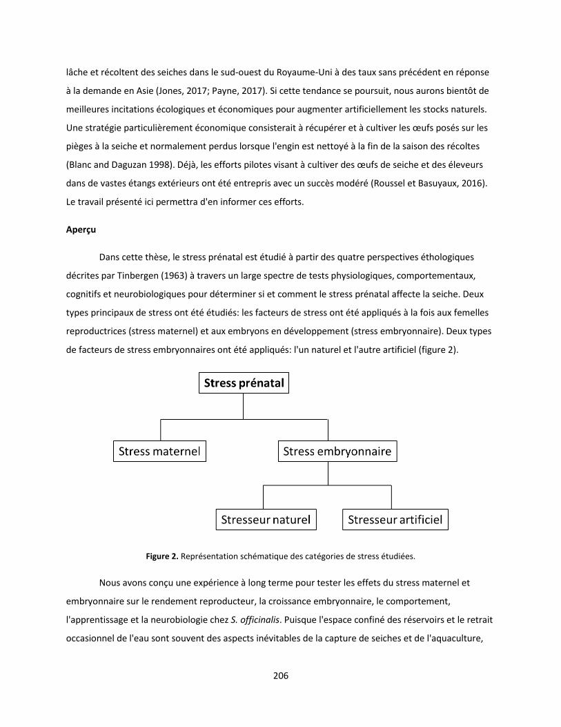
206
lâche et récoltent des seiches dans le sud-ouest du Royaume-Uni à des taux sans précédent en réponse
à la demande en Asie (Jones, 2017; Payne, 2017). Si cette tendance se poursuit, nous aurons bientôt de
meilleures incitations écologiques et économiques pour augmenter artificiellement les stocks naturels.
Une stratégie particulièrement économique consisterait à récupérer et à cultiver les œufs posés sur les
pièges à la seiche et normalement perdus lorsque l'engin est nettoyé à la fin de la saison des récoltes
(Blanc and Daguzan 1998). Déjà, les efforts pilotes visant à cultiver des œufs de seiche et des éleveurs
dans de vastes étangs extérieurs ont été entrepris avec un succès modéré (Roussel et Basuyaux, 2016).
Le travail présenté ici permettra d'en informer ces efforts.
Aperçu
Dans cette thèse, le stress prénatal est étudié à partir des quatre perspectives éthologiques
décrites par Tinbergen (1963) à travers un large spectre de tests physiologiques, comportementaux,
cognitifs et neurobiologiques pour déterminer si et comment le stress prénatal affecte la seiche. Deux
types principaux de stress ont été étudiés: les facteurs de stress ont été appliqués à la fois aux femelles
reproductrices (stress maternel) et aux embryons en développement (stress embryonnaire). Deux types
de facteurs de stress embryonnaires ont été appliqués: l'un naturel et l'autre artificiel (figure 2).
Figure 2. Représentation schématique des catégories de stress étudiées.
Nous avons conçu une expérience à long terme pour tester les effets du stress maternel et
embryonnaire sur le rendement reproducteur, la croissance embryonnaire, le comportement,
l'apprentissage et la neurobiologie chez S. officinalis. Puisque l'espace confiné des réservoirs et le retrait
occasionnel de l'eau sont souvent des aspects inévitables de la capture de seiches et de l'aquaculture,
207
nous avons utilisé ces facteurs comme facteurs de stress aigu chronique et répété pour créer un groupe
de traitement «mère stressée». Nous avons comparé ce groupe aux «mères témoins non stressées»
(UM-C). Dans certains tests, nous avons également comparé ces deux groupes à un groupe «mère
sauvage» (WM). Dans une deuxième expérience, nous avons examiné les effets du stress embryonnaire.
Nous avons subdivisé les œufs UM-C en trois groupes; un groupe témoin (UM-C), un groupe exposé à un
facteur de stress d'origine naturelle et un groupe exposé à un facteur de stress artificiel tout au long du
développement embryonnaire. En tant que facteur de stress naturel, nous avons sélectionné le bar
Dicentrarchus labrax, un prédateur commun dans la Manche qui se nourrit de petites seiches (UM-PE).
En tant que facteur de stress artificiel, nous avons sélectionné des épisodes de lumière artificielle
brillante (LED) chronométrés au hasard tout au long de la journée au cours du développement
embryonnaire. Nous avons ensuite appliqué une batterie de tests à la progéniture de ces groupes de
stress après l'éclosion afin d'évaluer ses effets.
Chapitre 1
Au chapitre 1, nous présentons des données démontrant que le stress maternel réduit
nettement la ponte des seiches. Cette différence n'a pas pu être expliquée par la taille des femelles ou le
temps de survie: Il n'y avait pas de différence de poids moyen ou de LMD entre les groupes de stress et
les femelles des deux groupes ont survécu un peu plus de deux semaines après leur capture avant de
mourir naturellement. En plus de réduire le nombre d'œufs pondus par SM, moins d'œufs SM ont éclos.
Le taux d'éclosion de l'UM-C (57,14%) est tombé à mi-portée des taux d'éclosion déclarés dans la
littérature sur l'aquaculture, alors que le taux de SM était très faible (22,27%). Une telle différence dans
le taux d'éclosion n'a pas été observée dans les groupes de stress embryonnaires, suggérant que les
facteurs de stress appliqués aux mères peuvent avoir un impact plus important sur l'éclosion que les
facteurs de stress appliqués directement à la progéniture. Ni le stress maternel ni embryonnaire n'a été
associé à des différences significatives dans la taille des éclosions.
Fait intéressant, 25% de SM et aucun des UM-C qui ont produit des œufs viables pondent
partiellement ou entièrement translucides. Dans la plupart des cas, la membrane de l'œuf de S.
officinalis est imprégnée d'encre noire de la mère, bien que des œufs translucides soient parfois
observés en aquaculture et dans la nature. Dans notre expérience, le ratio de SM affichant ce trait ne
diffère pas significativement de l'UM-C, mais il constitue une tendance statistique et nous croyons qu'il
peut être lié au traitement de stress. La présence d'œufs translucides dans une couvée pourrait servir de
208
marqueur de la présence de facteurs de stress pendant la ponte, donnant des indications indirectes sur
les conditions de ponte à l'état sauvage ou la pertinence d'un environnement d'élevage en captivité.
En plus de tester les effets du stress sur la reproduction, nous avons également cherché un
indicateur simple de stress chez les femelles pondeuses. Chez de nombreux autres animaux (par
exemple, les oiseaux, les mammifères, les poissons), le cortisol et / ou les glucocorticoïdes sont sécrétés
en réponse au stress et interviennent dans la plupart de ses effets (Moberg 1991). L'échantillonnage
fécal est relativement simple et non intrusif, nous avons donc prélevé des échantillons quotidiens et de
la corticostérone quantifiée. Malheureusement, les niveaux de corticostérone fécale ne se sont pas
avérés être un indicateur particulièrement bon du stress chez cette espèce (au moins pendant la
reproduction). Nous avons également tenté de relier le matériel de reproduction inutilisé des femelles
décédées à des niveaux de stress. Les œufs prêts à la fécondation, à l'encapsulation et à la ponte sont
stockés dans la cavité palléale qui, à la mort, peut contenir jusqu'à 500 ovocytes (Boletzky 1988;
Laptikhovsky et al. 2003). Puisque les ovocytes inutilisés dans la cavité palléale des femelles décédées
indiquent un potentiel de reproduction inutilisé, nous avons examiné le nombre restant au décès, en
supposant qu'il y aurait plus de résidus dans la SM au décès. Cependant, il n'y avait pas de différence
entre les deux groupes, et donc ce trait ne peut pas être utilisé comme un mesure de les niveaux de
stress. Bien que ces deux facteurs ne puissent constituer de simples marqueurs de stress chez les
seiches, les aquaculteurs et les biologistes des pêches pourraient un jour utiliser un nombre réduit
d'œufs et l'absence d'encre dans les œufs pour indiquer la présence de stress maternel chez les seiches.
Les données présentées au chapitre 1 montrent que le stress peut influencer la seiche par au
moins une des trois voies potentielles en réduisant la production reproductive totale des femelles et la
qualité des œufs produits. Cela fait allusion à l'existence de compromis entre la réponse du stress
maternel et sa production reproductive. Plus important encore, il montre que le stress peut avoir un
impact sur la seiche avant même que ses œufs ne soient pondus, un facteur que nous devons prendre
en compte lors de la formulation des explications mécaniques et adaptatives (première et troisième
perspective de Tinbergen) du comportement de la seiche. Il suggère également une ligne directrice très
spécifique pour l'aquaculture de la seiche: minimiser la manipulation des femelles reproductrices afin de
maximiser le rendement reproducteur et la qualité des œufs.
Chapitre 2
209
Après avoir examiné en profondeur les jeunes seiches au moyen de deux articles de synthèse, le
chapitre 2 présente les résultats de nombreuses expériences comportementales, de tests
d'apprentissage et de tests neurobiologiques.
Modelage du corps
Nous avons mesuré la «perturbation» de la structure du corps de la seiche en utilisant le
protocole «d'hétérogénéité» (HI) décrit dans (Di Poi et al. 2014) sur un fond uniforme en damier gris et
un damier noir et blanc. Dans tous les groupes, le HI moyen sur le fond perturbateur était constamment
plus élevé que celui sur l'uniforme, suggérant que toutes les seiches ont ajusté leurs modèles de corps à
l'arrière-plan. Des différences significatives ont également été observées entre les groupes: Dans notre
expérience, le stress maternel et artificiel (lumière LED) a augmenté la perturbation moyenne du
schéma corporel affiché, bien que les différences entre les groupes de stress maternels étaient plus
importantes et plus significatives qu'entre les groupes de stress embryonnaires. Nos résultats suggèrent
également que la captivité femelle pendant la ponte peut induire un biais de groupe pour une plus
grande perturbation chez sa progéniture, puisque la progéniture des mères sauvages avait l'HI le plus
élevé globalement.
Prédation
Les motivations de l'alimentation et les capacités prédatrices ont été testées 4 jours après
l'éclosion en plaçant la seiche dans une petite arène avec des crevettes pendant 15 minutes. Près de
deux fois plus de descendants UM-LE ont tenté de capturer que UM-C ou UM-PE. Les UM-LE étaient
également capables de détecter les proies à une distance significativement plus grande que les UM-PE,
bien qu'ils soient statistiquement identiques au groupe témoin. Il convient également de noter que
même si une plus grande proportion de crevettes capturées par UM-LE, elles n'étaient pas de meilleurs
prédateurs que les autres groupes, puisque la capture et le succès ne différaient pas significativement
entre les groupes (près de 100%).
Latéralité visuelle
Suite à des travaux démontrant l'existence d'un biais de rotation à gauche chez de jeunes
seiches, nous avons testé la latéralisation visuelle de la seiche de cinq jours dans un labyrinthe en forme
de T. Aucun biais au niveau du groupe n'a été trouvé dans le groupe témoin. Parmi tous les autres
groupes, seul le groupe WM a affiché une préférence au niveau du groupe vers le côté gauche le jour 5.
210
Le fait que la préférence oculaire existe dans le groupe MW suggère que lorsque la ponte et le
développement précoce se produisent dans la nature, la maturation de la préférence d'utilisation de
l'œil gauche est plus rapide.
Activité nocturne
Nous avons mesuré les niveaux d'activité nocturne de base chez les nouveau-nés en utilisant un
logiciel de suivi comportemental. Nous n'avons trouvé aucune différence entre les groupes de stress
embryonnaires, tandis que la progéniture mère stressée était associée à une plus grande activité que la
progéniture mère témoin: la distance totale parcourue et le temps passé à bouger étaient plus élevés en
moyenne que la progéniture UM-C. Nous avons également observé une tendance statistique pour les
nouveau-nés MW à parcourir une plus grande distance que les UM-C. Ces résultats indiquent que la
progéniture des mères stressées était plus active, passant plus de temps en mouvement et par
conséquent parcourant une plus grande distance que la progéniture de l'UM-C.
Réponse aux menaces
Nous avons également suivi l'activité en réponse à l'introduction d'odeurs de prédateurs ou
d'eau vierge dans leur environnement immédiat. Une réduction de l'activité a été observée dans tous les
groupes après l'ajout de l'un ou l'autre stimulus dans le prolongement d'une tendance à la diminution
progressive de l'activité au fil du temps mais aucun groupe n'a présenté de réponse à l'odeur du
prédateur différente de celle de l'eau vierge.
Tests d'apprentissage PIT
Nous avons appliqué un test d'apprentissage standard aux mineurs à trois et cinq semaines
d'âge. Il n'y avait pas de différences significatives entre les groupes dans la mesure de la performance du
test PIT entre le stress maternel et les groupes témoins. Parmi les groupes de stress embryonnaires à
l'âge de trois semaines, la progéniture exposée à la lumière (UM-LE) a pris beaucoup moins de temps
pour atteindre le critère que les deux autres groupes, suggérant une capacité à apprendre plus
rapidement. Outre les indications selon lesquelles UM-LE pourrait apprendre plus rapidement à trois
semaines, il n'y avait pas d'autres effets des traitements du stress prénatal sur les capacités
d'apprentissage et de mémoire dans la tâche PIT.
Monoamines
211
Nous avons quantifié la concentration de sept monoamines et de leurs principaux métabolites
dans le tissu neural des nouveau-nés. Tous les groupes de stress (SM, UM-PE et UM-LE) présentaient des
concentrations de sérotonine significativement plus faibles que le groupe témoin. Dans le même temps,
le taux de renouvellement de la sérotonine (5-HiAA / 5-HT) était significativement plus faible chez les
sujets SM et UM-LE que chez les témoins, mais significativement plus élevé chez les sujets UM-PE. Un
rapport plus élevé de la sérotonine à son métabolite principal indique un taux de renouvellement plus
élevé (Bidel 2015), ce qui signifie un taux plus rapide de synthèse de la sérotonine. Dans les
céphalopodes, la sérotonine agit comme un neurotransmetteur, avec des effets moteurs et
chromatiques, ainsi qu'un rôle important dans la transmission synaptique et l'induction de la
potentialisation à long terme (Messenger 1996; Shomrat et al. 2010). Ainsi, les concentrations plus
faibles de sérotonine mesurées dans tous les groupes de stress, plus les taux de renouvellement plus
faibles de SM et d'UM-LE pourraient être des signes de déficits neurologiques. Les concentrations de
noradrénaline (NA) étaient significativement plus faibles dans les SM et les UM-PE et significativement
plus élevées dans les UM-LE que chez les témoins. Dans les céphalopodes, la NA est associée à une
respiration plus rapide et à des niveaux plus élevés de conscience et de cognition (Messenger 1996;
Bidel 2015). Ainsi, les différences entre les groupes pourraient indiquer des niveaux différents de
«vigilance» en raison du stress prénatal, avec UM-LE en état d'alerte élevé et les autres groupes de
stress à un niveau réduit de vigilance. Ensemble, les différences dans la concentration de sérotonine, le
taux de renouvellement de la sérotonine et les concentrations de noradrénaline indiquent que le stress
prénatal affecte la chimie du cerveau de la seiche qui naît.
Volumétrie et neurogenèse
En utilisant la volumétrie et le marquage à la phosphohistone, nous avons quantifié la croissance
et la division cellulaire dans le cerveau. La mesure des différents lobes cérébraux par rapport à la taille
globale du cerveau a révélé des lobes verticaux (VL) significativement plus petits dans tous les groupes
de stress par rapport au groupe témoin. Dans le même temps, la coloration à la phosphohistone a
montré une neurogenèse plus élevée (c'est-à-dire une croissance) dans les lobes optiques (OL) et VL de
SM. Le VL et l'OL sont des sièges d'apprentissage et de mémoire chez les céphalopodes, et leur taille
relative et leur croissance sont corrélées avec l'apprentissage et la mémoire (Dickel, Chichery, et
Chichery 2001). La plus petite taille des LV dans tous les groupes de stress suggère qu'ils sont
cognitivement compromis à l'éclosion par rapport au groupe témoin. En outre, la réduction de la
neurogenèse dans la VL et l'OL de SM suggère que ce groupe a été retardé dans le
212
neurodéveloppement. La neurogenèse supérieure dans le VL et l'OL de SM suggère que le stress
transmis de la mère à la progéniture a un effet plus fort sur la taille de la VL et la croissance que le stress
embryonnaire.
L'existence de différences comportementales et neurobiologiques dans les groupes de stress
maternels et embryonnaires démontre que le stress peut être transféré de la mère à la progéniture ainsi
que vécu directement par les embryons eux-mêmes. Le manque général d'apprentissage et les
différences de mémoire étaient inattendus étant donné les effets du stress prénatal chez les autres
espèces et les différences de taille de la LV à l'éclosion.
Chapitre 3
Dans une expérience distincte mais liée, nous avons analysé les effets de l'environnement
d'incubation en comparant un groupe d'œufs recueillis en laboratoire («Lab») à un groupe laissé in situ
jusqu'à la quasi-éclosion («Mer»). Nous avons ensuite mesuré la taille, les modèles corporels et les
capacités de prédation de la progéniture. ML était similaire entre les deux groupes de traitement, et la
structuration du corps contre un fond quadrillé ne différait pas significativement entre les groupes. Nous
avons mesuré cinq aspects du comportement prédateur lors de la première rencontre des nouveau-nés
avec leurs proies préférées, la crevette (Wells 1958): Tentative de capture, taux de capture, taux de
réussite, temps de capture et distance de détection. Aucune de ces variables ne différait
significativement entre les groupes et le taux de capture était très élevé (entre 85% et 90% selon
Messenger 1968). Ceci suggère que les nouveau-nés élevés en laboratoire seraient également capables
de se nourrir pendant cette période critique de croissance. Une différence existait entre les groupes: les
nouveau-nés incubés en laboratoire présentaient un nombre significativement plus élevé d'individus HI
(plus perturbateurs) que les nouveau-nés «Mer». Cela suggère que l'environnement d'incubation peut
affecter la structure du corps dans certains contextes.
Le chapitre 3 démontre que l'incubation dans un environnement de laboratoire n'affecte pas
fortement trois aspects fondamentaux du comportement et de la survie chez les nouveau-nés de seiche:
la croissance embryonnaire, la structure du corps et la capacité de prédation. Le manque général de
différences entre les nouveau-nés naturellement et artificiellement incubés renforce la confiance dans
les données expérimentales existantes. Nos résultats sont également encourageants du point de vue de
la conservation; la seiche artificiellement incubée pourrait augmenter les stocks de poissons ou
remplacer la seiche sauvage dans certaines situations.
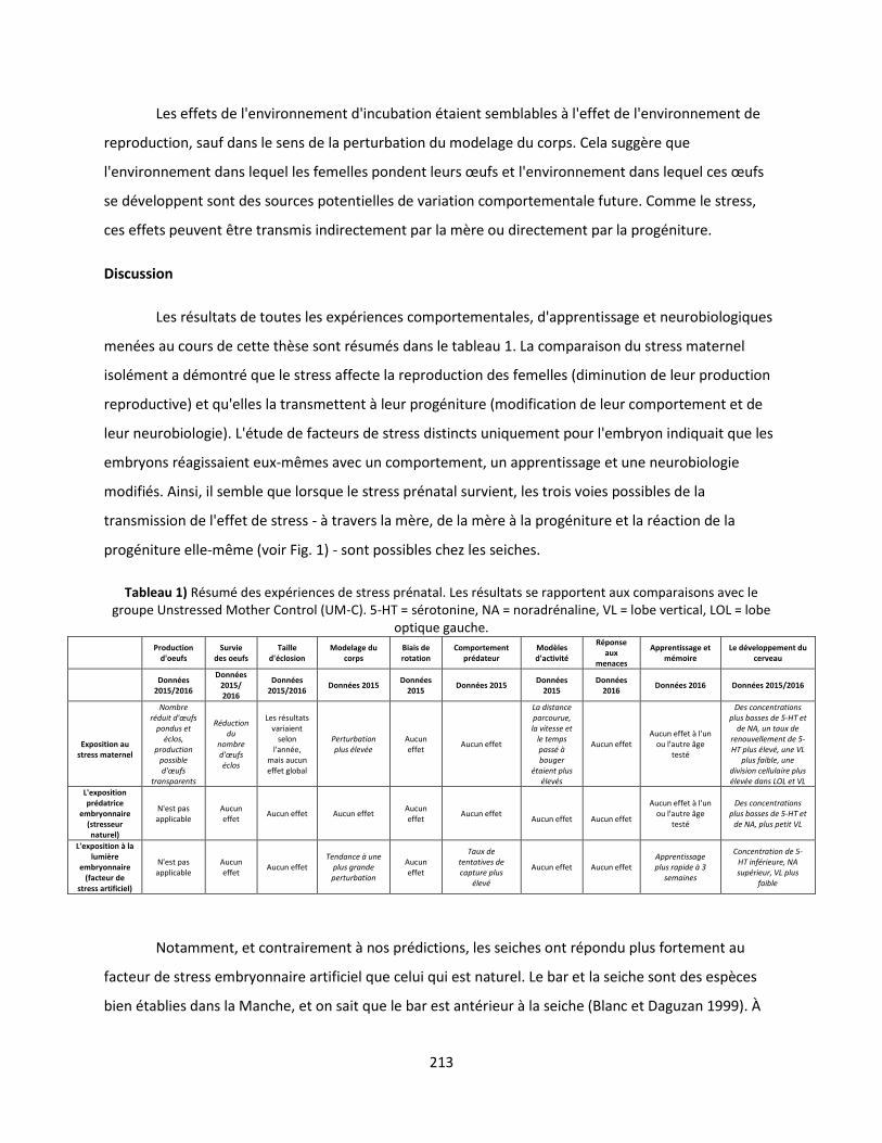
213
Les effets de l'environnement d'incubation étaient semblables à l'effet de l'environnement de
reproduction, sauf dans le sens de la perturbation du modelage du corps. Cela suggère que
l'environnement dans lequel les femelles pondent leurs œufs et l'environnement dans lequel ces œufs
se développent sont des sources potentielles de variation comportementale future. Comme le stress,
ces effets peuvent être transmis indirectement par la mère ou directement par la progéniture.
Discussion
Les résultats de toutes les expériences comportementales, d'apprentissage et neurobiologiques
menées au cours de cette thèse sont résumés dans le tableau 1. La comparaison du stress maternel
isolément a démontré que le stress affecte la reproduction des femelles (diminution de leur production
reproductive) et qu'elles la transmettent à leur progéniture (modification de leur comportement et de
leur neurobiologie). L'étude de facteurs de stress distincts uniquement pour l'embryon indiquait que les
embryons réagissaient eux-mêmes avec un comportement, un apprentissage et une neurobiologie
modifiés. Ainsi, il semble que lorsque le stress prénatal survient, les trois voies possibles de la
transmission de l'effet de stress - à travers la mère, de la mère à la progéniture et la réaction de la
progéniture elle-même (voir Fig. 1) - sont possibles chez les seiches.
Tableau 1) Résumé des expériences de stress prénatal. Les résultats se rapportent aux comparaisons avec le groupe Unstressed Mother Control (UM-C). 5-HT = sérotonine, NA = noradrénaline, VL = lobe vertical, LOL = lobe
optique gauche.
Production
d'oeufs Survie
des oeufs Taille
d'éclosion Modelage du
corps Biais de rotation
Comportement prédateur
Modèles d'activité
Réponse aux
menaces
Apprentissage et mémoire
Le développement du cerveau
Données
2015/2016
Données 2015/ 2016
Données 2015/2016
Données 2015 Données
2015 Données 2015
Données 2015
Données 2016
Données 2016 Données 2015/2016
Exposition au
stress maternel
Nombre réduit d'œufs
pondus et éclos,
production possible d'œufs
transparents
Réduction du
nombre d'œufs éclos
Les résultats variaient
selon l'année,
mais aucun effet global
Perturbation plus élevée
Aucun effet
Aucun effet
La distance parcourue, la vitesse et
le temps passé à bouger
étaient plus élevés
Aucun effet Aucun effet à l'un
ou l'autre âge testé
Des concentrations plus basses de 5-HT et
de NA, un taux de renouvellement de 5-HT plus élevé, une VL
plus faible, une division cellulaire plus élevée dans LOL et VL
L'exposition prédatrice
embryonnaire (stresseur naturel)
N'est pas applicable
Aucun effet
Aucun effet Aucun effet Aucun effet
Aucun effet
Aucun effet
Aucun effet
Aucun effet à l'un ou l'autre âge
testé
Des concentrations plus basses de 5-HT et
de NA, plus petit VL
L'exposition à la lumière
embryonnaire (facteur de
stress artificiel)
N'est pas applicable
Aucun effet
Aucun effet Tendance à une
plus grande perturbation
Aucun effet
Taux de tentatives de capture plus
élevé
Aucun effet Aucun effet Apprentissage plus rapide à 3
semaines
Concentration de 5-HT inférieure, NA supérieur, VL plus
faible
Notamment, et contrairement à nos prédictions, les seiches ont répondu plus fortement au
facteur de stress embryonnaire artificiel que celui qui est naturel. Le bar et la seiche sont des espèces
bien établies dans la Manche, et on sait que le bar est antérieur à la seiche (Blanc et Daguzan 1999). À
214
partir d'expériences antérieures, nous savions que la présentation prénatale de l'odeur du prédateur est
associée à des changements de latéralisation visuelle (Jozet-Alves et Hebert 2012). Ces faits, associés à
la flexibilité comportementale impressionnante des seiches, nous ont permis de prédire que la sélection
naturelle aurait produit une réaction comportementale à ce prédateur. Au lieu de cela, les seiches
exposées aux prédateurs n'ont montré aucune différence par rapport au groupe témoin dans aucun de
nos tests de comportement, bien qu'ils aient montré des changements dans la chimie et la morphologie
du cerveau. La présence de changements neurobiologiques dans les descendants des œufs exposés aux
prédateurs démontre que les embryons de seiche sont physiquement affectés par le stress prénatal,
alors que l'absence de réaction comportementale suggère une habituation aux indices de prédateurs
prénataux, comme indiqué à l'article 4. En effet, d'autres espèces peuvent adapter leurs réactions
innées anti-prédateurs en fonction du niveau de menace. De nombreux gastéropodes, un groupe proche
des céphalopodes, montrent des réactions aux odeurs de prédateur, et ces réactions sont plus fortes
lorsqu'elles sont associées à des signaux d'alarme de conspécifiques (p. Ex. McCarthy et Fisher 2000;
Jacobsen et Stabell 2004; Dalesman et al. 2006). Dans le même temps, nous savons que la seiche
s'habitue à certains stimuli prénataux (Romagny et al. 2012), il semble donc plausible que l'odeur
prédateur, associée à l'absence de signal d'alarme provenant des nombreux autres embryons de seiche
présents, n'a pas entraîne des changements de comportement parce que la seiche a appris à le
considérer comme non menaçant.
Contrairement au facteur de stress naturel, le facteur de stress artificiel a montré des
différences comportementales et d'apprentissage, ainsi que des différences dans la chimie et la
morphologie du cerveau. Étant donné qu'un décalage entre une réponse au stress et le facteur de stress
lui-même entraîne souvent des effets secondaires négatifs, nous avons prédit que le facteur de stress
artificiel (ce qui n'était pas quelque chose que les espèces auraient pu expérimenter historiquement
pour faire évoluer une réaction par sélection naturelle) aurait un effet "négatif" sur la progéniture.
Cependant, le groupe exposé à la lumière a montré des effets «positifs» dans les comportements testés:
de meilleures capacités d'apprentissage et une moindre motivation alimentaire conduisant à un taux de
prédation plus élevé. Une hypothèse d'expliquer cela est que l'exposition prénatale à un facteur de
stress sans précédent a induit une réponse d'alarme généralisée qui a entraîné une accélération du
développement. Alternativement, on sait que l'exposition prénatale à la lumière affecte le rythme
circadien dans une gamme d'animaux, y compris les seiches, qui peuvent éclore plus tôt ou plus tard en
fonction du régime léger (Paulij et al. 1991). Dans ce cas, la lumière peut avoir un développement
accéléré.
215
Enfin, nous avons également pu faire des comparaisons indirectes entre l'incubation artificielle
et la reproduction artificielle et avons constaté des impacts similaires sur les comportements testés (pas
d'effet sur le comportement prédateur, les effets mineurs sur le modelage du corps). Cela montre que
l'environnement, ainsi que les facteurs de stress spécifiques, peuvent avoir un impact sur le
comportement post-natal de la progéniture, ce qui doit être pris en compte lors de la conception et
l'interprétation d'expériences avec de jeunes seiches.
Les résultats présentés nous permettent de commencer à formuler un modèle approximatif de
stress prénatal dans les seiches des quatre perspectives de Tinbergen. En ce qui concerne les
mécanismes de transmission du stress prénatal (première perspective de Tinbergen), nous émettons
l'hypothèse que chez la seiche, les femelles pourraient transférer de la corticostérone à la progéniture
dans l'œuf. Cela a fait l'objet d'un intérêt particulier chez les vertébrés, en particulier les oiseaux, qui ont
été trouvés pour imprégner leur jaune d'œuf avec des hormones de stress qui affectent l'axe
hypothalamique-hypophysaire et surrénal (HPA) de leur progéniture (p. Ex. Charil et al. 2010). Nous
n'avons pas trouvé de preuve définitive à l'encontre de cette hypothèse ou contre cette hypothèse, mais
notre travail suggère que d'autres possibilités possibles de transmission du stress de la mère à la
progéniture sont possibles, puisque la progéniture de la mère stressée différait d'autres groupes de
stress néanmoins. Espérons que, au fur et à mesure que les techniques analytiques moléculaires et
neurologiques deviennent plus raffinées et largement disponibles, nous pourrons déterminer les
mécanismes exacts qui conduisent à des changements de comportement après le stress prénatal.
Nos expériences ont eu beaucoup de perspicacité pour se lancer sur les perspectives deuxième
et troisième de Tinbergen, l'ontogénique et l'adaptatif. D'après les différences de comportement du
groupe du stress artificiel, on voit que, de façon ontogénique, la période prénatale ne peut pas être
ignorée comme source d'influence comportementale, puisque les facteurs stressants subis par
l'embryon semblent modifier les comportements futurs. Les générations antérieures de chercheurs ont
largement négligé cette fois, car on présumait que les embryons ne pouvaient pas percevoir le monde
extérieur, bien que ce point de vue ait été renversé au cours des dernières décennies. Nos résultats
fournissent des preuves supplémentaires (avec Darmaillacq, Lesimple, et Dickel 2008; Romagny et al.
2012; Jozet-Alves et Hebert 2012), que la seiche peut percevoir le monde en dehors de la membrane de
l'œuf et modifier son comportement futur en réponse. D'un point de vue adaptatif, nous avons constaté
que le stress maternel pourrait être un meilleur indicateur des perspectives d'avenir pour les seiches
embryonnaires que l'expérience embryonnaire directe: beaucoup plus de différences comportementales
216
étaient apparentes en association avec le stress maternel qu'avec l'embryonnaire. D'autre part, on
pourrait soutenir que les réponses au facteur de stress embryonnaire étaient plus clairement
«adaptables» (meilleure apprentissage et prédation), ce qui suggère que l'expérience embryonnaire
directe est plus instructive pour la seiche après tout. De toute évidence, les réponses des seiches sont
complexes, et d'autres expériences sont nécessaires pour comprendre l'interaction des forces sélectives
conduisant le comportement post-natal induit par le stress.
Enfin, nous avons pu aborder le stress prénatal à partir de la quatrième perspective de
Tinbergen, le phylogénique, en comparant nos résultats de la seiche aux autres modèles PReSTO'Cog.
Les quatre modèles (poulets, caille, poisson zèbre et truite) ont montré des effets sur le stress maternel
ainsi que sur les facteurs de stress embryonnaires artificiels et naturels. Ces résultats impliquent que
l'existence de réponses au stress est fortement conservée dans les groupes phylogénétiques. En outre, il
semble que, comme les seiches, les effets du stress prénatal chez les animaux se manifestent sur une
multitude de niveaux et sont compliqués par de nombreuses interactions.
Résumé
Les résultats de cette thèse démontrent sans équivoque que le stress prénatal affecte les
seiches et suggère fortement que trois des trois avenues potentielles de stress (chez la mère, de la mère
à la progéniture et de la progéniture en développement directement) semblent fonctionner chez cette
espèce. La figure 3 illustre la gamme des influences prénatales qui ont été trouvées pour modifier le
comportement post-natal. En comparant les groupes de stress, nous avons fait plusieurs déductions
provisoires sur le stress prénatal chez les seiches des trois premières perspectives de Tinbergen. Du
point de vue mécaniste, nous n'avons trouvé aucune preuve de transfert de corticostérone, mais
certaines possibilités que les effets du stress prénatal sont médiées par des changements dans la
neurobiologie. Lors de l'examen du stress de la perspective ontogénique, nous avons constaté une
confirmation supplémentaire que l'expérience embryonnaire modifie les comportements futurs dans
cette espèce. D'un point de vue adaptatif, nous avions des indications mitigées sur le fait que le stress
maternel ou un facteur de stress embryonnaire artificiel avait un effet plus fort et plus positif sur le
comportement de la progéniture. Enfin, en comparant nos résultats de seiches à ceux des autres
modèles animaux de Presto'Cog et en utilisant la quatrième perspective de Tinbergen, nous voyons que
les effets de stress prénatal semblent être partagés par une large gamme de groupes d'animaux.
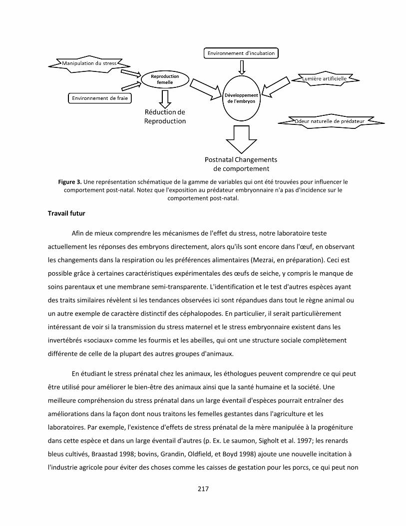
217
Figure 3. Une représentation schématique de la gamme de variables qui ont été trouvées pour influencer le comportement post-natal. Notez que l'exposition au prédateur embryonnaire n'a pas d'incidence sur le
comportement post-natal.
Travail futur
Afin de mieux comprendre les mécanismes de l'effet du stress, notre laboratoire teste
actuellement les réponses des embryons directement, alors qu'ils sont encore dans l'œuf, en observant
les changements dans la respiration ou les préférences alimentaires (Mezrai, en préparation). Ceci est
possible grâce à certaines caractéristiques expérimentales des œufs de seiche, y compris le manque de
soins parentaux et une membrane semi-transparente. L'identification et le test d'autres espèces ayant
des traits similaires révèlent si les tendances observées ici sont répandues dans tout le règne animal ou
un autre exemple de caractère distinctif des céphalopodes. En particulier, il serait particulièrement
intéressant de voir si la transmission du stress maternel et le stress embryonnaire existent dans les
invertébrés «sociaux» comme les fourmis et les abeilles, qui ont une structure sociale complètement
différente de celle de la plupart des autres groupes d'animaux.
En étudiant le stress prénatal chez les animaux, les éthologues peuvent comprendre ce qui peut
être utilisé pour améliorer le bien-être des animaux ainsi que la santé humaine et la société. Une
meilleure compréhension du stress prénatal dans un large éventail d'espèces pourrait entraîner des
améliorations dans la façon dont nous traitons les femelles gestantes dans l'agriculture et les
laboratoires. Par exemple, l'existence d'effets de stress prénatal de la mère manipulée à la progéniture
dans cette espèce et dans un large éventail d'autres (p. Ex. Le saumon, Sigholt et al. 1997; les renards
bleus cultivés, Braastad 1998; bovins, Grandin, Oldfield, et Boyd 1998) ajoute une nouvelle incitation à
l'industrie agricole pour éviter des choses comme les caisses de gestation pour les porcs, ce qui peut non
218
seulement diminuer le bien-être des animaux, mais aussi la quantité et la qualité du produit à base de
viande. De même, une meilleure compréhension du stress prénatal peut conduire à des moyens
d'atténuer ses effets négatifs sur les enfants et les adultes, et peut-être même pour empêcher qu'il ne
se produise même en premier lieu. Par exemple, les enfants humains de femmes qui ont été connus
pour avoir subi un stress pendant la grossesse pourraient être ciblés pour une attention maternelle
supplémentaire ou une thérapie prématurée, car les soins parentaux ont atténué certains des effets
négatifs du stress prénatal chez les rongeurs (examiné dans Francis et Meaney 1999). Cette recherche
nous aiderait également à prédire quelles sortes de choses peuvent être stressantes pour un fœtus. Ici,
nous avons vu une habituation possible en réponse à une odeur de prédateur que nous nous attendions
à être très stressante. Cela suggère que nous devrions être conscients de la possibilité que les embryons
humains puissent également s'habituer aux facteurs de stress. En effet, les fœtus humains sont habitués
à des stimuli vibriotactile répétés perçus dans l'utérus (Leader et al. 1982).

219
220
Resumé
Le stress prénatal est un sujet d'intérêt éthologique croissant en raison de ses effets sur la santé humaine et le bien-être des animaux. Cette thése de doctorat s’intéresse à la seiche Sepia officinalis, un modèle pratique dans lequel la progéniture en développement peut être séparée de leurs mères pour examiner diverses sources potentielles de stress en isolement expérimental. Plusieurs catégories de facteurs de stress ont été appliquées aux embryons et aux juvéniles et la progéniture résultante a été testée dans une série d'épreuves physiologiques et comportementales. L'objectif était de déterminer si différents types de stress prénatal affectent la seiche et, dans l'affirmative, comment ces effets se transmettent. Les données présentées démontrent que les stresseurs appliqués aux femelles reproductrices (stress maternel), ainsi que les stresseurs appliqués directement aux embryons (stress embryonnaire), affectent le comportement postnatal (y compris la structuration corporelle, la latéralisation cérébrale, la prédation et les schémas d'activité) la mémoire et / ou la neurobiologie (y compris les concentrations et le renouvellement de la monoamine, la taille des différents lobes cérébraux et la division cellulaire). Les résultats mettent en évidence la présence de trois voies par lesquelles le stress peut exercer des effets: sur le nombre de descendants produits par la femelle, la transmission de la femelle à sa progéniture et directement sur la progéniture elle-même. Les expériences ont également démontré qu'un facteur de stress complètement artificiel (lumière forte) affectait un éventail plus large de comportements chez la progéniture qu’un stress naturel (odeur de prédateur). Enfin, les données ont montré que l'environnement d'incubation et d’élevage peuvent également affecter la progéniture et méritent donc une attention particulière dans la formulation et l'interprétation des expériences avec cette espèce. Ces découvertes informent à la fois les pratiques de bien-être des seiches et d'autres céphalopodes (par exemple, réduisent la manipulation pour maximiser la reproduction) et élucident et renforcent les principes éthologiques qui s'appliquent au stress animal en général (par exemple la transmission des effets de stress de la mère à la progéniture). Compte tenu des informations fournies ici et dans de nombreuses autres études, la seiche et d'autres céphalopodes devraient continuer à servir de modèles comportementaux en éthologie et en biologie en général. Mots clés: odeur de prédateur, stress maternel, facteur de stress artificiel, structuration du corps, latéralisation cérébrale, activité, réponse à une menace, comportement prédateur, apprentissage, test PIT, monoamine, volumétrie, marquage phosphohistone
------------------------------------------------------------------------------------------------------
Abstract Prenatal stress is a subject of growing ethological interest due to its effects on human health and animal
welfare. This Ph.D. thesis utilizes the cuttlefish Sepia officinalis, a convenient model in which developing offspring can be separated from their mothers to examine various potential sources of stress in experimental isolation. Several categories of stressors were applied to cuttlefish and cuttlefish eggs and the resulting offspring were tested in a range of physiological and behavioral tests. The goal was to determine if various types of prenatal stress affect cuttlefish, and if so, how these effects are transmitted. The data presented demonstrate that both stressors applied to reproducing females (maternal stress), as well as stressors applied directly to embryos (embryonic stress), affected post-natal behavior (including body patterning, brain lateralization, predation and activity patterns), learning, memory and/or neurobiology (including monoamine concentrations and turnover, the size of various brain lobes and cell division). The results highlight the presence of three pathways by which stress can exert effects: on the number of offspring produced by the female, transmission from the female to her offspring and directly on the offspring themselves. The experiments also demonstrated that a completely artificial stressor (bright light) affected a wider range of behaviors in offspring than a natural-occurring one (predator odor). Finally, the data showed that incubation and spawning environment can also affect offspring, and thus deserve attention in the formulation and interpretation of experiments with this species. These findings inform both welfare practices for cuttlefish and other cephalopods (e.g. reduce handling to maximize reproduction) as well as elucidating and reinforcing ethological principles that apply to animal stress in general (e.g. the transmission of stress effects from mother to offspring). Given the insight provided here and in numerous other studies, cuttlefish and other cephalopods should continue to serve as behavioral models in ethology and biology in general. Keywords: Predator odor, maternal stress, artificial stressor, body patterning, brain lateralization, activity, threat response, predatory behavior, learning, PIT test, monoamine, volumetry, phosphohistone labeling
Related Documents


![SEPIA OFFICINALIS - Homepage | Vlaams … of S. oficinalis from stage 7 of Naef onwards (Naef, 1928). but they did not notice any difference between embryonic and adult hemocyanin.]](https://static.cupdf.com/doc/110x72/5ba0731309d3f242318ce6a8/sepia-officinalis-homepage-vlaams-of-s-oficinalis-from-stage-7-of-naef-onwards.jpg)