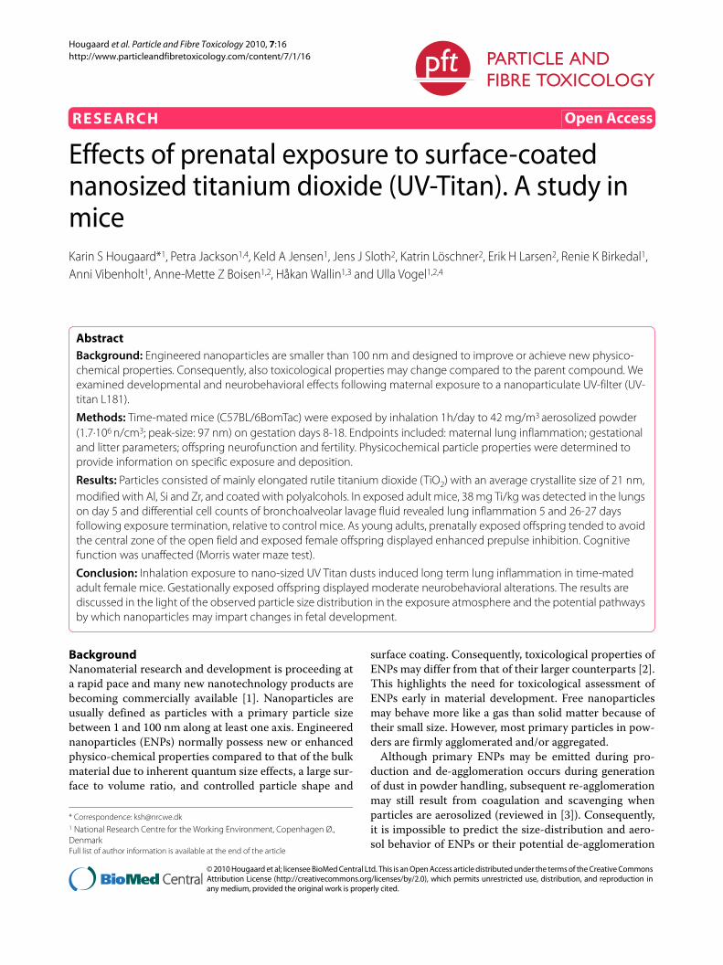
Hougaard et al. Particle and Fibre Toxicology 2010, 7:16 http://www.particleandfibretoxicology.com/content/7/1/16 Open Access RESEARCH © 2010 Hougaard et al; licensee BioMed Central Ltd. This is an Open Access article distributed under the terms of the Creative Commons Attribution License (http://creativecommons.org/licenses/by/2.0), which permits unrestricted use, distribution, and reproduction in any medium, provided the original work is properly cited. Research Effects of prenatal exposure to surface-coated nanosized titanium dioxide (UV-Titan). A study in mice Karin S Hougaard* 1 , Petra Jackson 1,4 , Keld A Jensen 1 , Jens J Sloth 2 , Katrin Löschner 2 , Erik H Larsen 2 , Renie K Birkedal 1 , Anni Vibenholt 1 , Anne-Mette Z Boisen 1,2 , Håkan Wallin 1,3 and Ulla Vogel 1,2,4 Abstract Background: Engineered nanoparticles are smaller than 100 nm and designed to improve or achieve new physico- chemical properties. Consequently, also toxicological properties may change compared to the parent compound. We examined developmental and neurobehavioral effects following maternal exposure to a nanoparticulate UV-filter (UV- titan L181). Methods: Time-mated mice (C57BL/6BomTac) were exposed by inhalation 1h/day to 42 mg/m 3 aerosolized powder (1.7·10 6 n/cm 3 ; peak-size: 97 nm) on gestation days 8-18. Endpoints included: maternal lung inflammation; gestational and litter parameters; offspring neurofunction and fertility. Physicochemical particle properties were determined to provide information on specific exposure and deposition. Results: Particles consisted of mainly elongated rutile titanium dioxide (TiO 2 ) with an average crystallite size of 21 nm, modified with Al, Si and Zr, and coated with polyalcohols. In exposed adult mice, 38 mg Ti/kg was detected in the lungs on day 5 and differential cell counts of bronchoalveolar lavage fluid revealed lung inflammation 5 and 26-27 days following exposure termination, relative to control mice. As young adults, prenatally exposed offspring tended to avoid the central zone of the open field and exposed female offspring displayed enhanced prepulse inhibition. Cognitive function was unaffected (Morris water maze test). Conclusion: Inhalation exposure to nano-sized UV Titan dusts induced long term lung inflammation in time-mated adult female mice. Gestationally exposed offspring displayed moderate neurobehavioral alterations. The results are discussed in the light of the observed particle size distribution in the exposure atmosphere and the potential pathways by which nanoparticles may impart changes in fetal development. Background Nanomaterial research and development is proceeding at a rapid pace and many new nanotechnology products are becoming commercially available [1]. Nanoparticles are usually defined as particles with a primary particle size between 1 and 100 nm along at least one axis. Engineered nanoparticles (ENPs) normally possess new or enhanced physico-chemical properties compared to that of the bulk material due to inherent quantum size effects, a large sur- face to volume ratio, and controlled particle shape and surface coating. Consequently, toxicological properties of ENPs may differ from that of their larger counterparts [2]. This highlights the need for toxicological assessment of ENPs early in material development. Free nanoparticles may behave more like a gas than solid matter because of their small size. However, most primary particles in pow- ders are firmly agglomerated and/or aggregated. Although primary ENPs may be emitted during pro- duction and de-agglomeration occurs during generation of dust in powder handling, subsequent re-agglomeration may still result from coagulation and scavenging when particles are aerosolized (reviewed in [3]). Consequently, it is impossible to predict the size-distribution and aero- sol behavior of ENPs or their potential de-agglomeration * Correspondence: [email protected] 1 National Research Centre for the Working Environment, Copenhagen Ø., Denmark Full list of author information is available at the end of the article

Welcome message from author
This document is posted to help you gain knowledge. Please leave a comment to let me know what you think about it! Share it to your friends and learn new things together.
Transcript
Hougaard et al. Particle and Fibre Toxicology 2010, 7:16http://www.particleandfibretoxicology.com/content/7/1/16
Open AccessR E S E A R C H
ResearchEffects of prenatal exposure to surface-coated nanosized titanium dioxide (UV-Titan). A study in miceKarin S Hougaard*1, Petra Jackson1,4, Keld A Jensen1, Jens J Sloth2, Katrin Löschner2, Erik H Larsen2, Renie K Birkedal1, Anni Vibenholt1, Anne-Mette Z Boisen1,2, Håkan Wallin1,3 and Ulla Vogel1,2,4
AbstractBackground: Engineered nanoparticles are smaller than 100 nm and designed to improve or achieve new physico-chemical properties. Consequently, also toxicological properties may change compared to the parent compound. We examined developmental and neurobehavioral effects following maternal exposure to a nanoparticulate UV-filter (UV-titan L181).
Methods: Time-mated mice (C57BL/6BomTac) were exposed by inhalation 1h/day to 42 mg/m3 aerosolized powder (1.7·106 n/cm3; peak-size: 97 nm) on gestation days 8-18. Endpoints included: maternal lung inflammation; gestational and litter parameters; offspring neurofunction and fertility. Physicochemical particle properties were determined to provide information on specific exposure and deposition.
Results: Particles consisted of mainly elongated rutile titanium dioxide (TiO2) with an average crystallite size of 21 nm, modified with Al, Si and Zr, and coated with polyalcohols. In exposed adult mice, 38 mg Ti/kg was detected in the lungs on day 5 and differential cell counts of bronchoalveolar lavage fluid revealed lung inflammation 5 and 26-27 days following exposure termination, relative to control mice. As young adults, prenatally exposed offspring tended to avoid the central zone of the open field and exposed female offspring displayed enhanced prepulse inhibition. Cognitive function was unaffected (Morris water maze test).
Conclusion: Inhalation exposure to nano-sized UV Titan dusts induced long term lung inflammation in time-mated adult female mice. Gestationally exposed offspring displayed moderate neurobehavioral alterations. The results are discussed in the light of the observed particle size distribution in the exposure atmosphere and the potential pathways by which nanoparticles may impart changes in fetal development.
BackgroundNanomaterial research and development is proceeding ata rapid pace and many new nanotechnology products arebecoming commercially available [1]. Nanoparticles areusually defined as particles with a primary particle sizebetween 1 and 100 nm along at least one axis. Engineerednanoparticles (ENPs) normally possess new or enhancedphysico-chemical properties compared to that of the bulkmaterial due to inherent quantum size effects, a large sur-face to volume ratio, and controlled particle shape and
surface coating. Consequently, toxicological properties ofENPs may differ from that of their larger counterparts [2].This highlights the need for toxicological assessment ofENPs early in material development. Free nanoparticlesmay behave more like a gas than solid matter because oftheir small size. However, most primary particles in pow-ders are firmly agglomerated and/or aggregated.
Although primary ENPs may be emitted during pro-duction and de-agglomeration occurs during generationof dust in powder handling, subsequent re-agglomerationmay still result from coagulation and scavenging whenparticles are aerosolized (reviewed in [3]). Consequently,it is impossible to predict the size-distribution and aero-sol behavior of ENPs or their potential de-agglomeration
* Correspondence: [email protected] National Research Centre for the Working Environment, Copenhagen Ø., DenmarkFull list of author information is available at the end of the article
© 2010 Hougaard et al; licensee BioMed Central Ltd. This is an Open Access article distributed under the terms of the Creative CommonsAttribution License (http://creativecommons.org/licenses/by/2.0), which permits unrestricted use, distribution, and reproduction inany medium, provided the original work is properly cited.
Hougaard et al. Particle and Fibre Toxicology 2010, 7:16http://www.particleandfibretoxicology.com/content/7/1/16
Page 2 of 15
during airway deposition. Therefore, experimental workis urgently required to assess these parameters as well asto determine the resulting biological effects in vivo.When inhaled, a considerable fraction of sub-μm sizeparticles may deposit in the deeper airways. Once depos-ited in the lung, material may be retained for a long time[4]. Nanoparticles can also translocate across the lungepithelium, although the rate of distribution to otherorgans varies [3,5-7]. Airborne particles released duringproduction or handling of ENPs are therefore of particu-lar concern.
The toxicological properties of nanosized particles aregenerally poorly understood, although knowledge insome areas (especially inflammation and particle translo-cation) is rapidly growing. Reproductive and develop-mental toxicity is integrated into the nanomaterialsresearch strategy of the U.S. Environmental ProtectionAgency [8] and recommended by the ReproductiveHealth Research Team under the National OccupationalResearch Agenda of the U.S. National Institute of Occu-pational Safety and Health [9]. Nanomaterials may affectthe developing fetus either directly or indirectly. Directeffects might occur after translocation of particles frommaternal lung to blood and then across the placenta. Bythe indirect pathway, maternal pulmonary inflammationorchestrates release of signaling molecules which poten-tially affect both mother and fetus. Preliminary work sug-gests that the fetal nervous system is specifically sensitiveto maternal particulate exposure during pregnancy[10,11]. Today very little is known on developmental tox-icity of nanomaterials.
Titanium dioxide (TiO2) has previously been used as ageneric model compound to illustrate potential toxiceffects of exposure to relatively inert nanoparticles. How-ever, TiO2 is also a widely-used industrial nanomaterial(e.g., sunscreens and lacquers with "invisible" UV-filters,and paints with photocatalytic-induced self-cleaningproperties). Thus, the exposure of consumers and factoryworkers who handle TiO2 nanomaterials and nanomate-rial-based products must be considered. Increasing evi-dence suggests that the toxicity of TiO2 not only dependson size, but also varies with crystalline polymorph, parti-cle shape, surface coating and functionalization(reviewed in [12]). Thus silica-coated TiO2 increased lunginflammation significantly compared to pure TiO2 andpure silica in the mouse [4].
The present study investigated developmental neuro-toxicity in offspring of mice that inhaled TiO2 (UV-titanL181, a coated and chemically modified rutile) duringpregnancy, in parallel with maternal inflammatoryresponse. Effects on the nervous system were evaluatedby use of a neurobehavioral test battery. Furthermore,
particle physicochemical properties and exposure werecharacterized in detail.
Materials and methodsAnimalsTime-mated, nulliparous mice (C57BL/6BomTac,Taconic Europe, Ejby, Denmark) arrived at gestation day(GD) 3 and were randomly grouped 5 or 6 in polypropyl-ene cages with bedding and enrichment (removed duringnursing). Animals were housed under controlled environ-mental conditions, with 12 hour light from 6.00 a.m. andaccess to food (Altromin 1324) and tap water ad libitum(further information in Additional file 1). On GD4, ani-mals were weighed and assigned to two groups of 22 and23 animals, respectively, with similar weight distribu-tions. For cross-over mating, naïve CBA/J mice (CharlesRiver Wiga, Sulzfeld, Germany) were supplied at nineweeks of age. Procedures complied with EC Directive 86/609/EEC and Danish regulations on experiments withanimals (Permission 2006/561-1123).
Material characterizationThis study used UV-titan L181 (Kemira, Pori, Finland), arutile modified with unspecified amounts of zirconium(Zr), silicon (Si), aluminum (Al) and coated with polyal-cohols.
Physical particle size, morphology and general state ofagglomeration/aggregation were determined by analysisof particles suspended on holey carbon-coated Cu TEM-grids using a 200 kV Transmission Electron Microscope(TEM) (Tecnai G20, FEI Company, Hillsboro, Oregon,USA). Sample preparation for TEM analysis is describedin Additional file 1.
Crystalline phases and crystallite sizes were determinedby powder X-ray diffraction (XRD) with a Bruker D8Advance diffractometer equipped with a Lynxeye CCDdetector (Bruker AXS Inc., Madison, WI 53711-5373,USA), using monochromated CuKα1 (1.540598 Å) rays.Results were obtained by Rietveld refinement of the X-raydiffractograms using Bruker TOPAS V4.1 software. Elon-gation was determined by analysis of reflections fromprincipal crystallographical axis using the Scherrer equa-tion.
Specific surface area was determined on a Quantach-rome Autosorp-1 (Quantachrome GmbH & Co. KG,Odelzhausen, Germany) using multipoint Brunauer,Emmett, and Teller (BET) nitrogen adsorption methodafter 1 h degassing at 300°C. Analysis was completedaccording to DIN ISO 9277 as a commercial service byQuantachrome GmbH & Co. KG.
Elemental composition was analyzed by X-ray Fluores-cence analysis on a Philips PW-2400 spectrometer as acommercial service by the Department of Earth Sciences,
Hougaard et al. Particle and Fibre Toxicology 2010, 7:16http://www.particleandfibretoxicology.com/content/7/1/16
Page 3 of 15
University of Aarhus, Denmark. Elemental concentra-tions were determined using their standard protocolusing rock standards for calibration.
The organic coating of the UV-titan particles wasextracted with methanol by Pressurized Liquid Extrac-tion (PLE) at 2000 psi and 200°C, followed by centrifuga-tion at 4000 rpm (3310 g) for 10 min. Chemicalcomposition of the supernatant was analyzed by laserdesorption ionization and time of flight MS (MALDI-TOF without matrix) on a stainless steel ground targetwith a Bruker AutoFlex II (Bruker Daltonics, Inc., Bre-men Germany). Acurate mass determination (1 ppm) wasperformed with electrospray-MS (ESI-MS) on a BrukermicroQ-TOF (Bruker Daltonics, Inc., Bremen Germany)with direct injection. The masses are reported as mass tocharge ratios (m/z) of the protonated compounds([M+H]+).
ExposureMice were exposed to filtered clean air or a target con-centration of 40 mg UV-Titan/m3 on GD8-18, one hr/dayas described [13,14]. Airflow in the exposure chamberwas dynamic (20 L/min) with evenly distributed exposureatmosphere. A microfeeder aerosolized powder particlesthrough a dispersion nozzle at a pressure of 5 bar (Fraun-hofer Institute für Toxicologie und Aerosolforschung,Hannover, Germany). The dose from one hour exposureto 40 mg TiO2/m3 corresponds to the 8-hr time weightedaverage (TWA) occupational exposure limit according toDanish Regulations [15]. Animals were placed separatelyin rooms of a "twelve-room-pie"; a cylindrical wire meshcage (? 29 cm, height 9 cm) with radical partitions.Females were observed for signs of toxicity and returnedto cages less than 5 min after exposure. Body weight wasrecorded before exposure on GD9, 11, 14, and 18.
Exposure monitoringMass-concentrations of total suspended dust was con-trolled periodically by filter sampling and adjusted tomaintain a concentration of ~40 mg/m3. Exposure air wassampled on pre-weighed Millipore Fluoropore filters (?2.5 cm; pore size 0.45 μm) at an airflow of 2 L/min usingMillipore cassettes, for 10 min. Filters were weighedimmediately on a Sartorius Microscale (Type M3P000V001). Final gravimetric data were obtained on accli-matized filters (50%RH and 20°C).
Particle number and size distribution in the exposureatmosphere were monitored using a GRIMM Sequential(Stepping) Mobility Particle Sizer (SMPS) system for sub-μm particles (12.8 to 486 nm; based on the rutile densityof 4.25 g/cm3 [16]) and a GRIMM Dustmonitor (Model1.106) for coarse particles (0.75 to > 15 μm). The SMPSconsisted of a Long Electrostatic Classifier (Model No.5.521) and a GRIMM Condensation Particle Counter
(Model 5.400). The time resolution was 218 and 6 s forthe SMPS and Dustmonitor data, respectively (see Addi-tional file 1 for further explanation on the on-line particleexposure monitoring).
Parturition and lactationAfter exposure on GD18, females were singly housed.Delivery was expected on GD20, and designated postna-tal day (PND) 0. Pups were counted and sexed on PND1.Dams and individual pups were weighed at PND1, 8, 11,16, 19, and 22. On PND2, one pup from litters with atleast 5 pups, and on PND23-24 one male and one femaleper litter, were sacrificed by decapitation. Lungs, liver,heart, brain, and on PND2, stomachs containing milk,were dissected, weighed, snap frozen in liquid N2 andstored at -80°C. At weaning (PND22), one male andfemale per litter were randomly chosen for behavioraltesting and housed as described.
Non-pregnant time-mated females without implanta-tions ("NP females") were euthanized on PND3 (i.e. 5days post exposure) and subjected to bronchoalveolarlavage (BAL), as were dams with litters at PND24-25 ("Pfemales"; 26-27 days post exposure). Females were anaes-thetized with Hypnorm and Dormicum and sacrificed bywithdrawal of heart blood (stabilized in 0.17 mol/lK2EDTA). BAL was performed as described below, fol-lowed by determination of uterine implantation sites anddissection of organs as described for offspring.
Titanium in tissue and milkApproximately 25-75 mg tissue (lung and liver for adults,liver for offspring) and 110-140 mg (milk) were weighedand analyzed for content of titanium (Ti). For PND2pups, milk and liver samples were pooled from 4-5 ani-mals. Maternal lung was included to determine remain-ing TiO2 and liver to assess systemic distribution in adults[17,18] and fetal animals [19]. Tissues were digested inconcentrated nitric acid (PlasmaPure, SCP Science, Que-bec, Canada) in a microwave oven (Multiwave, AntonPaar, Graz, Austria), and Ti content determined by qua-drupole-based inductively coupled plasma mass spec-trometer (ICPMS 7500ce, Agilent Technologies, Tokyo,Japan) equipped with a collision/reaction cell (CRC). TheCRC was pressurized with helium as collision gas toreduce polyatomic interferences on Ti isotopes. Settingsfor ICPMS measurements are given in (Additional file 1,Table S1). Sulphur-containing polyatomics (e.g. 32S16O+)strongly interfered with the most abundant Ti isotope,48Ti (abundance 73.8%). There was less interference with49Ti and 50Ti (abundance 5.5 and 5.4%, respectively),which were selected for quantitative analysis. The limit ofdetection (LOD) for Ti in tissues, based on three timesthe standard deviation of repeated blank measurements,
Hougaard et al. Particle and Fibre Toxicology 2010, 7:16http://www.particleandfibretoxicology.com/content/7/1/16
Page 4 of 15
was estimated to be 0.2-5 mg/kg depending on sampleintake and dilution.
BAL preparation and analysesWe used BAL cell composition and neutrophil influx toindicate lung inflammation. This has proven to be a rele-vant and sensitive marker of pulmonary inflammation(e.g. [20,21]). BAL was performed four times with 0.8 ml0.9% sterile saline ([20]; further information in Additionalfile 1). The total number of cells and of dead cells in BALsamples was determined in cell suspension B by Nucleo-Counter. Differential counts of macrophages, neutro-phils, lymphocytes, eosinophils, and epithelial cells weredetermined by counting 200 cells in cell supernatant fixedwith 96% ethanol and stained with May-Grünwald-Giemsa stain. All slides from both time points were ran-domized, blinded and scored on the same day. Total num-ber of cells was calculated by combining data fromdifferential cell counts with the total number of cells inBAL.
Behavioral testingInvestigations were performed during the light period.Exposed and control animals were tested alternately. Ani-mals were transferred to the experimental room 1 hrbefore the first test. Observers were blinded to exposurestatus of the animals, and the same observer was usedthroughout any specific test.
Learning and memory was tested in the Morris watermaze at age 11 and 15 weeks (males), and 12 and 16weeks (females) as described [13] with minor modifica-tions. A stable, invisible platform was submerged 1 cmbelow the water surface in a circular plastic pool (? 100cm). Animals were tested in four daily trials. Mice wereplaced at the designated starting position and completedthe trial when climbing onto the platform. When failingto locate the platform within 60 s, animals were led to theplatform. All animals spent 15 s on the platform beforereturning to the cage. The following scheme was used:Learning: Test for 5 consecutive days with platform incenter of the southeastern quadrant. Memory: Threeweeks later, test with platform in south-eastern quadrant,for 3 days. Reversal learning: The following day, test withplatform in north-western quadrant for 4 trials. Newlearning: The following day, test with platform in centerof pool for 4 trials. Noldus Ethovision (Version 5, NoldusInformation Technology, Wageningen, The Netherlands)was used to register latency and path length, and calcu-lated swimming velocity and relative occupancy in theeach of the quadrants.
Activity was assessed for 3 min at 14 weeks of age in anopen field using the dry water maze pool. Trials com-menced in the center of the field and the location of theanimal was registered by Noldus Ethovision XT version 5.
The tracking device calculated total ambulation, whichwas subsequently split into three time-bins of 1 min totest for habituation. Duration in the central and the outer9 cm peripheral zone of the field, as well as the number ofcrossings from the outer to the central zone wereextracted.
Acoustic startle reaction (ASR) and prepulse inhibition(PPI) were tested at 4 months as described [22] in twochambers (San Diego Instruments, San Diego, USA) with70 dB(A) white background noise. A piezoelectric accel-erometer transduced displacement of test tubes (? 3.6 cm)in response to movements of the animal. Animals wereacclimatized for 5 min in the tube before sessions startedand ended with 5 startle trials of 40 ms 120 dB(A) burstsof white noise. In between, 35 trials were delivered insemi-randomized order (10 trials of 120 dB(A); 5 each of4 prepulse + startle trials (prepulses of 72, 74, 78, and 86dB(A)); 5 trials with only background noise). Tube move-ments were averaged over 100 ms following onset of thestartle stimulus (AVG). The five AVGs for each prepulseintensity were averaged and used to calculate PPI, whichwas expressed as percent reduction in AVG compared tothe average of the 10 middle startle trials: %PPI =100*((AVG at prepulse+startle trial)/(AVG at startletrial))*100%.
Time-to-first F2 litterAt 19 weeks of age, control and exposed offspring werecross-mated to naïve CBA/J mice (12 weeks old) andtime-to-first-delivery of F2 litter, litter size, and genderratio were recorded.
StatisticsLitter was considered the statistical unit. Gestationalparameters were analyzed by Mann-Whitney U-test, andtime-to-first-delivery by log rank test (separately by gen-der). ANOVAs were applied to the remaining data whenrelevant with repeated measures in trials, days, or time-bins. In the analysis of weight gain in adult females, Ti inadult tissues, and BAL cell counts, the factor "Pregnancy"was added to distinguish (barren) NP females from (lit-tering) P females. Since these groups of adult females dif-fered with regard to both time after exposure andpregnancy, only pairwise comparisons related to expo-sure were explored. ANCOVA controlled for litter size inthe analyses of weight gain during exposure, birthweights, and pre-weaning pup weights. Behavioral datawere analyzed by two-way ANOVA, with Prenatal expo-sure and Gender as factors, apart from startle data, wherePPI was analyzed separately for each prepulse intensity[22]. Pairwise comparisons were performed by T-test orMann Whitney U-test (p < 0.1). Analyses were performedin SYSTAT Software Package 9, MINITAB 14, and SAS9.1.
Hougaard et al. Particle and Fibre Toxicology 2010, 7:16http://www.particleandfibretoxicology.com/content/7/1/16
Page 5 of 15
ResultsParticle characteristicsPhysicochemical characteristics of the UV-titan sampleare summarized in Table 1. Rutile was the only crystallinephase in the sample and TiO2 accounted for 70.8 wt%.Residual mass was composed of Zr, Si, Al, and a littlesodium (Na) as well as 5.2 wt% volatiles (loss on ignition).Stochiometric calculations show that the modifier ele-ments partly occurred in oxides, but presence of nativemetals or non-stochiometric amorphous compounds arealso possible. BET measurements show that the specificsurface area was ~38 m2/g higher (i.e. 107.7 m2/g) thanreported by the manufacturer (ca. 70 m2/g). This differ-ence in specific surface area may arise due to out-gassingof the powder at 300°C for 1 h before analysis. This mayhave volatilized the organic coating, thereby increasingthe accessible surface area.
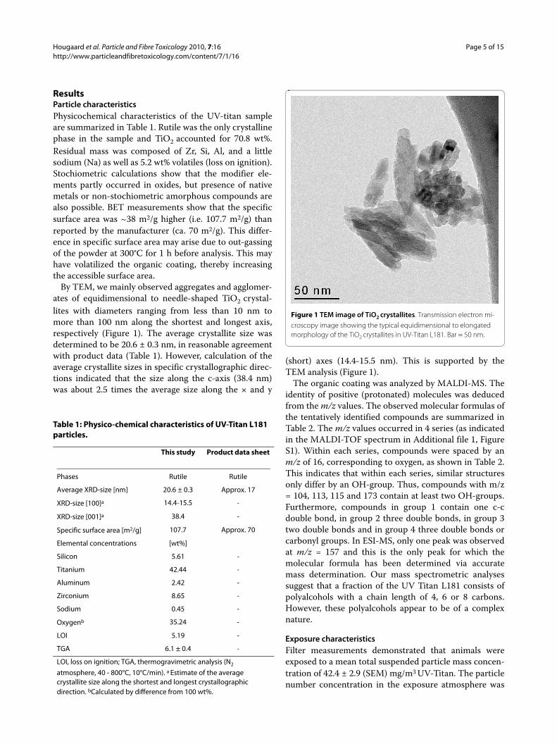
By TEM, we mainly observed aggregates and agglomer-ates of equidimensional to needle-shaped TiO2 crystal-lites with diameters ranging from less than 10 nm tomore than 100 nm along the shortest and longest axis,respectively (Figure 1). The average crystallite size wasdetermined to be 20.6 ± 0.3 nm, in reasonable agreementwith product data (Table 1). However, calculation of theaverage crystallite sizes in specific crystallographic direc-tions indicated that the size along the c-axis (38.4 nm)was about 2.5 times the average size along the × and y
(short) axes (14.4-15.5 nm). This is supported by theTEM analysis (Figure 1).
The organic coating was analyzed by MALDI-MS. Theidentity of positive (protonated) molecules was deducedfrom the m/z values. The observed molecular formulas ofthe tentatively identified compounds are summarized inTable 2. The m/z values occurred in 4 series (as indicatedin the MALDI-TOF spectrum in Additional file 1, FigureS1). Within each series, compounds were spaced by anm/z of 16, corresponding to oxygen, as shown in Table 2.This indicates that within each series, similar structuresonly differ by an OH-group. Thus, compounds with m/z= 104, 113, 115 and 173 contain at least two OH-groups.Furthermore, compounds in group 1 contain one c-cdouble bond, in group 2 three double bonds, in group 3two double bonds and in group 4 three double bonds orcarbonyl groups. In ESI-MS, only one peak was observedat m/z = 157 and this is the only peak for which themolecular formula has been determined via accuratemass determination. Our mass spectrometric analysessuggest that a fraction of the UV Titan L181 consists ofpolyalcohols with a chain length of 4, 6 or 8 carbons.However, these polyalcohols appear to be of a complexnature.
Exposure characteristicsFilter measurements demonstrated that animals wereexposed to a mean total suspended particle mass concen-tration of 42.4 ± 2.9 (SEM) mg/m3 UV-Titan. The particlenumber concentration in the exposure atmosphere was
Figure 1 TEM image of TiO2 crystallites. Transmission electron mi-croscopy image showing the typical equidimensional to elongated morphology of the TiO2 crystallites in UV-Titan L181. Bar = 50 nm.
Table 1: Physico-chemical characteristics of UV-Titan L181 particles.
This study Product data sheet
Phases Rutile Rutile
Average XRD-size [nm] 20.6 ± 0.3 Approx. 17
XRD-size [100]a 14.4-15.5 -
XRD-size [001]a 38.4 -
Specific surface area [m2/g] 107.7 Approx. 70
Elemental concentrations [wt%]
Silicon 5.61 -
Titanium 42.44 -
Aluminum 2.42 -
Zirconium 8.65 -
Sodium 0.45 -
Oxygenb 35.24 -
LOI 5.19 -
TGA 6.1 ± 0.4 -
LOI, loss on ignition; TGA, thermogravimetric analysis (N2
atmosphere, 40 - 800°C, 10°C/min). a Estimate of the average crystallite size along the shortest and longest crystallographic direction. bCalculated by difference from 100 wt%.
Hougaard et al. Particle and Fibre Toxicology 2010, 7:16http://www.particleandfibretoxicology.com/content/7/1/16
Page 6 of 15
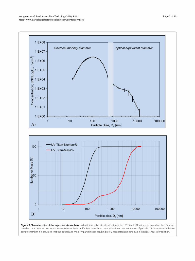
1.70 ± 0.20·106/cm3. The major particle size-mode was~100 nm (geometric mean number diameter 97 nm),with a coarser size mode at ~4 μm (Figure 2A). Smallersize modes were observed at ~20 nm and 1 μm. By num-ber, 80% of the particles were between 40 and 200 nm andno particles were coarser than 12.5 μm detected (Figure2B). The mass-size distribution was strongly dominatedby μm-size particles (geometric mean 3.2 μm) and 75% ofthe mass were represented by particles larger than 1.6 μm(Figure 2B). The fraction of sub-100-nm-size particlesamounted to 1% of the mass.
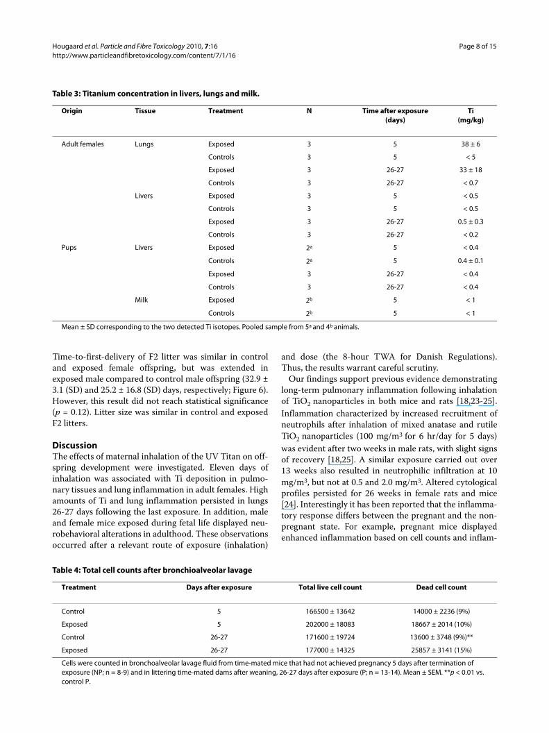
Ti concentration in tissues and milkTi concentration in tissue and milk samples is shown inTable 3. Lungs from exposed females contained 38 mg Ti/kg on day 5 after the exposure and 33 mg Ti/kg on days26-27. No Ti was detected in unexposed female lungs (p =0.0002). Values were similar between control and exposedanimals for all other samples.
Maternal and litter parametersSimilar numbers of control and exposed females deliv-ered litters, and none of the time-mated females withoutlitters displayed implantations. Gestational and litterparameters were similar, apart from a slight decrease inpup viability in TiO2 litters (p = 0.083, c.f. Table B, Addi-tional file 1, Table S2l). Only maternal lung weightshowed overall statistical significant variation with expo-sure, in both absolute (p = 0.04) and relative (p = 0.05)measures (data not shown). Pairwise comparisonsshowed both measures to be marginally increased in only
in P females (0.05 <p < 0.1). No effects related to exposurewere detected for offspring organ weights.
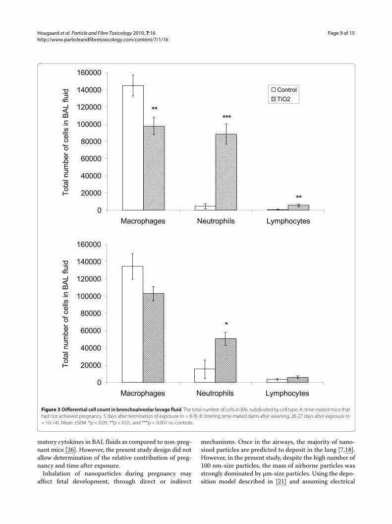
Lung inflammation in time-mated femalesLung inflammation was evaluated by cell counts of BALfluid (Table 4 and Figure 3). Overall, more neutrophilswere present in BAL in TiO2 exposed compared to unex-posed females (p < 0.001), with significant exposure-pregnancy interaction (p = 0.02). BAL from exposed NPfemales contained 19 times more neutrophils in BALthan did unexposed NP females (5 days after exposure, p< 0.001). The exposed P females displayed 3-fold moreneutrophils compared to unexposed P females (26-27days after exposure, p = 0.02). The exposure also resultedin overall change in macrophages (p = 0.002) and lym-phocytes (p = 0.007) compared to unexposed P females.In NP females, pairwise comparisons revealed fewermacrophages (p = 0.009) but more lymphocytes (p =0.008) in exposed compared to UNexposed NP females.No cell type showed significant change in exposed Pfemales compared to respective controls. Overall, a statis-tically significant increase in the total number of deadcells in BAL fluid (p = 0.03) was observed in BAL fromthe exposed P females (p = 0.004) but not in BAL fromexposed NP females. Total cell counts, total number ofeosinophils, and epithelial cells in BAL were did not varywith exposure.
Behavioral dataIn the Morris water maze, no change was observed inperformance as a result of prenatal TiO2 exposure ineither male or female offspring (data not shown).
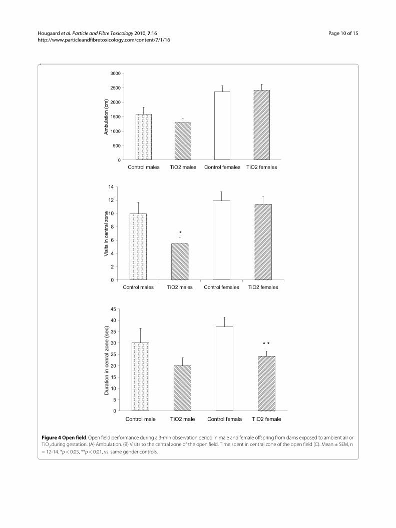
In the open field, ambulation differed by gender (p <0.001) but not exposure, as females moved approximately50% longer than males (Figure 4A). Prenatally exposedanimals spent significantly less time than controls in thecentral zone of the field (p = 0.009), and visited the cen-tral zone less frequently (Exposure: p = 0.056; Gender: p =0.003). Exposed males entered the central zone signifi-cantly less frequently than unexposed males (Figure 4B, p= 0.021) and exposed females spent less time in the cen-tral zone than did unexposed females (Figure 4C, p =0.009).
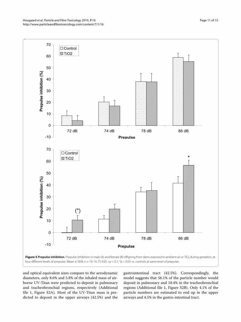
Analysis of acoustic startle demonstrated that exposedmale offspring startled less than control males and wereless inhibited by prepulse, whereas the opposite patternwas apparent for female offspring (Additional file 1, Fig-ure S3). Statistical analysis substantiated a stronger PPI inprenatally exposed females at the highest and lowest pre-pulse compared to control offspring (Figure 5B; p = 0.041and p = 0.089, respectively).
Time-to-first F2 litterAt termination of behavioral testing, control and exposedC57BL offspring were cross-mated to naïve CBA/J mice.
Table 2: Observed m/z values and tentative molecular formulas
m/z[M+H]+
Tentative molecular formular
1 72 C4H7N
88 C4H7NO
104 C4H7NO2
2 81 C6H8
97 C6H8O
113 C6H8O2
3 83 C6H10
99 C6H10O
115 C6H10O2
4 141 C8H13O2
157* C8H13O3
173 C8H13O4
* Molecular formula determined from exact mass measurement (mass accuracy 1 ppm)
Hougaard et al. Particle and Fibre Toxicology 2010, 7:16http://www.particleandfibretoxicology.com/content/7/1/16
Page 7 of 15
Figure 2 Characteristics of the exposure atmosphere. A) Particle number size distribution of the UV-Titan L181 in the exposure chamber. Data are based on nine one-hour exposure measurements. Mean ± SD. B) Accumulated number and mass concentration of particle concentrations in the ex-posure chamber. It is assumed that the optical and mobility particle sizes can be directly compared and data gap is filled by linear interpolation.
1,E+00
1,E+01
1,E+02
1,E+03
1,E+04
1,E+05
1,E+06
1,E+07
1,E+08
1 10 100 1000 10000 100000
Particle Size, Dp [nm]
Concentr
ation; dN
/dLo
gD
p [n/c
m3]
optical equivalent diameterelectrical mobility diameter
0
50
100
1 10 100 1000 10000 100000
Particle size, Dp [nm]
Num
be
r o
r M
ass [
%]
UV-Titan-Number%
UV Titan-Mass%
A)
B)
Hougaard et al. Particle and Fibre Toxicology 2010, 7:16http://www.particleandfibretoxicology.com/content/7/1/16
Page 8 of 15
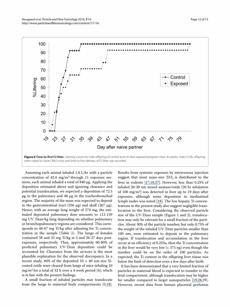
Time-to-first-delivery of F2 litter was similar in controland exposed female offspring, but was extended inexposed male compared to control male offspring (32.9 ±3.1 (SD) and 25.2 ± 16.8 (SD) days, respectively; Figure 6).However, this result did not reach statistical significance(p = 0.12). Litter size was similar in control and exposedF2 litters.
DiscussionThe effects of maternal inhalation of the UV Titan on off-spring development were investigated. Eleven days ofinhalation was associated with Ti deposition in pulmo-nary tissues and lung inflammation in adult females. Highamounts of Ti and lung inflammation persisted in lungs26-27 days following the last exposure. In addition, maleand female mice exposed during fetal life displayed neu-robehavioral alterations in adulthood. These observationsoccurred after a relevant route of exposure (inhalation)
and dose (the 8-hour TWA for Danish Regulations).Thus, the results warrant careful scrutiny.
Our findings support previous evidence demonstratinglong-term pulmonary inflammation following inhalationof TiO2 nanoparticles in both mice and rats [18,23-25].Inflammation characterized by increased recruitment ofneutrophils after inhalation of mixed anatase and rutileTiO2 nanoparticles (100 mg/m3 for 6 hr/day for 5 days)was evident after two weeks in male rats, with slight signsof recovery [18,25]. A similar exposure carried out over13 weeks also resulted in neutrophilic infiltration at 10mg/m3, but not at 0.5 and 2.0 mg/m3. Altered cytologicalprofiles persisted for 26 weeks in female rats and mice[24]. Interestingly it has been reported that the inflamma-tory response differs between the pregnant and the non-pregnant state. For example, pregnant mice displayedenhanced inflammation based on cell counts and inflam-
Table 3: Titanium concentration in livers, lungs and milk.
Origin Tissue Treatment N Time after exposure(days)
Ti(mg/kg)
Adult females Lungs Exposed 3 5 38 ± 6
Controls 3 5 < 5
Exposed 3 26-27 33 ± 18
Controls 3 26-27 < 0.7
Livers Exposed 3 5 < 0.5
Controls 3 5 < 0.5
Exposed 3 26-27 0.5 ± 0.3
Controls 3 26-27 < 0.2
Pups Livers Exposed 2a 5 < 0.4
Controls 2a 5 0.4 ± 0.1
Exposed 3 26-27 < 0.4
Controls 3 26-27 < 0.4
Milk Exposed 2b 5 < 1
Controls 2b 5 < 1
Mean ± SD corresponding to the two detected Ti isotopes. Pooled sample from 5a and 4b animals.
Table 4: Total cell counts after bronchioalveolar lavage
Treatment Days after exposure Total live cell count Dead cell count
Control 5 166500 ± 13642 14000 ± 2236 (9%)
Exposed 5 202000 ± 18083 18667 ± 2014 (10%)
Control 26-27 171600 ± 19724 13600 ± 3748 (9%)**
Exposed 26-27 177000 ± 14325 25857 ± 3141 (15%)
Cells were counted in bronchoalveolar lavage fluid from time-mated mice that had not achieved pregnancy 5 days after termination of exposure (NP; n = 8-9) and in littering time-mated dams after weaning, 26-27 days after exposure (P; n = 13-14). Mean ± SEM. **p < 0.01 vs. control P.
Hougaard et al. Particle and Fibre Toxicology 2010, 7:16http://www.particleandfibretoxicology.com/content/7/1/16
Page 9 of 15
matory cytokines in BAL fluids as compared to non-preg-nant mice [26]. However, the present study design did notallow determination of the relative contribution of preg-nancy and time after exposure.
Inhalation of nanoparticles during pregnancy mayaffect fetal development, through direct or indirect
mechanisms. Once in the airways, the majority of nano-sized particles are predicted to deposit in the lung [7,18].However, in the present study, despite the high number of100 nm-size particles, the mass of airborne particles wasstrongly dominated by μm-size particles. Using the depo-sition model described in [21] and assuming electrical
Figure 3 Differential cell count in bronchoalveolar lavage fluid. The total number of cells in BAL subdivided by cell type. A: time-mated mice that had not achieved pregnancy, 5 days after termination of exposure (n = 8-9). B: littering time-mated dams after weaning, 26-27 days after exposure (n = 10-14). Mean ±SEM. *p < 0.05, **p < 0.01, and ***p < 0.001 vs. controls.
0
20000
40000
60000
80000
100000
120000
140000
160000
Macrophages Neutrophils Lymphocytes
Tota
l num
ber
of cells
in B
AL flu
id Control
TiO2
*****
**
0
20000
40000
60000
80000
100000
120000
140000
160000
Macrophages Neutrophils Lymphocytes
Tota
l num
ber
of cells
in B
AL flu
id
*
Hougaard et al. Particle and Fibre Toxicology 2010, 7:16http://www.particleandfibretoxicology.com/content/7/1/16
Page 10 of 15
Figure 4 Open field. Open field performance during a 3-min observation period in male and female offspring from dams exposed to ambient air or TiO2 during gestation. (A) Ambulation. (B) Visits to the central zone of the open field. Time spent in central zone of the open field (C). Mean ± SEM, n = 12-14. *p < 0.05, **p < 0.01, vs. same gender controls.
0
500
1000
1500
2000
2500
3000
Control males TiO2 males Control females TiO2 females
Am
bula
tion (
cm
)
0
2
4
6
8
10
12
14
Control males TiO2 males Control females TiO2 females
Vis
its in c
entr
al zone
*
0
5
10
15
20
25
30
35
40
45
Control male TiO2 male Control femala TiO2 female
Dura
tion in c
enra
l zone (
sec)
* *
Hougaard et al. Particle and Fibre Toxicology 2010, 7:16http://www.particleandfibretoxicology.com/content/7/1/16
Page 11 of 15
and optical equivalent sizes compare to the aerodynamicdiameters, only 8.6% and 5.8% of the inhaled mass of air-borne UV-Titan were predicted to deposit in pulmonaryand tracheobronchial regions, respectively (Additionalfile 1, Figure S2A). Most of the UV-Titan mass is pre-dicted to deposit in the upper airways (42.5%) and the
gastrointestinal tract (42.5%). Correspondingly, themodel suggests that 56.1% of the particle number woulddeposit in pulmonary and 18.4% in the tracheobronchialregions (Additional file 1, Figure S2B). Only 4.1% of theparticle numbers are estimated to end up in the upperairways and 4.5% in the gastro-intestinal tract.
Figure 5 Prepulse inhibition. Prepulse inhibition in male (A) and female (B) offspring from dams exposed to ambient air or TiO2 during gestation, at four different levels of prepulse. Mean ± SEM, n = 10-14. (*) 0.05 <p < 0.1; *p < 0.05 vs. controls at same level of prepulse.
-10
0
10
20
30
40
50
60
70
72 dB 74 dB 78 dB 86 dB
Prepulse
Pre
puls
e in
hibi
tion
(%)
Control
TiO2
-10
0
10
20
30
40
50
60
70
72 dB 74 dB 78 dB 86 dB
Prepulse
Pre
pul
se in
hib
ition
(%)
Control
TiO2
(*)
*
Hougaard et al. Particle and Fibre Toxicology 2010, 7:16http://www.particleandfibretoxicology.com/content/7/1/16
Page 12 of 15
Assuming each animal inhaled 1.8 L/hr with a particleconcentration of 42.4 mg/m3 through 11 exposure ses-sions, each animal inhaled a total of 840 μg. Applying thedeposition estimated above and ignoring clearance andpotential translocation, we expected a deposition of 72.5μg in the pulmonary and 48 μg in the tracheobronchialregion. The majority of the mass was expected to depositin the gastrointestinal tract (356 μg) and skull (267 μg).Hence, with an average lung weight of 274 mg, the esti-mated deposited pulmonary dose amounts to 112-159mg UV Titan/kg lung depending on whether pulmonaryor bronchopulmonary regions are considered. This corre-sponds to 48-67 mg Ti/kg after adjusting for Ti concen-tration in the sample (Table 1). The lungs of femalescontained 38 and 33 mg Ti/kg at 5 and 26-27 days post-exposure, respectively. Thus, approximately 60-80% ofpredicted pulmonary UV-Titan deposition could beaccounted for. Clearance from the airways is the mostplausible explanation for the observed discrepancy. In arecent study, 60% of the deposited 10 × 40 nm-size Si-coated rutile were cleared from lungs of mice inhaling 10mg/m3 for a total of 32 h over a 4 week period [4], whichis in line with the present findings.
A small fraction of inhaled particles may translocatefrom the lungs to maternal body compartments [5,18].
Results from systemic exposure by intravenous injectionsuggest that most nano-size TiO2 is distributed to theliver in rodents [17,18,27]. However, less than 0.25% ofinhaled 20-30 nm mixed anatase/rutile (20 hr inhalationof 100 mg/m3) was detected in liver up to 19 days afterexposure, although some deposition in mediastinallymph nodes was noted [18]. The low hepatic Ti-concen-trations in the present study also suggest negligible trans-location to the liver. Considering the observed particlesize of the UV-Titan sample (Figure 1 and 2), transloca-tion may only be relevant for a small fraction of the parti-cles. About 30% of the particle number, but only 0.75% ofthe weight of the inhaled UV-Titan particles smaller than100 nm, were estimated to deposit in the pulmonaryregion. If translocation and accumulation in the liveroccur at an efficiency of 0.25‰, then the Ti concentrationin the liver would be very low (< 375 ng) even though thenumber could be on the order of 100 particles. Asexpected, the Ti content in the offspring liver tissue wasbelow the limit of detection even a few days after birth.
It has been demonstrated that a very limited fraction ofparticles in maternal blood is expected to transfer to thefetal compartment, although translocation may be higherfor smaller compared to larger nanoparticles [19,28,29].However, recent data from human placental perfusion
Figure 6 Time-to-first F2 litter. Littering curves for male offspring of control and UV-titan exposed pregnant mice. As adults, male C57BL offspring were mated to naïve CBA/J mice and time-to-first-delivery of F2 litter was recorded.
0
10
20
30
40
50
60
70
80
90
100
19 23 27 31 35 39 43 47 51 55 59 63 67 71 75 79
Day after naive partner
%Litte
ring Control
Exposed
Hougaard et al. Particle and Fibre Toxicology 2010, 7:16http://www.particleandfibretoxicology.com/content/7/1/16
Page 13 of 15
models showed that nearly 30% of polystyrene beads inthe maternal circuit were transferred to the fetal com-partment [30]. Takeda et al. (2009) also observed TiO2aggregates of particles in offspring testicle and brain tis-sue as long as six weeks after birth when pregnant micewere exposed subcutaneously to 25-70 nm particles at atotal dose of 16 mg/kg [10]. Thus, since nano-sized parti-cles may reach fetal tissues, direct exposure of the fetus toparticles is possible.
Direct exposure of the fetus in the present study isexpected to be low. However, indirect mechanisms couldlead to fetal effects. Indeed, developmental effects havebeen observed even following limited maternal exposure.For example, the offspring of mothers exposed intrana-sally to a single dose of 50 μg nano-sized particles (TiO2,carbon black, or diesel exhaust particles) during gestationdisplay a more pronounced asthmatic phenotype. Theunderlying mechanism for this outcome remainsunknown [26].
Engineered nanoparticles are often coated and/ororganically functionalized. In this study, rutile is modifiedby Zr, Si, Al, and Na and coated with complex polyalco-hols. Degradation or release of such coatings followed byplacental transfer presents an additional mechanism bywhich nanoparticles may influence fetal development.Also, metals leached or dissolved from the nanomaterialmay speciate into mobile ions and traverse the placenta[31,32]. For future studies it would be interesting to inves-tigate the effect of pure and coated particles to elucidatethe role of the particle surface in toxicity.
Thus, the literature, as well as our results, suggests thatsignaling cascades may be responsible for effects in ani-mals exposed in utero. This is corroborated by observa-tions of widespread changes in the expression of genesassociated with acute phase, inflammation and immuneresponse in NP females in the present study (Halappana-var S, personal communication). In the present experi-ment, maternal lung-inflammation following inhalationof UV-Titan may have resulted in cross-placental transferof inflammatory cytokines [33]. Also diesel exhaust hasbeen shown to increase placental mRNA levels of inflam-matory cytokines in pregnant mice [34]. It is well estab-lished that maternal inflammation may adverselyinterfere with fetal neurodevelopment. Thus activation ofthe maternal immune system (in absence of pathogens)during gestation may induce significant changes in thenervous system and behavior of the offspring, and admin-istration of exogenous pro-inflammatory cytokines mayinduce structural and functional abnormalities in theadult offspring (reviewed in [33,35]). Particle-inducedinflammation may therefore represent yet another path-way for interference with fetal development. Finally, post-natal transfer could potentially take place through
maternal milk [32], although we detected no Ti in milk afew days after delivery.
Offspring were evaluated in a neurobehavioral test bat-tery. Exposed offspring tended to avoid the central zoneof the open field. Furthermore, exposed female offspringdisplayed enhanced prepulse inhibition. To our knowl-edge this is the first study of prenatal (inhalation) expo-sure to nano-TiO2 to assess nervous system function afterbirth. As described above, one study of prenatal exposureto pure 20-70 nm anatase TiO2 reported particle aggre-gates in offspring brain tissue six weeks after birth. Inaddition, nervous tissue (olfactory bulb) showed someindications of increased apoptosis [10]. Another study,with an almost similar prenatal exposure regimen,reported gene expression changes related to apoptosis,development, and central neural system function inwhole brain homogenate [36]. Two older studies assessedfunction of the central nervous system after prenatalexposure, but to trace amounts of dissolved Ti rather thanparticles. Exposed male offspring displayed some signs ofdelayed reflex emergency and decreased ambulation inthe open field test, whereas female offspring showedincreased number of errors in a maze learning test[37,38]. However, limited information of study designs forall three studies renders interpretation of these findingsdifficult. The minimal database on neurodevelopmentfollowing prenatal exposure to nanoparticles does notprovide a background on which gender specificity ofeffects can be discussed. However, it is a common obser-vation in neurodevelopmental studies that male andfemale offspring display differential phenotypes after pre-natal insults (e.g. [39,40]), as is also reflected in the pres-ent study.
In a previous study, prenatal exposure to 20-70 nmanatase TiO2 particles were observed in Leydig and Ser-toli cells and in spermatids, 4 days and 6 weeks afterbirth. Furthermore, daily sperm production was signifi-cantly lower in exposed compared to control offspring[10]. Also exposure of pregnant mice to 14 nm carbonblack particles by intratracheal instillation has been asso-ciated with significantly decreased daily sperm produc-tion and seminiferous tubule damage in the maleoffspring [41]. Following the behavioral testing, fecunditywas therefore assessed by mating offspring to unexposedmice and recording time-to-first-litter. Male offspringthat had been exposed to particulate TiO2 during fetal lifedisplayed a (non-significant) delay in time-to-first-litter.With this endpoint we would recommend to increase sta-tistical power by increasing the number of breeding pairs.
ConclusionsInhalation of nano-sized coated TiO2 induced long-termlung inflammation in time-mated adult mice, and their
Hougaard et al. Particle and Fibre Toxicology 2010, 7:16http://www.particleandfibretoxicology.com/content/7/1/16
Page 14 of 15
gestationally exposed offspring displayed neurobehav-ioral alterations. Exposure was conducted at an exposurelevel approximating the 8-hour TWA in Denmark. Futureassessments of TiO2 toxicity would benefit from addingmore dose levels to aid risk assessment. Careful analysisof physicochemical characteristics of the nanomaterialand monitoring of the exposure atmosphere made esti-mation of actual dose possible. Although direct fetalexposure to UV-Titan was probably low, both direct andindirect pathways resulting from the exposure may inter-fere with fetal development and it is likely that severalpathways operate to determine the outcome. In futurestudies, mapping changes in e.g. the molecular pathwaysthat are altered in the brains of the descendents wouldhelp to shed light on the biological basis for the alteredbehavior. This would also reveal the molecular targets ofthe exposure and open up for understanding the potentialrelevance to human health.
Abbreviations and definitions?: Diameter; ANCOVA: analysis of covariance; ANOVA:analysis of variance; ASR: acoustic startle reaction; AVG:average of tube movements for 100 ms following onset ofstartle stimulus; BAL: bronchoalveolar lavage; BET:Brunauer, Emmett, and Teller; CRC: collision/reactioncell; dB(A): decibel, A-weighted; EC: European Commis-sion; ESI-MS: electrospray-MS; ENP: engineered nano-particles; F2 litter: second generation litter; GD: gestationday; ICPMS: inductively coupled plasma mass spectrom-eter; LDI-TOF: laser desorption ionization time of flightmass spectrometry (MALDI-TOF without matrix assis-tance); LOD: limit of detection; m/z : mass to chargeratios MBq, megabecquerel; MS: mass spectrometry; nm:nanometer; NP females: non-pregnant time-matedfemales without implantations; P females: time-matedfemales with litters; PND: postnatal day; PPI: prepulseinhibition; SD: standard deviation; SEM: standard errorof the mean; SMPS: sequential (stepping) mobility parti-cle sizer; TEM: transmission electron microscopy; TGA:thermogravimetric analysis; TiO2: titanium dioxide;TWA: time weighted average; UV: ultraviolet; XRD: X-ray diffraction.
Additional material
Competing interestsThe authors declare that they have no competing interests.
Authors' contributionsKSH was substantially involved in design of the study, acquisition and analysisof gestational and behavioral data, statistical analyses, interpretation of results,and drafted the manuscript. PJA was substantially involved in designing thestudy, acquisition of gestational data, and drafting of the manuscript regardingBAL data and revised the manuscript critically. KAJ made substantial contribu-tion to particle analysis, drafting of the manuscript regarding exposure charac-terization and discussion of data, and revised the manuscript critically. JJS, EHLand KAL analyzed Ti in tissues, and drafted the manuscript regarding this end-point. RKB carried out particulate X-ray diffraction and drafted the manuscriptregarding this endpoint. AV characterized the organic coating of the UV-titanand drafted the manuscript regarding this endpoint. HW contributed substan-tially to the study design, set up the particulate exposure and the exposureprotocol, and revised the manuscript critically. AMB carried out postnatalbreeding and contributed to the manuscript regarding this endpoint. UBVcontributed substantially to the study design, drafting and interpretation ofBAL data, and revised the manuscript critically. All authors have read andapproved the final manuscript.
AcknowledgementsSkilled technical assistance from Michael Guldbrandsen, Gitte Kristiansen, Signe Nielsen, Lourdes Petersen, and Birgitte Herbst is greatly appreciated. The Danish Working Environment Research Fund supported the study (Nanokem, grant #20060068816). The Danish Association for the Paint and Lacquer Indus-try supplied the particles.
Author Details1National Research Centre for the Working Environment, Copenhagen Ø., Denmark, 2National Food Institute, Technical University of Denmark, Søborg, Denmark, 3Institute of Public Health, University of Copenhagen. Copenhagen K, Denmark and 4Institute for Science, Systems and Models, Roskilde University, Roskilde, Denmark
References1. An inventory of nanotechnology-based consumer products currently
on the market [http://www.nanotechproject.org/inventories/consumer/analysis_draft/]
2. Borm PJ, Robbins D, Haubold S, Kuhlbusch T, Fissan H, Donaldson K, Schins R, Stone V, Kreyling W, Lademann J, Krutmann J, Warheit D, Oberdorster E: The potential risks of nanomaterials: a review carried out for ECETOC. Part Fibre Toxicol 2006, 3(11):11.
3. Schneider T, Jensen KA: Relevance of aerosol dynamics and dustiness for personal exposure to manufactured nanoparticles. Journal of nanoparticle research 2009, 11:1637-1650.
4. Rossi EM, Pylkkanen L, Koivisto AJ, Vippola M, Jensen KA, Miettinen M, Sirola K, Nykasenoja H, Karisola P, Stjernvall T, Vanhala E, Kiilunen M, Pasanan P, Mäkinen M, Hämeri K, Joutensaari J, Tuomi T, Jokiniemi J, Wolff H, Savolainen K, Matikainen S, Alenius H: Airway Exposure to Silica-Coated TiO2 Nanoparticles Induces Pulmonary Neutrophilia in Mice. Toxicol Sci 2010, 113:422-433.
5. Kreyling WG, Semmler-Behnke M, Seitz J, Scymczak W, Wenk A, Mayer P, Takenaka S, Oberdorster G: Size dependence of the translocation of inhaled iridium and carbon nanoparticle aggregates from the lung of rats to the blood and secondary target organs. Inhal Toxicol 2009, 21:55-60.
6. Sadauskas E, Jacobsen NR, Danscher G, Stoltenberg M, Vogel U, Larsen A, Kreyling W, Wallin H: Biodistribution of gold nanoparticles in mouse lung following intratracheal instillation. Chemical Cetral 2009, 3:16.
7. Maynard AD, Kumpel D: Airborne nanostuctured particles and occupational health. Journal of nanoparticle research 2005, 7:587-614.
8. US Environmental Protection Agency: Nanomaterials Research Strategy. EPA 620/K-09/011. Washington, D.C; 2009.
9. Lawson CC, Grajewski B, Daston GP, Frazier LM, Lynch D, McDiarmid M, Murono E, Perreault SD, Robbins WA, Ryan MA, Shelby M, Whelan EA: Workgroup report: Implementing a national occupational
Additional file 1 PDF-file, containing additional description of meth-ods, two tables and three figures. - Housing of animals. - BAL preparation and analysis. -Sample preparation for TEM analysis. - On-line particle expo-sure monitoring. Table S1: Settings for the ICPMS measurements. Table S2: Pregnancy and litter data. Figure S1: MALDI-TOF spectrum of methanol extract of UV-titan 181. Figure S2: Estimated deposition curves in the air-ways of UV-Titan in exposed mice. (A) Estimated accumulated mass deposi-tion curves in the airways of UV-Titan in exposed mice. (B) Estimated accumulated particle number deposition curves in the airways of UV-Titan in exposed mice. Figure S3. Basal startle reaction, in male (A) and female (B) offspring from dams exposed to ambient air or TiO2 during gestation.
Received: 26 March 2010 Accepted: 14 June 2010 Published: 14 June 2010This article is available from: http://www.particleandfibretoxicology.com/content/7/1/16© 2010 Hougaard et al; licensee BioMed Central Ltd. This is an Open Access article distributed under the terms of the Creative Commons Attribution License (http://creativecommons.org/licenses/by/2.0), which permits unrestricted use, distribution, and reproduction in any medium, provided the original work is properly cited.Particle and Fibre Toxicology 2010, 7:16
Hougaard et al. Particle and Fibre Toxicology 2010, 7:16http://www.particleandfibretoxicology.com/content/7/1/16
Page 15 of 15
reproductive research agenda--decade one and beyond. Environ Health Perspect 2006, 114:435-441.
10. Takeda K, Suzuki K, Ishihara A, Kubo-Irie M, Fujimoto R, Tabata M, Oshio S, Nihei Y, Ihara T, Sugamata M: Nanoparticles transferred from pregnant mice to their offspring can damage the genital and cranial nerve systems. Journal of Health Science 2009, 55:95-102.
11. Tsuchiya T, Oguri I, Yamakoshi YN, Miyata N: Novel harmful effects of [60]fullerene on mouse embryos in vitro and in vivo. FEBS Lett 1996, 393:139-145.
12. Johnston HJ, Hutchison GR, Christensen FM, Peters S, Hankin S, Stone V: Identification of the mechanisms that drive the toxicity of TiO(2) particulates: the contribution of physicochemical characteristics. Part Fibre Toxicol 2009, 6(33):33.
13. Hougaard KS, Jensen KA, Nordly P, Taxvig C, Vogel U, Saber AT, Wallin H: Effects of prenatal exposure to diesel exhaust particles on postnatal development, behavior, genotoxicity and inflammation in mice. Part Fibre Toxicol 2008, 5(3):3.
14. Hougaard KS, Saber AT, Jensen KA, Vogel U, Wallin H: Diesel exhaust particles: effects on neurofunction in female mice. Basic Clin Pharmacol Toxicol 2009, 105:139-143.
15. Arbejdstilsynet: Grænseværdier for stoffer og materialer. At-vejledning C.0.1. København: Arbejdstilsynet; 2007.
16. The Mineralogy Database: Rutile mineral data 2010.17. Sadauskas E, Wallin H, Stoltenberg M, Vogel U, Doering P, Larsen A,
Danscher G: Kupffer cells are central in the removal of nanoparticles from the organism. Part Fibre Toxicol 2007, 4(10):10.
18. van Ravenzwaay B, Landsiedel R, Fabian E, Burkhardt S, Strauss V, Ma-Hock L: Comparing fate and effects of three particles of different surface properties: nano-TiO(2), pigmentary TiO(2) and quartz. Toxicol Lett 2009, 186:152-159.
19. Challier JC, Panigel M, Meyer E: Uptake of colloidal 198Au by fetal liver in rat, after direct intrafetal administration. Int J Nucl Med Biol 1973, 1:103-106.
20. Saber AT, Bornholdt J, Dybdahl M, Sharma AK, Loft S, Vogel U, Wallin H: Tumor necrosis factor is not required for particle-induced genotoxicity and pulmonary inflammation. Arch Toxicol 2005, 79:177-182.
21. Jacobsen NR, Moller P, Jensen KA, Vogel U, Ladefoged O, Loft S, Wallin H: Lung inflammation and genotoxicity following pulmonary exposure to nanoparticles in ApoE-/-mice. Part Fibre Toxicol 2009, 6(2):2.
22. Hougaard KS, Andersen MB, Hansen AM, Hass U, Werge T, Lund SP: Effects of prenatal exposure to chronic mild stress and toluene in rats. Neurotoxicol Teratol 2005, 27:153-167.
23. Grassian VH, O'shaughnessy PT, mcakova-Dodd A, Pettibone JM, Thorne PS: Inhalation exposure study of titanium dioxide nanoparticles with a primary particle size of 2 to 5 nm. Environ Health Perspect 2007, 115:397-402.
24. Bermudez E, Mangum JB, Wong BA, Asgharian B, Hext PM, Warheit DB, Everitt JI: Pulmonary responses of mice, rats, and hamsters to subchronic inhalation of ultrafine titanium dioxide particles. Toxicol Sci 2004, 77:347-357.
25. Ma-Hock L, Burkhardt S, Strauss V, Gamer AO, Wiench K, van Ravenzwaay B, Landsiedel R: Development of a short-term inhalation test in the rat using nano-titanium dioxide as a model substance. Inhal Toxicol 2009, 21:102-118.
26. Fedulov AV, Leme A, Yang Z, Dahl M, Lim R, Mariani TJ, Kobzik L: Pulmonary exposure to particles during pregnancy causes increased neonatal asthma susceptibility. Am J Respir Cell Mol Biol 2008, 38:57-67.
27. Fabian E, Landsiedel R, Ma-Hock L, Wiench K, Wohlleben W, van RB: Tissue distribution and toxicity of intravenously administered titanium dioxide nanoparticles in rats. Arch Toxicol 2008, 82:151-157.
28. Takahashi S, Matsuoka O: Cross placental transfer of 198Au-colloid in near term rats. J Radiat Res (Tokyo) 1981, 22:242-249.
29. Semmler-Behnke M, Fertsch S, Schmid G, Wenk A, Keryling WG: Uptake of 1.4 nm versus 18 nm gold particles by secondary target organs is size dependent in control and prgnant rats after intratracheal or intravenous application [abstract]. Nanotoxicology Abstract Book 2007:14.
30. Wick P, Malek A, Manser P, Meili D, Maeder-Althaus X, Diener L, Diener PA, Zisch A, Krug HF, von MU: Barrier capacity of human placenta for nanosized materials. Environ Health Perspect 2010, 118:432-436.
31. Kopf-Maier P, Brauchle U, Heussler A: Transplacental passage of titanium after treatment with titanocene dichloride. Toxicology 1988, 48:253-260.
32. Tozuka Y, Watanabe N, Osawa M, Toriba A, Kizu R, Hayakawa K: Transfer of polycyclic aromatic hydrocarbons to fetuses and breast milk of rats exposed to diesel exhaust. J Health Sci 2004, 50:497-502.
33. Jonakait GM: The effects of maternal inflammation on neuronal development: possible mechanisms. Int J Dev Neurosci 2007, 25:415-425.
34. Fujimoto A, Tsukue N, Watanabe M, Sugawara I, Yanagisawa R, Takano H, Yoshida S, Takeda K: Diesel exhaust affects immunological action in the placentas of mice. Environ Toxicol 2005, 20:431-440.
35. Meyer U, Feldon J, Fatemi SH: In-vivo rodent models for the experimental investigation of prenatal immune activation effects in neurodevelopmental brain disorders. Neurosci Biobehav Rev 2009, 33:1061-1079.
36. Shimizu M, Tainaka H, Oba T, Mizuo K, Umezawa M, Takeda K: Maternal exposure to nanoparticulate titanium dioxide during the prenatal period alters gene expression related to brain development in the mouse. Part Fibre Toxicol 2009, 6(20):20.
37. Tsujii H, Hoshishima K: The effect of the administration of trace amounts of metals to pregnant mice upon the behavior and learning of their offspring. Journal of the Faculty of Agriculture Shinshu University 1979, 16:13-27.
38. Hoshishima K, Shimai S, Kano K: The combined administration of certain metals in trace dose upon the postnatal development of behavior in mice. Dev Toxicol Environ Sci 1983, 11:529-32. 529-532.
39. Hougaard KS, Hass U, Lund SP, Simonsen L: Effects of prenatal exposure to toluene on postnatal development and behavior in rats. Neurotoxicol Teratol 1999, 21:241-250.
40. Weinstock M: Gender differences in the effects of prenatal stress on brain development and behaviour. Neurochem Res 2007, 32:1730-1740.
41. Yoshida S, Hiyoshi K, Oshio S, Takano H, Takeda K, Ichinose T: Effects of fetal exposure to carbon nanoparticles on reproductive function in male offspring. Fertil Steril 2010, 15:1695-1699.
doi: 10.1186/1743-8977-7-16Cite this article as: Hougaard et al., Effects of prenatal exposure to surface-coated nanosized titanium dioxide (UV-Titan). A study in mice Particle and Fibre Toxicology 2010, 7:16
http://www.ncbi.nlm.nih.gov/entrez/query.fcgi?cmd=Retrieve&db=PubMed&dopt=Abstract&list_uids=8804443
http://www.ncbi.nlm.nih.gov/entrez/query.fcgi?cmd=Retrieve&db=PubMed&dopt=Abstract&list_uids=4803258
http://www.ncbi.nlm.nih.gov/entrez/query.fcgi?cmd=Retrieve&db=PubMed&dopt=Abstract&list_uids=6270325
http://www.ncbi.nlm.nih.gov/entrez/query.fcgi?cmd=Retrieve&db=PubMed&dopt=Abstract&list_uids=3344525
Related Documents