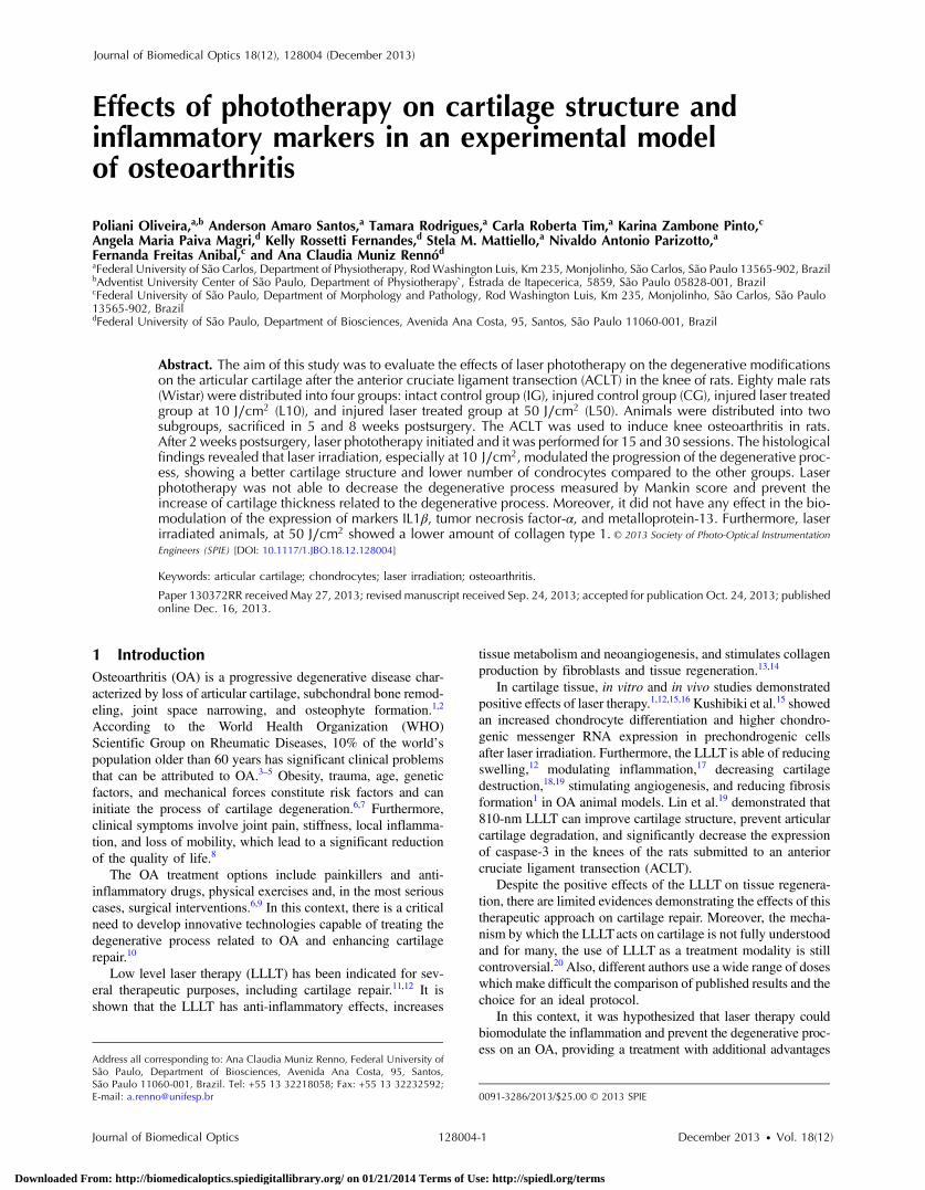
Effects of phototherapy on cartilage structure and inflammatory markers in an experimental model of osteoarthritis Poliani Oliveira Anderson Amaro Santos Tamara Rodrigues Carla Roberta Tim Karina Zambone Pinto Angela Maria Paiva Magri Kelly Rossetti Fernandes Stela M. Mattiello Nivaldo Antonio Parizotto Fernanda Freitas Anibal Ana Claudia Muniz Rennó Downloaded From: http://biomedicaloptics.spiedigitallibrary.org/ on 01/21/2014 Terms of Use: http://spiedl.org/terms

Welcome message from author
This document is posted to help you gain knowledge. Please leave a comment to let me know what you think about it! Share it to your friends and learn new things together.
Transcript
Effects of phototherapy on cartilagestructure and inflammatory markersin an experimental model ofosteoarthritis
Poliani OliveiraAnderson Amaro SantosTamara RodriguesCarla Roberta TimKarina Zambone PintoAngela Maria Paiva MagriKelly Rossetti FernandesStela M. MattielloNivaldo Antonio ParizottoFernanda Freitas AnibalAna Claudia Muniz Rennó
Downloaded From: http://biomedicaloptics.spiedigitallibrary.org/ on 01/21/2014 Terms of Use: http://spiedl.org/terms
Effects of phototherapy on cartilage structure andinflammatory markers in an experimental modelof osteoarthritis
Poliani Oliveira,a,b Anderson Amaro Santos,a Tamara Rodrigues,a Carla Roberta Tim,a Karina Zambone Pinto,cAngela Maria Paiva Magri,d Kelly Rossetti Fernandes,d Stela M. Mattiello,a Nivaldo Antonio Parizotto,aFernanda Freitas Anibal,c and Ana Claudia Muniz Rennód
aFederal University of São Carlos, Department of Physiotherapy, RodWashington Luis, Km 235, Monjolinho, São Carlos, São Paulo 13565-902, BrazilbAdventist University Center of São Paulo, Department of Physiotherapy`, Estrada de Itapecerica, 5859, São Paulo 05828-001, BrazilcFederal University of São Paulo, Department of Morphology and Pathology, Rod Washington Luis, Km 235, Monjolinho, São Carlos, São Paulo13565-902, BrazildFederal University of São Paulo, Department of Biosciences, Avenida Ana Costa, 95, Santos, São Paulo 11060-001, Brazil
Abstract. The aim of this study was to evaluate the effects of laser phototherapy on the degenerative modificationson the articular cartilage after the anterior cruciate ligament transection (ACLT) in the knee of rats. Eighty male rats(Wistar) were distributed into four groups: intact control group (IG), injured control group (CG), injured laser treatedgroup at 10 J∕cm2 (L10), and injured laser treated group at 50 J∕cm2 (L50). Animals were distributed into twosubgroups, sacrificed in 5 and 8 weeks postsurgery. The ACLT was used to induce knee osteoarthritis in rats.After 2 weeks postsurgery, laser phototherapy initiated and it was performed for 15 and 30 sessions. The histologicalfindings revealed that laser irradiation, especially at 10 J∕cm2, modulated the progression of the degenerative proc-ess, showing a better cartilage structure and lower number of condrocytes compared to the other groups. Laserphototherapy was not able to decrease the degenerative process measured by Mankin score and prevent theincrease of cartilage thickness related to the degenerative process. Moreover, it did not have any effect in the bio-modulation of the expression of markers IL1β, tumor necrosis factor-α, and metalloprotein-13. Furthermore, laserirradiated animals, at 50 J∕cm2 showed a lower amount of collagen type 1. © 2013 Society of Photo-Optical Instrumentation
Engineers (SPIE) [DOI: 10.1117/1.JBO.18.12.128004]
Keywords: articular cartilage; chondrocytes; laser irradiation; osteoarthritis.
Paper 130372RR received May 27, 2013; revised manuscript received Sep. 24, 2013; accepted for publication Oct. 24, 2013; publishedonline Dec. 16, 2013.
1 IntroductionOsteoarthritis (OA) is a progressive degenerative disease char-acterized by loss of articular cartilage, subchondral bone remod-eling, joint space narrowing, and osteophyte formation.1,2
According to the World Health Organization (WHO)Scientific Group on Rheumatic Diseases, 10% of the world’spopulation older than 60 years has significant clinical problemsthat can be attributed to OA.3–5 Obesity, trauma, age, geneticfactors, and mechanical forces constitute risk factors and caninitiate the process of cartilage degeneration.6,7 Furthermore,clinical symptoms involve joint pain, stiffness, local inflamma-tion, and loss of mobility, which lead to a significant reductionof the quality of life.8
The OA treatment options include painkillers and anti-inflammatory drugs, physical exercises and, in the most seriouscases, surgical interventions.6,9 In this context, there is a criticalneed to develop innovative technologies capable of treating thedegenerative process related to OA and enhancing cartilagerepair.10
Low level laser therapy (LLLT) has been indicated for sev-eral therapeutic purposes, including cartilage repair.11,12 It isshown that the LLLT has anti-inflammatory effects, increases
tissue metabolism and neoangiogenesis, and stimulates collagenproduction by fibroblasts and tissue regeneration.13,14
In cartilage tissue, in vitro and in vivo studies demonstratedpositive effects of laser therapy.1,12,15,16 Kushibiki et al.15 showedan increased chondrocyte differentiation and higher chondro-genic messenger RNA expression in prechondrogenic cellsafter laser irradiation. Furthermore, the LLLT is able of reducingswelling,12 modulating inflammation,17 decreasing cartilagedestruction,18,19 stimulating angiogenesis, and reducing fibrosisformation1 in OA animal models. Lin et al.19 demonstrated that810-nm LLLT can improve cartilage structure, prevent articularcartilage degradation, and significantly decrease the expressionof caspase-3 in the knees of the rats submitted to an anteriorcruciate ligament transection (ACLT).
Despite the positive effects of the LLLT on tissue regenera-tion, there are limited evidences demonstrating the effects of thistherapeutic approach on cartilage repair. Moreover, the mecha-nism by which the LLLTacts on cartilage is not fully understoodand for many, the use of LLLT as a treatment modality is stillcontroversial.20 Also, different authors use a wide range of doseswhich make difficult the comparison of published results and thechoice for an ideal protocol.
In this context, it was hypothesized that laser therapy couldbiomodulate the inflammation and prevent the degenerative proc-ess on an OA, providing a treatment with additional advantages
Address all corresponding to: Ana Claudia Muniz Renno, Federal University ofSão Paulo, Department of Biosciences, Avenida Ana Costa, 95, Santos,São Paulo 11060-001, Brazil. Tel: +55 13 32218058; Fax: +55 13 32232592;E-mail: [email protected] 0091-3286/2013/$25.00 © 2013 SPIE
Journal of Biomedical Optics 128004-1 December 2013 • Vol. 18(12)
Journal of Biomedical Optics 18(12), 128004 (December 2013)
Downloaded From: http://biomedicaloptics.spiedigitallibrary.org/ on 01/21/2014 Terms of Use: http://spiedl.org/terms
for clinical use. Consequently, the present study was carried out inorder to analyze the effects of lasertherapy at two different flu-encies, on cartilage damage in an OA experimental model inknees of rats. Histology analysis was used to evaluate the doseresponses of laser application in cartilage repair, after 5- and 8-weeks postsurgery. Also, immunohistochemistry evaluation ofinflammatory markers involved in the process of cartilage degen-eration [tumor necrosis factor (TNF-α) and interleukin-1 (IL-1β),proteolitic enzymes related to the matrix degradation metallopro-tein 13 (MMP 13) and Collagen type 1 (Col-1)].
2 Methods
2.1 Experimental Groups
This study was approved and conducted in accordance with theAnimal Care Committee guidelines of the Federal Universityof São Carlos (CEP 040/2010). Animals were maintained at19°C to 23°C on a 12:12 h light-dark cycle in the AnimalExperimentation Laboratory of the Federal University of SãoCarlos. Rats were housed in plastic cages and had free accessto water and standard food.
Eighty male Wistar rats (weighing 300� 20 g, 12 to13 weeks) were randomly distributed into four groups (n ¼ 20):intact control group (IG), injured control group (CG), injuredlaser treated group at 10 J∕cm2 (L10), and injured laser treatedgroup at 50 J∕cm2 (L50). The animals were distributed into twosubgroups, with different periods of sacrifice (5- and 8-weekspostsurgery).
2.2 Anterior Cruciate Ligament Transection (ACLT)
The animals were submitted to general anesthesia induced byintraperitoneal injection of xilazin (Syntec®, R. Soluções Lar,Cotia, São Paulo, Brasil, 20 mg∕kg, IP) and ketamin(Agener®, Av. do Café, São Paulo, Brasil at 40 mg∕kg, IP)and subjected to the ACLT of the left hind paw. The leftknee was shaved, then sterilized and draped in sterile fashion.A medial arthrotomy was performed. Then, the patella was dis-located and the anterior cruciate ligament was isolated and trans-ected. The ACLT was confirmed with Lachman testing by thesurgeon and an observer.21 After being irrigated with sterilesaline solution, the wounds were closed in layers and antisep-tically treated. Rats were given appropriate postoperative careand allowed free activities in individual cages.
2.3 Treatments
Laser treatment started 2 weeks after the surgery and it was per-formed for 15 and 30 sessions for each subgroup, using the fol-lowing protocol: five consecutive days of irradiation with aninterval of 2 days, for 3 and 6 weeks, respectively. The LLLTwas applied at two points (on the medial and lateral sides of thejoint), using the punctual contact technique. A low-energy Ga-Al-As laser (Theralaser, DMC® São Carlos, São Paulo, Brazil)was used at 830 nm, continuous wave diode, with a 0.028-cm2
beam diameter, a power output of 30 mW, fluence at 10 J∕cm2
(irradiation time of 10 s, energy per point 0.3 J), and fluence at50 J∕cm2 (irradiation time of 47 s, energy per point 1.4 J). Onthe respective days, animals were euthanized individually by car-bon dioxide asphyxia. The knee joints were removed for analysis.
2.4 Histological Analysis
After harvesting, the specimens were fixated in 4% formalde-hyde for 2 days, followed by decalcified in 4% ethylenediami-netetraacetic acid . The specimens were divided into two pieces,using a blade, at the mean point between both condyles,perpendicular to the articular surface. Samples were embeddedin paraffin blocks and histological sections were done.Therefore, thin sections (6 μm) were prepared in the sagittalplane, starting from the medial margin of the joint using amicrometer (Leica RM—2145, Germany). Laminas werestained with hematoxylin and eosin (HE, Merck, Germany),Safranin-O (Merck, Germany), and Picrossirius-Red (Merck,Germany). Moreover, three sections were obtained for the imu-nohistoquemical analysis.
2.5 Histological Descriptive Analysis
Histopathological alterations in the articular cartilage wereevaluated by two blinded observers. For descriptive analysis,the samples were stained with HE to evaluate cartilage structure,amount of cells and cellular organization. The specimens wereexamined using a light microscopy (100×) (Leica MicrosystemsAG, Wetzlar, Germany).
2.6 Semi-Quantitative Analysis
The severity of the OA lesion was graded on a scale using theMankin scoring system (Table 1).22,23 Samples were stainedwith Safranin- O and histological evaluation system was per-formed along all of the extension. At least three sections ofeach specimen were examined using light microscopy (100×)(Leica Microsystems AG, Wetzlar, Germany). The meanMankin score of the two observers was calculated.
2.7 Morphometric Analysis
The morphometric study was carried out using one randomizedslide stained with HE. The cartilage thickness and number ofchondrocytes in each area were quantitatively scored usingthe computer-based image analysis Axiovision 3.1 ImageAnalysis (Carl Zeiss, Oberkochen, Germany). To count thenumber of condrocytes, three areas of 80; 000 μm2, at the ante-rior, central, and posterior region of each slide were chosen.Within each area, cells were marked and the chondrocytes aver-age was calculated. Thickness was also measured in threeregions, one central and two laterals (300-mm left and rightfrom the first region), from subcondral bone to articularsurface.24
2.8 Picrossirius Red Analysis
Histological sections stained by the Picrosirius-polarizationmethod were viewed under polarized light (Carl Zeiss,Oberkochen, Germany) to assess the collagen organization inthe cartilage tissue.25–28 Similarly to the morphometric analysis,three areas of 80,000 μm2 (anterior, central, and posteriorregion) of each slide were chosen to quantify the amount of col-lagen using the software ImageJ (Version 1.45, NationalInstitutes of Health, Bethesda, Maryland). In each field, an indi-rect evaluation of the total collagen fibers organization based onthe birefringence of the collagen fiber bundles after staining withPicrosirius was performed. Two experienced observers (PO andAR) performed the scoring in a blinded manner.29
Journal of Biomedical Optics 128004-2 December 2013 • Vol. 18(12)
Oliveira et al.: Effects of phototherapy on cartilage structure and inflammatory markers. . .
Downloaded From: http://biomedicaloptics.spiedigitallibrary.org/ on 01/21/2014 Terms of Use: http://spiedl.org/terms
2.9 Immunohistochemistry
The TNF-α, IL-1β, MMP 13, and Col-1 immunoexpressionwere determined using the streptavidin-biotin-peroxidasemethod. Sections at 3 μm were deparaffinized in three changesof xylene and rehydrated in a graded series of ethanol to distilledwater. For antigen retrieval, slides were placed in 0.01 M citrate-buffer pH 6.0 and heated in a microwave for three cycles of5 min each at 850 W. Endogenous peroxidases were quenchedby incubation in 3% H2O2 for 20 min at room temperature. Sec-tions were incubated overnight at 4°C with primary antibody:TNF-α (polyclonal rabitt anti-rat, ab6671, Abcam, Cambrige,MA, UK), interleukin-1 (IL-1β) (polyclonal rabitt anti-rat, sc-7884, Sta Cruz biotechnology, California, USA), metalloprotein13 (MMP 13) (polyclonal rabitt anti-rat, ab75606, abcam, Cam-brige, MA, UK) and Collagen type 1 (Col-1) (anti-Col 1A mon-oclonal primary antibody, Sta Cruz Biotechnology, USA). Sub-sequently, sections were incubated. Which tissue was used aspositive control for each antibody tested in immunohistochemistry
technique with biotinylated secondary antibody (LSA B, Dakocytomation) for 30 min, washed in phosphate-buffered saline,and incubated with streptavidin peroxidase conjugate (LSA B,Dakocytomation) for 30 min. Finally, the reaction was devel-oped using 3,3′-Dianobenzidine tetrahydrocloride (Sigma,St. Louis, Missouri) for 5 min. Slides were briefly counter-stained in hematoxylin and rehydrated, and cover slipsadded. Negative and positive controls were to run simultane-ously. Positive controls were represented by mammary tissue.Negative controls were made by eliminating the primary anti-body as established in previous studies conducted by ourgroup.30 The TNF-α, IL-1β, MMP 13, and Col-1 immunoex-pressions were evaluated both qualitatively (presence of theimmunomarkers) and quantitatively in predetermined fieldsusing a light microscope ((Leica Microsystems AG, Wetzlar,Germany).
2.10 Immunohistochemical Semiquantitative Analysis
Sections stained using immunohistochemistry were analyzed forthe percentages of immunopositive cells in control and experi-mental animals. A total of 1000 cells were evaluated in 3 to 5fields at 400×magnification. These values were used as labelingindices.
2.11 Statistical Analysis
The normality of all variables’ distribution was verified usingthe Shapiro–Wilk W test. For the variable that exhibited normaldistribution, comparisons among the groups were made usingone-way analysis of variance (ANOVA) with pos hoc Tukey’stest. For the variable that exhibited nonnormal distribution,Kruskal Wallis test was used. STATISTICA version 7.0 wasused to carry out the statistics analysis. Values of p < 0.05 wereconsidered statistically significant.
3 Results
3.1 General Findings
Neither postoperative complications nor behavioral changeswere observed. The rats rapidly returned to their normal dietand showed no weight loss during the experimentation. Noneof the animals died during the experiment and no infection inthe surgical site was observed.
3.2 Histological Analysis
3.2.1 Descriptive analysis
Histopathological analysis revealed that after 5 weeks, cartilagetissue of IG presented a normal structure, without signs of fibril-lation. Moreover, in the superficial region, the chondrocyteswere displayed in a parallel arrangement and in the intermediateregion they were organized in columns [Fig. 1(a)]. The CGpresented signs of fibrillation, irregularities in the articular sur-face and chondrocytes displayed in a disorganized disposition[Fig. 1(c)]. In the laser treated groups, at both fluencies, thearticular surface showed initial signs of fibrillation and condro-cytes organized in a normal orientation, resembling the intactanimals [Figs. 1(e) and 1(g)].
After 8 weeks, the histological findings revealed thatIG presented normal cartilage structure [Fig. 1(b)]. In the controlanimals, the degenerative process progressed, presenting in-tense fibrillation, surface irregularities and hipercellularity, with
Table 1 Mankin score for the histological grading of cartilagedegeneration.
Grade
I. Structure
a. Normal 0
b. Surface irregularities 1
c. Pannus and surface irregularities 2
d. Clefts to transitional zone 3
e. Clefts to radial zone 4
f. Clefts to calcified zone 5
g. Complete disorganisation 6
II. Cells
a. Normal 0
b. Difuse hypercellularity 1
c. Cloning 2
d. Hipocellularity 3
III. Safranin-O staining
a. Normal 0
b. Slight reduction 1
c. Moderate reduction 2
d. Severe reduction 3
e. No dye noted 4
IV. Tidemark integrity
a. Intact 0
b. crossed by blood vessels 1
Journal of Biomedical Optics 128004-3 December 2013 • Vol. 18(12)
Oliveira et al.: Effects of phototherapy on cartilage structure and inflammatory markers. . .
Downloaded From: http://biomedicaloptics.spiedigitallibrary.org/ on 01/21/2014 Terms of Use: http://spiedl.org/terms
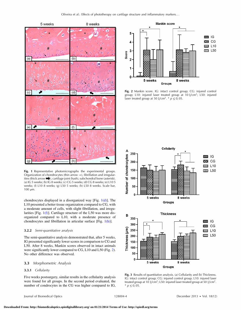
chondrocytes displayed in a disorganized way [Fig. 1(d)]. TheL10 presented a better tissue organization compared to CG, witha moderate amount of cells, with slight fibrillation, and irregu-larities [Fig. 1(f)]. Cartilage structure of the L50 was more dis-organized compared to L10, with a moderate presence ofchondrocytes and fibrillation in articular surface [Fig. 1(h)].
3.2.2 Semi-quantitative analysis
The semi-quantitative analysis demonstrated that, after 5 weeks,IG presented significantly lower scores in comparison to CG andL50. After 8 weeks, Mankin scores observed in intact animalswere significantly lower compared to CG, L10 and L50 (Fig. 2).No other difference was observed.
3.3 Morphometric Analysis
3.3.1 Cellularity
Five weeks postsurgery, similar results in the cellularity analysiswere found for all groups. In the second period evaluated, thenumber of condrocytes in the CG was higher compared to IG,
Fig. 1 Representative photomicrographs the experimental groups.Organization of chondrocytes (thin arrow →), fibrillation and irregular-ities (thick arrow ), cartilage joint (hash), subchondral bone (asterisk).(a) IG 5 weeks; (b) IG 8 weeks; (c) CG 5weeks; (d) CG 8 weeks; (e) L10 5weeks; (f) L10 8 weeks; (g) L50 5 weeks; (h) L50 8 weeks. Scale bar,100 μm.
Fig. 2 Mankin score. IG: intact control group; CG: injured controlgroup; L10: injured laser treated group at 10 J∕cm2; L50: injuredlaser treated group at 50 J∕cm2. * p ≤ 0; 05.
Fig. 3 Results of quantitative analysis. (a) Cellularity and (b) Thickness.IG: intact control group; CG: injured control group; L10: injured lasertreated group at 10 J∕cm2; L50: injured laser treated group at 50 J∕cm2.* p ≤ 0;05.
Journal of Biomedical Optics 128004-4 December 2013 • Vol. 18(12)
Oliveira et al.: Effects of phototherapy on cartilage structure and inflammatory markers. . .
Downloaded From: http://biomedicaloptics.spiedigitallibrary.org/ on 01/21/2014 Terms of Use: http://spiedl.org/terms
L10 and L50 (p ¼ 0.0052, 0.00017, 0.00015, respectively)[Fig. 3(a)].
3.3.2 Thickness
After 5 weeks, the IG presented lower cartilage thickness com-pared to CG (p ¼ 0.029) and L50 (p ¼ 0.00050) [Fig. 3(a)].After 8 weeks, IG had significantly lower cartilage thicknesscompared to the other groups (p ¼ 0.0031, 0.0021, 0.00019to CG, L10, and L50, respectively) [Fig. 3(b)].
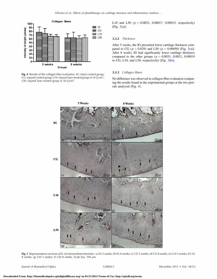
3.3.3 Collagen fibers
No difference was observed in collagen fiber evaluation compar-ing the results found in the experimental groups at the two peri-ods analyzed (Fig. 4).
Fig. 4 Results of the collagen fiber evaluation. IG: intact control group;CG: injured control group; L10: injured laser treated group at 10 J∕cm2;L50: injured laser treated group at 50 J∕cm2.
Fig. 5 Representatives sections of IL-1β immunohistochemistry. (a) IG 5 weeks; (b) IG 8 weeks; (c) CG 5 weeks; (d) CG 8 weeks; (e) L10 5 weeks; (f) L108 weeks; (g) L50 5 weeks; (f) L50 8 weeks. Scale bar, 100 μm.
Journal of Biomedical Optics 128004-5 December 2013 • Vol. 18(12)
Oliveira et al.: Effects of phototherapy on cartilage structure and inflammatory markers. . .
Downloaded From: http://biomedicaloptics.spiedigitallibrary.org/ on 01/21/2014 Terms of Use: http://spiedl.org/terms
3.4 Immunohistochemistry
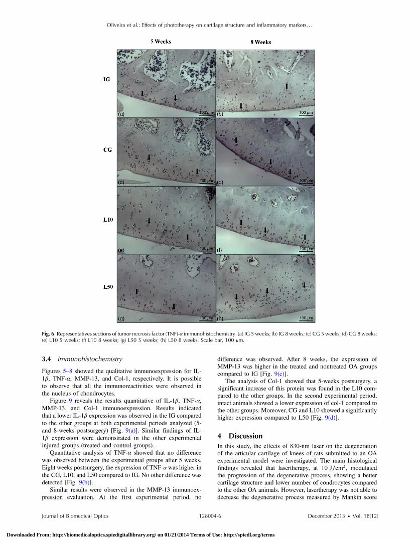
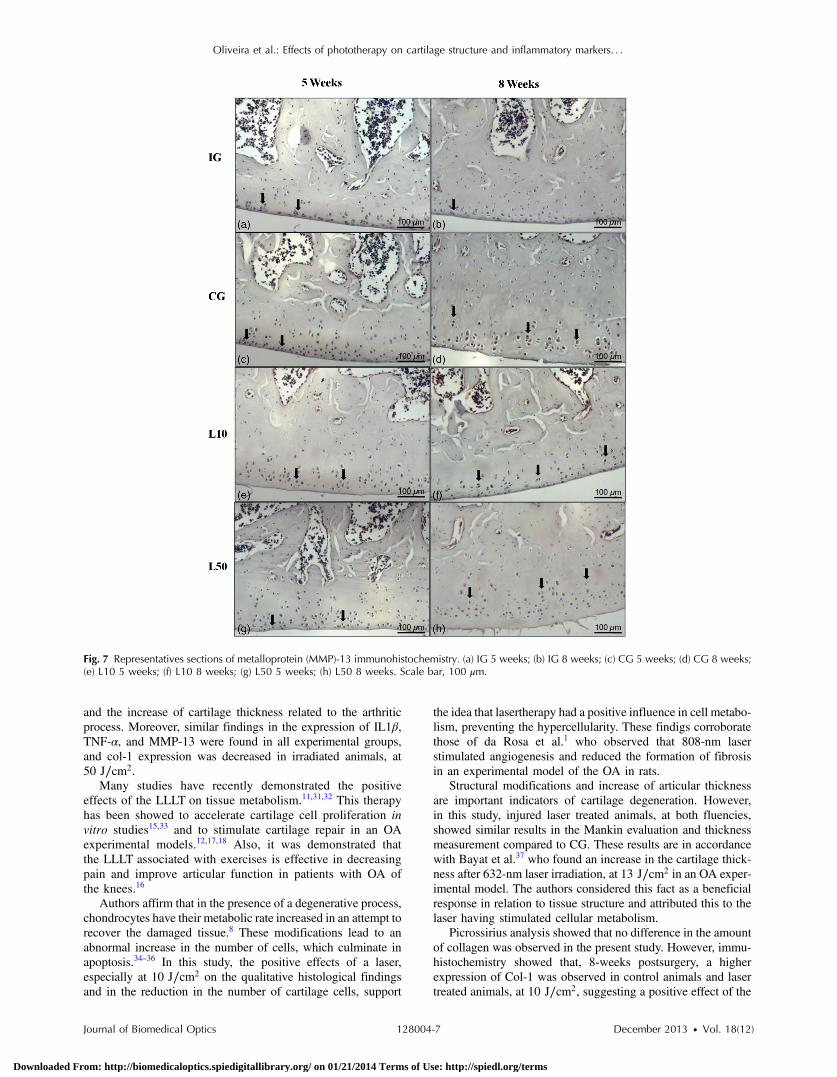
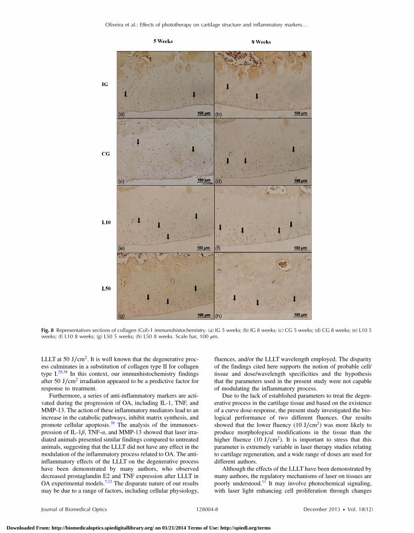
Figures 5–8 showed the qualitative immunoexpression for IL-1β, TNF-α, MMP-13, and Col-1, respectively. It is possibleto observe that all the immunoreactivities were observed inthe nucleus of chondrocytes.
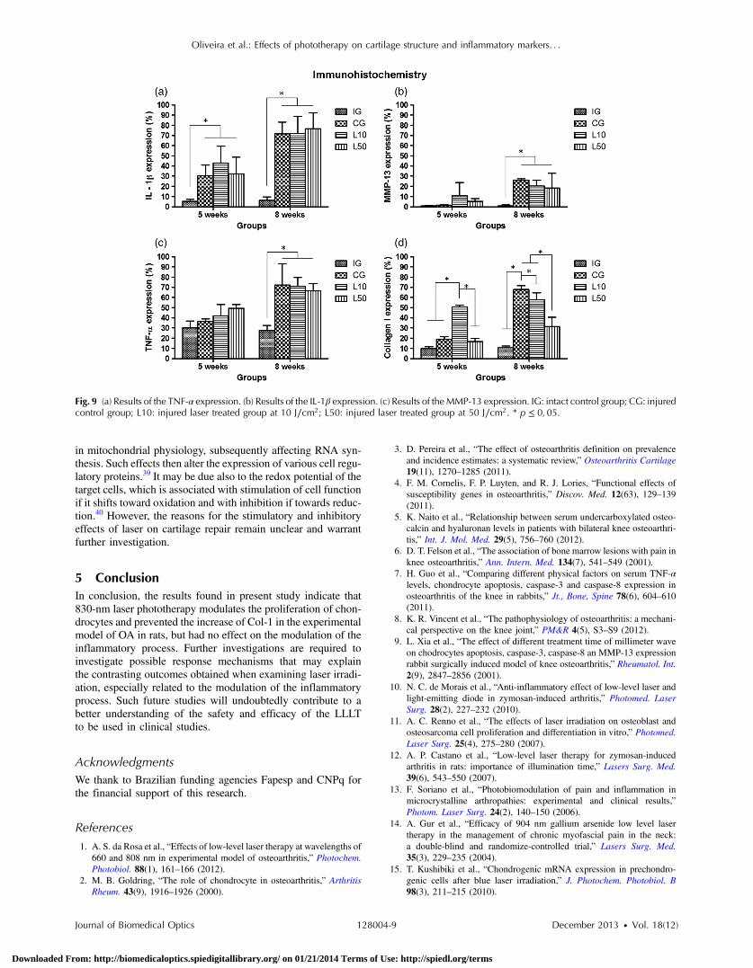
Figure 9 reveals the results quantitative of IL-1β, TNF-α,MMP-13, and Col-1 immunoexpression. Results indicatedthat a lower IL-1β expression was observed in the IG comparedto the other groups at both experimental periods analyzed (5-and 8-weeks postsurgery) [Fig. 9(a)]. Similar findings of IL-1β expression were demonstrated in the other experimentalinjured groups (treated and control groups).
Quantitative analysis of TNF-α showed that no differencewas observed between the experimental groups after 5 weeks.Eight weeks postsurgery, the expression of TNF-α was higher inthe CG, L10, and L50 compared to IG. No other difference wasdetected [Fig. 9(b)].
Similar results were observed in the MMP-13 immunoex-pression evaluation. At the first experimental period, no
difference was observed. After 8 weeks, the expression ofMMP-13 was higher in the treated and nontreated OA groupscompared to IG [Fig. 9(c)].
The analysis of Col-1 showed that 5-weeks postsurgery, asignificant increase of this protein was found in the L10 com-pared to the other groups. In the second experimental period,intact animals showed a lower expression of col-1 compared tothe other groups. Moreover, CG and L10 showed a significantlyhigher expression compared to L50 [Fig. 9(d)].
4 DiscussionIn this study, the effects of 830-nm laser on the degenerationof the articular cartilage of knees of rats submitted to an OAexperimental model were investigated. The main histologicalfindings revealed that lasertherapy, at 10 J∕cm2, modulatedthe progression of the degenerative process, showing a bettercartilage structure and lower number of condrocytes comparedto the other OA animals. However, lasertherapy was not able todecrease the degenerative process measured by Mankin score
Fig. 6 Representatives sections of tumor necrosis factor (TNF)-α immunohistochemistry. (a) IG 5 weeks; (b) IG 8 weeks; (c) CG 5weeks; (d) CG 8 weeks;(e) L10 5 weeks; (f) L10 8 weeks; (g) L50 5 weeks; (h) L50 8 weeks. Scale bar, 100 μm.
Journal of Biomedical Optics 128004-6 December 2013 • Vol. 18(12)
Oliveira et al.: Effects of phototherapy on cartilage structure and inflammatory markers. . .
Downloaded From: http://biomedicaloptics.spiedigitallibrary.org/ on 01/21/2014 Terms of Use: http://spiedl.org/terms
and the increase of cartilage thickness related to the arthriticprocess. Moreover, similar findings in the expression of IL1β,TNF-α, and MMP-13 were found in all experimental groups,and col-1 expression was decreased in irradiated animals, at50 J∕cm2.
Many studies have recently demonstrated the positiveeffects of the LLLT on tissue metabolism.11,31,32 This therapyhas been showed to accelerate cartilage cell proliferation invitro studies15,33 and to stimulate cartilage repair in an OAexperimental models.12,17,18 Also, it was demonstrated thatthe LLLT associated with exercises is effective in decreasingpain and improve articular function in patients with OA ofthe knees.16
Authors affirm that in the presence of a degenerative process,chondrocytes have their metabolic rate increased in an attempt torecover the damaged tissue.8 These modifications lead to anabnormal increase in the number of cells, which culminate inapoptosis.34–36 In this study, the positive effects of a laser,especially at 10 J∕cm2 on the qualitative histological findingsand in the reduction in the number of cartilage cells, support
the idea that lasertherapy had a positive influence in cell metabo-lism, preventing the hypercellularity. These findigs corroboratethose of da Rosa et al.1 who observed that 808-nm laserstimulated angiogenesis and reduced the formation of fibrosisin an experimental model of the OA in rats.
Structural modifications and increase of articular thicknessare important indicators of cartilage degeneration. However,in this study, injured laser treated animals, at both fluencies,showed similar results in the Mankin evaluation and thicknessmeasurement compared to CG. These results are in accordancewith Bayat et al.37 who found an increase in the cartilage thick-ness after 632-nm laser irradiation, at 13 J∕cm2 in an OA exper-imental model. The authors considered this fact as a beneficialresponse in relation to tissue structure and attributed this to thelaser having stimulated cellular metabolism.
Picrossirius analysis showed that no difference in the amountof collagen was observed in the present study. However, immu-histochemistry showed that, 8-weeks postsurgery, a higherexpression of Col-1 was observed in control animals and lasertreated animals, at 10 J∕cm2, suggesting a positive effect of the
Fig. 7 Representatives sections of metalloprotein (MMP)-13 immunohistochemistry. (a) IG 5 weeks; (b) IG 8 weeks; (c) CG 5 weeks; (d) CG 8 weeks;(e) L10 5 weeks; (f) L10 8 weeks; (g) L50 5 weeks; (h) L50 8 weeks. Scale bar, 100 μm.
Journal of Biomedical Optics 128004-7 December 2013 • Vol. 18(12)
Oliveira et al.: Effects of phototherapy on cartilage structure and inflammatory markers. . .
Downloaded From: http://biomedicaloptics.spiedigitallibrary.org/ on 01/21/2014 Terms of Use: http://spiedl.org/terms
LLLT at 50 J∕cm2. It is well known that the degenerative proc-ess culminates in a substitution of collagen type II for collagentype I.29,38 In this context, our immunhistochemistry findingsafter 50 J∕cm2 irradiation appeared to be a predictive factor forresponse to treatment.
Furthermore, a series of anti-inflammatory markers are acti-vated during the progression of OA, including IL-1, TNF, andMMP-13. The action of these inflammatory mediators lead to anincrease in the catabolic pathways, inhibit matrix synthesis, andpromote cellular apoptosis.39 The analysis of the immunoex-pression of IL-1β, TNF-α, and MMP-13 showed that laser irra-diated animals presented similar findings compared to untreatedanimals, suggesting that the LLLT did not have any effect in themodulation of the inflammatory process related to OA. The anti-inflammatory effects of the LLLT on the degenerative processhave been demonstrated by many authors, who observeddecreased prostaglandin E2 and TNF expression after LLLT inOA experimental models.7,12 The disparate nature of our resultsmay be due to a range of factors, including cellular physiology,
fluences, and/or the LLLT wavelength employed. The disparityof the findings cited here supports the notion of probable cell/tissue and dose/wavelength specificities and the hypothesisthat the parameters used in the present study were not capableof modulating the inflammatory process.
Due to the lack of established parameters to treat the degen-erative process in the cartilage tissue and based on the existenceof a curve dose-response, the present study investigated the bio-logical performance of two different fluences. Our resultsshowed that the lower fluency (10 J∕cm2) was more likely toproduce morphological modifications in the tissue than thehigher fluence (10 J∕cm2). It is important to stress that thisparameter is extremely variable in laser therapy studies relatingto cartilage regeneration, and a wide range of doses are used fordifferent authors.
Although the effects of the LLLT have been demonstrated bymany authors, the regulatory mechanisms of laser on tissues arepoorly understood.11 It may involve photochemical signaling,with laser light enhancing cell proliferation through changes
Fig. 8 Representatives sections of collagen (Col)-1 immunohistochemistry. (a) IG 5 weeks; (b) IG 8 weeks; (c) CG 5 weeks; (d) CG 8 weeks; (e) L10 5weeks; (f) L10 8 weeks; (g) L50 5 weeks; (h) L50 8 weeks. Scale bar, 100 μm.
Journal of Biomedical Optics 128004-8 December 2013 • Vol. 18(12)
Oliveira et al.: Effects of phototherapy on cartilage structure and inflammatory markers. . .
Downloaded From: http://biomedicaloptics.spiedigitallibrary.org/ on 01/21/2014 Terms of Use: http://spiedl.org/terms
in mitochondrial physiology, subsequently affecting RNA syn-thesis. Such effects then alter the expression of various cell regu-latory proteins.39 It may be due also to the redox potential of thetarget cells, which is associated with stimulation of cell functionif it shifts toward oxidation and with inhibition if towards reduc-tion.40 However, the reasons for the stimulatory and inhibitoryeffects of laser on cartilage repair remain unclear and warrantfurther investigation.
5 ConclusionIn conclusion, the results found in present study indicate that830-nm laser phototherapy modulates the proliferation of chon-drocytes and prevented the increase of Col-1 in the experimentalmodel of OA in rats, but had no effect on the modulation of theinflammatory process. Further investigations are required toinvestigate possible response mechanisms that may explainthe contrasting outcomes obtained when examining laser irradi-ation, especially related to the modulation of the inflammatoryprocess. Such future studies will undoubtedly contribute to abetter understanding of the safety and efficacy of the LLLTto be used in clinical studies.
AcknowledgmentsWe thank to Brazilian funding agencies Fapesp and CNPq forthe financial support of this research.
References1. A. S. da Rosa et al., “Effects of low-level laser therapy at wavelengths of
660 and 808 nm in experimental model of osteoarthritis,” Photochem.Photobiol. 88(1), 161–166 (2012).
2. M. B. Goldring, “The role of chondrocyte in osteoarthritis,” ArthritisRheum. 43(9), 1916–1926 (2000).
3. D. Pereira et al., “The effect of osteoarthritis definition on prevalenceand incidence estimates: a systematic review,” Osteoarthritis Cartilage19(11), 1270–1285 (2011).
4. F. M. Cornelis, F. P. Luyten, and R. J. Lories, “Functional effects ofsusceptibility genes in osteoarthritis,” Discov. Med. 12(63), 129–139(2011).
5. K. Naito et al., “Relationship between serum undercarboxylated osteo-calcin and hyaluronan levels in patients with bilateral knee osteoarthri-tis,” Int. J. Mol. Med. 29(5), 756–760 (2012).
6. D. T. Felson et al., “The association of bone marrow lesions with pain inknee osteoarthritis,” Ann. Intern. Med. 134(7), 541–549 (2001).
7. H. Guo et al., “Comparing different physical factors on serum TNF-αlevels, chondrocyte apoptosis, caspase-3 and caspase-8 expression inosteoarthritis of the knee in rabbits,” Jt., Bone, Spine 78(6), 604–610(2011).
8. K. R. Vincent et al., “The pathophysiology of osteoarthritis: a mechani-cal perspective on the knee joint,” PM&R 4(5), S3–S9 (2012).
9. L. Xia et al., “The effect of different treatment time of millimeter waveon chodrocytes apoptosis, caspase-3, caspase-8 an MMP-13 expressionrabbit surgically induced model of knee osteoarthritis,” Rheumatol. Int.2(9), 2847–2856 (2001).
10. N. C. de Morais et al., “Anti-inflammatory effect of low-level laser andlight-emitting diode in zymosan-induced arthritis,” Photomed. LaserSurg. 28(2), 227–232 (2010).
11. A. C. Renno et al., “The effects of laser irradiation on osteoblast andosteosarcoma cell proliferation and differentiation in vitro,” Photomed.Laser Surg. 25(4), 275–280 (2007).
12. A. P. Castano et al., “Low-level laser therapy for zymosan-inducedarthritis in rats: importance of illumination time,” Lasers Surg. Med.39(6), 543–550 (2007).
13. F. Soriano et al., “Photobiomodulation of pain and inflammation inmicrocrystalline arthropathies: experimental and clinical results,”Photom. Laser Surg. 24(2), 140–150 (2006).
14. A. Gur et al., “Efficacy of 904 nm gallium arsenide low level lasertherapy in the management of chronic myofascial pain in the neck:a double-blind and randomize-controlled trial,” Lasers Surg. Med.35(3), 229–235 (2004).
15. T. Kushibiki et al., “Chondrogenic mRNA expression in prechondro-genic cells after blue laser irradiation,” J. Photochem. Photobiol. B98(3), 211–215 (2010).
Fig. 9 (a) Results of the TNF-α expression. (b) Results of the IL-1β expression. (c) Results of theMMP-13 expression. IG: intact control group; CG: injuredcontrol group; L10: injured laser treated group at 10 J∕cm2; L50: injured laser treated group at 50 J∕cm2. * p ≤ 0;05.
Journal of Biomedical Optics 128004-9 December 2013 • Vol. 18(12)
Oliveira et al.: Effects of phototherapy on cartilage structure and inflammatory markers. . .
Downloaded From: http://biomedicaloptics.spiedigitallibrary.org/ on 01/21/2014 Terms of Use: http://spiedl.org/terms
16. P. P. Alfredo et al., “Efficacy of low level laser therapy associated withexercises in knee osteoarthritis: a randomized double-blind study,” Clin.Rehabil. 26(6), 523–533 (2012).
17. M. R. Guerino et al., “Laser treatment of experimentally inducedchronic arthritis,” Appl. Surf. Sci. 154(1), 561–564 (2000).
18. H. J. Cho et al., “Effect of low-level laser therapy on osteoarthropathy inrabbit,” In Vivo 18(5), 585–591 (2004).
19. Y. S. Lin, M. H. Huang, and C. Y. Chai, “Effects of helium-neon laser onthe mucopolysaccharide induction in experimental osteoarthritic carti-lage,” Osteoarthritis Cartilage 14(4), 377–383 (2006).
20. L. Brosseau et al., “Low level laser therapy for osteoarthritis and rheu-matoid arthritis: a metaanalysis,” J. Rheumatol. 27(8), 1961–1969 (2000).
21. J. M. Williams et al., “Effects of surgically induced instability on ratKnee articular cartilage,” J. Anat. 134(1), 103–109 (1982).
22. R. G. Pearson et al., “Histopathology grading systems for characterisa-tion of human knee osteoarthritis–reproducibility, variability, reliability,correlation, and validity,” Osteoarthritis Cartilage 19(3), 324–331(2011).
23. H. J. Mankin et al., “Bochemical and metabolic abnormalities inarticular cartilage from osteoarthritic human hips II. Correlation of mor-phology with biochemical and metabolic data,” J. Bone Joint Surg. Am.53(3), 523–537 (1971).
24. A. F. Renner et al., “The effect of a passive muscle stretching protocolon the articular cartilage,”Osteoarthritis Cartilage 14(2), 196–202 (2006).
25. L. C. Junqueira, G. S. Montes, and E. M. Sanchez, “The influence ofsection thickness on the study of collagen by the Pricrosirius-polariza-tion method,” Histochemistry 74(1), 153–156 (1982).
26. G. S. Montes, “Structural biology of the fibres of the collagenous andelastic systems,” Cell Biol. Int. 20(1), 15–27 (1996).
27. G. B. Andrade et al., “Use of the Picrosirius-polarization method to agefibrotic lesions in the hepatic granulomas produced in experimentalmurine schistosomiasis,” Ann. Trop. Med. Parasitol. 93(3), 265–272(1999).
28. I. Garavello-Freitas et al., “Low-power laser irradiation improves his-tomorphometrical parameters and bone matrix organization during
tibia wound healing in rats,” J. Photochem. Photobiol. B 70(2),81–89 (2003).
29. F. A. Vasilceac et al., “The remodeling of collagen fibers in rats anklessubmitted to immobilization and muscle stretch protocol,” Rheumatol.Int. 31(6), 737–742 (2011).
30. A. P. Paiotti et al., “Effect of COX-2 inhibitor lumiracoxib and the TNF-α antagonist etanercept on TNBS-induced colitis in Wistar rats,” J. Mol.Histol. 43(3), 307–317 (2012).
31. F. Kamali et al., “The therapeutic effect of low-level laser on repair ofosteochondral defects in rabbit knee,” J. Photochem. Photobiol. B 88(1),11–15 (2007).
32. J. M. Bjordal et al., “A systematic review of low level laser therapy withlocation-specific doses for pain from chronic joint disorders,” Aust. J.Physiother. 49(2), 107–116 (2003).
33. B. J. Wong et al., “Identification of chondrocyte proliferation followinglaser irradiation, thermal injury, and mechanical trauma,” Lasers Surg.Med. 37(1). 89–96 (2005).
34. I. Fujita et al., “Apoptosis of hypertrophic chondrocytes in rat cartilagi-nous growth plate,” J. Orthop. Sci. 2(5), 328–333 (1997).
35. K. Kuhnt et al., “Cell death in cartilage,” Osteoarthritis Cartilage 12(1),1–16 (2004).
36. K. P. Pritzker et al., “Osteoarthritis cartilage histopathology: gradingand staging,” Osteoarthritis Cartilage 14(1), 13–29 (2006).
37. M. Bayat et al., “Effect of low-level helium-neon laser therapy onhistological and ultrastructural features of immobilized rabbit articularcartilage,” J. Photochem. Photobiol. 87(2), 81–87 (2007).
38. C. N. K. Dias et al., “Progression of articular cartilage degeneration afterapplication of muscle stretch,” Connect. Tissue Res. 53(1), 39–47(2012).
39. T. I. Karu and S. F. Kolyakov, “Exact action spectra for cellular responserelevant to phototherapy,” Photomed. Laser Surg. 23(4), 355–361(2005).
40. M. Mognato et al., “Cell growth modulation of human cells irradiatedin vitro with low-level laser therapy,” Photomed. Laser Surg. 22(6),523–526 (2004).
Journal of Biomedical Optics 128004-10 December 2013 • Vol. 18(12)
Oliveira et al.: Effects of phototherapy on cartilage structure and inflammatory markers. . .
Downloaded From: http://biomedicaloptics.spiedigitallibrary.org/ on 01/21/2014 Terms of Use: http://spiedl.org/terms
Related Documents