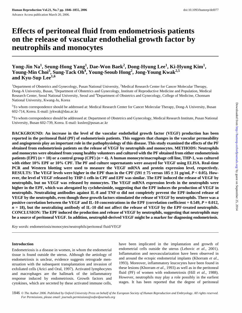
Human Reproduction Vol.21, No.7 pp. 1846–1855, 2006 doi:10.1093/humrep/del077 Advance Access publication March 20, 2006. 1846 © The Author 2006. Published by Oxford University Press on behalf of the European Society of Human Reproduction and Embryology. All rights reserved. For Permissions, please email: [email protected] Effects of peritoneal fluid from endometriosis patients on the release of vascular endothelial growth factor by neutrophils and monocytes Yong-Jin Na 1 , Seung-Hong Yang 1 , Dae-Won Baek 2 , Dong-Hyung Lee 1 , Ki-Hyung Kim 1 , Young-Min Choi 3 , Sung-Tack Oh 4 , Young-Seoub Hong 2 , Jong-Young Kwak 2,5 and Kyu-Sup Lee 1,6 1 Department of Obstetrics and Gynecology, Pusan National University, 2 Medical Research Center for Cancer Molecular Therapy, Dong-A University, Busan, 3 Department of Obstetrics and Gynecology, Institute of Reproductive Medicine and Population, Medical Research Center, Seoul National University, Seoul and 4 Department of Obstetrics and Gynecology, College of Medicine, Chonnam National University, Kwang-Ju, Korea 5 To whom correspondence should be addressed at: Medical Research Center for Cancer Molecular Therapy, Dong-A University, Busan 602-714, Korea. E-mail: [email protected] 6 To whom correspondence should be addressed at: Department of Obstetrics and Gynecology, Medical Research Institute, Pusan National University, Busan 602-739, Korea. E-mail: [email protected] BACKGROUND: An increase in the level of the vascular endothelial growth factor (VEGF) production has been reported in the peritoneal fluid (PF) of endometriosis patients. This suggests that changes in the vascular permeability and angiogenesis play an important role in the pathophysiology of this disease. This study examined the effects of the PF obtained from endometriosis patients on the release of VEGF by neutrophils and monocytes. METHODS: Neutrophils and monocytes were obtained from young healthy volunteers and cultured with the PF obtained from either endometriosis patients (EPF) (n = 18) or a control group (CPF) (n = 4). A human monocyte/macrophage cell line, THP-1, was cultured with either 10% EPF or 10% CPF. The PF and culture supernatants were assayed for VEGF using ELISA. Real-time PCR and Western blotting were used to measure the VEGF mRNA and protein expression level, respectively. RESULTS: The VEGF levels were higher in the EPF than in the CPF (591 ± 75 versus 185 ± 31 pg/ml, P < 0.05). How- ever, the level of VEGF released by THP-1 cells in CPF and EPF was similar. The EPF induced the release of VEGF by neutrophils, but no VEGF was released by monocytes. The VEGF mRNA expression levels in the neutrophils were higher in the EPF, which was abrogated by cycloheximide, suggesting that the EPF induces the production of VEGF in neutrophils. Neutralizing antibodies against IL-8 and TNF-a did not completely prevent the EPF-induced release of VEGF by the neutrophils, even though these growth factors stimulated the release of VEGF by neutrophils. There was a positive correlation between the VEGF and IL-10 concentrations in the EPF (correlation coefficient = 0.549, P = 0.012, n = 18), but the neutralizing antibody of IL-10 did not affect the release of VEGF by the EPF-treated neutrophils. CONCLUSION: The EPF induced the production and release of VEGF by neutrophils, suggesting that neutrophils may be a source of peritoneal VEGF. In addition, neutrophil-derived VEGF might be a marker for diagnosing endometriosis. Key words: endometriosis/monocytes/neutrophils/peritoneal fluid/VEGF Introduction Endometriosis is a disease in women, in whom the endometrial tissue is found outside the uterus. Although the aetiology of endometriosis is unclear, evidence suggests retrograde men- struation with the subsequent transplantation and invasion of exfoliated cells (Arici and Oral, 1997). Activated lymphocytes and macrophages are the hallmark of the inflammatory response induced by endometriosis. Growth factors and cytokines, which are secreted by these activated immune cells, have been implicated in the implantation and growth of endometrial cells outside the uterus (Lebovic et al., 2001). Inflammation and neovascularization have been observed in and around the ectopic endometrial implants (Khorram et al., 1993). Moreover, inflammatory leucocytes have been found in these lesions (Khorram et al., 1993) as well as in the peritoneal fluid (PF) of women with endometriosis (Hill et al., 1988). However, neutrophils may play a role possibly in the earliest stages. It has been reported that the degree of peritoneal by guest on September 25, 2014 http://humrep.oxfordjournals.org/ Downloaded from

Welcome message from author
This document is posted to help you gain knowledge. Please leave a comment to let me know what you think about it! Share it to your friends and learn new things together.
Transcript
Human Reproduction Vol.21, No.7 pp. 1846–1855, 2006 doi:10.1093/humrep/del077
Advance Access publication March 20, 2006.
1846 © The Author 2006. Published by Oxford University Press on behalf of the European Society of Human Reproduction and Embryology. All rights reserved.For Permissions, please email: [email protected]
Effects of peritoneal fluid from endometriosis patients on the release of vascular endothelial growth factor by neutrophils and monocytes
Yong-Jin Na1, Seung-Hong Yang1, Dae-Won Baek2, Dong-Hyung Lee1, Ki-Hyung Kim1, Young-Min Choi3, Sung-Tack Oh4, Young-Seoub Hong2, Jong-Young Kwak2,5 and Kyu-Sup Lee1,6
1Department of Obstetrics and Gynecology, Pusan National University, 2Medical Research Center for Cancer Molecular Therapy, Dong-A University, Busan, 3Department of Obstetrics and Gynecology, Institute of Reproductive Medicine and Population, Medical Research Center, Seoul National University, Seoul and 4Department of Obstetrics and Gynecology, College of Medicine, Chonnam National University, Kwang-Ju, Korea5To whom correspondence should be addressed at: Medical Research Center for Cancer Molecular Therapy, Dong-A University, Busan 602-714, Korea. E-mail: [email protected] whom correspondence should be addressed at: Department of Obstetrics and Gynecology, Medical Research Institute, Pusan National University, Busan 602-739, Korea. E-mail: [email protected]
BACKGROUND: An increase in the level of the vascular endothelial growth factor (VEGF) production has beenreported in the peritoneal fluid (PF) of endometriosis patients. This suggests that changes in the vascular permeabilityand angiogenesis play an important role in the pathophysiology of this disease. This study examined the effects of the PFobtained from endometriosis patients on the release of VEGF by neutrophils and monocytes. METHODS: Neutrophilsand monocytes were obtained from young healthy volunteers and cultured with the PF obtained from either endometriosispatients (EPF) (n = 18) or a control group (CPF) (n = 4). A human monocyte/macrophage cell line, THP-1, was culturedwith either 10% EPF or 10% CPF. The PF and culture supernatants were assayed for VEGF using ELISA. Real-timePCR and Western blotting were used to measure the VEGF mRNA and protein expression level, respectively.RESULTS: The VEGF levels were higher in the EPF than in the CPF (591 ± 75 versus 185 ± 31 pg/ml, P < 0.05). How-ever, the level of VEGF released by THP-1 cells in CPF and EPF was similar. The EPF induced the release of VEGF byneutrophils, but no VEGF was released by monocytes. The VEGF mRNA expression levels in the neutrophils werehigher in the EPF, which was abrogated by cycloheximide, suggesting that the EPF induces the production of VEGF inneutrophils. Neutralizing antibodies against IL-8 and TNF-a did not completely prevent the EPF-induced release ofVEGF by the neutrophils, even though these growth factors stimulated the release of VEGF by neutrophils. There was apositive correlation between the VEGF and IL-10 concentrations in the EPF (correlation coefficient = 0.549, P = 0.012,n = 18), but the neutralizing antibody of IL-10 did not affect the release of VEGF by the EPF-treated neutrophils.CONCLUSION: The EPF induced the production and release of VEGF by neutrophils, suggesting that neutrophils maybe a source of peritoneal VEGF. In addition, neutrophil-derived VEGF might be a marker for diagnosing endometriosis.
Key words: endometriosis/monocytes/neutrophils/peritoneal fluid/VEGF
Introduction
Endometriosis is a disease in women, in whom the endometrialtissue is found outside the uterus. Although the aetiology ofendometriosis is unclear, evidence suggests retrograde men-struation with the subsequent transplantation and invasion ofexfoliated cells (Arici and Oral, 1997). Activated lymphocytesand macrophages are the hallmark of the inflammatoryresponse induced by endometriosis. Growth factors andcytokines, which are secreted by these activated immune cells,
have been implicated in the implantation and growth ofendometrial cells outside the uterus (Lebovic et al., 2001).Inflammation and neovascularization have been observed inand around the ectopic endometrial implants (Khorram et al.,1993). Moreover, inflammatory leucocytes have been found inthese lesions (Khorram et al., 1993) as well as in the peritonealfluid (PF) of women with endometriosis (Hill et al., 1988).However, neutrophils may play a role possibly in the earlieststages. It has been reported that the degree of peritoneal
by guest on September 25, 2014
http://humrep.oxfordjournals.org/
Dow
nloaded from
Effects of EPF on the release of VEGF
1847
inflammation, as assessed by the total number of PF polymor-phonuclear leucocytes, decreases with the increasing stage ofthe endometriosis (Haney et al., 1991).
The vascular endothelial growth factor (VEGF) is ahomodimeric peptide growth factor with various biologicaleffects, such as the formation of vascular tubes and the perme-ability of blood vessels (Keck et al., 1989; Leung et al., 1989).Many types of cells, including macrophages (McLaren et al.,1996a), neutrophils (Taichman et al., 1997) and T cells (Freemanet al., 1995), produce VEGF. The induction of VEGF is stimu-lated by various growth factors, cytokines and hypoxia(Neufeld et al., 1999). VEGF has been suggested to play a rolein the progression of endometriosis, because this growth factorstimulates the angiogenic activity in endometriosis (Fascianiet al., 2000). Moreover, elevated VEGF levels have been foundin the PF of endometriosis patients (McLaren et al., 1996b),and a positive correlation has been reported between the sever-ity of endometriosis and the VEGF concentrations in the PF(Shifren et al., 1996).
On attraction, neutrophils are activated and secrete VEGF(Gaudry et al., 1997; Taichman et al., 1997; Webb et al.,1998). Neutrophils in the endometrium are a source of intra-vascular VEGF for vessels undergoing angiogenesis (Gargettet al., 2001), and the increased chemotactic activity for neu-trophils has been demonstrated in the PF of endometriosispatients (Leiva et al., 1993). Previous studies have shown thatthe PF and endometrial macrophages are significant sources ofVEGF (McLaren et al., 1996b). However, the biological sig-nificance of VEGF in neutrophils during the pathological pro-cess of endometriosis is unclear. Therefore, this studyexamined the effect of the PF on the release of VEGF by neu-trophils and monocytes.
Materials and methods
Reagents
The recombinant human VEGF, interleukin-8 (IL-8), tumour necrosisfactor-α (TNF-α), epithelial neutrophil-activating peptide-78 (ENA-78), granulocyte–macrophage colony-stimulating factor (GM-CSF)and neutralizing antibodies against IL-10, IL-8 and TNF-α were pur-chased from R&D Systems (Minneapolis, MN, USA). The RPMI-1640 medium was purchased from Gibco-BRL (Gaithersburg, MD,USA). The specific antibodies against VEGF165 and β-actin were pur-chased from Santa-Cruz Biotech (Santa Cruz, CA, USA). The activat-ing anti-Fas antibody (clone CH11, IgM) was obtained from UpstateBiotechnology (Lake Placid, NY, USA). The Histopaque-1077, dex-tran (500 000 Da), lipopolysaccharide (LPS), cycloheximide, pertus-sis toxin (PTX) and other chemicals were acquired from SigmaChemical Co. (St Louis, MO, USA).
Patients
The endometriosis patients (n = 18) in this study had either stage III orIV endometriosis (Kwak et al., 2002). The patients were stagedaccording to the revised American Fertility Society scoring system(The American Fertility Society, 1985). The presence of endometrio-sis was assessed at the time of surgery and was later confirmed histo-logically. Women taking oral contraceptives and gonadotrophin-releasing hormone analogues were excluded. The PF from theendometriosis group (EPF) was collected in a sterile environment
before any surgical manipulations. The control PF (CPF) (n = 4) wasobtained from fertile women undergoing laparoscopic surgery for vari-ous gynaecological indications other than endometriosis. These condi-tions included a dermoid cyst and carcinoma in situ of the uterinecervix. Patients with infertility without endometriosis were notincluded in the control group because there are many underlyingcauses of the infertility, and the patient selection in this study was notbased on the presence or absence of infertility. The PF was centrifugedat 800 × g for 10 min, and the supernatants were stored at −70°C.Figure 1 is a schematic drawing of the study design. Each CPF andEPF sample was assayed for the presence of VEGF and VEGF produc-tion in the THP-1 cells. Among the nine EPF samples containing ahigh level of VEGF, five EPF samples, which were sufficient for allexperiments in this study were used to determine the level of VEGFproduction in the neutrophils and monocytes (blanked triangle inFigure 2A). The mean VEGF concentrations in the CPF mixture and EPFmixture were 185 and 905 pg/ml, respectively (Figure 2A). Informedconsent was obtained from all subjects, and the Institutional ReviewBoard at Pusan National University Hospital approved this study.
Neutrophil isolation and culture
The peripheral blood neutrophils were isolated from 10 young healthydonors using a method involving dextran sedimentation and differen-tial centrifugation through a Ficoll–Hypaque density gradient (Kwaket al., 2002). Briefly, the blood was mixed with a solution containing3% dextran and 0.9% NaCl and stored for 45 min at 25°C. Theneutrophil-rich upper layer of the suspension was collected, the resid-ual erythrocytes were removed by hypotonic lysis, and the remainingpellet was suspended in HEPES-buffered saline. The suspension wascentrifuged on a Histopaque solution at 4°C. The isolated neutrophils(5 × 10⁶/ml) were maintained in a RPMI-1640 medium supplementedwith or without 10% EPF, or 10% CPF in the presence of 10% heat-inactivated fetal bovine serum (FBS), 1% glutamine, 100 U/ml of pen-icillin and 100 μg/ml of streptomycin in 24-well flat-bottomed platesat 37°C in a humidified atmosphere containing 5% CO2 (Kwak et al.,2002). Neutrophils with lobed nuclei and neutrally stained cytoplasmand granules were found to be 95% morphologically pure by Wright–Giemsa staining.
Monocyte isolation
The peripheral blood mononuclear cells were prepared by density-gradient centrifugation of the whole blood (n = 10) over a peripheralblood mononuclear cell separation medium (Histopaque-1077). The
Figure 1. Experimental use of the peritoneal fluid (PF) from theendometriosis group (EPF) and control group (CPF) and the assayperformed. See details in ‘Materials and methods’.
by guest on September 25, 2014
http://humrep.oxfordjournals.org/
Dow
nloaded from

Y.-J.Na et al.
1848
mononuclear cells were suspended in RPMI-1640 medium containing10% FBS and incubated for 1 h at 37°C to allow the monocytes toattach to the culture dish. The cells were washed five times with warmmedium to remove the lymphocytes, and the remaining cells were col-lected. The purity of the isolated monocytes was confirmed by flowcytometric analysis using the fluorescein-isothiocyanate-conjugatedanti-CD14 antibody (BD Pharmingen, NJ, USA) with >90% cell purity.
THP-1 cell culture
The THP-1 cells (Korean Cell Line Bank, Seoul National UniversityHospital, Korea), which were derived from a human monocyte/macro-phage cell line, were cultured in RPMI-1640 medium containing100 U/ml of penicillin, 100 μg/ml of streptomycin, 2 mM L-glutamine,20 mM HEPES and 10% FBS. The THP-1 cells were plated at 5 × 10⁶/mlin 24-well plates and treated with either 10% EPF or 10% CPF. After24 h at 37°C and 5% CO2, the THP-1 cells were centrifuged. The super-natants were then removed and assayed for VEGF.
Real-time PCR
The total cellular RNA from the neutrophils was isolated using the Trireagent (Sigma). All RNA samples were treated with DNase I, and thequality of the RNA was determined by measuring the optical densityat 260 nm (OD260). For each sample, 2 μg of the total RNA was firstconverted to cDNA using the reverse transcriptase and oligo (dT)primers. The real-time–PCR amplification reactions were then per-formed on aliquots of the cDNA using Assays-on-Demand TaqManGene Expression Products (p/n, 4335626) (Applied Biosystems,Foster City, CA, USA) and by continuous fluorescence monitoring(ABI PRISM® 7000; Sequence Detection System, Applied Biosystems).The following PCR reactions were used: an initial denaturation step(95°C for 120 s), followed by 40 cycles of denaturation at 95°C (15 s),
annealing at 58°C (30 s) and extension at 72°C (45 s). The primersand probes for VEGF and β-actin were proprietary oligonucleotidespurchased from Applied Biosystems ‘Assay-on-Demand’, and a 5´-end labelling with a reporter fluorochrome [6-carboxyfluoresceinreporter] was used.
ELISA assay
The neutrophils and monocytes were pretreated with or without 1 μg/mlof PTX for 4 h, or with 0.5 ng of the neutralizing antibodies againstTNF-α, IL-8 or IL-10 for 1 h. To determine whether the production ofVEGF is mediated by protein synthesis, we also pretreated the neu-trophils with an inhibitor of the translation, cycloheximide (10 μg/ml)for 1 h. The cells were cultured in RPMI-1640 media containing 10%EPF, 10% CPF, 10 ng/ml of IL-8, 10 ng/ml of ENA-78 or 20 ng/ml ofTNF-α for the indicated times. The IL-10 and VEGF concentrationsin the PFs and supernatants were measured in triplicate using standardELISA kits (R&D Systems). The VEGF detected in this study usingELISA represents the VEGF165 and VEGF121 as specified by the man-ufacturer. The sensitivity of the ELISA was ≤5 pg/ml. Standardcytokine preparations were used as the internal controls.
Western blot analysis
Freshly isolated neutrophils and cells (1 × 10⁷), which were culturedin the presence or absence of 10% CPF, 10% EPF or 1 μg/ml of LPSfor the indicated times, were pretreated with diisofluorophosphate for20 min and resuspended in a buffer containing 20 mM Tris–HCl, pH7.4, 50 mM NaCl, 1% Triton X-100 and protease inhibitors. Thelysates (75 μg) were subjected to 10% sodium dodecyl sulphate–poly-acrylamide gel electrophoresis and transferred to nitrocellulose mem-branes. The membranes were blocked for 1 h at 25°C with a blockingbuffer (10 mM Tris–HCl, 0.15 M NaCl, 0.1% NaN3 and 5% skimmilk) and incubated with the primary antibodies directed againsteither VEGF165 or β-actin (1:1000 dilution) in a blocking buffer over-night at 4°C. The secondary antibodies conjugated to horseradish per-oxidase were diluted 1:5000 in a blocking buffer and incubated for 1 h.The signal was detected using ECL chemiluminescence.
Neutrophil apoptosis assay
The neutrophils, which had been incubated in the presence or absenceof GM-CSF (10 ng/ml), LPS (1 μg/ml) or agonistic anti-Fas antibody(1 μg/ml) for 24 h, were spun down on a glass slide in a cytospin(Shandon, Pittsburgh, PA, USA). The cells were stained with aGiemsa staining solution (Fluka, Bushs, Switzerland), and the per-centage of apoptotic cells showing condensed and fragmented nucleiwith a diminished cell volume was determined by counting at least300 cells per slide (Kwak et al., 2002).
Statistical analysis
The results are presented as mean ± SE. Mann–Whitney U-test wasused to compare the individual groups. The correlation coefficientswere calculated using the Spearman’s correlation test. A P-value<0.05 was considered significant. The number of samples is repre-sented by n. Each experiment was carried out either in duplicate or intriplicate. The analyses were performed using the Statistics Packagefor Social Sciences version 10.1.
Results
VEGF levels in EPF and supernatants from cultured THP-1 cells with EPF
The VEGF concentration was found to be significantly higherin the EPF than in the CPF (591 ± 75 versus 185 ± 31 pg/ml,
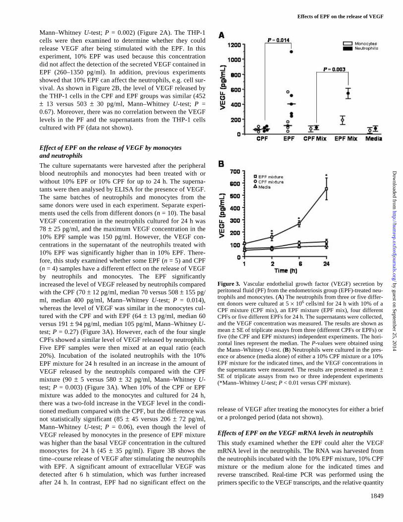
Figure 2. Concentrations of vascular endothelial growth factor(VEGF) in the peritoneal fluid (PF) and the release of VEGF by thePF-treated THP-1 cells. (A) The PF from control group (CPF; n = 4)and the endometriosis group (EPF; n = 18) were collected, and theVEGF concentrations were measured as described in ‘Materials andmethods’. The dots indicate the mean of triplicate assays from twoindependent experiments. The EPF as shown in blanked triangle wasused for a further assay and mixed together in the EPF mixture. (B)The cultured THP-1 cells were treated with or without media, either a10% CPF (+CPF) or a 10% EPF (+EPF) for 24 h, and the supernatantswere collected. The VEGF concentration in the supernatants wasmeasured using an ELISA kit. The results of the untreated culture(media) are shown as mean ± SE of triplicate assays from four inde-pendent experiments. The horizontal lines represent the median. TheP-values were obtained using the Mann–Whitney U-test.
by guest on September 25, 2014
http://humrep.oxfordjournals.org/
Dow
nloaded from
Effects of EPF on the release of VEGF
1849
Mann–Whitney U-test; P = 0.002) (Figure 2A). The THP-1cells were then examined to determine whether they couldrelease VEGF after being stimulated with the EPF. In thisexperiment, 10% EPF was used because this concentrationdid not affect the detection of the secreted VEGF contained inEPF (260–1350 pg/ml). In addition, previous experimentsshowed that 10% EPF can affect the neutrophils, e.g. cell sur-vival. As shown in Figure 2B, the level of VEGF released bythe THP-1 cells in the CPF and EPF groups was similar (452± 13 versus 503 ± 30 pg/ml, Mann–Whitney U-test; P =0.67). Moreover, there was no correlation between the VEGFlevels in the PF and the supernatants from the THP-1 cellscultured with PF (data not shown).
Effect of EPF on the release of VEGF by monocytes and neutrophils
The culture supernatants were harvested after the peripheralblood neutrophils and monocytes had been treated with orwithout 10% EPF or 10% CPF for up to 24 h. The superna-tants were then analysed by ELISA for the presence of VEGF.The same batches of neutrophils and monocytes from thesame donors were used in each experiment. Separate experi-ments used the cells from different donors (n = 10). The basalVEGF concentration in the neutrophils cultured for 24 h was78 ± 25 pg/ml, and the maximum VEGF concentration in the10% EPF sample was 150 pg/ml. However, the VEGF con-centrations in the supernatant of the neutrophils treated with10% EPF was significantly higher than in 10% EPF. There-fore, this study examined whether some EPF (n = 5) and CPF(n = 4) samples have a different effect on the release of VEGFby neutrophils and monocytes. The EPF significantlyincreased the level of VEGF released by neutrophils comparedwith the CPF (70 ± 12 pg/ml, median 70 versus 508 ± 155 pg/ml, median 400 pg/ml, Mann–Whitney U-test; P = 0.014),whereas the level of VEGF was similar in the monocytes cul-tured with the CPF and with EPF (64 ± 13 pg/ml, median 60versus 191 ± 94 pg/ml, median 105 pg/ml, Mann–Whitney U-test; P = 0.27) (Figure 3A). However, each of the four singleCPFs showed a similar level of VEGF released by neutrophils.Five EPF samples were then mixed at an equal ratio (each20%). Incubation of the isolated neutrophils with the 10%EPF mixture for 24 h resulted in an increase in the amount ofVEGF released by the neutrophils compared with the CPFmixture (90 ± 5 versus 580 ± 32 pg/ml, Mann–Whitney U-test; P = 0.003) (Figure 3A). When 10% of the CPF or EPFmixture was added to the monocytes and cultured for 24 h,there was a two-fold increase in the VEGF level in the condi-tioned medium compared with the CPF, but the difference wasnot statistically significant (85 ± 45 versus 206 ± 72 pg/ml,Mann–Whitney U-test; P = 0.06), even though the level ofVEGF released by monocytes in the presence of EPF mixturewas higher than the basal VEGF concentration in the culturedmonocytes for 24 h (45 ± 35 pg/ml). Figure 3B shows thetime–course release of VEGF after stimulating the neutrophilswith EPF. A significant amount of extracellular VEGF wasdetected after 6 h stimulation, which was further increasedafter 24 h. In contrast, EPF had no significant effect on the
release of VEGF after treating the monocytes for either a briefor a prolonged period (data not shown).
Effects of EPF on the VEGF mRNA levels in neutrophils
This study examined whether the EPF could alter the VEGFmRNA level in the neutrophils. The RNA was harvested fromthe neutrophils incubated with the 10% EPF mixture, 10% CPFmixture or the medium alone for the indicated times andreverse transcribed. Real-time PCR was performed using theprimers specific to the VEGF transcripts, and the relative quantity
Figure 3. Vascular endothelial growth factor (VEGF) secretion byperitoneal fluid (PF) from the endometriosis group (EPF)-treated neu-trophils and monocytes. (A) The neutrophils from three or five differ-ent donors were cultured at 5 × 10⁶ cells/ml for 24 h with 10% of aCPF mixture (CPF mix), an EPF mixture (EPF mix), four differentCPFs or five different EPFs for 24 h. The supernatants were collected,and the VEGF concentration was measured. The results are shown asmean ± SE of triplicate assays from three (different CPFs or EPFs) orfive (the CPF and EPF mixtures) independent experiments. The hori-zontal lines represent the median. The P-values were obtained usingthe Mann–Whitney U-test. (B) Neutrophils were cultured in the pres-ence or absence (media alone) of either a 10% CPF mixture or a 10%EPF mixture for the indicated times, and the VEGF concentrations inthe supernatants were measured. The results are presented as mean ±SE of triplicate assays from two or three independent experiments(*Mann–Whitney U-test; P < 0.01 versus CPF mixture).
by guest on September 25, 2014
http://humrep.oxfordjournals.org/
Dow
nloaded from

Y.-J.Na et al.
1850
of each transcript was determined after normalization to theβ-actin level. The fold change in the expression level of thesetranscripts in the PF-treated neutrophils compared with that inthe media-treated neutrophils is shown (Figure 4A). Stimulat-ing the neutrophils with the EPF for 2 and 6 h resulted in a 2.5-and 4.2-fold increase, respectively. The EPF-induced release ofVEGF by the neutrophils was abrogated by pretreating thesecells with 10 μg/ml of cycloheximide for 1 h (Figure 4B).These results suggest that the release of VEGF is the result ofde novo synthesis by a certain PF factor in neutrophils. West-ern blot analysis of the VEGF in the neutrophil lysates showedthat the 2 h EPF treatment decreased the protein level of the43 kDa band, but the amount of the intracellular VEGF proteinswas maintained for up to 12 h (Figure 4C). In comparison, the2 h LPS treatment decreased the protein level of VEGF in thecell lysates. The release of VEGF by neutrophils in the pres-ence of the EPF was not abrogated by adding an LPS inhibitor,polymyxin B, indicating that this effect was not the result ofLPS contamination (data not shown). This suggests that theEPF induces both the production and secretion of VEGF byneutrophils.
IL-8 and TNF-a-independent release of VEGF by EPF
CXCL8 (IL-8) was recently reported to induce VEGF secretionby human neutrophils (Cullen et al., 2000), and the IL-8 con-centration in the EPF was shown to be higher than that in theCPF (Ryan et al., 1995; Arici et al., 1996). Therefore, this studyexamined whether the effect of EPF on the release of VEGF byneutrophils is mediated by this cytokine. The CXC chemokinesactivate neutrophils via an interaction with the chemokinereceptors CXCR1 and CXCR2. Therefore, the neutrophils werepretreated with the PTX, which inhibits signalling through theGi and Go family of G-proteins, and cultured in a medium con-taining either EPF or IL-8. As shown in Figure 5A, thereappears to be an approximately 20% reduction in VEGF in theconditioned medium in the cells treated with PTX, but therewas no significant difference, whereas the release of IL-8-induced VEGF was significantly inhibited by PTX. The neu-tralizing antibody of IL-8 caused a marked reduction in theability of IL-8 to induce the release of VEGF by neutrophils(data not shown), but it had no effect on the EPF-inducedrelease of VEGF by neutrophils (Figure 5B). This indicatesthat the IL-8-induced activation of the PTX sensitive receptorstimulates the release of VEGF in neutrophils. However, IL-8in the EPF may be insufficient for releasing VEGF in neu-trophils, or other unknown factor(s) in EPF down-regulate therelease of VEGF by the EPF-treated neutrophils.
Next, the cells were treated with 20 ng/ml of TNF-α for 24 h.As expected, TNF-α induced the release of VEGF by neu-trophils (Figure 5A). In contrast, TNF-α was unable to stimulatethe release of VEGF by monocytes. It is possible that thiscytokine is the factor in EPF responsible for inducing the releaseof VEGF by neutrophils because there is a high level of TNF-αin the EPF. As shown in Figure 5B, the TNF-α-neutralizingantibody marginally inhibited the release of VEGF by theEPF mixture-treated neutrophils. A significantly lower level ofVEGF was observed as a result of the combined treatment with
Figure 4. Vascular endothelial growth factor (VEGF) production inperitoneal fluid (PF) from the endometriosis group (EPF)-treatedneutrophils. (A) Neutrophils (1 × 10⁶) (n = 3) were treated with orwithout the 10% EPF or PF from control group (CPF) mixture forthe indicated times. The total RNA was isolated, and the VEGFmRNA was quantified using real-time PCR as described in ‘Materi-als and methods’. The mRNA level of VEGF in each sample wasnormalized to that of β-actin. The values are represented as mean ±SE of three separate experiments performed in triplicate. (B) Theneutrophils were pretreated with 10 μg/ml of cycloheximide (CHX)for 1 h and further incubated for 24 h with or without the 10% EPFmixture (EPF). The VEGF concentrations of the supernatants areexpressed as mean ± SE and are representative of three separateexperiments. (C) Neutrophils were treated with lipopolysaccharide(LPS) (1 μg/ml), 10% CPF mixture (CPF) or EPF mixture (EPF) forthe indicated times. The VEGF proteins (43 kDa) were detectedusing the anti-VEGF165 antibody. β-Actin was used as the positivecontrol. The molecular masses of the protein standards are indi-cated in kilodaltons (kDa). *P < 0.001, **P < 0.01 by Mann–WhitneyU-test.
by guest on September 25, 2014
http://humrep.oxfordjournals.org/
Dow
nloaded from

Effects of EPF on the release of VEGF
1851
the neutralizing antibody of the TNF-α and IL-8 than the treat-ment with each antibody (598 ± 32 versus 380 ± 25 pg/ml,Mann–Whitney U-test; P < 0.05). However, treating the neu-trophils with both antibodies simultaneously did not com-pletely inhibit the release of VEGF. As a control, these twoneutralizing antibodies significantly blocked the IL-8- andTNF-α-induced release of VEGF by the neutrophils (data notshown). This suggests that factors other than IL-8 and TNF-αin the EPF may also be responsible for the release of VEGF byneutrophils.
Effects of IL-10 on EPF-induced release of VEGF by neutrophils
A comparison was made between the IL-10 and VEGF levelsdetected in the EPF. As shown in Figure 6A, the IL-10 level inthe EPF from the 18 patients correlated with the VEGF level
(correlation coefficient = 0.549, P = 0.012). Therefore, thisstudy examined whether the neutralizing antibody of IL-10could either down-regulate or up-regulate the release of VEGFby neutrophils and monocytes. The IL-10-neutralizing anti-body alone had little or no effect on the release of VEGF by theEPF-treated cells (Figure 6B).
Effect of neutrophil survival on the VEGF release
This study examined whether neutrophil survival could affectthe release of VEGF by neutrophils. This is because thenumber of neutrophils in the culture condition decreased as aresult of apoptosis. However, aged cells might affect therelease of cytokines, and the EPF reduces the rate of apoptosis
Figure 5. Effect of IL-8 and TNF-α on the release of vascularendothelial growth factor (VEGF) by peritoneal fluid from theendometriosis group (EPF)-treated neutrophils. (A) Neutrophils (5 ×10⁶/ml) (n = 3) were pre-incubated in the culture medium with orwithout 1 μg/ml of the pertussis toxin (PTX) for 4 h and further cul-tured with or without the CPF mixture (CPF mix, 10%), EPF mixture(EPF mix, 10%), IL-8 (10 ng/ml) or ENA-78 (10 ng/ml) for 24 h. Theneutrophils were also treated with 20 ng/ml of TNF-α for 24 h. (B)The neutrophils (n = 5) were pretreated with the TNF-α-neutralizingantibody (0.5 ng), IL-8-neutralizing antibody (0.5 ng) or both anti-bodies for 1 h and further cultured with the 10% EPF mixture for 24 h.The release of VEGF was quantified using ELISA. The data areshown as mean ± SE and are representative of three or five separateexperiments. *P < 0.05 by Mann–Whitney U-test.
Figure 6. Effect of IL-10 in peritoneal fluid from the endometriosisgroup (EPF) on the release of vascular endothelial growth factor(VEGF) by neutrophils. (A) The peritoneal IL-10 and VEGF concen-trations were measured by ELISA, as described in ‘Materials andmethods’. The results are presented as the mean of triplicate assays.The Pearson correlation coefficient was used to determine the correla-tion between the IL-10 and VEGF levels. (B) The neutrophils werepretreated with 0.5 ng of IL-10-neutralizing antibody for 1 h and incu-bated with or without the 10% EPF mixture (EPF mix) for 24 h. Theresults represent the mean ± SE of three independent experiments.There was no significant difference between the two groups with orwithout the IL-10 antibody using the Mann–Whitney U-test.
by guest on September 25, 2014
http://humrep.oxfordjournals.org/
Dow
nloaded from
Y.-J.Na et al.
1852
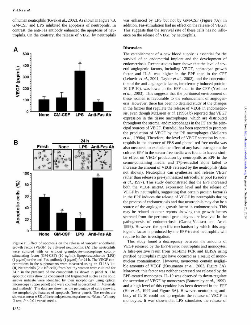
of human neutrophils (Kwak et al., 2002). As shown in Figure 7B,GM-CSF and LPS inhibited the apoptosis of neutrophils. Incontrast, the anti-Fas antibody enhanced the apoptosis of neu-trophils. On the contrary, the release of VEGF by neutrophils
was enhanced by LPS but not by GM-CSF (Figure 7A). Inaddition, Fas-stimulation had no effect on the release of VEGF.This suggests that the survival rate of these cells has no influ-ence on the release of VEGF by neutrophils.
Discussion
The establishment of a new blood supply is essential for thesurvival of an endometrial implant and the development ofendometriosis. Recent studies have shown that the level of sev-eral angiogenic factors, including VEGF, hepatocyte growthfactor and IL-8, was higher in the EPF than in the CPF(Lebovic et al., 2001; Taylor et al., 2002), and the concentra-tion of the anti-angiogenic factor, interferon-γ-induced protein-10 (IP-10), was lower in the EPF than in the CPF (Yoshinoet al., 2003). This suggests that the peritoneal environment ofthese women is favourable to the enhancement of angiogen-esis. However, there has been no detailed study of the changesin the factors that regulate the release of VEGF in endometrio-sis, even though McLaren et al. (1996a,b) reported that VEGFexpression in the tissue macrophages, which are distributedthroughout the stroma, and macrophages in the PF are the prin-cipal sources of VEGF. Estradiol has been reported to promotethe production of VEGF by the PF macrophages (McLarenet al., 1996a). Therefore, the level of VEGF secretion by neu-trophils in the absence of FBS and phenol red-free media wasalso measured to exclude the effect of any basal estrogen in theculture. EPF in the serum-free media was found to have a simi-lar effect on VEGF production by neutrophils as EPF in theserum-containing media, and 17β-estradiol alone failed toincrease the amount of VEGF released by the neutrophils (datanot shown). Neutrophils can synthesize and release VEGFrather than release a pre-synthesized intracellular pool (Gaudryet al., 1997). This study demonstrated that the EPF increasedboth the VEGF mRNA expression level and the release ofVEGF by neutrophils, suggesting that certain protein factor(s)in the EPF induces the release of VEGF by neutrophils duringthe process of endometriosis and that neutrophils may also be asource of the angiogenic growth factor in endometriosis. Thismay be related to other reports showing that growth factorssecreted from the peritoneal granulocytes are involved in thepathogenesis of endometriosis (Garcia-Velasco and Arici,1999). However, the specific mechanism by which this ang-iogenic factor is produced by the EPF-treated neutrophils willrequire further investigation.
This study found a discrepancy between the amounts ofVEGF released by the EPF-treated neutrophils and monocytes.A false-positive result from real-time PCR and ELISA usingpurified neutrophils might have occurred as a result of mono-nuclear contamination. However, monocytes contain negligi-ble amounts of VEGF (Kusumanto et al., 2003, Figure 3A).Moreover, this factor was neither expressed nor released by theEPF-treated monocytes. IL-10 was observed to down-regulatethe secretion of VEGF by monocytes (Bottomley et al., 1999),and a high level of this cytokine has been detected in the EPF(Ho et al., 1997 and Figure 6A). However, neutralizing anti-body of IL-10 could not up-regulate the release of VEGF inmonocytes. It was shown that LPS stimulates the release of
Figure 7. Effect of apoptosis on the release of vascular endothelialgrowth factor (VEGF) by cultured neutrophils. (A) The neutrophilswere cultured with or without granulocyte–macrophage colony-stimulating factor (GM-CSF) (10 ng/ml), lipopolysaccharide (LPS)(1 μg/ml) or the anti-Fas antibody (1 μg/ml) for 24 h. The VEGF con-centrations in the supernatants were measured using an ELISA kit.(B) Neutrophils (2 × 10⁶ cells) from healthy women were cultured for24 h in the presence of the compounds as shown in panel A. Theapoptotic cells showing condensed and fragmented nuclei as the solidarrows indicate were identified by their morphology using opticalmicroscopy (upper panel) and were counted as described in ‘Materialsand methods’. The data are shown as the percentage of cells showingthe morphologic features of apoptosis (lower panel). The results areshown as mean ± SE of three independent experiments. *Mann–WhitneyU-test; P < 0.01 versus media.
by guest on September 25, 2014
http://humrep.oxfordjournals.org/
Dow
nloaded from
Effects of EPF on the release of VEGF
1853
VEGF as a result of a contact co-culture of the monocytes withendothelial cells in the presence of a lipoprotein, but not in theabsence of a contact co-culture (Pakala et al., 2002). Therefore,normal monocytes/macrophages might be sensitized or primedby a certain factor in the PF and then be able to secrete VEGFby other growth factors.
VEGF exists as one of four different molecular species, 121,165, 189 and 206, which are produced by alternative exonsplicing of a single VEGF gene. Real-time PCR (Webb et al.,1998) and Northern blotting (Scapini et al., 1999) showed thatneutrophils express the mRNA species encoding the two mostcommon VEGF splice variants, VEGF121 and VEGF165. Theregulation of VEGF expression has been shown to occur inother cells via both a transcriptional and a post-transcriptionalmechanism (Cohen et al., 1996; Levy et al., 1997). VEGF hasbeen shown to be synthesized, be present in specific granulesand be released by neutrophils (Gaudry et al., 1997; Taichmanet al., 1997; Webb et al., 1998). This study demonstrated theup-regulation of VEGF expression at the mRNA level as wellas the release of the VEGF protein in neutrophils cultured withthe EPF. Moreover, the time–course experiments showed thatthe increased release of VEGF occurred for up to 24 h. Webbet al. (1998) reported that majority of the VEGF productionoccurred within the first hour of stimulation with TNF-α, andonly relatively small amounts of VEGF were subsequentlyreleased. However, the mechanism involved in the expressionof VEGF mRNA in neutrophils is unknown.
A series of cytokines known to stimulate the release ofVEGF by neutrophils were examined to determine the prima-rily responsible molecule(s) for the release of VEGF. VEGFexpression is regulated by various cytokines (Ferrara andDavis-Smyth, 1997). Several growth factors that may haveangiogenic activity such as IL-8 and TNF-α have been foundto be elevated in the EPF (Taylor et al., 2002). These factorsalso stimulate for the production and release of VEGF by neu-trophils (Webb et al., 1998; Cullen et al., 2000), and a combi-nation of VEGF and TNF-α might have an additive effect onangiogenesis. This study focused on IL-8 and TNF-α as can-didate molecules in the EPF to stimulate neutrophils. This isbecause monocytes lack the required CXCR2 receptors, cannotrespond to IL-8 and do not secrete VEGF in the presence ofTNF-α. CXCL5 (ENA-78), which binds CXCR2 with a highaffinity, did not induce the release of VEGF by neutrophils,whereas IL-8 has a high affinity for both CXCR1 and CXCR2(Murphy et al., 2000). This suggests that the release of VEGFby neutrophils in the presence of IL-8 might be due to CXCR1activation and not due to CXCR2 activation. However, neitherPTX nor the neutralizing antibody of IL-8 significantly affec-ted the EPF-induced release of VEGF by the neutrophils.Therefore, it is possible that this cytokine may not be the majorVEGF-releasing factor in the EPF. The TNF-α-treated neu-trophils showed considerable accumulation of VEGF (Scapiniet al., 1999), but TNF-α failed to stimulate the production ofVEGF by monocytes (Pakala et al., 2002). Webb et al. (1998)reported that the TNF-α-induced secretion of VEGF is inde-pendent of the de novo synthesis of VEGF and acts on a pre-formed pool of the VEGF molecule. In contrast, Ryuto et al.(1996) reported that the TNF-α-dependent induction of VEGF
is mediated via the activation of the transcription factor inhuman glioma cells. Because the EPF-stimulated release ofVEGF by neutrophils was partially but not completely abro-gated by the combined treatment of the neutralizing antibodiesof TNF-α and IL-8, it is possible that several growth factors orcytokines may act in concert to stimulate the release of VEGFby neutrophils.
The induction of VEGF release from neutrophils byPF might play a role in the inflammatory process of endome-triosis. It was demonstrated that neutrophils augment theextravasation of tumour cells via the neutrophil-inducedtransmigration of tumour cells across the endothelial cellmonolayers (Wu et al., 2000). The observation that the EPFinduces the release of VEGF from the neutrophils suggestsa role for neutrophil-derived VEGF in modulating theendothelial permeability and angiogenesis. Moreover, VEGFwas shown to induce monocyte chemotaxis and transmigra-tion via the endothelial monolayers (Clauss et al., 1990; Heilet al., 2000). Therefore, the chemoattractant properties of theneutrophil-derived VEGF might reinforce the recruitment ofmonocytes to the focal endometrial site in the peritoneumfollowing the initial activation of neutrophils.
VEGF provides an attractive target for therapeutic interven-tion in endometriosis. Monocyte/macrophages, polymorphonu-clear leucocytes and natural killer (NK) cells are found in theperitoneal cavity of normal subjects as well as in patients suf-fering from various disorders (Melichar and Freedman, 2002).However, it has been suggested that peritoneal monocytes andNK cells play an important role in endometriosis (Lebovicet al., 2001). It has been reported that various blood or ovarianfactors have an influence on deep endometriosis and cysticovarian endometriosis, whereas the PF concentrations have aninfluence on endometrial and superficially implanted cells(Koninckx et al., 1998). This means that a superficial endome-trial implant might be influenced by the PF factor(s).
Because neutrophils are conspicuous in the peripheral bloodand stimulated neutrophils release a significant amount ofVEGF, the neutrophil-derived VEGF might be sufficient toaccount for the VEGF levels in the PF or in local lesions ofendometriotic implants, even though there is a lower numberof infiltrating neutrophils in the PF than mononuclear cells. Itis possible that increased vascular permeability contributes toendometrial angiogenesis, and the increased level of the PFcomponents might occur through the release of VEGF-containinggranules to the vessels by activated endometrial neutrophils(Williams and Solomkin, 1999), or through the action of theadherent junction proteins in neutrophils (Tinsley et al., 1999).Recently, Shimoya et al. (2000) reported that the secretory leu-cocyte protease inhibitor concentration was higher in the PF ofendometriosis patients than in the control women, which hasan anti-inflammatory effect on the EPF through the inhibitionof elastase-induced cytokine production via the peritonealmacrophages as a result of the consequent prevention of neu-trophil accumulation in the PF. Marginating and adherent neu-trophils in the endometrium provide a source for VEGF in theendometrial vessels undergoing proliferation (Gargett et al.,2001; Heryanto et al., 2004), and neutrophils have been shownto be a source of VEGF in the PF. Therefore, blocking the
by guest on September 25, 2014
http://humrep.oxfordjournals.org/
Dow
nloaded from
Y.-J.Na et al.
1854
activation of neutrophils and modulating the functions of mac-rophages will be an attractive target for an anti-angiogenicstrategy.
AcknowledgementsThis work was supported by a grant from the Korea Health 21 R&DProject, Ministry of Health and Welfare, Republic of Korea (01-PJ10-PG6-01GN13-0002).
References
The American Fertility Society (1985) Revised American Fertility Societyclassification of endometriosis. Fertil Steril 43,351–352.
Arici A and Oral E (1997) The peritoneal environment in endometriosis. InDiamond MP and Osteen KG (eds) Endometrium and Endometriosis. Black-well Science, Massachusetts, USA, pp. 161–173.
Arici A, Tazuke SI, Attar E, Kliman HJ and Olive DL (1996) Interleukin-8concentration in peritoneal fluid of patients with endometriosis and modula-tion of interleukin-8 expression in human mesothelial cells. Mol HumReprod 2,40–45.
Bottomley MJ, Webb NJ, Watson CJ, Holt PJ, Freemont AJ and Brenchley PE(1999) Peripheral blood mononuclear cells from patients with rheumatoidarthritis spontaneously secrete vascular endothelial growth factor (VEGF):specific up-regulation by tumour necrosis factor-α (TNF-α) in synovialfluid. Clin Exp Immunol 117,171–176.
Clauss M, Gerlach M, Gerlach H, Brett J, Wang F, Familletti PC, Pan YC,Olander JV, Connolly DT and Stern D (1990) Vascular permeability fac-tor: a tumor-derived polypeptide that induces endothelial cell and mono-cyte procoagulant activity, and promotes monocyte migration. J Exp Med172,1535–1545.
Cohen T, Nahari D, Cerem LW, Neufeld G and Levi BZ (1996) Interleukin 6induces the expression of vascular endothelial growth factor. J Biol Chem271,736–741.
Cullen VC, Mackarel AJ, Hislip SJ, O’Connor CM and Keenan AK (2000)Investigation of vascular endothelial growth factor effects on pulmonaryendothelial monolayer permeability and neutrophil transmigration. GenPharmacol 35,149–157.
Fasciani A, D’Ambrogio G, Bocci G, Monti M, Genazzani AR and Artini PG(2000) High concentrations of the vascular endothelial growth factor andinterleukin-8 in ovarian endometriomata. Mol Hum Reprod 6,50–54.
Ferrara N and Davis-Smyth T (1997) The biology of vascular endothelialgrowth factor. Endocr Rev 18,4–25.
Freeman MR, Schneck FX, Gagnon ML, Corless C, Soker S, Niknejad K, PeoplesGE and Klagsbrun M (1995) Peripheral blood T lymphocytes and lymphocytesinfiltrating human cancers express vascular endothelial growth factor: a poten-tial role for T cells in angiogenesis. Cancer Res 55,4140–4145.
Garcia-Velasco JA and Arici A (1999) Chemokines and human reproduction.Fertil Steril 71,983–993.
Gargett CE, Lederman F, Heryanto B, Gambino LS and Rogers PA (2001) Focalvascular endothelial growth factor correlates with angiogenesis in humanendometrium. Role of intravascular neutrophils. Hum Reprod 16,1065–1075.
Gaudry M, Bregerie O, Andrieu VEI, Benna J, Pocidalo MA and Hakim J(1997) Intracellular pool of vascular endothelial growth factor in humanneutrophils. Blood 90,4153–4161.
Haney AF, Jenkins S and Weinberg JB (1991) The stimulus responsible for theperitoneal fluid inflammation observed in infertile women with endometrio-sis. Fertil Steril 56,408–413.
Heil M, Clauss M, Suzuki K, Buschmann IR, Willuweit A, Fischer S andSchaper W (2000) Vascular endothelial growth factor (VEGF) stimulatesmonocyte migration through endothelial monolayers via increased integrinexpression. Eur J Cell Biol 79,850–857.
Heryanto B, Girling JE and Rogers PA (2004) Intravascular neutrophils par-tially mediate the endometrial endothelial cell proliferative response to oes-trogen in ovariectomised mice. Reproduction 127,613–620.
Hill JA, Faris HM, Schiff I and Anderson DJ (1988) Characterization of leuko-cyte subpopulations in the peritoneal fluid of women with endometriosis.Fertil Steril 50,216–222.
Ho HN, Wu MY, Chao KH, Chen CD, Chen SU and Yang YS (1997) Perito-neal interleukin-10 increases with decrease in activated CD4+ T lym-phocytes in women with endometriosis. Hum Reprod 12,2528–2533.
Keck PJ, Hauser SD, Krivi G, Sanzo K, Warren T, Feder J and Connolly DT(1989) Vascular permeability factor, an endothelial cell mitogen related toPDGF. Science 246,1309–1312.
Khorram O, Taylor RN, Ryan IP, Schall TJ and Landers DV (1993) Peritonealfluid concentrations of the cytokine RANTES correlate with the severity ofendometriosis. Am J Obstet Gynecol 169,1545–1549.
Koninckx PR, Kennedy SH and Barlow DH (1998) Endometriotic disease: therole of peritoneal fluid. Hum Reprod Update 4,741–751.
Kusumanto YH, Dam WA, Hospers GA, Meijer C and Mulder NH (2003)Platelets and granulocytes, in particular the neutrophils, form importantcompartments for circulating vascular endothelial growth factor. Angiogen-esis 6,283–287.
Kwak JY, Park SW, Kim KH, Na YJ and Lee KS (2002) Modulation of neu-trophil apoptosis by plasma and peritoneal fluid from patients with advancedendometriosis. Hum Reprod 17,595–600.
Lebovic DI, Mueller MD and Taylor RN (2001) Immunobiology of endometri-osis. Fertil Steril 75,1–10.
Leiva MC, Hasty LA, Pfeifer S, Mastrianni L Jr and Lyttle CR (1993)Increased chemotactic activity of peritoneal fluid in patients with endometri-osis. Am J Obstet Gynecol 168,592–598.
Leung DW, Cachianes G, Kuang WJ, Goeddel DV and Ferrara N (1989) Vas-cular endothelial growth factor is a secreted angiogenic mitogen. Science246,1306–1309.
Levy AP, Levy NS, Iliopoulos O, Jiang C, Kaplin WG Jr and Goldberg MA(1997) Regulation of vascular endothelial growth factor by hypoxia and itsmodulation by the von Hippel-Lindau tumor suppressor gene. Kidney Int51,575–578.
McLaren J, Prentice A, Charnock-Jones DS, Millican SA, Muller KH, SharkeyAM and Smith SK (1996a) Vascular endothelial growth factor is producedby peritoneal fluid macrophages in endometriosis and is regulated by ovar-ian steroids. J Clin Invest 98,482–489.
McLaren J, Prentice A, Charnock-Jones DS and Smith SK (1996b) Vascularendothelial growth factor (VEGF) concentrations are elevated in peritonealfluid of women with endometriosis. Hum Reprod 11,220–223.
Melichar B and Freedman RS (2002) Immunology of the peritoneal cavity: rel-evance for host-tumor relation. Int J Gynecol Cancer 12,3–17.
Murphy PM, Baggiolini M, Charo IF, Hebert CA, Horuk R, Matsushima K,Miller LH, Oppenheim JJ and Power CA (2000) International union of phar-macology. XXII. Nomenclature for chemokine receptors. Pharmacol Rev52,145–176.
Neufeld G, Cohen T, Gengrinovitch S and Poltorak Z (1999) Vascularendothelial growth factor (VEGF) and its receptors. FASEB J 13,9–22.
Pakala R, Watanabe T and Benedict CR (2002) Induction of endothelial cellproliferation by angiogenic factors released by activated monocytes. Cardio-vasc Radiat Med 3,95–101.
Ryan IP, Tseng JF, Schriock ED, Khorram O, Landers DV and Taylor RN(1995) Interleukin-8 concentrations are elevated in peritoneal fluid ofwomen with endometriosis. Fertil Steril 63,929–932.
Ryuto M, Ono M, Izumi H, Yoshida S, Weich HA, Kohno K and Kuwana M(1996) Induction of vascular endothelial growth factor by tumor necrosisfactor-α in human glioma cells. Possible roles of SP-1. J Biol Chem271,28220–28228.
Scapini P, Calzetti F and Cassatella MA (1999) On the detection of neutrophil-derived vascular endothelial growth factor (VEGF). J Immunol Methods232,121–129.
Shifren JL, Tseng JF, Zaloudek CJ, Ryan IP, Meng YG, Ferrara N, Jaffe RBand Taylor RN (1996) Ovarian steroid regulation of vascular endothelialgrowth factor in the human endometrium: implications for angiogenesis dur-ing the menstrual cycle and in the pathogenesis of endometriosis. J ClinEndocrinol Metab 81,3112–3118.
Shimoya K, Moriyama A, Ogata I, Nobunaga T, Koyama M, Azuma C andMurata T (2000) Increased concentrations of secretory leukocyte proteaseinhibitor in peritoneal fluid of women with endometriosis. Mol Hum Reprod6,829–834.
Taichman NS, Young S, Cruchley AT, Taylor P and Paleolog E (1997) Humanneutrophils secrete vascular endothelial growth factor. J Leukoc Biol62,397–400.
Taylor RN, Lebovic DI and Mueller MD (2002) Angiogenic factors inendometriosis. Ann N Y Acad Sci 955,89–100.
Tinsley JH, Wu MH, Ma W, Taulman AC and Yuan SY (1999) Activatedneutrophils induce hyperpermeability and phosphorylation of adherensjunction proteins in coronary venular endothelial cells. J Biol Chem274,24930–24934.
by guest on September 25, 2014
http://humrep.oxfordjournals.org/
Dow
nloaded from

Effects of EPF on the release of VEGF
1855
Webb NJ, Myers CR, Watson CJ, Bottomley MJ and Brenchley PE (1998)Activated human neutrophils express vascular endothelial growth factor(VEGF). Cytokine 10,254–257.
Williams MA and Solomkin JS (1999) Integrin-mediated signaling in humanneutrophil functioning. J Leukoc Biol 65,725–736.
Wu QD, Wang JH, Bouchier-Hayes D and Redmond HP (2000) Neutrophil-induced transmigration of tumour cells treated with tumour-conditionedmedium is facilitated by granulocyte-macrophage colony-stimulating factor.Eur J Surg 166,361–366.
Yoshino O, Osuga Y, Koga K, Hirota Y, Tsutsumi O, Yano T, Morita Y,Momoeda M, Fujiwara T, Kugu K et al. (2003) Concentrations of inter-feron-γ-induced protein-10 (IP-10), an antiangiogenic substance, aredecreased in peritoneal fluid of women with advanced endometriosis. Am JReprod Immunol 50,60–65.
Submitted on May 4, 2005; resubmitted on October 7, 2005, December 15,2005, February 1, 2006; accepted on February 21, 2006
by guest on September 25, 2014
http://humrep.oxfordjournals.org/
Dow
nloaded from
Related Documents