Effects of Feeding Spodoptera littoralis on Lima Bean Leaves. III. Membrane Depolarization and Involvement of Hydrogen Peroxide 1 Massimo E. Maffei*, Axel Mitho ¨fer, Gen-Ichiro Arimura, Hannes Uchtenhagen, Simone Bossi, Cinzia M. Bertea, Laura Starvaggi Cucuzza, Mara Novero, Veronica Volpe, Stefano Quadro, and Wilhelm Boland Department of Plant Biology and Centre of Excellence CEBIOVEM, University of Turin, I–10125 Turin, Italy (M.E.M, S.B., C.M.B., L.S.C., M.N., V.V., S.Q.); and Max Planck Institute for Chemical Ecology, Bioorganic Chemistry, D–07745 Jena, Germany (A.M., G.-I.A., H.U., W.B.) In response to herbivore (Spodoptera littoralis) attack, lima bean (Phaseolus lunatus) leaves produced hydrogen peroxide (H 2 O 2 ) in concentrations that were higher when compared to mechanically damaged (MD) leaves. Cellular and subcellular localization analyses revealed that H 2 O 2 was mainly localized in MD and herbivore-wounded (HW) zones and spread throughout the veins and tissues. Preferentially, H 2 O 2 was found in cell walls of spongy and mesophyll cells facing intercellular spaces, even though confocal laser scanning microscopy analyses also revealed the presence of H 2 O 2 in mitochondria/peroxisomes. Increased gene and enzyme activations of superoxide dismutase after HW were in agreement with confocal laser scanning microscopy data. After MD, additional application of H 2 O 2 prompted a transient transmembrane potential (V m ) depolarization, with a V m depolarization rate that was higher when compared to HW leaves. In transgenic soybean (Glycine max) suspension cells expressing the Ca 21 - sensing aequorin system, increasing amounts of added H 2 O 2 correlated with a higher cytosolic calcium ([Ca 21 ] cyt ) concentration. In MD and HW leaves, H 2 O 2 also triggered the increase of [Ca 21 ] cyt , but MD-elicited [Ca 21 ] cyt increase was more pronounced when compared to HW leaves after addition of exogenous H 2 O 2 . The results clearly indicate that V m depolarization caused by HW makes the membrane potential more positive and reduces the ability of lima bean leaves to react to signaling molecules. In response to pathogen invasion, plants mount a broad range of defense responses, including the gen- eration of reactive oxygen species (ROS; Lamb and Dixon, 1997; Mur et al., 2005). ROS are also generated in plant tissues in response to wounding (Angelini et al., 1990; Bradley et al., 1992; Olson and Varner, 1993; Felton et al., 1994; Bi and Felton, 1995; Orozco-Ca ´rdenas and Ryan, 1999), mechanical stimulation of isolated cells (Yahraus et al., 1995; Gus-Mayer et al., 1998), and the treatment of cell suspension cultures with elicitors (Legendre et al., 1993; Mitho ¨fer et al., 1997; Stennis et al., 1998). ROS also have been associated with plant herbivore interactions (Mitho ¨fer et al., 2004; Leitner et al., 2005), and oxidative changes in the plants correspond with oxidative damage in the midguts of insects feeding on previously wounded plants (Orozco-Ca ´rdenas and Ryan, 1999). In pathogenic interactions, wound-induced ROS accumulation, in particular hydrogen peroxide (H 2 O 2 ), is observed both locally and systemically in leaves of several plant species (Mehdy et al., 1996; Bergey et al., 1999; Orozco- Ca ´rdenas and Ryan, 1999; Orozco-Ca ´rdenas et al., 2001). In some cases, especially under stress condi- tions, a rapid and transient production of high levels of ROS may reach intracellular concentrations of up to 1 M H 2 O 2 in about 13 min (Jacks and Davidonis, 1996). In response to herbivores, H 2 O 2 levels are likely to be elevated as long as the attacks persist. Bi and Felton (1995) have proposed that herbivore attacks can cause a localized oxidative response in soybean (Glycine max) leaves and have identified some potential functions of ROS that might affect plant-herbivore interactions. Furthermore, the presence of H 2 O 2 in the plant in response to herbivory, before any subsequent second- ary pathogen invasion, could be advantageous because timing of the induction of defense responses can be an important factor in the success or failure of plants to defend against pathogen attacks (Dangl et al., 1996), and wound-generated H 2 O 2 that occurs in the veins also could have a defensive role against bacteria, fungi, or viruses, as they may invade leaves wounded by herbivores (Orozco-Ca ´rdenas and Ryan, 1999). ROS also represent second messengers that eventually acti- vate downstream defense reactions (Foyer and Noctor, 2005), such as synthesis of pathogenesis-related proteins (Chen et al., 1993), glutathione S-transferase, glutathione 1 This work was supported by the Fonds der Chemischen Industrie (Frankfurt a.M.) from the Centre of Excellence for Plant and Microbial Biosensing (Turin) and local research grants from the University of Turin, Italy. * Corresponding author; e-mail [email protected]; fax 390–112365967. The author responsible for distribution of materials integral to the findings presented in this article in accordance with the policy described in the Instructions for Authors (www.plantphysiol.org) is: Massimo E. Maffei ([email protected]). Article, publication date, and citation information can be found at www.plantphysiol.org/cgi/doi/10.1104/pp.105.071993. 1022 Plant Physiology, March 2006, Vol. 140, pp. 1022–1035, www.plantphysiol.org Ó 2006 American Society of Plant Biologists www.plant.org on February 17, 2016 - Published by www.plantphysiol.org Downloaded from Copyright © 2006 American Society of Plant Biologists. All rights reserved.

Welcome message from author
This document is posted to help you gain knowledge. Please leave a comment to let me know what you think about it! Share it to your friends and learn new things together.
Transcript
Effects of Feeding Spodoptera littoralis on Lima BeanLeaves. III. Membrane Depolarization and Involvementof Hydrogen Peroxide1
Massimo E. Maffei*, Axel Mithofer, Gen-Ichiro Arimura, Hannes Uchtenhagen, Simone Bossi,Cinzia M. Bertea, Laura Starvaggi Cucuzza, Mara Novero, Veronica Volpe, Stefano Quadro,and Wilhelm Boland
Department of Plant Biology and Centre of Excellence CEBIOVEM, University of Turin, I–10125 Turin,Italy (M.E.M, S.B., C.M.B., L.S.C., M.N., V.V., S.Q.); and Max Planck Institute for Chemical Ecology,Bioorganic Chemistry, D–07745 Jena, Germany (A.M., G.-I.A., H.U., W.B.)
In response to herbivore (Spodoptera littoralis) attack, lima bean (Phaseolus lunatus) leaves produced hydrogen peroxide (H2O2) inconcentrations that were higher when compared to mechanically damaged (MD) leaves. Cellular and subcellular localizationanalyses revealed that H2O2 was mainly localized in MD and herbivore-wounded (HW) zones and spread throughout the veinsand tissues. Preferentially, H2O2 was found in cell walls of spongy and mesophyll cells facing intercellular spaces, even thoughconfocal laser scanning microscopy analyses also revealed the presence of H2O2 in mitochondria/peroxisomes. Increased geneand enzyme activations of superoxide dismutase after HW were in agreement with confocal laser scanning microscopy data. AfterMD, additional application of H2O2 prompted a transient transmembrane potential (Vm) depolarization, with aVm depolarizationrate that was higher when compared to HW leaves. In transgenic soybean (Glycine max) suspension cells expressing the Ca21-sensing aequorin system, increasing amounts of added H2O2 correlated with a higher cytosolic calcium ([Ca21]cyt) concentration.In MD and HW leaves, H2O2 also triggered the increase of [Ca21]cyt, but MD-elicited [Ca21]cyt increase was more pronounced whencompared to HW leaves after addition of exogenous H2O2. The results clearly indicate that Vm depolarization caused by HWmakes the membrane potential more positive and reduces the ability of lima bean leaves to react to signaling molecules.
In response to pathogen invasion, plants mount abroad range of defense responses, including the gen-eration of reactive oxygen species (ROS; Lamb andDixon, 1997; Mur et al., 2005). ROS are also generatedin plant tissues in response to wounding (Angeliniet al., 1990; Bradley et al., 1992; Olson and Varner, 1993;Felton et al., 1994; Bi and Felton, 1995; Orozco-Cardenasand Ryan, 1999), mechanical stimulation of isolatedcells (Yahraus et al., 1995; Gus-Mayer et al., 1998), andthe treatment of cell suspension cultures with elicitors(Legendre et al., 1993; Mithofer et al., 1997; Stenniset al., 1998). ROS also have been associated with plantherbivore interactions (Mithofer et al., 2004; Leitneret al., 2005), and oxidative changes in the plantscorrespond with oxidative damage in the midgutsof insects feeding on previously wounded plants(Orozco-Cardenas and Ryan, 1999). In pathogenic
interactions, wound-induced ROS accumulation, inparticular hydrogen peroxide (H2O2), is observed bothlocally and systemically in leaves of several plantspecies (Mehdy et al., 1996; Bergey et al., 1999; Orozco-Cardenas and Ryan, 1999; Orozco-Cardenas et al.,2001). In some cases, especially under stress condi-tions, a rapid and transient production of high levels ofROS may reach intracellular concentrations of up to 1 M
H2O2 in about 13 min (Jacks and Davidonis, 1996). Inresponse to herbivores, H2O2 levels are likely to beelevated as long as the attacks persist. Bi and Felton(1995) have proposed that herbivore attacks can causea localized oxidative response in soybean (Glycine max)leaves and have identified some potential functions ofROS that might affect plant-herbivore interactions.Furthermore, the presence of H2O2 in the plant inresponse to herbivory, before any subsequent second-ary pathogen invasion, could be advantageous becausetiming of the induction of defense responses can be animportant factor in the success or failure of plants todefend against pathogen attacks (Dangl et al., 1996),and wound-generated H2O2 that occurs in the veinsalso could have a defensive role against bacteria,fungi, or viruses, as they may invade leaves woundedby herbivores (Orozco-Cardenas and Ryan, 1999). ROSalso represent second messengers that eventually acti-vate downstream defense reactions (Foyer and Noctor,2005), such as synthesis of pathogenesis-related proteins(Chen et al., 1993), glutathione S-transferase, glutathione
1 This work was supported by the Fonds der ChemischenIndustrie (Frankfurt a.M.) from the Centre of Excellence for Plantand Microbial Biosensing (Turin) and local research grants from theUniversity of Turin, Italy.
* Corresponding author; e-mail [email protected]; fax390–112365967.
The author responsible for distribution of materials integral to thefindings presented in this article in accordance with the policydescribed in the Instructions for Authors (www.plantphysiol.org) is:Massimo E. Maffei ([email protected]).
Article, publication date, and citation information can be found atwww.plantphysiol.org/cgi/doi/10.1104/pp.105.071993.
1022 Plant Physiology, March 2006, Vol. 140, pp. 1022–1035, www.plantphysiol.org � 2006 American Society of Plant Biologists www.plant.org on February 17, 2016 - Published by www.plantphysiol.orgDownloaded from
Copyright © 2006 American Society of Plant Biologists. All rights reserved.
peroxidase (GPX), and ubiquitin (Levine et al., 1994), aswell as phytoalexin accumulation (Devlin and Gustine,1992; Mithofer et al., 2004) and production of herbivore-induced volatile products (Mithofer et al., 2004).
The induction of the oxidative burst is often de-pendent on Ca21, which has been demonstrated sev-eral times (e.g. Price et al., 1994; Low and Merida,1996; Cazale et al., 1998). Electrophysiological studiesrevealed the existence of plasma membrane Ca21-permeable channels activated by membrane depolariza-tion or hyperpolarization in response to environmentalstimuli (for review, see White, 2000; Sanders et al., 2002)and at the very edge of herbivore wounding in plant-insect interactions (Maffei et al., 2004). ROS can interactwith ion channel activity, and oxidase-dependent elec-tron transfer could drive a transmembrane potential(Vm) depolarization; both effects lead to channel acti-vation. This regulation of ion channels by H2O2 inplants has been indicated (Cazale et al., 1998; Pei et al.,2000; Foreman et al., 2003; Overmyer et al., 2003) buthas never been analyzed in plant herbivore interaction,to our knowledge. Thus, the aim of this study was toanalyze the role of H2O2 in plant-herbivore interactions.
By taking advantage of the model system consist-ing of Spodoptera littoralis larvae feeding on lima bean(Phaseolus lunatus) leaves (Maffei et al., 2004; Mithoferet al., 2005), we used several microscopic, physiolog-ical, biochemical, and molecular biological tools to eval-uate the role of H2O2 in plant-herbivore interactions.Thus, we investigated (1) the cellular and subcellularH2O2 localization in mechanically damaged (MD) andS. littoralis herbivore-wounded (HW) tissues by meansof fluorescence light microscopy, confocal laser-scanningmicroscopy (CLSM), and transmission electron micros-copy; (2) the effect of H2O2 on [Ca21]cyt using CLSMand apoaequorin-transformed plants; (3) the effect ofH2O2 on Vm and cytosolic calcium ([Ca21]cyt) variationsusing specific ion channel inhibitors in MD and HWtissues; and (4) the enzyme activity and quantitativegene expression of some ROS scavenging systems. Weshow that H2O2 is released upon HW and MD in planttissues. We also demonstrate that H2O2 release causedVm depolarization involving Ca21 channels. Further-more, we show that the HW-induced Vm depolarizationis a crucial event that, by depolarizing the membranepotential, reduces the ability of lima bean leaves toreact to signaling molecules. Finally, we show that in-creased H2O2 accumulation in HW leaves is correlatedto increased superoxide dismutase (SOD) enzyme ac-tivity and gene expression.
RESULTS
Cellular and Subcellular Localization of H2O2 in MD
and HW Lima Bean Tissues
ROS play two divergent roles in plant adaptation tothe changing environment: enhancement of damageby a highly oxidizing microenvironment or mediatingthe activation of other defense responses under certain
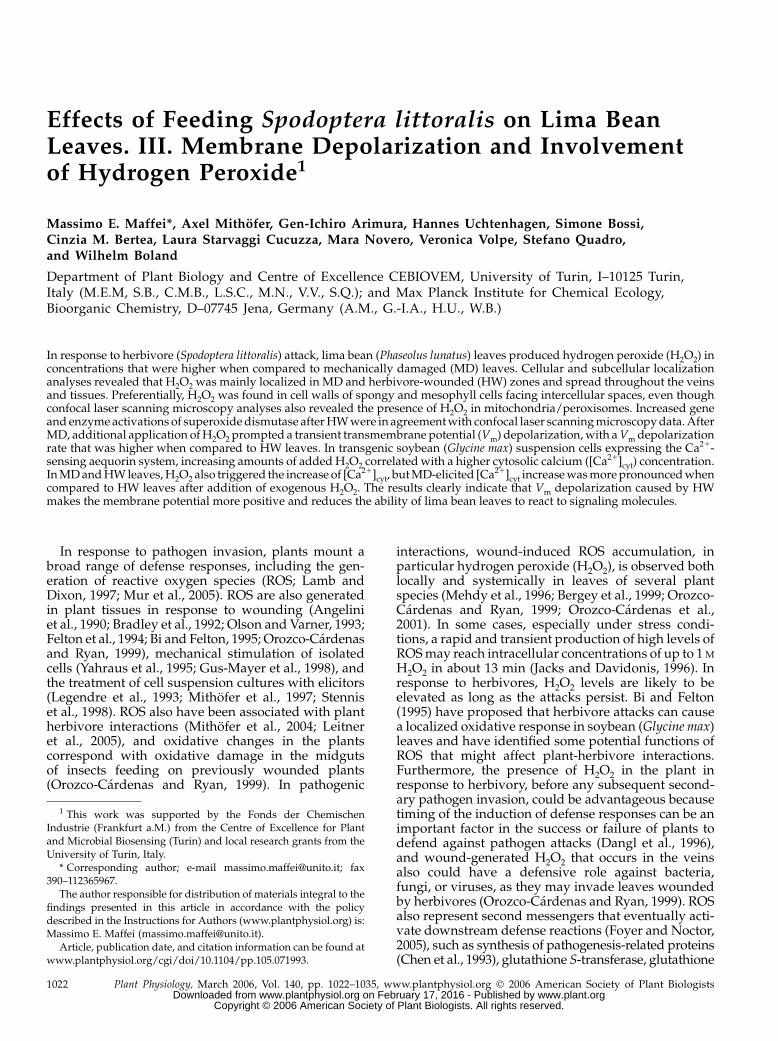
biotic and abiotic stresses (Lamb and Dixon, 1997;Yang and Poovaiah, 2002). In contrast to most animalcells, plant cells are able to produce ROS, mainly H2O2,constitutively in significant amounts, and this produc-tion is developmentally regulated by light, phytohor-mones, or wounding and predominantly associatedwith the cell’s exocellular matrix (ECM; Bolwell andWojtaszek, 1997; Bolwell, 1999). Mechanical damage oflima bean leaves triggered the production of H2O2 ascan be seen after incubation of tissues with the dye 3,3-diaminobenzidine (DAB; Thordal-Christensen et al.,1997; Orozco-Cardenas and Ryan, 1999). DAB stainingincreased in time by spreading throughout the veinsand tissues and was mainly localized in the damagedzone (Fig. 1, C and E). The accumulation of H2O2 oc-curred near wound sites and also in distal unwoundedleaves, indicating that the process might be regulatedby a systemic signaling system. When S. littoralis wasallowed to feed on DAB-incubated leaves, the samereaction was observed and increased with time, with aslightly increased staining with respect to MD leaves(Fig. 1, D and F). Figure 1 also shows the edge of a leafafter 6 h of MD (Fig. 1A) and HW (Fig. 1B) immedi-ately cut before mounting the slides; here, it is clearlyvisible that in both cases the slicing of leaves causesan immediate H2O2 release. DAB H2O2 assay is basedon endogenous peroxidases, the levels of which areassumed to be constant. As this is not always the case(as shown, for instance, by Hiraga et al., 2000), direct
Figure 1. Histochemical localization of H2O2 in MD and HW limabean leaves after incubation of tissues with the dye DAB. As a control,the first top pictures show the edge of leaf samples of 6 h after MD andHW immediately cut before mounting the slides; it is clearly visible thatthe preparation of samples causes an immediate H2O2 release, but thestaining is lower if compared to MD and HW tissues after a longerexposure. Metric bar 5 1 mm.
A Role for Hydrogen Peroxide following Herbivore Attack
Plant Physiol. Vol. 140, 2006 1023 www.plant.org on February 17, 2016 - Published by www.plantphysiol.orgDownloaded from
Copyright © 2006 American Society of Plant Biologists. All rights reserved.
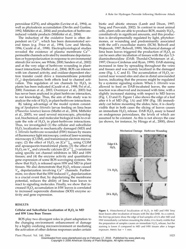
measurements using cerium chloride have been usedas a more accurate approach (Bestwick et al., 1998;Mur et al., 2000). Figure 2 shows subcellular localiza-tion of H2O2 in HW (Fig. 2, A–C) and MD (Fig. 2, D–F)lima bean leaves as a function of the distance from thewound zone. In general, the appearance of CeCl3 de-posits of variable intensity occurred in the cell walls ofspongy and mesophyll cells facing intercellular spaces.A strong and evident CeCl3 precipitation was visible inboth HW and MD tissues at the very edge of wound(Fig. 2, A and D). At about 500 mm from the bite (Fig.2B) or MD (Fig. 2E) zone, accumulation of H2O2 wasstill at high levels; however, at longer distances stain-ing fades in MD tissues (Fig. 2F), whereas it remainedhigh in HW tissues (Fig. 2C).
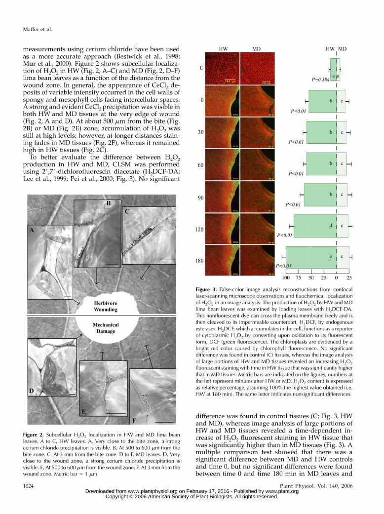
To better evaluate the difference between H2O2production in HW and MD, CLSM was performedusing 2#,7#-dichlorofluorescin diacetate (H2DCF-DA;Lee et al., 1999; Pei et al., 2000; Fig. 3). No significant
difference was found in control tissues (C; Fig. 3, HWand MD), whereas image analysis of large portions ofHW and MD tissues revealed a time-dependent in-crease of H2O2 fluorescent staining in HW tissue thatwas significantly higher than in MD tissues (Fig. 3). Amultiple comparison test showed that there was asignificant difference between MD and HW controlsand time 0, but no significant differences were foundbetween time 0 and time 180 min in MD leaves and
Figure 2. Subcellular H2O2 localization in HW and MD lima beanleaves. A to C, HW leaves. A, Very close to the bite zone, a strongcerium chloride precipitation is visible. B, At 500 to 600 mm from thebite zone. C, At 3 mm from the bite zone. D to F, MD leaves. D, Veryclose to the wound zone, a strong cerium chloride precipitation isvisible. E, At 500 to 600 mm from the wound zone. F, At 3 mm from thewound zone. Metric bar 5 1 mm.
Figure 3. False-color image analysis reconstructions from confocallaser-scanning microscope observations and fluochemical localizationof H2O2 in an image analysis. The production of H2O2 by HW and MDlima bean leaves was examined by loading leaves with H2DCF-DA.This nonfluorescent dye can cross the plasma membrane freely and isthen cleaved to its impermeable counterpart, H2DCF, by endogenousesterases. H2DCF, which accumulates in the cell, functions as a reporterof cytoplasmic H2O2 by converting upon oxidation to its fluorescentform, DCF (green fluorescence). The chloroplasts are evidenced by abright red color caused by chlorophyll fluorescence. No significantdifference was found in control (C) tissues, whereas the image analysisof large portions of HW and MD tissues revealed an increasing H2O2
fluorescent staining with time in HW tissue that was significantly higherthat in MD tissues. Metric bars are indicated on the figures; numbers atthe left represent minutes after HW or MD. H2O2 content is expressedas relative percentage, assuming 100% the highest value obtained (i.e.HW at 180 min). The same letter indicates nonsignificant differences.
Maffei et al.
1024 Plant Physiol. Vol. 140, 2006 www.plant.org on February 17, 2016 - Published by www.plantphysiol.orgDownloaded from
Copyright © 2006 American Society of Plant Biologists. All rights reserved.
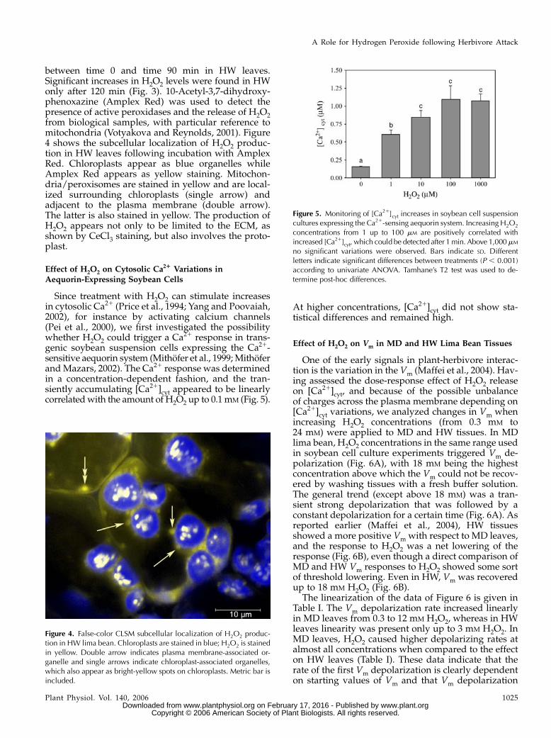
between time 0 and time 90 min in HW leaves.Significant increases in H2O2 levels were found in HWonly after 120 min (Fig. 3). 10-Acetyl-3,7-dihydroxy-phenoxazine (Amplex Red) was used to detect thepresence of active peroxidases and the release of H2O2from biological samples, with particular reference tomitochondria (Votyakova and Reynolds, 2001). Figure4 shows the subcellular localization of H2O2 produc-tion in HW leaves following incubation with AmplexRed. Chloroplasts appear as blue organelles whileAmplex Red appears as yellow staining. Mitochon-dria/peroxisomes are stained in yellow and are local-ized surrounding chloroplasts (single arrow) andadjacent to the plasma membrane (double arrow).The latter is also stained in yellow. The production ofH2O2 appears not only to be limited to the ECM, asshown by CeCl3 staining, but also involves the proto-plast.
Effect of H2O2 on Cytosolic Ca21 Variations inAequorin-Expressing Soybean Cells
Since treatment with H2O2 can stimulate increasesin cytosolic Ca21 (Price et al., 1994; Yang and Poovaiah,2002), for instance by activating calcium channels(Pei et al., 2000), we first investigated the possibilitywhether H2O2 could trigger a Ca21 response in trans-genic soybean suspension cells expressing the Ca21-sensitive aequorin system (Mithofer et al., 1999; Mithoferand Mazars, 2002). The Ca21 response was determinedin a concentration-dependent fashion, and the tran-siently accumulating [Ca21]cyt appeared to be linearlycorrelated with the amount of H2O2 up to 0.1 mM (Fig. 5).
At higher concentrations, [Ca21]cyt did not show sta-tistical differences and remained high.
Effect of H2O2 on Vm in MD and HW Lima Bean Tissues
One of the early signals in plant-herbivore interac-tion is the variation in the Vm (Maffei et al., 2004). Hav-ing assessed the dose-response effect of H2O2 releaseon [Ca21]cyt, and because of the possible unbalanceof charges across the plasma membrane depending on[Ca21]cyt variations, we analyzed changes in Vm whenincreasing H2O2 concentrations (from 0.3 mM to24 mM) were applied to MD and HW tissues. In MDlima bean, H2O2 concentrations in the same range usedin soybean cell culture experiments triggered Vm de-polarization (Fig. 6A), with 18 mM being the highestconcentration above which the Vm could not be recov-ered by washing tissues with a fresh buffer solution.The general trend (except above 18 mM) was a tran-sient strong depolarization that was followed by aconstant depolarization for a certain time (Fig. 6A). Asreported earlier (Maffei et al., 2004), HW tissuesshowed a more positive Vm with respect to MD leaves,and the response to H2O2 was a net lowering of theresponse (Fig. 6B), even though a direct comparison ofMD and HW Vm responses to H2O2 showed some sortof threshold lowering. Even in HW, Vm was recoveredup to 18 mM H2O2 (Fig. 6B).
The linearization of the data of Figure 6 is given inTable I. The Vm depolarization rate increased linearlyin MD leaves from 0.3 to 12 mM H2O2, whereas in HWleaves linearity was present only up to 3 mM H2O2. InMD leaves, H2O2 caused higher depolarizing rates atalmost all concentrations when compared to the effecton HW leaves (Table I). These data indicate that therate of the first Vm depolarization is clearly dependenton starting values of Vm and that Vm depolarization
Figure 4. False-color CLSM subcellular localization of H2O2 produc-tion in HW lima bean. Chloroplasts are stained in blue; H2O2 is stainedin yellow. Double arrow indicates plasma membrane-associated or-ganelle and single arrows indicate chloroplast-associated organelles,which also appear as bright-yellow spots on chloroplasts. Metric bar isincluded.
Figure 5. Monitoring of [Ca21]cyt increases in soybean cell suspensioncultures expressing the Ca21-sensing aequorin system. Increasing H2O2
concentrations from 1 up to 100 mM are positively correlated withincreased [Ca21]cyt, which could be detected after 1min. Above 1,000mM
no significant variations were observed. Bars indicate SD. Differentletters indicate significant differences between treatments (P , 0.001)according to univariate ANOVA. Tamhane’s T2 test was used to de-termine post-hoc differences.
A Role for Hydrogen Peroxide following Herbivore Attack
Plant Physiol. Vol. 140, 2006 1025 www.plant.org on February 17, 2016 - Published by www.plantphysiol.orgDownloaded from
Copyright © 2006 American Society of Plant Biologists. All rights reserved.
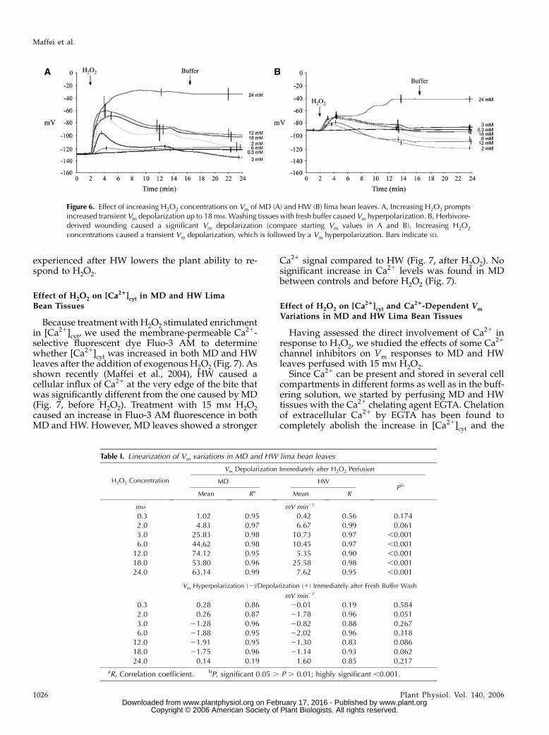
experienced after HW lowers the plant ability to re-spond to H2O2.
Effect of H2O2 on [Ca21]cyt in MD and HW LimaBean Tissues
Because treatment with H2O2 stimulated enrichmentin [Ca21]cyt, we used the membrane-permeable Ca21-selective fluorescent dye Fluo-3 AM to determinewhether [Ca21]cyt was increased in both MD and HWleaves after the addition of exogenous H2O2 (Fig. 7). Asshown recently (Maffei et al., 2004), HW caused acellular influx of Ca21 at the very edge of the bite thatwas significantly different from the one caused by MD(Fig. 7, before H2O2). Treatment with 15 mM H2O2caused an increase in Fluo-3 AM fluorescence in bothMD and HW. However, MD leaves showed a stronger
Ca21 signal compared to HW (Fig. 7, after H2O2). Nosignificant increase in Ca21 levels was found in MDbetween controls and before H2O2 (Fig. 7).
Effect of H2O2 on [Ca21]cyt and Ca21-Dependent Vm
Variations in MD and HW Lima Bean Tissues
Having assessed the direct involvement of Ca21 inresponse to H2O2, we studied the effects of some Ca21
channel inhibitors on Vm responses to MD and HWleaves perfused with 15 mM H2O2.
Since Ca21 can be present and stored in several cellcompartments in different forms as well as in the buff-ering solution, we started by perfusing MD and HWtissues with the Ca21 chelating agent EGTA. Chelationof extracellular Ca21 by EGTA has been found tocompletely abolish the increase in [Ca21]cyt and the
Figure 6. Effect of increasing H2O2 concentrations on Vm of MD (A) and HW (B) lima bean leaves. A, Increasing H2O2 promptsincreased transient Vm depolarization up to 18mM.Washing tissues with fresh buffer caused Vm hyperpolarization. B, Herbivore-derived wounding caused a significant Vm depolarization (compare starting Vm values in A and B). Increasing H2O2
concentrations caused a transient Vm depolarization, which is followed by a Vm hyperpolarization. Bars indicate SD.
Table I. Linearization of Vm variations in MD and HW lima bean leaves
Vm Depolarization Immediately after H2O2 Perfusion
H2O2 Concentration MD HWP b
Mean Ra Mean R
mM mV min21
0.3 1.02 0.95 0.42 0.56 0.1742.0 4.83 0.97 6.67 0.99 0.0613.0 25.83 0.98 10.73 0.97 ,0.0016.0 44.62 0.98 10.45 0.97 ,0.001
12.0 74.12 0.95 5.35 0.90 ,0.00118.0 53.80 0.96 25.58 0.98 ,0.00124.0 63.14 0.99 7.62 0.95 ,0.001
Vm Hyperpolarization (2)/Depolarization (1) Immediately after Fresh Buffer Wash
mV min21
0.3 0.28 0.86 20.01 0.19 0.5842.0 0.26 0.87 21.78 0.96 0.0513.0 21.28 0.96 20.82 0.88 0.2676.0 21.88 0.95 22.02 0.96 0.318
12.0 21.91 0.95 21.30 0.83 0.08618.0 21.75 0.96 21.14 0.93 0.06224.0 0.14 0.19 1.60 0.85 0.217
aR, Correlation coefficient. bP, significant 0.05 . P . 0.01; highly significant ,0.001.
Maffei et al.
1026 Plant Physiol. Vol. 140, 2006 www.plant.org on February 17, 2016 - Published by www.plantphysiol.orgDownloaded from
Copyright © 2006 American Society of Plant Biologists. All rights reserved.
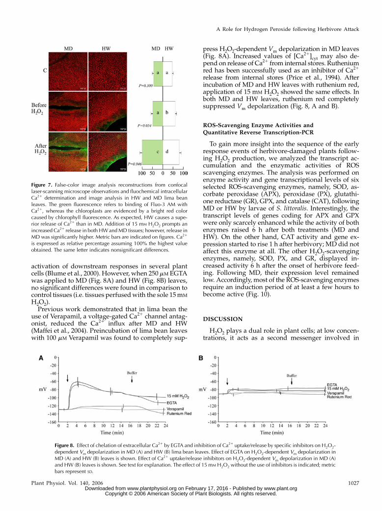
activation of downstream responses in several plantcells (Blume et al., 2000). However, when 250 mM EGTAwas applied to MD (Fig. 8A) and HW (Fig. 8B) leaves,no significant differences were found in comparison tocontrol tissues (i.e. tissues perfused with the sole 15 mM
H2O2).Previous work demonstrated that in lima bean the
use of Verapamil, a voltage-gated Ca21 channel antag-onist, reduced the Ca21 influx after MD and HW(Maffei et al., 2004). Preincubation of lima bean leaveswith 100 mM Verapamil was found to completely sup-
press H2O2-dependent Vm depolarization in MD leaves(Fig. 8A). Increased values of [Ca21]cyt may also de-pend on release of Ca21 from internal stores. Rutheniumred has been successfully used as an inhibitor of Ca21
release from internal stores (Price et al., 1994). Afterincubation of MD and HW leaves with ruthenium red,application of 15 mM H2O2 showed the same effects. Inboth MD and HW leaves, ruthenium red completelysuppressed Vm depolarization (Fig. 8, A and B).
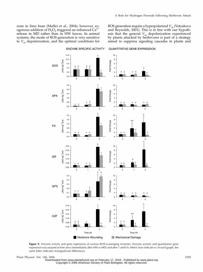
ROS-Scavenging Enzyme Activities andQuantitative Reverse Transcription-PCR
To gain more insight into the sequence of the earlyresponse events of herbivore-damaged plants follow-ing H2O2 production, we analyzed the transcript ac-cumulation and the enzymatic activities of ROSscavenging enzymes. The analysis was performed onenzyme activity and gene transcriptional levels of sixselected ROS-scavenging enzymes, namely, SOD, as-corbate peroxidase (APX), peroxidase (PX), glutathi-one reductase (GR), GPX, and catalase (CAT), followingMD or HW by larvae of S. littoralis. Interestingly, thetranscript levels of genes coding for APX and GPXwere only scarcely enhanced while the activity of bothenzymes raised 6 h after both treatments (MD andHW). On the other hand, CAT activity and gene ex-pression started to rise 1 h after herbivory; MD did notaffect this enzyme at all. The other H2O2-scavengingenzymes, namely, SOD, PX, and GR, displayed in-creased activity 6 h after the onset of herbivore feed-ing. Following MD, their expression level remainedlow. Accordingly, most of the ROS-scavenging enzymesrequire an induction period of at least a few hours tobecome active (Fig. 10).
DISCUSSION
H2O2 plays a dual role in plant cells; at low concen-trations, it acts as a second messenger involved in
Figure 7. False-color image analysis reconstructions from confocallaser-scanning microscope observations and fluochemical intracellularCa21 determination and image analysis in HW and MD lima beanleaves. The green fluorescence refers to binding of Fluo-3 AM withCa21, whereas the chloroplasts are evidenced by a bright red colorcaused by chlorophyll fluorescence. As expected, HW causes a supe-rior release of Ca21 than in MD. Addition of 15 mM H2O2 prompts anincreased Ca21 release in both HWandMD tissues; however, release inMD was significantly higher. Metric bars are indicated on figures. Ca21
is expressed as relative percentage assuming 100% the highest valueobtained. The same letter indicates nonsignificant differences.
Figure 8. Effect of chelation of extracellular Ca21 by EGTA and inhibition of Ca21 uptake/release by specific inhibitors on H2O2-dependent Vm depolarization in MD (A) and HW (B) lima bean leaves. Effect of EGTA on H2O2-dependent Vm depolarization inMD (A) and HW (B) leaves is shown. Effect of Ca21 uptake/release inhibitors on H2O2-dependent Vm depolarization in MD (A)and HW (B) leaves is shown. See text for explanation. The effect of 15 mM H2O2 without the use of inhibitors is indicated; metricbars represent SD.
A Role for Hydrogen Peroxide following Herbivore Attack
Plant Physiol. Vol. 140, 2006 1027 www.plant.org on February 17, 2016 - Published by www.plantphysiol.orgDownloaded from
Copyright © 2006 American Society of Plant Biologists. All rights reserved.
signaling, and at high concentrations it is part of directdefense and may also lead to programmed cell death(Vandenabeele et al., 2003). One of the questions thatremains unresolved is how H2O2 levels can triggerdifferent responses (Kovtun et al., 2000; Neill et al.,2002); however, the ultimate question on how H2O2 isperceived and transmitted within plant cells remainsunanswered, even though several candidate genes havebeen identified that are involved in oxidative stresssensing and signal transduction (Vandenabeele et al.,2003). In plant-pathogen interactions, plant receptorproteins perceive pathogen-derived or interaction-dependent signals, followed by downstream signalingevents, including ion fluxes (Ebel and Mithofer, 1998).Induced defense response includes the generation ofROS and NO as well as direct induction of genes,whereas amplification of the signal occurs through thegeneration of additional molecules (such as Ca21,oxylipins, or ethylene). Redox status alterations triggermechanisms for cell protection, lipid peroxidationleads to new signaling molecules, while cross talkbetween the various activated signaling cascades ap-pears to coordinate the response (Hammond-Kosackand Jones, 2000). Undoubtedly, wounding is one of thecommon events in plant interactions with pathogensand herbivores. Nevertheless, herbivores can causelarger and faster damage in attacked leaves where cellwalls are damaged and the remaining tissues experi-ence all the consequences of cell disruption. H2O2 isoften generated or even overproduced in response towounding in the cell walls and in the vascular bundlecells. It can be readily transported in water through theapoplast and diffuse initially into the cells adjacent toeach vein (Orozco-Cardenas et al., 2001). In MD andHW lima bean leaves, the accumulation of H2O2 athigh levels in cell walls adjacent to intercellular spacesin the spongy mesophyll could be explained by therapid evaporation of water in these air-filled spaces,together with the lower ROS-scavenging activity atthese sites. This also might represent a defense strat-egy for the plant because wounds and the intercellularspaces are paths for subsequent secondary invasion bymicrobial pathogens (Bestwick et al., 1997; Orozco-Cardenas et al., 2001). HW leaves showed higherproduction of H2O2 with respect to MD leaves. ROShave been associated with plant herbivore interac-tions, and oxidative changes in the plants correspondwith oxidative damage in the midguts of insectsfeeding on previously wounded plants (Bi and Felton,1995). The finding that mitochondria and/or peroxi-somes are stained by Amplex Red indicates that inHW H2O2 can be produced also in the protoplast (Fig.4). Besides, peroxisomes, which are well known to beinvolved in H2O2 breakdown through the action ofCAT, have recently been found to be an importantsource of ROS (Corpas et al., 2001), while accumula-tion of ROS in mitochondria has been found to play acrucial role in programmed cell death (Maxwell et al.,2002; Foyer and Noctor, 2005). In response to herbi-vores, H2O2 levels are likely to be elevated as long as
the attacks persist. In tomato plants (Lycopersiconesculentum) that constitutively express prosystemin, aprecursor of systemin that functions in the cascades ofa long-distance systemic signaling, the levels of H2O2are constitutively elevated and may provide an earlydefense barrier (Orozco-Cardenas and Ryan, 1999).Direct oxidative injury to insect midguts and damageto the nutritive and antioxidant components of theplants may be one of the ways plant counterattackherbivore feeding activity. According to Dangl et al.(1996), the presence of H2O2 in the plant in response toherbivory, before any pathogen invasion, could be ad-vantageous because timing of the induction of defenseresponses can be an important factor in the success orfailure of plants to defend against pathogen attacks.This is particularly true in S. littoralis-lima bean inter-action since feeding insects introduce regurgitate fromthe foregut containing microorganisms into the freshlydamaged leaf.
Signaling during HR does not only involve thepassage of H2O2 from the apoplast, across the mem-brane into cytosol. This suggests that apoplast-specificcomponents might be the source of additional signals,the generation of which requires locally high ROSlevels that cannot be supplied by diffusion of intracel-lular H2O2 (Foyer and Noctor, 2005; Mur et al., 2005).MD lima bean leaves reacted fast and dramatically toH2O2 by inducing a strong Vm depolarization. How-ever, HW leaves already showed a reduced starting Vmwith the consequence of a dramatically lower or evenno responsiveness to H2O2 application (Fig. 8). The de-polarization of the Vm by the action of HW is thuslinked to a reduction of downstream responses ofattacked leaves to signaling molecules such as H2O2,and the increased production of H2O2 in HW leavesindicates a possible threshold heightening of the plantH2O2-sensing system, with a consequent increasedproduction of H2O2.
H2O2 burst often has been associated with the acti-vation of cascade signaling events. A close interactionexists between H2O2 and cytosolic calcium in responseto biotic and abiotic stimuli both in plants (Sanderset al., 1999; Murata et al., 2001; Sagi and Fluhr, 2001;Yang and Poovaiah, 2002; Foreman et al., 2003) andanimals (Castro et al., 2004; Redondo et al., 2004;Rosado et al., 2004; Tabet et al., 2004) systems (seeHepler, 2005, for a historical perspective assay). Thesame independent assay using transgenic soybean cellsuspensions that was successfully applied to analyzethe activities of herbivore regurgitate components(Maffei et al., 2004) was fundamental to demonstratethat H2O2 is also able to elicit [Ca21]cyt release (Fig. 5).The upper level of soybean cell culture responsivenessto H2O2 corresponds to the lower level in both MD andHW lima bean leaves. This situation reflects the highersensitivity of cell suspension cultures compared toplant tissues and should be considered for further com-parisons when aequorin is used to evaluate activitiesof molecules involved in signaling processes. A rapidincrease in [Ca21]cyt has also been observed at the bite
Maffei et al.
1028 Plant Physiol. Vol. 140, 2006 www.plant.org on February 17, 2016 - Published by www.plantphysiol.orgDownloaded from
Copyright © 2006 American Society of Plant Biologists. All rights reserved.
zone in lima bean (Maffei et al., 2004); however, ex-ogenous addition of H2O2 triggered an enhanced Ca21
release in MD rather than in HW leaves. In animalsystems, the mode of ROS generation is very sensitiveto Vm depolarization, and the optimal conditions for
ROS generation require a hyperpolarized Vm (Votyakovaand Reynolds, 2001). This is in line with our hypoth-esis that the general Vm depolarization experiencedby plants attacked by herbivores is part of a strategyaimed to suppress signaling cascades in plants and
Figure 9. Enzyme activity and gene expression of various ROS-scavenging enzymes. Enzyme activity and quantitative geneexpressionwas assayed at time zero (immediately after HWorMD) and after 1 and 6 h. Metric bars indicate SD. In each graph, thesame letter indicates nonsignificant differences.
A Role for Hydrogen Peroxide following Herbivore Attack
Plant Physiol. Vol. 140, 2006 1029 www.plant.org on February 17, 2016 - Published by www.plantphysiol.orgDownloaded from
Copyright © 2006 American Society of Plant Biologists. All rights reserved.
eventually reduce the capability to induce defenses. Inthis context, the parallel increase of H2O2 after HW onthe one hand and the herbivore-induced Vm depolar-ization on the other hand may represent a strategy ofthe plant to produce toxic H2O2 and of the herbivore toreduce H2O2 production by Vm depolarization.
In plants, membrane depolarization is involved invarious signal transduction pathways (Ward et al., 1995;White, 2000; Kurusu et al., 2004). Electrophysiologicalstudies revealed the existence of plasma membrane-localized Ca21-permeable channels activated by membranede- or hyperpolarization in response to external stim-uli (for review, see White, 2000). These channels arepostulated to play pivotal roles in early steps of avariety of signal transduction networks, such as ab-scisic acid-induced stomatal closure (Hamilton et al.,2000; Pei et al., 2000), defense responses (Kluseneret al., 2002), tip growth in rhizoid cells (Taylor et al.,1996), and growth of root apex (Kiegle et al., 2000).To better elucidate the role of Ca21 variations and theexperienced Vm depolarization upon H2O2 treatment,
we used two Ca21 channel inhibitors and a Ca21 che-lating agent. In a previous work we observed that theuse of the voltage-gated Ca21 channel antagonist ve-rapamil was able to reduce calcium influx in HW limabeans (Maffei et al., 2004). In MD leaves, verapamiland ruthenium red, a potent inhibitor of the release ofCa21 from internal stores, completely suppressedH2O2-induced Vm depolarization (Fig. 8A), whereas theuse of the Ca21 chelator EGTA did not exert any effecton both MD and HW leaves. Evidently, the removal offree Ca21 from the extracellular space (and buffersolution) was not sufficient to abolish Vm depolariza-tion. On the other hand, the blockage of voltage-gatedchannels and internal store release of Ca21 indicatedthat these two sources of Ca21 could be associated withVm depolarization. However, we cannot exclude thatboth Verapamil and ruthenium red may target thesame calcium pool in this system.
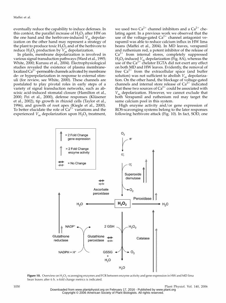
High enzyme activity and/or gene expression ofROS-scavenging systems belong to the later responsesfollowing herbivore attack (Fig. 10). In fact, SOD, one
Figure 10. Overview on H2O2-scavenging enzymes and FCR between enzyme activity and gene expression in HWandMD limabean leaves after 6 h. x-fold change metrics is indicated.
Maffei et al.
1030 Plant Physiol. Vol. 140, 2006 www.plant.org on February 17, 2016 - Published by www.plantphysiol.orgDownloaded from
Copyright © 2006 American Society of Plant Biologists. All rights reserved.
of the early enzymes involved in the reduction of thesuperoxide anion generated either by Mehler reactionand photorespiration or by the reduction of molecularoxygen in mitochondria and oxidase reaction (Ishikawaet al., 1996), is activated at the gene transcription andenzyme activity levels only after 6 h. On the otherhand, the basal activity of SOD was high at zero and1 h after treatment in both HW and MD leaves, butdropped in MD leaves after 6 h. The latter finding isconsistent with the increase of H2O2 in HW leaves withtime (compare Figs. 2, 3, and 9). Gene activation ofCAT started earlier (after 1 h) and reached the highestactivity at 6 h. This enzyme, along with PX, is directlyinvolved in the disruption of H2O2, and their activa-tion indicates the effort of attacked plants to reduceoxidative damage. However, a direct comparison be-tween SOD activity and CAT-PX activities explainswhy H2O2 increases with time. In fact, after 6 h SODactivity is about 7- and 37-fold higher than that of thePX and CAT, respectively (Fig. 9). The highest x-foldchange ratio (FCR) between HW and MD activitiesconcerning gene up-regulation was found for CAT,which also showed the highest FCR for enzyme activ-ity. For CAT, PX, and GR, FCR gene up-regulation wasmore evident than FCR enzyme activity, whereas SODshowed a superior FCR enzyme activity when com-pared to FCR gene up-regulation.
The low activity of APX (a typical chloroplasticenzyme; Ishikawa et al., 1996) is in agreement with thelack of Amplex Red staining observed in chloroplastswith CLSM (Fig. 4) and suggests a major role of non-photosynthetic H2O2 production after herbivore at-tack. Glutathione directly reduces most active oxygenspecies and also scavenges H2O2 via GPX, which is in-volved in the detoxification of lipid peroxides ratherthan H2O2 per se (Noctor and Foyer, 1998; Nagalakshmiand Prasad, 2001; Chen et al., 2004; Foyer and Noctor,2005). This might explain the almost identical GPX ac-tivity between HW and MD, even though it is gener-ally believed that wounding caused increased GPXactivity (Fig. 10).
In conclusion, these results clearly demonstrate thatone of the strategies of successful herbivore attack byS. littoralis on lima bean is the immediate lowering ofVm to a significant depolarized state, which in turnreduces the ability of the plant to react to wound-induced signals. The depolarized Vm reduces the abilityof the leaf to respond to at least one of the ROS, H2O2,and despite lowered Vm this molecule is overpro-duced. An open question remains the characterizationof the origin and the nature of the molecule responsi-ble for Vm depolarization after HW.
MATERIALS AND METHODS
Plant and Animal Material
Feeding experiments were carried out using the lima bean (Phaseolus
lunatus cv Ferry Morse var. Jackson Wonder Bush). Individual plants were
grown from seed in a plastic pot with sterilized potting soil at 23�C and 60%
humidity using daylight fluorescent tubes at approximately 270 mE m22 s21
with a photophase of 16 h. Experiments were conducted with 12- to 16-d-old
seedlings showing two fully developed primary leaves, which were found to
be the most responsive leaves.
Larvae of Spodoptera littoralis (Boisd.; Lepidoptera, Noctuidae) were grown
in petri dishes at long photoperiod (14–16 h photophase) and 22�C to 24�C as
described (Maffei et al., 2004).
Chemicals
H2O2 (30%), cerium chloride, H2DCF-DA, Fluo-3 AM, and Verapamil were
purchased from Fluka Biochemika; DAB, EGTA, and ruthenium red were
from Sigma/Aldrich. Synthetic coelenterazine was from Calbiochem. Amplex
Red Hydrogen Peroxide/Peroxidase Assay kit (A-22188) was purchased from
Molecular Probes.
Membrane Potentials
Membrane potentials were determined in leaf segments. The Vm was
determined with glass micropipettes with a tip resistance of 4 to 10 MV and
filled with 3 M KCl. Micropipettes were used as microsalt bridges to Ag/AgCl
electrodes obtained with a Narishighe PE-21 puller and inserted vertically in
the tissue by means of a micromanipulator (Maffei et al., 2004). Measurements
were always performed within 15 min after MD or HW. Leaves for Vm
measurements were sliced after MD or HW treatment and were always
equilibrated for 60 to 120 min in 5 mM MES-NaOH, pH 6.0. Having assessed
after several trials no significant differences between controls (undamaged
leaves sliced and immediately used for Vm measurement) and MD (leaves
damaged and sliced after 15 min for Vm measurement), all Vm data are
referred only to MD. Perfusion of solutions was granted by a multichannel
Ismatec Reglo peristaltic pump (flow rate 1 mL min21). Based on topograph-
ical and temporal determination of Vm performed in a previous work, the
electrode was inserted between 0.5 and 1.5 mm from the wound/bite zone,
where a significant Vm depolarization occurs in HW (Maffei et al., 2004). Vm
variations were recorded both on a pen recorder and through a digital port of a
PC using a data logger. Measurements were performed after perfusion with
increasing concentrations of H2O2 in both MD and HW lima bean leaves.
Perfusion with 15 mM H2O2 was also performed in the presence of the
inhibitors of Ca21 uptake/release, which were applied after MD and HW.
All chemicals were dissolved in water, which was present in the control
solutions, and perfused in a 50 mM MES-Na-buffered solution, pH 6.0,
containing 0.5 mM calcium sulfate and 2.5 mM dichlorophenyldimethylurea
(DCMU), used to poison photosynthetic electron transfer. Several trials
demonstrated that DCMU has no effect on Vm when used at 2.5 mM and
does not alter the ROS production during the short time of Vm detection. After
a period of Vm stabilization, saturation of the well where leaf tissues have been
placed occurred in 2 min, after which perfusion was carried out for a variable
time (until stabilization of the Vm). Washing of the well was done by perfusing
with fresh buffer. Saturation with fresh buffer took 10 to 12 min, and then the
solution was allowed to perfuse until Vm reached a constant value.
Cellular and Subcellular Localization of H2O2 in HWand MD Lima Bean Leaves
Histochemical Localization of H2O2 Using DAB
Cuttings of lima bean plants were placed in 15-mL Falcon tubes containing
1 mg mL21 DAB dissolved in HCl-acidified (pH 3.2) distilled water. Plants
were then placed overnight under moderate vacuum to allow penetration of
the dye. Leaves were then mechanically damaged with a cork borer or
herbivore wounded with S. littoralis. Leaves were sampled 1, 3, 6, and 24 h
after MD or HW treatment, bleached in boiling 90% ethanol for 10 min, and
mounted on glass slides. To improve staining visibility, samples were observed
with the epifluorescence Nikon Eclipse E400 microscope equipped with a
450-nm filter, and pictures are presented as black and white inversed images.
Subcellular Localization of H2O2 Using CeCl3
Following MD or HW treatment, small lima bean tissue pieces were
excised from leaves and vacuum infiltrated with freshly prepared 5 mM CeCl3
in 50 mM MOPS at pH 7.2 for 1 h. After fixation in 1.25% (v/v) glutaraldehyde
A Role for Hydrogen Peroxide following Herbivore Attack
Plant Physiol. Vol. 140, 2006 1031 www.plant.org on February 17, 2016 - Published by www.plantphysiol.orgDownloaded from
Copyright © 2006 American Society of Plant Biologists. All rights reserved.
and 1.25% (v/v) paraformaldehyde in 50 mM sodium cacodylate buffer, pH
7.2, for 2 h under vacuum at room temperature, samples were kept overnight
at 4�C. Subsequently, tissues were washed twice, under vacuum, for 10 min in
cacodylate buffer, postfixed for 1 h in osmium tetroxide 1% (v/v) in cacodylate
buffer, and washed twice in cacodylate buffer for 10 min. Tissues were then
dehydrated in a graded acetone series, incubated in rising concentration of
acetone-resin mixture, and then embedded and polymerized for 24 h at 60�Cin Epon/Araldite resin. Thin sections were then cut with a Reichert Ultracut
ultramicrotome. Staining was not performed in order not to overlay CeCl3
precipitation, and thin sections mounted on 400 mesh grids were observed
under the Philips CM10 transmission electron microscope.
CLSM Subcellular Localization of H2O2 and Active PXsUsing Amplex Red
Lima bean leaves from rooted plants in pots were herbivore wounded and
mechanically damaged after incubation with the dye Amplex Red. The
Molecular Probes Amplex Red Hydrogen Peroxide/Peroxidase Assay kit
(A-22188) was used and dissolved in MES-Na buffer 50 mM, pH 6.0, con-
taining 0.5 mM calcium sulfate and 5 mM DCMU, to obtain a 50 mM solution.
Leaves where then mounted on a Leica TCS SP2 multiband confocal laser-
scanning microscope stative without separating the leaf from the plant.
Scannings were recorded from time 0 up to 180 min using the HCX PL APO
633/1.20 W Corr/0.17CS objective. The microscope was operated with a
Laser Ar (458 nm/5 mW; 476 nm/5 mW; 488 nm/20 mW; 514 nm/20 mW), a
Laser HeNe 543 nm/1.2 mW, and a Laser HeNe 633 nm/10 mW.
Fluorimetric Determination of H2O2 Using H2DCF-DA
Lima bean leaves from rooted plants in pots were herbivore wounded and
mechanically damaged after incubation with the dye H2DCF-DA. This non-
fluorescent dye can cross the plasma membrane freely, and is then cleaved to
its impermeable counterpart, dichlorofluorescin (H2DCF), by endogenous
esterases. H2DCF, which accumulates in the cell, functions as a reporter of
cytoplasmic H2O2 by converting upon oxidation to its fluorescent form, DCF
(green fluorescence; Lee et al., 1999). H2DCF-DA was dissolved in MES-Na
buffer 50 mM, pH 6.0, containing 0.5 mM CaSO4 and 5 mM DCMU, to obtain a
50-mM solution. Leaves were then mounted on an Olympus FLUOview
confocal laser-scanning microscope stative without separating the leaf from
the plant. Scannings were recorded from time 0 up to 180 min. The microscope
is operated with a Krypton/Argon laser at 488 nm and 568 nm wavelengths:
the first wavelength excitates the dye, resulting in emission of green light, and
the second mostly excitates chloroplasts, which emit red fluorescence. Images
generated by the FluoView software were analyzed using the public domain
NIH Image J program (developed at the United States National Institutes of
Health and available on the Internet at http://rsb.info.nih.gov/nih-image).
Intracellular Calcium Variation Determination
Fluo-3 AM (more permeant for cells), purchased as a stock solution in
dimethyl sulfoxide, was diluted in 50 mM MES-Na buffer, pH 6.0, by addition of
0.5 mM CaSO4, 2.5 mM DCMU to reach the concentration of 5 mM. This resulting
solution was used for an initial treatment of lima bean leaves not separated
from the plant: The leaf was gently fixed over a glass slide, and a drop (about
20 mL) of 5 mM Fluo-3 AM solution was applied and covered with another glass
slide. Thirty minutes after treatment with Fluo-3 AM, the leaf was mounted on
an Olympus FLUOview confocal laser-scanning microscope stative without
separating the leaf from the plant. After 30 min, 15 mM H2O2 were added to MD
and HW, and Fluo-3 AM fluorescence was recorded. Measurements were taken
in intact leaves, with leaves wounded mechanically and after herbivore
feeding, both in the presence and absence of exogenous calcium. The Olympus
confocal laser-scanning microscope is operated as above. Images generated by
the FluoView software were analyzed using the public domain NIH Image J
program (developed at the United States National Institutes of Health and
available on the Internet at http://rsb.info.nih.gov/nih-image).
Aequorin-Dependent Luminescence Measurements
Transgenic soybean (Glycine max) 6.6.12 cell lines carrying the stably
integrated plasmid pGNAequ/neo2 and expressing apoaequorin (Mithofer et al.,
1999) were used to reconstitute aequorin in vivo with 10 mM synthetic
coelenterazine on a shaker (125 rpm) in the dark for 24 h. The Ca21-specific
luminescence (470 nm) was measured in a final volume of 200 mL using a
digital luminometer (Bio-Orbit 1250) as described (Mithofer et al., 1999;
Mithofer and Mazars, 2002). Treatments with various compounds were
performed by adding 1 to 10 mL of different concentrations of aqueous stock
solutions to the cell suspension culture. Mixing time for the addition of any
compound was 5 to 7 s. In each experiment, the concentration of reconstituted
aequorin was not limiting under any of the experimental conditions, with a
maximal consumption not exceeding 10%. The residual aequorin was com-
pletely discharged by adding 200 mL of 20% ethanol containing 2 M CaCl2
(final concentration 10% and 1 M, respectively). The resulting luminescence
was used to estimate the total amount of aequorin present in various
experiments to determine the rate of aequorin consumption for the calculation
of the cytosolic Ca21 concentrations according to Allen et al. (1977).
ROS-Scavenging Enzyme Activity
Leaves were collected immediately after and at 1 h and 6 h after HW or
MD. Intact leaves pooled from five plants were frozen in liquid N2 and stored
at 280�C before enzyme extraction. ROS scavenger enzymes were extracted
following the method of Zhang and Kirkham (1996) with some modifications.
All operations were carried out at 4�C. Plant material was ground with mortar
and pestle under liquid nitrogen in cold 50 mM sodium phosphate, pH 7.5,
containing 250 mM Suc, 1.0 mM EDTA, 10 mM KCl, 1 mM MgCl2, 0.5 mM
phenylmethylsulfonyl fluoride, 0.1 mM dithiothreitol, and 1% (w/v) poly-
vinylpolypyrrolidone in a 6:1 proportion (w/v). The homogenate was then
filtered through eight layers of cheesecloth and centrifuged at 25,000g for
20 min at 4�C. The supernatant was brought to 80% saturation with addition of
solid ammonium sulfate (NH4)2SO4 and allowed to stir gently for several
hours at 4�C. After centrifugation at 28,000g for 45 min at 4�C, pellets, con-
taining most of enzyme activity, were resuspended in a small volume of 50 mM
sodium phosphate, pH 7.5, and used directly for enzyme assays.
SOD (EC 1.15.1.1)
The activity of SOD was measured according to Krishnan et al. (2002). This
method tests the ability of SOD to inhibit the reduction of nitro blue
tetrazolium by the superoxide anion generated photochemically. One milli-
liter of assay mixture consisted of 50 mM Na-P buffer, pH 7.8, 13 mM Met, 75 mM
nitro blue tetrazolium, 2 mM riboflavin, 0.1 mM EDTA, and enzyme extract.
Riboflavin was added last, the samples were placed 30 cm below a light source
(4,000 lux), and the reaction was allowed to run for 15 min. The reaction was
stopped by switching off the light. A nonirradiated reaction mixture, which
was run in parallel, did not develop color and served as a control. The ab-
sorbance was read at 560 nm.
APX (EC 1.11.1.11)
APX activity was determined from the decrease in A290, due to the H2O2-
dependent oxidation of ascorbate (e 5 2.8 mM21 cm21; Zhang and Kirkham,
1996). The 1-mL reaction mixture contained 50 mM Na-P, pH 7.0, 0.5 mM
ascorbic acid, 0.1 mM H2O2, 0.1 mM EDTA, and enzyme.
PX (EC 1.11.1.7)
PX activities were measured as oxidation of guaiacol (e5 26.6 mM21 cm21)
in the presence of H2O2 (Zhang and Kirkham, 1996). The reaction mixture
contained 50 mM Na-P, pH 7.0, 0.33 mM guaiacol, 0.27 mM H2O2, and enzyme
extract in 1.0 mL final volume. The reaction was started by addition of
guaiacol and followed spectrophotometrically at 470 nm.
GR (EC 1.8.1.10)
GR activity was determined at 340 nm by following the oxidation of
NADPH (e 5 6.2 mM21 cm21) at 340 nm (Zhang and Kirkham, 1996). The
reaction mixture contained 100 mM Na-P, pH 7.5, 0.2 mM NADPH, 0.5 mM
glutathione disulfide, and enzyme extract in a 1.0 mL final volume at 30�C.
The reaction was started by adding glutathione disulfide.
Maffei et al.
1032 Plant Physiol. Vol. 140, 2006 www.plant.org on February 17, 2016 - Published by www.plantphysiol.orgDownloaded from
Copyright © 2006 American Society of Plant Biologists. All rights reserved.
GPX (EC 1.11.1.9)
GPX was assayed indirectly, as described by Anderson and Davis (2004).
The reaction contained 125 mM K-P buffer, pH 7.0, 1.2 mM cumene hydroper-
oxide, 1.25 mM EDTA, 1.25 mM sodium azide, 1.0 mM glutathione, 0.25 mM
NADPH, 0.6 IU of yeast GR (Sigma Type III), and enzyme extract in a final
volume of 1 mL. After incubation for 10 min at 25�C with all the reagents
except cumene hydroperoxide, the reaction was started with cumene
hydroperoxide. Decrease of NADPH at 340 nm was corrected for nonenzy-
matic controls.
CAT (EC 1.11.1.6)
CAT activities were assayed spectrophotometrically by monitoring the
change in A240 due to the decreased absorption of H2O2 (e H2O2 5 39.4 mM21
cm21; Zhang and Kirkham, 1996). The reaction mixture in 1 mL final volume
contained 50 mM Na-P, pH 7.0, 15 mM H2O2, and enzyme extract. The reaction
was initiated by addition of H2O2.
Soluble Protein Determination
Soluble protein concentration was evaluated by the method of Bradford
(1976) using bovine serum albumin as a standard.
Real-Time PCR
Total RNA was isolated from leaf tissues of MD and HW plants at the time
zero, and after 1 and 6 h following MD or HW using the Concert Plant RNA
Reagent (Invitrogen) following the manufacturer’s protocol. The total RNA
was purified to eliminate genomic DNA using Qiagen RNeasy Plant RNA kit
and RNase-Free DNase set (Qiagen). First-strand cDNAwas synthesized using
SuperScript III Reverse Transcriptase (Invitrogen), oligo(dT)12-18 primer, and
1 mg of total RNA at 50�C for 50 min. Primers for real-time PCR were designed
using the Primer 3 software (http://frodo.wi.mit.edu/cgi-bin/primer3/
primer3_www.cgi) for a length of the resulting PCR product of approximately
200 bp (see below). The real-time PCR was done on an Mx3000P Real-Time PCR
system (Stratagene). The process was performed with 25mL of reaction mixture
consisting of 12.5 mL of 23 Brilliant SYBR Green QPCR Master Mix (Stra-
tagene), cDNA (1 mL from 20 mL of each reverse transcription [RT] product
pool), 100 nM primers, and 30 mM ROX as a reference dye. The following
protocol was applied: initial polymerase activation of 10 min at 95�C; and 40
cycles of 30 s at 95�C, 60 s at 55�C, and 30 s at 72�C. PCR conditions were
determined by comparing threshold values in dilution series of the RT product,
followed by non-RT template control and nontemplate control for each primer
pair. Relative RNA levels were calibrated and normalized with the level of actin
mRNA (GenBank accession no. DQ159907).
Primers used for real-time PCR were as follows: ACT1, accession number
DQ159907, forward primer 5#-AGGCTCCTCTTAACCCCAAG-3#, reverse
primer 5#-GTGGGAGAGCATAACCCTCA-3#; AP1, accession number
DQ004738, forward primer 5#-AGGAGCGTTCTGGATTTGAG-3#, reverse
primer 5#-AATCAGCGAAGAACGCATCT-3#; CAT1, accession number
DQ004737, forward primer 5#-GCCGCATGGTCTTGAATAAG-3#, reverse
primer 5#-ATTTGGGTGCATTAGCAGGA-3#; GP1, accession number
DQ004739, forward primer 5#-AACAGGAAACTCAGCCTTGAA-3#, reverse
primer 5#-ATTGTCAACGTTGCCTCACA-3#; GR1, accession number
DQ159906, forward primer 5#-GCCCCTTCATTCCTGATATTC-3#, reverse
primer 5#-TCGTCAAATCCCCTCAGAAC-3#; PER1, accession number
DQ159909, forward primer 5#-GACCCTGTCATGGACCAAAC-3#, reverse
primer 5#-CAATGCCCTTGGTCCTCTTA-3#; and SOD1, accession number
DQ159910, forward primer 5#-AAGCACCACAGGGCTTATGT-3#, reverse
primer 5#-ATGGTTTTCCACCTCCACCT-3#.
Statistics
At least five repetitions were used for the statistical treatment of the data.
More than five repetitions contributed to the mean values given in Figures 6
and 8. The data are expressed as mean values; metric bars indicate the SD. To
evaluate the difference significance of the control and the treatments at the
given concentrations, ANOVA was performed. Tamhane’s T2 test was used to
determine post-hoc differences.
ACKNOWLEDGMENTS
We are indebted to Dr. G. Trautmann (Bayer AG) for supplying us with
egg clutches of lepidopteran larvae and A. Berg for caterpillar rearing. We
also thank H. Maischak for help with statistical analysis.
Received September 29, 2005; revised January 12, 2006; accepted January 12,
2006; published January 27, 2006.
LITERATURE CITED
Allen D, Blinks JR, Prendergast FG (1977) Aequorin luminescence: rela-
tion of light emission to calcium concentration—a calcium-independent
component. Science 195: 996–998
Anderson J, Davis DG (2004) Abiotic stress alters transcript profiles and
activity of glutathione-S-transferase, glutathione peroxidase and gluta-
thione reductase in Euphorbia esula. Physiol Plant 120: 421–433
Angelini R, Manes F, Federico R (1990) Spatial and functional correlation
between diamine-oxidase and peroxidase activities and their depen-
dence upon de-etiolation and wounding in chick-pea stems. Planta 182:
89–96
Bergey DR, Orozco-Cardenas ML, de Moura DS, Ryan CA (1999) A
wound- and systemic-inducible polygalacturonase in tomato leaves.
Proc Natl Acad Sci USA 96: 1756–1760
Bestwick CS, Brown IR, Bennett MHR, Mansfield JW (1997) Localization
of hydrogen peroxide accumulation during the hypersensitive reaction
of lettuce cells to Pseudomonas syringae pv phaseolicola. Plant Cell 9:
209–221
Bestwick CS, Brown IR, Mansfield JW (1998) Localized changes in
peroxidase activity accompany hydrogen peroxide generation during
the development of a nonhost hypersensitive reaction in lettuce. Plant
Physiol 118: 1067–1078
Bi JL, Felton GW (1995) Foliar oxidative stress and insect herbiv-
ory: primary compounds, secondary metabolites, and reactive oxygen
species as components of induced resistance. J Chem Ecol 21:
1511–1530
Blume B, Nurnberger T, Nass N, Scheel D (2000) Receptor-mediated
increase in cytoplasmic free calcium required for activation of pathogen
defense in parsley. Plant Cell 12: 1425–1440
Bolwell DJ (1999) Role of active oxygen species and NO in plant defence
responses. Curr Opin Plant Biol 2: 287–294
Bolwell GP, Wojtaszek P (1997) Mechanisms for the generation of reactive
oxygen species in plant defence: a broad perspective. Physiol Mol Plant
Pathol 51: 347–366
Bradford MM (1976) A rapid and sensitive method for the quantitation of
microgram quantities of protein utilizing the principle of protein-dye
binding. Anal Biochem 72: 248–254
Bradley DJ, Kjellbom P, Lamb CJ (1992) Elicitor- and wound-induced
oxidative cross-linking of a proline-rich plant cell wall protein: a novel,
rapid defense response. Cell 70: 21–30
Castro J, Bittner CX, Humeres A, Montecinos VP, Vera JC, Barros LF
(2004) A cytosolic source of calcium unveiled by hydrogen peroxide
with relevance for epithelial cell death. Cell Death Differ 11: 468–478
Cazale A-C, Rouet-Mayer M-A, Barbier-Brygoo H, Mathieu Y, Lauriere C
(1998) Oxidative burst and hypoosmotic stress in tobacco cell suspen-
sions. Plant Physiol 116: 659–669
Chen K-M, Gong H-J, Chen G-C, Wang S-M, Zhang C-L (2004) Gradual
drought under field conditions influences the glutathione metabolism,
redox balance and energy supply in spring wheat. J Plant Growth Regul
23: 20–28
Chen Z, Silva H, Klessig DF (1993) Active oxygen species in the induc-
tion of plant systemic acquired resistance by salicylic acid. Science 262:
1883–1886
Corpas FJ, Barros JB, del Rio LA (2001) Peroxisomes as a source of reactive
oxygen species and nitric oxide signal molecules in plants. Trends Plant
Sci 6: 145–150
Dangl JL, Dietrich RA, Richberg MH (1996) Death don’t have no mercy:
cell death programs in plant-microbe interactions. Plant Cell 8:
1793–1807
Devlin WS, Gustine DL (1992) Involvement of the oxidative burst
in phytoalexin accumulation and the hypersensitive reaction. Plant
Physiol 100: 1189–1195
A Role for Hydrogen Peroxide following Herbivore Attack
Plant Physiol. Vol. 140, 2006 1033 www.plant.org on February 17, 2016 - Published by www.plantphysiol.orgDownloaded from
Copyright © 2006 American Society of Plant Biologists. All rights reserved.
Ebel J, Mithofer A (1998) Early events in the elicitation of plant defence.
Planta 206: 335–348
Felton G, Summers CB, Mueller AJ (1994) Oxidative responses in soybean
foliage to herbivory by bean leaf beetle and three-cornered alfalfa
hopper. J Chem Ecol 20: 639–649
Foreman J, Demidchik V, Bothwell JHF, Mylona P, Miedema H, Torres
MA, Linstead P, Costa S, Brownlee C, Jones JDG, et al (2003) Reactive
oxygen species produced by NADPH oxidase regulate plant cell
growth. Nature 422: 442–446
Foyer CH, Lopez Delgado H, Dat JF, Scott IM (1997) Hydrogen peroxide
and glutathione-associated mechanisms of acclimatory stress tolerance
and signalling. Physiol Plant 100: 241–254
Foyer CH, Noctor G (2005) Redox homeostasis and antioxidant signaling: a
metabolic interface between stress perception and physiological re-
sponses. Plant Cell 17: 1866–1875
Gus-Mayer S, Naton B, Hahlbrock K, Schmelzer E (1998) Local mechan-
ical stimulation induces components of the pathogen defense response
in parsley. Proc Natl Acad Sci USA 95: 8398–8403
Hamilton DWA, Hills A, Kohler B, Blatt MR (2000) Ca21 channels at
the plasma membrane of stomatal guard cells are activated by hyper-
polarization and abscisic acid. Proc Natl Acad Sci USA 97: 4967–
4972
Hammond-Kosack K, Jones JDG (2000) Responses to plant pathogens. In B
Buchanan, W Gruissem, R Jones, eds, Biochemistry and Molecular
Biology of Plants. American Society of Plant Physiologists, Rockville,
MD, pp 1102–1156
Hepler PK (2005) Calcium: a central regulator of plant growth and
development. Plant Cell 17: 2142–2155
Hiraga S, Ito H, Yamakawa H, Ohtsubo N, Seo S, Mitsuhara I, Matsui H,
Honma M, Ohashi Y (2000) A HR-induced tobacco peroxidase gene is
responsive to spermine, but not to salicylate, methyl jasmonate, and
ethephon. Mol Plant Microbe Interact 13: 210–216
Ishikawa T, Takeda T, Kohno H, Shigeoka S (1996) Molecular character-
ization of Euglena ascorbate preoxidase using monoclonal antibody.
Biochim Biophys Acta 1290: 69–75
Jacks TJ, Davidonis GH (1996) Superoxide, hydrogen peroxide, and the res-
piratory burst of fungally infected plant cells. Mol Cell Biochem 158: 77–79
Kiegle E, Gilliham M, Haseloff J, Tester M (2000) Hyperpolarization
activated calcium currents found only in cells from the elongation zone
of Arabidopsis thaliana roots. Plant J 21: 225–229
Klusener B, Young JJ, Murata Y, Allen GJ, Mori IC, Hugouvieux V,
Schroeder JI (2002) Convergence of calcium signaling pathways of
pathogenic elicitors and abscisic acid in Arabidopsis guard cells. Plant
Physiol 130: 2152–2163
Kovtun Y, Chiu WL, Tena G, Sheen J (2000) Functional analysis of
oxidative stress-activated mitogen-activated protein kinase cascade in
plants. Proc Natl Acad Sci USA 97: 2940–2945
Krishnan N, Chattopadhyay S, Kundu JK, Chaudhuri A (2002) Superox-
ide dismutase activity in haemocytes and haemolymph of Bombix mori
following bacterial infection. Curr Sci 83: 321–325
Kurusu T, Sakurai Y, Miyao A, Hirochika H, Kuchitsu K (2004) Identi-
fication of a putative voltage-gated Ca21-permeable channel (OsTPC1)
involved in Ca21 influx and regulation of growth and development in
rice. Plant Cell Physiol 45: 693–702
Lamb C, Dixon RA (1997) The oxidative burst in plant disease resistance.
Annu Rev Plant Physiol Plant Mol Biol 48: 251–275
Lee S, Choi H, Suh S, Doo I-S, Oh K-Y, Choi EJ, Schroeder Taylor AT,
Low PS, Lee Y (1999) Oligogalacturonic acid and chitosan reduce sto-
matal aperture by inducing the evolution of reactive oxygen species from
guard cells of tomato and Commelina communis. Plant Physiol 121: 147–152
Legendre L, Rueter S, Heinstein PF, Low PS (1993) Characterization of the
oligogalacturonide-induced oxidative burst in cultured soybean (Gly-
cine max) cells. Plant Physiol 102: 233–240
Leitner M, Boland W, Mithofer A (2005) Direct and indirect defences
induced by piercing-sucking and chewing herbivores in Medicago
truncatula. New Phytol 167: 597–606
Levine A, Tenhaken R, Dixon R, Lamb C (1994) H2O2 from the oxidative
burst orchestrates the plant hypersensitive disease resistance response.
Cell 79: 583–593
Low PS, Merida JR (1996) The oxidative burst in plant defense: function
and signal transduction. Physiol Plant 96: 533–542
Maffei M, Bossi S, Spiteller D, Mithofer A, Boland W (2004) Effects of
feeding Spodoptera littoralis on lima beans leaves. I. Membrane poten-
tials, intracellular calcium variations, oral secretions, and regurgitate
components. Plant Physiol 134: 1752–1762
Maxwell DP, Nickels R, McIntosh L (2002) Evidence of mitochondrial
involvement in the transduction of signals required for the induction
of genes associated with pathogen attack and senescence. Plant J 29:
269–279
Mehdy MC, Sharma YK, Sathasivan K, Bays NW (1996) The role of
activated oxygen species in plant disease resistance. Physiol Plant 98:
365–374
Mithofer A, Daxberger A, Fromhold-Treu D, Ebel J (1997) Involvement of
an NAD(P)H oxidase in the elicitor-inducible oxidative burst in soy-
bean. Phytochemistry 45: 1101–1107
Mithofer A, Ebel J, Bhagwat AA, Boller T, Neuhaus-Url G (1999)
Transgenic aequorin monitors cytosolic calcium transients in soy-
bean cells challenged with b-glucan or chitin elicitors. Planta 207:
566–574
Mithofer A, Mazars C (2002) Aequorin-based measurements of intracel-
lular Ca21-signatures in plant cells. Biol Proced Online 4: 105–118
Mithofer A, Schulze B, Boland W (2004) Biotic and heavy metal stress
response in plants: evidence for common signals. FEBS Lett 566: 1–5
Mithofer A, Wanner G, Boland W (2005) Effects of feeding Spodoptera
littoralis on lima bean leaves. II. Continuous mechanical wounding
resembling insect feeding is sufficient to elicit herbivory-related volatile
emission. Plant Physiol 137: 1160–1168
Mur L, Kenton P, Draper J (2005) In planta measurements of oxidative
bursts elicited by avirulent and virulent bacterial pathogens suggests
that H2O2 is insufficient to elicit cell death in tobacco. Plant Cell Environ
28: 548–561
Mur LAJ, Brown IR, Darby RM, Bestwick CS, Bi YM, Mansfield JW,
Draper J (2000) A loss of resistance to avirulent bacterial pathogens in
tobacco is associated with the attenuation of a salicylic acid-potentiated
oxidative burst. Plant J 23: 609–621
Murata Y, Pei Z-M, Mori IC, Schroeder J (2001) Abscisic acid activation of
plasma membrane Ca21 channels in guard cells requires cytosolic
NAD(P)H and is differentially disrupted upstream and downstream
of reactive oxygen species production in abi1-1 and abi2-1 protein
phosphatase 2C mutants. Plant Cell 13: 2513–2523
Nagalakshmi N, Prasad MNV (2001) Responses of glutathione cycle
enzymes and glutathione metabolism to copper stress in Scenedesmus
bijugatus. Plant Sci 160: 291–299
Neill S, Desikan R, Hancock J (2002) Hydrogen peroxide signaling. Curr
Opin Plant Biol 5: 388–395
Noctor G, Foyer CH (1998) Ascorbate and glutathione: keeping active
oxygen under control. Annu Rev Plant Physiol Plant Mol Biol 49:
249–279
Olson PD, Varner JE (1993) Hydrogen peroxide and lignification. Plant J 4:
887–892
Orozco-Cardenas M, Narvaez-Vasquez J, Ryan CA (2001) Hydrogen
peroxide acts as a second messenger for the induction of defense genes
in tomato plants in response to wounding, systemin, and methyl
jasmonate. Plant Cell 13: 179–181
Orozco-Cardenas ML, Ryan C (1999) Hydrogen peroxide is generated
systemically in plant leaves by wounding and systemin via the octade-
canoid pathway. Proc Natl Acad Sci USA 96: 6553–6557
Overmyer K, Brosche M, Kangasjarvi J (2003) Reactive oxygen species and
hormonal control of cell death. Trends Plant Sci 8: 335–342
Pei Z-M, Murata Y, Benning G, Thomine S, Klusener B, Allen GJ,
Grill E, Schroeder JI (2000) Calcium channels activated by hydrogen
peroxide mediate abscisic acid signalling in guard cells. Nature 406:
731–734
Price AH, Taylor A, Ripley SJ, Griffiths A, Trewavas AJ, Knight MR (1994)
Oxidative signals in tobacco increase cytosolic calcium. Plant Cell 6:
1301–1310
Redondo PC, Salido GM, Pariente JA, Rosado JA (2004) Dual effect of
hydrogen peroxide on store-mediated calcium entry in human platelets.
Biochem Pharmacol 67: 1065–1076
Rosado JA, Ridondo PC, Salido GM, Gomez-Arteta E, Sage SO, Pariente
JA (2004) Hydrogen peroxide generation induces pp60(src) activation in
human platelets: evidence for the involvement of this pathway in store-
mediated calcium entry. J Biol Chem 279: 1665–1675
Sagi M, Fluhr R (2001) Superoxide production by plant homologues of the
gp91phox NADPH oxidase: modulation of activity by calcium and by
tobacco mosaic virus infection. Plant Physiol 126: 1281–1290
Maffei et al.
1034 Plant Physiol. Vol. 140, 2006 www.plant.org on February 17, 2016 - Published by www.plantphysiol.orgDownloaded from
Copyright © 2006 American Society of Plant Biologists. All rights reserved.
Sanders D, Brownlee C, Harper JF (1999) Communicating with calcium.
Plant Cell 11: 691–706
Sanders D, Pelloux J, Brownlee C, Harper JF (2002) Calcium and the
crossroads of signalling. Plant Cell (Suppl) 14: S401–S417
Stennis MJ, Chandra S, Ryan CA, Low PS (1998) Systemin potentiates
the oxidative burst in cultured tomato cells. Plant Physiol 117: 1031–
1036
Tabet F, Savoia C, Schiffrin EL, Touyz RM (2004) Differential calcium
regulation by hydrogen peroxide and superoxide in vascular smooth
muscle cells from spontaneously hypersensitive rats. J Cardiovasc
Pharmacol 44: 200–208
Taylor AR, Manison NF, Fernandez C, Wood J, Brownlee C (1996) Spatial
organization of calcium signaling involved in cell volume control in the
Fucus rhizoids. Plant Cell 8: 2015–2031
Thordal-Christensen H, Zhang ZG, Wei YD, Collinge DB (1997) Subcel-
lular localization of H2O2 in plants: H2O2 accumulation in papillae and
hypersensitive response during the barley-powdery mildew interaction.
Plant J 11: 1187–1194
Vandenabeele S, Van Der Kelen K, Dat J, Gadjev I, Boonefaes T, Morsa S,
Rottiers P, Slooten L, Van Montagu M, Zabeau M, et al (2003) A
comprehensive analysis of hydrogen peroxide-induced gene expression
in tobacco. Proc Natl Acad Sci USA 23: 16113–16118
Votyakova TV, Reynolds IJ (2001) DCm-dependent and -independent
production of reactive oxygen species by rat brain mitochondria.
J Neurochem 79: 266–277
Ward JM, Pei ZM, Schroeder JI (1995) Roles of ion channels in initiation of
signal transduction in higher plants. Plant Cell 7: 833–844
White PJ (2000) Calcium channels in higher plants. Biochim Biophys Acta
1465: 171–189
Yahraus T, Chandra S, Legendre L, Low PS (1995) Evidence for mechan-
ically induced oxidative burst. Plant Physiol 109: 1259–1266
Yang T, Poovaiah BW (2002) Hydrogen peroxide homeostasis: activation
of plant catalase by calcium/calmodulin. Proc Natl Acad Sci USA 99:
4097–4102
Zhang J, Kirkham MB (1996) Antioxidant responses to drought in sun-
flower and sorghum seedlings. New Phytol 132: 361–373
A Role for Hydrogen Peroxide following Herbivore Attack
Plant Physiol. Vol. 140, 2006 1035 www.plant.org on February 17, 2016 - Published by www.plantphysiol.orgDownloaded from
Copyright © 2006 American Society of Plant Biologists. All rights reserved.
Related Documents


![Spodoptera frugiperda (J.E.Smith) [Lepidoptera: …...G20 Discussion group on ‘Fall Armyworm Spodoptera frugiperda (J.E.Smith) [Lepidoptera: Noctuidae]’ Sengottaiyan Vennila1,](https://static.cupdf.com/doc/110x72/5fd516c8ccd4990891330f0c/spodoptera-frugiperda-jesmith-lepidoptera-g20-discussion-group-on-afall.jpg)
