RESEARCH Open Access Effects of excess sugars and lipids on the growth and development of Caenorhabditis elegans Xiong Wang, Lin Zhang, Lei Zhang, Wenli Wang, Sihan Wei, Jie Wang, Huilian Che and Yali Zhang * Abstract Background: Excessive intake of carbohydrates and fats causes over-nutrition, leading to a variety of diseases and complications. Here, we characterized the effects of different types of sugar and lipids on the growth and development of Caenorhabditis elegans. Methods: We measured the lifespan, reproductive capacity, and length of nematodes after sugars and lipids treatment alone and co-treatment of sugars and lipids. Furthermore, we studied the mechanisms underlying the damage caused by high-sucrose and high-stearic acid on C.elegans by using transcriptome sequencing technology. Results: The results showed that a certain concentration of sugar and lipid promoted the growth and development of nematodes. However, excessive sugars and lipids shortened the lifespan and length of nematodes and destroyed their reproductive capacity. Based on the results of the orthogonal test, we selected 400 mmol/L sucrose and 500 μg/mL stearic acid to model a high-sugar and high-lipid diet for C. elegans. Conclusion: High-sugar and high-lipid intake altered the expression of genes involved in biofilm synthesis, genes that catalyze the synthesis and degradation of endogenous substances, and genes involved in innate immunity, resulting in physiological damage. Furthermore, we explored the protective effect of resveratrol on high-sugar and high-lipid damage to nematodes. Resveratrol plays a role in repairing by participating in the metabolism of foreign substances and reducing cellular oxidative stress. Keywords: Over-nutrition, Sugar and lipids, C. elegans, Growth and development Background All animals require energy to sustain basic life activities, such as survival, growth, and reproduction. Digested and absorbed, dietary nutrients are important precursors for the synthesis and metabolism of cells. Carbohydrates and fats are the main organic material sources to sustain life activities. Carbohydrates are present in all living or- ganisms and have a variety of basic functions, providing energy for all non-photosynthetic organisms. Lipids per- form many essential functions in cells. Due to their highly reduced state, they are effective energy storage molecules. They are bilayered hydrophobic units that form cells and organelle membranes, and act as effective signaling molecules to facilitate communication between cells [1]. Reasonable carbohydrate and fat intake have a positive impact on human life activities, but excessive in- take may be harmful to the human body, leading to dia- betes, high blood pressure, and tumors [2, 3]. In recent years, with the prevalence of human obesity and dia- betes, interest in lipid and carbohydrate metabolism has become increasingly prominent. With the improvement in people’s living standards, the dietary structure has gradually developed towards high sugar and high lipid. Continued high sugar and high lipid intake can lead to several abnormal condi- tions, such as obesity and type 2 diabetes. High fat and high lipid intake lead to over-nutrition, which, in turn, causes obesity. In the past 40 years, the world’s obese population has increased from 105 million in 1975 to 641 million in 2014. Almost 1 in every 8 adults in the © The Author(s). 2020 Open Access This article is distributed under the terms of the Creative Commons Attribution 4.0 International License (http://creativecommons.org/licenses/by/4.0/), which permits unrestricted use, distribution, and reproduction in any medium, provided you give appropriate credit to the original author(s) and the source, provide a link to the Creative Commons license, and indicate if changes were made. The Creative Commons Public Domain Dedication waiver (http://creativecommons.org/publicdomain/zero/1.0/) applies to the data made available in this article, unless otherwise stated. * Correspondence: [email protected] Lin Zhang co-author Beijing Advanced Innovation Center for Food Nutrition and Human Health, China Agricultural University, College of Food Science and Nutritional Engineering, Beijing, China Wang et al. Genes & Nutrition (2020) 15:1 https://doi.org/10.1186/s12263-020-0659-1

Welcome message from author
This document is posted to help you gain knowledge. Please leave a comment to let me know what you think about it! Share it to your friends and learn new things together.
Transcript
RESEARCH Open Access
Effects of excess sugars and lipids on thegrowth and development of CaenorhabditiselegansXiong Wang, Lin Zhang, Lei Zhang, Wenli Wang, Sihan Wei, Jie Wang, Huilian Che and Yali Zhang*
Abstract
Background: Excessive intake of carbohydrates and fats causes over-nutrition, leading to a variety of diseases andcomplications. Here, we characterized the effects of different types of sugar and lipids on the growth anddevelopment of Caenorhabditis elegans.
Methods: We measured the lifespan, reproductive capacity, and length of nematodes after sugars and lipidstreatment alone and co-treatment of sugars and lipids. Furthermore, we studied the mechanisms underlying thedamage caused by high-sucrose and high-stearic acid on C.elegans by using transcriptome sequencing technology.
Results: The results showed that a certain concentration of sugar and lipid promoted the growth anddevelopment of nematodes. However, excessive sugars and lipids shortened the lifespan and length of nematodesand destroyed their reproductive capacity. Based on the results of the orthogonal test, we selected 400 mmol/Lsucrose and 500 μg/mL stearic acid to model a high-sugar and high-lipid diet for C. elegans.
Conclusion: High-sugar and high-lipid intake altered the expression of genes involved in biofilm synthesis, genesthat catalyze the synthesis and degradation of endogenous substances, and genes involved in innate immunity,resulting in physiological damage. Furthermore, we explored the protective effect of resveratrol on high-sugar andhigh-lipid damage to nematodes. Resveratrol plays a role in repairing by participating in the metabolism of foreignsubstances and reducing cellular oxidative stress.
Keywords: Over-nutrition, Sugar and lipids, C. elegans, Growth and development
BackgroundAll animals require energy to sustain basic life activities,such as survival, growth, and reproduction. Digested andabsorbed, dietary nutrients are important precursors forthe synthesis and metabolism of cells. Carbohydratesand fats are the main organic material sources to sustainlife activities. Carbohydrates are present in all living or-ganisms and have a variety of basic functions, providingenergy for all non-photosynthetic organisms. Lipids per-form many essential functions in cells. Due to theirhighly reduced state, they are effective energy storagemolecules. They are bilayered hydrophobic units thatform cells and organelle membranes, and act as effective
signaling molecules to facilitate communication betweencells [1]. Reasonable carbohydrate and fat intake have apositive impact on human life activities, but excessive in-take may be harmful to the human body, leading to dia-betes, high blood pressure, and tumors [2, 3]. In recentyears, with the prevalence of human obesity and dia-betes, interest in lipid and carbohydrate metabolism hasbecome increasingly prominent.With the improvement in people’s living standards,
the dietary structure has gradually developed towardshigh sugar and high lipid. Continued high sugar andhigh lipid intake can lead to several abnormal condi-tions, such as obesity and type 2 diabetes. High fat andhigh lipid intake lead to over-nutrition, which, in turn,causes obesity. In the past 40 years, the world’s obesepopulation has increased from 105 million in 1975 to641 million in 2014. Almost 1 in every 8 adults in the
© The Author(s). 2020 Open Access This article is distributed under the terms of the Creative Commons Attribution 4.0International License (http://creativecommons.org/licenses/by/4.0/), which permits unrestricted use, distribution, andreproduction in any medium, provided you give appropriate credit to the original author(s) and the source, provide a link tothe Creative Commons license, and indicate if changes were made. The Creative Commons Public Domain Dedication waiver(http://creativecommons.org/publicdomain/zero/1.0/) applies to the data made available in this article, unless otherwise stated.
* Correspondence: [email protected] Zhang co-authorBeijing Advanced Innovation Center for Food Nutrition and Human Health,China Agricultural University, College of Food Science and NutritionalEngineering, Beijing, China
Wang et al. Genes & Nutrition (2020) 15:1 https://doi.org/10.1186/s12263-020-0659-1
world has obesity problems; China is a country with thegreatest number of obese people [4]. Obesity increasesthe risk of type 2 diabetes, cardiovascular disease, stroke,high blood pressure, and cancers, affecting physicalhealth. Studies have found that obesity is affected by age,diet, living environment, and genes [5]. Obesity is essen-tially an energy balance disorder caused by excessive en-ergy intake over energy consumption [6]. Energy balanceis highly regulated and complexly related to energy con-sumption by food sensory, nutrient intake signals, nutri-ent delivery and storage, eating behavior, growth,reproduction, basal metabolism, and physical activity.The integrated metabolic system inside the human bodyis highly complex and redundant, and it is difficult tofully elucidate the mechanisms underlying human obes-ity in a short period of time [7]. In addition, mammaliangenetic experiments take a long period of time. There-fore, many researchers are trying to study obesity-relatedmetabolism in lower model organisms. Caenorhabditiselegans has been widely used to study obesity-relatedmetabolism due to several advantages: (1) knowledge ofthe complete genome sequence; (2) the core genes in-volved in lipid and sugar metabolism pathways arehighly conserved and align with higher organisms; and(3) low price, short life cycle, operability, transparent,and easy to observe [8–10].Resveratrol, a polyphenolic plant antitoxin, is pro-
duced when plants encounter external stimuli, such asfungi and ultraviolet radiation, and plays an importantrole in protecting plants [11]. Resveratrol has been de-rived from various parts of several plants, including thefruits, skin, and seeds. Numerous studies have shownthat resveratrol exhibits various biological activities, suchas blood fat-lowering, antioxidative, anti-aging, anti-tumor, anti-thrombosis, and immunoregulatory effects[12]. In terms of lipid metabolism, resveratrol inhibits fataccumulation by reducing the synthesis of lipids andcholesterol, while promoting fat decomposition by en-hancing fatty acid oxidation and glucose transport [13].Resveratrol ameliorates the abnormal lipid metabolisminduced by dietary fat. The greater the concentration ofresveratrol within a certain range, the better the recoveryof antioxidant capacity in mice and the better the abilityto improve lipid metabolism. However, after a certainrange, resveratrol causes pre-oxidation in the body anddoes not improve hepatic redox status and lipid metab-olism [14].In the present study, we evaluated the effects of sugars
and lipids on the damage caused in C. elegans and selectedthe appropriate sugar and lipid concentration to model ahigh-sugar and high-fat diet. In addition, we explored therole of resveratrol in protecting C. elegans against high-sugar and high-lipid damage. Moreover, by using tran-scriptome sequencing technology, we studied the damage
mechanism of high sucrose and high stearic acid on C. ele-gans and the repair mechanism of resveratrol.
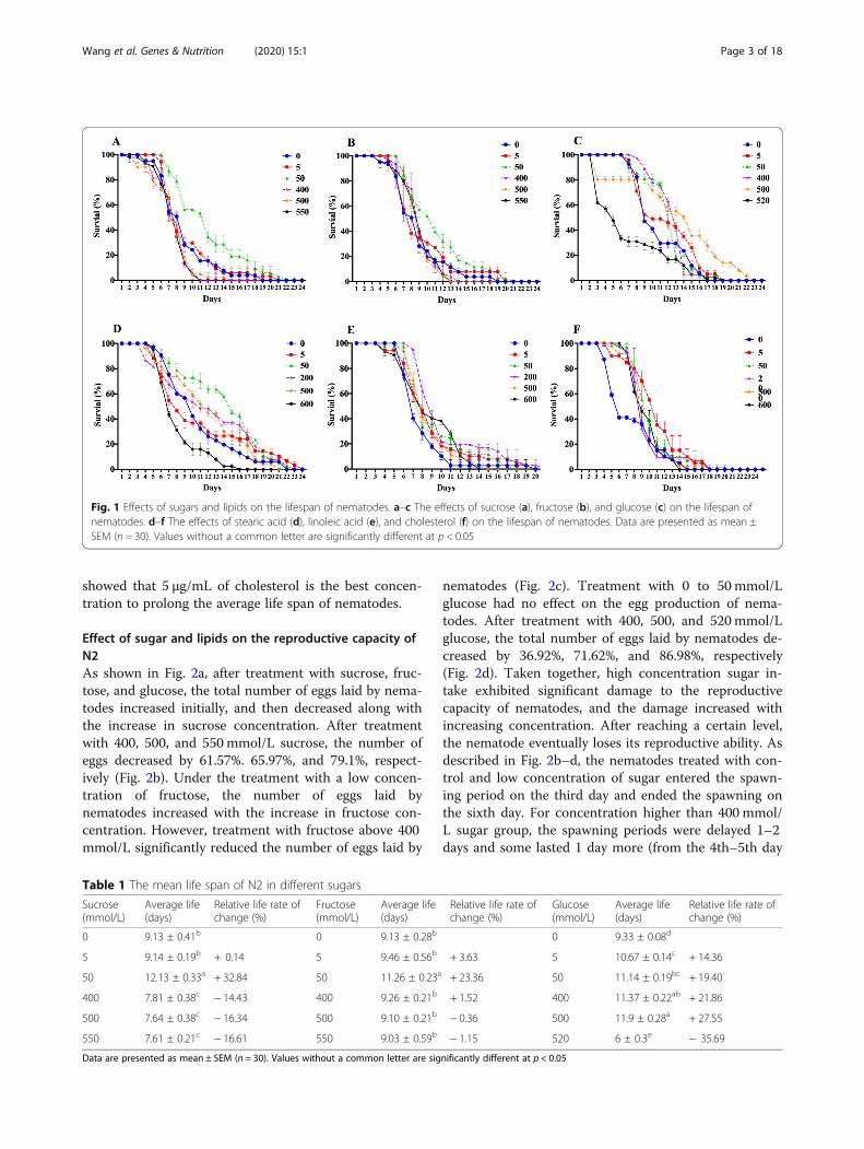
ResultsEffect of sugar and lipids on lifespan of N2Nematodes were treated with sugar at concentrationsranging from 0 to 550 mmol/L and lipid at concentra-tions ranging from 0 to 600 μg/mL. As shown inFig. 1a–c and Table 1, the average lifespan of nematodestreated with different concentrations of sucrose, fructose,and glucose increased initially and then decreased.Treatment with sucrose and fructose at a concentrationof 5 mmol/L had a weak effect on the average lifespan ofnematodes, whereas treatment with 5mmol/L glucosesignificantly prolonged the average lifespan of nema-todes. Treatment with 50mmol/L sucrose, fructose, andglucose significantly prolonged the average lifespan ofnematodes and delayed the onset of death. Treatmentwith sucrose at concentrations above 400 mmol/L sig-nificantly shortened the average lifespan of nematodes,whereas for fructose and glucose, the turning pointswere 500 mmol/L and 520 mmol/L, respectively. This in-dicates that treatment with low concentrations of su-crose, fructose, and glucose prolonged the averagelifespan of nematodes, whereas when the concentrationof sugar reached a certain level, the average lifespan ofnematodes was significantly shortened. Among the threekinds of sugar, sucrose exhibited a relatively narrowrange of concentration that prolonged the lifespan ofnematodes, but glucose had a wider range of said con-centration—5 mmol/L to 500 mmol/L.As shown in Fig. 1d–f and Table 2, the average life-
span of nematodes treated with different concentrationsof stearic acid, linoleic acid, and cholesterol increasedinitially and then decreased. Treatment with 5 μg/mL ofstearic acid and linoleic acid had a weak effect on theaverage lifespan of nematodes, whereas treatment with5 μg/mL of cholesterol significantly prolonged the aver-age lifespan of nematodes. Treatment with 50 μg/mLand 200 μg/mL of stearic acid and linoleic acid signifi-cantly prolonged the average lifespan of nematodes. Asexpected, high concentrations of lipid began to shortenthe lifespan of nematodes. Stearic acid can prolong theaverage life span of nematodes by up to 31.82% at a con-centration of 50 μg/mL, but it decreases the average lifespan severely at a concentration of 600 μg/mL. Althoughlinoleic acid also shows a similar pattern as the othertest substances, it did not decrease the average life spanof nematodes at any higher concentration we used in theexperiments compared to that of controls. As C. eleganscannot synthesize cholesterol itself, 5 μg/mL of choles-terol was added to the control medium in every experi-ment except in the cholesterol test, in which nocholesterol was added to the control medium. Our result
Wang et al. Genes & Nutrition (2020) 15:1 Page 2 of 18
showed that 5 μg/mL of cholesterol is the best concen-tration to prolong the average life span of nematodes.
Effect of sugar and lipids on the reproductive capacity ofN2As shown in Fig. 2a, after treatment with sucrose, fruc-tose, and glucose, the total number of eggs laid by nema-todes increased initially, and then decreased along withthe increase in sucrose concentration. After treatmentwith 400, 500, and 550 mmol/L sucrose, the number ofeggs decreased by 61.57%. 65.97%, and 79.1%, respect-ively (Fig. 2b). Under the treatment with a low concen-tration of fructose, the number of eggs laid bynematodes increased with the increase in fructose con-centration. However, treatment with fructose above 400mmol/L significantly reduced the number of eggs laid by
nematodes (Fig. 2c). Treatment with 0 to 50 mmol/Lglucose had no effect on the egg production of nema-todes. After treatment with 400, 500, and 520mmol/Lglucose, the total number of eggs laid by nematodes de-creased by 36.92%, 71.62%, and 86.98%, respectively(Fig. 2d). Taken together, high concentration sugar in-take exhibited significant damage to the reproductivecapacity of nematodes, and the damage increased withincreasing concentration. After reaching a certain level,the nematode eventually loses its reproductive ability. Asdescribed in Fig. 2b–d, the nematodes treated with con-trol and low concentration of sugar entered the spawn-ing period on the third day and ended the spawning onthe sixth day. For concentration higher than 400 mmol/L sugar group, the spawning periods were delayed 1–2days and some lasted 1 day more (from the 4th–5th day
Fig. 1 Effects of sugars and lipids on the lifespan of nematodes. a–c The effects of sucrose (a), fructose (b), and glucose (c) on the lifespan ofnematodes. d–f The effects of stearic acid (d), linoleic acid (e), and cholesterol (f) on the lifespan of nematodes. Data are presented as mean ±SEM (n = 30). Values without a common letter are significantly different at p < 0.05
Table 1 The mean life span of N2 in different sugars
Sucrose(mmol/L)
Average life(days)
Relative life rate ofchange (%)
Fructose(mmol/L)
Average life(days)
Relative life rate ofchange (%)
Glucose(mmol/L)
Average life(days)
Relative life rate ofchange (%)
0 9.13 ± 0.41b 0 9.13 ± 0.28b 0 9.33 ± 0.08d
5 9.14 ± 0.19b + 0.14 5 9.46 ± 0.56b + 3.63 5 10.67 ± 0.14c + 14.36
50 12.13 ± 0.33a + 32.84 50 11.26 ± 0.23a + 23.36 50 11.14 ± 0.19bc + 19.40
400 7.81 ± 0.38c − 14.43 400 9.26 ± 0.21b + 1.52 400 11.37 ± 0.22ab + 21.86
500 7.64 ± 0.38c − 16.34 500 9.10 ± 0.21b − 0.36 500 11.9 ± 0.28a + 27.55
550 7.61 ± 0.21c − 16.61 550 9.03 ± 0.59b − 1.15 520 6 ± 0.3e − 35.69
Data are presented as mean ± SEM (n = 30). Values without a common letter are significantly different at p < 0.05
Wang et al. Genes & Nutrition (2020) 15:1 Page 3 of 18
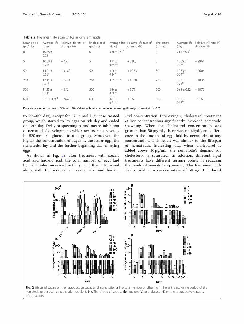
to 7th–8th day), except for 520 mmol/L glucose treatedgroup, which started to lay eggs on 8th day and endedon 12th day. Delay of spawning period means inhibitionof nematodes’ development, which occurs most severelyin 520 mmol/L glucose treated group. Moreover, thehigher the concentration of sugar is, the lesser eggs thenematodes lay and the further beginning day of layingeggs.As shown in Fig. 3a, after treatment with stearic
acid and linoleic acid, the total number of eggs laidby nematodes increased initially, and then, decreasedalong with the increase in stearic acid and linoleic
acid concentration. Interestingly, cholesterol treatmentat low concentrations significantly increased nematodespawning. When the cholesterol concentration wasgreater than 50 μg/mL, there was no significant differ-ence in the amount of eggs laid by nematodes at anyconcentration. This result was similar to the lifespanof nematodes, indicating that when cholesterol isadded above 50 μg/mL, the nematode’s demand forcholesterol is saturated. In addition, different lipidtreatments have different turning points in reducingthe levels of nematode spawning. The treatment withstearic acid at a concentration of 50 μg/mL reduced
Table 2 The mean life span of N2 in different lipids
Stearic acid(μg/mL)
Average life(days)
Relative life rate ofchange (%)
linoleic acid(μg/mL)
Average life(days)
Relative life rate ofchange (%)
cholesterol(μg/mL)
Average life(days)
Relative life rate ofchange (%)
0 10.78 ±0.21c
0 8.36 ± 0.41c 0 7.64 ± 0.3d
5 10.88 ±0.24c
+ 0.93 5 9.11 ±0.65abc
+ 8.96, 5 10.85 ±0.26a
+ 29.61
50 14.21 ±0.52a
+ 31.82 50 9.26 ±0.34ab
+ 10.83 50 10.33 ±0.34ab
+ 26.04
200 12.11 ±0.66b
+ 12.34 200 9.79 ± 0.5a + 17.20 200 9.73 ±0.21bc
+ 10.36
500 11.15 ±0.21c
+ 3.42 500 8.84 ±0.38bc
+ 5.79 500 9.68 ± 0.42c + 10.76
600 8.15 ± 0.36d − 24.40 600 8.83 ±0.21bc
+ 5.60 600 9.77 ±0.36bc
+ 9.96
Data are presented as mean ± SEM (n = 30). Values without a common letter are significantly different at p < 0.05
Fig. 2 Effects of sugars on the reproduction capacity of nematodes. a The total number of offspring in the entire spawning period of thenematode under each concentration gradient. b, c The effects of sucrose (b), fructose (c), and glucose (d) on the reproductive capacityof nematodes
Wang et al. Genes & Nutrition (2020) 15:1 Page 4 of 18
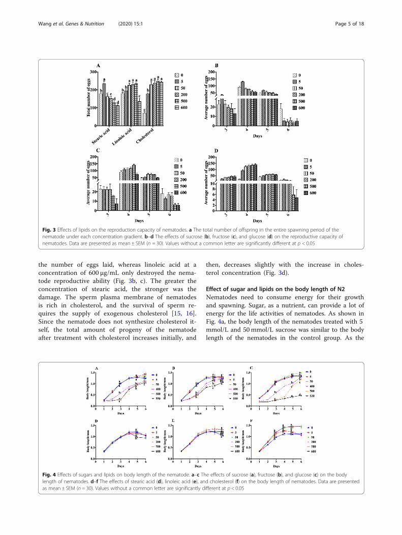
the number of eggs laid, whereas linoleic acid at aconcentration of 600 μg/mL only destroyed the nema-tode reproductive ability (Fig. 3b, c). The greater theconcentration of stearic acid, the stronger was thedamage. The sperm plasma membrane of nematodesis rich in cholesterol, and the survival of sperm re-quires the supply of exogenous cholesterol [15, 16].Since the nematode does not synthesize cholesterol it-self, the total amount of progeny of the nematodeafter treatment with cholesterol increases initially, and
then, decreases slightly with the increase in choles-terol concentration (Fig. 3d).
Effect of sugar and lipids on the body length of N2Nematodes need to consume energy for their growthand spawning. Sugar, as a nutrient, can provide a lot ofenergy for the life activities of nematodes. As shown inFig. 4a, the body length of the nematodes treated with 5mmol/L and 50mmol/L sucrose was similar to the bodylength of the nematodes in the control group. As the
Fig. 3 Effects of lipids on the reproduction capacity of nematodes. a The total number of offspring in the entire spawning period of thenematode under each concentration gradient. b–d The effects of sucrose (b), fructose (c), and glucose (d) on the reproductive capacity ofnematodes. Data are presented as mean ± SEM (n = 30). Values without a common letter are significantly different at p < 0.05
Fig. 4 Effects of sugars and lipids on body length of the nematode. a–c The effects of sucrose (a), fructose (b), and glucose (c) on the bodylength of nematodes. d–f The effects of stearic acid (d), linoleic acid (e), and cholesterol (f) on the body length of nematodes. Data are presentedas mean ± SEM (n = 30). Values without a common letter are significantly different at p < 0.05
Wang et al. Genes & Nutrition (2020) 15:1 Page 5 of 18
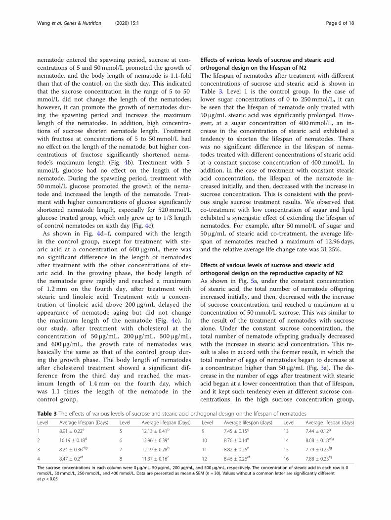
nematode entered the spawning period, sucrose at con-centrations of 5 and 50 mmol/L promoted the growth ofnematode, and the body length of nematode is 1.1-foldthan that of the control, on the sixth day. This indicatedthat the sucrose concentration in the range of 5 to 50mmol/L did not change the length of the nematodes;however, it can promote the growth of nematodes dur-ing the spawning period and increase the maximumlength of the nematodes. In addition, high concentra-tions of sucrose shorten nematode length. Treatmentwith fructose at concentrations of 5 to 50 mmol/L hadno effect on the length of the nematode, but higher con-centrations of fructose significantly shortened nema-tode’s maximum length (Fig. 4b). Treatment with 5mmol/L glucose had no effect on the length of thenematode. During the spawning period, treatment with50mmol/L glucose promoted the growth of the nema-tode and increased the length of the nematode. Treat-ment with higher concentrations of glucose significantlyshortened nematode length, especially for 520 mmol/Lglucose treated group, which only grew up to 1/3 lengthof control nematodes on sixth day (Fig. 4c).As shown in Fig. 4d–f, compared with the length
in the control group, except for treatment with ste-aric acid at a concentration of 600 μg/mL, there wasno significant difference in the length of nematodesafter treatment with the other concentrations of ste-aric acid. In the growing phase, the body length ofthe nematode grew rapidly and reached a maximumof 1.2 mm on the fourth day, after treatment withstearic and linoleic acid. Treatment with a concen-tration of linoleic acid above 200 μg/mL delayed theappearance of nematode aging but did not changethe maximum length of the nematode (Fig. 4e). Inour study, after treatment with cholesterol at theconcentration of 50 μg/mL, 200 μg/mL, 500 μg/mL,and 600 μg/mL, the growth rate of nematodes wasbasically the same as that of the control group dur-ing the growth phase. The body length of nematodesafter cholesterol treatment showed a significant dif-ference from the third day and reached the max-imum length of 1.4 mm on the fourth day, whichwas 1.1 times the length of the nematode in thecontrol group.
Effects of various levels of sucrose and stearic acidorthogonal design on the lifespan of N2The lifespan of nematodes after treatment with differentconcentrations of sucrose and stearic acid is shown inTable 3. Level 1 is the control group. In the case oflower sugar concentrations of 0 to 250 mmol/L, it canbe seen that the lifespan of nematode only treated with50 μg/mL stearic acid was significantly prolonged. How-ever, at a sugar concentration of 400 mmol/L, an in-crease in the concentration of stearic acid exhibited atendency to shorten the lifespan of nematodes. Therewas no significant difference in the lifespan of nema-todes treated with different concentrations of stearic acidat a constant sucrose concentration of 400mmol/L. Inaddition, in the case of treatment with constant stearicacid concentration, the lifespan of the nematode in-creased initially, and then, decreased with the increase insucrose concentration. This is consistent with the previ-ous single sucrose treatment results. We observed thatco-treatment with low concentration of sugar and lipidexhibited a synergistic effect of extending the lifespan ofnematodes. For example, after 50 mmol/L of sugar and50 μg/mL of stearic acid co-treatment, the average life-span of nematodes reached a maximum of 12.96 days,and the relative average life change rate was 31.25%.
Effects of various levels of sucrose and stearic acidorthogonal design on the reproductive capacity of N2As shown in Fig. 5a, under the constant concentrationof stearic acid, the total number of nematode offspringincreased initially, and then, decreased with the increaseof sucrose concentration, and reached a maximum at aconcentration of 50 mmol/L sucrose. This was similar tothe result of the treatment of nematodes with sucrosealone. Under the constant sucrose concentration, thetotal number of nematode offspring gradually decreasedwith the increase in stearic acid concentration. This re-sult is also in accord with the former result, in which thetotal number of eggs of nematodes began to decrease ata concentration higher than 50 μg/mL (Fig. 3a). The de-crease in the number of eggs after treatment with stearicacid began at a lower concentration than that of lifespan,and it kept such tendency even at different sucrose con-centrations. In the high sucrose concentration group,
Table 3 The effects of various levels of sucrose and stearic acid orthogonal design on the lifespan of nematodes
Level Average lifespan (Days) Level Average lifespan (Days) Level Average lifespan (days) Level Average lifespan (days)
1 8.91 ± 0.22e 5 12.13 ± 0.41b 9 7.45 ± 0.15g 13 7.44 ± 0.12g
2 10.19 ± 0.18d 6 12.96 ± 0.39a 10 8.76 ± 0.14e 14 8.08 ± 0.18efg
3 8.24 ± 0.36efg 7 12.19 ± 0.28b 11 8.82 ± 0.26e 15 7.79 ± 0.25fg
4 8.47 ± 0.2ef 8 11.37 ± 0.16c 12 8.46 ± 0.26ef 16 7.88 ± 0.23fg
The sucrose concentrations in each column were 0 μg/mL, 50 μg/mL, 200 μg/mL, and 500 μg/mL, respectively. The concentration of stearic acid in each row is 0mmol/L, 50 mmol/L, 250 mmol/L, and 400 mmol/L. Data are presented as mean ± SEM (n = 30). Values without a common letter are significantly differentat p < 0.05
Wang et al. Genes & Nutrition (2020) 15:1 Page 6 of 18
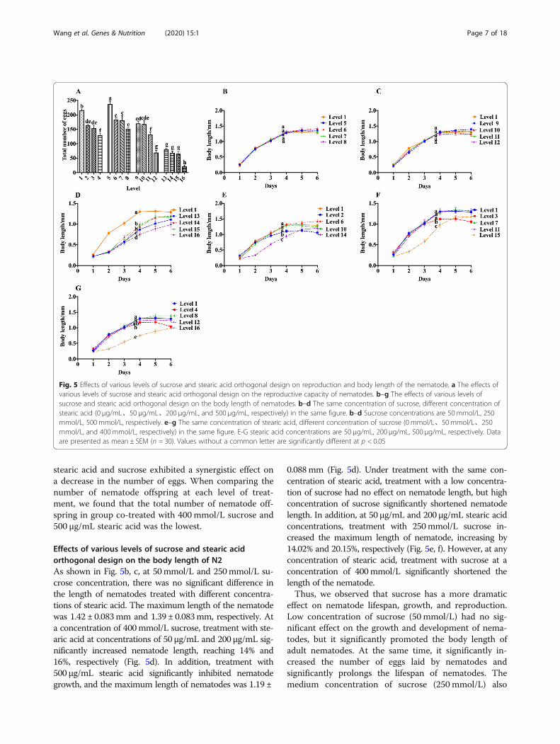
stearic acid and sucrose exhibited a synergistic effect ona decrease in the number of eggs. When comparing thenumber of nematode offspring at each level of treat-ment, we found that the total number of nematode off-spring in group co-treated with 400 mmol/L sucrose and500 μg/mL stearic acid was the lowest.
Effects of various levels of sucrose and stearic acidorthogonal design on the body length of N2As shown in Fig. 5b, c, at 50mmol/L and 250mmol/L su-crose concentration, there was no significant difference inthe length of nematodes treated with different concentra-tions of stearic acid. The maximum length of the nematodewas 1.42 ± 0.083mm and 1.39 ± 0.083mm, respectively. Ata concentration of 400mmol/L sucrose, treatment with ste-aric acid at concentrations of 50 μg/mL and 200 μg/mL sig-nificantly increased nematode length, reaching 14% and16%, respectively (Fig. 5d). In addition, treatment with500 μg/mL stearic acid significantly inhibited nematodegrowth, and the maximum length of nematodes was 1.19 ±
0.088mm (Fig. 5d). Under treatment with the same con-centration of stearic acid, treatment with a low concentra-tion of sucrose had no effect on nematode length, but highconcentration of sucrose significantly shortened nematodelength. In addition, at 50 μg/mL and 200 μg/mL stearic acidconcentrations, treatment with 250mmol/L sucrose in-creased the maximum length of nematode, increasing by14.02% and 20.15%, respectively (Fig. 5e, f). However, at anyconcentration of stearic acid, treatment with sucrose at aconcentration of 400mmol/L significantly shortened thelength of the nematode.Thus, we observed that sucrose has a more dramatic
effect on nematode lifespan, growth, and reproduction.Low concentration of sucrose (50 mmol/L) had no sig-nificant effect on the growth and development of nema-todes, but it significantly promoted the body length ofadult nematodes. At the same time, it significantly in-creased the number of eggs laid by nematodes andsignificantly prolongs the lifespan of nematodes. Themedium concentration of sucrose (250 mmol/L) also
Fig. 5 Effects of various levels of sucrose and stearic acid orthogonal design on reproduction and body length of the nematode. a The effects ofvarious levels of sucrose and stearic acid orthogonal design on the reproductive capacity of nematodes. b–g The effects of various levels ofsucrose and stearic acid orthogonal design on the body length of nematodes. b–d The same concentration of sucrose, different concentration ofstearic acid (0 μg/mL、50 μg/mL、200 μg/mL, and 500 μg/mL, respectively) in the same figure. b–d Sucrose concentrations are 50 mmol/L, 250mmol/L, 500 mmol/L, respectively. e–g The same concentration of stearic acid, different concentration of sucrose (0 mmol/L、50 mmol/L、250mmol/L, and 400mmol/L, respectively) in the same figure. E-G stearic acid concentrations are 50 μg/mL, 200 μg/mL, 500 μg/mL, respectively. Dataare presented as mean ± SEM (n = 30). Values without a common letter are significantly different at p < 0.05
Wang et al. Genes & Nutrition (2020) 15:1 Page 7 of 18
promoted the growth of nematode adults, but has nosignificant effect on the number of eggs. However, highconcentrations of sucrose (400 mmol/L) significantly re-duced the number of eggs and shortened the lifespan ofnematodes.The effect of stearic acid on nematodes is less promin-
ent than that of sucrose. It also prolonged the lifespan ofnematodes at low concentrations (50 μg/mL) andworked synergistically with 50 mmol/L sucrose. More-over, it showed inhibition of nematode reproductionability at each gradient sucrose concentration. Further-more, its effect on the growth and development of nem-atodes and adult body length was not significant. Thedecrease in the lifespan of nematodes after treatmentwith a high concentration of stearic acid (400 μg/mL)was much less than that after sucrose treatment (4.94%vs. 16.5%). However, when it is co-treated with sucrose,the growth and development of nematodes, the length ofadult worms, and the number of eggs laid, are more sig-nificantly inhibited.
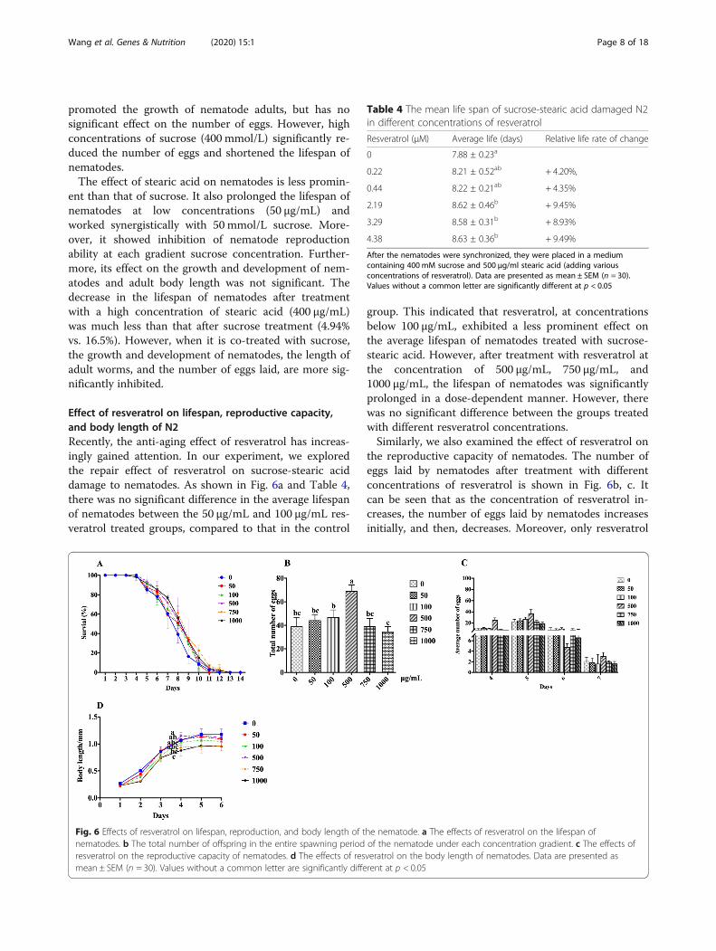
Effect of resveratrol on lifespan, reproductive capacity,and body length of N2Recently, the anti-aging effect of resveratrol has increas-ingly gained attention. In our experiment, we exploredthe repair effect of resveratrol on sucrose-stearic aciddamage to nematodes. As shown in Fig. 6a and Table 4,there was no significant difference in the average lifespanof nematodes between the 50 μg/mL and 100 μg/mL res-veratrol treated groups, compared to that in the control
group. This indicated that resveratrol, at concentrationsbelow 100 μg/mL, exhibited a less prominent effect onthe average lifespan of nematodes treated with sucrose-stearic acid. However, after treatment with resveratrol atthe concentration of 500 μg/mL, 750 μg/mL, and1000 μg/mL, the lifespan of nematodes was significantlyprolonged in a dose-dependent manner. However, therewas no significant difference between the groups treatedwith different resveratrol concentrations.Similarly, we also examined the effect of resveratrol on
the reproductive capacity of nematodes. The number ofeggs laid by nematodes after treatment with differentconcentrations of resveratrol is shown in Fig. 6b, c. Itcan be seen that as the concentration of resveratrol in-creases, the number of eggs laid by nematodes increasesinitially, and then, decreases. Moreover, only resveratrol
Fig. 6 Effects of resveratrol on lifespan, reproduction, and body length of the nematode. a The effects of resveratrol on the lifespan ofnematodes. b The total number of offspring in the entire spawning period of the nematode under each concentration gradient. c The effects ofresveratrol on the reproductive capacity of nematodes. d The effects of resveratrol on the body length of nematodes. Data are presented asmean ± SEM (n = 30). Values without a common letter are significantly different at p < 0.05
Table 4 The mean life span of sucrose-stearic acid damaged N2in different concentrations of resveratrol
Resveratrol (μM) Average life (days) Relative life rate of change
0 7.88 ± 0.23a
0.22 8.21 ± 0.52ab + 4.20%,
0.44 8.22 ± 0.21ab + 4.35%
2.19 8.62 ± 0.46b + 9.45%
3.29 8.58 ± 0.31b + 8.93%
4.38 8.63 ± 0.36b + 9.49%
After the nematodes were synchronized, they were placed in a mediumcontaining 400 mM sucrose and 500 μg/ml stearic acid (adding variousconcentrations of resveratrol). Data are presented as mean ± SEM (n = 30).Values without a common letter are significantly different at p < 0.05
Wang et al. Genes & Nutrition (2020) 15:1 Page 8 of 18
treatment at a concentration of 500 mg/mL led to sig-nificant differences in the number of eggs laid comparedto those in the control group.We determined the reparative effect of resveratrol on
the sucrose-stearic acid effect to shorten the length ofnematodes. Contrary to what we expected, co-treatmentwith different concentrations of resveratrol had a syner-gistic inhibitory effect on the inhibition of nematodegrowth, which is particularly evident in the growth phaseof the nematode (Fig. 6d). In addition, we observed thatresveratrol treatment at a concentration of 1000 μg/mLnot only severely inhibited nematode development, butalso significantly shortened the body length of adults.
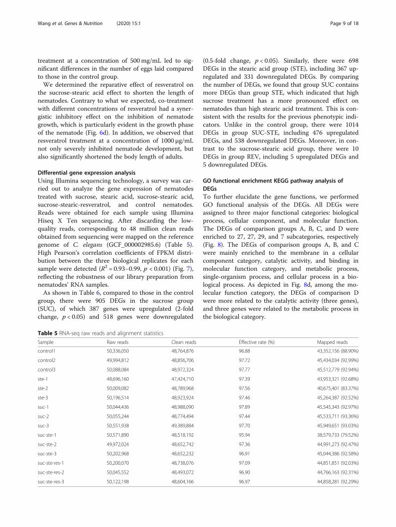
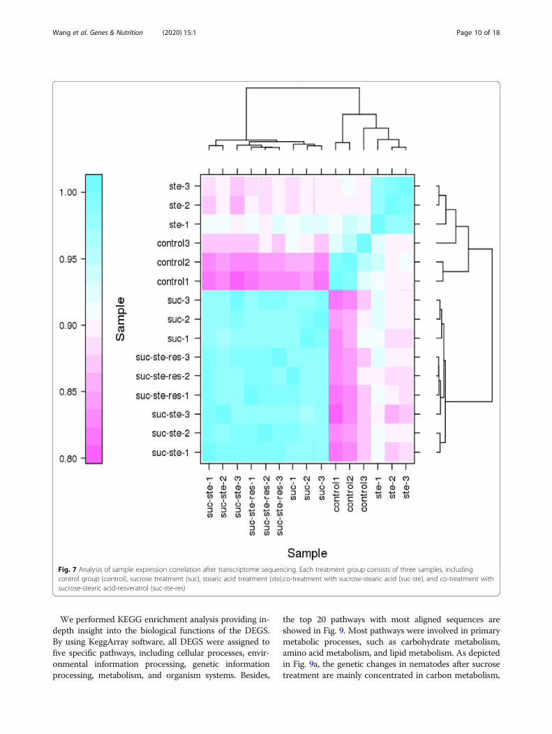
Differential gene expression analysisUsing Illumina sequencing technology, a survey was car-ried out to analyze the gene expression of nematodestreated with sucrose, stearic acid, sucrose-stearic acid,sucrose-stearic-resveratrol, and control nematodes.Reads were obtained for each sample using IlluminaHiseq X Ten sequencing. After discarding the low-quality reads, corresponding to 48 million clean readsobtained from sequencing were mapped on the referencegenome of C. elegans (GCF_000002985.6) (Table 5).High Pearson’s correlation coefficients of FPKM distri-bution between the three biological replicates for eachsample were detected (R2 = 0.93–0.99, p < 0.001) (Fig. 7),reflecting the robustness of our library preparation fromnematodes’ RNA samples.As shown in Table 6, compared to those in the control
group, there were 905 DEGs in the sucrose group(SUC), of which 387 genes were upregulated (2-foldchange, p < 0.05) and 518 genes were downregulated
(0.5-fold change, p < 0.05). Similarly, there were 698DEGs in the stearic acid group (STE), including 367 up-regulated and 331 downregulated DEGs. By comparingthe number of DEGs, we found that group SUC containsmore DEGs than group STE, which indicated that highsucrose treatment has a more pronounced effect onnematodes than high stearic acid treatment. This is con-sistent with the results for the previous phenotypic indi-cators. Unlike in the control group, there were 1014DEGs in group SUC-STE, including 476 upregulatedDEGs, and 538 downregulated DEGs. Moreover, in con-trast to the sucrose-stearic acid group, there were 10DEGs in group REV, including 5 upregulated DEGs and5 downregulated DEGs.
GO functional enrichment KEGG pathway analysis ofDEGsTo further elucidate the gene functions, we performedGO functional analysis of the DEGs. All DEGs wereassigned to three major functional categories: biologicalprocess, cellular component, and molecular function.The DEGs of comparison groups A, B, C, and D wereenriched to 27, 27, 29, and 7 subcategories, respectively(Fig. 8). The DEGs of comparison groups A, B, and Cwere mainly enriched to the membrane in a cellularcomponent category, catalytic activity, and binding inmolecular function category, and metabolic process,single-organism process, and cellular process in a bio-logical process. As depicted in Fig. 8d, among the mo-lecular function category, the DEGs of comparison Dwere more related to the catalytic activity (three genes),and three genes were related to the metabolic process inthe biological category.
Table 5 RNA-seq raw reads and alignment statistics
Sample Raw reads Clean reads Effective rate (%) Mapped reads
control1 50,336,050 48,764,876 96.88 43,352,156 (88.90%)
control2 49,994,812 48,856,706 97.72 45,434,034 (92.99%)
control3 50,088,084 48,972,324 97.77 45,512,779 (92.94%)
ste-1 48,696,160 47,424,710 97.39 43,953,321 (92.68%)
ste-2 50,009,082 48,789,968 97.56 40,675,401 (83.37%)
ste-3 50,196,514 48,923,924 97.46 45,264,387 (92.52%)
suc-1 50,044,436 48,988,090 97.89 45,545,343 (92.97%)
suc-2 50,055,244 48,774,494 97.44 45,533,711 (93.36%)
suc-3 50,551,938 49,389,884 97.70 45,949,651 (93.03%)
suc-ste-1 50,571,890 48,518,192 95.94 38,579,733 (79.52%)
suc-ste-2 49,972,024 48,652,742 97.36 44,991,273 (92.47%)
suc-ste-3 50,202,968 48,652,232 96.91 45,044,386 (92.58%)
suc-ste-res-1 50,200,070 48,738,076 97.09 44,851,851 (92.03%)
suc-ste-res-2 50,045,552 48,493,072 96.90 44,766,163 (92.31%)
suc-ste-res-3 50,122,198 48,604,166 96.97 44,858,281 (92.29%)
Wang et al. Genes & Nutrition (2020) 15:1 Page 9 of 18
We performed KEGG enrichment analysis providing in-depth insight into the biological functions of the DEGS.By using KeggArray software, all DEGS were assigned tofive specific pathways, including cellular processes, envir-onmental information processing, genetic informationprocessing, metabolism, and organism systems. Besides,
the top 20 pathways with most aligned sequences areshowed in Fig. 9. Most pathways were involved in primarymetabolic processes, such as carbohydrate metabolism,amino acid metabolism, and lipid metabolism. As depictedin Fig. 9a, the genetic changes in nematodes after sucrosetreatment are mainly concentrated in carbon metabolism,
Fig. 7 Analysis of sample expression correlation after transcriptome sequencing. Each treatment group consists of three samples, includingcontrol group (control), sucrose treatment (suc), stearic acid treatment (ste),co-treatment with sucrose-stearic acid (suc-ste), and co-treatment withsucrose-stearic acid-resveratrol (suc-ste-res)
Wang et al. Genes & Nutrition (2020) 15:1 Page 10 of 18
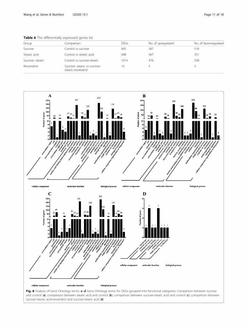
Fig. 8 Analysis of Gene Ontology terms. a–d Gene Ontology terms for DEGs grouped into functional categories. Comparison between sucroseand control (a); comparison between stearic acid and control (b); comparison between sucrose-stearic acid and control (c); comparison betweensucrose-stearic acid-resveratrol and sucrose-stearic acid (d)
Table 6 The differentially expressed genes list
Group Comparison DEGs No. of upregulated No. of downregulated
Sucrose Control vs sucrose 905 387 518
Stearic acid Control vs stearic acid 698 367 331
Sucrose- stearic Control vs sucrose-stearic 1014 476 538
Resveratrol Sucrose- stearic vs sucrose-stearic-resveratrol
10 5 5
Wang et al. Genes & Nutrition (2020) 15:1 Page 11 of 18
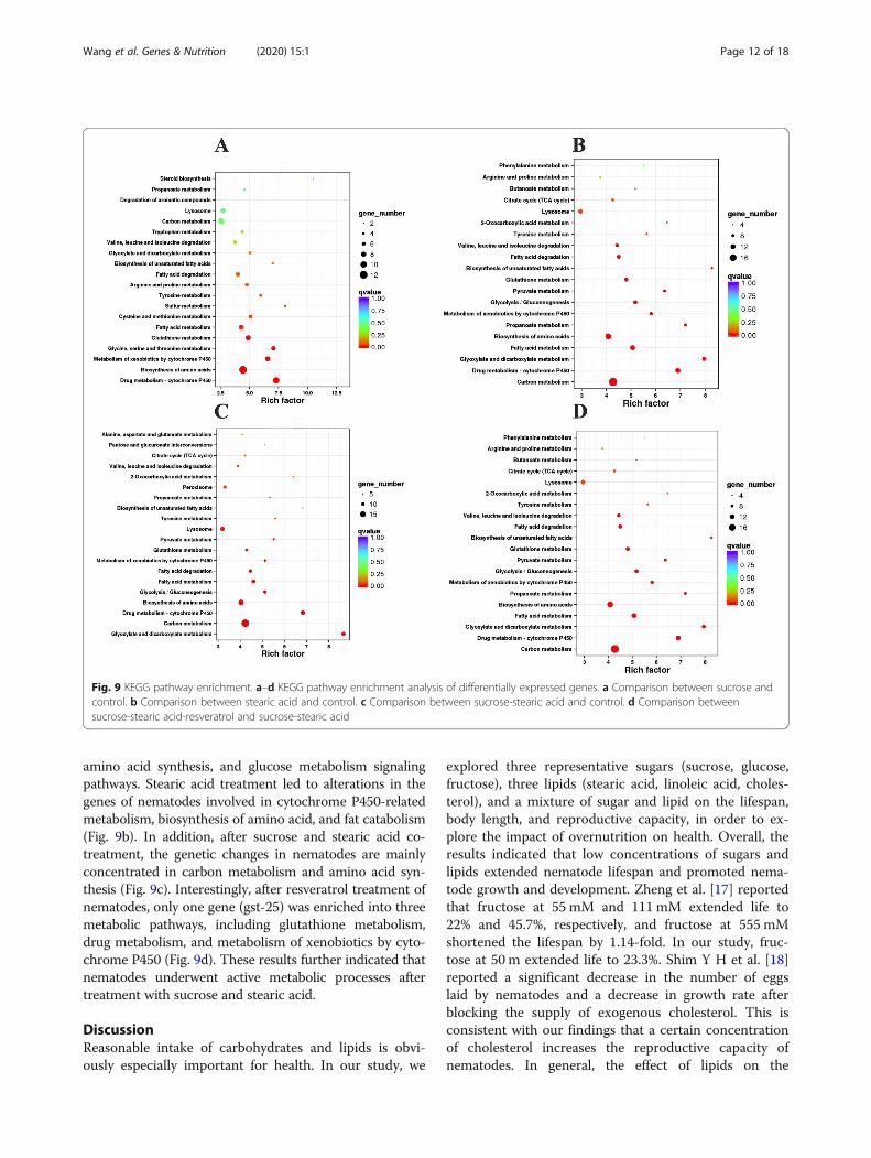
amino acid synthesis, and glucose metabolism signalingpathways. Stearic acid treatment led to alterations in thegenes of nematodes involved in cytochrome P450-relatedmetabolism, biosynthesis of amino acid, and fat catabolism(Fig. 9b). In addition, after sucrose and stearic acid co-treatment, the genetic changes in nematodes are mainlyconcentrated in carbon metabolism and amino acid syn-thesis (Fig. 9c). Interestingly, after resveratrol treatment ofnematodes, only one gene (gst-25) was enriched into threemetabolic pathways, including glutathione metabolism,drug metabolism, and metabolism of xenobiotics by cyto-chrome P450 (Fig. 9d). These results further indicated thatnematodes underwent active metabolic processes aftertreatment with sucrose and stearic acid.
DiscussionReasonable intake of carbohydrates and lipids is obvi-ously especially important for health. In our study, we
explored three representative sugars (sucrose, glucose,fructose), three lipids (stearic acid, linoleic acid, choles-terol), and a mixture of sugar and lipid on the lifespan,body length, and reproductive capacity, in order to ex-plore the impact of overnutrition on health. Overall, theresults indicated that low concentrations of sugars andlipids extended nematode lifespan and promoted nema-tode growth and development. Zheng et al. [17] reportedthat fructose at 55 mM and 111mM extended life to22% and 45.7%, respectively, and fructose at 555 mMshortened the lifespan by 1.14-fold. In our study, fruc-tose at 50 m extended life to 23.3%. Shim Y H et al. [18]reported a significant decrease in the number of eggslaid by nematodes and a decrease in growth rate afterblocking the supply of exogenous cholesterol. This isconsistent with our findings that a certain concentrationof cholesterol increases the reproductive capacity ofnematodes. In general, the effect of lipids on the
Fig. 9 KEGG pathway enrichment. a–d KEGG pathway enrichment analysis of differentially expressed genes. a Comparison between sucrose andcontrol. b Comparison between stearic acid and control. c Comparison between sucrose-stearic acid and control. d Comparison betweensucrose-stearic acid-resveratrol and sucrose-stearic acid
Wang et al. Genes & Nutrition (2020) 15:1 Page 12 of 18
spawning of nematodes was not as severe as the effect ofsugar. Since nematodes need to consume energy duringgrowth and development, a certain amount of sugar andlipids was used to provide nutrients for nematode life ac-tivities. As we guessed, excessive sugars and lipids treat-ment severely shortened the lifespan, body length of thenematode (Additional file 1: Figure S1) and destroys itsreproductive capacity. Interestingly, we found that sugarhad an adverse effect on nematodes at low to mediumconcentrations, while lipids damaged to nematodes atmoderate to high concentrations. In other words, sugarhas a stronger effect on nematodes than lipids. Sequen-cing data also corroborated this result, because thedifferential genes generated by sugar treatment are sig-nificantly more than lipids (Table 6). In addition, the re-sults of orthogonal tests indicated that co-treatmentwith high concentrations of sucrose and stearic acid hada synergistic effect on nematode damage. We examinedthe effects of excess sucrose and stearic acid on intes-tinal autophagy in nematodes. Immunofluorescence re-sults show nematode cells were negative with anti-Caspase (Additional file 1: Figure S6). Moreover, we fur-ther explored the mechanism of high sucrose and highstearic acid damage on C. elegans and the repair effect ofresveratrol using transcriptome sequencing technology.After treatment with sucrose, stearic acid, and sucrose-stearic acid, a total of 905,698 and 1014 DEGS wereidentified, respectively. It suggests that high-sucrose andhigh-stearic acid treatment causes an imbalance innematode glycolipid metabolism by altering the expres-sion of several genes.
Over-nutrition converts fat storage and exacerbates β-oxidations of fatty acidsThe regulation of lipid metabolism in C. elegans is influ-enced by the environment, such as temperature and nu-trient deficiencies, as well as its own physiological state,including growth, reproduction, development, and aging[19]. Moreover, the nematode body undergoes rapidchanges to produce an adaptive response to this stimu-lus. In our experiments, high sugar and high fat provideexcess nutrients to the nematodes. In addition to diges-tion and absorption to meet basic life activities, nema-todes store excess energy in the form of lipid droplets.In early embryos, lipid droplets are abundant, providingprecursors for membrane synthesis during rapid cell div-ision as well as energy for cellular processes until hatch-ing. TAGs are the major component of lipid droplets, aseffective energy storage molecules due to their reducedstate. During fat synthesis, diacylglycerol acyltransferaseencoded by dgat-2 catalyzes the formation of TAG fromfatty acyl-CoA and diacylglycerols [20]. After high-sucrose treatment and high-stearic acid treatment, nem-atodes convert excess nutrients into fat, by upregulating
dgat-2 genes involved in TAGs synthesis. In addition,vit-2, which is involved in the transfer of dietary lipids tolipid droplets, promoting fat accumulation, was upregu-lated after high-sucrose treatment and high-stearic acidtreatment.Fatty acids are separated from TAGs molecules, releas-
ing energy by β-oxidations. Fatty acids need to be acti-vated before they enter the mitochondria for oxidation,which involves four enzymes, which are acyl-CoA de-hydrogenase in the mitochondria or acyl-CoA oxidase inperoxisomes, enoyl-CoA hydratase, 3-hydroxylacyl-CoAdehydrogenase, and 3-ketoacyl-CoA thiolase [21]. Thegenes encoding these enzymes, including acox-1, maoc-1,dhs-28, and daf-22, were upregulated in our results, whichindicated that high-intensity energy metabolism was beingcarried out in the nematode (Additional file 1: Figure S2).
Fatty acids are synthesized de novo for growth,development, spawning, and signaling moleculesThe characteristic of lipid metabolism in C. elegans is thesynthesis of fatty acids de novo from acetyl-CoA. Inaddition to the oxidation of fatty acids to produce acetyl-CoA, other nutrients, such as carbohydrates and aminoacids, can be broken down into acetyl-CoA for de novofatty acid synthesis. During fatty acid synthesis, the pod-2encoded ACC enzyme limits acetyl-CoA to malonyl-CoAtransformation [22]. In the second step, the de novo synthe-sis of the fatty acyl chain by the two-carbon subunit acetyl-CoA is accomplished by the catalysis of a fatty acid synthaseencoded by fasn-1 [23]. In our study, high-sucrose treat-ment, high-stearic acid treatment, high-sucrose, and high-stearic acid co-treatment did not affect the expression ofpod-2 and fasn-1 genes.Nematodes are rich in polyunsaturated fatty acids
(PUFAs), produced by desaturation. There are fourfatty acid desaturases that convert 18:1n-9 into a seriesof C18 and C20 PUFAs, including FAT1 (Δ12), FAT2(Δ12), FAT3 (Δ12), and FAT4 (Δ5) [24]. These poly-unsaturated fatty acids provide precursors for thegrowth and reproduction of nematodes and are usedto synthesize fat. C. elegans, which is severely deficientin polyunsaturated fatty acids, exhibits many growth,reproduction, and neurological deficits. The Δ12 desa-turase fat-2 mutant contained only 1% PUFAs. Thesemutants grow slowly, have smaller embryos, and ex-hibit less coordinated motion than wild-type individ-uals [25]. The Δ6 desaturase fat-3 mutant containsC18 PUFAs but does not contain C20 PUFAs. Al-though they grew better than the fat-2 mutant andshowed a higher brood size, they showed many defectscompared to the wild type [26]. Fat-4 and fat-1 mu-tants contain different types of PUFAs and differentproportions of omega-6 and omega-3, although theirgrowth, development, and reproduction are largely
Wang et al. Genes & Nutrition (2020) 15:1 Page 13 of 18
unaffected [27]. In our experiments, high sucrosetreatment and high-stearic acid treatment significantlyupregulated genes encoding desaturase, including fat-1, fat-2, fat-3, fat-4, and fat-5 (Additional file 1: FigureS3). This indicated that nematodes produce largeamounts of PUFAs for growth and developmentthrough desaturation. The results of KEGG alsodemonstrated that high-stearic acid treatment leads tonematode lipid metabolism and decompositiondisorders, affecting the growth and development ofnematodes.In addition to affecting the growth and development of
nematodes, PUFAs are also used as signal molecules, re-leased from the membrane by phospholipase hydrolysisand further metabolized to form signaling molecules, col-lectively known as eicosanoids [28]. In mammals, thesynthesis of eicosanoids requires the participation of cyclo-oxygenase, lipoxygenase, and cytochrome P450 enzymes[29]. The cyp-gene family is reported to be responsible forencoding cytochrome P450s, NADPH-dependent monoox-ygenases that metabolize endogenous and exogenous com-pounds [30]. Sucrose treatment, stearic acid treatment, andsucrose-stearic acid co-treatment of nematodes downregu-lated cyp-gene expression, such as cyp-29A3, cyp-14A3, andcyp-35A4 and interfered with metabolism of nematodes.
Increased glucose metabolism shortens nematode lifeMonosaccharides are directly absorbed in the body’s me-tabolism. The disaccharide or polysaccharide is hydro-lyzed into glucose, which participates in glycolysis toprovide energy to the body, or is stored as a glycogen. Inmammals, glucose transport and absorption are medi-ated by GLUTs and insulin signaling. The fgt-1 gene isassociated with nematode glucose uptake, and there havebeen reports that RNAi-mediated knockdown of fgt-1extends lifespan of nematodes [31, 32]. Previous studiesindicated that inhibition of the glycolytic enzyme, glu-cose phosphate isomerase 1 (GPI-1), prolongs lifespan.Feng et al. [31] showed that disrupting glucose transport,by inhibiting fgt-1, is associated with age-1 and daf-2 sig-naling to extend nematode lifespan. These previousstudies also suggested that reduced glucose metabolismpromotes longevity. In our experiments, the expressionof fgt-1 and daf-2 genes was upregulated in the sucrosetreatment group and sucrose-stearic acid co-treatmentgroup, while stearic acid treatment had no effect. Thisindicated that the addition of sucrose increased themetabolic burden of nematodes, resulting in a shortenedlife.
Genes involved in the TGF-β signaling pathwayTransforming growth factor-β (TGF-β) superfamily li-gands participate in cell identify, growth, and develop-ment. In C. elegans, five such ligands have been
identified, including dbl-1, daf-7, unc-129, tig-2, andtig-3. Here, we only discussed dbl-1 and daf-7 signal-ing pathway, because their function has beenexplained more clearly. The core components of thedbl-1 pathway are the dbl-1 ligand, daf-4 and sma-6receptors, and sma-2, sma-3, and sma-4 intercellularsignals. Studies have shown that the lack of dbl-1 sig-naling pathway leads to small body size and male tailabnormal morphology [33]. In our gene expressionprofile, high-stearic acid treatment upregulated geneexpression levels of dbl-1, daf-4, sma-10, and sma-6,and high-sucrose treatment slightly upregulated theexpression of these genes, which suggested that dbl-1signal was enhanced. Furthermore, the expression ofthe intercellular signals sma-2, sma-3, and sma-4 wasupregulated after high-stearic acid treatment, whereassma-2 and sma-4 were downregulated in the high-sucrose treatment group (Additional file 1: Figure S4).This may be related to high-sucrose-induced shorten-ing of nematode length. In addition, it has been re-ported that overexpression of the dbl-1 gene shortensthe lifespan of nematodes [34]. Both high-sucrose andhigh-stearic acid treatment enhanced the dbl-1 signal-ing pathway, which may be responsible for the short-ened lifespan of nematodes caused by high sucroseand high stearic acid. Daf-7, a ligand for the TGF-βsignaling pathway, is involved in regulating nematodesentering the dauer phase. The core components ofdaf-7 pathway are the daf-7 ligand, daf-1 and daf-4 re-ceptors, daf-8, daf-3, and daf-14 transcription factors[35]. Our results indicated that high-stearic acid treat-ment significantly upregulated the gene expression ofdaf-7, but high sucrose significantly inhibited its ex-pression, suggesting that high sucrose may cause somestress on nematodes. There was no significant changein the expression levels of receptors daf-1 of daf-7,and there was a significant increase in daf-4 expres-sion. Daf-8 and daf-14 act as signaling molecules, bothof which are upregulated under high-stearic acid treat-ment and downregulated under high sucrose treat-ment, similar to daf-7 (Additional file 1: Figure S5).This indicated that high stearic acid inhibited nema-todes from entering the dauer phase, while high su-crose may cause certain stress, which might promoteentry of nematodes into the dauer phase.
Genes involved in the insulin signaling pathwayThe C. elegans insulin signaling pathway links energymetabolism with life activities, including growth, devel-opment, reproductive, longevity, and behavior [36]. Thisfundamental pathway is regulated by insulin-like peptide(ILPs) ligands that bind to the insulin/IGF-1 transmem-brane receptor (IGFR) ortholog daf-2. The main compo-nents of the C. elegans insulin signaling pathway include
Wang et al. Genes & Nutrition (2020) 15:1 Page 14 of 18
ILPs [37]. Several ILPs have been shown to be involvedin growth, longevity, and dauer formation of nematodes,such as daf-28 and ins gene family. In our study, wefound that sucrose treatment and stearic acid treatment,sucrose-stearic acid co-treatment, and resveratrol treat-ment had no effect on insulin signaling pathway-relatedgenes (daf-2, age-1, akt-1, ddl-1, hsf-1, and daf-16), ex-cept for the lipid treatment which upregulated daf-2.We only evaluated the gene expression profile of anematode before it entered the spawning stage, andmore experiments are needed to further investigate howsugar and lipids affect the lifespan of the nematode. InC. elegans, skn-1, the ortholog of Nrf-2, downstreamregulator of daf-2, is required for both oxidative stressresistance and anti-aging through its accumulation inthe intestinal nuclei to promote the detoxication targetgenes [38]. Stearic acid treatment significantly upregu-lated the expression of ins-27, ins-33, daf-2, and skn-1genes. Intriguingly, sucrose treatment and sucrose-stearic acid co-treatment significantly downregulatedskn-1 gene expression. This was also consistent with thephenotypic results where stearic acid was less harmful tonematode life, reproductive capacity, and body length,compared to sucrose. In addition, sugar and lipid treat-ments downregulated genes (gst gene family and ugtgene family) related to oxidative stress. Furthermore, inour gene expression profile, the acdh-1 gene encodingthe short-chain acyl-CoA dehydrogenase in mitochon-dria was upregulated after high glucose and high fattreatment. This may result in increased mitochondrialactivity, increased rate of oxidative phosphorylation, in-creased metabolism, and reduced lifespan.
Resveratrol protects sugar and lipid damage tonematodesThe natural active substance resveratrol has been provedto have antioxidant, delay aging, antibacterial, anti-inflammatory, and other biological activities [39]. How-ever, the protective effect of resveratrol on sugar andlipid damage and its mechanism of action are still un-clear. Therefore, we explored the protective effect of res-veratrol using a high sugar and high lipid model in C.elegans. Nevertheless, a certain concentration of resvera-trol can alleviate the damage of nematodes treated withhigh concentrations of sucrose and stearic acid.UDP-glycosyltransferase catalyzes the transfer of glycosyl
groups from activated donor molecules to receptor mole-cules and participates in several activities, such as detoxifi-cation, defense response, and regulation of hormone levels[40]. Glutathione S-transferase reduces cellular oxidativestress. Comparing differential gene analysis of sucrose-stearic acid co-treatment group and resveratrol group, wefound that the repair effect of resveratrol on damage causedby sucrose-stearic acid treatment on nematodes may be
related to UDP-glycosyltransferase and glutathione S-transferase. KEGG analysis showed that the repair of res-veratrol may be related to the metabolism of cytochromeP450 to foreign substances and glutathione metabolism(Fig. 9d). Our results were consistent with previous studieswhich reported that resveratrol acts against oxidative stressby regulating cytochromes involved in the metabolism ofexogenous substances [41]. Taken together, we speculatedthat the repair effect of resveratrol on damage due to highsucrose-stearic acid is mainly manifested in two aspects:one is to reduce the oxidative stress of cells and the other isto participate in the metabolism of exogenous substances(Additional files 2, 3, 4, and 5).
Materials and methodsMaterial and reagentsResveratrol (99%) was purchased from Sigma (Sigma,America). The sucrose, fructose, glucose, stearic acid,cholesterol, and linoleic acid used in the tests were ofanalytical grade and purchased from Sigma (Sigma,America). Stock solutions (200 mM) of resveratrol in di-methyl sulfoxide (DMSO) were stored at − 20 °C.
Animals, culture, and treatment with resveratrolWild type N2 strains were obtained from the Caenor-habditis Genetics Center and maintained on nematodegrowth medium (NGM) with concentrated Escherichiacoli OP50 as a food resource, at 20 °C. Age-synchronizedworms were generated in all experiments using the so-dium hypochlorite method. Stearic acid, linoleic acid,and cholesterol (0.1 g) completely dissolved in 2 ml ofethanol and configured to 100 ml of 1 mg/ml stock solu-tion, which was then added to different media. Resvera-trol was dissolved in DMSO to a final concentration of50 mg/mL and added at an appropriate ratio to moltenagar NGM.
Life spanLife span analyses were performed as previously de-scribed, at 20 °C [42]. L1 larvae were placed onto asugar-containing NGM plate, resveratrol-containingNGM or a lipid-containing NGM plate, and then, thelive nematodes in the plate were transferred to a freshplate every day. The number of nematodes survivingwas recorded each day until all died. The death of nem-atodes was defined as no reflection when gently prod-ded with a platinum wire. Lost nematodes and deadnematodes as they climb to the wall of the culturemedium were excluded from the statistics. Each experi-mental group consisted of 10 nematodes.
Reproduction capacityThe reproduction capacity was analyzed as previously de-scribed. L4 larvae from the synchronized L1 generation were
Wang et al. Genes & Nutrition (2020) 15:1 Page 15 of 18
placed onto an individual NGM plate. Nematodes weretransferred to a new medium every day until the end ofreproduction. Approximately after 12 h, the number of eggson the old medium was counted. Finally, the total amountof eggs laid by nematodes in the whole life was counted.Each experimental group consisted of 10 nematodes.
Measurements of body lengthAnimals were grown at 20 °C. After the treatment of thesample, the synchronic larvae were picked from theNGM culture plate and placed under a stereomicro-scope. The culture dish was rotated to make the body ofthe nematode closer to the scale and the length of thebody was evaluated. According to the ratio of the scaleto the actual length, the body length of the nematodewas calculated. The length of the nematode was mea-sured and recorded every 24 h until the sixth day. Eachexperimental group consisted of 10 nematodes.
ImmunofluorescenceNematodes were washed twice with M9 buffer and thenfixed in 4% formaldehyde (0.5 ml) for 15 min. After, add1 ml of frozen methanol and place at − 20 °C for 5 min.Permeabilized with 0.1% Triton-X 100 for 1 h and thenblocked with 5% BSA in PBS for 30 min at roomtemperature. Nematodes were then probed with themonoclonal rabbit anti-Caspase-3 (CST, cat 9664 s) (1:800) diluted in antibody dilution, overnight at 4 °C.Washed three times in PBSTB, and then labeled withAlexa Fluor® 488 goat anti-rabbit Igg (Invitrogen, Cat A-11070) (1:1000) for 2 h in the dark. Nematodes werethen washed three times in PBSTB. Coverslips weremounted onto slides, and cell staining was visualizedusing a Leica SP5.
Total RNA extraction, library preparation, and RNA-seqTrizol method was used to extract total RNA from nem-atodes, including control group (control), after sucrosetreatment at concentration of 400 mmol/L (suc), stearicacid treatment at concentration of 500 μg/mL (ste), co-treatment with 400 mmol/L concentration of sucroseand 500 μg/mL concentration of stearic acid (suc-ste),and co-treatment with sucrose-stearic acid-500 μg/mLresveratrol (suc-ste-res). Each group was analyzed intriplicates. Total RNA was quantified using Nanodropspectrophotometer. The RNA of each sample thatpassed the quality control test was used for library con-struction. The cDNA library construction and sequen-cing on Illumina Hiseq X Ten were performed at BeijingMega Genomic Technology (Beijing, China), followingthe manufacturer’s standard protocol.
Analysis of RNA-seqBy filtering rRNA reads, sequencing adapters, short-fragment reads, and other low-quality reads, clean readswere obtained. The clean reads were mapped to the nema-todes reference genome (National Center BiotechnologyInformation reference sequence: GCF_000002985.6) byTophat v2.1.0.In order to assess the quality of the sequencing, gene
coverage and sequencing saturation were analyzed. Aftergenome mapping, the open-source suite of the tool Cuf-finks was run with a reference annotation to generatefragments per kilo base of exon per million mapped read(FPKM) values for standardized calculation of the gene-expression levels. Differentially expressed genes (DEGs)were identified using Cuffdiff software. The calculatedgene expression levels could thus be used for comparinggene expression directly between the different samples.The significance threshold of the p-value of multipletests was set by the false discovery rate (FDR). Fold-change in expression was also estimated according tothe FPKM in each sample. Differentially expressed geneswere selected using the following filter criteria: FDR ≤0.05 and fold-change ≥ 2.The DEGs were subjected to enrichment analysis of
Gene Ontology (GO) and Kyoto Encyclopedias ofGenes and Genomes (KEGG). GO functions and KEGGpathways were analyzed by Blast2GO software (https://www.blast2go.com/) and Blastall software (http://www.kegg.jp/). (A) Comparison between sucrose and control;(B) comparison between stearic acid and control; (C)comparison between sucrose-acid and control; (D)comparison between sucrose-stearic acid-resveratroland sucrose-stearic acid. All transcriptome data hasbeen uploaded to NCBI. Gene Expression Omnibus ac-cession number is GSE141668, and the URL link ishttps://www.ncbi.nlm.nih.gov/geo/query/acc.cgi?acc=GSE141668.
Statistical analysesResults are expressed as mean ± SEM. Statistical signifi-cance was determined using one-way analysis of variance(ANOVA) followed by Tukey’s multiple-comparison testwith SPSS version 19.0. Differences were considered sig-nificant when p < 0.05.
ConclusionIntake of a certain amount of sugar and lipid promotesthe growth and development of nematodes and prolongstheir life to some extent. However, excess sugar and lipidintake disrupts the metabolism of nematodes, causing acertain degree of damage to their longevity, growth, andreproduction. Moreover, the high sugar phase causesmore severe damaged than the high lipid phase, mainlydue to an increase in the metabolic burden of nematodes
Wang et al. Genes & Nutrition (2020) 15:1 Page 16 of 18
and interference with normal metabolic function. Theprotective effect of resveratrol on nematodes is mani-fested as follows: reduction of cellular oxidative stressand participation in the metabolism of exogenous sub-stances. Resveratrol is expected to be used to alleviatedamage to the body due to over-nutrition.
Supplementary informationSupplementary information accompanies this paper at https://doi.org/10.1186/s12263-020-0659-1.
Additional file 1: Figure S1. Body length of nematodes in the controlgroup and sucrose (400 mM), stearic acid(500 μg/mL), sucrose and stearicacid co-treated groups (400 mM -500 μg/mL) on 6th day. Figure S2. FPKMvalue of different genes related to fat storage and exacerbates β-oxidations of fatty acids. Values without common letter are significantlydifferent at p < 0.05. Figure S3. FPKM value of different genes related tofatty acids synthesized. Values without common letter are significantly dif-ferent at p < 0.05. FPKM value of different genes involved in the DBL-1signaling pathway. Values without common letter are significantly differ-ent at p < 0.05. Figure S5. FPKM value of different genes involved in theDAF-7 signaling pathway. Values without common letter are significantlydifferent at p < 0.05. Figure S6. Immunocytochemistry images ofCaspase-3 and DAPI with or without treated on 7th day.
Additional file 2: File S1. Genes significantly up-regulated and down-regulated in nematodes after 400 mM sucrose treatment compared tothe untreated group.
Additional file 3: File S2. Genes significantly up-regulated and down-regulated in nematodes after 500 μg/mL stearic acid treatment comparedto the untreated group.
Additional file 4: File S3. Genes significantly up-regulated and down-regulated in nematodes after 400 mM sucrose- 500 μg/mL stearic acidtreatment compared to the untreated group.
Additional file 5: File S4. Genes significantly up-regulated and down-regulated in nematodes after 3.29 μM resveratrol treatment compared tothe 400 mM sucrose- 500 μg/mL stearic acid treated group.
AbbreviationsC. elegans: Caenorhabditis elegans; DEG: Differentially expressed genes;DMSO: Dimethyl sulfoxide; GO: Gene Ontology; GPI-1: Glucose phosphateisomerase 1; ILPs: Insulin-like peptide; KEGG: Kyoto Encyclopedias of Genesand Genomes; NGM: Nematode growth medium; RES: Resveratrol;STE: Stearic acid; SUC: Sucrose; TGF-β: Transforming growth factor-β
AcknowledgementsThis study was supported by the National Natural Science Foundation ofChina (grant no. 31790412).
Authors’ contributionsYZ acquired the funding. XW and LZ contributed to the methodology. XW,LinZ, LeiZ, SW, WW, and JW contributed to the resources. HC and YZsupervised the study. XW wrote the original draft, and reviewed and editedthe manuscript. All authors read and approved the final manuscript.
Competing interestsThe authors declare that they do not have competing interests.
Received: 22 August 2019 Accepted: 15 January 2020
References1. Satouchi K, Hirano K, Sakaguchi M, Takehara H, Matsuura F. Phospholipids
from the free-living nematode Caenorhabditis elegans. Lipids. 1993;28(9):837–40.
2. Cui X, Rosner B, Willett WC, Hankinson SE. Dietary Fat, Fiber, andCarbohydrate Intake in Relation to Risk of Endometrial Cancer. CancerEpidemiol Biomarkers Prev. 2011;20(5):978–89.
3. Sacks FM, Katan M. Randomized clinical trials on the effects of dietary fatand carbohydrate on plasma lipoproteins and cardiovascular disease. Am JMed. 2002;113(9):13–24.
4. Ashrafi K. Obesity and the regulation of fat metabolism. Wormbook theOnline Review of C Elegans Biology. 2007;9(10):1–20.
5. Ezzati M, Bentham J, Di Cesare M, Bilano V, Bixby H, Zhou B, Stevens GA,Riley LM, Taddei C, Hajifathalian K, et al. Worldwide trends in body-massindex, underweight, overweight, and obesity from 1975 To 2016: a pooledanalysis of 2416 population-based measurement studies in 128.9 millionchildren, adolescents, and adults. Lancet. 2017;390(10113):2627–42.
6. Collaboration NCDRF. Trends in adult body-mass index in 200 countriesfrom 1975 To 2014: a pooled analysis of 1698 population-basedmeasurement studies with 19.2 million participants. Lancet. 2016;387(10026):1377–96.
7. Badman MK, Flier JS. The adipocyte as an active participant in energybalance and metabolism. Gastroenterology. 2007;132(6):2103–15.
8. Zhang Y, Zou X, Ding Y, Wang H, Wu X, Liang B. Comparative genomicsand functional study of lipid metabolic genes in Caenorhabditis elegans.BMC Genomics. 2013;14(1):164.
9. Zheng J, Greenway FL. Caenorhabditis elegans as a model for obesityresearch. Int J Obes. 2012;36(2):186–94.
10. Martorell P, Llopis S, González N, Montón F, Ortiz P, Genovés S, Ramón D.Caenorhabditis elegans as a model to study the effectiveness and metabolictargets of dietary supplements used for obesity treatment: The Specific Caseof a Conjugated Linoleic Acid Mixture (Tonalin). J Agric Food Chem. 2012;60(44):11071–9.
11. Pangeni R, Sahni JK, Ali J, Sharma S, Baboota S. Resveratrol: review ontherapeutic potential and recent advances in drug delivery. Expert OpinionDrug Delivery. 2014;11(8):1–14.
12. Aschemann-Witzel J, Grunert KG. Resveratrol and health from a consumerperspective: perception, attitude, and adoption of a new functionalingredient. Ann N Y Acad Sci. 2015;1348(1):171–9.
13. Gómezzorita S, Fernándezquintela A, Macarulla MT, Aguirre L, Hijona E,Bujanda L, Milagro F, Martínez JA, Portillo MP. Resveratrol attenuatessteatosis in obese Zucker rats by decreasing fatty acid availability andreducing oxidative stress. Br J Nutr. 2012;107(2):202–10.
14. Ran G, Ying L, Li L, Yan QQ, Yi WJ, Ying CJ, Wu HM, Ye XL. Resveratrolameliorates diet-induced dysregulation of lipid metabolism in zebrafish(Danio rerio). PLoS One. 2017;12(7):e0180865.
15. Matyash V, Geier C, Henske A, Mukherjee S, Hirsh D, Thiele C, Grant B,Maxfield FR, Kurzchalia TV. Distribution and transport of cholesterol inCaenorhabditis elegans. Mol Biol Cell. 2001;12(6):1725–36.
16. Dou J, Chen L, Hu Y, Miao L. Cholesterol and the biosynthesis ofglycosphingolipids are required for sperm activation in Caenorhabditiselegans. Biochim Biophys Acta. 2012;1821(6):934–42.
17. Zheng J, Gao C, Wang M, Tran P, Mai N, Finley JW, Heymsfield SB,Greenway FL, Li Z, Heber D, et al. Lower doses of fructose extend lifespanin Caenorhabditis elegans. J Diet Suppl. 2017;14(3):264–77.
18. Shim YH, Chun JH, Lee EY, Paik YK. Role of cholesterol in germ-linedevelopment of Caenorhabditis elegans. Mol Reprod Dev. 2002;61(3):358–66.
19. Watts JL, Ristow M. Lipid and carbohydrate metabolism in Caenorhabditiselegans. Genetics. 2017;207(2):413–46.
20. Xu N, Zhang SO, Cole RA, McKinney SA, Guo F, Haas JT, Bobba S, Farese RVJr, Mak HY. The FATP1-DGAT2 complex facilitates lipid droplet expansion atthe ER-lipid droplet interface. J Cell Biol. 2012;198(5):895–911.
21. von Reuss SH, Bose N, Srinivasan J, Yim JJ, Judkins JC, Sternberg PW,Schroeder FC. Comparative metabolomics reveals biogenesis of Ascarosides,a modular library of small-molecule signals in C. elegans. J Am Chem Soc.2012;134(3):1817–24.
22. Kniazeva M, Crawford QT, Seiber M, Wang CY, Han M. Monomethylbranched-chain fatty acids play an essential role in Caenorhabditis elegansdevelopment. PLoS Biol. 2004;2(9):E257.
23. Jia F, Cui M, Than MT, Han M. Developmental defects of Caenorhabditiselegans lacking branched-chain alpha-Ketoacid dehydrogenase are mainlycaused by Monomethyl branched-chain fatty acid deficiency. J Biol Chem.2016;291(6):2967–73.
24. Watts JL. UsingCaenorhabditis elegansto uncover conserved functions ofOmega-3 and Omega-6 fatty acids. J Clin Med. 2016;5(2):1–14.
Wang et al. Genes & Nutrition (2020) 15:1 Page 17 of 18
25. Lesa GM, Palfreyman M, Hall DH, Clandinin MT, Rudolph C, Jorgensen EM,Schiavo G. Long chain polyunsaturated fatty acids are required for efficientneurotransmission in C. elegans. J Cell Sci. 2003;116(Pt 24):4965–75.
26. Watts JL, Phillips E, Griffing KR, Browse J. Deficiencies in C20polyunsaturated fatty acids cause behavioral and developmental defects inCaenorhabditis elegans fat-3 mutants. Genetics. 2003;163(2):581–9.
27. Watts JL, Browse J. Genetic dissection of polyunsaturated fatty acidsynthesis in Caenorhabditis elegans. Proc Natl Acad Sci U S A. 2002;99(9):5854–9.
28. Deline ML, Vrablik TL, Watts JL. Dietary supplementation of polyunsaturatedfatty acids in Caenorhabditis elegans. J Vis Exp. 2013;81:50879.
29. Spector AA, Kim HY. Cytochrome P450 epoxygenase pathway ofpolyunsaturated fatty acid metabolism. Biochim Biophys Acta. 2015;1851(4):356–65.
30. Menzel R, Rödel M, Kulas J, Steinberg CE. CYP35: xenobiotically inducedgene expression in the nematode Caenorhabditis elegans. Archives ofBiochemistry & Biophysics. 2005;438(1):93–102.
31. Feng Y, Williams BG, Koumanov F, Wolstenholme AJ, Holman GD. FGT-1 isthe major glucose transporter in C. elegans and is central to agingpathways. Biochem J. 2013;456(2):219–29.
32. Kitaoka S, Morielli AD, Zhao FQ. FGT-1 is a mammalian GLUT2-like facilitativeglucose transporter in Caenorhabditis elegans whose malfunction inducesfat accumulation in intestinal cells. PLoS One. 2013;8(6):e68475.
33. Padgett RW, Das P, Krishna S. TGF-beta signaling, Smads, and tumorsuppressors. Bioessays. 1998;20(5):382–90.
34. Luo S, Shaw WM, Ashraf J, Murphy CT. TGF-beta Sma/Mab signalingmutations uncouple reproductive aging from somatic aging. PLoS Genet.2009;5(12):e1000789.
35. Gumienny TL, Savage-Dunn C. TGF-beta signaling in C. elegans. WormBook.2013:1–34.
36. Murphy CT, Hu PJ: Insulin/insulin-like growth factor signaling in C. elegans.WormBook 2013:1–43.
37. Hua QX, Nakagawa SH, Wilken J, Ramos RR, Jia W, Bass J, Weiss MA. Adivergent INS protein in Caenorhabditis elegans structurally resembleshuman insulin and activates the human insulin receptor. Genes Dev. 2003;17(7):826–31.
38. Li H, Liu X, Wang D, Su L, Zhao T, Li Z, Lin C, Zhang Y, Huang B, Lu J, et al.O-GlcNAcylation of SKN-1 modulates the lifespan and oxidative stressresistance in Caenorhabditis elegans. Sci Rep. 2017;7:43601.
39. Chen W, Rezaizadehnajafi L, Wink M. Influence of resveratrol on oxidativestress resistance and life span in Caenorhabditis elegans. J PharmPharmacol. 2013;65(5):682–8.
40. Zhou XY, Zhao Y, Wang J, Wang X, Chen CX, Yin D, Zhao F, Yin JL, Guo M,Zhang LZ, et al. Resveratrol represses estrogen-induced mammarycarcinogenesis through NRF2-UGT1A8-estrogen metabolic axis activation.Biochem Pharmacol. 2018;155:252–63.
41. Fischer N, Buchter C, Koch K, Albert S, Csuk R, Watjen W. The resveratrolderivatives trans-3,5-dimethoxy-4-fluoro-4-hydroxystilbene and trans-2,4,5-trihydroxystilbene decrease oxidative stress and prolong lifespan inCaenorhabditis elegans. J Pharm Pharmacol. 2017;69(1):73–81.
42. Lee SJ, Murphy CT, Kenyon C. Glucose shortens the life span of C. elegansby downregulating DAF-16/FOXO activity and aquaporin gene expression.Cell Metab. 2009;10(5):379–91.
Publisher’s NoteSpringer Nature remains neutral with regard to jurisdictional claims inpublished maps and institutional affiliations.
Wang et al. Genes & Nutrition (2020) 15:1 Page 18 of 18
Related Documents


![Ch22: Carbohydrate Metabolism - Laney College · 2016-08-16 · [Handout] Catabolism Flow Chart CARBOHYDRATES Glucose 6-phosphate LIPIDS 5-C sugars + NAPDH Pentose Phosphate Pathway](https://static.cupdf.com/doc/110x72/5e671972a27e0160873de82f/ch22-carbohydrate-metabolism-laney-college-2016-08-16-handout-catabolism.jpg)