RESEARCH ARTICLE Effects of Estrogens on Adipokines and Glucose Homeostasis in Female Aromatase Knockout Mice Michelle L. Van Sinderen 1,3 , Gregory R. Steinberg 4¤a , Sebastian B. Jørgensen 4¤b , Jane Honeyman 4 , Jenny D. Chow 1,3 , Kerrie A. Herridge 1 , Amy L. Winship 1 , Evdokia Dimitriadis 1 , Margaret E. E. Jones 1,3¤c , Evan R. Simpson 1 , Wah Chin Boon 1,2,3 * 1 MIMR-PHI Institute of Medical Research, Clayton Vic 3180 Australia, 2 The Florey Institute of Neuroscience and Mental Health, University of Melbourne, Parkville Vic 3000, Australia, 3 Dept of Anatomy and Developmental Biology, Monash University, Clayton Vic 3800, Australia, 4 St Vincent’s Institute of Medical Research and Dept of Medicine, University of Melbourne, Fitzroy, Vic 3065, Australia ¤a Current Address: Division of Endocrinology and Metabolism, Dept of Medicine, McMaster University, Hamilton ON, Canada ¤b Current Address: Diabetes Research Unit, Novo Nordisk A/S, Maaloev, Denmark ¤c Current Address: Edith Cowan University, Joondalup, WA 6027, Australia * [email protected] Abstract The maintenance of glucose homeostasis within the body is crucial for constant and precise performance of energy balance and is sustained by a number of peripheral organs. Estro- gens are known to play a role in the maintenance of glucose homeostasis. Aromatase knockout (ArKO) mice are estrogen-deficient and display symptoms of dysregulated glu- cose metabolism. We aim to investigate the effects of estrogen ablation and exogenous estrogen administration on glucose homeostasis regulation. Six month-old female wildtype, ArKO, and 17β-estradiol (E2) treated ArKO mice were subjected to whole body tolerance tests, serum examination of estrogen, glucose and insulin, ex-vivo muscle glucose uptake, and insulin signaling pathway analyses. Female ArKO mice display increased body weight, gonadal (omental) adiposity, hyperinsulinemia, and liver triglycerides, which were amelio- rated upon estrogen treatment. Tolerance tests revealed that estrogen-deficient ArKO mice were pyruvate intolerant hence reflecting dysregulated hepatic gluconeogenesis. Analyses of skeletal muscle, liver, and adipose tissues supported a hepatic-based glucose dysregula- tion, with a down-regulation of Akt phosphorylation (a key insulin signaling pathway mole- cule) in the ArKO liver, which was improved with E2 treatment. Concurrently, estrogen treatment lowered ArKO serum leptin and adiponectin levels and increased inflammatory adipokines such as tumour necrosis factor alpha (TNFα) and interleukin 6 (IL6). Further- more, estrogen deficiency resulted in the infiltration of CD45 macrophages into gonadal adi- pose tissues, which cannot be reversed by E2 treatment. This study describes the effects of estrogens on glucose homeostasis in female ArKO mice and highlights a primary pheno- type of hepatic glucose dysregulation and a parallel estrogen modified adipokine profile. PLOS ONE | DOI:10.1371/journal.pone.0136143 August 28, 2015 1 / 16 OPEN ACCESS Citation: Van Sinderen ML, Steinberg GR, Jørgensen SB, Honeyman J, Chow JD, Herridge KA, et al. (2015) Effects of Estrogens on Adipokines and Glucose Homeostasis in Female Aromatase Knockout Mice. PLoS ONE 10(8): e0136143. doi:10.1371/journal.pone.0136143 Editor: Xiaoli Chen, University of Minnesota - Twin Cities, UNITED STATES Received: November 30, 2014 Accepted: July 31, 2015 Published: August 28, 2015 Copyright: © 2015 Van Sinderen et al. This is an open access article distributed under the terms of the Creative Commons Attribution License, which permits unrestricted use, distribution, and reproduction in any medium, provided the original author and source are credited. Data Availability Statement: All relevant data are within the paper and its Supporting Information files. Funding: This research was supported by Australian National Health and Medical Research Council project grants ID 338510, and 628553 and program grant 441100. MLS was supported by Monash Postgraduate Scholarship; WCB was supported by a Fred P. Archer Fellowship. The authors acknowledge the support of the Victorian Government through the Operational Infrastructure Scheme. The funders had no role in study design, data collection and analysis, decision to publish, or preparation of the manuscript.

Welcome message from author
This document is posted to help you gain knowledge. Please leave a comment to let me know what you think about it! Share it to your friends and learn new things together.
Transcript
RESEARCH ARTICLE
Effects of Estrogens on Adipokines andGlucose Homeostasis in Female AromataseKnockout MiceMichelle L. Van Sinderen1,3, Gregory R. Steinberg4¤a, Sebastian B. Jørgensen4¤b,Jane Honeyman4, Jenny D. Chow1,3, Kerrie A. Herridge1, Amy L. Winship1,Evdokia Dimitriadis1, Margaret E. E. Jones1,3¤c, Evan R. Simpson1, Wah Chin Boon1,2,3*
1 MIMR-PHI Institute of Medical Research, Clayton Vic 3180 Australia, 2 The Florey Institute ofNeuroscience and Mental Health, University of Melbourne, Parkville Vic 3000, Australia, 3 Dept of Anatomyand Developmental Biology, Monash University, Clayton Vic 3800, Australia, 4 St Vincent’s Institute ofMedical Research and Dept of Medicine, University of Melbourne, Fitzroy, Vic 3065, Australia
¤a Current Address: Division of Endocrinology and Metabolism, Dept of Medicine, McMaster University,Hamilton ON, Canada¤b Current Address: Diabetes Research Unit, Novo Nordisk A/S, Maaloev, Denmark¤c Current Address: Edith Cowan University, Joondalup, WA 6027, Australia* [email protected]
AbstractThe maintenance of glucose homeostasis within the body is crucial for constant and precise
performance of energy balance and is sustained by a number of peripheral organs. Estro-
gens are known to play a role in the maintenance of glucose homeostasis. Aromatase
knockout (ArKO) mice are estrogen-deficient and display symptoms of dysregulated glu-
cose metabolism. We aim to investigate the effects of estrogen ablation and exogenous
estrogen administration on glucose homeostasis regulation. Six month-old female wildtype,
ArKO, and 17β-estradiol (E2) treated ArKO mice were subjected to whole body tolerance
tests, serum examination of estrogen, glucose and insulin, ex-vivo muscle glucose uptake,
and insulin signaling pathway analyses. Female ArKO mice display increased body weight,
gonadal (omental) adiposity, hyperinsulinemia, and liver triglycerides, which were amelio-
rated upon estrogen treatment. Tolerance tests revealed that estrogen-deficient ArKO mice
were pyruvate intolerant hence reflecting dysregulated hepatic gluconeogenesis. Analyses
of skeletal muscle, liver, and adipose tissues supported a hepatic-based glucose dysregula-
tion, with a down-regulation of Akt phosphorylation (a key insulin signaling pathway mole-
cule) in the ArKO liver, which was improved with E2 treatment. Concurrently, estrogen
treatment lowered ArKO serum leptin and adiponectin levels and increased inflammatory
adipokines such as tumour necrosis factor alpha (TNFα) and interleukin 6 (IL6). Further-
more, estrogen deficiency resulted in the infiltration of CD45 macrophages into gonadal adi-
pose tissues, which cannot be reversed by E2 treatment. This study describes the effects of
estrogens on glucose homeostasis in female ArKO mice and highlights a primary pheno-
type of hepatic glucose dysregulation and a parallel estrogen modified adipokine profile.
PLOS ONE | DOI:10.1371/journal.pone.0136143 August 28, 2015 1 / 16
OPEN ACCESS
Citation: Van Sinderen ML, Steinberg GR,Jørgensen SB, Honeyman J, Chow JD, Herridge KA,et al. (2015) Effects of Estrogens on Adipokines andGlucose Homeostasis in Female AromataseKnockout Mice. PLoS ONE 10(8): e0136143.doi:10.1371/journal.pone.0136143
Editor: Xiaoli Chen, University of Minnesota - TwinCities, UNITED STATES
Received: November 30, 2014
Accepted: July 31, 2015
Published: August 28, 2015
Copyright: © 2015 Van Sinderen et al. This is anopen access article distributed under the terms of theCreative Commons Attribution License, which permitsunrestricted use, distribution, and reproduction in anymedium, provided the original author and source arecredited.
Data Availability Statement: All relevant data arewithin the paper and its Supporting Information files.
Funding: This research was supported by AustralianNational Health and Medical Research Councilproject grants ID 338510, and 628553 and programgrant 441100. MLS was supported by MonashPostgraduate Scholarship; WCB was supported by aFred P. Archer Fellowship. The authors acknowledgethe support of the Victorian Government through theOperational Infrastructure Scheme. The funders hadno role in study design, data collection and analysis,decision to publish, or preparation of the manuscript.
IntroductionGlucose homeostasis is maintained throughout the body via essential cross talk betweenperipheral tissues. Elevated blood glucose levels following energy intake stimulate the release ofinsulin from the pancreas which in-turn, stimulates glucose uptake into peripheral organs suchas skeletal muscle and adipose tissue. Additionally, the liver can produce glucose by glycogenol-ysis (i.e. the breaking down of glycogen) and by gluconeogenesis (i.e. the de novo synthesis ofglucose from non-carbohydrate precursors such as lactate, pyruvate, glycerol and alanine). Theformer occurs more rapidly, beginning within two to three hours after a meal in humans, butthe latter assumes a much greater importance with prolonged fasting [1,2]. Gluconeogenesiscan also be stimulated by glucagon or inhibited by insulin to maintain homeostasis [3]. Dysre-gulated glucose homeostasis and obesity are key components of the metabolic syndrome(MetS). Excess adipose tissue produces and secretes multiple adipokines including leptin, adi-ponectin, resistin, visfatin; and in addition pro-inflammatory peptides, tumour necrosis factoralpha (TNFα), interleukin (IL) 6 and monocyte chemoattractant protein (MCP) 1, which arealso released by invading macrophages commonly associated with obesity [4,5]. Adipokinesand free fatty acids (FFA) released from adipocytes can also play a role in maintaining glucosehomeostasis [3,6,7].
Both estrogens and androgens have been implicated in the modulation of various adipo-kines. This can be observed when investigating clinical changes involved with menopause andpolycystic ovarian syndrome (PCOS). Postmenopausal women have low estrogens and this isassociated with dyslipidemia, increased central adiposity, serum leptin and inflammatorymarkers with a decline in adiponectin [8]. The main source of estrogens after ovary senescencein postmenopausal women is from aromatase conversion of androgens in peripheral tissuessuch as adipose tissue and bone. Conversely, women afflicted with PCOS endure a state ofhyperandrogenism which results in increased adiposity, leptin, inflammatory markers TNFαand IL6, and obesity independent decreases in adiponectin [9,10].
To elucidate the consequence of estrogen deficiency on glucose homeostasis and adipokineprofiles, we utilized the aromatase knockout (ArKO) mouse model, which was generated viathe deletion of exon 9 of the Cyp19A1 gene. Aromatase is the rate-limiting enzyme for convert-ing C19 androgens to C18 estrogens and therefore the ArKO mouse is unable to produce estro-gens; yet it is still responsive to estrogens through active estrogen receptors (ER). This model isimportant because other models of estrogen deficiency, such as ovariectomy, retain the abilityto produce extra-gonadal estrogen due to aromatization at extra-gonadal sites. This makes theArKO a key model for investigating the effect of exogenous estrogen administration withoutuncontrolled endogenous interference.
Materials and MethodsAll efforts were made to minimize animal suffering and procedures were approved by the Mon-ash Medical Centre Animal Ethics Committee (Permit Number: MMCB2008/08).
MiceAromatase Knockout (ArKO) mice (C57Black6 X J129) were generated by disruption of theCyp19A1 gene [11]. Homozygous null or wild-type (WT) offspring were bred by crossing het-erozygous ArKO mice and were genotyped by PCR [12]. Mice were housed in groups underpathogenic free conditions, fed a soy-free mouse chow (Glen Forest Stock feeders, Perth, Aus-tralia) and water ad libitum as previously described [12]. Female WT and ArKO mice at 6months of age were used in these studies (n = 7–20 as stated in figure legends).
Estrogens, Adipokines and Glucose Homeostasis in Female ArKOMice
PLOS ONE | DOI:10.1371/journal.pone.0136143 August 28, 2015 2 / 16
Competing Interests: The authors have declaredthat no competing interests exist. SBJ conducted theexperiments in St Vincent's Institute, beforeemployment in Novo Nordis.

Estrogen and Sham TreatmentsEach ArKO mouse was implanted with a 17β-estradiol pellet (E2; 0.15 mg in 60 days i.e. 2.5 μg/day, for 6-weeks; Innovative Research of America, Toledo, OH, USA). No differences in bodymass, organ weights or glucose tolerance were detected between 6 month-old untreated ArKOand littermates implanted with a placebo pellet (saline 60 day slow release; Innovative Researchof America, Toledo, OH, USA; see S1 Table and S1 Fig), hence untreated 6 month-old ArKOfemale mice were used in this study. After treatment, mice were killed using a lethal dose ofanesthetic (100mg/ml Ketamine and 20mg/ml Xylazine in PBS). Blood was collected by cardiacpuncture and serum was separated, and stored at –20°C. Adipose, liver and muscle tissues wereremoved, weighed and snap frozen in liquid nitrogen and stored at -80°C.
Insulin Stimulated Tissue CollectionMice were anaesthetized (6mg pentobarbital sodium/100g body weight; Sigma, St Louis USA),and injected with insulin (150 U/kg; Actrapid; Novo Nordisk, Bagsvaerd, Denmark) after fast-ing overnight. Following a 5 min incubation, skeletal muscles (gastrocnemius, extensor digi-torum longus, tibialis anterior and soleus), liver and gonadal fat were excised and rapidlyfrozen in liquid nitrogen for storage at -80°C. Fasting blood samples were collected, serum sep-arated and stored at -20°C.
Serum Estrogen AssayLevels of 17β-estradiol in serum samples were measured in duplicates in a single RIA assayusing commercially available kits and protocols from Diagnostic Systems Laboratories (E2,DSL-4800 ultra-sensitive; Webster, TX, USA). The theoretical sensitivity of detection was2.2 pg/ml and the antibody used in this immunoassay was highly specific for 17β-estradiol.The intra-assay coefficients of variation (CV) for E2 were�8.9%.
Insulin AssayInsulin was measured by ELISA (#EZRMI-13K, Linco Research, St. Charles, MO, USA) follow-ing manufacturer’s protocol. Briefly, a 96-well microtitre plate pre-coated with monoclonalmouse anti-rat insulin antibodies was washed thrice with kit wash buffer before incubationwith 10μl of control, standards or sample serum, plus a biotinylated anti-insulin antibody atroom temperature. Unbound material was washed away and horseradish peroxidase wasadded to wells. Free enzyme conjugates were washed away and a light sensitive 3,3’,5,5’tetra-methylbenzidine substrate added. Enzyme activity was measured spectrophotometrically at450nm on the Envision Plate reader v 1.09 (Perkin Elmer, Waltham, MA, USA).
Tolerance TestsAll mouse cohorts were subjected to glucose tolerance test (GTT– 1g glucose/kg of body weighti.p, after 8 h of fasting; Sigma, St Louis USA), insulin tolerance test (ITT– 0.5U insulin/kg ofbody weight i.p., after 8 h of fasting; Actrapid; Novo Nordisk, Bagsvaerd, Denmark) and pyru-vate tolerance test (PTT– 1g pyruvate/kg of body weight i.p., after ~16-20h of fasting; Sigma, StLouis USA). At least five days of recovery were allowed between each test. Tail bleeding at spe-cific time point was used to obtain blood samples which were analyzed for glucose content(AccuChek Performer, Roche, Mannheim, Germany) immediately before, and at 20, 40, 60, 90and 120 min after an intraperitoneal injection.
Estrogens, Adipokines and Glucose Homeostasis in Female ArKOMice
PLOS ONE | DOI:10.1371/journal.pone.0136143 August 28, 2015 3 / 16
Ex-vivo Muscle 2-deoxyglucose (2-DG) Uptake AssaySoleus and extensor digitorum longus (EDL) muscles fromWT and ArKO mice were dissectedfrom anesthetized 6 month-old female mice and ex vivo 2-DG uptake was measured with orwithout insulin (2.8 nM Actrapid; Novo Nordisk, Bagsvaerd, Denmark) as previouslydescribed [13]. Muscle 2-DG uptake was measured by liquid scintillation counting (Tri-Carb2000, Packard Instrument) of the muscle lysate/supernatant which was prepared by homogeni-zation of the tissue sample in ice-cold protein lysis buffer (50mMHEPES, pH 7.4, 150mMNaCl, 10mM NaF, 1mM sodium pyrophosphate, 0.5mM EDTA, 250mM sucrose, 1mM DTT,1mMNa3VO4, 1% Triton X-100 and 1 Roche protease inhibitor tablet per 50ml) followed bycentrifugation at 4°C, 13,000 rpm for 30 min and collected.
Western AnalysisIce-cold protein lysis buffer (2.5mMHEPES, 68.5mM NaCl, 0.5mMMgCl2, 0.5mM CaCl2,5mMNaF, 1mM EDTA, 5mMNa pyrophosphate, 1mMNaVO4, 0.5% Nonidet P40, and 5%glycerol) containing protease inhibitors (Complete Mini; Roche, Mannheim, Germany) wasincubated with tissue samples for 45 min on ice as a form of homogenization. Lysate superna-tants were collected after centrifugation at 14,000g for 15 min, and stored at -20°C. Proteincontent was determined by the bicinchoninic acid (BCA) method (Pierce Biotechnology, Rock-ford, IL, USA). Protein samples (50μg) added to 1× sample loading buffer (0.125M Tris-Cl, 4%SDS, 20% v/v Glycerol, 0.2M DTT, 0.02% Bromophenol Blue, pH 6.8), heated for 5 min at95°C, separated by SDS-PAGE (10% polyacrylamide resolving gel and 4% stacking gel) andtransferred to a nitrocellulose membrane (Amersham GE Healthcare, Piscataway USA). Pri-mary antibodies diluted in 1% BSA in Tris Buffered Saline with 1% Tween were incubatedrocking overnight at 4°C (mouse anti-Phospho-Akt (Ser473) 1:1000, rabbit anti-Akt 1:1000;Cell Signaling Technologies, Beverly MA, USA). Appropriate Alexa Fluor secondary antibodieswere incubated at room temperature for 1 h. Protein band intensities were quantified using theOdyssey infrared imaging system (Licor Biosciences, Lincoln, NE, USA). All protein quantifi-cations were normalized to housekeeping protein β-tubulin (mouse anti-β-tubulin; 1:10,000,Millipore, California USA).
Real-time PCRTotal RNA was extracted from mouse gonadal adipose and liver tissues using RNeasy RNAextraction kit (QIAGEN, Duesseldorf, Germany) and Ultraspec RNA isolation system (FisherBiotec, WA, Australia) following manufacturers’ protocols, respectively. All RNA samples wereDNase I treated (DNAfree, Ambion, Foster City, CA, USA). Total RNA concentrations werequantified spectrophotometrically using the NanoDrop1000 (Thermo, Willmington, USA).cDNA derived from reverse transcription (Roche, Mannheim, Germany) of 1μg of RNA wasdiluted 10-fold and amplified by real-time PCR in the ABI7900H (Applied Biosystems, Califor-nia) using a SYBR ‘Master Mix’ (DMSO (Sigma) 10X Gold PCR Buffer, AmpliTaqGold(Applied Biosystems), 1MMg(oAC)2 (Sigma), 100mM dACT, dCCT, dGCT and dTCT (Bio-line) 1μg/μl 6-ROX and 10,000X con SYBR (Invitrogen)0, with specific oligonucleotide pairs:
Adiponectin (F:5’TGTTGGAATGACAGGAGCTGA3’, R:5’CACACTGAAGCCTGAGCGATAC3’); Interleukin-6 (IL6) (F:5’ATGGATGCTACCAAACTGGAT3’, R:5’TGAAGGACTCTGGCTTTGTCT3’); Leptin (F:5’TCCAGAAAGTCCAGGATGACAC3’, R:5’CACATTTTGGGAAGGCAGG3’);
Monocyte chemoattractant protein 1 (MCP-1; also referred to as chemokine (C-C motif)ligand 2, CCL2) (F:5’CCACTCACCTGCTGCTACTCA3’, R:5’TGGTGATCCTCTTGTAGCTCTCC3’); tumour necrosis factor α (TNFα) (F:5’CTCTTCAAGGGACAAGGCTG3’, R:5’GGAC
Estrogens, Adipokines and Glucose Homeostasis in Female ArKOMice
PLOS ONE | DOI:10.1371/journal.pone.0136143 August 28, 2015 4 / 16
TCCGCAAAGTCTAAG3’). Real-time PCR data were calculated using ΔΔCT method of analy-sis. All samples were normalized to housekeeping gene cyclophilin (F:5’CTTGGGCCGCGTCTCCTTC3’, R:5’TGCCGCCAGTGCCATTAT3’) transcript levels.
Adipokine Level AssaysAdipokine levels in mouse serum samples were analysed using the adipokine LINCOplex Kit96 well assay according to manufacturer’s protocol (Mouse Serum single-plex adiponectinpanel #MADPK-71-ADPN; Mouse Serum Adipokine kit #MADPK-71K –leptin, MCP1,TNFα, IL-6, PAI and insulin; Millipore, California USA). Thawed serum samples were vor-texed and centrifuged before use. Mouse adipokine standards and quality control cocktails pro-vided were reconstituted in deionized water. Briefly, standards, quality controls and serumsamples were pipetted into individual wells of the 96-well microtitre filter assay plate in dupli-cates after assay plates had been washed in a 1× wash buffer with a vacuum manifold filtrationunit (Millipore, California USA). After 4°C overnight incubation with antibody immobilizedbeads, plates were washed before mouse adipokine detection antibodies were added, and incu-bated for 30 min. Unhybridized antibodies were washed off before Streptavidin-Phycoerythr-ine were added and incubated at room temperature for 30 min. After Luminex sheath fluid wasadded, plates were scanned and analysed on the Luminex 100 instrument and software (Lumi-nex Corporation, Texas, USA).
Liver Tissue Triglyceride (TG) ContentSaponified extracts were prepared from frozen liver samples, and the triglyceride content wasquantified by comparison to a glycerol standard curve as previously described [14]. Hepatic tri-glycerides (TG) were extracted by digesting 100–300 mg liver tissues overnight at 55°C in etha-nolic potassium hydroxide (2:1 vol/vol of 100% ethanol and 30% potassium hydroxide) [15].Samples were neutralized with water:ethanol (1:1 vol/vol). After centrifugation (8,000×g,5 min), supernatant was collected and further diluted to 1.2ml with water:ethanol, of which200μl proceeded to saponification with 215μl 1MMgCl2 by vortexing briefly and incubating at4°C for 10 min. Saponified liver extracts (the upper phase) were separated by centrifugation,and the glycerol content was quantified by mixing 6μl extract with 200μl Free Glycerol Reagent(Sigma-Aldrich, Missouri, USA), which would react with free endogenous glycerol to generatea dye with an absorbance at 540nm. Absorbance (measured using the Wallac 1420 Victor PlateReader, LabX, Midland ON Canada) is directly proportional to free glycerol concentration ofthe sample. All blanks (6μl water), glycerol standards (6μl diluted 1:3; Sigma-Aldrich, Missouri,USA) and samples were quantified in duplicates.
ImmunohistochemistryParaffin embedded gonadal adipose tissues were sectioned (5μm), dewaxed in histosol (Sigma-Aldrich) and rehydrated in a graded series of ethanol. Sections were microwaved at high power(700 W) in 0.01 M citric acid buffer (pH 6.0) for 5 min. Endogenous peroxidase activity wasquenched with 6% H2O2 in 100% methanol (1:1 v/v) for 10 min. Tissues were incubated withnon-immune blocking solution (10% normal horse serum, 2% normal mouse serum) dilutedin 1×Tris-buffered saline for 30 min. The primary antibody; rabbit polyclonal CD45 (Abcam#ab10558) or non-immune goat IgG (isotype negative control, DakoCytomation) were appliedat 1μg/ml in blocking solution for 18 h at 4°C, followed by biotinylated goat anti-rabbit IgG(Dako 1:200) for 30 min, then streptavidin–biotin–peroxidase complex ABC (DakoCytoma-tion Glostrup, Denmark) according to the manufacturer's instructions. Peroxidase activity wasvisualized by application of diaminobenzidine substrate (DakoCtyomation) for 2 min. Tissues
Estrogens, Adipokines and Glucose Homeostasis in Female ArKOMice
PLOS ONE | DOI:10.1371/journal.pone.0136143 August 28, 2015 5 / 16

were counterstained with Harris haematoxylin (Sigma), air dried and mounted. A quality con-trol slide was present in each immunohistochemistry run. Five representative photographs at20× magnification were taken from each tissue section (n = 5 animals per group) using Cell-Sense software (Olympus). The number of CD45 positive cells per field were counted and aver-aged for each tissue by an observer who was blind to the genotypes and treatment.
Statistical AnalysisAll statistical analyses were performed using GraphPad Prism version 5.04 for Windows (Graph-Pad Software Inc). Data were analyzed using a two-tailed t-test or Mann-Whitney test dependingon the outcome of Shapiro-Wilk normality test. Since the data of body weight, liver triglyceride,serum glucose, adipocyte leptin and adiponectin, and ex-vivo muscle glucose uptake passed theShapiro-Wilk normality test, they were analyzed by two-tailed t-test. Other data were analyzedby two-tailed Mann-Whitney test. Data are expressed as mean ± standard deviation (SD). Differ-ences between groups are considered statistically significant if p-value<0.05.
Results
The effects of estrogens on body and tissue weightsUntreated female ArKOmice were significantly heavier thanWT controls (Table 1), and, theirgonadal (omental) adipose tissue accumulation was higher (p<0.05, Table 1) thanWT. 17β-estra-diol (E2) treatment (2.5μg/day) led to a reduction in body weight and gonadal adipose tissueweights compared to both untreated ArKO (p<0.05, p<0.01; Table 1) andWT littermates(p<0.05, p<0.01; Table 1). Untreated ArKO uteri were significantly reduced in wet weight(p<0.01) as compared to theWT counterparts. Upon E2 treatment, ArKO uterus weight surpassedthat of WT size (p<0.0001 vs. ArKO). Female ArKO livers tended to be heavier than theWT livers(p = 0.053, Table 1) but no differences were seen in liver weights between ArKO+E2 andWT.
Serum estrogens levelsSerum estrogens in untreated ArKO mice are undetectable. After 6 weeks of E2 treatment,serum estrogen levels in the ArKO mice were increased compared to both the untreated ArKOandWT mice (p = 0.05; Table 1).
Table 1. FemaleWT, ArKOmouse body, tissue weights, serum 17β-estradiol levels and liver triglycerides levels.
WT KO KOE
Number of mice 7–10 7–10 6–13
Body weight (g) 26.74±2.606 32.21±9.85* 27.14±3.78#
Gonadal/Omental adipose weight (g) 0.71±0.19 1.14±0.5* 0.36±0.35**##
Uterus weight (g) 0.11±0.01 0.01±0.003** 0.27±0.25###
Liver weight (g) 1.04±0.18 1.47±0.76 (p = 0.053 vs WT) 1.21±0.13
Serum 17β-estradiol (pM) 13.02±4.51 Not detectable 136.7±209 (p = 0.051 vs WT)
Liver Triglycerides (mg/ml) 0.84±0.33 1.96±1.42* 0.63±0.22#
Body, gonadal adipose tissue, and uterus weights in grams (g) of 6 month-old female wild type (WT), Aromatase knockout (KO) and 2.5μg/day 17β-
estradiol-treated ArKO (KOE). Serum estrogen levels were measured (pM) in 6 month-old female WT, KO and KOE animals. Liver triglycerides were
measured (mg/ml) in 6 month old female WT, KO and KOE. Data are expressed as mean ± SD
* p<0.05, and
**p<0.01 versus WT mice.#p<0.05##p<0.01###p<0.001 versus ArKO.
doi:10.1371/journal.pone.0136143.t001
Estrogens, Adipokines and Glucose Homeostasis in Female ArKOMice
PLOS ONE | DOI:10.1371/journal.pone.0136143 August 28, 2015 6 / 16

Liver triglyceride levelsMean liver triglyceride levels of untreated ArKO (1.96 mg/ml) are more than 2-fold higher(p<0.05) than that of WT (0.84 mg/ml), which were returned to WT levels upon E2 treatment(0.63 mg/ml; p<0.05 vs untreated ArKO; Table 1).
Whole body glucose, pyruvate and insulin tolerance in the ArKOmiceBasal fasted serum glucose levels were similar amongst the 3 groups of mice (Fig 1A). However,20 min after glucose challenge, untreated ArKO mice serum glucose levels were significantlyhigher (p<0.05) thanWT counterparts, and E2 treatment attenuated this increase (Fig 1A).Basal fasted serum insulin levels were increased in the ArKO mice (p<0.05) and was reducedupon E2 treatment (p<0.05; Fig 1B).
No significant differences were seen in area under the curve (AUC) for whole body glucoseand insulin tolerance tests between all 3 groups (data not shown). Untreated female ArKOmice displayed significantly higher pyruvate intolerance (p<0.001 AUC, Fig 2) compared toWT. Upon E2 treatment, the ArKO mice no longer show higher pyruvate intolerance as com-pared to WT, suggesting that the primary metabolic defect in ArKO mice is related to elevatedhepatic gluconeogenesis.
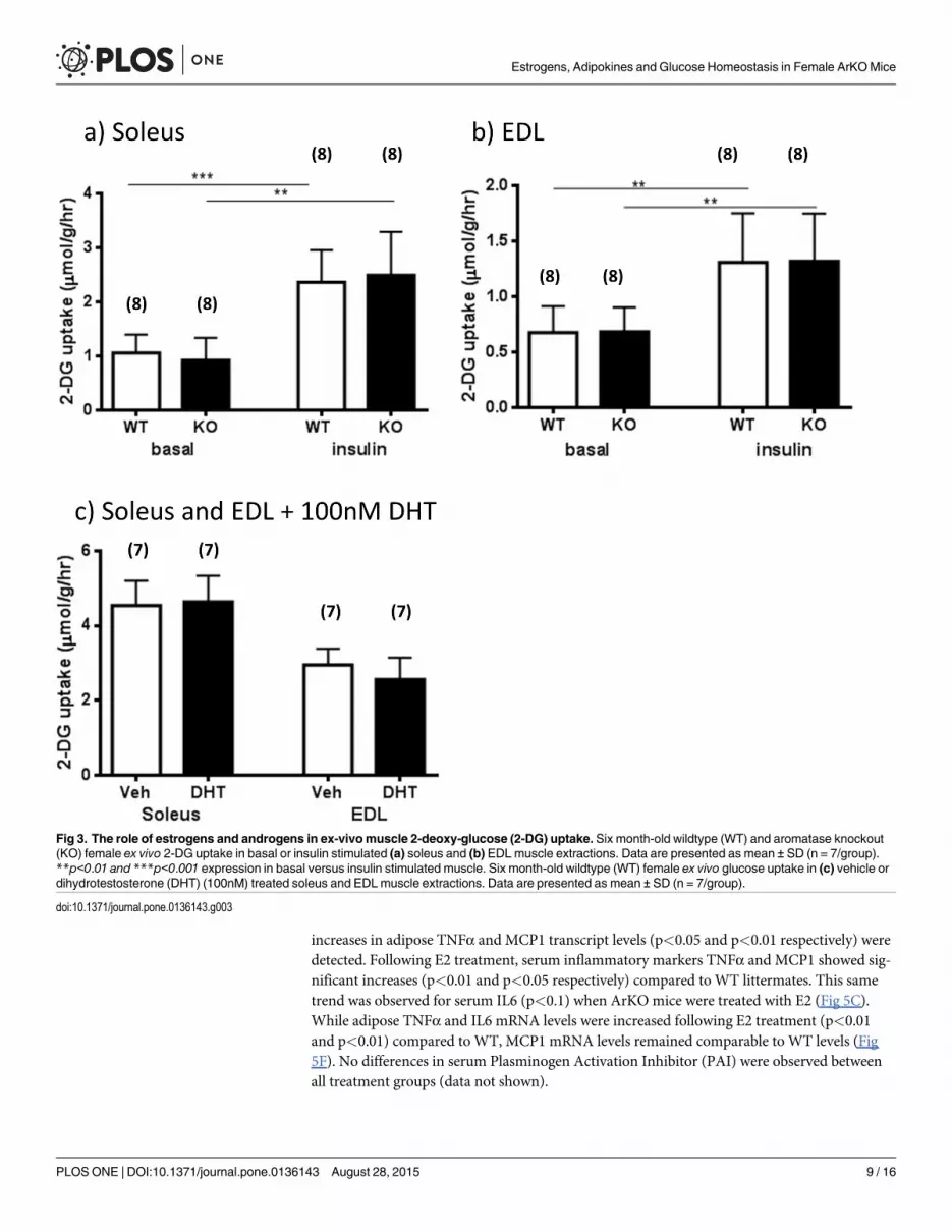
The role of estrogens and androgens in ex-vivo muscle glucose uptakeConsistent with the idea that the liver was the primary site of metabolic dysfunction, muscles(EDL and soleus) from both untreated ArKO and WTmice showed similar efficacy in basal orinsulin stimulated glucose uptake ex vivo (Fig 3A and 3B). The validity of this experiment wasconfirmed by significant increases of the glucose uptake observed in insulin stimulated musclescompared to vehicle treated muscles in both WT and KO genotypes (soleus, p<0.001 and0.001 respectively; EDL, p<0.01 and 0.01 respectively). Since ArKO mice have higher serumandrogen levels [16], we further examined the direct effects of dihydrotestosterone (DHT,100nM) on the glucose-uptake of six month-old WT soleus and EDL muscles in the ex vivo
Fig 1. Serum glucose and insulin levels. (a) Fasted basal serum glucose and 20 min after glucose challenge serum glucose levels; (b) Fasted basal seruminsulin (from 6 month-old female WT (wild type), KO (aromatase knockout) and KOE (aromatase knockout treated with 2.5µg/day 17β-estradiol) mice. Dataare expressed as mean ± SD (n = 5–13 per group).
doi:10.1371/journal.pone.0136143.g001
Estrogens, Adipokines and Glucose Homeostasis in Female ArKOMice
PLOS ONE | DOI:10.1371/journal.pone.0136143 August 28, 2015 7 / 16

experiment. No differences were found in soleus nor EDL insulin stimulated glucose uptakebetween vehicle and DHT treated muscles (Fig 3C, P>0.05) supporting the idea that androgenshave little effect on muscle insulin sensitivity.
Gonadal adipose tissue, liver and muscle Akt protein phosphorylationTo determine the potential mechanisms responsible for differences in glucose homeostasis, theexpression and phosphorylation levels of Akt (a critical component of canonical insulin signal-ing) was measured in gonadal adipose, skeletal muscle and liver tissues of six month-old WTand ArKO female mice after an i.p. injection of insulin. Consistent with our metabolic dataindicating defects in liver glucose homeostasis, we found that liver Akt phosphorylation wassignificantly decreased (p<0.05) in the untreated ArKO mice compared to the WT and wasrestored with E2 administration (Fig 4A). There were no differences in Akt phosphorylation inadipose tissue or skeletal muscle (Fig 4B and 4C).
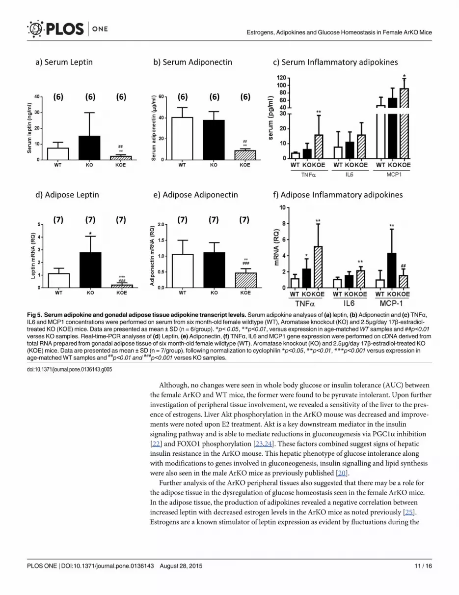
Adipokine expression levels in female ArKOmiceTo examine the mechanisms potentially contributing to the development of liver insulin resis-tance in ArKO mice, we examined serum adipokines and markers of inflammation. Serum lep-tin levels in untreated ArKO mice tended to be increased as compared to WT, whilst leptintranscript levels were significantly increased in the gonadal adipose tissue (p<0.05; Fig 5A and5D). After E2 treatment, ArKO serum leptin levels (p<0.01 and p<0.01) as well as transcriptlevels (p<0.001 and p<0.001) were lowered as compared to both ArKO and WT respectively(Fig 5A and 5D).
Both serum adiponectin and adipose adiponectin transcript levels were similar in theuntreated ArKO and WT littermates (Fig 5B and 5E). Unexpectedly, E2-treatment significantlydecreased serum adiponectin and adipose adiponectin mRNA levels compared to both WTand untreated ArKO mice (p<0.01 and p<0.01 respectively).
Levels of serum inflammatory adipokines TNFα, IL6 and MCP1 remained comparablebetween untreated female ArKO and their WT littermates (Fig 5C) although significant
Fig 2. Whole body glucose, insulin and pyruvate tolerance. (a)Whole body tolerance tests were completed on fasted 6 month-old female wildtype (WT)and aromatase knockout (KO) and 2.5μg/day 17β-estradiol-treated KO (KOE) pyruvate tolerance test and (b) corresponding area under curve. Data arepresented as the mean ± SD. (n = shown on corresponding bar/ group) *p<0.05, **p<0.01, and ***p<0.001 versus expression in WT samples and # p<0.05and ##p<0.01 versus KO samples.
doi:10.1371/journal.pone.0136143.g002
Estrogens, Adipokines and Glucose Homeostasis in Female ArKOMice
PLOS ONE | DOI:10.1371/journal.pone.0136143 August 28, 2015 8 / 16
increases in adipose TNFα and MCP1 transcript levels (p<0.05 and p<0.01 respectively) weredetected. Following E2 treatment, serum inflammatory markers TNFα and MCP1 showed sig-nificant increases (p<0.01 and p<0.05 respectively) compared to WT littermates. This sametrend was observed for serum IL6 (p<0.1) when ArKO mice were treated with E2 (Fig 5C).While adipose TNFα and IL6 mRNA levels were increased following E2 treatment (p<0.01and p<0.01) compared to WT, MCP1 mRNA levels remained comparable to WT levels (Fig5F). No differences in serum Plasminogen Activation Inhibitor (PAI) were observed betweenall treatment groups (data not shown).
Fig 3. The role of estrogens and androgens in ex-vivomuscle 2-deoxy-glucose (2-DG) uptake. Six month-old wildtype (WT) and aromatase knockout(KO) female ex vivo 2-DG uptake in basal or insulin stimulated (a) soleus and (b) EDL muscle extractions. Data are presented as mean ± SD (n = 7/group).**p<0.01 and ***p<0.001 expression in basal versus insulin stimulated muscle. Six month-old wildtype (WT) female ex vivo glucose uptake in (c) vehicle ordihydrotestosterone (DHT) (100nM) treated soleus and EDLmuscle extractions. Data are presented as mean ± SD (n = 7/group).
doi:10.1371/journal.pone.0136143.g003
Estrogens, Adipokines and Glucose Homeostasis in Female ArKOMice
PLOS ONE | DOI:10.1371/journal.pone.0136143 August 28, 2015 9 / 16

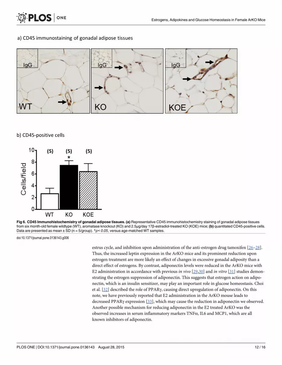
Macrophages in gonadal/omental adipose tissueA possible contributing source of inflammatory adipokines besides adipocytes is adipose asso-ciated macrophages which are CD45 positive [17]. Thus, we used CD45 immunostaining toexamine the extent of macrophage invasion into the gonadal adipose tissue (Fig 6A). CD45positive cells were significantly increased in untreated ArKO mice as compared to WT(p<0.05; Fig 6B). E2 treatment in the ArKO mice was unable to reduce CD45 positive cell pop-ulation (p = 0.058 vs WT).
DiscussionThe role of estrogens in the regulation of glucose homeostasis has been extensively studied asrecently reviewed by Kim et al. and Mauvais-Jarvis et al. [18,19]. However, defining the mecha-nisms by which exogenous estrogens affect glucose homeostasis, without the interference ofendogenous production, has yet to be elucidated. This is important when considering theeffects of hormone replacement therapy for menopause (when the ovaries, the main source ofestrogens within the body, have ceased hormone production).
This is the first study to use whole body tolerance tests in female ArKO mice to measurewhole-body glucose homeostasis. Female ArKO mice showed an increase in gonadal adiposetissue, serum glucose 20 min after glucose challenge, and fasted basal insulin levels comparedto WT counterparts, indicating defects in glucose homeostasis. Pyruvate tolerance tests indi-cated that the primary defect in glucose control was attributed to elevated hepatic gluconeogen-esis in female ArKO mice. In comparison, previous studies of themale ArKO mice showedsimilarly increased adiposity and serum glucose as well as glucose and pyruvate intolerance[20].
Higher liver triglycerides, which were reduced upon estrogen treatment, in the femaleArKO mice also support recent finding that liver fat is the best predictor of liver insulin sensi-tivity [21]. Consistent with this finding, treatment of female ArKO mice with insulin revealedsignificant reduction in liver phosphorylation of Akt, the primary signal controlling hepaticgluconeogenesis. These effects appear to be independent of changes in systemic inflammation.
Fig 4. Insulin stimulated Akt protein phosphorylation in liver, adipose and skeletal tissues. Six month-old female wildtype (WT) and aromataseknockout (KO) and 2.5μg/day 17β-estradiol-replaced KO (KOE) mice were used. Protein phosphorylation analyses of Akt levels were performed on proteinextracted from insulin stimulated (a) liver, (b) adipose tissue and (c) skeletal muscle. Data are presented as mean ± SD (n = 6-8/group). **p<0.01 versusexpression in age-matchedWT samples and #p<0.05 versus expression in age-matched ArKO untreated samples.
doi:10.1371/journal.pone.0136143.g004
Estrogens, Adipokines and Glucose Homeostasis in Female ArKOMice
PLOS ONE | DOI:10.1371/journal.pone.0136143 August 28, 2015 10 / 16
Although, no changes were seen in whole body glucose or insulin tolerance (AUC) betweenthe female ArKO and WTmice, the former were found to be pyruvate intolerant. Upon furtherinvestigation of peripheral tissue involvement, we revealed a sensitivity of the liver to the pres-ence of estrogens. Liver Akt phosphorylation in the ArKO mouse was decreased and improve-ments were noted upon E2 treatment. Akt is a key downstream mediator in the insulinsignaling pathway and is able to mediate reductions in gluconeogenesis via PGC1α inhibition[22] and FOXO1 phosphorylation [23,24]. These factors combined suggest signs of hepaticinsulin resistance in the ArKO mouse. This hepatic phenotype of glucose intolerance alongwith modifications to genes involved in gluconeogenesis, insulin signalling and lipid synthesiswere also seen in the male ArKO mice as previously published [20].
Further analysis of the ArKO peripheral tissues also suggested that there may be a role forthe adipose tissue in the dysregulation of glucose homeostasis seen in the female ArKO mice.In the adipose tissue, the production of adipokines revealed a negative correlation betweenincreased leptin with decreased estrogen levels in the ArKO mice as noted previously [25].Estrogens are a known stimulator of leptin expression as evident by fluctuations during the
Fig 5. Serum adipokine and gonadal adipose tissue adipokine transcript levels. Serum adipokine analyses of (a) leptin, (b) Adiponectin and (c) TNFα,IL6 and MCP1 concentrations were performed on serum from six month-old female wildtype (WT), Aromatase knockout (KO) and 2.5μg/day 17β-estradiol-treated KO (KOE) mice. Data are presented as mean ± SD (n = 6/group). *p< 0.05, **p<0.01, versus expression in age-matchedWT samples and ##p<0.01verses KO samples. Real-time-PCR analyses of (d) Leptin, (e) Adiponectin, (f) TNFα, IL6 and MCP1 gene expression were performed on cDNA derived fromtotal RNA prepared from gonadal adipose tissue of six month-old female wildtype (WT), Aromatase knockout (KO) and 2.5μg/day 17β-estradiol-treated KO(KOE) mice. Data are presented as mean ± SD (n = 7/group). following normalization to cyclophilin *p<0.05, **p<0.01, ***p<0.001 versus expression inage-matchedWT samples and ##p<0.01 and ###p<0.001 verses KO samples.
doi:10.1371/journal.pone.0136143.g005
Estrogens, Adipokines and Glucose Homeostasis in Female ArKOMice
PLOS ONE | DOI:10.1371/journal.pone.0136143 August 28, 2015 11 / 16
estrus cycle, and inhibition upon administration of the anti-estrogen drug tamoxifen [26–28].Thus, the increased leptin expression in the ArKO mice and its prominent reduction uponestrogen treatment are more likely an effect of changes in excessive gonadal adiposity than adirect effect of estrogens. By contrast, adiponectin levels were reduced in the ArKO mice withE2 administration in accordance with previous in vivo [29,30] and in vitro [31] studies demon-strating the estrogen suppression of adiponectin. This suggests that estrogen action on adipo-nectin, which is an insulin sensitizer, may play an important role in glucose homeostasis. Choiet al. [32] described the role of PPARγ, causing direct upregulation of adiponectin. On thisnote, we have previously reported that E2 administration in the ArKO mouse leads todecreased PPARγ expression [33], which may cause the reduction in adiponectin we observed.Another possible mechanism for reducing adiponectin in the E2 treated ArKO was theobserved increases in serum inflammatory markers TNFα, IL6 and MCP1, which are allknown inhibitors of adiponectin.
Fig 6. CD45 Immunohistochemistry of gonadal adipose tissues. (a)Representative CD45 immunohistochemistry staining of gonadal adipose tissuesfrom six month-old female wildtype (WT), aromatase knockout (KO) and 2.5μg/day 17β-estradiol-treated KO (KOE) mice; (b) quantitated CD45-positive cells.Data are presented as mean ± SD (n = 5/group). *p< 0.05, versus age-matchedWT samples.
doi:10.1371/journal.pone.0136143.g006
Estrogens, Adipokines and Glucose Homeostasis in Female ArKOMice
PLOS ONE | DOI:10.1371/journal.pone.0136143 August 28, 2015 12 / 16

Although E2 replacement in many cases of estrogen deficiency, such as menopause andovariectomy, results in reduced adiposity and inflammation [34,35]; there are cases wherebyestrogens can play a negative role in the development of inflammation. High or supra-physio-logical levels of estrogens as seen in third trimester pregnancy [36] are associated with increasesin TNFα and leptin concentrations in addition to decreased adiponectin [37]. Riant and col-leagues [38] also found increase in TNFα, IL6, MCP1 and F480 in ovariectomised mice on ahigh fat diet when treated with estrogens. Macrophage density was also increased in adiposetissue in this model which may account for the increased capacity of macrophages to producecytokines [38]. Direct modulation of inflammation by estrogens is corroborated by the locationof estrogen receptors (ERs) on cells of the immune system [39] and have been associated withmodulating immune cell recruitment into estrogen sensitive tissue such as the uterus [40,41].
Subsequent increased MCP1 concentration observed in the female ArKO serum with E2treatment and increased CD45 positive cells in the adipose tissue of female ArKO and ArKO+E2 mice suggest enhanced macrophage infiltration from the periphery which may explain theincreases in serum and adipocytes TNFα and IL6 in these animals. However, the discrepanciesbetween serum and adipose tissue MCP1 levels could be explained by the fact that MCP1 is notexclusively produced by adipose tissue [42]. Indeed, our data support the observation thatestrogen actions on inflammation are still dependent on a myriad of factors such as targetorgan, concentration (biphasic effects) and the ER isoforms present [43]. Estrogens may be act-ing through ERα instead of ERβ to exert changes in adiposity since ERαKOmice of both sexespresent increased adiposity but not ERβKOmice (for review see reference [15]); the formerpresents similar adiposity phenotypes to our estrogen-deficient ArKO mouse model [25].
Estrogen deficiency had neither inhibitory modifications in muscle or adipose Akt phos-phorylation nor insulin stimulated ex vivo skeletal muscle glucose uptake. The high HDL levels[44,45] in female ArKO mice may counteract any reductions in Akt associated with insulinresistance in muscle and adipose tissues, leading to no significant changes in Akt phosphoryla-tion in these ArKO tissues when compared to the WT.
ConclusionThe ArKO mouse is an important estrogen responsive model which allows us to examine exog-enous estrogen dosage on the relationship between hormones, adiposity and insulin resistancewithout the interference of uncontrolled endogenous estrogen production. Our observationsprovide an understanding into the complex mechanisms, including changes in Akt, TNFα, IL6and MCP1 by which estrogen regulates obesity and insulin resistance. Estrogen administrationin the ArKO mice leads to improvements in liver glucose homeostasis. However, simultaneousdisplays of increased inflammatory processes in the adipose tissue adipokines may be responsi-ble for the lack of improved whole body glucose recovery after estrogen replacement.
Supporting InformationS1 Table. Body and gonadal adipose tissue weights of untreated ArKO (KO) versus pla-cebo-treated ArKOmice. Body and adipose tissue weights in grams (g) six month-old femalearomatase knockout untreated (KO, n = 10) compared to six-week placebo-treated KO (KOP,n = 4). Data are expressed as mean ± SD, no significant differences are detected.(DOCX)
S1 Fig. Glucose tolerance tests of 6 month-old untreated ArKO (KO) vs. placebo treatedArKO (KOP).Whole body glucose tolerance tests (GTT) were completed on fasted 6 month-old female untreated aromatase knockout (KO) and 6 weeks of placebo treated KO (KOP). (A)
Estrogens, Adipokines and Glucose Homeostasis in Female ArKOMice
PLOS ONE | DOI:10.1371/journal.pone.0136143 August 28, 2015 13 / 16
glucose tolerance test and (B) corresponding area under curve. Data are presented asmean ± SD, no significant differences are detected; sample sizes in brackets.(PDF)
Author ContributionsConceived and designed the experiments: MLVS GRS MEEJ ED ERS WCB. Performed theexperiments: MLVS GRS SBJ JH JDC KAH ALWWCB. Analyzed the data: MLVS GRS SBJALW ED ERSWCB. Contributed reagents/materials/analysis tools: GRS ED ERSWCB. Wrotethe paper: MLVS GRS SBJ ERS WCB.
References1. Nordlie RC, Foster JD, Lange AJ. Regulation of glucose production by the liver. Annu Rev Nutr 1999;
19: 379–406. PMID: 10448530
2. Pilkis SJ, Granner DK. Molecular physiology of the regulation of hepatic gluconeogenesis and glycoly-sis. Annu Rev Physiol 1992; 54: 885–909. PMID: 1562196
3. Saltiel AR, Kahn CR. Insulin signalling and the regulation of glucose and lipid metabolism. Nature2001; 414: 799–806. PMID: 11742412
4. Gabriely I, Ma XH, Yang XM, Atzmon G, Rajala MW, Berg AH et al. Removal of visceral fat preventsinsulin resistance and glucose intolerance of aging: an adipokine-mediated process? Diabetes 2002;51: 2951–2958. PMID: 12351432
5. Wellen KE, Hotamisligil GS. Obesity-induced inflammatory changes in adipose tissue. J Clin Invest2003; 112: 1785–1788. PMID: 14679172
6. Lindmark S, Buren J, Eriksson JW. Insulin resistance, endocrine function and adipokines in type 2 dia-betes patients at different glycaemic levels: potential impact for glucotoxicity in vivo. Clin Endocrinol(Oxf) 2006; 65: 301–309.
7. Nuttall FQ, Ngo A, Gannon MC Regulation of hepatic glucose production and the role of gluconeogene-sis in humans: is the rate of gluconeogenesis constant? Diabetes Metab Res Rev 2008; 24: 438–458.doi: 10.1002/dmrr.863 PMID: 18561209
8. Tanko LB, Christiansen C. Adipose tissue, insulin resistance and low-grade inflammation: implicationsfor atherogenesis and the cardiovascular harm of estrogen plus progestogen therapy. Climacteric2006; 9: 169–180. PMID: 16766431
9. Samy N, HashimM, Sayed M, Said M. Clinical significance of inflammatory markers in polycystic ovarysyndrome: their relationship to insulin resistance and body mass index. Dis Markers 2009; 26: 163–170. doi: 10.3233/DMA-2009-0627 PMID: 19729797
10. Ardawi MS, Rouzi AA. Plasma adiponectin and insulin resistance in women with polycystic ovary syn-drome. Fertil Steril 2005; 83: 1708–1716. PMID: 15950640
11. Fisher C, Graves K, Parlow A, Simpson E.Characterization of mice deficient in aromatase (ArKO)because of targeted disruption of the cyp19 gene. Proc Natl Acad Sci U S A 1998; 95: 6965–6970.PMID: 9618522
12. Robertson KM, O'Donnell L, Jones ME, Meachem SJ, BoonWC, Fisher CR, et al. Impairment of sper-matogenesis in mice lacking a functional aromatase (cyp 19) gene. Proc Natl Acad Sci U S A 1999; 96:7986–7991. PMID: 10393934
13. Jorgensen SB, Honeyman J, Oakhill JS, Fazakerley D, Stockli J, Kemp BE, et al. Oligomeric resistinimpairs insulin and AICAR-stimulated glucose uptake in mouse skeletal muscle by inhibiting GLUT4translocation. Am J Physiol Endocrinol Metab 2009; 297: E57–66. doi: 10.1152/ajpendo.90744.2008PMID: 19435854
14. Chow JD, Jones ME, Prelle K, Simpson ER, BoonWC. A selective estrogen receptor alpha agonistameliorates hepatic steatosis in the male aromatase knockout mouse. J Endocrinol 2001; 210: 323–334.
15. Norris AW, Chen L, Fisher SJ, Szanto I, Ristow M, Jozsi AC, et al. Muscle-specific PPAR{gamma}-defi-cient mice develop increased adiposity and insulin resistance but respond to thiazolidinediones. J ClinInvest 2003; 112: 608–618. PMID: 12925701
16. Jones ME, BoonWC, Proietto J, Simpson ER. Of mice and men: the evolving phenotype of aromatasedeficiency. Trends Endocrinol Metab 2006; 17: 55–64. PMID: 16480891
Estrogens, Adipokines and Glucose Homeostasis in Female ArKOMice
PLOS ONE | DOI:10.1371/journal.pone.0136143 August 28, 2015 14 / 16

17. Davies LC, Jenkins SJ, Allen JE, Taylor PR. Tissue-resident macrophages. Nat Immunol 2013; 14:986–995. doi: 10.1038/ni.2705 PMID: 24048120
18. Mauvais-Jarvis F, Clegg DJ, Hevener AL. The role of estrogens in control of energy balance and glu-cose homeostasis. Endocr Rev 2013; 34: 309–338. doi: 10.1210/er.2012-1055 PMID: 23460719
19. Kim JH, Cho HT, Kim YJ. The role of estrogen in adipose tissue metabolism: insights into glucosehomeostasis regulation. Endocr J. 2014; 61(11):1055–67. PMID: 25109846
20. Van Sinderen ML, Steinberg GR, Jorgensen SB, To SQ, Knower KC, Clyne CD, et al. Hepatic GlucoseIntolerance Precedes Hepatic Steatosis in the Male Aromatase Knockout (ArKO) Mouse. PMID: PLoSOne 2014; 9(2): e87230. doi: 10.1371/journal.pone.0087230
21. Fullerton MD, Galic S, Marcinko K, Sikkema S, Pulinilkunnil T, Chen ZP, et al. Single phosphorylationsites in Acc1 and Acc2 regulate lipid homeostasis and the insulin-sensitizing effects of metformin. NatMed 2013; 19: 1649–1654. doi: 10.1038/nm.3372 PMID: 24185692
22. Li X, Monks B, Ge Q, BirnbaumMJ. Akt/PKB regulates hepatic metabolism by directly inhibiting PGC-1alpha transcription coactivator. Nature 2007; 447: 1012–1016. PMID: 17554339
23. BiggsWH 3rd, Meisenhelder J, Hunter T, CaveneeWK, Arden KC. Protein kinase B/Akt-mediatedphosphorylation promotes nuclear exclusion of the winged helix transcription factor FKHR1. Proc NatlAcad Sci U S A 1999; 96: 7421–7426. PMID: 10377430
24. Wang Q, Wang N, Dong M, Chen F, Li Z, Chen Y. GdCl3 reduces hyperglycaemia through Akt/FoxO1-induced suppression of hepatic gluconeogenesis in Type 2 diabetic mice. Clin Sci (Lond) 2014; 127(2):91–100.
25. Jones ME, Thorburn AW, Britt KL, Hewitt KN, Wreford NG, Proietto J, et al. Aromatase-deficient (ArKO)mice have a phenotype of increased adiposity. Proc Natl Acad Sci U S A 2000; 97: 12735–12740.PMID: 11070087
26. Tanaka M, Nakaya S, Kumai T, Watanabe M, Tateishi T, Shimizu H, et al. Effects of estrogen on serumleptin levels and leptin mRNA expression in adipose tissue in rats. Horm Res 2001; 56: 98–104.
27. Kristensen K, Pedersen SB, Richelsen B. Regulation of leptin by steroid hormones in rat adipose tis-sue. Biochem Biophys Res Commun 1999; 259: 624–630. PMID: 10364468
28. Machinal F, Dieudonne MN, Leneveu MC, Pecquery R, Giudicelli Y. In vivo and in vitro ob gene expres-sion and leptin secretion in rat adipocytes: evidence for a regional specific regulation by sex steroid hor-mones. Endocrinology 1999; 140: 1567–1574. PMID: 10098489
29. Macotela Y, Boucher J, Tran TT, Kahn CR. Sex and depot differences in adipocyte insulin sensitivityand glucose metabolism. Diabetes 2009; 58: 803–812. doi: 10.2337/db08-1054 PMID: 19136652
30. D'Eon TM, Souza SC, Aronovitz M, Obin MS, Fried SK, Greenberg AS. Estrogen regulation of adiposityand fuel partitioning. Evidence of genomic and non-genomic regulation of lipogenic and oxidative path-ways. J Biol Chem 2005; 280: 35983–35991. PMID: 16109719
31. Combs TP, Berg AH, Rajala MW, Klebanov S, Iyengar P, Jimenez-Chillaron JC, et al. Sexual differenti-ation, pregnancy, calorie restriction, and aging affect the adipocyte-specific secretory protein adiponec-tin. Diabetes 2003; 52: 268–276. PMID: 12540596
32. Choi JH, Banks AS, Kamenecka TM, Busby SA, Chalmers MJ, Kumar N, et al. Antidiabetic actions of anon-agonist PPARgamma ligand blocking Cdk5-mediated phosphorylation. Nature 2001; 477: 477–481.
33. Misso ML, Murata Y, BoonWC, Jones ME, Britt KL, Simpson ER. Cellular and molecular characteriza-tion of the adipose phenotype of the aromatase-deficient mouse. Endocrinology 2003; 144: 1474–1480. PMID: 12639931
34. Jelenik T, Roden M How estrogens prevent from lipid-induced insulin resistance. Endocrinology 2013;154: 989–992. doi: 10.1210/en.2013-1112 PMID: 23429711
35. Stubbins RE, Najjar K, Holcomb VB, Hong J, Nunez NP. Oestrogen alters adipocyte biology and pro-tects female mice from adipocyte inflammation and insulin resistance. Diabetes Obes Metab 2012; 14:58–66. doi: 10.1111/j.1463-1326.2011.01488.x PMID: 21834845
36. Winkler G, Cseh K, Baranyi E, Melczer Z, Speer G, Hajós P, et al. Tumor necrosis factor system in insu-lin resistance in gestational diabetes. Diabetes Res Clin Pract 2002; 56: 93–99. PMID: 11891016
37. Gao XL, Yang HX, Zhao Y. Variations of tumor necrosis factor-alpha, leptin and adiponectin in mid-tri-mester of gestational diabetes mellitus. Chin Med J (Engl) 2008; 121: 701–705.
38. Riant E, Waget A, Cogo H, Arnal JF, Burcelin R, Gourdy P. Estrogens protect against high-fat diet-induced insulin resistance and glucose intolerance in mice. Endocrinology 2009; 150: 2109–2117. doi:10.1210/en.2008-0971 PMID: 19164473
Estrogens, Adipokines and Glucose Homeostasis in Female ArKOMice
PLOS ONE | DOI:10.1371/journal.pone.0136143 August 28, 2015 15 / 16

39. Cutolo M, Accardo S, Villaggio B, Barone A, Sulli A, Coviello DA, et al. Androgen and estrogen recep-tors are present in primary cultures of human synovial macrophages. J Clin Endocrinol Metab 1996;81: 820–827. PMID: 8636310
40. Wood GW, Hausmann E, Choudhuri R. Relative role of CSF-1, MCP-1/JE, and RANTES in macro-phage recruitment during successful pregnancy. Mol Reprod Dev 1997; 46: 62–69; discussion 69–70.PMID: 8981365
41. Tibbetts TA, Conneely OM, O'Malley BW. Progesterone via its receptor antagonizes the pro-inflamma-tory activity of estrogen in the mouse uterus. Biol Reprod 1999; 60: 1158–1165. PMID: 10208978
42. Dahlman I, KaamanM, Olsson T, Tan GD, Bickerton AS, Wåhlén K, et al. A unique role of monocytechemoattractant protein 1 among chemokines in adipose tissue of obese subjects. J Clin EndocrinolMetab 2005; 90: 5834–5840. PMID: 16091493
43. Straub RH. The complex role of estrogens in inflammation. Endocr Rev 2007; 28: 521–574. PMID:17640948
44. Jones ME, Thorburn AW, Britt KL, Hewitt KN, Misso ML, Wreford NG et al. Aromatase-deficient (ArKO)mice accumulate excess adipose tissue. J Steroid BiochemMol Biol 2001; 79: 3–9. PMID: 11850201
45. Van Sinderen ML, BoonWC, Ederveen AG, Kloosterboer HJ, Simpson ER, Jones ME. The estrogeniccomponent of tibolone reduces adiposity in female aromatase knockout mice. Menopause 2009; 16:582–588. doi: 10.1097/gme.0b013e31818fb20b PMID: 19182696
Estrogens, Adipokines and Glucose Homeostasis in Female ArKOMice
PLOS ONE | DOI:10.1371/journal.pone.0136143 August 28, 2015 16 / 16
Related Documents