Effects of changes in ambient PAR and UV radiation on the nutritional quality of an Arctic diatom (Thalassiosira antarctica var. borealis) Eva Leu a,d, ⁎ , Sten-Åke Wängberg b , Angela Wulff c , Stig Falk-Petersen d , Jon Børre Ørbæk d , Dag O. Hessen a a University of Oslo, Department of Biology, P.O. Box 1066, N-0316 Oslo, Norway b Göteborg University, Botanical Institute, P.O. Box 461, SE-40530 Göteborg, Sweden c Göteborg University, Department of Marine Ecology, Marine Botany, P.O. Box 461, SE-40530 Göteborg, Sweden d Norwegian Polar Institute, Polar Environmental Centre, N-9296 Tromsø, Norway Received 21 October 2005; received in revised form 16 May 2006; accepted 6 June 2006 Abstract Polyunsaturated fatty acids (PUFAs) are essential macromolecules that are synthesized by phytoplankton during spring bloom, and they play a key role in the Arctic food web. They are, however, considered to be sensitive to oxidation by UV radiation (280–400 nm). Changes in the food quality of primary producers may affect the transport of biomass and energy in the whole ecosystem. Using a common Arctic diatom, we looked at the effect of ambient and increased UV radiation on its nutritional quality, specifically, the fatty acid composition and elemental ratios. In May 2004, in the archipelago of Svalbard (79° N), a unialgal culture of Thalassiosira antarctica var. borealis was subjected to a 17-day experiment in outdoor aquaria. The diatoms were kept in semi-continuous culture (40 1) and exposed to three treatments with different levels of UV radiation: none (UV-shielded), ambient, and enhanced. Fatty acid composition, C:N:P ratios, photosynthetic pigment composition, optimum quantum yield of PSII, and cell numbers were analysed over the experimental period. An initial increase in PAR (photosynthetically active radiation, 400–700 nm) intensities profoundly affected the fatty acid composition and substantially inhibited the synthesis of PUFAs, but the relative amounts of PUFAs were not reduced by UV radiation. Enhanced UV radiation did, however, cause a significant reduction in optimum quantum yield of PSII and affected some fatty acids, mainly 18:0 and 16:1 n-7, during the first week of the experiment. Both ambient and enhanced UV radiation caused significantly lower C:P and N:P ratios. At the same time, these treatments elicited a higher relative content of the photoprotective pigments diadinoxanthin and diatoxanthin. After acclimation to the new light levels these effects faded off. Thus, brief periods with high light exposure may cause significant changes in photosynthetic activity and food quality, but the capacity for photo-acclimation seems high. The impact of UV radiation seems to be less important for food quality than that of PAR during a sudden rise in total light intensity. © 2006 Elsevier B.V. All rights reserved. Keywords: Arctic; Fatty acid composition; PAR; Stoichiometry; Thalassiosira antarctica var. borealis; UV radiation 1. Introduction In Arctic marine ecosystems the light climate is continuously changing—both in absolute intensity, due to variation in sea ice extent and thickness (Aas et al., Journal of Experimental Marine Biology and Ecology 337 (2006) 65 – 81 www.elsevier.com/locate/jembe ⁎ Corresponding author. University of Oslo, Department of Biology, P.O. Box 1066, N-0316 Oslo, Norway. Tel.: +47 22 85 42 98; fax: +47 22 85 44 38. E-mail address: [email protected] (E. Leu). 0022-0981/$ - see front matter © 2006 Elsevier B.V. All rights reserved. doi:10.1016/j.jembe.2006.06.005

Welcome message from author
This document is posted to help you gain knowledge. Please leave a comment to let me know what you think about it! Share it to your friends and learn new things together.
Transcript
Effects of changes in ambient PAR and UV radiationon the nutritional quality of an Arctic diatom
(Thalassiosira antarctica var. borealis)
Eva Leu a,d,!, Sten-Åke Wängberg b, Angela Wulff c, Stig Falk-Petersen d,Jon Børre Ørbæk d, Dag O. Hessen a
a University of Oslo, Department of Biology, P.O. Box 1066, N-0316 Oslo, Norwayb Göteborg University, Botanical Institute, P.O. Box 461, SE-40530 Göteborg, Sweden
c Göteborg University, Department of Marine Ecology, Marine Botany, P.O. Box 461, SE-40530 Göteborg, Swedend Norwegian Polar Institute, Polar Environmental Centre, N-9296 Tromsø, Norway
Received 21 October 2005; received in revised form 16 May 2006; accepted 6 June 2006
Abstract
Polyunsaturated fatty acids (PUFAs) are essential macromolecules that are synthesized by phytoplankton during spring bloom, andthey play a key role in the Arctic food web. They are, however, considered to be sensitive to oxidation by UV radiation (280–400 nm).Changes in the food quality of primary producersmay affect the transport of biomass and energy in thewhole ecosystem.Using a commonArctic diatom, we looked at the effect of ambient and increased UV radiation on its nutritional quality, specifically, the fatty acidcomposition and elemental ratios. InMay 2004, in the archipelago of Svalbard (79° N), a unialgal culture of Thalassiosira antarctica var.borealiswas subjected to a 17-day experiment in outdoor aquaria. The diatomswere kept in semi-continuous culture (40 1) and exposed tothree treatments with different levels of UVradiation: none (UV-shielded), ambient, and enhanced. Fatty acid composition, C:N:P ratios,photosynthetic pigment composition, optimum quantum yield of PSII, and cell numbers were analysed over the experimental period. Aninitial increase in PAR (photosynthetically active radiation, 400–700 nm) intensities profoundly affected the fatty acid composition andsubstantially inhibited the synthesis of PUFAs, but the relative amounts of PUFAs were not reduced by UV radiation. Enhanced UVradiation did, however, cause a significant reduction in optimum quantum yield of PSII and affected some fatty acids, mainly 18:0 and16:1 n-7, during the first week of the experiment. Both ambient and enhancedUVradiation caused significantly lower C:P andN:P ratios.At the same time, these treatments elicited a higher relative content of the photoprotective pigments diadinoxanthin and diatoxanthin.After acclimation to the new light levels these effects faded off. Thus, brief periodswith high light exposuremay cause significant changesin photosynthetic activity and food quality, but the capacity for photo-acclimation seemshigh. The impact of UVradiation seems to be lessimportant for food quality than that of PAR during a sudden rise in total light intensity.© 2006 Elsevier B.V. All rights reserved.
Keywords: Arctic; Fatty acid composition; PAR; Stoichiometry; Thalassiosira antarctica var. borealis; UV radiation
1. Introduction
In Arctic marine ecosystems the light climate iscontinuously changing—both in absolute intensity, dueto variation in sea ice extent and thickness (Aas et al.,
Journal of Experimental Marine Biology and Ecology 337 (2006) 65–81www.elsevier.com/locate/jembe
! Corresponding author. University of Oslo, Department of Biology,P.O. Box 1066, N-0316 Oslo, Norway. Tel.: +47 22 85 42 98; fax: +4722 85 44 38.
E-mail address: [email protected] (E. Leu).
0022-0981/$ - see front matter © 2006 Elsevier B.V. All rights reserved.doi:10.1016/j.jembe.2006.06.005
2002; Sakshaug, 2004) and in spectral composition,following dynamic and seasonal depletion events in thestratospheric ozone layer (Dahlback, 2002). In winter andspring 2005, ozone values above theArctic Circle droppedto the lowest values ever recorded; at 18 km altitude morethan 50% of the ozone had been destroyed (http://toms.gsfc.nasa.gov/ozone/ozone.html, Press release SCOUT-03 project, EU/WMO, 25 April 2005). This event waslinked to extremely low stratospheric temperatures, whichcould be attributed, partly, to tropospheric warming.Further global warming could thus accentuate such pe-riods of major ozone losses.
Heightened levels of UVB radiation (280–320 nm),expected from such large-scale depletion events, clearlypose a threat to the biota living at these latitudes. Foodquality—especially fatty acids—at the base of the Arcticmarine food web forms the foundation for the transport ofbiomass and energy to higher trophic levels. Most or-ganisms use lipids as a primary energy storage for survivalin times before and after the short productive season(Falk-Petersen et al., 1990; Klein Breteler et al., 2005).The transfer of lipids from diatoms to herbivorous zoo-plankton (mainly represented by threeCalanus species) isof particular importance in high Arctic systems (Falk-Petersen et al., 2006). Polyunsaturated fatty acids (PUFAs)play a key role in food webs, because they are synthesizedde novo only by photosynthetic organisms, yet they areessential for the many heterotrophs (Sargent and Whittle,1981). Thus, any changes at the base of the food web maypropagate to higher trophic levels. PUFAs are also ofmajor importance in regulating membrane fluidity underlow temperatures; using these, organisms ensure the func-tioning of vital physiological processes (Hall et al., 2002and references therein).
Several studies have documented a negative impactof UV radiation on PUFAs in marine phytoplanktonspecies; this impact has been attributed either tooxidation of previously synthesized fatty acids (Skerrattet al., 1998; Wang and Chai, 1994) or to disruption oftheir synthesis (Döhler and Biermann, 1994; Goes et al.,1994). Only one of these studies (Skerratt et al., 1998)dealt with polar phytoplankton species, utilizingsimilarly low temperatures. Temperature is a crucialparameter, since it may have a substantial impact onfatty acid composition itself (Thompson et al., 1992) aswell as on the dynamics of repair mechanisms (Roosand Vincent, 1998). Most of the previous reports aboutUV-mediated effects on fatty acids were laboratorystudies using artificial light and UV radiation sources;thus they differed with respect to both spectralcomposition and light intensity, compared to outdoorambient conditions.
We sought to investigate the impact of ambient andenhanced UV radiation on the fatty acid composition ofThalassiosira antarctica var. borealis under realisticlight and temperature conditions in the high Arctic. Anoutdoor experiment with three different treatments (UV-shielded, ambient, and enhanced UV radiation) wascarried out in the high Arctic archipelago of Svalbard,Norway (79° N) in spring 2004. In addition to fatty acidcomposition, we analysed C:N:P ratios (i.e. cellular stoi-chiometry) as another proxy of food quality for consumers(Sterner and Elser, 2002). Other quantitative effects ofUV radiation on algal biomass development, photosyn-thetic pigment composition, and photosynthetic efficiency(measured as optimum quantum yield of PSII) wereincluded in order to evaluate the sensitivity of differentphysiological parameters to these light regimes.
2. Materials and methods
2.1. Location,weatherconditions, and lightmeasurements
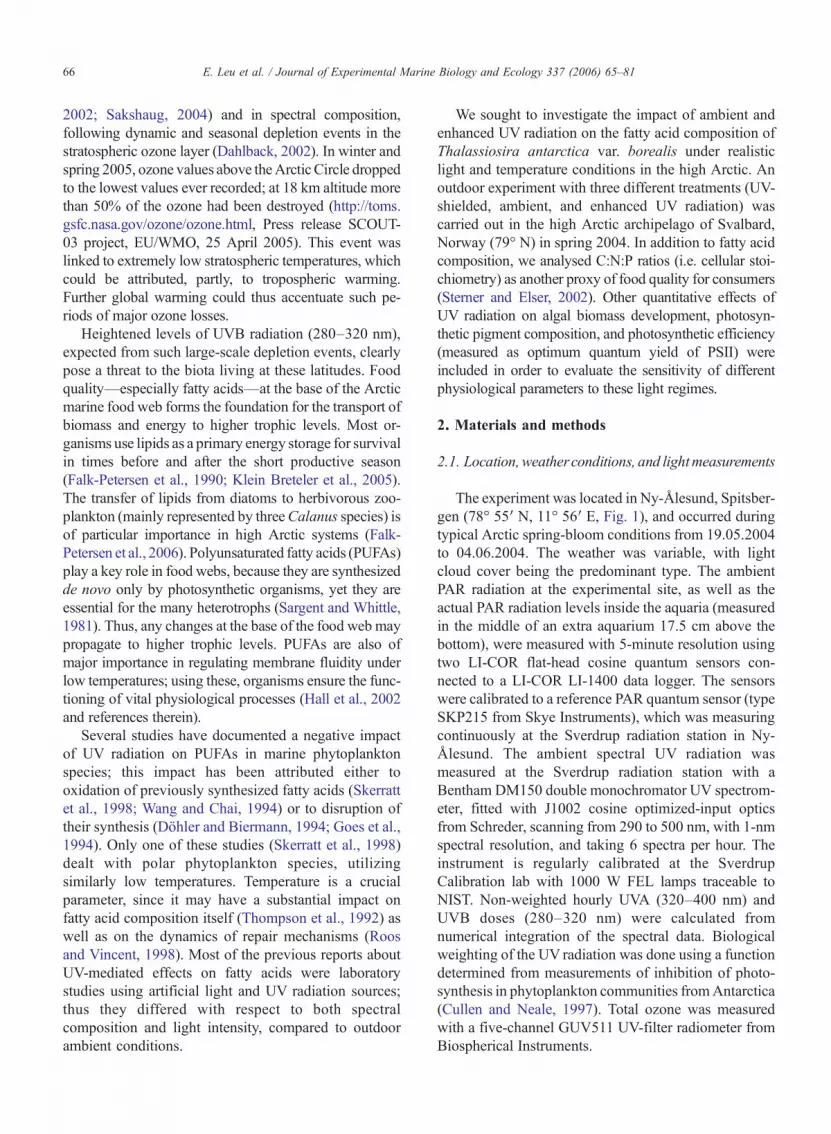
The experiment was located in Ny-Ålesund, Spitsber-gen (78° 55" N, 11° 56" E, Fig. 1), and occurred duringtypical Arctic spring-bloom conditions from 19.05.2004to 04.06.2004. The weather was variable, with lightcloud cover being the predominant type. The ambientPAR radiation at the experimental site, as well as theactual PAR radiation levels inside the aquaria (measuredin the middle of an extra aquarium 17.5 cm above thebottom), were measured with 5-minute resolution usingtwo LI-COR flat-head cosine quantum sensors con-nected to a LI-COR LI-1400 data logger. The sensorswere calibrated to a reference PAR quantum sensor (typeSKP215 from Skye Instruments), which was measuringcontinuously at the Sverdrup radiation station in Ny-Ålesund. The ambient spectral UV radiation wasmeasured at the Sverdrup radiation station with aBentham DM150 double monochromator UV spectrom-eter, fitted with J1002 cosine optimized-input opticsfrom Schreder, scanning from 290 to 500 nm, with 1-nmspectral resolution, and taking 6 spectra per hour. Theinstrument is regularly calibrated at the SverdrupCalibration lab with 1000 W FEL lamps traceable toNIST. Non-weighted hourly UVA (320–400 nm) andUVB doses (280–320 nm) were calculated fromnumerical integration of the spectral data. Biologicalweighting of the UV radiation was done using a functiondetermined from measurements of inhibition of photo-synthesis in phytoplankton communities fromAntarctica(Cullen and Neale, 1997). Total ozone was measuredwith a five-channel GUV511 UV-filter radiometer fromBiospherical Instruments.
66 E. Leu et al. / Journal of Experimental Marine Biology and Ecology 337 (2006) 65–81
No direct measurements of UV radiation could betaken from inside the aquaria. Due to shading from theexperimental setup, only diffuse radiation reached themiddle of the aquaria, except for very short periodsaround noon on clear sunny days. Since the ratio be-tween diffuse and global radiation depends on solarelevation and wavelength, and since it is higher forUV radiation compared to PAR, the calculation ofrealistic UV doses inside the aquaria used the followingequation:
UVaq ! PARaq " #UVD=PARD$; #1$
where UVaq is the calculated UV dose inside the aquaria,PARaq is the measured PAR radiation inside the aquaria,UVD is the diffuse ambient UV radiation, and PARD isthe diffuse ambient PAR radiation.
Since the only measurements available were ambientglobal radiation for both PAR and UV, Eq. (1) can berewritten:
UVaq ! PARaq " #UVG=PARG$ " PARG=D " UVD=G; #2$
where PARG is the ambient measured global PAR radia-tion, UVG is the ambient measured global UV radiation,PARG/D=PARG/PARD, UVD/G=UVD/UVG. The 2 lastterms, PARG/D and UVD/G, which are both functions ofsolar elevation and wavelength, were empirically derivedat the Sverdrup radiation station during 2 clear sunny dayswith standard sun/shade methods. Both of these last 2terms can be simplified: they are a smooth function ofsolar elevation when there is direct solar radiation (sun-shine); and they are equal to 1 when there is no sun(PARG=PARD, UVG=UVD). As a final adjustment in the
Fig. 1. Ny-Ålesund (Kongsfjorden) on Svalbard.
67E. Leu et al. / Journal of Experimental Marine Biology and Ecology 337 (2006) 65–81
derivation of UVaq, we weighted Eq. (2) with a numbercorresponding to the actual fraction of minutes withsunshine in each hour; the data for this came from thedirect radiation measurements at the Sverdrup radiationstation:
UVaq !PARaq " #UVG=PARG$
"X60
1
#PARG=D " UVD=G;$=60;#3$
For the hourly sums, the minute values of PARG/D
and UVD/G are given different values depending onwhether there is direct sun or not. In this way, the briefoccurrences of direct radiation inside the aquaria aroundmidday could also be corrected.
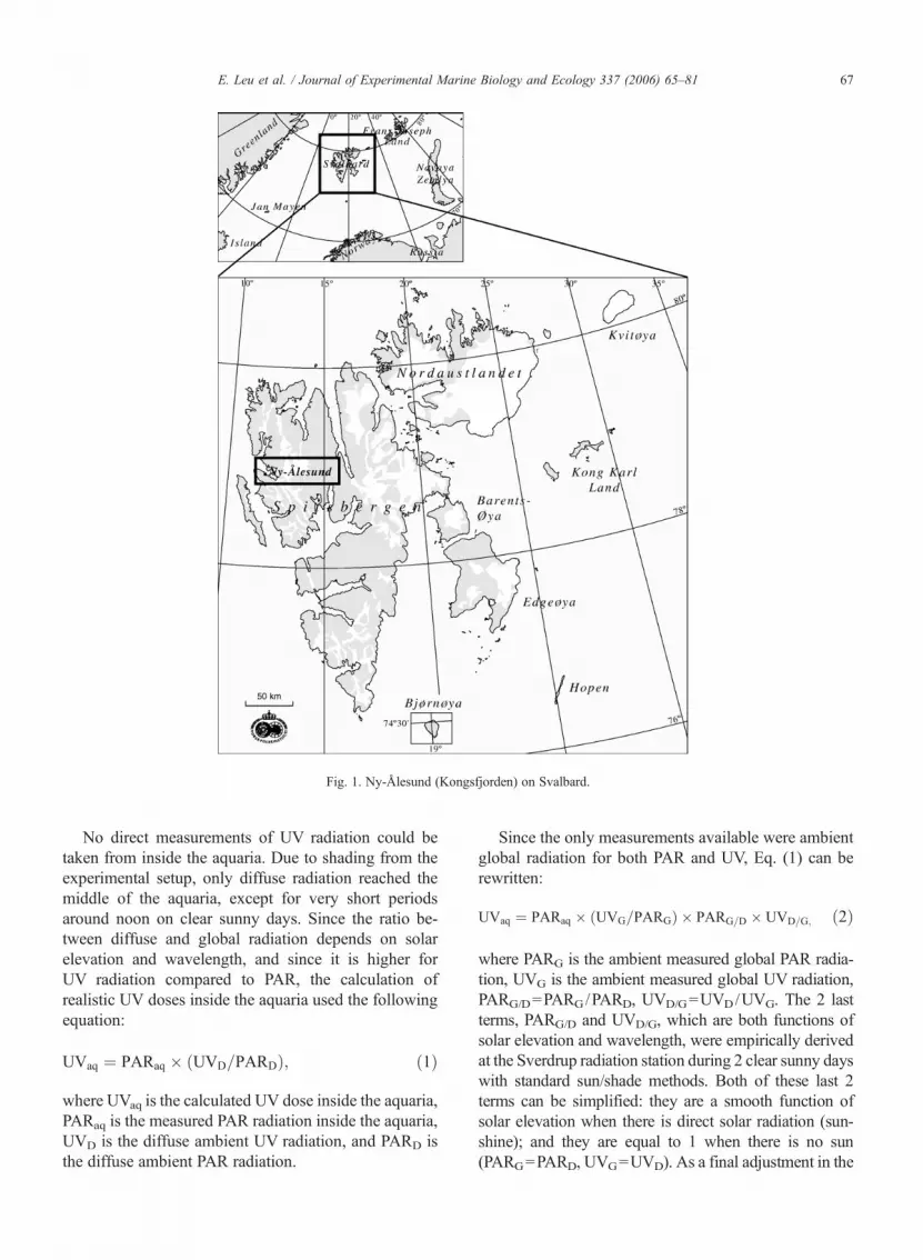
2.2. Experimental setup
The outdoor model ecosystem was modified from thesetup described in Wängberg et al. (2001), see Fig. 2.It consisted of 12 aquaria with 40 l volume each(39.0!28.7!37.9 cm [length /width /height]), made ofUV-transparent Plexiglas. The aquaria were placed inopaque plastic boxes and surrounded by a continuousflow of seawater for temperature control. The boxeswere covered by UV-transparent Plexiglas lids, com-bined with different cut-off foils. Three differenttreatments were applied in four replicates each:
(1) UV-shielded (referred to as PAR), protected fromUV radiation by an Ultraphan 400 foil (Digefra,Munich, Germany);
(2) ambient UV (referred to as UV), covered withcellulose acetate (Tamboer and Co Chemie B.V.,Heemstede, Netherlands) to correct for the ca.10% absorption of Ultraphan 400 in the PARspectrum;
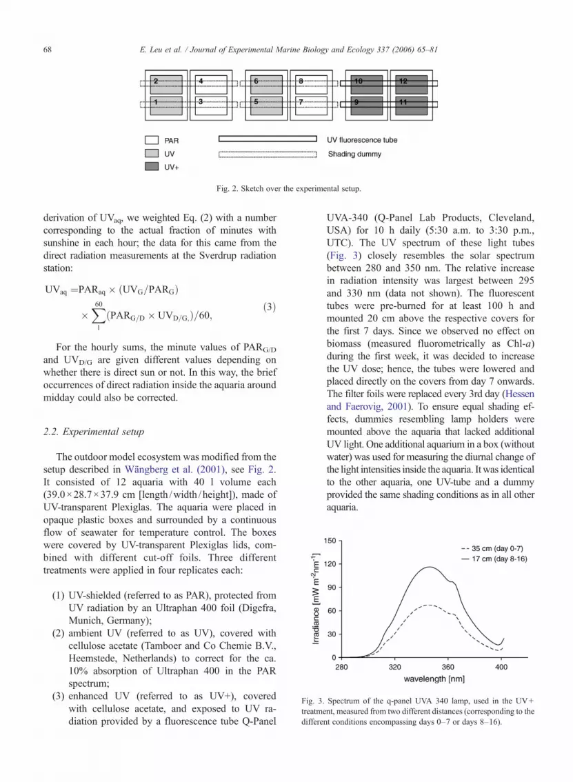
(3) enhanced UV (referred to as UV+), coveredwith cellulose acetate, and exposed to UV ra-diation provided by a fluorescence tube Q-Panel
UVA-340 (Q-Panel Lab Products, Cleveland,USA) for 10 h daily (5:30 a.m. to 3:30 p.m.,UTC). The UV spectrum of these light tubes(Fig. 3) closely resembles the solar spectrumbetween 280 and 350 nm. The relative increasein radiation intensity was largest between 295and 330 nm (data not shown). The fluorescenttubes were pre-burned for at least 100 h andmounted 20 cm above the respective covers forthe first 7 days. Since we observed no effect onbiomass (measured fluorometrically as Chl-a)during the first week, it was decided to increasethe UV dose; hence, the tubes were lowered andplaced directly on the covers from day 7 onwards.The filter foils were replaced every 3rd day (Hessenand Faerovig, 2001). To ensure equal shading ef-fects, dummies resembling lamp holders weremounted above the aquaria that lacked additionalUV light. One additional aquarium in a box (withoutwater) was used for measuring the diurnal change ofthe light intensities inside the aquaria. It was identicalto the other aquaria, one UV-tube and a dummyprovided the same shading conditions as in all otheraquaria.
Fig. 3. Spectrum of the q-panel UVA 340 lamp, used in the UV+treatment, measured from two different distances (corresponding to thedifferent conditions encompassing days 0–7 or days 8–16).
Fig. 2. Sketch over the experimental setup.
68 E. Leu et al. / Journal of Experimental Marine Biology and Ecology 337 (2006) 65–81

Due to technical problems, the UV intensities in theUV+ treatment could not be recorded continuously.Since the output of the UV fluorescent tubes depends onair temperature and wind chill, it is possible to calculateonly an estimate of the amount of extra radiation used inthat treatment. For this, we measured the UV radiation atdifferent distances from the UV tubes at 3 °C (mean airtemperature during the experimental period: !0.5±1.6 °C). Therefore, the values given here have to beconsidered as a theoretical maximum value that couldhave been achieved under stable temperature conditionsand without the influence of wind, i.e. a considerableover-estimation. The maximum intensities of UV radia-tion in the ambient UV treatment around noon inside theaquaria corresponded to underwater values measured inKongsfjorden at about 4 m depth on a sunny day forUVA, and at 1–2 m depth for UVB. PAR intensitiesinside the aquaria corresponded to 8–12 m depth in thefjord.
Seawater from the littoral zone of Kongsfjorden closeto the experimental site was filtered through a SNAP-RING filter bag with 1 !m mesh size (Hayward Indus-trial Products Inc., New Jersey, USA). Each aquariumwas filled with 32 l of it, then we added 3 l of a unialgalculture of T. antarctica var. borealis, and amended thiswith macronutrients in the following concentrations:88 !M NaNO3, 3.6 !M NaH2PO4, 10.6 !M Na2SiO3,and micronutrients, corresponding to a tenfold diluted f/2medium (Guillard, 1975), which is referred to as f/20.The monocultures had been isolated at the NorwegianCollege of Fishery Sciences in Tromsø, Norway, fromgerminating resting spores contained in bottom (60 m)surface sediments collected in May 2001 in sub-ArcticAustnesfjorden (Lofoten). Culturing prior to the start ofthe experiment took place in a laboratory in Ny–Ålesundin f/20 medium at 3 °C and a light intensity of 100 !molm!2 s!1, provided by fluorescent tubes (OSRAMLumilux de luxe 36W/950 daylight) for 14 h daily. Thealgaewere exposed toweak ambient light during the nightto adjust them tomidnight-sun conditions. During the first7 days of the experiment, 3 l (8.5%) of water from eachaquarium were replaced daily by filtered seawater. Fromday 8 onwards, the volume in each aquarium was in-creased to 40 l, and 10 l (25%) were exchanged daily. Thelower dilution rate during the first period was due to lowbiomass in the aquaria. The seawater added was takendirectly fromKongsfjorden close to the experimental site.Sufficient nutrient supply was assured by regular additionof nutrients corresponding to f/20. To prevent the algaefrom sedimentation, all aquaria were stirred thoroughlythree times daily. In four randomly selected aquaria, tem-perature loggers (Tinytag-12, Intab, Stenkullen, Sweden)
were placed to measure the water temperature throughoutthe entire experimental period every 10th minute. Watersampled daily was not only used for a variety of analysesof the phytoplankton culture itself, but also as a foodsolution in a feeding experiment with the calanoidcopepod,Calanus glacialis (Leu and Falk-Petersen inms).
2.3. Parameters analysed
2.3.1. Fatty acid compositionFor lipid analysis, daily, 1 l from each aquarium was
filtered onto glass fibre filters (GF/C, which were pre-washed with chloroform : methanol (2:1, v/v)). Sampleswere stored at !80 °C in 8 ml chloroform : methanol(2:1, v/v) until analysis. Total lipid extraction was car-ried out according to the procedure described in Folchet al. (1957). Afterwards, a known amount of 21:0, asinternal standard, was added, and an acid-catalysedtransesterification was carried out using 1% sulphuricacid in methanol (Christie, 1982). The extract was thencleaned using a silica column (Christie, 1982). The per-centage composition of fatty acid methyl esters (FAME)was determined in an Agilent 6890 N gas chromato-graph, equipped with a fused-silica, wall-coated, capil-lary column (50 m!0.25 mm i.d., Varian Select FAME),with an Agilent 7683 injector, and with flame ionizationdetection. Hydrogen was the carrier gas; the oven thermalgradient rose from an initial 60 °C, to 150 °C at 30 °Cmin!1, and then to a final temperature of 230 °C, increasing1.5 °C min!1. Individual components were identified bycomparisonwith two known standards andwere quantifiedusing HPChemStation software (Hewlett-Packard).
2.3.2. Nutrients and cellular stoichiometryTo control the amount of nutrients available in the
aquaria, samples for total nitrogen and phosphorus weretaken on days 0, 7, and 16. They were fixed with 1% (v/v)4 M sulphuric acid and stored in the dark at 4 °C untilanalysis. Samples for analysis of particulate C, N, and Pwere taken on days 7, 10, 13, and 16 by filtering 250ml ona pre-combusted (3 h, 500 °C) GF/C filter. For particulatephosphate analysis, the filters were, in addition, pre-washed with HCl (0.1M). All samples were stored frozen(!20 °C) until analysis. Particulate C and N were ana-lysed on a Carlo-Erba CHN 1106 elemental analyser.Particulate Pwasmeasured on corresponding filters placedin 15 ml distilled water that was acidified (150 !l 4.0 MH2SO4), peroxodisulfate was added (0.15 g K2S2O8), andthe filters were autoclaved (121 °C, 1 h). The particulatefraction of P was then analysed spectrophotometrically bythe standard ammonium–molybdate method (Hessenet al., 2002). Total P and N were analysed on unfiltered
69E. Leu et al. / Journal of Experimental Marine Biology and Ecology 337 (2006) 65–81
water samples. Total N (P) were measured as NO3 (PO4)after wet oxidation with alkaline (acid) with peroxodisul-fate. NO3 and SiO2 were analyzed colorimetrically in asegmented flow autoanalyzer, while PO4 was analyzed bymanual spectrophotometry.
2.3.3. Chlorophyll a and photosynthetic pigmentsChl-a concentration was analysed daily in order to
calculate the appropriate dilution rate and to calculate theconcentration of the feeding solution. At noon, 100-mlsamples were taken from all aquaria, filtered on a GF/Cfilter, and extracted in 10 ml 90% acetone (v/v) for 2 h atroom temperature in darkness. Chl-a concentrations werethen determined using a Turner field fluorometer(TURNER 10-AU-005-CE, Turner Designs Inc., Sunny-vale, CA, USA). Unless stated otherwise, Chl-a valuesgiven in the Results section are obtained by this method.
On 6 days, additional samples were taken for a com-plete analysis of all photosynthetic pigments by high-performance liquid chromatography (HPLC). Thus,250 ml were filtered onto a GF/C filter, put in liquidnitrogen (!196 °C), and transferred to !80 °C. The pig-ments were extracted in 1.5 ml 100% MeOH. The extractwas sonicated for 30 s using a Vibra-cell sonicator,equipped with a 3 mm diameter probe (cf. Wright andJeffrey, 1997). The extraction and HPLC analysis con-tinued according to Wright and Jeffrey (1997), usingan absorbance diode-array detector (SpectraphysicsUV6000LP). The column used was a C18 PhenomenexUltracarb 3 !m ODS (150!3.20 mm), with a guard col-umn, SecurityGuard Phenomenex C18 (4!3.0 mm).Chlorophyll breakdown products were integrated andquantified at 668 nm. Apart from the absorbance peaksfrom Chl-c1+c2 and Chl-a, the remaining absorbancepeaks at 668 nm were considered chlorophyll breakdownproducts, the absorbance units (area under peaks) werepooled and calculated as ratios to Chl-a (absorbance unitsof pooled peaks at 668 nm subtracted by absorbance unitsfromChl-c1+c2 and Chl-a and then divided by absorbanceunits of Chl-a at 668 nm). The HPLC system was cali-brated with pigment standards fromDHI, Water and Envi-ronment, Denmark. Peak identities were further confirmedby online recording of absorbance spectra (400–700 nm)as described in Wright and Jeffrey (1997). Pigments areexpressed as ratios to Chl-a (weight/weight).
2.3.4. Cell numbersFor counting of cells, 20-ml samples were taken from
all aquaria and fixed with 2% formaldehyde buffered withborax. The counting was done according to the methoddescribed by Utermöhl (1958). From each sample, about1000 cells were counted to obtain a sufficient accuracy.
2.3.5. Optimum quantum yield of PSIIAt 11 a.m. UTC, in the middle of the daily irradiation
period, the algal cultures were stirred and a small sample(about 50 ml) was taken for measurements of in vivochlorophyll fluorescence using a pulse-amplitude mod-ulation fluorometer (Water-PAM, Walz, Effeltrich,Germany), coupled to a PC with WinControl software(Walz, Effeltrich, Germany). Measurements were madeusing round quartz cuvettes at ambient (outdoor) tem-peratures according to a procedure modified after Hanelt(1998). Optimum quantum yield of PSII (cf. Maxwelland Johnson, 2000) was calculated as the ratio of vari-able to maximum fluorescence (Fv/Fm) of dark-adaptedalgae. After application of a 5-s far-red pulse, thesamples were kept in darkness inside the cuvette for3 min. The initial fluorescence F0 was measured byapplying weak, red, measuring-light pulses; and Fm wasdetermined by means of a saturating pulse of high lightintensity (0.6 s). Fv was then calculated by subtractingthe initial fluorescence from the maximum fluorescence:Fv=Fm−F0.
2.4. Statistical analysis
Effects of UV radiation on optimum quantum yield ofPSII, chlorophyll concentration, photosynthetic pigments,and cell number were analysed by one-way repeated mea-sures analysis of variance (RM-ANOVA) for the wholeexperimental period, andwith one-wayANOVA for singledays. All data were tested for homogeneity of varianceusing Cochran's test. There were three treatment levels(PAR, UV, UV+) and different levels of time (dependingon the parameter measured). Due to the unbalanced dataset(three replicates for the PAR treatment, but four replicatesinUVandUV+ treatments), UnequalNHSDwas used as apost hoc test. All univariate analyses were performed withnon-transformed data, using STATISTICA 7 (StatSoftInc.). A multivariate indirect gradient analysis (principalcomponent analysis, PCA) of changes in fatty acid com-position was performed using CANOCO forWindows 4.5(ter Braak and Smilauer, 2002). All fatty acids accountingfor less than 1% of total fatty acids in all samples wereremoved from the dataset before analysis. All analyseswere run with non-transformed percentage values of fattyacid composition.
3. Results
3.1. Radiation conditions and temperature
Noon values of photosynthetically active radiation(PAR), measured in air, varied between 700 and
70 E. Leu et al. / Journal of Experimental Marine Biology and Ecology 337 (2006) 65–81
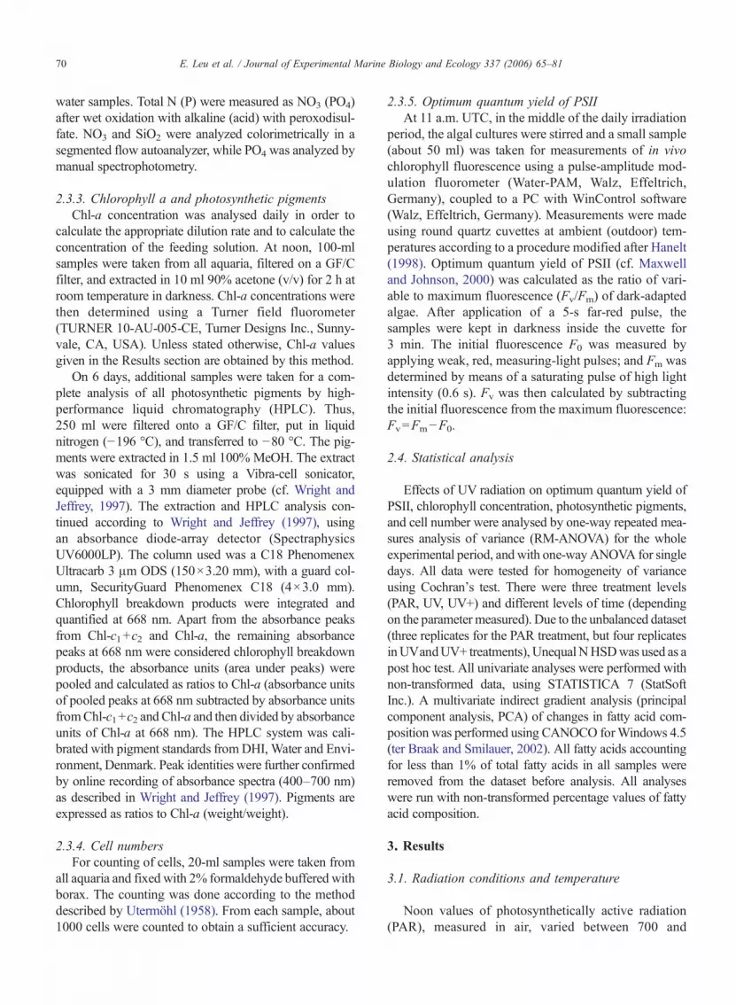
1200 !mol m!2 s!1. PAR intensities (logged in themiddle of one empty aquarium) exerted a pronounceddiurnal cycle, with noon values ranging from 200 to540 !mol m!2 s!1 (mean: 360 !mol m!2 s!1). Corre-sponding values measured near midnight, depending oncloud conditions, were between 20 and 70 !mol m!2 s!1.In themiddle of the aquarium, under cloudless conditions,the PAR intensity was 20% of the above-surface PAR,while the corresponding percentage during overcastweather was between 50% and 60% (Fig. 4A).
Stratospheric ozone values were high throughout theexperimental period, but showed a decreasing trend—from 400 to 360DU—during the experiment (R2 =0.74).Measured in the middle of one aquarium in theambient UV treatment, daily doses of unweightedUVA and UVB ranged from 334 to 685 kJ m!2, and 9to 28 kJ m!2, respectively (Fig. 4B). Inside the aquariaof the ambient UV treatment, there were no clear timetrends with regard to daily doses. In the UV+ treatment,after lowering the fluorescent tubes on day 7, the daily
Fig. 4. A. Temporal variations of incident PAR (!mol m!2 s!1) 19.05.–04.06.2004, measured inside the aquarium and outside. Shown are averagevalues per hour, calculated from measurements every 5th minute. B. Unweighted daily doses for UVA and UVB (kJ m!2), measured in the air andcalculated for the aquarium (ambient) and the UV+treatment (enhanced). Missing values are due to incomplete measuring series for the respectivedays.
71E. Leu et al. / Journal of Experimental Marine Biology and Ecology 337 (2006) 65–81
doses of UVB were significantly higher (t-Test, p<0.05);but for UVA, the difference was not significant. Duringthe first week of the experiment, with the UV+ treatment,daily doses of UVA and UVB were 18% and 48% higher,respectively, than the average ambient doses. Uponweighting of the UV radiation data (Cullen and Neale,1997), the increases were seen to be 22% and 69% higherthan average ambient doses. By day 7, the unweightedincreases were 37% and 86%, respectively, for UVA andUVB. But on all days the daily dose in the UV+ treatmentremained at lower values than those measured at thesurface.
Compared to underwater measurements in the fjordaround noon on a sunny day, the noon UVA intensities intheUVambient treatment (mean 15Wm!2) correspondedto values measured at 4 m depth (min 6.4 W m!2=9 m;max 24.5 W m!2=2 m). For UVB, the correspondingvalues were: mean=0.62 W m!2; min=0.21 W m!2;max=1.0Wm!2, whichwere equal to fieldmeasurementsat 2m, 4m, and 0mdepth. Themaximum intensities in theUV+ treatment (for day 0–7, and day 8–16, respectively)were 2.6 and 4.6 W m!2 higher than UVA in the ambienttreatment; for UVB, the added intensities were 0.2Wm!2
during the first week and 0.34 W m!2 during the second.The water temperature during the entire experimentdisplayed diurnal cycles, fluctuating mainly between 0and +2 °C. This was close to ambient surface conditions inthe fjord, where temperatures ranged from !1 to +1.5 °Cduring the time of the experiment.
3.2. General phytoplankton development
All aquaria underwent the same pronounced changesin phytoplankton responses over the experimental
period. Thus for clarity, we have chosen to first presentthe general time course development of the majorparameters before proceeding to the specific responseson the irradiation treatments.
3.2.1. Fluorometrically determined Chl-a concentra-tions and cell numbers
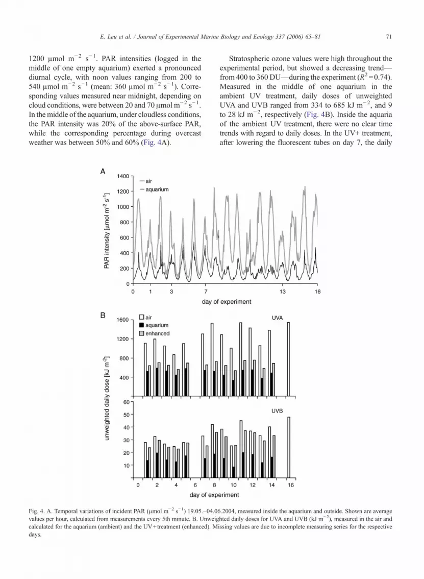
In all aquaria, Chl-a concentrations were between 1and 2 !g l!1 at the start of the experiment, and increasedslowly towards 10 !g l!1 on day 7. During the secondpart of the experimental period (day 8–16), Chl-a variedbetween 20 and 30 !g l!1. The cumulative biomassincrease was calculated from the fluorometrically deter-mined Chl-a values (Fig. 5). Note that these estimatedconcentrations did by far exceed the real Chl-a concen-trations (given above), since this estimate also includesthe biomass removed daily by dilution. Hence, Fig. 5shows an exponential increase of Chl-a over the durationof the experiment that would have occurred ideallywithout diluting the cultures. One aquarium from thePAR treatment differed markedly from this develop-ment: Here, concentrations remained at 2 !g l!1 for theduration of the experiment; data from this replicate weretherefore excluded from further analysis. Cell numbersincreased from 0.7–1!106 cells per litre on day 3 tovalues between 3 and 6!106 cells per litre on days 13and 16. There were no significant differences betweenthe different treatments on any day. Contamination of theaquaria with species other than T. antarctica was foundto be negligible.
3.2.2. Fatty acid compositionThe fatty acid composition showed distinct changes
during the course of the experiment, irrespective of the
Fig. 5. Cumulative biomass increase of T. antarctica during the experiment; shown are theoretical Chl-a concentrations that would have beenmeasured without dilution. Calculations are based upon fluorometrically measured values (mean values and standard deviation, n=3 in PAR andn=4 in UV and UV+).
72 E. Leu et al. / Journal of Experimental Marine Biology and Ecology 337 (2006) 65–81
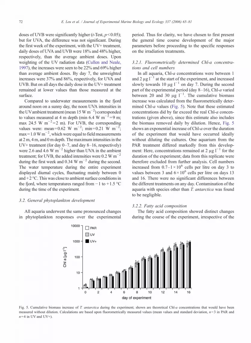
different radiation treatments (Table 1, Fig. 6). The mostconspicuous alteration of the profile was observed at thebeginning (between day 0 and day 1), and samples takenon the last day of the experiment (day 16) were mostsimilar to the day 0 samples (see Table 1). The per-centage fatty acid composition of the total lipids of T.antarctica before the onset of the experiment wasdominated by 16:0 (22%) and 16:1 n-7 (39%). Amongthe saturated fatty acids, 14:0 accounted for 9% and 18:0for 4%; while the predominant polyunsaturated fattyacids (PUFAs) were 16:3 n-4, 16:4 n-1, 18:4 n-3, and20:5 n-3. Of these, the last two n-3 fatty acids were mostabundant, at 5.5% and 7.2%, respectively. The PUFA22:6 n-3 accounted for less than 1% of the total fattyacid composition. Over the first day, the proportion of16:1 n-7 decreased to between 14% and 20%. Also overthis first day, percentages of total PUFAs were halvedcompared to their initial levels (Fig. 6). At the sametime, 18:0 increased from 4% to values between 21%and 26% in all treatments, and 16:0 increased from 22%to 29%. A monounsaturated fatty acid, 18:1 n-9, ac-counted for only 1% on day 0, but increased to 7–11%on day 1. Results from day 3 differed from day 1 onlyslightly: some PUFAs were present in even lower
Table1
Changes
infatty
acid
compo
sitio
nof
T.an
tarcticadu
ring
theexperiment
Day
0Day
1Day
3Day
7Day
13Day
16
PAR
UV
UV+
PAR
UV
UV+
PAR
UV
UV+
PAR
UV
UV+
PAR
UV
UV+
14:0
9.0±0.0
5.4±0.2
5.5±1.0
5.0±0.5
6.7±0.4
6.7±0.4
6.3±0.3
5.5±0.2
6.2±0.7
5.8±0.5
11.0±2.5
10.3±1.6
10.0±0.8
7.1±1.5
8.4±1.1
9.1±0.4
16:0
22.4±0.2
28.2±0.2
29.5±3.2
28.9±0.7
31.9±1.2
30.1±2.9
33.5±1.9
25.2±1.1
24.3±1.7
25.6±1.0
29.7±5.7
28.8±5.4
24.7±1.2
27.5±1.8
30.0±1.1
25.9±1.6
16:1
n-9
0.6±0.0
1.0±0.2
0.8±0.1
1.2±0.1
1.9±0.4
1.2±0.1
1.9±0.2
1.4±0.3
1.2±0.3
1.3±0.1
1.1±0.6
1.5±0.8
0.7±0.1
1.4±1.5
1.0±0.4
0.4±0.1
16:1
n-7
38.8±0.2
20.2±2.0
17.8±5.3
14.3±5.7
20.8±1.0
20.7±3.8
13.7±2.1
17.6±1.1
18.3±3.3
14.8±2.8
23.0±12
.123
.9±6.5
26.3±3.6
27.8±13
.425
.6±5.8
30.1±3.0
16:2
n-7
1.1±0.0
0.4±0.0
0.5±0.1
0.3±0.1
0.5±0.0
0.7±0.1
0.4±0.1
0.6±0.1
0.8±0.2
0.7±0.2
1.1±0.4
1.0±0.5
1.3±0.1
0.6±0.3
0.7±0.2
1.0±0.1
16:3
n-4
3.0±0.1
1.0±0.1
0.9±0.3
0.7±0.3
1.2±0.0
1.2±0.2
0.7±0.2
0.7±0.6
1.0±0.4
0.7±0.2
2.4±0.9
1.2±0.4
1.7±0.2
1.3±0.7
1.0±0.3
1.7±0.3
16:4
n-1
2.5±0.1
1.0±0.1
1.2±0.4
0.9±0.3
1.5±0.1
2.3±0.4
1.6±0.2
3.8±0.5
5.1±1.1
4.5±1.2
5.7±1.4
4.8±1.4
6.9±1.2
2.5±0.8
2.9±0.4
4.3±0.5
17:0
0.9±0.0
0.8±0.1
0.8±0.1
0.8±0.1
0.9±0.1
1.0±0.0
0.9±0.0
1.1±0.1
1.2±0.1
1.2±0.2
1.3±0.4
1.6±0.4
1.7±0.4
0.9±0.1
1.0±0.1
1.2±0.2
18:0
4.2±0.1
22.7±1.5
21.1±3.2
25.5±3.0
16.8±1.2
18.6±2.6
20.8±1.5
22.0±2.0
19.7±5.1
24.1±4.0
6.3±2.5
9.1±5.2
8.1±1.4
12.6±13
.112
.0±7.1
6.4±1.9
18:1
n-9
1.1±0.0
6.7±1.2
5.8±0.6
11.0±4.7
6.7±0.6
5.4±0.6
10.9±2.4
5.6±0.4
4.7±1.3
6.7±1.5
2.4±1.5
4.2±2.9
2.3±0.4
4.1±4.6
3.0±1.8
1.7±0.6
18:1
n-7
0.3±0.0
0.6±0.0
0.5±0.1
0.8±0.1
0.5±0.0
0.5±0.1
0.7±0.0
0.5±0.0
0.4±0.0
0.5±0.0
0.4±0.1
0.3±0.1
0.3±0.0
0.5±0.1
0.4±0.0
0.3±0.0
18:2
n-6
0.4±0.0
2.8±0.3
2.1±0.1
3.3±0.6
1.9±0.4
1.6±0.3
2.2±0.8
1.7±0.1
1.2±0.3
1.4±0.4
0.8±0.0
1.4±0.7
0.9±0.1
1.0±0.5
1.7±0.6
0.9±0.1
18:4
n-3
5.5±0.1
3.0±0.3
2.7±0.9
2.1±0.8
2.8±0.4
3.1±0.6
2.0±0.3
3.8±0.4
3.9±1.0
3.1±0.5
6.3±2.2
3.7±0.7
5.1±0.7
4.7±1.9
3.7±0.6
5.7±1.0
20:5
n-3
7.2±0.2
3.2±0.3
3.2±1.0
2.5±1.0
1.8±0.1
3.3±1.3
1.4±0.9
6.7±1.0
8.2±1.9
6.5±1.7
4.9±2.6
4.4±3.4
5.9±1.9
5.2±2.5
5.2±1.2
7.9±0.8
22:6
n-3
0.8±0.0
0.6±0.1
0.5±0.1
0.4±0.2
0.3±0.2
0.3±0.1
0.2±0.1
1.2±0.2
1.3±0.3
1.1±0.3
0.8±0.3
1.0±0.5
1.0±0.4
0.8±0.3
0.9±0.2
1.3±0.2
Fatty
acidsaregivenas
percentage
values
oftotallipid;sho
wnaremeans
±SD
,n=3forPA
R,n
=4forUVandUV+.
Fig. 6. Changes in polyunsaturated (PUFA), monounsaturated(MUFA), and saturated fatty acids (SFA) during the experiment andin the three different treatments (as % of total lipid).
73E. Leu et al. / Journal of Experimental Marine Biology and Ecology 337 (2006) 65–81

proportions, and there was significantly less 18:0 (17–22%) and higher average percentages of 16:0. After1 week, 16:0 decreased slightly in all aquaria, 18:0 in-creased again to the levels that had been observed on day1, while 18:1 n-9 declined. All PUFAs increased sub-stantially: 20:5 n-3 reached values similar to day 0 (7–8%); 16:4 n-1 and 22:6 n-3 were even higher than in thebeginning (4–5% and 1.1–1.3%, respectively). On day13, 14:0 had doubled compared to day 7 and accounted formore than 10%. Both 16:0 and 16:1 n-7 increased as well.18:0 dropped to values below 10% in all treatments, and18:1 n-9 showed lower values than before. 20:5 n-3 and22:6 n-3 decreased slightly, while 18:4 n-3 and 16:4 n-1increased. Results from the last sampling of the experi-
ment showed general resemblance to the starting fatty acidcomposition.
3.2.3. Stoichiometry and nutrientsMolar ratios of particulate C:N were between 12 and
14 in all treatments on day 7; they then decreased to lessthan 10 during the latter part of the experiment (Table 2).In contrast, the particulate C:P ratios increased duringthe experiment, from around 100 on day 7 to 150 on day16. The ratio between N and P increased more markedly,from between 6 and 10 on day 7 to nearly 20 on day 16.Total nitrogen and phosphorus concentrations were mea-sured on days 0, 7, and 16, and were >1500 and 150 !gl!1, respectively.
3.2.4. Photosynthetic pigmentsThe ratios of fucoxanthin to Chl-a (weight/weight) and
of diadinoxanthin+diatoxanthin toChl-a (Dd+Dt/Chl-a)decreased during the experiment (Table 3); but betacar-otene to Chl-a ratios were stable, around 0.02 on all days.The total sum of chlorophyll breakdown products to Chl-aincreased over time, from 0.4 to 0.5–0.6.
3.2.5. Optimum quantum yield of PSIIOptimum quantum yield measurements on 9 days
(day 6 to day 15) showed values between 0.35 and 0.56for Fv /Fm (Fig. 7). In all aquaria, the optimum quantumyield of PSII decreased toward the end of the ex-periment. A measurement of the culture before the startof the experiment yielded Fv /Fm=0.55.
3.3. Effects of UV radiation
An overview of the changes that UV had on thedifferent parameters is given in Table 4. Effects of UVradiation on fatty acid composition, photosynthetic
Table 2Molar ratios and concentrations [!g l!1] of C, N, and P in T.antarctica; !: UV treatments which differed from the PAR control in astatistically significant way (One-way ANOVA, Unequal N HSD,p<0.05); shown are means±SD, n=3 for PAR, n=4 for UV, UV+
Treatment Day 7 Day 13 Day 16
C:N PAR 12.8±1.2 10.7±2.8 9.0±2.2UV 13.0±2.3 10.1±1.9 7.1±0.6UV+ 13.8±3.0 10.1±1.3 8.5±2.2
C:P PAR 117.8±3.4 120.5±30.3 175.5±88.8UV 75.8±11.3! 136.9±51.1 132.6±46.8UV+ 81.6±4.1! 136.3±21.0 151.9±30.1
N:P PAR 9.3±1.2 11.3±0.7 18.7±4.6UV 5.9±0.4! 13.4±4.1 18.4±4.9UV 6.1±1.5! 13.7±2.6 19.0±7.4
C PAR 2035±230 2120±152 2408±1181UV 2032±110 2745±497 2526±503UV+ 2149±137 1950±333 2197±501
N PAR 188±41 219±24 300±70UV 185±26 338±63 413±48UV+ 189±50 236±79 317±115
P PAR 45±3.8 39±5.7 36±0.7UV 70±7.6! 60±14.8 52±14.2UV+ 68±4.6! 39±5.1 38±9.3
Table 3Photosynthetic pigments as ratios to Chl-a; !: UV treatments which differed from the PAR control in a statistically significant way (One-wayANOVA, Unequal N HSD, p<0.05), shown are means±SD, n=3 for PAR, n=4 for UV and UV+
Treatment Day 1 Day 7 Day 13
Dd+Dt /Chl-a PAR 0.049±0.003 0.043±0.002 0.031±0.004UV 0.058±0.008 0.057±0.004 0.036±0.003UV+ 0.084±0.011 0.071±0.006! 0.029±0.001
Fucoxanthin /Chl-a PAR 0.393±0.094 0.348±0.045 0.232±0.023UV 0.496±0.044 0.437±0.054 0.206±0.017UV+ 0.610±0.055 0.452±0.021 0.221±0.005
Betacarotene /Chl-a PAR 0.019±0.008 0.018±0.001 0.024±0.001UV 0.023±0.003 0.017±0.001 0.019±0.001UV+ 0.026±0.002 0.018±0.001 0.017±0.001!
Chl-a breakdown/Chl-a PAR 0.278±0.031 0.353±0.045 0.398±0.033UV 0.298±0.020 0.285±0.017 0.301±0.008!
UV+ 0.314±0.024 0.317±0.036 0.316±0.020!
74 E. Leu et al. / Journal of Experimental Marine Biology and Ecology 337 (2006) 65–81
pigments, and particulate C:N:P were found only ondays 1–7; whereas optimum quantum yield of PSII wasalso affected later in the experiment, as measured ondays 8 to 15.
3.3.1. Fatty acid compositionIn general, only the algae exposed to artificially
enhanced UV radiation showed treatment-dependentchanges in their fatty acid composition. An overviewover the significance of UV effects on the fatty acidcomposition for the analysed days is given in Table 5.Although it is mainly on day 3 that significant changesin the fatty acid composition were detectable, the sametrends (though not statistically significant) were seen on
days 1 and 7 as well. Thus, for clarity, we have chosen todescribe the effects of UV radiation on fatty acid com-position as divided into two periods: day 1–7 and day13–16. We believe this represents well the transitionfrom “initial” to “acclimated” conditions.
Results from the first week of the experiment showedtreatment-dependent effects, with the UV+ treatmentdiffering from the other two. But on day 13 and 16, nosignificant treatment effects were detectable. A principlecomponent analysis (PCA) of the fatty acid compositionpercentage data (Fig. 8) shows also the effect of bothtime and treatment for the period day 1–7. While theamount of PUFAs changed significantly with time, the
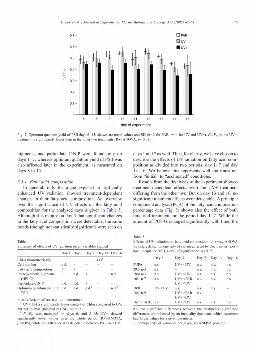
Fig. 7. Optimum quantum yield of PSII days 6–15; shown are mean values and SD (n=3 for PAR, n=4 for UV and UV+). Fv /Fm in the UV+treatment is significantly lower than in the other two treatments (RM ANOVA, p<0.05).
Table 4Summary of effects of UV radiation on all variables studied
Day 1 Day 3 Day 7 Day 13 Day 16
Chl-a fluorometrically ! ! ! (+)a !Cell number n.d. ! ! ! !Fatty acid composition + + ! ! !Photosynthetic pigments
(HPLC)! n.d. + ! n.d.
Particulate C:N:P n.d. n.d. + ! !Optimum quantum yield of
PSIIn.d. n.d. n.d.b + n.d.b
!: no effect; +: effect; n.d.: not determined.a UV+ had a significantly lower content of Chl-a compared to UV,
but not to PAR (unequal N HSD: p=0.03).b Fv /Fm was measured on days 6, and 8–15; UV+ showed
significantly lower values over the whole period (RM-ANOVA,p<0.05), while no difference was detectable between PAR and UV.
Table 5Effects of UV radiation on fatty acid composition: one-way ANOVAfor single days, homogeneity of variances tested by Cochran-test, post-hoc: unequal N HSD, Level of significance: p<0.05
Day 1 Day 3 Day 7 Day 13 Day 16
PUFA n.s. UV+<UV n.s. n.s. n.s.20:5 n-3 n.s. – n.s. n.s. n.s.18:4 n-3 n.s. UV+<UV n.s. n.s. n.s.16:1 n-7 n.s. UV+<PAR
UV+<UVn.s. n.s. n.s.
18:0 UV<UV+ n.s. n.s. n.s. –18:1 n-9 – UV+>PAR
UV+>UVn.s. – –
16:1 / 16:0 n.s. UV+<UV n.s. n.s. n.s.
n.s.: no significant differences between the treatments; significantdifferences are indicated by an inequality that states which treatmenthad larger values for a given parameter.–: homogeneity of variances not given; no ANOVA possible.
75E. Leu et al. / Journal of Experimental Marine Biology and Ecology 337 (2006) 65–81
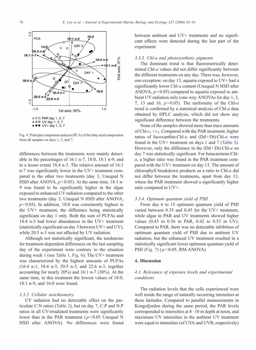
differences between the treatments were mainly detect-able in the percentages of 16:1 n-7, 18:0, 18:1 n-9, andto a lesser extent 18:4 n-3. The relative amount of 16:1n-7 was significantly lower in the UV+ treatment com-pared to the other two treatments (day 3, Unequal NHSD after ANOVA, p<0.05). At the same time, 18:1 n-9 was found to be significantly higher in the algaeexposed to enhanced UVradiation compared to the othertwo treatments (day 3, Unequal N HSD after ANOVA,p<0.05). In addition, 18:0 was consistently highest inthe UV+ treatment, the difference being statisticallysignificant on day 1 only. Both the sum of PUFAs and18:4 n-3 had lower abundances in the UV+ treatment(statistically significant on day 3 between UV+ and UV),while 20:5 n-3 was not affected by UV radiation.
Although not statistically significant, the tendenciesfor treatment-dependent differences on the last samplingday of the experiment were contrary to the situationduring week 1 (see Table 1, Fig. 6): The UV+ treatmentwas characterized by the highest amounts of PUFAs(16:4 n-1, 18:4 n-3, 20:5 n-3, and 22:6 n-3, togetheraccounting for nearly 20%) and 16:1 n-7 (30%). At thesame time, in this treatment the lowest values of 18:0,18:1 n-9, and 16:0 were found.
3.3.2. Cellular stoichiometryUV radiation had no detectable effect on the par-
ticulate C:N ratios (Table 2), but on day 7, C:P and N:Pratios in all UV-irradiated treatments were significantlylower than in the PAR treatment ( p<0.05 Unequal NHSD after ANOVA). No differences were found
between ambient and UV+ treatments and no signifi-cant effects were detected during the last part of theexperiment.
3.3.3. Chl-a and photosynthetic pigmentsThe dominant trend is that fluorometrically deter-
mined Chl-a values did not differ significantly betweenthe different treatments on any day. There was, however,one exception: on day 13, aquaria exposed to UV+ had asignificantly lower Chl-a content (Unequal N HSD afterANOVA, p<0.05) compared to aquaria exposed to am-bient UV radiation only (one-way ANOVAs for day 1, 3,7, 13 and 16, p>0.05). The uniformity of the Chl-atrend is confirmed by a statistical analysis of Chl-a dataobtained by HPLC analysis, which did not show anysignificant difference between the treatments.
None of the samples showedmore than trace amountsof Chl-c1+c2. Compared with the PAR treatment, higherratios of fucoxanthin:Chl-a and (Dd+Dt):Chl-a werefound in the UV+ treatment on days 1 and 7 (Table 3).However, only the difference in the (Dd+Dt):Chl-a onday 7 was statistically significant. For betacarotene:Chl-a, a higher ratio was found in the PAR treatment com-pared with the UV+ treatment on day 13. The amount ofchlorophyll breakdown products as a ratio to Chl-a didnot differ between the treatments, apart from day 13,where the PAR treatment showed a significantly higherratio compared to UV+.
3.3.4. Optimum quantum yield of PSIIFrom day 6 to 15 optimum quantum yield of PSII
varied between 0.35 and 0.45 for the UV+ treatment,while algae in PAR and UV treatments showed highervalues (0.43 to 0.56 in PAR, 0.42 to 0.53 in UV).Compared to PAR, there was no detectable inhibition ofoptimum quantum yield of PSII due to ambient UVradiation, but the enhanced UV treatment resulted in astatistically significant lower optimum quantum yield ofPSII (Fig. 7) ( p<0.05, RM-ANOVA).
4. Discussion
4.1. Relevance of exposure levels and experimentalconditions
The radiation levels that the cells experienced werewell inside the range of naturally occurring intensities atthese latitudes. Compared to parallel measurements inKongsfjorden during the same period, the PAR levelscorresponded to intensities at 8–10 m depth at noon, andmaximum UV intensities in the ambient UV treatmentwere equal to intensities (of UVA and UVB, respectively)
Fig. 8. Principal component analysis (PCA) of the fatty acid compositionfrom all samples on days 1, 3, and 7.
76 E. Leu et al. / Journal of Experimental Marine Biology and Ecology 337 (2006) 65–81

measured at 2–4 and 1–2m depth. In all cases, even in theUV+ treatment, the daily doses in the experiment werelower than the corresponding daily doses measured in air.The transition in light intensity between the pre-experimental phase and the experimental conditions wasmade as smooth as possible, with the transfer taking placeat 8:00 p.m. local time. However, especially at these highlatitudes, algae are experiencing rapid changes in theirnatural habitat as well (see discussion later). A relativelyshort exposure time resulted in the major effects that wenote here, but upon extended exposure to high PAR andUV radiation, the phytoplankton seemed to be able toadapt to it. Similar acclimation to UV radiation (after14 days) was found in a chlorophyte from Antarctica(Lesser et al., 2002), whereas acclimation took placemuch faster in a study of the subtropical diatom Chaeto-ceros gracilis (Hazzard et al., 1997). Nutrient levelsduring the experiment were even higher than thoseusually reported during winter–early-spring in Kongsf-jorden (Leu et al., submitted for publication-b; Hop et al.,2002) and certainly well above limiting conditions.Temperatures were close to ambient values measured inthe fjord at the same time.
4.2. Treatment effects: UV radiation vs. PAR
The UV radiation, which was similar to naturalconditions found in the high Arctic, affected the fattyacid composition and the cellular stoichiometry of T.antarctica var. borealis. The algae also responded byreducing the optimum quantum yield of PSII andincreasing the ratio (compared to Chl-a) of the photo-protective pigments diadinoxanthin and diatoxanthin.However, these effects occurred during only the firstpart of the experiment. During the later part of theexperiments, no effects of the applied UV radiation werefound in any of the investigated parameters, apart fromoptimum quantum yield of PSII. This held true eventhough the cells that were given enhanced UV radiationreceived increased doses from day 7 onwards.
The largest changes in the fatty acid composition of T.antarctica occurred immediately after the start of theexperiment, and were independent of the different treat-ments. Compared to the conditions during pre-cultiva-tion in the laboratory, light intensity was the parameterthat changed most profoundly. At the outset, tempera-tures decreased from + 2.6 °C (average for last 24 h priorto experiment start) to ! 0.2 °C (average for the first 24 hof experiment). According to the homeoviscous adapta-tion theory (Sinensky, 1974), we might have expectedthat with a decrease in temperature, the fatty acidcomposition would have shown an increase in PUFAs,
but we observed the opposite. Thus, we assume that thedifferences in fatty acid composition were causedprimarily by the change in irradiation intensity. Support-ing this view, the pattern of changes in the fatty acidcomposition due to enhanced PAR resembled closely thepattern we observed after UV exposure. In addition,PUFAs were substantially reduced by increased PAR.According to Thompson et al. (1990), for differentspecies of marine phytoplankton the content of PUFAsand the relative amounts of 16:0 and 16:1 n-7 aredependent on light intensity. This is in accordance withour results for T. antarctica, in that at the start of theexperiment, the decrease in the relative fatty acid contentwas strongest in 16:1 n-7 and in the PUFAs. At the sametime, 18:0, 18:1 n-9, and 18:2 n-6 increased consider-ably. An increase in 18:0, 18:1 n-9, and 18:2 n-6, incombination with a drop in the content of PUFAs, pointsto an inhibition in the synthesis of PUFAs. Arao andYamada (1994) describe different possible pathways forthe synthesis of 20:5 n-3 in a marine diatom; in thesepathways, the first products were the three C18 fattyacids mentioned above. In our experiment, PUFAsynthesis resumed between day 3 and day 7; by thattime PUFA levels were essentially back to startconditions. On the other hand, it was only on day 16that 16:1 n-7 and 18:0 reached values similar to thosefound on day 0.
The close coupling between PAR-and UV-inducedeffects on algal physiology and biochemistry in theseresults underlines the need for outdoor experimentsunder natural light intensities; this work also highlightsthe importance of a realistic UV:PAR ratio duringexposure. Not only can these major spectral componentscause similar changes in the investigated organisms, butexposure to high PAR intensities has also been shown todecrease phytoplankton sensitivity towards UV stress ingeneral (see Vernet, 2000 and references therein; Roosand Vincent, 1998). In Isochrysis galbana, an increasein PAR intensity, both during pre-cultivation and UVexposure, led to a reduced effect of UV radiation on n-3fatty acids (Wang and Chai, 1994).
4.3. The effect of UV radiation
4.3.1. Fatty acid compositionThe development of the fatty acid composition of T.
antarctica var. borealis that we observed did not supportearlier reports about UV radiation causing specificdamage or reduced synthesis of PUFAs. The only n-3fatty acid responding negatively to UV radiation was18:4 n-3. However, this fatty acid is of minor impor-tance in diatoms (Dalsgaard et al., 2003), and accounted
77E. Leu et al. / Journal of Experimental Marine Biology and Ecology 337 (2006) 65–81
for only 2–6% of the total fatty acids in T. antarctica.But 20:5 n-3, the most abundant PUFA in diatoms, wasnot reduced by UV radiation. Nor was 16:4 n-1, which,due to its high number of double bonds, a priori, shouldbe most sensitive to oxidative stress caused by UVradiation (Halliwell and Gutteridge, 1989; Cosgrove etal., 1987). Rather, these two fatty acids were even higherin the algae exposed to enhanced UV radiation (com-pared to the other two treatments at the end of theexperiment, although this was statistically significantonly for 16:4 n-1). Our results for 16:4 n-1 contradict thefindings of Goes et al. (1994), who suggested that theratio between 16:0 and 16:4 could be used as a markerfor UVB stress, after observing a selective reduction ofthe latter in the marine prasinophyte Tetraselmis sp. Insupport of our findings, Döhler and Biermann (1994)also described an increase of 20:5 n-3 in response toirradiation with UVB. There have been reports of spe-cies-specific variation in the response of 20:5 n-3 to UVradiation (Skerratt et al., 1998; Wang and Chai, 1994)with a decrease of 20:5 n-3 in some species.
As an alternative to the idea that radiation could havea direct effect on a specific type of fatty acid, we favourthe concept that the changes are downstream effectsresulting from a generally weakened state. For this, wehave other indications: First, if oxidative stress were themechanism responsible for the observed changes, fattyacids with a higher number of double bonds should bemore severely affected thanmonounsaturated fatty acids.And second, 16:1 n-7 has been shown to be generallycorrelated with growth rate (e.g. Kattner and Brock-mann, 1990; Thompson et al., 1990). From this point ofview, a decrease in this fatty acid could be seen as merelya general indicator of stress due to shifting conditions,rather than a specific target of high radiation levels.
4.3.2. Cellular stoichiometryA significant decrease in the molar ratio of particulate
C:P and N:P under both ambient and UV+ radiation wasfound on day 7 (we have no data for day 1), while the C:Nratio was not affected. This supports findings regardingfreshwater algae (Tank et al., 2003; Watkins et al., 2001;Xenopoulos et al., 2002) and is verified by several tests(laboratory experiments) with various marine and fresh-water species (Leu et al., submitted for publication-a, Leuet al., in press). The reduction of C:P and N:P ratios wascaused by higher P concentrations in the UV exposedtreatments, whereas C concentrationswere not affected byUV radiation (Table 2, Unequal N HSD). We suggesttherefore that these changes in C:P ratios can be attributedto an increase in cellular P, rather than to a decrease in C,which agrees with data fromWängberg et al. (1998). The
most likely explanation for the UV-dependent decrease ofC:P and N:P is therefore an increased need for nutrientsfor cellular repair, a view suggested by Hessen et al.(1995).
The C:N ratios on day 7 were higher than most (butnot all) values reported in the literature (e.g. Shifrin andChisholm, 1981; Berges et al., 2002; Viso and Marty,1993). This high ratio could indicate nitrogen limitation,but this is highly unlikely, due to the addition of N inexcess. We therefore suggest that high, or rapidly in-creasing PAR intensities probably caused an inhibitionof nitrogen assimilation, which disappeared after someperiod of adaptation to the new radiation regime (thiscould also explain the decrease in C:N and increase inN:P and C:P on days 13 and 16). It has previously beenshown that nitrogen metabolism can be affected in thismanner by UV radiation (Döhler et al., 1995), and it isnot unlikely that high intensities of PAR could have asimilar effect. Interestingly, Mock and Kroon (2002)found that for Antarctic phytoplankton, the physiolog-ical response to nitrogen limitation is very similar to areaction towards light stress. Here, we see that highirradiation levels or a rapid light increase probablycaused effects similar to nutrient stress.
4.3.3. Photosynthetic pigmentsUV radiation has been found to stimulate the
diadinoxanthin cycle (i.e. the de-epoxidation of Dd toDt under strong light) usually as an increased conversionof Dd to Dt (Goss et al., 1999). But there are differentopinions regarding time scales for the conversion of Ddto Dt—from seconds, to minutes (Olaizola and Yama-moto, 1994; Willemoes and Monas, 1991), and hours(Demers et al., 1991). Our sampling design did not allowfor estimating the conversion between Dd to Dt,particularly not if the conversion takes place withinminutes. Therefore we used the ratio of (Dd+Dt) /Chl-ato indicate a photoadaptive response to high lightintensities (Brunet et al., 1993). The treatment effect onday 7—a higher ratio of these photoprotective pigmentsin both UV treatments—disappeared on day 13 andparalleled the results for stoichiometry and fatty acidcomposition (partly), indicating that over the experi-mental period, the microalgae accomplished at leastpartially an acclimation to UV radiation.
High concentrations of Chl-a degradation productsindicate senescence in microalgae and might be aconsequence of light stress. But the ratios found herewere so high that we assume a contamination of oursamples by dead algal cells; these probably had sedi-mented to the bottom of the aquaria, and were re-suspended when the water was stirred before sampling.
78 E. Leu et al. / Journal of Experimental Marine Biology and Ecology 337 (2006) 65–81
4.3.4. Optimum quantum yield of PSIIFor optimum quantum yield of PSII, we found no
differences between the PAR treatment and the ambientUV treatment. This confirms the results by Lesser et al.(1996), who undertook an outdoor experiment with adiatom-dominated phytoplankton community at asimilarly high latitude (78° S near McMurdo Stationin Antarctica). In addition, the values they found foroptimum quantum yield of PSII were very similar toours, and optimum quantum yield of PSII declined as aresponse to high PAR intensities (increased radiationfrom 17% to 41% of ambient). In a field study north ofSvalbard in May 2003, for near-surface phytoplankton,optimum quantum yield of PSII values of approximately0.5 were also found (McMinn and Hegseth, 2004). Inour UV+ treatment, however, we observed a decrease inoptimum quantum yield of PSII over the measurementperiod (day 6–15), indicating that the applied level of extraUV posed a stress factor to the algae. This is particularlyinteresting because none of the other parameters werenegatively affected by UV radiation beyond day 7. Op-timum quantum yield of PSII, therefore, seems to be arather sensitive parameter for documenting UV stress.
4.4. Changes in food quality vs. food quantity
Neither Chl-a concentrations nor cell numbers weresignificantly affected by UV radiation. In general, thenutritional quality was more sensitive to light manip-ulations than biomass. However, since the food qualitywas not affected negatively (no decrease in PUFAs,and no increase in C:P ratios) and the quantityremained unchanged, it seems to be unlikely that ambientUV radiation has severe food-web effects. In a feedingexperiment, in which the herbivorous copepod C.glacialis was fed the algal cultures from the studydescribed here, no treatment-dependent changes ofzooplankton fatty acid composition, growth, survival, ormoulting rate were found (Leu and Falk-Petersen in ms).The effect of increased PAR on the nutritional quality, bycontrast, was considerable.
4.5. Ecological considerations
The sudden change and exposure to ambient radia-tion conditions at the start of this experiment resemblesthe situation in the field during ice breakup in Arcticspring (compared to pre-experimental conditions, lightintensity increased several-fold). The onset of thespring bloom in the Marginal Ice Zone is tightly linkedto the retreat of the ice edge (Sakshaug and Slagstad,1991; Sakshaug, 2004). The formation of a halocline
between the surface melt water and deeper, more salinewater masses stabilizes the water column throughstratification. Consequently, phytoplankton biomassduring the bloom phase is concentrated in theuppermost water layer (Engelsen et al., 2004), whereit is exposed to high intensities of both PAR and UVradiation. Satellite data revealed a strong phytoplank-ton bloom in the Marginal Ice Zone of the northernBarents Sea about 2 weeks after the ice edge hadreceded (Engelsen et al., 2002). This matches the timerequired for adaptation that we observed in our study.Furthermore, T. antarctica var. borealis is a speciesoften dominating the early phase of a bloom and seemsto be associated with sea ice (Falk-Petersen et al.,2000; Wiktor, 1999). It is likely that it belongs to thegroup of pioneers during a spring bloom, starting thespring production from ice-associated seed material(Michel et al., 2002; Syvertsen, 1991). We suggest,therefore, that the observed reaction patterns in thebiochemistry and physiology of this diatom areimportant for our understanding of the timing and thedevelopment of an ice-edge bloom. Turbulent mixingor sudden variation in cloud cover or in stratosphericozone concentrations are further possible causes forrapid changes in radiation exposure, changes thatphytoplankton in the Arctic are frequently subjected to.Thus, it is important to consider the brief and dynamicnature of the high Arctic spring bloom and to take intoaccount the meteorological conditions in order to fullyunderstand the role of PAR and UV in the ecology ofthe autotrophs. While the capacity for adaptation seemshigh, sudden light exposure after a long period of lowlight appears to affect quality more than quantity, withPAR having a stronger impact on these changes thanUV.
Acknowledgements
We are sincerely grateful to Anette Wold, WojtekMoskal, and Wojciech Walkusz for their invaluable as-sistance in carrying out the experiment. Our thanks goalso to Roberto Sparapani, Roberto Azzolini, and Guidodi Prisco, who generously enabled us to use the facilitiesat the Italian Research Station Dirigibile Italia in Ny–Ålesund. We gratefully acknowledge the help from KåreEdvardsen and Carl-Petter Nielsen with the UVmeasurements and calculations. We furthermore wishto thank Hans Christian Eilertsen for supplying theThalassiosira culture. Vegard Lyngmo, Berit Kaasa, andBirger Skjelbred carried out some of the laboratoryanalyses. This study was financed by the NorwegianResearch Council, project no. 150 331/720. [SS]
79E. Leu et al. / Journal of Experimental Marine Biology and Ecology 337 (2006) 65–81

References
Aas, E., Høkedal, J., Højerslev, N.K., Sandvik, R., Sakshaug, E., 2002.Spectral properties and UV-attenuation in Arctic marine waters. In:Hessen, D.O. (Ed.), UV Radiation and Arctic Ecosystems.Springer, Berlin, pp. 23–56.
Arao, T., Yamada, M., 1994. Biosynthesis of polyunsaturated fattyacids in the marine diatom, Phaeodactylum tricornutum. Phyto-chemistry 35, 1177–1181.
Berges, J.A., Varela, D.E., Harrison, P.J., 2002. Effects of temperatureon growth rate, cell composition and nitrogen metabolism in themarine diatom Thalassiosira pseudonana (Bacillariophyceae).Mar. Ecol. Prog. Ser. 225, 139–146.
Brunet, C., Brylinski, J.M., Lemoine, Y., 1993. In-situ variations of thexanthophylls diatoxanthin and diadinoxanthin — photoadaptationand relationships with a hydrodynamical system in the EasternEnglish-Channel. Mar. Ecol. Prog. Ser. 102, 69–77.
Christie, W.W., 1982. Lipid Analysis. Pergamon Press, New York.Cosgrove, J.P., Church, D.F., Pryor, W.A., 1987. The kinetics of the
autoxidation of polyunsaturated fatty acids. Lipids 22, 299–304.Cullen, J.J., Neale, P.J., 1997. Biological weighting functions for
describing the effects of ultraviolet radiation on aquatic systems.In: Häder, D.-P. (Ed.), Effects of Ozone Depletion on AquaticEcosystems. R.G.Landes, Austin, TX, pp. 97–118.
Dahlback, A., 2002. Recent changes in surface ultraviolet solarradiation and stratospheric ozone at a high Arctic site. In: Hessen,D.O. (Ed.), Radiation and Arctic Ecosystems. Springer-Verlag,Berlin, pp. 1–22.
Dalsgaard, J., St John, M., Kattner, G., Muller-Navarra, D., Hagen, W.,2003. Fatty acid trophic markers in the pelagic marine environ-ment. Adv. Mar. Biol. 46, 225–340.
Demers, S., Roy, S., Gagnon, R., Vignault, C., 1991. Rapid light-induced changes in cell fluorescence and in xanthophyll—cyclepigments of Alexandrium excavatum (Dinophyceae) and Thalas-siosira pseudonana (Bacillariophyceae)— a photo-protectionmechanism. Mar. Ecol. Prog. Ser. 76, 185–193.
Döhler, G., Biermann, T., 1994. Impact of UV-B radiation on the lipidand fatty acid composition of synchronized Ditylum brightwellii(West) Grunow. Z. Nat.Forsch., C J. Biosci. 49, 607–614.
Döhler, G., Hagmeier, E., David, C., 1995. Effects of solar andartificial UV irradiation on pigments and assimilation of N-15ammonium and N-15 nitrate by macroalgae. J. Photochem.Photobiol., B Biol. 30, 179–187.
Engelsen, O., Hegseth, E.N., Hop, H., Hansen, E., Falk-Petersen, S.,2002. Spatial variability of chlorophyll-a in the Marginal Ice Zoneof the Barents Sea, with relations to sea ice and oceanographicconditions. J. Mar. Syst. 35, 79–97.
Engelsen, O., Hop, H., Hegseth, E.N., Hansen, E., Falk-Petersen, S.,2004.Deriving phytoplankton biomass in theMarginal Ice Zone fromsatellite observable parameters. Int. J. Remote Sens. 25, 1453–1457.
Falk-Petersen, S., Hopkins, C.C.E., Sargent, J.R., 1990. Trophicrelationships in the pelagic, arctic food web. In: Barnes, M.,Gibson, R.N. (Eds.), 24th Europ. Mar. Biol. Symp. AberdeenUniversity Press, pp. 315–333.
Falk-Petersen, S., Hop, H., Budgell, W.P., Hegseth, E.N., Korsnes, R.,Loyning, T.B., Orbaek, J.B., Kawamura, T., Shirasawa, K., 2000.Physical and ecological processes in the marginal ice zone of thenorthernBarents Sea during the summermelt period. J.Mar. Syst. 27,131–159.
Falk-Petersen, S., Timofeev, S., Pavlov, V., Sargent, J.R., 2006.Climate variability and possible effects on arctic food chains. The
role of Calanus. In: Ørbæk, J.B., Tombre, I., Kallenborn, R.,Hegseth, E., Falk-Petersen, S., Hoel (Eds.), EnvironmentalChallenges in Arctic–Alpine Regions. . Arctic–Alpine Ecosystemsand People in a Changing Environment. Springer Verlag, p. 250.
Folch, J., Lees, M., Sloane Stanley, G.H., 1957. A simple method forisolation and purification of total lipids from animal tissue. J. Biol.Chem. Baltim 226, 497–509.
Goes, J.I., Handa, N., Taguchi, S., Hama, T., 1994. Effect of UV-Bradiation on the fatty acid composition of the marine phytoplankterTetraselmis sp.: relationship to cellular pigments. Mar. Ecol. Prog.Ser. 114, 259–274.
Goss, R., Mewes, H., Wilhelm, C., 1999. Stimulation of thediadinoxanthin cycle by UV-B radiation in the diatom Phaeodac-tylum tricornutum. Photosynth. Res. 59, 73–80.
Guillard, R.R.L., 1975. Culture of phytoplankton for feeding marineinvertebrates. In: Smith, W.L., Chanley, M.H. (Eds.), Culture ofMarine Invertebrate Animals. Plenum Press, pp. 29–60.
Hall, J.M., Parrish, C.C., Thompson, R.J., 2002. Eicosapentaenoicacid regulates scallop (Placopecten magellanicus) membranefluidity in response to cold. Biol. Bull. 202, 201–203.
Halliwell, B., Gutteridge, J.M.C., 1989. Free Radicals in Biology andMedicine. Clarendon Press, Oxford.
Hanelt, D., 1998. Capability of dynamic photoinhibition in Arcticmacroalgae is related to their depth distribution. Mar. Biol. 131,361–369.
Hazzard, C., Lesser, M.P., Kinzie, R.A., 1997. Effects of ultravioletradiation on photosynthesis in the subtropical marine diatom,Chaetoceros gracilis (Bacillariophyceae). J. Phycol. 33, 960–968.
Hessen, D.O., Faerovig, P.J., 2001. The photoprotective role of humus—DOC for Selenastrum and Daphnia. Plant Ecol. 154, 263–273.
Hessen, D.O., Van Donk, E., Andersen, T., 1995. Growth-responses,P-uptake and loss of flagellae in Chlamydomonas reinhardtiiexposed to UV-B. J. Plankton Res. 17, 17–27.
Hessen, D.O., Faerovig, P.J., Andersen, T., 2002. Light, nutrients, andP : C ratios in algae: grazer performance related to food quality andquantity. Ecology 83, 1886–1898.
Hop, H., Pearson, T., Hegseth, E.N., Kovacs, K.M., Wiencke, C.,Kwasniewski, S., Eiane, K., Mehlum, F., Gulliksen, B., Wlo-darska-Kowalczuk, M., Lydersen, C., Weslawski, J.M., Cochrane,S., Gerland, S., et al., 2002. The marine ecosystem ofKongsfjorden, Svalbard. Polar Res. 21, 167–208.
Kattner, G., Brockmann, U.H., 1990. Particulate and dissolved fattyacids in an enclosure containing a unialgal Skeletonema costatum(Greve) Cleve culture. J. Exp. Mar. Biol. Ecol. 141, 1–13.
Klein Breteler, W.C.M., Schogt, N., Rampen, S., 2005. Effect ofdiatom nutrient limitation on copepod development: role ofessential lipids. Mar. Ecol. Prog. Ser. 291, 125–133.
Lesser, M.P., Neale, P.J., Cullen, J.J., 1996. Acclimation of Antarcticphytoplankton to ultraviolet radiation: ultraviolet-absorbing com-pounds and carbon fixation. Mol. Mar. Biol. Biotechnol. 5,314–325.
Lesser, M.P., Barry, T.M., Banaszak, A.T., 2002. Effects of UV radiationon a chlorophyte alga (Scenedesmus sp.) isolated from the fumarolefields of Mt. Erebus, Antarctica. J. Phycol. 38, 473–481.
Leu, E., Faeroevig, P., Hessen, D.O., in press. UV effects onstoichiometry and PUFAs of Selenastrum capricornutum andtheir consequences for the grazer Daphnia magna. Freshw. Biol.
Leu, E., Falk-Petersen, S., Hessen, D.O., submitted for publication-a.No negative effects of ultraviolet radiation on food quality ofArctic diatoms. Limnol. Oceanogr.
Leu, E., Falk-Petersen, S., Kwasniewski, S., Wulff, A., Edvardsen, K.,Hessen, D.O., submitted for publication-b. Fatty acid dynamics
80 E. Leu et al. / Journal of Experimental Marine Biology and Ecology 337 (2006) 65–81

during the spring bloom in a high Arctic fjord: importance ofabiotic factors vs. community changes. Can. J. Fish. Aquat. Sci.
Maxwell, K., Johnson, G.N., 2000. Chlorophyll fluorescence—apractical guide. J. Exp. Bot. 51 (345), 659–668.
McMinn, A., Hegseth, E.N., 2004. Quantum yield and photosyntheticparameters of marine microalgae from the southern Arctic Ocean,Svalbard. J. Mar. Biol. Assoc. U.K. 84, 865–871.
Michel, C., Nielsen, T.G., Nozais, C., Gosselin, M., 2002. Significanceof sedimentation and grazing by ice micro-and meiofauna forcarbon cycling in annual sea ice (northern Baffin Bay). Aquat.Microb. Ecol. 30, 57–68.
Mock, T., Kroon, B.M.A., 2002. Photosynthetic energy conversionunder extreme conditions- I: important role of lipids as structuralmodulators and energy sink under N-limited growth in Antarcticsea ice diatoms. Phytochemistry 61, 41–51.
Olaizola, M., Yamamoto, H.Y., 1994. Short-term response of thediadinoxanthin cycle and fluorescence yield to high irradiance inChaetoceros muelleri (Bacillariophyceae). J. Phycol. 30, 606–612.
Roos, J.C., Vincent, W.F., 1998. Temperature dependence of UVradiation effects on Antarctic cyanobacteria. J. Phycol. 34, 118–125.
Sakshaug, E., 2004. Primary and secondary production in the ArcticSeas. In: Stein, R., Macdonald, R.W. (Eds.), The Organic CarbonCycle in the Arctic Ocean. Springer-Verlag, Berlin, pp. 57–82.
Sakshaug, E., Slagstad, D., 1991. Light and productivity of phytoplank-ton in Polar marine ecosystems — a physiological view. Polar Res.10, 69–85.
Sargent, J.R., Whittle, K.J., 1981. Lipids and Hydrocarbons in theMarine Food Web. In: Longhurst, A.R. (Ed.), Analysis of MarineEcosystems. Academic Press, London, pp. 491–533.
Shifrin, N.S., Chisholm, S.W., 1981. Phytoplankton lipids— inter-specific differences and effects of nitrate, silicate and light–darkcycles. J. Phycol. 17, 374–384.
Sinensky, M., 1974. Homeoviscous adaptation— homeostatic processthat regulates viscosity of membrane lipids in Escherichia-Coli.Proc. Natl. Acad. Sci. U. S. A. 71, 522–525.
Skerratt, J.H., Davidson, A.D., Nichols, P.D., McMeekin, T.A., 1998.Effect of UV-B on lipid content of three Antarctic marinephytoplankton. Phytochemistry 49, 999–1007.
Sterner, R.W., Elser, J.J., 2002. Ecological Stoichiometry. PrincetonUniversity Press, Princeton.
Syvertsen, E.E., 1991. Ice algae in the Barents Sea — types ofassemblages, origin, fate and role in the ice-edge phytoplanktonbloom. Polar Res. 10, 277–287.
Tank, S.E., Schindler, D.W., Arts, M.T., 2003. Direct and indirecteffects of UV radiation on benthic communities: epilithic food
quality and invertebrate growth in four montane lakes. Oikos 103,651–667.
ter Braak, C.F.J., Smilauer, P., 2002. CANOCO Reference manual andCanoDraw for Windows User's guide: Software for CanonicalCommunity Ordination (version 4.5). Microcomputer Power,Ithaca, NY, USA. 500 pp.
Thompson, P.A., Harrison, P.J., Whyte, J.N.C., 1990. Influence ofirradiance on the fatty acid composition of phytoplankton. J. Phycol.26, 278–288.
Thompson, P.A., Guo, M.X., Harrison, P.J., Whyte, J.N.C., 1992.Effects of variation in temperature on the fatty acid composition of8 species of marine phytoplankton. J. Phycol. 28, 488–497.
Utermöhl, H., 1958. Zur Vervollkommnung der quantitativenPhytoplankton Methodik. Mitt.Int. Ver. Limnol., vol. 9, pp. 1–38.
Vernet, M., 2000. Effects of UV radiation on the physiology and ecologyof marine phytoplankton. In: de Mora, S., Demers, S., Vernet, M.(Eds.), The Effects of UV Radiation in the Marine Environment.Cambridge University Press, Cambridge, pp. 237–278.
Viso, A.C., Marty, J.C., 1993. Fatty acids from 28 marine microalgae.Phytochemistry 34, 1521–1533.
Wang, K.S., Chai, T.J., 1994. Reduction in omega-3 fatty acids by UV-B irradiation in microalgae. J. Appl. Phycol. 6, 415–421.
Wängberg, S.-A., Selmer, J.-S., Gustavson, K., 1998. Effect of UV-Bradiation on carbon and nutrient dynamics in marine planktoncommunities. J. Photochem. Photobiol., B Biol. 45, 19–24.
Wängberg, S.-A., Wulff, A., Nilsson, C., Stagell, U., 2001. Impact ofUV-B radiation on microalgae and bacteria: a mesocosm studywith computer modulated UV-B radiation addition. Aquat. Microb.Ecol. 25, 75–86.
Watkins, E.M., Schindler, D.W., Turner, M.A., Findlay, D., 2001.Effects of solar ultraviolet radiation on epilithic metabolism, andnutrient and community composition in a clear-water boreal lake.Can. J. Fish Aquat. Sci. 58, 2059–2070.
Wiktor, J., 1999. Early spring microplankton development under fastice covered fjords of Svalbard, Arctic. Oceanologia 41, 51–72.
Willemoes, M., Monas, E., 1991. Relationship between growth,irradiance and the xanthophyll cycle pool in the diatom Nitzschiapalea. Physiol. Plant. 83, 449–456.
Wright, S., Jeffrey, S., 1997. High-resolution HPLC system forchlorophylls and carotenoids of marine phytoplankton. In: Jeffrey,S., Mantoura, R., Wright, S. (Eds.), Phytoplankton Pigments inOceanography. UNESCO, Paris.
Xenopoulos, M.A., Frost, P.C., Elser, J.J., 2002. Joint effects of UVradiation and phosphorus supply on algal growth rate and elementalcomposition. Ecology 83, 423–435.
81E. Leu et al. / Journal of Experimental Marine Biology and Ecology 337 (2006) 65–81
Related Documents


