Effects of Atorvastatin on the Intracellular Stability and Secretion of Apolipoprotein B in HepG2 Cells Abbas Mohammadi, Joseph Macri, Roger Newton, Tanya Romain, Daisy Dulay, Khosrow Adeli Abstract—We investigated the effects of atorvastatin, a new 3-hydroxy-3-methylglutaryl coenzyme A (HMG-CoA) reductase inhibitor, on the biogenesis of apolipoprotein B (apoB) in intact and permeabilized HepG2 cells. Intact cells were pretreated either with single or multiple doses of atorvastatin (0.1 to 20 mmol/L) for periods of 6 to 20 hours and pulsed with [ 35 S]methionine. In some cases the cells were permeabilized with digitonin. Experiments were performed to investigate the effects of atorvastatin on (1) the rates of lipid synthesis and secretion, (2) the synthesis and accumulation of apoB, (3) the intracellular stability of apoB, (4) the amount of apoB-containing lipoprotein particles assembled in HepG2 microsomes, and (5) the secretion and accumulation of apoB into the culture medium. ApoB synthesis, degradation, and secretion were measured by pulse-chase experiments with [ 35 S]methionine in both intact and permeabilized HepG2 cells. Lipid synthesis was assessed by pulse-labeling experiments with [ 3 H]acetate or [ 3 H]oleate bound to bovine serum albumin. Comparisons were made under basal conditions and in the presence of oleate (0.36 mmol/L). Atorvastatin acutely inhibited the synthesis of cholesterol and cholesterol ester but did not have a significant effect on triglyceride or phospholipid synthesis. Atorvastatin did not affect the uptake of [ 35 S]methionine by the cells nor did it influence the synthesis of apoB or a control protein, albumin. However, atorvastatin reduced the secretion of apoB into the culture medium, apparently by enhancing the degradation of apoB in the cell under basal and induced conditions with oleate. The stability of apoB associated with the lipoprotein particles was also significantly lowered by atorvastatin. The stimulated degradation of apoB in atorvastatin-treated cells was sensitive to MG132, a proteasome inhibitor. The net effect of atorvastatin was a reduction in the number of apoB-containing lipoprotein particles of different sizes isolated from microsomes and a reduction in apoB secretion into the culture medium. The data suggest that atorvastatin may impair the translocation of apoB into the lumen of the endoplasmic reticulum, thus increasing the amount of apoB degraded intracellularly. It is hypothesized that atorvastatin alters these parameters primarily as a result of inhibiting cholesterol synthesis and limiting the availability of cholesterol and/or cholesterol ester for the normal assembly of apoB-containing lipoprotein particles. (Arterioscler Thromb Vasc Biol. 1998;18:783-793.) Key Words: apolipoprotein B n atorvastatin n HMG-CoA reductase inhibitor n degradation n translocation n secretion C ompetitive inhibitors of HMG-CoA reductase can dra- matically reduce plasma total and LDL cholesterol as well as LDL-apoB in subjects with either familial hypercho- lesterolemia 1,2 or nonfamilial hypercholesterolemia. 3 It is well accepted that these inhibitors act at least in part by upregu- lating LDL receptors in the liver, thereby facilitating the clearance of atherogenic apoB-containing lipoproteins. Re- cent evidence suggests that HMG-CoA reductase inhibitors may also reduce apoB secretion by limiting the availability of cholesterol derived from de novo synthesis. 4 Because free cholesterol is necessary as a substrate for ACAT-derived cholesteryl esters in the hydrophobic core of lipoproteins, it has been hypothesized that either triglycerides or cholesteryl esters are obligatory for protecting apoB from intracellular proteolytic degradation. 5–8 If certain inhibitors chronically inhibit cholesterol biosynthesis, there exists the potential for this pharmacological action to decrease apoB secretion. Indeed, apoB turnover studies in patients with combined hyperlipidemia and moderate hyperlipidemia have shown that HMG-CoA reductase inhibitors significantly reduce the in vivo production rate of apoB-containing lipoproteins by influencing both VLDL apoB secretion as well as direct production of LDL apoB. 9,10 Human apoB 100 is expressed exclusively in the liver. ApoB 100 is essential for the hepatic assembly and secretion of the triglyceride-rich lipoprotein particle VLDL. The forma- tion of VLDL-apoB particles is a very complex process that requires the coordinate synthesis and assembly of apoB, triglycerides, cholesterol esters, phospholipids, and other components. Ample evidence suggests that acute modulation Received June 10, 1997; revision accepted December 12, 1997. From the Department of Chemistry and Biochemistry, University of Windsor, Windsor, Ontario, Canada (A.M., J.M., T.R., D.D., K.A.); and Parke-Davis Pharmaceutical Research, Warner-Lambert Co, Ann Arbor, Mich (R.N.). Correspondence to Khosrow Adeli, Department of Chemistry and Biochemistry, University of Windsor, 401 Sunset Ave, Windsor, Ontario, Canada N9B 3P4. E-mail [email protected] © 1998 American Heart Association, Inc. 783 by guest on March 4, 2016 http://atvb.ahajournals.org/ Downloaded from

Welcome message from author
This document is posted to help you gain knowledge. Please leave a comment to let me know what you think about it! Share it to your friends and learn new things together.
Transcript

Effects of Atorvastatin on the Intracellular Stability andSecretion of Apolipoprotein B in HepG2 Cells
Abbas Mohammadi, Joseph Macri, Roger Newton, Tanya Romain, Daisy Dulay, Khosrow Adeli
Abstract—We investigated the effects of atorvastatin, a new 3-hydroxy-3-methylglutaryl coenzyme A (HMG-CoA)reductase inhibitor, on the biogenesis of apolipoprotein B (apoB) in intact and permeabilized HepG2 cells. Intact cellswere pretreated either with single or multiple doses of atorvastatin (0.1 to 20mmol/L) for periods of 6 to 20 hours andpulsed with [35S]methionine. In some cases the cells were permeabilized with digitonin. Experiments were performedto investigate the effects of atorvastatin on (1) the rates of lipid synthesis and secretion, (2) the synthesis andaccumulation of apoB, (3) the intracellular stability of apoB, (4) the amount of apoB-containing lipoprotein particlesassembled in HepG2 microsomes, and (5) the secretion and accumulation of apoB into the culture medium. ApoBsynthesis, degradation, and secretion were measured by pulse-chase experiments with [35S]methionine in both intact andpermeabilized HepG2 cells. Lipid synthesis was assessed by pulse-labeling experiments with [3H]acetate or [3H]oleatebound to bovine serum albumin. Comparisons were made under basal conditions and in the presence of oleate(0.36 mmol/L). Atorvastatin acutely inhibited the synthesis of cholesterol and cholesterol ester but did not have asignificant effect on triglyceride or phospholipid synthesis. Atorvastatin did not affect the uptake of [35S]methionine bythe cells nor did it influence the synthesis of apoB or a control protein, albumin. However, atorvastatin reduced thesecretion of apoB into the culture medium, apparently by enhancing the degradation of apoB in the cell under basal andinduced conditions with oleate. The stability of apoB associated with the lipoprotein particles was also significantlylowered by atorvastatin. The stimulated degradation of apoB in atorvastatin-treated cells was sensitive to MG132, aproteasome inhibitor. The net effect of atorvastatin was a reduction in the number of apoB-containing lipoproteinparticles of different sizes isolated from microsomes and a reduction in apoB secretion into the culture medium. The datasuggest that atorvastatin may impair the translocation of apoB into the lumen of the endoplasmic reticulum, thusincreasing the amount of apoB degraded intracellularly. It is hypothesized that atorvastatin alters these parametersprimarily as a result of inhibiting cholesterol synthesis and limiting the availability of cholesterol and/or cholesterol esterfor the normal assembly of apoB-containing lipoprotein particles.(Arterioscler Thromb Vasc Biol. 1998;18:783-793.)
Key Words: apolipoprotein Bn atorvastatinn HMG-CoA reductase inhibitorn degradationn translocationn secretion
Competitive inhibitors of HMG-CoA reductase can dra-matically reduce plasma total and LDL cholesterol as
well as LDL-apoB in subjects with either familial hypercho-lesterolemia1,2 or nonfamilial hypercholesterolemia.3 It is wellaccepted that these inhibitors act at least in part by upregu-lating LDL receptors in the liver, thereby facilitating theclearance of atherogenic apoB-containing lipoproteins. Re-cent evidence suggests that HMG-CoA reductase inhibitorsmay also reduce apoB secretion by limiting the availability ofcholesterol derived from de novo synthesis.4 Because freecholesterol is necessary as a substrate for ACAT-derivedcholesteryl esters in the hydrophobic core of lipoproteins, ithas been hypothesized that either triglycerides or cholesterylesters are obligatory for protecting apoB from intracellularproteolytic degradation.5–8 If certain inhibitors chronically
inhibit cholesterol biosynthesis, there exists the potential forthis pharmacological action to decrease apoB secretion.Indeed, apoB turnover studies in patients with combinedhyperlipidemia and moderate hyperlipidemia have shown thatHMG-CoA reductase inhibitors significantly reduce the invivo production rate of apoB-containing lipoproteins byinfluencing both VLDL apoB secretion as well as directproduction of LDL apoB.9,10
Human apoB100 is expressed exclusively in the liver.ApoB100 is essential for the hepatic assembly and secretion ofthe triglyceride-rich lipoprotein particle VLDL. The forma-tion of VLDL-apoB particles is a very complex process thatrequires the coordinate synthesis and assembly of apoB,triglycerides, cholesterol esters, phospholipids, and othercomponents. Ample evidence suggests that acute modulation
Received June 10, 1997; revision accepted December 12, 1997.From the Department of Chemistry and Biochemistry, University of Windsor, Windsor, Ontario, Canada (A.M., J.M., T.R., D.D., K.A.); and
Parke-Davis Pharmaceutical Research, Warner-Lambert Co, Ann Arbor, Mich (R.N.).Correspondence to Khosrow Adeli, Department of Chemistry and Biochemistry, University of Windsor, 401 Sunset Ave, Windsor, Ontario, Canada
N9B 3P4.E-mail [email protected]© 1998 American Heart Association, Inc.
783 by guest on March 4, 2016http://atvb.ahajournals.org/Downloaded from

of apoB secretion is posttranscriptionally regulated.11–15 Con-trol at the level of apoB translocation across the ER mem-brane has been suggested to be the key regulatory motifcontrolling the secretion of apoB-containing lipoproteins.Translocation efficiency and the rate of transport out of theER may determine whether apoB is secreted or shunted intodegradative pathways.16,17 Any apoB that is not translocatedacross the ER membrane and assembled into a lipoproteinparticle is subsequently diverted for intracellular degrada-tion.18,19 ApoB translocation is blocked in Chinese hamsterovary cells, suggesting that apoB requires a unique processfor complete translocation that is not expressed in nonhepaticcells.20 More recently, Rusinol and coworkers21,22showed thatapoB translocation into the lumen of microsomes of rathepatocytes is disrupted in membranes enriched in phosphati-dyl-monomethylethanolamine and demonstrated that im-paired translocation leads to increased degradation of theprotein.22 Protease protection studies in permeabilized HepG2cells23 recently provided direct evidence that apoB transloca-tion can be altered by oleate treatment of HepG2 cells as wellas by agents such as DTT and cyclosporin A that affect thecorrect conformation of the apoB molecule.
Inefficient translocation of apoB appears to lead to itsintracellular degradation. A significant proportion of newlysynthesized apoB is rapidly degraded in rat hepatocytes17,18,24
and HepG2 cells.25 ApoB degradation may occur early in theER,18 and degradation of freshly translated apoB may regulatethe proportion of apoB that enters the secretory path-way.17,26–28 In permeabilized HepG2 cells, degradation ofapoB occurs by a pH- and temperature-dependent, calcium-independent, and ALLN-sensitive process.8 Degradation ofapoB generates a distinct 70-kD fragment that is N-terminalin nature and is detectable in the lumen of the ER.8,29 The siteof apoB degradation is generally thought to be the ER or aclosely associated compartment,8,17,18,28although three recentstudies have reported post-ER degradation of apoB in insulin-treated rat hepatocytes,30 choline-deficient rat hepatocytes,31
and glucagon-stimulated rat hepatocytes.32 Few data areavailable on the turnover of different intracellular pools ofapoB100 and the mechanisms involved. Studies by Boren etal33,34and Cartwright et al35 suggest that the membrane-boundapoB, as well as a fraction of the luminal apoB, may be sortedto degradation. Recent data from our laboratory confirm thatboth membrane-bound apoB and lipoprotein-associated apoBare subject to intracellular degradation, possibly involvingdistinct degradative mechanisms.36 Yeung and coworkers37
have recently demonstrated the involvement of the ubiquitin-dependent proteasome system in degradation of apoB. Recentdata from two other laboratories provide further evidence forthe role of the proteasome in apoB degradation.38,39
In the present study, intact and permeabilized HepG2 cellshave been used to investigate the effect of atorvastatin, a newHMG-CoA reductase inhibitor, on the intracellular degrada-tion of apoB as well as the formation of apoB-containinglipoprotein particles in the lumen of the ER. Measurements oflipid synthetic rates, apoB degradation, the amount of luminalapoB particles, and secretion into the media provide evidenceto support the possibility that atorvastatin may act to limit thehepatic production of apoB-containing lipoproteins, possiblyby reducing the intracellular availability of cholesterol orcholesterol esters.
MethodsMaterialsHepG2 cells (ATCC HB 8065) were obtained from American TypeCulture Collection. Cell culture media, reagents, and fetal bovineserum (certified grade) were from Life Technologies. Culture dishesand flasks were obtained from Corning or Falcon. Digitonin (50%purity), leupeptin, pepstatin, ALLN, and other common laboratoryreagents were from Sigma Chemical Co. Digitonin of a higher purity(100%) was obtained from Calbiochem. Ready Safe was fromBeckman. Trasylol (aprotinin) was from Bayer. Ultrapure electro-phoresis reagents were from Bio-Rad.L-[35S]methionine (specificactivity .1000 Ci/mmol), [35S]protein labeling mixture (specificactivity of .1000 Ci/mmol), and prestained protein standards(rainbow markers) were purchased from Dupont Canada. Monospe-cific apoB antibody was obtained from Medix-Biotech. Rabbitanti-goat IgG was from Sigma. Immunoprecipitin was obtained fromLife Technologies. Enhance (fluorographic reagent) was from Du-pont Canada and Amplify was from Amersham International.
Cell CultureMonolayer cell cultures were maintained ina-MEM in culture flasksor multiwell dishes containing 10% fetal calf serum.40 Cells weregrown in 35-, 60-, or 100-mm dishes at 37°C, 5% CO2 in completemedium (a-MEM, 10% fetal bovine serum). Cultures were allowedto reach 80% to 100% confluence before any experiment wasperformed.
Determination of Synthesis and Secretion ofCellular and Secreted LipidsHepG2 cells were pulsed for 3 or 18 hours with 5mCi/mL[3H]acetate to assess the rates of synthesis and secretion of choles-terol, cholesterol ester, and phospholipids. Triglyceride synthesis andsecretion were monitored by labeling HepG2 cells for 3 to 5 hourswith 5 mCi/mL [3H]oleate bound to BSA. After labeling wasperformed, cells were extracted with hexane:isopropanol (3:2), andthe total lipid extract was dried, suspended in hexane, and applied toa thin-layer chromatogram. The TLC plates were developed using atwo-solvent system to separate polar lipids with chloroform:meth-anol:acetic acid:formic acid:H2O (70:30:12:4:2) and neutral lipidswith petroleum ether:ethyl ether:acetic acid (90:10:1). The lipidswere stained with iodine vapor and identified based on the use of aset of known lipid standards (Sigma). The spots identified on theTLC plates were cut and counted using a scintillation counter.
Note that the doses of atorvastatin used in this study (0.1 to10 mmmol/L) relate closely to blood levels of atorvastatin in patientstreated with 10 to 80 mg of the drug. Assuming complete absorptionof the drug, 10 to 80 mg of atorvastatin (calcium salt) is estimated toresult in blood levels of'1.8 to 18mmol/L (in a 70-kg man with ablood volume of 5 L). Although the blood levels would decreasegradually after the absorption phase, it is likely that the concentrationof the drug would remain high in hepatic tissue considering the liverselective nature of the drug.
Selected Abbreviations and Acronyms
a-MEM 5 a modification of Eagle’s minimal essential mediumACAT 5 acyl CoA:cholesterol acyltransferaseALLN 5 N-acetylleucylleucylnorleucinal
ER 5 endoplasmic reticulumHMG-CoA 5 3-hydroxy-3-methylglutaryl coenzyme A
TLC 5 thin-layer chromatography
784 Effect of Atorvastatin on ApoB Secretion
by guest on March 4, 2016http://atvb.ahajournals.org/Downloaded from

Metabolic Labeling and Permeabilization ofHepG2 CellsConfluent HepG2 cultures grown in 100-mm dishes were depleted ofmethionine by incubation in methionine-free MEM for 60 minutes at37°C under 5% CO2. HepG2 cells were pulse-chased and madesemipermeable as described below. Cells were incubated with 50 to100 mCi/mL of [35S]methionine, washed in Earle’s balanced saltsolution three times, and chased in complete medium containing 5 to10 mmol/L methionine. After a washing with MEM, the cells wereincubated in cytoskeletal (CSK) buffer (0.3 mol/L sucrose, 0.1 mol/LKCl, 2.5 mmol/L MgCl2, 1 mmol/L Na-free EDTA, and 10 mmol/LPIPES, pH 6.8) containing 50mg/mL of digitonin for 10 minutes.Digitonized cells were washed three times in CSK buffer and wereused for the degradation studies.
Determination of ApoB Translocation EfficiencyTo determine the efficiency of apoB translocation across the mem-brane of the ER, we used a protocol recently published by ourlaboratory.23 Briefly, HepG2 cells were pulsed for 5 minutes with[35S]methionine and then chased for various periods of time. At eachtime point, cells were permeabilized with digitonin (75mg/mL) for5 minutes, washed, and then treated with trypsin (200mg/mL). Aftertrypsinization for 10 minutes at room temperature, proteolysis wasinhibited by the addition of a 10-fold excess of soybean trypsininhibitor and the cells were solubilized for immunoprecipitation,which was performed as below. The protease inhibitor cocktail wasincluded in all steps to prevent any residual trypsin activity.
Isolation and Analysis ofApoB-Containing LipoproteinsSubcellular fractionation was performed as described.33,34,36,41,42
Briefly, permeabilized cells incubated for 0 to 2 hours were washedonce with 250 mmol/L sucrose and 3 mmol/L imidazole, pH 7.4, andonce with 50 mmol/L sucrose and 3 mmol/L imidazole, pH 7.4. Thecells were then scraped in 0.5 mL of 50-mmol/L sucrose solutionsupplemented with a cocktail of protease inhibitors (0.1 mmol/Lleupeptin, 1 mmol/L PMSF, 100 KIU/mL Trasylol (aprotinin),1 mmol/L pepstatin A, and 5mmol/L ALLN) and homogenized witha glass Dounce homogenizer as described.36,41,42The homogenate wascentrifuged for 10 minutes at 2200g, and the supernatant containingthe crude microsomes was subjected to carbonate extraction fol-lowed by fractionation of membrane and luminal fractions byultracentrifugation, as described.32 In some experiments, luminalcontents isolated from microsomes were supplemented with proteaseinhibitors (0.1 mmol/L leupeptin, 1 mmol/L PMSF, 100 KIU/mLTrasylol, 1 mmol/L pepstatin A, and 5mmol/L ALLN) and thensubjected to ultracentrifugation on a step sucrose gradient (1.5 mL49%/3.0 mL 25%/2.0 mL 20%/3.5 mL sample/1.9 mL 5%/0.9 mL0% sucrose) at 35 000 rpm in a SW41 rotor for 65 hours at 12°C. Allsolutions contained the protease inhibitor cocktail as above. Gradi-ents were fractionated into 1-mL fractions, and the density of thefractions was determined to ensure the linearity of the sucrosegradient.
ImmunoprecipitationCell lysates or fractions collected from sucrose gradients werediluted with 800mL of the solubilization buffer containing 360mL5XC buffer (250 mmol/L Tris/HCl [pH 7.4], 750 mmol/L NaCl,25 mmol/L EDTA, 5 mmol/L PMSF, and 5% Triton X-100), 410mLPBS, 20mL Trasylol, and 10mL PMSF and were preimmunopre-cipitated by the addition of 2mL preimmune serum. After 2 hours ofincubation at room temperature, 60mL immunoprecipitin was added,and samples were further incubated for 1 hour. Samples were clearedby centrifuging in a microfuge for 2 minutes, and the supernatantwas subjected to specific immunoprecipitation with antibodies toapoB or albumin. Immunoprecipitation was performed by adding 10mL of antibody or antiserum to each sample and incubating overnightat 4°C. The amount of antibody required was optimized to ensurequantitative immunoprecipitation of apoB from the HepG2 lysates.By testing increasing concentrations of the primary antibody, we
found that 10mL of apoB antiserum was sufficient to precipitate allof the radiolabeled apoB in the HepG2 lysate from a 35-mm culturedish. Immunoprecipitin (100mL) was then added to each sample andfurther incubated at room temperature for 1 hour. Samples werecentrifuged for 2 minutes at 14 000 rpm to pellet the immunopre-cipitates. Immunoprecipitates were washed three times with thewashing buffer (10 mmol/L Tris/HCl [pH 7.4], 2 mmol/L EDTA,0.1% SDS, and 1% Triton X-100). Finally, the immunoprecipitateswere prepared for SDS-PAGE by suspending and boiling in 100mLof electrophoresis sample buffer (see below).
SDS-PAGE and FluorographySDS-PAGE was performed essentially as described.43 Gels werecomposed of 5% (wt/vol) stacking and 6% (wt/vol) resolving gels or3% to 15% gradient gels with a 5% stacking layer. Electrophoresiswas at 66 V for 16 hours. The gels were fixed, stained, andfluorographed by incubation in Enhance or Amplify. The gels weredried and exposed to Kodak X-Omat AR5 film at280°C for 1 to 4days. Molecular weight markers (Sigma) were carbonic anhydrase(29 000), egg albumin (45 000), BSA (66 000), phosphorylase b(97 400),b-galactosidase (116 000), and myosin (205 000).
To determine the radioactivity in apoB100, the bands correspondingto the proteins visualized by fluorography were cut out of the gel anddigested44 and radioactivity was counted. In some cases, the apoBbands were scanned using a Bio-Rad imaging densitometer.
ResultsAtorvastatin Acutely Inhibits Synthesis andSecretion of Cholesterol and Cholesterol Ester inHepG2 CellsThe effects of atorvastatin on the intracellular synthesis andextracellular secretion of lipids were determined by [3H]ac-etate- or [3H]oleate-labeling of HepG2 cells pretreated for 18hours with various concentrations of atorvastatin. The radio-activity in various lipid fractions was determined by solventextraction, TLC analysis, and scintillation counting. Fig 1shows the level of newly synthesized lipids at variousconcentrations of atorvastatin. There was a dramatic decreasein the intracellular levels of cholesterol (Fig 1a) and choles-
Figure 1. Effect of atorvastatin on intracellular lipid synthesis inHepG2 cells. Near-confluent HepG2 cells were treated with 0.1to 10 mmol/L atorvastatin for 18 hours and labeled for 3 hourswith [3H]acetate or [3H]oleate bound to BSA. Lipids wereextracted from cells and subjected to TLC analysis. The amountof radioactivity in various lipid fractions was estimated by scintil-lation counting of lipid spots identified on TLC plates(mean6SD, n53). a, Cholesterol (CH); b, cholesterol ester (CE);c, triglyceride (TG); and d, phosphatidylethanolamine (PE).
Mohammadi et al May 1998 785
by guest on March 4, 2016http://atvb.ahajournals.org/Downloaded from

terol esters (Fig 1b) with increasing doses of atorvastatin(significantly lower at 1mmol/L compared with 0 control,P,.05, n53). Maximal inhibition of both free cholesterol andcholesterol ester levels was achieved at 1mmol/L atorvasta-tin, and further increases in the dose did not have a furtherlowering effect on these lipid fractions. To assess the synthe-sis and secretion of triglycerides, cells were labeled with[3H]oleate bound to BSA. Interestingly, atorvastatin did nothave an inhibitory effect on triglyceride accumulation in thecell (triglyceride levels at different doses not significantlydifferent from 0 control,P..05, n53) (Fig 1c). Furthermore,atorvastatin at doses up to 5mmol/L had no significant effecton the intracellular levels of phosphotidylethanolamine(P..05, n53), suggesting that the drug does not induce anysignificant change in phospholipid accumulation in HepG2cells (Fig 1d). Note that although there was an apparentincrease at 10mmol/L, this change was not significant whencompared with the level at 2.5mmol/L (P..05). Similarly, nosignificant changes were observed in phosphotidylcholine(data not shown).
Analysis of lipids in the culture media of HepG2 cells (Fig2) showed that pretreatment with atorvastatin caused a similarpattern of changes as observed in the cells. Atorvastatin at aconcentration of 1mmol/L significantly (P,.05, n53) inhib-ited the secretion of cholesterol and cholesterol ester (Fig 2aand 2b). However, atorvastatin had no appreciable effects onsecretion of triglyceride (Fig 2c), phosphotidylethanolamine(Fig 2d), or phosphatidylcholine (Fig 2e). The data suggestthat the atorvastatin-induced inhibition of intracellular cho-lesterol and cholesterol ester levels also results in a signifi-cant inhibition of their extracellular secretion into the culturemedium.
Atorvastatin Does Not Alter IntracellularSynthesis of ApoB or Control Protein AlbuminTo investigate the effect of atorvastatin treatment on thesynthesis of apoB, a series of pulse-chase labeling experi-ments were performed using [35S]methionine. First, the up-take of [35S]methionine was compared between control cellsand cells pretreated with atorvastatin for 18 hours. Fig 3ashows the total trichloroacetic acid–precipitable radioactivityrecovered from HepG2 cells after a 10-minute pulse. Therewas no significant difference (P..05, n56) between the totalamount of radiolabeled protein accumulated in control versusdrug-treated cells, suggesting that atorvastatin treatment didnot significantly influence the uptake of [35S]methionine byHepG2 cells. Similarly, there was no appreciable effect onalbumin synthesis, as shown in Fig 3b and 3c. The amount ofradiolabeled albumin immunoprecipitated from the cells(quantified by cutting and scintillation counting of the bands)was similar between control cells and those treated withatorvastatin (Fig 3c) (P..05, n56). This again confirmed thenotion that atorvastatin treatment of HepG2 cells does notalter total protein synthesis or the specific synthesis of
Figure 2. Effect of atorvastatin on extracellular lipid secretion inHepG2 cells. Near-confluent HepG2 cells were treated with1 mmol/L atorvastatin for 18 hours. Lipid secretion was moni-tored by labeling for either 18 hours with [3H]acetate or 5 hourswith [3H]oleate bound to BSA. Lipids were extracted from mediaand subjected to TLC analysis. The amount of radioactivity invarious lipid fractions was estimated by scintillation counting oflipid spots identified on TLC plates (mean6SD, n53). a, Choles-terol (CH); b, cholesterol ester (CE); c, triglyceride (TG); d, phos-phatidylethanolamine (PE); and e, phosphatidylcholine (PC).*Significantly different from control (P,.05, n53).
Figure 3. Atorvastatin does not alter the synthesis of apoB or ofthe control protein albumin. Near-confluent HepG2 cells weretreated with 10 mmol/L atorvastatin for 18 hours and thenlabeled for 15 minutes with [35S]methionine. a, Total amount ofradiolabeled proteins was assessed by solubilizing the cells anddetermining the amount of trichloroacetic acid–precipitableradioactivity. b, Cell extracts were also subjected to immunopre-cipitation with a monospecific antibody against albumin orapoB, and the immunoprecipitates were analyzed by SDS-PAGEand fluorography. c and d, The albumin or apoB bands were cutout of the gel, solubilized, and counted using a scintillationcounter. The quantification of albumin and apoB bands(mean6SD, n56) is shown in c and d, respectively.
786 Effect of Atorvastatin on ApoB Secretion
by guest on March 4, 2016http://atvb.ahajournals.org/Downloaded from

individual proteins such as albumin. In addition, pulse-labeling of control and atorvastatin-treated HepG2 cells andimmunoprecipitation of apoB also showed that the synthesisof apoB was not significantly affected with atorvastatintreatment (Fig 3b and 3d). There was no significant change(P..05, n56) in cellular apoB (quantified by cutting andscintillation counting of the bands), suggesting that atorva-statin does not alter the level of apoB synthesis under theseconditions.
Atorvastatin Inhibits Secretion of ApoB andReduces Its Intracellular StabilityWe initially determined the effect of atorvastatin on thesecretion of apoB in intact HepG2 cells that were treatedovernight with a single dose of the drug. Fig 4a shows the
amount of apoB secreted over 2 hours in the presence andabsence of atorvastatin. There was a small but significantdecrease in the amount of apoB secreted in drug-treated cells(P,.05, n53). A similar pulse-chase labeling experimentwas performed to assess the stability of apoB in the presenceof atorvastatin. ApoB stability was determined by comparingthe amount of apoB recovered from control and atorvastatin-treated cells after a 2-hour chase as a percentage of theamount recovered at the beginning of the chase (percentageof the 0 time for control and atorvastatin-treated cells,respectively). As shown in Fig 4b, there was a substantialdecrease (significantly different,P,.05, n53) in the amountof apoB recovered in atorvastatin-treated cells, suggesting ahigher level of intracellular degradation in the presence ofthe drug.
Atorvastatin Partially Prevents the StimulatoryEffect of Oleate on ApoBWe also determined the effect of atorvastatin on the secretionof apoB in oleate-treated HepG2 cells that had been treatedovernight with a single dose of atorvastatin. Fig 5a and 5bdemonstrate the amount of apoB secreted over 2 hours in thepresence and absence of atorvastatin. ApoB secretion anddegradation were assessed in the presence of media alone,media1BSA, media1BSA/oleate, and media1BSA/oleate1atorvastatin (a representative autoradiograph isshown in Fig 5a, and quantification is shown in Fig 5b).Treatment with BSA did not significantly affect the secretionof apoB compared with control cells. However, there was a2.3-fold stimulation of apoB secretion in cells incubated withBSA/oleate (13.1960.87, n54) compared with BSA-treatedcells (5.7960.37, n54). Incubation with both oleate/BSA andatorvastatin resulted in a lower stimulation of apoB secretion(10.4660.43, n54). There was a significant decrease(P,.005) in the amount of immunoprecipitable apoB in cellstreated with both the drug and oleate/BSA when comparedwith that in cells treated with oleate/BSA alone. Treatment
Figure 5. Effect of atorvastatin on apoB secretion and degradation in oleate-treated HepG2 cells.Near-confluent HepG2 cells were treated with 10 mmol/L atorvastatin for 18 hours in the pres-ence or absence of BSA-bound oleate (360 mmol/L). Cells were pulsed for 15 minutes with[35S]methionine and chased for 2 hours with excess cold methionine. Media and cells were col-lected, and apoB was immunoprecipitated with a specific anti-apoB antibody followed by SDS-PAGE and fluorography. a, Representative fluorograph showing the apoB immunoprecipitatedfrom cells treated with media, media1BSA, media1BSA/oleate, and media1BSA/oleate1atorvastatin in duplicate. Quantification of apoB was performed either by scintillationcounting of the apoB band or by densitometric scanning. b, The amount of apoB secreted incells treated with media, media1BSA, media1BSA/oleate, and media1BSA/oleate1atorvastatin.c, Percentage of apoB remaining after a 2-hour chase in intact cells treated with media,media1BSA, media1BSA/oleate, and media1BSA/oleate1atorvastatin (mean6SD, n54). *Signifi-cantly different from cells treated with BSA/oleate but not atorvastatin (P,.05).
Figure 4. Effect of atorvastatin on apoB secretion and stabilityin intact HepG2 cells. Near-confluent HepG2 cells were treatedwith 10 mmol/L atorvastatin for 18 hours in complete MEM.Cells were pulsed for 15 minutes with [35S]methionine and werechased for 2 hours with excess cold methionine. Media andcells were collected, and apoB was immunoprecipitated with aspecific anti-apoB antibody followed by SDS-PAGE and fluorog-raphy. Quantification of apoB was performed either by scintilla-tion counting of the apoB100 band or by densitometric scanning.a, Immunoprecipitable apoB recovered in media of control andatorvastatin-treated HepG2 cells. b, ApoB stability in HepG2cells expressed as the percentage of apoB remaining after a2-hour chase in control and atorvastatin-treated cells(mean6SD, n53). *Significantly different from control (P,.05).
Mohammadi et al May 1998 787
by guest on March 4, 2016http://atvb.ahajournals.org/Downloaded from

with the inhibitor therefore appeared to partially abolish thestimulatory effect of oleate on apoB secretion.
To determine whether the effect of atorvastatin was par-tially at the level of apoB degradation, we performed apulse-chase experiment in intact HepG2 cells (Fig 5c). Cellswere pretreated with oleate (with or without a single dose ofthe inhibitor), pulsed for 10 minutes, and then chased for 0 or2 hours. ApoB was immunoprecipitated at both 0 time and 2hours, and the extent of apoB degradation was estimated bydetermining the percentage of apoB remaining after the2-hour chase. As expected, oleate treatment resulted in asignificant elevation (4.1-fold) in the percentage of apoBremaining in cells chased for 2 hours versus that in controlcells. This suggests oleate-triggered inhibition of apoB deg-radation. However, the percentage of apoB remaining wassignificantly lower in cells treated with BSA/oleate1atorvastatin compared with BSA/oleate-treated cells(P,.05, n53). This suggests that atorvastatin destabilizes thenewly synthesized apoB in oleate-treated cells, resulting in ahigher rate of degradation in the cell and a decreased rate ofsecretion.
Effect of Atorvastatin on ApoB Stability inPermeabilized HepG2 CellsTo further investigate the effect of atorvastatin on apoBdegradation, we performed several degradation experimentsin cells permeabilized with digitonin. We previously usedpermeabilized HepG2 cells to investigate apoB degradationand showed the usefulness of this system for studying thedegradation process. It is important to note that the necessarycontrol experiments showing the integrity of microsomalmembranes in digitonin-permeabilized HepG2 cells previ-ously have been published.8,23,36 Intact HepG2 cells weretreated with atorvastatin, pulsed with [35S]methionine, brieflychased, and permeabilized with digitonin. Permeabilized cellswere incubated in buffer, and degradation was monitored byimmunoprecipitation of apoB before and after a 2-hour chase.Atorvastatin-treated cells contained significantly loweramounts of immunoprecipitable apoB at the end of the 2-hourchase, suggesting that atorvastatin reduced stability and
increased intracellular degradation of apoB. There was a 42%decrease (average of two experiments) in immunoprecipitableapoB remaining in atorvastatin-treated cells (apoB recoveredin control cells was 2250676 cpm per dish versus 1226684cpm per dish in atorvastatin-treated cells.
To assess whether the effect of atorvastatin on apoBdegradation in permeabilized cells is dose dependent, HepG2cells were treated with multiple doses of atorvastatin at twodifferent concentrations (10 and 20mmol/L). Fig 6a and 6bshow the effect of different doses of atorvastatin on theimmunoprecipitable apoB recovered in permeabilized cells(Fig 6a, representative fluorograph; Fig 6b, quantification ofapoB degradation expressed as percentage of apoB remain-ing). Drug treatment at 20mmol/L every 3 hours (4 doses)decreased the percentage of apoB remaining by 47%, com-pared with a 24% decrease at a multiple dose of 10mmol/L(significantly different,P,.05, n53). Atorvastatin-mediatedstimulation of apoB degradation was therefore concentrationdependent.
The effect of atorvastatin on apoB degradation was alsodetermined in cells treated for 6 versus 20 hours. Cellspretreated for either 6 or 20 hours were pulsed, briefly chased,and permeabilized with digitonin. Permeabilized cells wereincubated in buffer to monitor apoB degradation. Fig 6c and6d show the effect of atorvastatin at different times of drugpretreatment (Fig 6c, representative fluorograph; Fig 6d,quantification of apoB degradation expressed as the percent-age of apoB remaining). Treatment at either 6 or 20 hoursresulted in a significant decrease (P,.05, n53) in thepercentage of apoB remaining (expressed as the apoB radio-activity remaining at 2 hours in control and atorvastatin-treated cells as a percentage of the 0 time radioactivity forcontrol and atorvastatin-treated cells, respectively). The datasuggest an enhancement of apoB degradation with atorvasta-tin. Drug treatment for both time periods appeared to havesimilar effects on apoB stability, indicating that atorvastatinelicited its effect within 6 hours of preincubation.
The rate of apoB degradation in permeabilized HepG2cells was also monitored in both control and atorvastatin-treated cells. Treatment with the inhibitor appeared to accel-
Figure 6. Effect of atorvastatin on apoB degradation in per-meabilized HepG2 cells. Near-confluent HepG2 cells weretreated with 10 or 20 mmol/L atorvastatin for 6 or 18 hours.Cells were pulsed for 15 minutes with [35S]methionine andchased for 10 minutes with excess cold methionine. Cellswere then permeabilized with digitonin (50 mg/mL) for 10minutes, and the permeabilized cells were incubated in acytoskeletal buffer for 1 and 2 hours before immunoprecipita-tion of the cells with a specific anti-apoB antibody. Immuno-precipitates were analyzed by SDS-PAGE and fluorography.a and c, Representative fluorographs of apoB recovered incells treated with atorvastatin at different doses (a) or differenttimes (c). ApoB radioactivity was quantified by cutting andscintillation counting of the bands, and apoB degradationwas assessed by calculating the percentage of apoB remain-ing in cells under various conditions. b, Percentage of apoBremaining after a 2-hour chase in control cells and cellstreated with atorvastatin at 2 different doses (10 and20 mmol/L) (mean6SD, n53). d, Percentage of apoB remain-ing in cells treated with 10 mmol/L atorvastatin for either 6 or20 hours (mean6SD, n53). *Significantly different from con-trol (P,.05).
788 Effect of Atorvastatin on ApoB Secretion
by guest on March 4, 2016http://atvb.ahajournals.org/Downloaded from
erate the degradation rate of apoB. Over the first hour ofchase, the percentage of apoB radioactivity remaining was72.7563.93 (n53) in control cells and 49.6161.96 (n53) indrug-treated cells. The rate of degradation during the firsthour of chase was approximately twofold greater in atorvas-tatin-treated cells (based on regression analysis, the slope ofapoB loss in control cells was –27.2 compared with –50.4 fordrug-treated cells). The rise in apoB degradation rate alsocorrelated well with the data presented above, indicating a24.7% decline in the total apoB radioactivity after a 2-hourchase period.
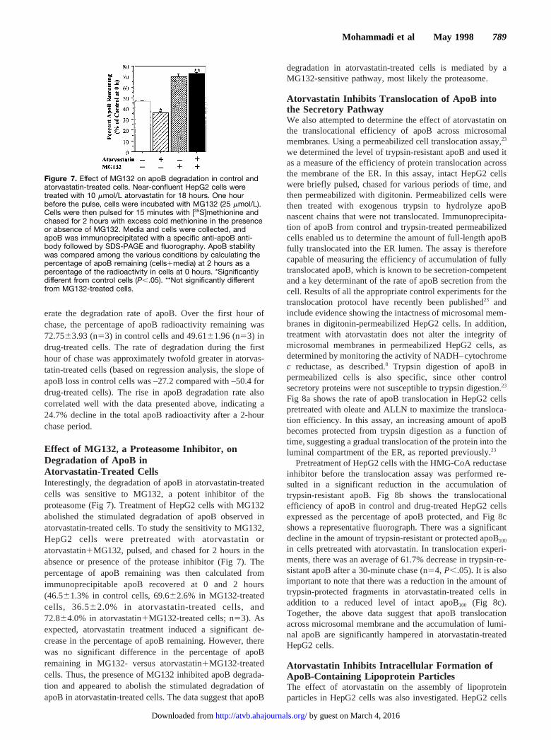
Effect of MG132, a Proteasome Inhibitor, onDegradation of ApoB inAtorvastatin-Treated CellsInterestingly, the degradation of apoB in atorvastatin-treatedcells was sensitive to MG132, a potent inhibitor of theproteasome (Fig 7). Treatment of HepG2 cells with MG132abolished the stimulated degradation of apoB observed inatorvastatin-treated cells. To study the sensitivity to MG132,HepG2 cells were pretreated with atorvastatin oratorvastatin1MG132, pulsed, and chased for 2 hours in theabsence or presence of the protease inhibitor (Fig 7). Thepercentage of apoB remaining was then calculated fromimmunoprecipitable apoB recovered at 0 and 2 hours(46.561.3% in control cells, 69.662.6% in MG132-treatedcells, 36.562.0% in atorvastatin-treated cells, and72.864.0% in atorvastatin1MG132-treated cells; n53). Asexpected, atorvastatin treatment induced a significant de-crease in the percentage of apoB remaining. However, therewas no significant difference in the percentage of apoBremaining in MG132- versus atorvastatin1MG132-treatedcells. Thus, the presence of MG132 inhibited apoB degrada-tion and appeared to abolish the stimulated degradation ofapoB in atorvastatin-treated cells. The data suggest that apoB
degradation in atorvastatin-treated cells is mediated by aMG132-sensitive pathway, most likely the proteasome.
Atorvastatin Inhibits Translocation of ApoB intothe Secretory PathwayWe also attempted to determine the effect of atorvastatin onthe translocational efficiency of apoB across microsomalmembranes. Using a permeabilized cell translocation assay,23
we determined the level of trypsin-resistant apoB and used itas a measure of the efficiency of protein translocation acrossthe membrane of the ER. In this assay, intact HepG2 cellswere briefly pulsed, chased for various periods of time, andthen permeabilized with digitonin. Permeabilized cells werethen treated with exogenous trypsin to hydrolyze apoBnascent chains that were not translocated. Immunoprecipita-tion of apoB from control and trypsin-treated permeabilizedcells enabled us to determine the amount of full-length apoBfully translocated into the ER lumen. The assay is thereforecapable of measuring the efficiency of accumulation of fullytranslocated apoB, which is known to be secretion-competentand a key determinant of the rate of apoB secretion from thecell. Results of all the appropriate control experiments for thetranslocation protocol have recently been published23 andinclude evidence showing the intactness of microsomal mem-branes in digitonin-permeabilized HepG2 cells. In addition,treatment with atorvastatin does not alter the integrity ofmicrosomal membranes in permeabilized HepG2 cells, asdetermined by monitoring the activity of NADH–cytochromec reductase, as described.8 Trypsin digestion of apoB inpermeabilized cells is also specific, since other controlsecretory proteins were not susceptible to trypsin digestion.23
Fig 8a shows the rate of apoB translocation in HepG2 cellspretreated with oleate and ALLN to maximize the transloca-tion efficiency. In this assay, an increasing amount of apoBbecomes protected from trypsin digestion as a function oftime, suggesting a gradual translocation of the protein into theluminal compartment of the ER, as reported previously.23
Pretreatment of HepG2 cells with the HMG-CoA reductaseinhibitor before the translocation assay was performed re-sulted in a significant reduction in the accumulation oftrypsin-resistant apoB. Fig 8b shows the translocationalefficiency of apoB in control and drug-treated HepG2 cellsexpressed as the percentage of apoB protected, and Fig 8cshows a representative fluorograph. There was a significantdecline in the amount of trypsin-resistant or protected apoB100
in cells pretreated with atorvastatin. In translocation experi-ments, there was an average of 61.7% decrease in trypsin-re-sistant apoB after a 30-minute chase (n54, P,.05). It is alsoimportant to note that there was a reduction in the amount oftrypsin-protected fragments in atorvastatin-treated cells inaddition to a reduced level of intact apoB100 (Fig 8c).Together, the above data suggest that apoB translocationacross microsomal membrane and the accumulation of lumi-nal apoB are significantly hampered in atorvastatin-treatedHepG2 cells.
Atorvastatin Inhibits Intracellular Formation ofApoB-Containing Lipoprotein ParticlesThe effect of atorvastatin on the assembly of lipoproteinparticles in HepG2 cells was also investigated. HepG2 cells
Figure 7. Effect of MG132 on apoB degradation in control andatorvastatin-treated cells. Near-confluent HepG2 cells weretreated with 10 mmol/L atorvastatin for 18 hours. One hourbefore the pulse, cells were incubated with MG132 (25 mmol/L).Cells were then pulsed for 15 minutes with [35S]methionine andchased for 2 hours with excess cold methionine in the presenceor absence of MG132. Media and cells were collected, andapoB was immunoprecipitated with a specific anti-apoB anti-body followed by SDS-PAGE and fluorography. ApoB stabilitywas compared among the various conditions by calculating thepercentage of apoB remaining (cells1media) at 2 hours as apercentage of the radioactivity in cells at 0 hours. *Significantlydifferent from control cells (P,.05). **Not significantly differentfrom MG132-treated cells.
Mohammadi et al May 1998 789
by guest on March 4, 2016http://atvb.ahajournals.org/Downloaded from

were pulsed, briefly chased, and permeabilized with digito-nin. Permeabilized cells were incubated for various times andthen subjected to subcellular fractionation. Luminal lipopro-tein particles were isolated from total microsomes and thenanalyzed by sucrose gradient ultracentrifugation and immu-noprecipitation. As depicted in Fig 8, there was a consider-ably lower amount of apoB-containing lipoprotein particles inthe lumen of microsomes isolated from atorvastatin-treatedcells both at 0 time (Fig 9a) and after a 2-hour chase (Fig 9b),suggesting that (1) fewer apoB lipoprotein particles accumu-lated in the microsomal lumen and (2) luminal apoB lipopro-tein particles were less stable in drug-treated cells. Fractions2 through 5 represent high-density apoB lipoprotein particles(apoB lipoproteins with density similar to that of HDL, peakdensity 1.065 to 1.170 g/mL), and fractions 6 through 12represent the lower-density apoB lipoprotein particles (LDL/VLDL-apoB, peak density 1.011 to 1.045 g/mL) (see Refer-ences 33 and 34). Therefore, we also observed a reduction inthe accumulation of apoB-containing lipoprotein particles inthe lumen of the ER associated with the observed increase inapoB degradation and a decrease in apoB translocation.
DiscussionNumerous evidence suggests that, once synthesized, apoB issubjected to posttranslational regulation, a process that is
closely linked to the lipid status of the cell. The supply of oneor more of the core lipids (triglycerides and cholesterol ester)in apoB-containing lipoproteins plays an important role inregulating the assembly and secretion of these lipoproteins.Human liver can apparently “vary the type and quantity ofapoB particles secreted in response to the load and type oflipid it has received.”45 Increased delivery of either fatty acidsor sterols, or both, to the liver may result in overproduction ofapoB particles.46 The amount of newly synthesized apoB usedfor secretion may be modulated by the available supply oftriglycerides,26,46–48 cholesterol ester,4,5,6 or possibly specificpools of phospholipids.49 Some studies have suggested thatincreased synthesis of triglyceride may upregulate apoBsecretion by increasing the recruitment of the ER-translocatedapoB to form mature lipoproteins,47,48 while in other studiesincreased cholesterol ester synthesis rather than triglyceridesynthesis has been suggested as the immediate regulator ofapoB secretion.4,5,25 Overall, these studies suggest that apoBmay be made in surplus and its secretion rate may be decidedby the availability of the intracellular lipid supply.
Intracellular lipid pools most likely regulate apoB produc-tion via posttranslational mechanisms that may involve facil-itating the translocation of newly synthesized apoB across theER membrane and reducing apoB degradation or enhancingits assembly into secretion-competent lipoprotein particles. In
Figure 8. Determination of the efficiencyof translocation of apoB in control andatorvastatin-treated cells. a, Basal rateof apoB translocation across the ERmembrane. Near-confluent HepG2 cellswere pulsed for 5 minutes with [35S]me-thionine and chased for various timeperiods with excess cold methionine.Cells were then permeabilized with digi-tonin (75 mg/mL) for 5 minutes, and thepermeabilized cells were incubated withtrypsin to digest any untranslocatedapoB chains. ApoB was immunoprecipi-tated, the immunoprecipitates were ana-lyzed by SDS-PAGE and fluorography,and apoB radioactivity was quantified bycutting and scintillation counting of theapoB100 band. b, Near-confluent HepG2cells were treated with or without10 mmol/L atorvastatin for 24 hours andwere pulsed for 5 minutes, chased for30 minutes, and permeabilized as inpanel a. Cells were trypsinized, andapoB was immunoprecipitated and ana-lyzed by SDS-PAGE and fluorography.ApoB radioactivity was quantified bycutting and scintillation counting of theapoB100 band. *Significantly differentfrom control cells treated with trypsinbut not atorvastatin (P,.05) (mean6SD,n54). c, Representative fluorograph ofthe apoB translocation experiment incontrol and atorvastatin-treated cells.Arrowhead indicates the position of the550-kD apoB100 band.
790 Effect of Atorvastatin on ApoB Secretion
by guest on March 4, 2016http://atvb.ahajournals.org/Downloaded from

the present study, we used atorvastatin, a new HMG-CoAreductase inhibitor with potent inhibitory effects on theintracellular rate of cholesterol synthesis, to investigate theeffect of inhibition of cholesterol synthesis on apoB translo-cation into the ER and its intracellular degradation. Atorva-statin has been shown to decrease cholesterol synthesis to aconsiderable extent in vitro, ex vivo, and in vivo.50–53 Resultsobtained in our laboratory also demonstrated that treatment ofHepG2 cells with atorvastatin under basal or lipid-richconditions results in a significant reduction in cholesterol,cholesterol ester, and apoB secretion. Several previous invitro studies have been performed to investigate the effect ofHMG-CoA reductase inhibitors on apoB secretion and haveyielded conflicting results. Ribeiro et al54 studied the effect ofsimvastatin on primary cultures of rat hepatocytes and re-ported stimulation of apoB secretion. On the other hand, Satoet al25 found that treatment of HepG2 cells by CS-514 did notinfluence the synthesis and secretion of apoB. Our observa-tions that atorvastatin inhibits apoB secretion in HepG2 cellscompare with the results of two recent in vivo studies inminiature pigs and guinea pigs. Huff and coworkers55 recentlyused miniature pigs to demonstrate that atorvastatin reduced
plasma cholesterol by inhibiting the hepatic secretion ofVLDL apoB.55 Similarly, studies by Fernandez and cowork-ers56 also showed a significant reduction in hepatic VLDLapoB secretion rates in guinea pigs treated with atorvastatin.These in vivo studies support our in vitro evidence showinga reduction in apoB secretion with atorvastatin treatment.
Previous in vitro studies of statins have mostly focused onthe effect of these inhibitors on the secretion of apoB andhave not explored the intracellular mechanisms by whichthese drugs may exert their effects on the production ofapoB-containing lipoproteins. To further investigate themechanism of atorvastatin action on apoB secretion, we usedboth intact cells as well as a semipermeable HepG2 sys-tem.8,23,29,36Results from these experiments appeared to sug-gest that atorvastatin decreased apoB secretion by stimulatingits degradation in HepG2 cells. Further investigation showedthat atorvastatin may stimulate apoB secretion by inhibitingapoB translocation across the ER membrane, which in turnmay result from depletion of cholesterol or cholesterol ester.Atorvastatin showed its stimulatory effect on apoB degrada-tion under basal as well as oleate-treated conditions. How-ever, the drug was clearly more effective in reducing apoBsecretion when HepG2 cells were enriched in lipids bypretreatment with oleate. This is not unexpected becauseapoB secretion, which is very low under basal conditions inHepG2 cells, is significantly increased with oleate treatment.Atorvastatin appears to interfere more effectively with li-poprotein secretion under such stimulated conditions.
Our results regarding the effect of atorvastatin on thetranslocation of apoB are consistent with current research,which suggests that translocation may be a key regulatorypoint in apoB secretion.17–20,22,57 Evidence that the actualprocess of apoB translocation across the ER may be a crucialregulatory point in the production of lipoproteins has recentlybeen suggested by Rusinol and Vance.22 They have shownthat the supplementation of primary rat hepatocytes withphosphatidyl-monomethylethanolamine decreased the secre-tion of apoB. This decrease was attributed to a decrease in thetranslocation of apoB and was independent of lipid availabil-ity. Furthermore, Bonnardel and Davis57 recently used HepG2cells to demonstrate that apoB translocation rather thandegradation may be the primary mechanism that regulatedapoB secretion. Recent data from our laboratory23 have alsoshown that the efficiency of apoB translocation can bemodulated by lipid availability as well as by altering theconformation of the nascent protein with agents such as DTTand cyclosporin. There is considerable evidence to suggestthe existence of two distinct pools of apoB in the ER: aluminal (trypsin-resistant) apoB pool, which is used for theassembly of lipoproteins, and a membrane-bound (trypsin-susceptible) apoB pool, which is thought to be shunted to adegradative pathway.17,26,55 The membrane-bound apoB poolmay also act as a precursor for the formation of dense apoBparticles in the ER lumen.36 Our data in the present reportdemonstrate that atorvastatin reduces the amount of apoB thatis translocated into the lumen of the ER. An alternativeexplanation for the apparent effect on apoB translocation isthat atorvastatin induces intraluminal degradation of apoBand therefore reduces the amount of luminal apoB. Thispossibility is unlikely given that the translocation experiment
Figure 9. Distribution of luminal apoB-containing lipoproteins incontrol and atorvastatin-treated cells. HepG2 cells were prein-cubated with or without atorvastatin (10 mmol/L) for 18 hours.Cells were then pulsed for 15 minutes with [35S]methionine,briefly chased with excess cold methionine, and permeabilizedwith digitonin (50 mg/mL) for 10 minutes. Permeabilized cellswere incubated in a cytoskeletal buffer for 0 (a) or 2 hours (b)before homogenization and fractionation of microsomes. Lumi-nal lipoproteins were extracted from microsomes by carbonatetreatment and separated from the membrane fraction by centrif-ugation (SW55 rotor; 35 000 rpm, 93 minutes). Fractionation ofluminal lipoproteins was performed by sucrose gradient centrifu-gation (SW41 rotor; 35 000 rpm, 65 hours). After centrifugation,gradient fractions were collected and immunoprecipitated with amonospecific anti-apoB antibody. Immunoprecipitates were an-alyzed by SDS-PAGE and fluorography, and apoB radioactivitywas quantified by cutting and scintillation counting of theapoB100 band. a, Luminal lipoproteins at 0 hours; b, luminallipoproteins after 2-hour chase. Both a and b show the resultsof a representative experiment (two others performed).
Mohammadi et al May 1998 791
by guest on March 4, 2016http://atvb.ahajournals.org/Downloaded from

was performed in the presence of ALLN, which appears toinhibit both proteasome-mediated apoB degradation37 as wellas degradation of apoB in the ER lumen.8,36 We suggest thatthe reduced translocation may result from the depletion ofcholesterol or cholesterol ester from HepG2 cells. Thisdepletion of lipid may reduce the number of apoB-containinglipoprotein particles and increase the amount of superfluousapoB. Several studies have demonstrated the importance oflipid availability for the translocation of apoB.23,33,34 Thepresence of oleate has been suggested to facilitate thetranslocation of apoB into the lumen and thereby reduce thepercentage of apoB that becomes prone to degradation.26,36
The data demonstrating that HepG2 cells incubated in thepresence of atorvastatin contained a significantly loweramount of luminal apoB-containing lipoproteins further sup-port the suggestion that atorvastatin may hamper apoBtranslocation across the ER membrane and reduce the amountof apoB molecules available for lipoprotein assembly. Frac-tionation of luminal apoB into dense (HDL-like) and light(LDL/VLDL-like) particles33,34 showed that both fractionsundergo a higher level of degradation in the presence ofatorvastatin. However, the HDL-like particles showed a moreprofound susceptibility to degradation under conditions ofatorvastatin treatment. This effect could possibly be exertedthrough inhibition of cholesterol synthesis and reduction inthe level of cholesterol ester pool available for lipoproteinassembly. The exact mechanism(s) for the effect of atorva-statin on apoB degradation remains to be elucidated throughfurther investigations. In preliminary experiments, we foundthat apoB degradation in atorvastatin-treated cells was sensi-tive to MG132, a potent inhibitor of the proteasome. Thisprotease inhibitor appeared to normalize the stability of apoBin atorvastatin-treated cells. The data suggest that the stimu-lated degradation of apoB on atorvastatin treatment can beprevented by inhibition of the proteasome. This finding is inagreement with the hypothesis that atorvastatin treatmentmay result in inhibition of apoB translocation leading to anincreased proportion of membrane-associated apoB chainsand their degradation by the cytosolic proteasome.
Overall, the results of our study further support the clinicalevidence that atorvastatin decreases the production of LDL-apoB. This effect may be occurring at several levels. Ator-vastatin has been shown to decrease the translocation of apoBinto the lumen and increase its intracellular degradation. Thisincreased degradation may be the result of the impairedtranslocation. Taken together, the data suggest that atorvasta-tin may decrease plasma LDL-apoB not only through upregu-lation of LDL receptors but also through a decrease in thehepatic production of apoB-containing lipoproteins.
AcknowledgmentsThis study was supported by grants from the Heart and StrokeFoundation of Ontario and Parke-Davis (to K.A.). We gratefullyacknowledge the excellent technical assistance of Debbie Rudy andAndrea Aiton.
References1. Havel R, Hunninghake D, Illingworth R, Lees R, Stein E, Tobert J, Lees
A, Leon A, Gardner K, Johnson G, Mellies M, Rhymers P, Tun P.Lovastatin (mevinolin) in the treatment of heterozygous familial hyper-
cholesterolemia: a multicenter study.Ann Intern Med. 1987;107:609–615.
2. Illingworth D, Sexton G. Hypercholesterolemic effects of mevinolin inpatients with heterozygous familial hypercholesterolemia.J Clin Invest.1984;74:1972–1978.
3. Lovastatin Study Group II. Therapeutic response to lovastatin (mevinolin)in nonfamilial hypercholesterolemia: a multicenter study.JAMA. 1986;256:2829–2834.
4. Kohen-Avramoglu RK, Cianflone KM, Sniderman AD. The role ofneutral lipid accessible pool in the regulation of secretion of apoB100lipoprotein particles by HepG2 cells.J Lipid Res. 1995;36:2513–2528.
5. Cianflone K, Yasruel Z, Rodriguez M, Vas D, Sniderman A. Regulationof apoB secretion from HepG2 cells: evidence for a critical role forcholesteryl ester synthesis in the response to a fatty acid challenge.J LipidRes. 1990;31:2045–2055.
6. Dashti N. The effect of low density lipoproteins, cholesterol and25-hydroxycholesterol on apolipoprotein B gene expression in HepG2cells.J Biol Chem. 1992;267:7160–7169.
7. Tanaka M, Jingami H, Otani H, Cho M, Ueda Y, Arai H, Nagano Y, DoiT, Yokode M, Kita T. Regulation of apolipoprotein B production andsecretion in response to the change of intracellular cholesteryl estercontents in rabbit hepatocytes.J Biol Chem. 1993;268:12713–12718.
8. Adeli K. Regulated intracellular degradation of apolipoprotein B in semi-permeable HepG2 cells.J Biol Chem. 1994;269:9166–9175.
9. Grundy SM, Vega GL. Influence of mevinolin on metabolism of lowdensity lipoproteins in primary moderate hypercholesterolemia.J LipidRes. 1985;26:1464–1475.
10. Arad Y, Ramakrishnan R, Ginsberg H. Lovastatin therapy reduces lowdensity lipoprotein apoB levels in subjects with combined hyperlipidemiaby reducing the production of apoB-containing lipoproteins: implicationsfor the pathophysiology of apoB production.J Lipid Res. 1990;31:567–582.
11. Lusis A, Taylor B, Quon D, Zollman S, LeBoeuf RC. Genetic factorscontrolling structure and expression of apolipoproteins B and E in mice.J Biol Chem. 1987;262:7594–7604.
12. Pullinger CR, North JD, Teng BB, Rifici VA, Ronhild de Brito AE, ScottJ. The apolipoprotein B gene is constitutively expressed in HepG2 cells:regulation of secretion by oleic acid, albumin, and insulin, and mea-surement of the mRNA half-life.J Lipid Res. 1989;30:1065–1077.
13. Dashti N, Williams D, Alaupovic P. Effects of oleate and insulin on theproduction rates and cellular mRNA concentrations of apolipoproteins inHepG2 cells.J Lipid Res. 1989;30:1365–1373.
14. Moberly J, Cole T, Alpers D, Moberly JB, Cole TG, Alpers DH,Schonfeld G. Oleic acid stimulation of apolipoprotein B secretion fromHepG2 and Caco-2 cells occurs post-transcriptionally.Biochim BiophysActa. 1990;1042:70–80.
15. Kaptein A, Roodenburg L, Princen H. Butyrate stimulates the secretion ofapolipoprotein (apo) A-I and apo B100 by the human hepatoma cell lineHepG2.Biochem J. 1991;278:557–564.
16. Borchardt R, Davis R. Intrahepatic assembly of very low densitylipoproteins.J Biol Chem. 1987;262:16394–16402.
17. Davis RA, Thrift RN, Wu CC, Howell KE. Apolipoprotein B is bothintegrated into and translocated across the endoplasmic reticulummembrane.J Biol Chem. 1990;265:10005–10011.
18. Davis R, Prewett A, Thompson J, Chan P, Borchardt R, Gallaher W.Intrahepatic assembly of very low density lipoproteins: immunologiccharacterization of apolipoprotein B in lipoproteins and hepaticmembrane fractions and its intracellular distribution.J Lipid Res. 1989;30:1185–1196.
19. Du EZ, Kurth J, Wang SL, Humiston P, Davis RA. Proteolysis-coupledsecretion of the N-terminus of apolipoprotein B.J Biol Chem. 1994;269:24169–24176.
20. Thrift RN, Drisko J, Dueland S, Trawick JD, Davis RA. Translocation ofapolipoprotein B across the endoplasmic reticulum is blocked in a non-hepatic cell line.Proc Natl Acad Sci U S A. 1992;89:9161–9165.
21. Rusinol AE, Chan EYW, Vance JE. Movement of apolipoprotein B intothe lumen of microsomes from hepatocytes is disrupted in membranesenriched in phosphatidylmonoethylethanolamine.J Biol Chem. 1993;268:25168–25175.
22. Rusinol AE, Vance JE. Inhibition of secretion of truncated apolipoproteinB by monomethylethanolamine is independent of the length of the apo-lipoprotein.J Biol Chem. 1995;270:13318–13325.
23. Macri J, Adeli K. Studies on intracellular translocation of apolipoproteinB in a permeabilized HepG2 system.J Biol Chem. 1997;272:7328–7337.
792 Effect of Atorvastatin on ApoB Secretion
by guest on March 4, 2016http://atvb.ahajournals.org/Downloaded from

24. Sparks J, Sparks C. Insulin modulation of hepatic synthesis and secretionof apolipoprotein B by rat hepatocytes.J Biol Chem. 1990;265:8854–8862.
25. Sato R, Imanaka T, Takatsuki A, Takano A. Degradation of newlysynthesized apolipoprotein B-100 in a pre-Golgi compartment.J BiolChem. 1990;265:11880–11884.
26. Dixon J, Furukawa S, Ginsberg H. Oleate stimulates secretion of apoli-poprotein B-containing lipoproteins from Hep G2 cells by inhibiting earlyintracellular degradation of apolipoprotein B.J Biol Chem. 1991;266:5080–5086.
27. White A, Graham D, LeGros J, Pease R, Scott J. Oleate-mediated stim-ulation of apolipoprotein B secretion from rat hepatoma cells.J BiolChem. 1992;267:15657–15664.
28. Furukawa S, Sakata N, Ginsberg HN, Dixon JL. Studies of the sites ofintracellular degradation of apolipoprotein B in HepG2 cells.J BiolChem. 1992;267:22630–22638.
29. Sallach S, Adeli K. Intracellular degradation of apolipoprotein B gen-erates an N-terminal 70 kDa fragment in the endoplasmic reticulum.Biochim Biophys Acta. 1995;1265:29–32.
30. Sparks JD, Sparks CE. Hormonal regulation of lipoprotein assembly andsecretion.Curr Opin Lipidol. 1993;44:177–186.
31. Verkade HJ, Fast DG, Rusinol AE, Scraba DG, Vance DE. Impairedbiosynthesis of phosphatidylcholine causes a decrease in the number ofvery low density lipoprotein particles in the Golgi but not in the endo-plasmic reticulum of rat liver.J Biol Chem. 1993;268:24990–24996.
32. Wang C-N, Hobman TC, Brindly DN. Degradation of apolipoprotein B incultured rat hepatocytes occurs in a post-endoplasmic reticulum com-partment.J Biol Chem. 1995;270:24924–24931.
33. Boren J, Wettesten M, Sjoberg A, Thorlin T, Bondjers G, Wiklund O,Olofsson SO. The assembly and secretion of apoB 100 containinglipoproteins in Hep G2 cells.J Biol Chem. 1990;265:10556–10564.
34. Boren J, Graham L, Wettesten M, Scott J, White A, Olofsson S. Theassembly and secretion of ApoB 100-containing lipoproteins in Hep G2cells.J Biol Chem. 1992;267:9858–9867.
35. Cartwright IJ, Hebbachi AM, Higgins JA. Transit and sorting of apoli-poprotein B within the endoplasmic reticulum and Golgi compartments ofisolated hepatocytes from normal and orotic acid-fed rats.J Biol Chem.1993;268:20937–20952.
36. Adeli K, Wettesten M, Asp L, Mohammadi A, Macri J, Olofsson SO.Intracellular assembly and degradation of apolipoprotein B in digitonin-permeabilized HepG2 cells.J Biol Chem. 1997;272:5031–5039.
37. Yeung SJ, Chen SH, Chan L. Ubiquitin-proteasome pathway mediatesintracellular degradation of apolipoprotein B.Biochemistry. 1996;35:13843–13848.
38. Benoist F, Grand-Perret T. Co-translational degradation of apolipoproteinB100 by the proteasome is prevented by microsomal triglyceride transferprotein: synchronized translation studies on HepG2 cells treated with aninhibitor of microsomal triglyceride transfer protein.J Biol Chem. 1997;272:20435–20442.
39. Fisher EA, Zhou M, Mitchell DM, Wu X, Omura S, Wang H, GoldbergAL, Ginsberg HN. Degradation of apolipoprotein B100 is mediated bythe ubiquitin-proteasome and involves heat shock protein 70.J BiolChem. 1997;272:20427–20434.
40. Adeli K, Sinkevitch C. Secretion of apolipoprotein B in serum-freecultures of HepG2 cells.FEBS Lett. 1990;2:345–348.
41. Bostrom K, Boren J, Wettesten M, Sjorberg A, Bondjers G, Wiklund O,Carlsson P, Olofsson S-O. Studies on the assembly of apo B-100-containing lipoproteins in HepG2 cells.J Biol Chem. 1988;263:4434–4442.
42. Bostrom K, Wettesten M, Boren J, Bondjers G, Wiklund O, OlofssonS-O. Pulse-chase studies of the synthesis and intracellular transport ofapolipoprotein B-100 in HepG2 cells.J Biol Chem. 1986;261:13800–13806.
43. Laemmli UK. Cleavage of structural proteins during the assembly of thehead of bacteriophage T4.Nature (Lond). 1970;227:680–685.
44. Wettesten M, Bostrom K, Bondjers G, Jarfeldt M, Norfeldt P-I, CarellaM, Wiklund O, Boren J, Olofsson S-O. Pulse-chase studies of the syn-thesis of apolipoprotein B in a human hepatoma cell line, HepG2.EurJ Biochem. 1985;149:461–466.
45. Sniderman AD, Cianflone K. Substrate delivery as a determinant ofhepatic apoB secretion.Arterioscler Thromb. 1993;13:629–636.
46. Wu X, Sakata N, Dixon J, Ginsberg HN. Exogenous VLDL stimulatesapolipoprotein B secretion from HepG2 cells by both pre- and post-translational mechanisms.J Lipid Res. 1994;35:1200–1210.
47. Boren J, Rustaeus S, Wettesten M, Andersson M, Wiklund A, OlofssonS-O. Influence of triacylglycerol biosynthesis rate on the assembly ofapoB-100–containing lipoproteins in HepG2 cells.Arterioscler Thromb.1993;13:1743–1754.
48. Wu X, Sakata N, Lui E, Ginsberg HN. Evidence for a lack of regulationof the assembly and secretion of apolipoprotein B-containing lipoproteinfrom HepG2 cells by cholesteryl ester.J Biol Chem. 1994;269:12375–12382.
49. Vance JE, Vance DE. Lipoprotein assembly and secretion by hepatocytes.Annu Rev Nutr. 1990;10:337–356.
50. Shaw MK, Newton RS, Sliskovic DR, Roth BD, Ferguson E, Krause R.Hep-G2 cells and primary rat hepatocytes differ in their response toinhibitors of HMG-CoA reductase.Biochem Biophys Res Commun. 1990;170:726–734.
51. Krause BR, Newton RS. Animal models for the evaluation of inhibitorsof HMG-CoA reductase.Adv Lipid Res. 1991;1:57–72.
52. Bocan TMA, Ferguson E, McNally W, Uhlendorf PD, Mueller SB,Dehart P, Sildovic DR, Roth BD, Krause BR, Newton R. Hepatic andnonhepatic sterol synthesis and tissue distribution following adminis-tration of liver selective HMG-CoA reductase inhibitor, CI-981: com-parison with selected HMG-CoA reductase inhibitors.Biochim BiophysActa. 1992;1123:133–144.
53. Bocan TMA, Mazur MJ, Mueller SB, Brown EQ, Sliskovic DR, O’BrienPM, Creswell MW, Lee H, Uhlendorf PD, Roth BD, Newton R. Anti-atherosclerotic activity of inhibitors of 3-hydroxy-3-methylglutarylcoenzyme A reductase in cholesterol-fed rabbits: a biochemical andmorphological evaluation.Atherosclerosis. 1994;111:127–142.
54. Ribeiro A, Mangeney M, Loriette C, Thomas G, Pepin D, Janvier B,Chambaz J, Bereziat G. Effect of simvastatin on the synthesis andsecretion of lipoproteins in relation to the metabolism of cholesterol incultured hepatocytes.Biochim Biophys Acta. 1991;1086:279–286.
55. Burnett JR, Wilcox LJ, Telford DE, Kleinstiver SJ, Barrett PHR, NewtonRS, Huff MW. Inhibition of HMG-CoA reductase by atorvastatindecreases both VLDL and LDL apolipoprotein B production in miniaturepigs.Arterioscler Thromb Vasc Biol.1997;17:2589–2600.
56. Conde K, Marcela V-J, Krause BR, Newton RS, Fernandez ML. Hypo-cholesterolemic actions of atorvastatin are associated with alterations onhepatic cholesterol metabolism and lipoprotein composition in the guineapig. J Lipid Res. 1996;37:2372–2382.
57. Bonnardel JA, Davis RA. In HepG2 cells, translocation, not degradation,determines the fate of the de novo synthesized apolipoprotein B.J BiolChem. 1995;270:28892–28896.
Mohammadi et al May 1998 793
by guest on March 4, 2016http://atvb.ahajournals.org/Downloaded from

AdeliAbbas Mohammadi, Joseph Macri, Roger Newton, Tanya Romain, Daisy Dulay and Khosrow
HepG2 CellsEffects of Atorvastatin on the Intracellular Stability and Secretion of Apolipoprotein B in
Print ISSN: 1079-5642. Online ISSN: 1524-4636 Copyright © 1998 American Heart Association, Inc. All rights reserved.
Greenville Avenue, Dallas, TX 75231is published by the American Heart Association, 7272Arteriosclerosis, Thrombosis, and Vascular Biology
doi: 10.1161/01.ATV.18.5.7831998;18:783-793Arterioscler Thromb Vasc Biol.
http://atvb.ahajournals.org/content/18/5/783World Wide Web at:
The online version of this article, along with updated information and services, is located on the
http://atvb.ahajournals.org//subscriptions/
at: is onlineArteriosclerosis, Thrombosis, and Vascular Biology Information about subscribing to Subscriptions:
http://www.lww.com/reprints
Information about reprints can be found online at: Reprints:
document. AnswerPermissions and Rights Question andunder Services. Further information about this process is available in the
permission is being requested is located, click Request Permissions in the middle column of the Web page whichCopyright Clearance Center, not the Editorial Office. Once the online version of the published article for
can be obtained via RightsLink, a service of theArteriosclerosis, Thrombosis, and Vascular Biologyin Requests for permissions to reproduce figures, tables, or portions of articles originally publishedPermissions:
by guest on March 4, 2016http://atvb.ahajournals.org/Downloaded from
Related Documents


![Interaction of a recombinant form of apolipoprotein[a ... · Interaction of a recombinant form of apolipoprotein[a] with human fibroblasts and with the human hepatoma cell line HepG2](https://static.cupdf.com/doc/110x72/5d0ce32c88c993064c8b69eb/interaction-of-a-recombinant-form-of-apolipoproteina-interaction-of-a-recombinant.jpg)