Effects of 10 Hz individualized repetitive transcranial magnetic stimulation on patients with disorders of consciousness : a study protocol for an exploratory double-blind crossover randomized sham- controlled trial Chengwei Xu Zhujiang Hospital https://orcid.org/0000-0003-4636-3424 Zhaohua Zhu Zhujiang Hospital Wanchun Wu Zhujiang Hospital Xiaochun Zheng Zhujiang Hospital Haili Zhong Zhujiang Hospital Xinyi Qian Gannan Medical University Qiuyou Xie Zhujiang Hospital Xiyan Huang ( [email protected] ) Zhujiang Hospital Research Article Keywords: disorders of consciousness, repetitive transcranial magnetic stimulation, randomized control trial Posted Date: May 4th, 2022 DOI: https://doi.org/10.21203/rs.3.rs-1288848/v1 License: This work is licensed under a Creative Commons Attribution 4.0 International License. Read Full License

Welcome message from author
This document is posted to help you gain knowledge. Please leave a comment to let me know what you think about it! Share it to your friends and learn new things together.
Transcript
Effects of 10 Hz individualized repetitivetranscranial magnetic stimulation on patients withdisorders of consciousness : a study protocol for anexploratory double-blind crossover randomizedsham- controlled trialChengwei Xu
Zhujiang Hospital https://orcid.org/0000-0003-4636-3424Zhaohua Zhu
Zhujiang HospitalWanchun Wu
Zhujiang HospitalXiaochun Zheng
Zhujiang HospitalHaili Zhong
Zhujiang HospitalXinyi Qian
Gannan Medical UniversityQiuyou Xie
Zhujiang HospitalXiyan Huang ( [email protected] )
Zhujiang Hospital
Research Article
Keywords: disorders of consciousness, repetitive transcranial magnetic stimulation, randomized controltrial
Posted Date: May 4th, 2022
DOI: https://doi.org/10.21203/rs.3.rs-1288848/v1
License: This work is licensed under a Creative Commons Attribution 4.0 International License. Read Full License
Page 1 of 18
Trials structured Study Protocol template
Trials guidance: the numbers in curly brackets (e.g. {5a}) are SPIRIT item identifiers. Please do not remove the numbers in curly brackets, or any heading that contains them. The item identifiers are
slightly out of sequence to make the document flow more easily but it is important that they remain in the
document to allow electronic searches by SPIRIT item number.
If you are certain that an item does not apply, please state "n/a" and provide a short explanation. Leaving an
item blank or stating “n/a” without an explanation will lead to your manuscript being returned before review.
NB: All text in green can be removed once you have finished creating your Study Protocol. Text in black is
mandatory. Please also read the submission guidelines for Study Protocols prior to submitting to Trials:
https://trialsjournal.biomedcentral.com/submission-guidelines/preparing-your-manuscript/study-protocoll
Title
Effects of 10 Hz individualized repetitive transcranial magnetic stimulation on patients with disorders of consciousness: a study protocol for an exploratory double-blind crossover randomized sham-controlled trial
Names protocol contributors
Chengwei Xu, Zhaohua Zhu, Wanchun Wu, Xiaochun Zheng, Haili Zhong, Xinyi Qian, Qiuyou Xie & Xiyan Huang Abstract Trials guidance: The Abstract should not exceed 350 words. Please minimize the use of abbreviations and
do not cite references in the abstract. The abstract must include the following separate sections:
• Background: Repetitive transcranial magnetic stimulation (rTMS), as a non-invasive brain stimulation
technique, has shown potentials for consciousness recovery of patients with disorders of consciousness
(DoC), as it is effective in regulating the excitability of central nervous system. However it is difficult to
achieve satisfactory effect with “one size fits all” rTMS treatment due to different clinical conditions of
patients. There is an urgent need to develop individualized strategy to improve the effectiveness rTMS
on patients with DoC.
• Methods: Our protocol is a randomized double-blind sham-controlled crossover trial that includes 30
DoC patients. Each patient will received 20 sessions, in which 10 sessions will be rTMS-active stimulus
and the other 10 sessions will be sham stimulus, separated by no less than 10 days’ washout period.
The rTMS-active will include 10 Hz rTMS over the individualized-targeted selection area for each patient
according to the different insult regions of the brain. Coma Recovery Scale-Revised (CRS-R) will be
used as primary outcome at baseline, after the first stage of stimulation, at the end of the washout period
Page 2 of 18
and after the second stage of stimulation. Secondary outcomes will be measured at the same time,
including efficiency, relative spectral power and functional connectivity of high-density electroencephalo-
graph (EEG). Adverse events will be recorded during the study.
• Discussion: rTMS has obtained Grade A evidence in treating patients with several central nervous
system diseases, and there has been some evidence showing partial improvement on level of
consciousness in DoC patients. However, the effectiveness of rTMS in DoC is only 30%~36%,
mostly due to the non-specific target selection. In this protocol, we present a double-blind crossover
randomized sham-controlled trial based on the individualized-targeted selection strategy that aims
to study the effectiveness of rTMS therapy for DoC, and the result may provide new insights to non-
invasive brain stimulation.
• Trial registration: ClinicalTrials.gov: NCT05187000. Registered on January 10, 2022.
Keywords disorders of consciousness, repetitive transcranial magnetic stimulation, randomized control trial.
Administrative information Trials guidance: please include this text in your protocol just above the Administrative information table:
Note: the numbers in curly brackets in this protocol refer to SPIRIT checklist item numbers. The order of the
items has been modified to group similar items (see http://www.equator-network.org/reporting-
guidelines/spirit-2013-statement-defining-standard-protocol-items-for-clinical-trials/).
Title {1} Effects of 10 Hz individualized repetitive transcranial magnetic
stimulation on patients with disorders of consciousness: a study
protocol for an exploratory double-blind crossover randomized sham-
controlled trial
Trial registration {2a and 2b}. ClinicalTrials.gov: NCT05187000. Registered on January 10, 2022.
Protocol version {3} November 1, 2021, version 8 (original)
Page 3 of 18
Funding {4} National Natural Science Foundation of China (No. 82171174,
81974154, 81801119), and Key Realm R&D Program of Guangzhou
(No. 202007030005).
Author details {5a} CWX and ZHZ: conceived and designed the study protocol and
contributed to drafting the manuscript. CWX and WCW: wrote the
manuscript and participated in the coordination and implementation of
the study. XYH and QYX: revised the study protocol and wrote several
sections of the manuscript. XCZ, HLZ and XYQ: helped develop the
study measures and data collection. All authors contributed to drafting
the manuscript and approved the final manuscript.
Name and contact information for
the trial sponsor {5b} Zhujiang Hospital of Southern Medical University.
Principal Investigator Dr. Qiuyou Xie, M.D., Ph.D.
Address: 253 Industrial Avenue, Haizhu District, Guangzhou city,
Guangdong Province, China
Email: [email protected]
Tel: +86 13903019604.
Role of sponsor {5c} This is a researcher-driven study carried out in Zhujiang Hospital of
Southern Medical University, with no outside sponsor or funding. The
Principal Investigator is actively involved in planning and executing of
the study.
Introduction
Background and rationale {6a} Disorders of consciousness (DoC) are a series of arousal and cognitive disorders secondary to severe brain
injury[1], encompassing a spectrum of conditions ranging from vegetative state (VS)[2] [also called
unresponsive conscious state (UWS)] to minimally conscious state (MCS)[3]. Long-term hospitalization of
such patients brings up enormous mental suffering and medical burden to their family and society[4]. So far,
there is lack of effective treatments for patients with DoC[5]. As one of the non-pharmacological treatments, the neuromodulation technology has been developed
rapidly in DoC treatments since 2007[6]. Repetitive transcranial magnetic stimulation (rTMS) is a form of
non-invasive brain stimulation (NIBS) technology which has been recommended to be applied in the
treatment of depression, obsessive-compulsive disorder, post-stroke movement disorder and other
neurological or psychiatric diseases[7]. Compared to other NIBS, rTMS can be combined with MRI
Page 4 of 18
navigation technology to precisely excite or inhibit specific cerebral areas[8] and has a great advantage in
exploring connections between different cerebral functional areas[9]. Previously, the application of rTMS in DoC patients was mainly based on the intervention theories related
to stroke, such as the interhemispheric competition model, vicariation model and the bimodal balance-
recovery model[10]. Since stroke models mostly focus on brain function reorganization after focal brain
injury, it is unknown whether they can cope with the changes in network function caused by extensive brain
injury in DoC patients. At present, there are several theories which concern different aspects about neural
activity and consciousness. The most prominent maybe Global Workspace Theory (GWT)[11] and
Integrated Information Theory (IIT)[12], which highlight different areas such as prefrontal or posterior areas
of the brain are crucial for consciousness[13]. According to GWT, dorsal lateral prefrontal cortex (DLPFC)
plays an important role in executive control network (ECN), especially in improving mood and cognitive
function[14]. Most studies have selected the left DLPFC area as the intervention target, and believed that
stimulating left DLPFC can strengthen thalamo-cortical and cortico-cortical connections and significantly
improve behavioral performance and EEG power spectrum, particularly in MCS patients[15-19]. Meanwhile,
according to IIT, the posterior parietal cortex (PPC) is considered as a critical hub in consciousness recovery
in default model network (DMN), and also selected as a stimulation target area for DoC patients in some
studies[20, 21]. However, until now the effective rate of rTMS in DoC patients is only 30% to 36%[6]. The
most important reason may be that they used the same rTMS stimulation target area for all DoC patients
who have different consciousness states and different cortical injury positions.
Therefore, we propose an individualized-targeted selection strategy of rTMS intervention program for DoC
patients according to their levels of consciousness and sites of injury. We aim to conducted a randomized
double-blind sham-controlled crossover clinical trial that could evaluate the effects of lindividualized rTMS on
DoC patients. The detailed strategies will be described in subsequent sections.
Objectives {7} Primary objective To examine whether10 Hz individualized rTMS is more effective to improve CRS-R scores in DoC patients
than the rTMS-sham control.
Secondary objective
(1) To examine whether 10 Hz individualized rTMS is more efficiency in DoC patients than the rTMS-
sham control;
(2) To examine whether 10 Hz individualized rTMS is more effective to improve relative spectral power
of EEG in DoC patients than the rTMS-sham control;
(3) To examine whether 10 Hz individualized rTMS is more effective to improve functional connectivity of
EEG in DoC patients than the rTMS-sham control.
Trial design {8}
Page 5 of 18
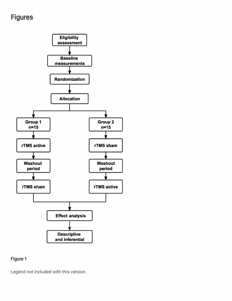
This study is a crossover randomized, double-blind,sham-controlled clinical trial. 30 DoC patients will be
recruited and divided into two groups in a 1:1 ratio. Group 1 will start with 10 sessions (once a day) of rTMS-
active. After no less than 10 days’ washout, this group will be given another 10 sessions (once a day) with
rTMS-sham. In contrast, group 2 will do the opposite protocol, participants will start with 10 sessions (once a
day) of rTMS-sham, and after no less than 10 days’ washout period will be received 10 sessions (once a
day) of rTMS-active. This trial includes a 20-day intervention and a no less than 10 days’ washout period. 5
to 10 days washout period has been used in some crossover studies[22, 23] and was shown to be enough to
reset the effects[24]. The study protocol flow chart and time of collection of outcomes are described in Figs.
1 and 2, respectively.
Methods: Participants, interventions and outcomes
Study setting {9} Department of Rehabilitation Medicine and Clinical Research Center (CRC), Zhujiang hospital of Southern
Medical University (SMU) will be responsible for training physiotherapists in a standard operating procedure,
and Data Monitoring Committee (DMC) will be responsible for supervising this trial.
The study protocol was developed according to the guidelines of the standard items of the protocol: Recommendations for interventional trials (SPIRIT) guidelines[25].
Eligibility criteria {10} Patients who meet all the following enrollment criteria will enter this study after signing the informed consent:
(1) Age: 18-70 years old, acquired brain injuries less than 1 year and more than 28 days in DoC; (2)
Diagnosed as VS/UWS or MCS by CRS-R; (3) No medical history of neuropsychiatric diseases; (4) No
sedatives in use or other drugs that might interfere with brain stimulation, such as Na+ or Ca2+ channel
blockers or N Methyl D Aspartate receptor antagonists; (5) Stable state of disease and vital signs; (6) The
families of the patients volunteer to participate in this study and provided signed informed consent; and (7)
The integrity of the individual-targeted selection area are verified by MRI.
Patients who meet the following criteria will be excluded: (1) Patients in other non-invasive or invasive
neuroregulation trials; (2) Motor evoked potential (MEP) in M1 region cannot be induced by TMS pulse; (3)
Uncontrolled epilepsy, seizure within 4 weeks before enrollment; (4) Contraindications for rTMS or EEG,
such as metallic implant in the skull, pacemaker, craniotomy under the stimulated site and implanted brain
devices.
Who will take informed consent? {26a} Informed consent will be obtained before trial baseline evaluation at the inpatient clinic by the rehab doctor.
Additional consent provisions for collection and use of participant data and biological specimens {26b} This study did not collect additional biological samples.
Page 6 of 18
Interventions Explanation for the choice of comparators {6b} rTMS is a form of NIBS technology in clinic treatment[26]. Compared to other NIBS, rTMS can be combined
with MRI navigation technology to precisely excite or inhibit specific cerebral areas[8] and has a great
advantage in exploring connections between different cerebral functional areas[27].
Intervention description {11a} rTMS
All participants will receive both rTMS-active and rTMS-sham interventions, separated by at least 10-day
washout period. Stimulation intensity varies across this experiment and will be determined by the resting
motor threshold (RMT) which is defined as the lowest TMS intensity applied to the M1 region. It can evoke
electromyography (EMG) with an amplitude >50 µV peak-to-peak from the right first dorsal interosseous
(FDI) muscle.at least five out of 10 pulses. If the RMT was above 67%, then the actual intensity of the
stimulus was set to 60% of the maximum output of the stimulation device[28]. The researchers will be
trained to use the neuronavigation system to mark, and the coil surface will be positioned at a tangent angle
of 45° to the scalp[29] on the individualized-targeted selection area to perform rTMS interventions. rTMS
pulses will be delivered using an NTK-TMS-Ⅱ300 stimulator with an IIB502 97-mm figure-of-eight coil. There
are two identical surfaces in this coil, one of which can output rTMS-active pluses, and other surface can
output rTMS-sham pluses (Brain Modulation Technology Development CO, LDT, JiangXi, CHN). It can
produce a biphasic waveform with a pulse width of ~0.32 ms.
rTMS-active During the rTMS-active stage, treatment will be given for 10 consecutive sessions (one session daily). The
participants will be placed in the semi-reclining position on either a normal chair or a wheelchair and each
stimulation session will last 20 minutes with a frequency of 10 Hz (train duration: 1s; inter-train interval: 5s;
200 effective stimulation series; 2000 pulses at 90% of RMT). The magnetic stimulation is administered
following safety guidelines[30].
rTMS-sham
During the rTMS-sham stage, treatment will be given for 10 consecutive sessions (one session daily) also.
The sham coil surface has no magnetic field to send to the cerebral cortex while appearing to be the same
shape as the active coil, with good approximation of auditory feedback[31]. In our study, the parameters and
targeted area of the rTMS-sham are the same as that of the rTMS-active. The only difference is that the
rTMS-sham stimulation has no pulse magnetic field sent to the target area but will generate noise and
vibration.
Individualized-targeted selection strategy of rTMS for DOC We propose an individualized-targeted selection strategy of rTMS as the highlight of this protocol. First, two
experienced doctors will choose a relatively intact brain hemisphere (left or right hemisphere) through
patients’ MRI images. Then, the doctors will select the DLPFC for MCS patients and PPC for VS/UWS
patients respectively as their individualized-targeted selection areas. For example, if a patient is VS
Page 7 of 18
assessed by CRS-R and his/her left hemisphere is damaged seriously than the right hemisphere, then the
right PPC will be selected. Furthermore, if there is no obvious difference between bilateral hemisphere injury,
such as hypoxic-ischemic encephalopathy (HIE), diffuse axonal injury, we will prefer to select the left
hemisphere as the stimulation area, because the left DLPFC and left PPC areas have better therapeutic
effects in previous studies[16, 19, 32-34].
Criteria for discontinuing or modifying allocated interventions {11b} Patients who meet the following criteria will be discontinued intervention: (1) Any indication of epilepsy that
occurred during the trial; (2) High fever (≥38°) more than 3 consecutive day.
Strategies to improve adherence to interventions {11c} Free rTMS treatment and EEG examination for participants as our strategy to improve adherence to
interventions.
Relevant concomitant care permitted or prohibited during the trial {11d} The relevant concomitant care include the use of drugs (such as amantadine), prevention of complications
and arranging routine rehabilitation treatments. All of the routine rehabilitation programs are provided by
qualified rehabilitation therapists from the Department of Rehabilitation Medicine, Zhujiang Hospital of SMU,
which include passive limb range-of-motion training, limb electrical stimulation, barometric therapy,
respiratory rehabilitation, swallowing therapy, gastrointestinal rehabilitation, speech therapy and hyperbaric
oxygen therapy.
Provisions for post-trial care {30} Treatment of the participant will be conducted by standard care protocols regardless of the trial participation.
Outcomes {12} Primary outcomes JFK Coma Recovery Scale-Revised CRS-R[35], as a gold standard, is widely used to define the level of consciousness and assess
neurobehavioral recovery in DoC patients. It includes six subscales that assess auditory, visual, motor,
motor/speech, communication, and arousal processes. Each item of CRS-R is in good agreement with the
diagnostic and differential diagnostic criteria of VS/UWS, MCS and emerge minimally conscious state
(EMCS)[1]. CRS-R will be recorded at four time points: before the experiment (-t1) and after the end of the
first rTMS stage (t2), after washout period (t4), and after the second rTMS stage (t6).
Secondary outcomes Efficiency Base on CRS-R, participant’s level of consciousness from VS/UWS to MCS, MCS to MCS+, or MCS+ to
Page 8 of 18
EMCS are considered effective. New MCS manifestations in participants with MCS participant are also
considered effective[36] (e.g. patients with autonomous motor responses showed new visual tracking after
treatment).
Relative spectral power Relative spectral power will be calculated by the selected artifact-free EEG epochs at five frequency bands:
δ (1–4 Hz), θ (4–8 Hz), α (8–13 Hz), β (13–30 Hz), and γ (30–45 Hz). The relative power of each given band
is calculated as follows:
𝑅𝑃(𝑓 , 𝑓 ) = 𝑃(𝑓 ,𝑓 )𝑃(1,45) × 100%
where P (f1, f2) indicates the absolute power between low f1 and high f2 frequency. P (1, 45) is the sum of
power (1–45 Hz). Then, the relative power for each band was averaged across channels[37].
Functional connectivity Coherence For further describing the functional interaction between brain regions, we will perform coherence[38, 39]
analysis. Coherence is the measure of synchrony of brain activity across different brain regions. We will
analyzed the coherence of paired channels using 60 channels rest-EEG data, while the values of channel x
and y are calculated by the absolute power spectral density and the cross power spectral density: 𝐶 (𝑓) = 𝑃 (𝑓)𝑃 ( ) ( )
Where f stands for frequency. After obtaining the coherence values of the full frequency band (1-45Hz), a
coherence matrix can be obtained in each brain region by calculating the coherence of the paired electrodes.
Then the coherence values in each frequency band can be obtained by averaging all channel pairs:
𝐴 = 1𝐹 − 𝐹 𝐶 (𝑓)𝑑𝑓
F2 and F1 distributions represent the upper and lower frequencies of each frequency band. Finally, the
average value of coherence matrix on all data segments is used as the coherence of this frequency band.
Participant timeline {13} Fig. 2
Sample size {14} This is a crossover exploratory study. The CRS-R total score after two-stage treatments will be considered
as the primary outcome, while high-density EEG analysis (relative spectral power and functional
connectivity) will be the secondary outcome. In this context[40], the sample size calculation will be based on
Page 9 of 18
an expected difference between the treatment groups of 20%, setting a significance level of 0.05% and a
power of 80%. Considering patients’ compliance and approximately 20% dropout rate during the study, the
final sample size of 30 participants is needed. We consider this is an achievable sample size and adequate
to allow for the dropout rate while still leaving a reasonable final sample.
Recruitment {15} Participants will be recruited from the Department of Rehabilitation Medicine, Zhujiang Hospital of SMU , with
varying degrees of DoC[41] caused by traumatic brain injury (TBI) or non-traumatic brain injury (nTBI),
including VS/UWS and MCS. Our recruitment advertisement has been posted on the bulletin board of
Zhujiang Hospital in November 2021. Patients’ families can contact our principal investigator. After the
informed consent is obtained from family members, doctors will conduct screening according to the inclusion
and exclusion criteria. Only those who meet all the recruitment requirements and do not meet any exclusion
criteria will participate this study.
Assignment of interventions: allocation
Sequence generation {16a} Participants will be randomly divided into two groups in a 1:1 ratio according to computer-generated
randomization using the Random Numbers Function of the statistical software SPSS 23.0 (IBM, USA).
Randomization will be performed under the control of a blinded worker from DMC who will be the only
person allowed to manage the electronic coding of the randomization to assign the individuals. The
researchers will be blind to the group in which the participant is allocated to.
Concealment mechanism {16b} In order to perform the allocation concealment process, the coded groups will be placed in a closed opaque
envelope, which will be marked as the code for each participant and held by the staff responsible for
randomization. The envelope will only be opened during active or sham rTMS. To ensure proper blinding,
participants will be given a code and will be concealed from the allocation process by a independent staff of
the DMC who is responsible for randomization. The rTMS experimenter (responsible for applying the
intervention) does not know the group allocation either. The rTMS coil will be wrapped in identical opaque
plastic papers and labeled A or B. In addition, the rTMS experimenter will be told by one DMC staff
(responsible for randomization) to use surface A or B first.
Implementation {16c}
The sealed envelopes will be opened and concealed again in the first rTMS stage prior to the intervention by
the staff responsible for randomization. Then, the code will inform the rTMS experimenter.
Page 10 of 18
Assignment of interventions: Blinding
Who will be blinded {17a} Both participants and clinic staffs (outcome assessors, caregivers, nurses, physical therapists and statistical
analysts, etc) will remain blind to group allocation. Whether the intervention is rTMS-active or rTMS-sham
will not be revealed throughout the study.
Procedure for unblinding if needed {17b} A sealed copy of the cipher will also be prepared for the situation of emergency unblinding.
Data collection and management
Plans for assessment and collection of outcomes {18a} All variables in the protocol will be documented in a Case Report Form (CRF). CRS-R and rest-EEG will be
collected at the baseline, after the first stage of stimulation, at the end of the washout period and after
the second stage of stimulation. The investigators who enter information into the CRF are responsible for
ensuring the accuracy and completeness of information. When the data collection are completed, each CRF
will be validated for integrity, consistency and rationality by the DMC.
Plans to promote participant retention and complete follow-up {18b} Not applicable. Study participants will complete all evaluations and treatment during their stay in the hospital.
Data management {19} All data will be handled with utmost care and confidentiality. Data will be stored electronically with passwords
and CRF will be stored for the duration of the study, then archived in locked filing cabinets at the Department
of Rehabilitation Medicine of Zhujiang Hospital of SMU, for a minimum period of 15 years after the end of the
project.
Confidentiality {27} Each participant will be identified by a identification code, the first letter of the last name, the first letter of the
first name. An identification list of participants will be kept in the researcher’s file. Information will be collected
for each participant in a CRF filled out by the investigator. Every precaution will be taken to respect the
privacy of participants in the conduct of the study.
Plans for collection, laboratory evaluation and storage of biological specimens for
Page 11 of 18
genetic or molecular analysis in this trial/future use {33}
Not applicable. No biological specimens will be collected.
Statistical methods
Statistical methods for primary and secondary outcomes {20a} SPSS 23.0 statistical software will be used to analyze the results. All the statistical hypotheses will be tested
by two-side test, with the statistically significant test level set at 0.05 and the confidence interval estimation of
the parameters set to 95%. When the data does not meet the condition of the parameter test, the data
transformation can be used; if it still does not meet the condition, the non-parameter test can be considered.
In the descriptive analysis of sample, the baseline mean and standard deviation between two groups will
be compared for the normally distributed measurement data, and the minimum, maximum, P25, P50 and
P75 will be given for the non-normal distributed data. In the final data analysis, the ANOVA by two stage
crossover design will be used if data conform to normal distribution, otherwise the rank sum test will be used.
The enumeration data will be expressed as frequency or percentage, and the chi-square test or Fisher's
exact test will be used to compare the baseline differences between the two groups.
Interim analyses {21b} No any interim analysis will be conducted.
Methods for additional analyses (e.g. subgroup analyses) {20b} No any additional analysis will be conducted.
Methods in analysis to handle protocol non-adherence and any statistical methods to handle missing data {20c} Both Intention-to-treat (ITT) analysis set and Per-protocol (PP) analysis set will be used. ITT set includes all
participants who have been randomized. When a participant is randomly assigned to a rTMS-active followed
by a sham-rTMS, he or she should be included in the rTMS-active followed by a sham analysis. For PP set,
only participants who complied with the intervention will be analyzed. In this study, for various reasons, the
interruption of rTMS-active no more than 3 days, and the total times of real-stimulation no less than 8 times
will all be included in the PP. The ITT analysis will be compared with the PP analysis to determine whether
two results are consistent.
Plans to give access to the full protocol, participant level-data and statistical code {31c}
Page 12 of 18
Participants data is sensitive data and cannot be delivered even if name code, but will be available from the
corresponding author on reasonable request.
Oversight and monitoring
Composition of the coordinating centre and trial steering committee {5d} Not applicable. This study is a single-center clinical trial with low risks for the participating patients.
Composition of the data monitoring committee, its role and reporting structure {21a} Administered by DMC of Zhujiang Hospital of SMU for this single-center study.
Adverse event reporting and harms {22} Possible adverse events and other unintended effects of the trial will be documented on CRF and medical
records. All significant adverse events will be listed specifically. Lethal or severe adverse events will be
reported to the Health Commission of Guangdong Province as soon as possible or within 7 days from getting
informed of the adverse event.
Frequency and plans for auditing trial conduct {23} Not applicable. No frequency and procedures for auditing trial conduct in this study.
Plans for communicating important protocol amendments to relevant parties (e.g. trial participants, ethical committees) {25} In case of possible future protocol modifications, The Ethical Committee at Zhujiang Hospital of SUM will be
informed.
Dissemination plans {31a} The trial results will be published in international peer-reviewed journals focusing on the investigatory field in
question.
Discussion
TMS is a powerful NIBS developed in recent years. The principle is to create a magnetic field energy through
the skull, which induces electric field in the cortex to generate an induced current, the membrane
depolarization, to modulate the function of key cortical locations[42]. The effect is spread out via structural
Page 13 of 18
connectivity to other areas belonging to the same network, rebalancing abnormal activity levels between
their nodes[43]. Some related studies have also concluded that rTMS had certain effects on DoC arousal.
rTMS can improve the excitability of cerebral cortical neurons[17] and functional connectivity of brain
network[23, 44-46] , promote the release of molecules in the brain, and activate the regenerative
neuropeptide genes such as C-fos and ZIF268[46]. However, most published trials evaluating TMS as an
intervention for DoC patients are characterized by insufficient sample size[7]. Moreover, various etiology,
damaged area and severity of injury are the possible reasons of unsatisfactory effect of fixed rTMS target[24,
26]. In this study we will improve the reliability of the evidence with a two-stage crossover randomized
controlled trial using 30 participants, which is expected to be the most solid evidence available when
completed. Compare with some of the drawbacks of fixing targets in previous studies could be avoided, such
as selecting a structurally damaged brain area for stimulation. Meanwhile, we hope to further verify the
existing consciousness model, i.e, GWT and ITT through the individualized-targeted selection strategy.
Up to now, the retention/restoration of DMN activity is a basic/intrinsic attribute to maintain/enter the function
of MCS[47], but restoration of DMN connectivity alone is not enough to fully restore consciousness of
patients after severe brain injury[48-51]. Since DMN is believed to participate in mind wandering[52] and self-
referential processes[53, 54], it is thus acceptable that MCS patients have partially retained self-awareness
or daydreaming cognition, or at least residual functional structures[55]. Compared with VS/UWS, their brain
network features contain better DMN but decreased ECN activity[37, 55]. Studies have found significant
differences between MCS and VS/UWS in the left-side executive control network when comparing the
percentage of patients with corresponding independent neuronal activity components[56]. Also the difference
of hub node sets between MCS and VS/UWS was tested by using the index of thresholded connectome
intactness, and found that MCS patients retained more normal hub nodes than VS/UWS patients[57]. It is
suggested that these brain areas can be used as candidate targets for noninvasive stimulation to improve
patients’ conditions.
The protocol described herein is expected to be the first randomized controlled double-blind crossover trial
to assess the effect of rTMS intervention on DoC patients. We hope to prove the effectiveness of rTMS in
DoC patients by prioritizing individualized-targeted selection strategy, and could also apply this intervention
strategy to other NIBS regulations in DoC, such as transcranial direct current stimulation (tDCS). In our
assessment, CRS-R is used as a major evaluation index, and EEG is used as a supplement. JFK CRS-R
estimators are all qualified with unified training. We will also actively explore the EEG methods for clinical
diagnosis and cognitive function assessment. Therefore, it will provide reliable evidence for the application of
individualized rTMS in patients with DoC.
Trial status
The study is currently ongoing at the time of submitting this manuscript (January 2022), using protocol
version 8 (1 November 2021). Recruitment started on June 2021, and the study is expected to be completed
in December 2022.
Page 14 of 18
Abbreviations DoC: Disorders of Consciousness; rTMS: Repetitive transcranial magnetic stimulation; NIBS: non-invasive
brain stimulation; CRS-R: Coma Recovery Scale-Revised; VS: Vegetative State; UWS: Unresponsive
Wakefulness Syndrome ; MCS: Minimally Conscious State; EMCS: Emerge Minimally Conscious State EEG:
Electroencephalogram; ICA: Independent Component Analysis; GWT: Global Workspace Theory; IIT:
Integrated Information Theory; DLPFC: dorsal lateral prefrontal cortex; ECN: Executive Control Network;
DMN: Default Mode Network; PPC: Posterior Parietal Cortex; TBI: traumatic brain injury; nTBI:non-traumatic
brain injury; HIE: hypoxic-ischemic encephalopathy; MEP: Motor evoked potential; RMT: resting motor
threshold; EMG: electromyography; FDI: first dorsal interosseous; CRF: Case Report Form; PP: Per-
protocol; ITT: Intention-to-treat; CRC: Clinical Research Center; SMU: Southern Medical University; DMC:
Data Monitoring Committee; tDCS: transcranial direct current stimulation.
Declarations Trials guidance: All manuscripts must contain the following subheadings:
• Acknowledgements
• Authors' contributions
• Funding
• Availability of data and material
• Ethics approval and consent to participate
• Consent for publication
• Competing interests
• Authors’ information (optional)
Acknowledgements
The authors thank Department of Rehabilitation Medicine, Zhujiang Hospital of Southern Medical University
for the availability of the Central Laboratory of Disorders of Consciousness and Clinical Research Center,
Zhujiang Hospital of Southern Medical University for the support to.
Authors’ contributions {31b} CWX and ZHZ: conceived and designed the study protocol and contributed to drafting the manuscript. CWX and WCW: wrote the manuscript and participated in the coordination and implementation of the study. XYH and QYX: revised the study protocol and wrote several sections of the manuscript. XCZ, HLZ and XYQ: helped develop the study measures and data collection. All authors contributed to drafting the manuscript and approved the final manuscript.
Page 15 of 18
Funding {4} The study was supported by grants from the National Natural Science Foundation of China (No. 82171174,
81974154, 81801119), and Key Realm R&D Program of Guangzhou (No. 202007030005). The funding
organizations played no further role in study design, data collection, analysis and interpretation, or paper
writing.
Availability of data and materials {29} The datasets generated and analyzed during the current study are not publicly available because the
protocol has not been completed at the time of submission (see “Trial status” above), but will be available
from the corresponding author on reasonable request.
Ethics approval and consent to participate {24} The study protocol is conducted in accordance with the Declaration of Helsinki and relevant Chinese
regulations on clinical trials. It has been approved by the Ethical Committee of Zhujiang Hospital of SMU
(NO. 2021-KY-092-02). Data will be published in peer-reviewed journals and presented at conferences, both
nationally and internationally. Informed consent will be obtained by main researcher from each participant or
his legal guardian before data collection starts.
Consent for publication {32} Not applicable.
Competing interests {28} The authors declare that they have no competing interests.
Authors’ information (optional) Department of Rehabilitation Medicine, Zhujiang Hospital of Southern Medical University, Guangzhou, Guangdong Province 510280, People’s Republic of China. Chengwei Xu, Wanchun Wu, Xiaochun Zheng, Haili Zhong, Qiuyou Xie& Xiyan Huang
Clinical Research Center, Zhujiang Hospital of Southern Medical University, Guangzhou, Guangdong Province 510280, People’s Republic of China. Zhaohua Zhu
School of Rehabilitation Medicine, Gannan Medical University, Ganzhou, Jiangxi province 341000, People’s Republic of China. Xinyi Qian
Page 16 of 18
Chengwei Xu and Zhaohua Zhu contributed equally to this study, and should be considered as co-first
authors.
Corresponding authors Correspondence to Xiyan Huang or Qiuyou Xie.
References 1. Schnakers C. Update On Diagnosis in Disorders of Consciousness. EXPERT REV NEUROTHER. 2020; 20(10):997-1004. https://doi.org/10.1080/14737175.2020.1796641. 2. PVS MTFO. Medical Aspects of the Persistent Vegetative State (1). The New England journal of medicine. 1994; 330(21):1499-1508. 3. Giacino JT, Ashwal S, Childs N, et al. The Minimally Conscious State: Definition and Diagnostic Criteria. In., vol. 58; 2002: 349-353. 4. FINS JJ. Disorders of Consciousness, Past, Present, and Future. CAMB Q HEALTHC ETHIC. 2019; 28(04):603-615. https://doi.org/10.1017/S0963180119000719. 5. Bernat JL. Chronic Disorders of Consciousness. Lancet (London, England). 2006; 367(9517):1181-1192. 6. Thibaut A, Schiff N, Giacino J, Laureys S, Gosseries O. Therapeutic Interventions in Patients with Prolonged Disorders of Consciousness. LANCET NEUROL. 2019; 18(6):600-614. https://doi.org/10.1016/s1474-4422(19)30031-6. 7. Lefaucheur J, Aleman A, Baeken C, et al. Evidence-Based Guidelines On the Therapeutic Use of Repetitive Transcranial Magnetic Stimulation (rTMS): An Update (2014–2018). CLIN NEUROPHYSIOL. 2020; 131(2):474-528. https://doi.org/10.1016/j.clinph.2019.11.002. 8. Herwig U, Padberg F, Unger J, Spitzer M, Schönfeldt-Lecuona C. Transcranial Magnetic Stimulation in Therapy Studies: Examination of the Reliability of "Standard" Coil Positioning by Neuronavigation. In., vol. 50; 2001: 58-61. 9. Julkunen P, Saisanen L, Danner N, Niskanen E, Hukkanen T, Mervaala E, Kononen M. Comparison of Navigated and Non-Navigated Transcranial Magnetic Stimulation for Motor Cortex Mapping, Motor Threshold and Motor Evked Potentials. NEUROIMAGE. 2009; 44(3):790-795. https://doi.org/10.1016/j.neuroimage.2008.09.040. 10. Lin Y, Potter-Baker KA, Cunningham DA, et al. Stratifying Chronic Stroke Patients Based On the Influence of Contralesional Motor Cortices: An Inter-Hemispheric Inhibition Study. CLIN NEUROPHYSIOL. 2020; 131(10):2516-2525. https://doi.org/10.1016/j.clinph.2020.06.016. 11. Dehaene S, Lau H, Kouider S. What is Consciousness, and Could Machines Have It? SCIENCE. 2017; 358(6362):486-492. https://doi.org/10.1126/science.aan8871. 12. Tononi G, Boly M, Massimini M, Koch C. Integrated Information Theory: From Consciousness to its Physical Substrate. NAT REV NEUROSCI. 2016; 17(7):450-461. https://doi.org/10.1038/nrn.2016.44. 13. Northoff G, Lamme V. Neural Signs and Mechanisms of Consciousness: Is there a Potential Convergence of Theories of Consciousness in Sight? NEUROSCI BIOBEHAV R. 2020; 118:568-587. https://doi.org/10.1016/j.neubiorev.2020.07.019. 14. Balconi M, Ferrari C. RTMS Stimulation On Left DLPFC Increases the Correct Recognition of Memories for Emotional Target and Distractor Words. Cognitive, Affective, & Behavioral Neuroscience. 2012; 12(3):589-598. https://doi.org/10.3758/s13415-012-0090-1. 15. Wu M, Wu Y, Yu Y, Gao J, Meng F, He F, Shi J, Luo B. Effects of Theta Burst Stimulation of the Left Dorsolateral Prefrontal Cortex in Disorders of Consciousness. BRAIN STIMUL. 2018; 11(6):1382-1384. https://doi.org/10.1016/j.brs.2018.07.055. 16. Xia X, Bai Y, Zhou Y, Yang Y, Xu R, Gao X, Li X, He J. Effects of 10 Hz Repetitive Transcranial Magnetic Stimulation of the Left Dorsolateral Prefrontal Cortex in Disorders of Consciousness. FRONT NEUROL. 2017; 8:182. https://doi.org/10.3389/fneur.2017.00182. 17. Naro A, Russo M, Leo A, Bramanti P, Quartarone A, Calabrò RS. A Single Session of Repetitive Transcranial Magnetic Stimulation Over the Dorsolateral Prefrontal Cortex in Patients with Unresponsive Wakefulness Syndrome. NEUROREHAB NEURAL RE. 2014; 29(7):603-613. https://doi.org/10.1177/1545968314562114.
Page 17 of 18
18. Xie Y, Zhang T, Chen AC. Repetitive Transcranial Magnetic Stimulation for the Recovery of Stroke Patients with Disturbance of Consciousness. BRAIN STIMUL. 2015; 8(3):674-675. https://doi.org/10.1016/j.brs.2015.01.406. 19. He R, Fan J, Wang H, Zhong Y, Ma J. Differentiating Responders and Non-Responders to rTMS Treatment for Disorder of Consciousness Using EEG After-Effects. FRONT NEUROL. 2020; 11:583268. https://doi.org/10.3389/fneur.2020.583268. 20. Legostaeva L, Poydasheva A, Iazeva E, et al. Stimulation of the Angular Gyrus Improves the Level of Consciousness. Brain Sciences. 2019; 9(5):103. https://doi.org/10.3390/brainsci9050103. 21. Lin Y, Liu T, Huang Q, et al. Electroencephalography and Functional Magnetic Resonance Imaging-Guided Simultaneous Transcranial Direct Current Stimulation and Repetitive Transcranial Magnetic Stimulation in a Patient with Minimally Conscious State. FRONT NEUROSCI-SWITZ. 2019; 13:746. https://doi.org/10.3389/fnins.2019.00746. 22. He F, Wu M, Meng F, et al. Effects of 20 Hz Repetitive Transcranial Magnetic Stimulation On Disorders of Consciousness: A Resting-State Electroencephalography Study. NEURAL PLAST. 2018; 2018:1-8. https://doi.org/10.1155/2018/5036184. 23. Liu X, Meng F, Gao J, Zhang L, Zhou Z, Pan G, Luo B. Behavioral and Resting State Functional Connectivity Effects of High Frequency rTMS on Disorders of Consciousness: A Sham-Controlled Study. FRONT NEUROL. 2018; 9:982. https://doi.org/10.3389/fneur.2018.00982. 24. O'Neal CM, Schroeder LN, Wells AA, Chen S, Stephens TM, Glenn CA, Conner AK. Patient Outcomes in Disorders of Consciousness Following Transcranial Magnetic Stimulation: A Systematic Review and Meta-Analysis of Individual Patient Data. In., vol. 12; 2021: 694970. 25. Chan A, Tetzlaff JM, Gøtzsche PC, et al. SPIRIT 2013 Explanation and Elaboration: Guidance for Protocols of Clinical Trials. BMJ (Clinical research ed.). 2013; 346:e7586. https://doi.org/10.1136/bmj.e7586. 26. Lefaucheur J, Aleman A, Baeken C, et al. Evidence-Based Guidelines On the Therapeutic Use of Repetitive Transcranial Magnetic Stimulation (rTMS): An Update (2014–2018). CLIN NEUROPHYSIOL. 2020; 131(2):474-528. https://doi.org/10.1016/j.clinph.2019.11.002. 27. Julkunen P, Saisanen L, Danner N, Niskanen E, Hukkanen T, Mervaala E, Kononen M. Comparison of Navigated and Non-Navigated Transcranial Magnetic Stimulation for Motor Cortex Mapping, Motor Threshold and Motor Evoked Potentials. NEUROIMAGE. 2009; 44(3):790-795. https://doi.org/10.1016/j.neuroimage.2008.09.040. 28. Rossi S, Hallett M, Rossini PM, Pascual-Leone A. Safety, Ethical Considerations, and Application Guidelines for the Use of Transcranial Magnetic Stimulation in Clinical Practice and Research. In., vol. 120; 2009: 2008-2039. 29. Hannah R, Rothwell JC. Pulse Duration as Well as Current Direction Determines the Specificity of Transcranial Magnetic Stimulation of Motor Cortex during Contraction. BRAIN STIMUL. 2017; 10(1):106-115. https://doi.org/10.1016/j.brs.2016.09.008. 30. Rossi S, Antal A, Bestmann S, et al. Safety and Recommendations for TMS Use in Healthy Subjects and Patient Populations, with Updates On Training, Ethical and Regulatory Issues: Expert Guidelines. Clinical neurophysiology : official journal of the International Federation of Clinical Neurophysiology. 2021; 132(1):269-306. https://doi.org/10.1016/j.clinph.2020.10.003. 31. Duecker F, Sack AT. Rethinking the Role of Sham TMS. FRONT PSYCHOL. 2015; 6:210. https://doi.org/10.3389/fpsyg.2015.00210. 32. Xie Y, Zhang T, Chen AC. Repetitive Transcranial Magnetic Stimulation for the Recovery of Stroke Patients with Disturbance of Consciousness. BRAIN STIMUL. 2015; 8(3):674-675. https://doi.org/10.1016/j.brs.2015.01.406. 33. Xia X, Liu Y, Bai Y, et al. Long-Lasting Repetitive Transcranial Magnetic Stimulation Modulates Electroencephalography Oscillation in Patients with Disorders of Consciousness. NEUROREPORT. 2017; 28(15):1022-1029. https://doi.org/10.1097/WNR.0000000000000886. 34. Legostaeva L, Poydasheva A, Iazeva E, et al. Stimulation of the Angular Gyrus Improves the Level of Consciousness. Brain Sciences. 2019; 9(5):103. https://doi.org/10.3390/brainsci9050103. 35. Giacino JT, Kalmar K, Whyte J. The JFK Coma Recovery Scale-Revised: Measurement Characteristics and Diagnostic Utility. ARCH PHYS MED REHAB. 2004; 85(12):2020-2029. 36. Huang W, Wannez S, Fregni F, et al. Repeated Stimulation of the Posterior Parietal Cortex in Patients in Minimally Conscious State: A Sham-Controlled Randomized Clinical Trial. BRAIN STIMUL. 2017; 10(3):718-720. https://doi.org/10.1016/j.brs.2017.02.001. 37. Mencarelli L, Biagi MC, Salvador R, Romanella S, Ruffini G, Rossi S, Santarnecchi E. Network Mapping of Connectivity Alterations in Disorder of Consciousness: Towards Targeted Neuromodulation. J CLIN MED. 2020; 9(3):828. https://doi.org/10.3390/jcm9030828.
Page 18 of 18
38. Davey MP, Victor JD, Schiff ND. Power Spectra and Coherence in the EEG of a Vegetative Patient with Severe Asymmetric Brain Damage. Clinical neurophysiology : official journal of the International Federation of Clinical Neurophysiology. 2000; 111(11):1949-1954. 39. Schorr B, Schlee W, Arndt M, Bender A. Coherence in Resting-State EEG as a Predictor for the Recovery From Unresponsive Wakefulness Syndrome. J NEUROL. 2016; 263(5):937-953. https://doi.org/10.1007/s00415-016-8084-5. 40. Julious SA. Sample Size of 12 Per Group Rule of Thumb for a Pilot Study. PHARM STAT. 2005; 4(4):287-291. https://doi.org/10.1002/pst.185. 41. Giacino JT, Katz DI, Schiff ND, et al. Comprehensive Systematic Review Update Summary: Disorders of Consciousness. ARCH PHYS MED REHAB. 2018; 99(9):1710-1719. https://doi.org/10.1016/j.apmr.2018.07.002. 42. Barker AT, Jalinous R, Freeston IL. Non-Invasive Magnetic Stimulation of Human Motor Cortex. LANCET. 1985; 1(8437):1106-1107. https://doi.org/10.1016/s0140-6736(85)92413-4. 43. Sale MV, Mattingley JB, Zalesky A, Cocchi L. Imaging Human Brain Networks to Improve the Clinical Efficacy of Non-Invasive Brain Stimulation. NEUROSCI BIOBEHAV R. 2015; 57:187-198. https://doi.org/10.1016/j.neubiorev.2015.09.010. 44. Xia X, Wang Y, Li C, Li X, He J, Bai Y. Transcranial Magnetic Stimulation-Evoked Connectivity Reveals Modulation Effects of Repetitive Transcranial Magnetic Stimulation On Patients with Disorders of Consciousness. NEUROREPORT. 2019; 30(18):1307-1315. https://doi.org/10.1097/WNR.0000000000001362. 45. Jang SH, Kwon YH. Effect of Repetitive Transcranial Magnetic Stimulation On the Ascending Reticular Activating System in a Patient with Disorder of Consciousness: A Case Report. In., vol. 20; 2020: 37. 46. Caballero-Villarraso J, Medina FJ, Escribano BM, Agüera E, Santamaría A, Pascual-Leone A, Túnez I. Mechanisms Involved in Neuroprotective Effects of Transcranial Magnetic Stimulation. CNS & neurological disorders drug targets. 2021. https://doi.org/10.2174/1871527320666210809121922. 47. Vincent JL, Patel GH, Fox MD, et al. Intrinsic Functional Architecture in the Anaesthetized Monkey Brain. NATURE. 2007; 447(7140):83-86. https://doi.org/10.1038/nature05758. 48. NORTON L, HUTCHISON RM, YOUNG GB, LEE DH, SHARPE MD, MIRSATTARI SM. Disruptions of Functional Connectivity in the Default Mode Network of Comatose Patients. NEUROLOGY. 2012; 78(3):175-181. https://doi.org/10.1212/WNL.0b013e31823fcd61. 49. Fernández-Espejo D, Soddu A, Cruse D, et al. A Role for the Default Mode Network in the Bases of Disorders of Consciousness. ANN NEUROL. 2012; 72(3):335-343. https://doi.org/10.1002/ana.23635. 50. Rosazza C, Andronache A, Sattin D, et al. Multimodal Study of Default-Mode Network Integrity in Disorders of Consciousness. ANN NEUROL. 2016; 79(5):841-853. https://doi.org/10.1002/ana.24634. 51. Threlkeld ZD, Bodien YG, Rosenthal ES, Giacino JT, Nieto-Castanon A, Wu O, Whitfield-Gabrieli S, Edlow BL. Functional Networks Reemerge During Recovery of Consciousness After Acute Severe Traumatic Brain Injury. Cortex; a journal devoted to the study of the nervous system and behavior. 2018; 106:299-308. https://doi.org/10.1016/j.cortex.2018.05.004. 52. Mason MF, Norton MI, Van Horn JD, Wegner DM, Grafton ST, Macrae CN. Wandering Minds: The Default Network and Stimulus-Independent Thought. SCIENCE. 2007; 315(5810):393-395. https://doi.org/10.1126/science.1131295. 53. Cavanna AE, Trimble MR. The Precuneus: A Review of its Functional Anatomy and Behavioural Correlates. Brain : a journal of neurology. 2006; 129(Pt 3):564-583. 54. Cavanna AE. The Precuneus and Consciousness. CNS SPECTRUMS. 2007; 12(7):545-552. 55. Vanhaudenhuyse A, Noirhomme Q, Tshibanda LJF, et al. Default Network Connectivity Reflects the Level of Consciousness in Non-Communicative Brain-Damaged Patients. BRAIN. 2010; 133(1):161-171. https://doi.org/10.1093/brain/awp313. 56. Demertzi A, Gómez F, Crone JS, et al. Multiple fMRI System-Level Baseline Connectivity is Disrupted in Patients with Consciousness Alterations. Cortex; a journal devoted to the study of the nervous system and behavior. 2014; 52:35-46. https://doi.org/10.1016/j.cortex.2013.11.005. 57. Sinitsyn DO, Legostaeva LA, Kremneva EI, et al. Degrees of Functional Connectome Abnormality in Disorders of Consciousness. HUM BRAIN MAPP. 2018; 39(7):2929-2940. https://doi.org/10.1002/hbm.24050.
Figures
Figure 1
Legend not included with this version.
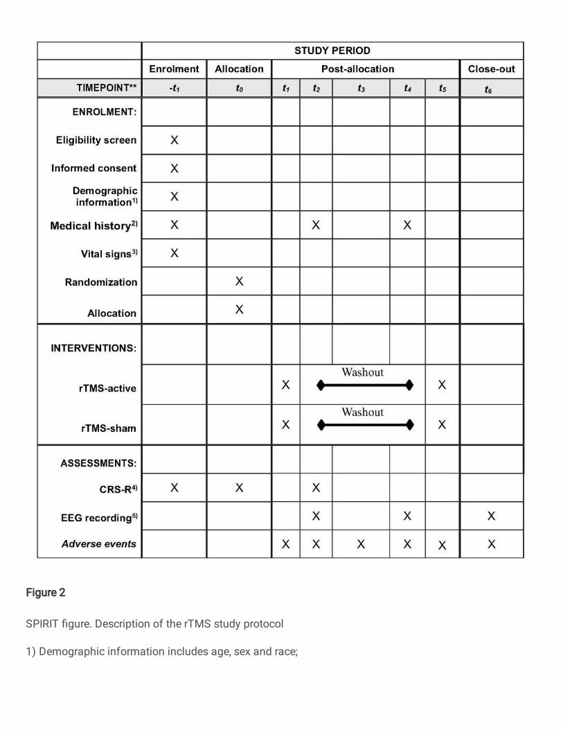
Figure 2
SPIRIT �gure. Description of the rTMS study protocol
1) Demographic information includes age, sex and race;
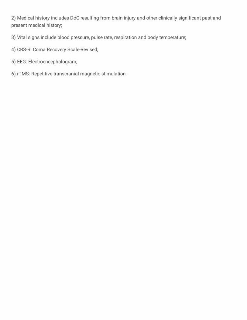
2) Medical history includes DoC resulting from brain injury and other clinically signi�cant past andpresent medical history;
3) Vital signs include blood pressure, pulse rate, respiration and body temperature;
4) CRS-R: Coma Recovery Scale-Revised;
5) EEG: Electroencephalogram;
6) rTMS: Repetitive transcranial magnetic stimulation.
Related Documents