A Japanese Boy with H-ABC 163 163 Tohoku J. Exp. Med., 2006, 209, 163-167 Received December 19, 2005; revision accepted for publication March 27, 2006. Correspondence: Keisuke Wakusawa, Department of Pediatrics, Tohoku University School of Medicine, 1-1 Seiryomachi, Aoba-ku, Sendai 980-8574, Japan. e-mail: k-wakusawa@nifty. com Case Report Effective Treatment with Levodopa and Carbidopa for Hypomyelination with Atrophy of the Basal Ganglia and Cerebellum KEISUKE WAKUSAWA, KAZUHIRO HAGINOYA, T ARO KITAMURA, NORIKO T OGASHI, MAMIKO ISHITOBI, HIROYUKI Y OKOYAMA, SHUICHI HIGANO, 1 AKIRA ONUMA, 2 T AKAHIRO NARA 3 and KAZUIE IINUMA Department of Pediatrics, 1 Department of Diagnostic Radiology, Tohoku University School of Medicine, Sendai, Japan, 2 Department of Pediatrics, Takutou Rehabilitation Center for Disabled Children, Sendai, Japan, and 3 Department of Rehabilitation, Miyagi Children’s Hospital, Sendai, Japan WAKUSAWA, K., HAGINOYA, K., KITAMURA, T., T OGASHI, N., ISHITOBI, M., Y OKOYAMA, H., HIGANO, S., ONUMA, A., NARA, T. and IINUMA, K. Effective Treatment with Levodopa and Carbidopa for Hypomyelination with Atrophy of the Basal Ganglia and Cerebellum. Tohoku J. Exp. Med., 2006, 209 (2), 163-167 ── Hypomyelination with atrophy of the basal ganglia and cerebellum (H-ABC) is a rare leukoencephalopathy presenting in the in- fantile period and characterized by diffuse cerebral hypomyelination, and atrophy of the basal ganglia and cerebellum. As patients with H-ABC lack remarkable laboratory find- ings, the diagnosis is based on brain magnetic resonance imaging findings alone. Only eight cases have been reported in the literature, and thus the natural course and treatment of this disease are not fully understood. We report a 35-month-old boy with H-ABC who had hemidystonia, hypomyelination, and cerebellar ataxia. We diagnosed H-ABC after considering a thorough differential diagnosis, excluding other diseases involving hemidys- tonia, hypomyelination, and cerebellar ataxia. Furthermore, technetium-99m ethyl cyste- inate dimmer-single-photon emission computerized tomography (Tc-ECD-SPECT) and positron emission tomography with fluorodeoxyglucose 18 F (FDG-PET) revealed decreased blood flow and glucose metabolism in the bilateral lenticular nucleus, thalamus, and cerebellum. A peroral levodopa preparation containing carbidopa (levodopa-carbidopa) was effective at ameliorating and stopping the progression of the patient’s dystonia (final effective doses: levodopa, 200 mg/day and carbidopa, 20 mg/day). This is the first case report of a Japanese patient with H-ABC and treatment for this disease. Levodopa- carbidopa may be an effective treatment for H-ABC. ──── H-ABC; levodopa- carbidopa; hemidystonia; hypomyelination; cerebellar ataxia © 2006 Tohoku University Medical Press

Welcome message from author
This document is posted to help you gain knowledge. Please leave a comment to let me know what you think about it! Share it to your friends and learn new things together.
Transcript

A Japanese Boy with H-ABC 163
163
Tohoku J. Exp. Med., 2006, 209, 163-167
Received December 19, 2005; revision accepted for publication March 27, 2006.Correspondence: Keisuke Wakusawa, Department of Pediatrics, Tohoku University School of Medicine, 1-1
Seiryomachi, Aoba-ku, Sendai 980-8574, Japan.e-mail: k-wakusawa@nifty. com
Case Report
Effective Treatment with Levodopa and Carbidopa for Hypomyelination with Atrophy of the Basal Ganglia and Cerebellum
KEISUKE WAKUSAWA, KAZUHIRO HAGINOYA, TARO KITAMURA, NORIKO TOGASHI, MAMIKO ISHITOBI, HIROYUKI YOKOYAMA, SHUICHI HIGANO,1 AKIRA ONUMA,2 TAKAHIRO NARA
3 and KAZUIE IINUMA
Department of Pediatrics, 1Department of Diagnostic Radiology, Tohoku University School of Medicine, Sendai, Japan,2Department of Pediatrics, Takutou Rehabilitation Center for Disabled Children, Sendai, Japan, and3Department of Rehabilitation, Miyagi Children’s Hospital, Sendai, Japan
WAKUSAWA, K., HAGINOYA, K., KITAMURA, T., TOGASHI, N., ISHITOBI, M., YOKOYAMA, H., HIGANO, S., ONUMA, A., NARA, T. and IINUMA, K. Effective Treatment with Levodopa and Carbidopa for Hypomyelination with Atrophy of the Basal Ganglia and Cerebellum. Tohoku J. Exp. Med., 2006, 209 (2), 163-167 ── Hypomyelination with atrophy of the basal ganglia and cerebellum (H-ABC) is a rare leukoencephalopathy presenting in the in-fantile period and characterized by diffuse cerebral hypomyelination, and atrophy of the basal ganglia and cerebellum. As patients with H-ABC lack remarkable laboratory find-ings, the diagnosis is based on brain magnetic resonance imaging findings alone. Only eight cases have been reported in the literature, and thus the natural course and treatment of this disease are not fully understood. We report a 35-month-old boy with H-ABC who had hemidystonia, hypomyelination, and cerebellar ataxia. We diagnosed H-ABC after considering a thorough differential diagnosis, excluding other diseases involving hemidys-tonia, hypomyelination, and cerebellar ataxia. Furthermore, technetium-99m ethyl cyste-inate dimmer-single-photon emission computerized tomography (Tc-ECD-SPECT) and positron emission tomography with fluorodeoxyglucose 18F (FDG-PET) revealed decreased blood flow and glucose metabolism in the bilateral lenticular nucleus, thalamus, and cerebellum. A peroral levodopa preparation containing carbidopa (levodopa-carbidopa) was effective at ameliorating and stopping the progression of the patient’s dystonia (final effective doses: levodopa, 200 mg/day and carbidopa, 20 mg/day). This is the first case report of a Japanese patient with H-ABC and treatment for this disease. Levodopa-carbidopa may be an effective treatment for H-ABC. ──── H-ABC; levodopa-carbidopa; hemidystonia; hypomyelination; cerebellar ataxia© 2006 Tohoku University Medical Press

K. Wakusawa et al.164 A Japanese Boy with H-ABC 165
In 2002, Van der Knaap (2002) first reported patients who had hypomyelination with atrophy of the basal ganglia and cerebellum (H-ABC). H-ABC is an infantile-onset, progressive leukoen-cephalopathy. As patients with H-ABC lack remarkable laboratory findings, the diagnosis is based on three brain magnetic resonance imaging findings: diffuse cerebral hypomyelination, atro-phy of the basal ganglia, and atrophy of the cere-bellum. Only eight cases have been reported (van der Knaap 2002; Mercimek-Mahmutoglu et al. 2005), and most of the details of this disease remains unknown. More patients are needed to establish this condition as a clinical disease entity. Furthermore, there is no known treatment for this condition, and there have been no reports of Japanese patients with the same clinical and radiological findings. Here, we report a Japanese boy who appeared to have H-ABC and who was effectively treated with levodopa-carbidopa.
CASE REPORT
A 35-month-old boy was born by normal delivery after 40 weeks of gestation. His birth weight was 3,032 g. He had no contributory fam-ily history. Head control was achieved at 2.5 months of age; rolling, standing, and walking alone were achieved at 4, 12, and 15 months, respectively. However, because he could not walk for more than a few steps until he was 18 months old, he was referred to another hospital for evalu-ation of delayed development. Brain magnetic resonance imaging (MRI) revealed delayed myelination. Dystonic posture of the right leg had been evident upon walking since the age of 2 years. The gait disturbance gradually progressed, and he could not walk alone at 34 months, when his right arm also began to show a dystonic pos-ture. He could barely use his right hand. He was referred to our hospital for close evaluation of right hemidystonia at 35 months of age.
At the first examination, he was 85 cm tall (−2 S.D.) and weighed 12 kg (−1 S.D.); his head circumference was 47 cm (within the normal range). The right upper and lower extremities showed rigidity and abnormally flexed posture and were thought to have dystonia. His right
hand could not grasp anything. The deep tendon reflexes were exaggerated in the right extremities, and the Babinski reflex was positive on the right. These clinical findings suggested that his right extremities showed both pyramidal and extrapy-ramidal signs. Muscle power and tonus and deep tendon reflexes were normal on the left side. He could not creep alternately; therefore, he moved using his upper and lower limbs symmetrically like a jumping frog. He had neither surface anomalies nor a dysmorphic face. No abnormali-ties of the sensory systems were observed. The liver and spleen were not palpable. His mental and motor development was delayed, and the developmental quotient evaluated at age 2 was 53.
On brain MRI, the deep cerebral white mat-ter, including the internal capsule and corpus cal-losum, showed mildly elevated signal intensity on T2-weighted images and low signal intensity on T1-weighted images, compared with age-matched controls (Figs. 1a and 1c). The pyramidal tracts in the brain stem also showed mildly high signal intensity on T2-weighted images and low signal intensity on T1-weighted images. These findings were consistent with hypomyelination. The puta-men was very small bilaterally, and these regions had high signal intensity on T2-weighted images. In contrast, the globus pallidus and thalamus appeared normal. The cerebellar vermis and hemisphere were atrophic, whereas the pons showed no atrophy.
Magnetic resonance spectroscopy (MRS) demonstrated a decreased N-acetylaspartate/creatinine (NAA/Cr) ratio in the basal ganglia and thalamus (0.67; age-matched normal range: 1.33-1.65; Martin et al. 2001). There was no lac-tate peak. These MRS findings might suggest the decrease or poor development of neurons in the basal ganglia. Magnetic resonance angiography (MRA) was unremarkable. Technetium-99m eth-yl cysteinate dimmer-single-photon emission computerized tomography (Tc-ECD-SPECT) revealed decreased cerebral blood flow (CBF) at the basal ganglia, thalamus, and cerebellum (Figs. 2a and 2b). Positron emission tomography with fluorodeoxyglucose 18F (FDG-PET) showed decreased FDG uptake at the basal ganglia and

K. Wakusawa et al.164 A Japanese Boy with H-ABC 165
cerebellum (Figs. 2c and 2d).Electroencephalography, somatosensory
evoked potentials of the median nerve, and flush visual evoked potentials were normal. The brain stem auditory evoked potentials showed an abnor-mally prolonged interval of waves I to V (right: 5.04 milliseconds, left: 4.88 milliseconds; normal: 3.78-4.62 milliseconds), suggesting dysfunction of the brain stem.
The following laboratory results were all unremarkable: blood cell karyotype; blood gas analysis; serum levels of uric acid, ammonia, cop-per (Cu), ceruloplasmin, pyruvate, lactate, IgG, IgA, IgM, CMV-IgM·IgG, HSV-IgM·IgG, and rubella-IgM·IgG; anti-streptolysin O; antinuclear antibody; anti-cardiolipin antibody; anti-double-stranded DNA antibody; white blood cell arylsulfatase A; the coagulation system, including protein C, protein S, and anti-thrombin III; plasma very-long-chain fatty acids (VLCFA); free biotin;
total biotin; biotinidase; neopterin; biopterin; the neopterin/biopterin (N/B) ratio; transferrin iso-electric focusing; acylcarnitine; and proteolipid protein (PLP) gene analysis for Pelizaeus-Merzbacher disease. The serum amino acid anal-ysis showed an increased level of glycine (4.80 mg/dl; normal 1.23-2.83), although the glycine level in the cerebrospinal fluid (CSF) was not increased (0.03 mg/dl, normal). The CSF/serum glycine ratio was 0.0062 (normal < 0.03; Hamash 1995). Urinary levels of organic acids analyzed with gas chromatography-mass spectroscopy (GC-MS), urinary electrolytes, creatinine, uric acid, neopterin, biopterin, and the N/B ratio were normal.
In the CSF, the cell count, glucose and pro-tein levels, lactate, pyruvate, amino acids, homovanillic acid (HVA), 5-hydroxyindole acetic acid (5-HIAA), γ -aminobutyric acid, myelin-basic protein, neopterin, biopterin, and N/B ratio were
Fig. 1. The Brain MR images on admission (at 35 months) and at 55 months. T2-weighted (TR3600, TE95.9) axial MR images taken at 35 (a and b) and 55 (d and e) months and
T1-weighted (TR440, TE14.0) sagittal MR images taken at 35 (c) and 55 (f) months. The cerebral white matter, including the internal capsule and corpus callosum (thin white arrows), had mildly increased signal intensity on the T2-weighted images compared to the age-matched controls. The putamen was very small bilaterally (black arrows), and this region had high signal intensity on T2-weighted images. The cerebellar vermis and hemisphere were atrophic (thick white arrows).

K. Wakusawa et al.166 A Japanese Boy with H-ABC 167
unremarkable. The bone marrow (BM) and oph-thalmologic examinations were unremarkable.
Given these findings, the patient was diag-nosed with H-ABC. A peroral levodopa prepara-tion containing carbidopa (levodopa-carbidopa), vitamin B1, and biotin were all tried as treatments. Soon after hospitalization, he began to have athe-totic movement of the left fingers and ataxia of the trunk. The drugs did not initially appear to be effective (initial doses: levodopa; 1 mg/day and carbidopa, 0.1 mg/day). However, because his dystonic posture became worse when he did not take levodopa-carbidopa, we decided to increase the dose. His ability to stand and walk with sup-port improved and equinovarus decreased on daily doses of 200 mg levodopa, 20 mg carbidopa and 10 mg trihexyphenidyl HCl. He still could not alternatively creep, and the ataxia of the trunk persisted.
At present, he is 5 years and 6 months old. His hemidystonia has improved and he can use his right hand and grasp objects, although his right arm sometimes shows a dystonic or athetotic posture. Mental retardation is still evident, but he is able to use a few meaningful words. The increased deep tendon reflexes of both legs with a positive Babinski reflex, truncal ataxia, and loss of alterative creeping persist. Repeat MRI taken at 55 months of age showed no progression of myelination compared with the previous study (Figs. 1d and 1f).
DISCUSSION
The clinical and laboratory findings in our patient were characterized as hemidystonia, hypo-myelination, and cerebellar atrophy. FDG-PET and Tc-ECD-SPECT studies showed that the basal ganglia and cerebellum were severely impaired in
Fig. 2. ECD-SPECT (a, b) and FDG-PET studies (c, d) of Brain. Slices including the basal ganglia (a, c) and cerebellar hemisphere (b, d). The CBF and FDG uptake
were decreased in the basal ganglia (thin black arrows), thalamus (thick black arrows), and cerebel-lum (white arrows).
K. Wakusawa et al.166 A Japanese Boy with H-ABC 167
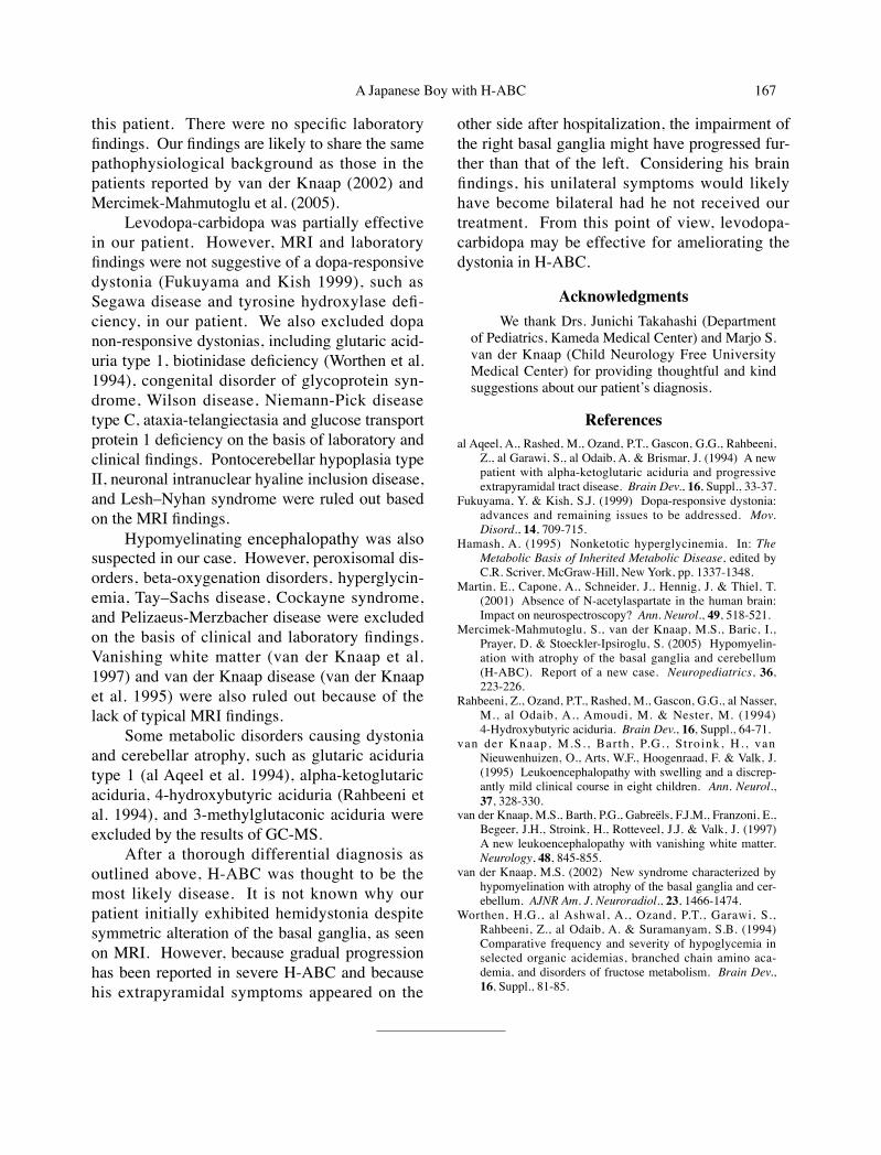
this patient. There were no specific laboratory findings. Our findings are likely to share the same pathophysiological background as those in the patients reported by van der Knaap (2002) and Mercimek-Mahmutoglu et al. (2005).
Levodopa-carbidopa was partially effective in our patient. However, MRI and laboratory findings were not suggestive of a dopa-responsive dystonia (Fukuyama and Kish 1999), such as Segawa disease and tyrosine hydroxylase defi-ciency, in our patient. We also excluded dopa non-responsive dystonias, including glutaric acid-uria type 1, biotinidase deficiency (Worthen et al. 1994), congenital disorder of glycoprotein syn-drome, Wilson disease, Niemann-Pick disease type C, ataxia-telangiectasia and glucose transport protein 1 deficiency on the basis of laboratory and clinical findings. Pontocerebellar hypoplasia type II, neuronal intranuclear hyaline inclusion disease, and Lesh–Nyhan syndrome were ruled out based on the MRI findings.
Hypomyelinating encephalopathy was also suspected in our case. However, peroxisomal dis-orders, beta-oxygenation disorders, hyperglycin-emia, Tay–Sachs disease, Cockayne syndrome, and Pelizaeus-Merzbacher disease were excluded on the basis of clinical and laboratory findings. Vanishing white matter (van der Knaap et al. 1997) and van der Knaap disease (van der Knaap et al. 1995) were also ruled out because of the lack of typical MRI findings.
Some metabolic disorders causing dystonia and cerebellar atrophy, such as glutaric aciduria type 1 (al Aqeel et al. 1994), alpha-ketoglutaric aciduria, 4-hydroxybutyric aciduria (Rahbeeni et al. 1994), and 3-methylglutaconic aciduria were excluded by the results of GC-MS.
After a thorough differential diagnosis as outlined above, H-ABC was thought to be the most likely disease. It is not known why our patient initially exhibited hemidystonia despite symmetric alteration of the basal ganglia, as seen on MRI. However, because gradual progression has been reported in severe H-ABC and because his extrapyramidal symptoms appeared on the
other side after hospitalization, the impairment of the right basal ganglia might have progressed fur-ther than that of the left. Considering his brain findings, his unilateral symptoms would likely have become bilateral had he not received our treatment. From this point of view, levodopa-carbidopa may be effective for ameliorating the dystonia in H-ABC.
AcknowledgmentsWe thank Drs. Junichi Takahashi (Department
of Pediatrics, Kameda Medical Center) and Marjo S. van der Knaap (Child Neurology Free University Medical Center) for providing thoughtful and kind suggestions about our patient’s diagnosis.
Referencesal Aqeel, A., Rashed, M., Ozand, P.T., Gascon, G.G., Rahbeeni,
Z., al Garawi, S., al Odaib, A. & Brismar, J. (1994) A new patient with alpha-ketoglutaric aciduria and progressive extrapyramidal tract disease. Brain Dev., 16, Suppl., 33-37.
Fukuyama, Y. & Kish, S.J. (1999) Dopa-responsive dystonia: advances and remaining issues to be addressed. Mov. Disord., 14, 709-715.
Hamash, A. (1995) Nonketotic hyperglycinemia. In: The Metabolic Basis of Inherited Metabolic Disease, edited by C.R. Scriver, McGraw-Hill, New York, pp. 1337-1348.
Martin, E., Capone, A., Schneider, J., Hennig, J. & Thiel, T. (2001) Absence of N-acetylaspartate in the human brain: Impact on neurospectroscopy? Ann. Neurol., 49, 518-521.
Mercimek-Mahmutoglu, S., van der Knaap, M.S., Baric, I., Prayer, D. & Stoeckler-Ipsiroglu, S. (2005) Hypomyelin-ation with atrophy of the basal ganglia and cerebellum (H-ABC). Report of a new case. Neuropediatrics, 36, 223-226.
Rahbeeni, Z., Ozand, P.T., Rashed, M., Gascon, G.G., al Nasser, M., al Odaib, A., Amoudi, M. & Nester, M. (1994) 4-Hydroxybutyric aciduria. Brain Dev., 16, Suppl., 64-71.
van der Knaap, M.S. , Barth, P.G. , Stroink, H. , van Nieuwenhuizen, O., Arts, W.F., Hoogenraad, F. & Valk, J. (1995) Leukoencephalopathy with swelling and a discrep-antly mild clinical course in eight children. Ann. Neurol., 37, 328-330.
van der Knaap, M.S., Barth, P.G., Gabreëls, F.J.M., Franzoni, E., Begeer, J.H., Stroink, H., Rotteveel, J.J. & Valk, J. (1997) A new leukoencephalopathy with vanishing white matter. Neurology, 48, 845-855.
van der Knaap, M.S. (2002) New syndrome characterized by hypomyelination with atrophy of the basal ganglia and cer-ebellum. AJNR Am. J. Neuroradiol., 23, 1466-1474.
Worthen, H.G., al Ashwal, A., Ozand, P.T., Garawi, S., Rahbeeni, Z., al Odaib, A. & Suramanyam, S.B. (1994) Comparative frequency and severity of hypoglycemia in selected organic acidemias, branched chain amino aca-demia, and disorders of fructose metabolism. Brain Dev., 16, Suppl., 81-85.
Related Documents