Effective gene therapy for haemophilic mice with pathogenic factor IX antibodies David M. Markusic 1 , Brad E. Hoffman 1 , George Q. Perrin 1 , Sushrusha Nayak 1 , Xiaomei Wang 1 , Paul A. LoDuca 1 , Katherine A. High 2,3,4 , Roland W. Herzog 1 * Keywords: adeno-associated virus; factor IX; haemophilia B; inhibitors; liver gene transfer DOI 10.1002/emmm.201302859 Received April 08, 2013 Revised August 15, 2013 Accepted August 19, 2013 GSee accompanying article http://dx.doi.org/10.1002/emmm.201302857 Formation of pathogenic antibodies is a major problem in replacement therapies for inherited protein deficiencies. For example, antibodies to coagulation factors (‘inhibitors’) seriously complicate treatment of haemophilia. While immune tolerance induction (ITI) protocols have been developed, inhibitors against factor IX (FIX) are difficult to eradicate due to anaphylactic reactions and nephrotic syndrome and thus substantially elevate risks for morbidity and mortality. However, hepatic gene transfer with an adeno-associated virus (AAV) serotype 8 vector expressing FIX (at levels of 4% of normal) rapidly reversed pre-existing high-titre inhibitors in haemophilia B mice, eliminated antibody production by B cells, desensitized from anaphylaxis (even if protein therapy was resumed) and provided long-term correction. High levels of FIX protein suppressed memory B cells and increased Treg induction, indicating direct and indirect mechanisms of suppression of inhibitor formation. Persistent presence of Treg was required to prevent relapse of antibodies. Together, these data suggest that hepatic gene transfer-based ITI provides a safe and effective alternative to eradicate inhibitors. This strategy may be broadly applicable to reversal of antibodies in different genetic diseases. INTRODUCTION Treatment of inherited protein deficiency may be complicated by pathogenic antibody responses during replacement therapy, highlighting the need for development of suitable immune tolerance protocols. One example is haemophilia B, which results from the loss of functional coagulation factor IX (FIX) with an estimated incidence of 1 in 30,000 male births worldwide. Untreated, patients can develop spontaneous bleeds into the joints and closed spaces resulting in high morbidity and mortality. Disease severity is based on residual FIX activity (severe <1%, moderate 1–5% and mild >5%), which impacts the frequency and severity of bleeds. Patients are currently treated with exogenous FIX protein concentrate, which is plasma‐derived or recombinant. A fraction of patients (2–5%), predominantly those with severe haemophilia B, develop neutralizing antibodies to the FIX, termed inhibitors, requiring treatment with expensive bypassing agents to maintain haemostasis. Most of the available bypassing products are designated for short‐term treatment on‐demand use, and thus haemophilia B patients with inhibitors experience increased morbidity. Unfortunately, haemophilia B patients have a poor response rate to immune tolerance induction (ITI) protocols that require frequent high levels of factor adminis- tration. ITI often has to be stopped because of anaphylaxis or nephrotic syndrome (Chitlur et al, 2009; DiMichele, 2007; DiMichele, 2012; Ewenstein et al, 1997; Jadhav & Warrier, 2000; Recht et al, 2011). IgE formation has been identified as a cause for anaphylactic reactions against FIX, which occur in 25–50% of inhibitor patients (Jadhav & Warrier, 2000; Recht et al, 2011; Thorland et al, 1999; Warrier et al, 1997). Because of the severity of the immune response and lack of effective tolerance protocols, inhibitor formation in haemophilia B has been described as an ‘orphan disease in need of attention’ (DiMichele, 2007). (1) Department of Pediatrics, University of Florida, Gainesville, FL, USA (2) Department of Pediatrics, The Children’s Hospital of Philadelphia and University of Pennsylvania Medical Center, Philadelphia, PA, USA (3) Center for Cellular and Molecular Therapeutics, The Children’s Hospital of Philadelphia, Philadelphia, PA, USA (4) Howard Hughes Medical Institute, Philadelphia, PA, USA *Corresponding author: Tel: þ1 352 273 8113; Fax: þ1 352 273 8342; E-mail: rherzog@ufl.edu Research Article OPEN ACCESS TRANSPARENT PROCESS Reversing inhibitors with AAV liver gene transfer 1698 ß 2013 The Authors. Published by John Wiley and Sons, Ltd on behalf of EMBO. This is an open access article under the terms of the Creative Commons Attribution License, which permits use, distribution and reproduction in any medium, provided the original work is properly cited. EMBO Mol Med (2013) 5, 1698–1709

Welcome message from author
This document is posted to help you gain knowledge. Please leave a comment to let me know what you think about it! Share it to your friends and learn new things together.
Transcript
Research ArticleOPENACCESS
TRANSPARENTPROCESS Reversing inhibitors with AAV liver gene transfer
1698
Effective gene therapy for haemophilic micewith pathogenic factor IX antibodies
David M. Markusic1, Brad E. Hoffman1, George Q. Perrin1, Sushrusha Nayak1, Xiaomei Wang1,Paul A. LoDuca1, Katherine A. High2,3,4, Roland W. Herzog1*
Keywords: adeno-associated virus; factor
IX; haemophilia B; inhibitors; liver gene
transfer
DOI 10.1002/emmm.201302859
Received April 08, 2013
Revised August 15, 2013
Accepted August 19, 2013
GSee accompanying article
http://dx.doi.org/10.1002/emmm.201302857
(1) Department of Pediatrics, University of Florida, Gai
(2) Department of Pediatrics, The Children’s Hospital
University of Pennsylvania Medical Center, Philadel
(3) Center for Cellular and Molecular Therapeutics, The C
Philadelphia, Philadelphia, PA, USA
(4) Howard Hughes Medical Institute, Philadelphia, PA,
*Corresponding author: Tel: þ1 352 273 8113; Fax: þE-mail: [email protected]
� 2013 The Authors. Published by John Wiley and Sons,the terms of the Creative Commons Attribution License, wprovided the original work is properly cited.
Formation of pathogenic antibodies is a major problem in replacement therapies
for inherited protein deficiencies. For example, antibodies to coagulation factors
(‘inhibitors’) seriously complicate treatment of haemophilia. While immune
tolerance induction (ITI) protocols have been developed, inhibitors against factor
IX (FIX) are difficult to eradicate due to anaphylactic reactions and nephrotic
syndrome and thus substantially elevate risks for morbidity and mortality.
However, hepatic gene transfer with an adeno-associated virus (AAV) serotype 8
vector expressing FIX (at levels of �4% of normal) rapidly reversed pre-existing
high-titre inhibitors in haemophilia B mice, eliminated antibody production by B
cells, desensitized from anaphylaxis (even if protein therapy was resumed) and
provided long-term correction. High levels of FIX protein suppressed memory B
cells and increased Treg induction, indicating direct and indirect mechanisms of
suppression of inhibitor formation. Persistent presence of Treg was required
to prevent relapse of antibodies. Together, these data suggest that hepatic
gene transfer-based ITI provides a safe and effective alternative to eradicate
inhibitors. This strategy may be broadly applicable to reversal of antibodies in
different genetic diseases.
INTRODUCTION
Treatment of inherited protein deficiency may be complicatedby pathogenic antibody responses during replacement therapy,highlighting the need for development of suitable immunetolerance protocols. One example is haemophilia B, whichresults from the loss of functional coagulation factor IX (FIX)with an estimated incidence of 1 in 30,000 male birthsworldwide. Untreated, patients can develop spontaneousbleeds into the joints and closed spaces resulting in highmorbidity and mortality. Disease severity is based on residualFIX activity (severe <1%, moderate 1–5% and mild >5%),which impacts the frequency and severity of bleeds. Patients
nesville, FL, USA
of Philadelphia and
phia, PA, USA
hildren’s Hospital of
USA
1 352 273 8342;
Ltd on behalf of EMBO. Thhich permits use, distribut
are currently treated with exogenous FIX protein concentrate,which is plasma‐derived or recombinant. A fraction of patients(2–5%), predominantly those with severe haemophilia B,develop neutralizing antibodies to the FIX, termed inhibitors,requiring treatment with expensive bypassing agents tomaintain haemostasis. Most of the available bypassingproducts are designated for short‐term treatment on‐demanduse, and thus haemophilia B patients with inhibitors experienceincreased morbidity. Unfortunately, haemophilia B patientshave a poor response rate to immune tolerance induction (ITI)protocols that require frequent high levels of factor adminis-tration. ITI often has to be stopped because of anaphylaxis ornephrotic syndrome (Chitlur et al, 2009; DiMichele, 2007;DiMichele, 2012; Ewenstein et al, 1997; Jadhav & Warrier,2000; Recht et al, 2011). IgE formation has been identified as acause for anaphylactic reactions against FIX, which occur in25–50% of inhibitor patients (Jadhav & Warrier, 2000; Rechtet al, 2011; Thorland et al, 1999;Warrier et al, 1997). Because ofthe severity of the immune response and lack of effectivetolerance protocols, inhibitor formation in haemophilia B hasbeen described as an ‘orphan disease in need of attention’(DiMichele, 2007).
is is an open access article underion and reproduction in any medium,
EMBO Mol Med (2013) 5, 1698–1709
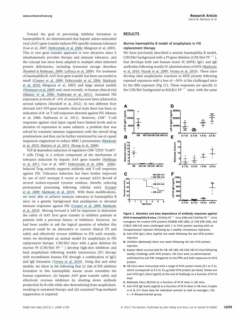
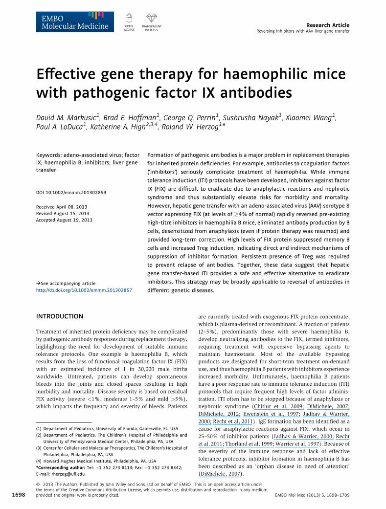
Figure 1. Mutation and dose dependence of antibody responses against
hFIX in hemophilia Bmice. C3H/HeJ F9�/�mice (HB) and C3H/HeJ F9�/�mice
transgenic for mutant hFIX proteins R180W (HB-180), LS 338 (HB-338), and
G381E (HB-CH) were challenged with 1 IU hFIX protein starting with an
intraperitoneal injection following by 5 weekly intravenous injections.
A. Anti-hFIX IgG1 titers (ng/ml) one week following the last hFIX protein
injection.
B. Inhibitor (Bethesda) titers one week following the last hFIX protein
injection.
C. Kaplan-Meier survival plot for HB, HB-180, HB-338, HB-CH mice following
weekly challenge with hFIX protein. HB mice were co-administered
antihistamine and PAF antagonist on the fifth and sixth exposures to hFIX
protein.
D. HB mice were immunized with a range of hFIX protein levels (0.1 to 3 IU,
which corresponds to 0.5 to 15mg total hFIX protein per dose). Shown are
anti-hFIX IgG1 titers (ng/ml) at the end of challenge as a function of hF.IX
dose.
E. Bethesda titers (BU/ml) as a function of hF.IX dose in HB mice.
F. Anti-hFIX IgE levels (ng/ml) as a function of hF.IX dose in HB mice. Graphs
in A, B, D-F show data for individual animals as well as averages� SD;
n¼4–6/experimental group.
Research Articlewww.embomolmed.orgDavid M. Markusic et al.
Toward the goal of preventing inhibitor formation inhaemophilia B, we demonstrated that hepatic adeno‐associatedviral (AAV) gene transfer induces FIX‐specific immune tolerance(Cao et al, 2007; Dobrzynski et al, 2006; Mingozzi et al, 2003).This in vivo gene transfer approach is very attractive since itsimultaneously provides therapy and immune tolerance, andthe concept has since been adapted to multiple other inheritedprotein deficiencies, including lysosomal storage disorders(Koeberl & Kishnani, 2009; LoDuca et al, 2009). For treatmentof haemophilia B, AAV liver gene transfer has been successful insmall (Cooper et al, 2009; Dobrzynski et al, 2006; Markusicet al, 2010; Mingozzi et al, 2003) and large animal models(Niemeyer et al, 2009) and, most recently, in human clinical trial(Manno et al, 2006; Nathwani et al, 2011). Sustained FIXexpression at levels of�6% of normal has now been achieved inseveral subjects (Davidoff et al, 2012). In two different liverdirected AAV‐hF9 gene transfer clinical trials there has been noindication of B‐ or T‐cell responses directed against FIX (Mannoet al, 2006; Nathwani et al, 2011). However, CD8þ T‐cellresponses against viral input capsid have limited levels and/orduration of expression in some subjects, a problem that wassolved by transient immune suppression with the steroid drugprednisolone and that can be further minimized by use of capsidsequences engineered to reduce MHC I presentation (Markusicet al, 2010; Martino et al, 2013; Zhong et al, 2008).
TGF‐b‐dependent induction of regulatory CD4þCD25þFoxP3þ
T cells (Treg) is a critical component of the mechanism oftolerance induction by hepatic AAV gene transfer (Hoffmanet al, 2011; Cao et al, 2007; Dobrzynski et al, 2004, 2006).Induced Treg actively suppress antibody and T‐cell responsesagainst FIX. Tolerance induction has been further improvedby use of AAV serotype 8 vector or mutant AAV2 devoid ofseveral surface‐exposed tyrosine residues, thereby reducingproteasomal processing following cellular entry (Cooperet al, 2009; Markusic et al, 2010). With these modifications,we were able to achieve immune tolerance in haemophilia Bmice on a genetic background that predisposes to elevatedimmune responses against FIX (Cooper et al, 2009; Markusicet al, 2010). Moving forward it will be important to determinethe safety of AAV liver gene transfer in inhibitor patients orpatients with a previous history of inhibitors. However, wehad been unable to ask the logical question of whether thisprotocol could be an alternative to current clinical ITI andsafely and effectively reverse inhibitors to FIX until recently,when we developed an animal model for anaphylaxis in FIXreplacement therapy. C3H/HeJ mice with a gene deletion formurine F9 (C3H/HeJ F9�/�) develop high‐titre inhibitors andfatal anaphylaxis following weekly intravenous (IV) therapywith recombinant human FIX through a combination of IgG1and IgE formation (Verma et al, 2010). Using this and othermodels, we show in the following that (i) risk of FIX inhibitorformation in this haemophilic mouse strain resembles thehuman experience; (ii) hepatic AAV gene transfer safely andeffectively reverses inhibitors by shutting down antibodyproduction by B cells while also desensitizing from anaphylaxis,resulting in sustained therapy and (iii) sustained Treg‐mediatedsuppression is required.
EMBO Mol Med (2013) 5, 1698–1709 �
RESULTS
Murine haemophilia B model of anaphylaxis in FIXreplacement therapyWe have previously described a murine haemophilia B model,C3H/HeJ background with a F9 gene deletion (C3H/HeJ F9�/�),that develops both anti human factor IX (hFIX) IgG1 and IgEantibodies followingweekly IV administration of hFIX (Markusicet al, 2010; Nayak et al, 2009; Verma et al, 2010). These micedevelop fatal anaphylactic reactions to hFIX protein followingrepeated exposures with a loss of �50% of the challenged miceby the fifth exposure (Fig 1C). These responses are specific tothe C3H/HeJ background as BALB/c F9�/� mice, with the same
2013 The Authors. Published by John Wiley and Sons, Ltd on behalf of EMBO. 1699
Research Article www.embomolmed.orgReversing inhibitors with AAV liver gene transfer
1700
gene deletion, tolerated up to eight weekly challenges of hFIXprotein with only mild IgG1 responses (Lozier et al, 2005) andabsence of fatal anaphylaxis (unpublished observations).Clinically, the risk of developing inhibitors or anaphylaxisin haemophilia B patients is greater in patients with genedeletion or complex chromosomal rearrangements, followedby nonsense mutations, and then missense mutations (Chitluret al, 2009). We maintain three different C3H/HeJ F9�/� linestransgenic for hFIX mutants, two of which are cross reactiveimmunological material negative (crim�) (late stop codon AA338 and a G381E missense mutation analogous to a spontaneousmutation in the canine haemophilia B colony at Chapel Hill)and a crimþmutant (R180W), referred to as HB‐338, HB‐CH andHB‐180, respectively. In contrast to control C3H/HeJ F9�/�mice,all three transgenic mutant hFIX mice groups tolerated weeklyhFIX challenge with no evidence for antibody/inhibitor forma-tion against hFIX IgG1 (Fig 1A and B). Cumulatively, only 1 of15mice with endogenous hFIX expression died during the courseof the experiment, and no signs of anaphylaxis were observed,while only 40% of gene deletion mice survived (Fig 1C), which
Figure 2. Reversal of inhibitors and of anaphylaxis by hepatic AAV8‐hF9 gene
generate pre-existing high-titer inhibitors. A control group of immunized mice we
Following confirmation of inhibitors, mice were treated with 1�1011 vg of an AAV8
vector. Samples from immunized control HB (n¼5) and vector-treated HB mice
A. Experimental time line.
B. Anti-hFIXIgG1 titers (ng/ml).
C. IgE titers (ng/ml).
D. Inhibitor titers (BU/ml).
E. Circulating levels of hFIX protein (ng/ml).
F. Restoration of hemostasis was assessed bymeasurement of coagulation activity
are average� SD.
G. Kaplan-Meier survival plot of 1�1011 vg AAV8-hF9 vector treated mice follow
� 2013 The Authors. Published by John Wiley and Sons, Ltd on behalf of EMBO.
showed anaphylactic reactions as previously published (Vermaet al, 2010). Thus, the model mirrors the human situation in thatendogenous FIX expression confers a level of tolerance that isprotective of pathogenic antibody responses.
Although haemophilia A patients treated with factor VIII(F.VIII) protein have a higher incidence of inhibitors (20–30%),remarkably there are few reports of patients with anaphylaxis(Jadhav &Warrier, 2000). One hypothesis is that the higher hFIXsystemic levels of total protein needed for therapeutic effect mayact as trigger (1 IU hFIX is 5000 ng/ml compared to 200 ng/mlfor hF.VIII) (Warrier et al, 1997). Therefore, we evaluatedIgE and IgG1 levels in response to weekly challenge with 0.1,0.3, 1 and 3 IU/ml of hFIX protein (with the two higher dosesbeing in the range clinically used to treat moderate to severebleeds). We observed a trend towards higher IgG1 andBethesda inhibitors titres with increasing hFIX protein (Fig 1Dand E), while IgE levels strongly correlated with hFIX levels(Fig 1F). Importantly, mice challenged with 0.1 and 0.3 IU hFIXdid not require pharmacological intervention to survive thechallenge.
transfer. C3H/HeJ F9�/� mice (HB) were immunized with hFIX protein to
re then left alone to follow hFIX antibodies over the course of the experiment.
-hF9 vector (n¼8) and were bled 4, 8, 16, and 18 weeks following injection of
were used for ELISA measurements.
by activated partial thromboplastin time (aPTT, in seconds). Data points in A–F
ing weekly challenge with hFIX protein.
EMBO Mol Med (2013) 5, 1698–1709
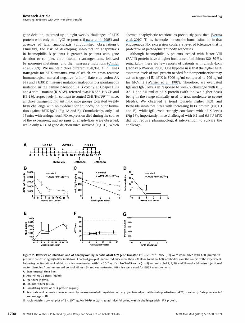
Figure 3. Reversal of B‐ and T‐cell responses against hFIX by hepatic
AAV8‐hF9 gene transfer. B-cell ELISpot assays specific for hFIX antibody
secretingwere performed on both bonemarrow and spleen cells from C3H/HeJ
F9�/� mice immunized against hF.X. Results are presented as spots forming
units (SFU) per 106 cells with representative wells presented to the right.
A. B-cell ELISpots for hFIX antibody secreting bone marrow cells (AAV8 F9
versus Early Con p¼0.0022 and AAV8 F9 versus Late Con p¼0.0286).
B. B-cell ELISpots for hFIX antibody secreting splenocytes (AAV8 F9 versus
Early Con p¼0.0043 and AAV8 F9 versus Late Con p¼0.0195). Early con
(early controls) refers to mice sacrificed shortly after the immunization
protocol, i.e. the time point of vector treatment. Late Con (late controls)
are immunized untreated mice that were followed in parallel to vector
treated mice.
C. Cytokine responses specific to hF.IX. Splenocytes (1�105 perwell) were
cultured in vitro without or with hFIX protein (10mg/ml) and harvested
48 h later for mRNA extraction and cDNA synthesis and ran on qPCR array
with genes associated with Th1, Th2, or Treg responses. Results are
presented as fold change compared to unstimulated cells. Bar graphs
indicate average� SD with n¼4/experimental group (IL-4 Control versus
AAV8 F9 p¼0.0196). The students 2-tailed T test was used to determine
statistical significance.
Research Articlewww.embomolmed.orgDavid M. Markusic et al.
AAV8‐hF9 liver gene transfer reverses hFIX inhibitors, preventsamnestic immune responses including anaphylaxisNext, we asked if C3H/HeJ F9�/� mice with high‐titre pre‐existing inhibitors could be safely treated with liver genetransfer (or experience anaphylaxis as with continued proteininjections) and what would happen to inhibitor levels?We chosean AAV8 vector because it has strong tropism to the liver afterperipheral vein administration, avoiding the need for aninvasive procedure in the presence of an inhibitor, and failsto transduce professional antigen presenting cells (APCs).Additionally, the construct includes a strong and highlyhepatocyte‐specific promoter (see published data and Support-ing Information Fig S1) (Cooper et al, 2009; Lu & Song, 2009;Manno et al, 2006; Vandendriessche et al, 2007). In naïveanimals, efficient hepatic AAV gene transfer can establish adominant state of immune tolerance that suppresses immuneresponses to the transgene product in other tissues (Hoffmanet al, 2007; Passini et al, 2007; Zhang et al, 2012).
In the first set of experiments, we established a group of micethat had developed high‐titre inhibitors (average anti‐hFIX IgG1level of 27mg/ml, IgE of 330 ng/ml, and Bethesda titre of 16,n¼ 8) after protein therapy (Fig 2A–D). A dose of 1� 1011 vgAAV8‐hF9 vector was delivered via the tail vein 1 week afterthe last hFIX protein injection. Previous studies conducted innaïve C3H/HeJ F9�/� mice showed significant correction with>20% normal hFIX levels using a similar vector dose anddelivery route (Cooper et al, 2009). One month following genetransfer mice were bled and challenged for an additional monthwith weekly IV recombinant hFIX protein in the absence ofanti‐histamine/PAF antagonist (Fig 2A). Remarkably, 1 monthfollowing gene transfer anti‐hFIX titres declined to undetectableby IgE and IgG ELISA and Bethesda assays in all vector treatedanimals (Fig 2B–D). No fatalities or anaphylactic reactions wereobserved after vector treatment or subsequent hFIX proteininjections (Fig 2G). Vector treated mice showed stable hFIXprotein expression and complete correction of the aPTTcoagulation time through the duration of the experiment (upto 20 weeks post‐gene transfer, Fig 2E and F). In the absence ofgene transfer and further exposure to hFIX protein, anti‐hFIXand Bethesda titres decline much more slowly (Fig 2B and D).
AAV8‐hF9 liver gene transfer reverses cellular responses tohFIXVector treated animals were given a final challenge with hFIXprotein and sacrificed the following week to characterizedifferences in B‐ and T‐cell responses against hFIX protein.Bone marrow and splenocyte cells were harvested for thedetection of anti‐hFIX secreting cells (ACS) using a B‐cellELISpot. Vector treated mice had no detectable spots (Fig 3A andB), in line with no detectable circulating antibodies for over3months (Fig 2B). In contrast, we detected ACS in splenocytes ofcontrol mice that had formed inhibitors after protein therapy butdid not receive gene transfer (Fig 3A and B). Additional controlmice, whichwere analyzed early after protein therapy (i.e. closerto the time when gene transfer was given), showed ACS inspleen at similar frequencies and, additionally, in bone marrow(Fig 3A). Therefore, anti‐hFIX producing plasma cells form in the
EMBO Mol Med (2013) 5, 1698–1709 �
spleen and bone marrow in this model but are persistent in thespleen, and hepatic expression eliminated anti‐hFIX production.
Upon in vitro stimulation with hFIX antigen, no evidence for aT‐cell response in vector treated mice was obtained usingquantitative RT‐PCR array that included cytokines and markersfor Th1, Th2, Th17 and Treg. Control mice (no vector treatment)showed IL‐4 expression and additional, albeit with more
2013 The Authors. Published by John Wiley and Sons, Ltd on behalf of EMBO. 1701
Research Article www.embomolmed.orgReversing inhibitors with AAV liver gene transfer
1702
variability, IL‐13 and IFN‐g (Fig 3C). IL‐4 and IL‐13 are Th2cytokines known to promote IgG1 and IgE class switch in B cells.
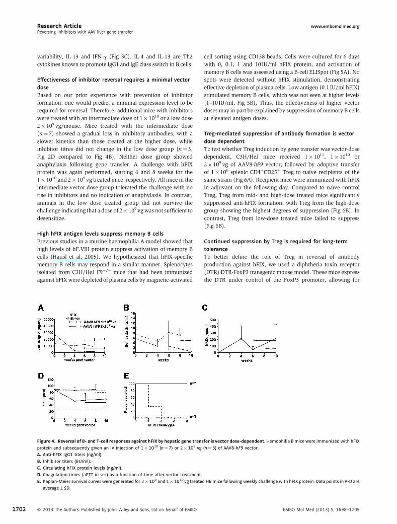
Effectiveness of inhibitor reversal requires a minimal vectordoseBased on our prior experience with prevention of inhibitorformation, one would predict a minimal expression level to berequired for reversal. Therefore, additional mice with inhibitorswere treated with an intermediate dose of 1� 1010 or a low dose2� 109 vg/mouse. Mice treated with the intermediate dose(n¼ 7) showed a gradual loss in inhibitory antibodies, with aslower kinetics than those treated at the higher dose, whileinhibitor titres did not change in the low dose group (n¼ 3,Fig 2D compared to Fig 4B). Neither dose group showedanaphylaxis following gene transfer. A challenge with hFIXprotein was again performed, starting 6 and 8 weeks for the1� 1010 and 2� 109 vg treated mice, respectively. All mice in theintermediate vector dose group tolerated the challenge with norise in inhibitors and no indication of anaphylaxis. In contrast,animals in the low dose treated group did not survive thechallenge indicating that a dose of 2� 109 vgwas not sufficient todesensitize.
High hFIX antigen levels suppress memory B cellsPrevious studies in a murine haemophilia A model showed thathigh levels of hF.VIII protein suppress activation of memory Bcells (Hausl et al, 2005). We hypothesized that hFIX‐specificmemory B cells may respond in a similar manner. Splenocytesisolated from C3H/HeJ F9�/� mice that had been immunizedagainst hFIXwere depleted of plasma cells bymagnetic‐activated
Figure 4. Reversal of B‐ and T‐cell responses against hFIX by hepatic gene trans
protein and subsequently given an IV injection of 1�1010 (n¼7) or 2�109 vg
A. Anti-hFIX IgG1 titers (ng/ml).
B. Inhibitor titers (BU/ml).
C. Circulating hFIX protein levels (ng/ml).
D. Coagulation times (aPTT in sec) as a function of time after vector treatment.
E. Kaplan-Meier survival curves were generated for 2�109 and 1�1010 vg treate
average� SD.
� 2013 The Authors. Published by John Wiley and Sons, Ltd on behalf of EMBO.
cell sorting using CD138 beads. Cells were cultured for 6 dayswith 0, 0.1, 1 and 10 IU/ml hFIX protein, and activation ofmemory B cells was assessed using a B‐cell ELISpot (Fig 5A). Nospots were detected without hFIX stimulation, demonstratingeffective depletion of plasma cells. Low antigen (0.1 IU/ml hFIX)stimulated memory B cells, which was not seen at higher levels(1–10 IU/ml, Fig 5B). Thus, the effectiveness of higher vectordoses may in part be explained by suppression of memory B cellsat elevated antigen doses.
Treg‐mediated suppression of antibody formation is vectordose dependentTo test whether Treg induction by gene transfer was vector‐dosedependent, C3H/HeJ mice received 1� 1011, 1� 1010 or2� 109 vg of AAV8‐hF9 vector, followed by adoptive transferof 1� 106 splenic CD4þCD25þ Treg to naive recipients of thesame strain (Fig 6A). Recipient mice were immunized with hFIXin adjuvant on the following day. Compared to naïve controlTreg, Treg from mid‐ and high‐dose treated mice significantlysuppressed anti‐hFIX formation, with Treg from the high‐dosegroup showing the highest degrees of suppression (Fig 6B). Incontrast, Treg from low‐dose treated mice failed to suppress(Fig 6B).
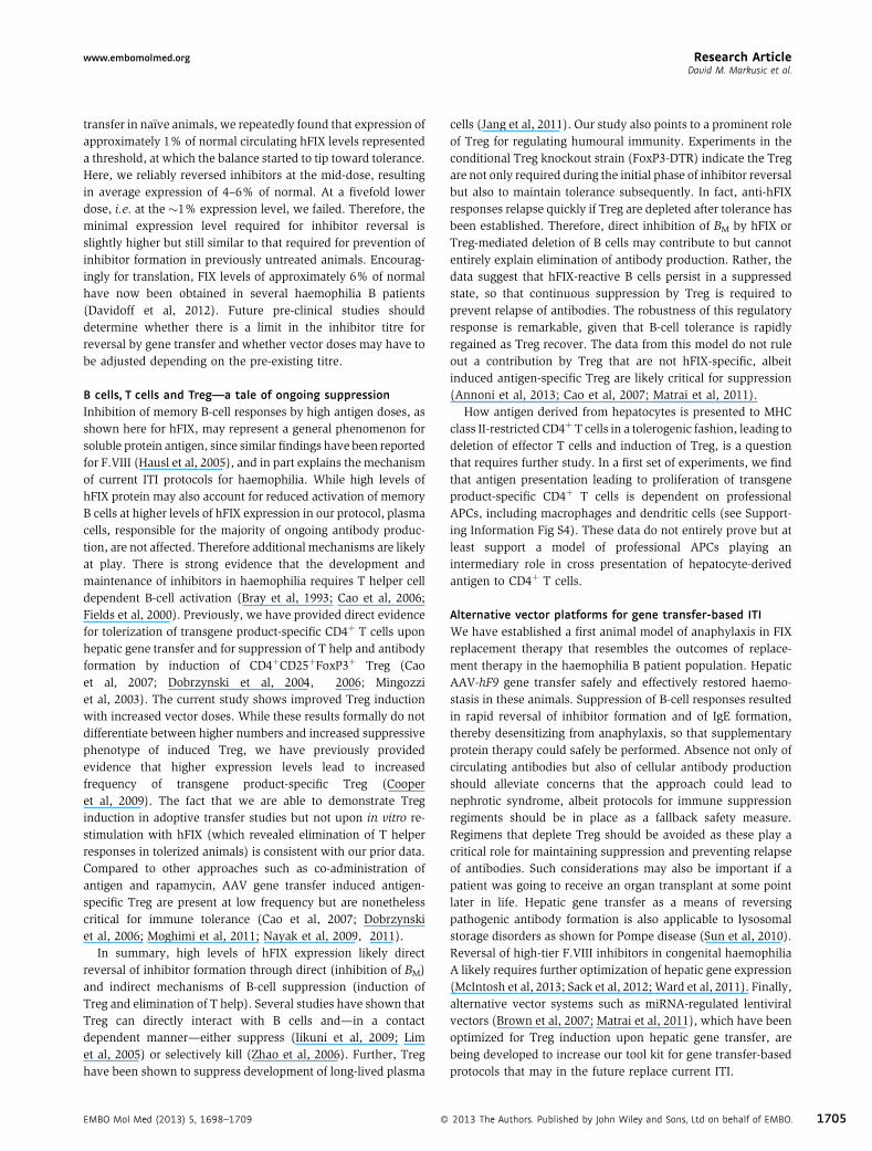
Continued suppression by Treg is required for long‐termtoleranceTo better define the role of Treg in reversal of antibodyproduction against hFIX, we used a diphtheria toxin receptor(DTR) DTR‐FoxP3 transgenic mouse model. These mice expressthe DTR under control of the FoxP3 promoter, allowing for
fer is vector dose‐dependent. Hemophilia B mice were immunized with hFIX
(n¼3) of AAV8-hF9 vector.
d HBmice following weekly challenge with hFIX protein. Data points in A-D are
EMBO Mol Med (2013) 5, 1698–1709
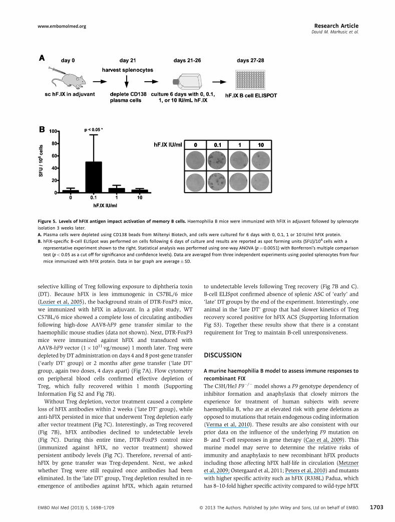
Figure 5. Levels of hFIX antigen impact activation of memory B cells. Haemophilia B mice were immunized with hFIX in adjuvant followed by splenocyte
isolation 3 weeks later.
A. Plasma cells were depleted using CD138 beads from Miltenyi Biotech, and cells were cultured for 6 days with 0, 0.1, 1 or 10 IU/ml hFIX protein.
B. hFIX-specific B-cell ELISpot was performed on cells following 6 days of culture and results are reported as spot forming units (SFU)/106 cells with a
representative experiment shown to the right. Statistical analysis was performed using one-way ANOVA (p¼0.0051) with Bonferroni’s multiple comparison
test (p<0.05 as a cut off for significance and confidence levels). Data are averaged from three independent experiments using pooled splenocytes from four
mice immunized with hFIX protein. Data in bar graph are average� SD.
Research Articlewww.embomolmed.orgDavid M. Markusic et al.
selective killing of Treg following exposure to diphtheria toxin(DT). Because hFIX is less immunogenic in C57BL/6 mice(Lozier et al, 2005), the background strain of DTR‐FoxP3 mice,we immunized with hFIX in adjuvant. In a pilot study, WTC57BL/6 mice showed a complete loss of circulating antibodiesfollowing high‐dose AAV8‐hF9 gene transfer similar to thehaemophilic mouse studies (data not shown). Next, DTR‐FoxP3mice were immunized against hFIX and transduced withAAV8‐hF9 vector (1� 1011 vg/mouse) 1 month later. Treg weredepleted by DT administration on days 4 and 8 post‐gene transfer(‘early DT’ group) or 2 months after gene transfer (‘late DT’group, again two doses, 4 days apart) (Fig 7A). Flow cytometryon peripheral blood cells confirmed effective depletion ofTreg, which fully recovered within 1 month (SupportingInformation Fig S2 and Fig 7B).
Without Treg depletion, vector treatment caused a completeloss of hFIX antibodies within 2 weeks (‘late DT’ group), whileanti‐hFIX persisted in mice that underwent Treg depletion earlyafter vector treatment (Fig 7C). Interestingly, as Treg recovered(Fig 7B), hFIX antibodies declined to undetectable levels(Fig 7C). During this entire time, DTR‐FoxP3 control mice(immunized against hFIX, no vector treatment) showedpersistent antibody levels (Fig 7C). Therefore, reversal of anti‐hFIX by gene transfer was Treg‐dependent. Next, we askedwhether Treg were still required once antibodies had beeneliminated. In the ‘late DT’ group, Treg depletion resulted in re‐emergence of antibodies against hFIX, which again returned
EMBO Mol Med (2013) 5, 1698–1709 �
to undetectable levels following Treg recovery (Fig 7B and C).B‐cell ELISpot confirmed absence of splenic ASC of ‘early’ and‘late’ DT groups by the end of the experiment. Interestingly, oneanimal in the ‘late DT’ group that had slower kinetics of Tregrecovery scored positive for hFIX ACS (Supporting InformationFig S3). Together these results show that there is a constantrequirement for Treg to maintain B‐cell unresponsiveness.
DISCUSSION
A murine haemophilia B model to assess immune responses torecombinant FIXThe C3H/HeJ F9�/� model shows a F9 genotype dependency ofinhibitor formation and anaphylaxis that closely mirrors theexperience for treatment of human subjects with severehaemophilia B, who are at elevated risk with gene deletions asopposed to mutations that retain endogenous coding information(Verma et al, 2010). These results are also consistent with ourprior data on the influence of the underlying F9 mutation onB‐ and T‐cell responses in gene therapy (Cao et al, 2009). Thismurine model may serve to determine the relative risks ofimmunity and anaphylaxis to new recombinant hFIX productsincluding those affecting hFIX half‐life in circulation (Metzneret al, 2009; Ostergaard et al, 2011; Peters et al, 2010) andmutantswith higher specific activity such as hFIX (R338L) Padua, whichhas 8–10‐fold higher specific activity compared to wild‐type hFIX
2013 The Authors. Published by John Wiley and Sons, Ltd on behalf of EMBO. 1703
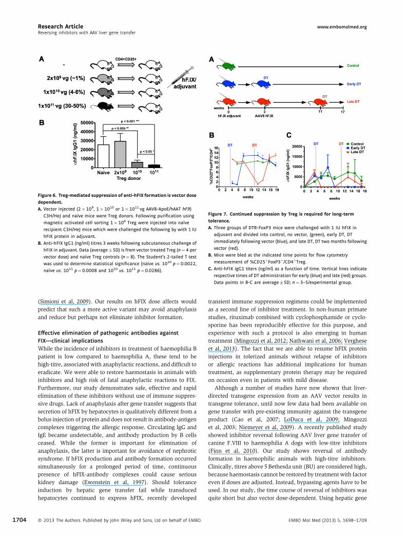
Figure 6. Treg‐mediated suppression of anti‐hFIX formation is vector dose
dependent.
A. Vector injected (2�109, 1�1010 or 1�1011 vg AAV8-ApoE/hAAT hF9)
C3H/HeJ and naı̈ve mice were Treg donors. Following purification using
magnetic activated cell sorting 1�106 Treg were injected into naı̈ve
recipient C3H/HeJ mice which were challenged the following by with 1 IU
hFIX protein in adjuvant.
B. Anti-hFIX IgG1 (ng/ml) titres 3 weeks following subcutaneous challenge of
hFIX in adjuvant. Data (average� SD) is from vector treated Treg (n¼4 per
vector dose) and naı̈ve Treg controls (n¼8). The Student’s 2-tailed T test
was used to determine statistical significance (naı̈ve vs. 1010 p¼0.0022,
naı̈ve vs. 1011 p¼0.0008 and 1010 vs. 1011 p¼0.0286).
Figure 7. Continued suppression by Treg is required for long‐term
tolerance.
A. Three groups of DTR-FoxP3 mice were challenged with 1 IU hFIX in
adjuvant and divided into control, no vector, (green), early DT, DT
immediately following vector (blue), and late DT, DT two months following
vector (red).
B. Mice were bled at the indicated time points for flow cytometry
measurement of %CD25þFoxP3þ/CD4þTreg.C. Anti-hFIX IgG1 titers (ng/ml) as a function of time. Vertical lines indicate
respective times of DT administration for early (blue) and late (red) groups.
Data points in B-C are average� SD; n¼3–5/experimental group.
Research Article www.embomolmed.orgReversing inhibitors with AAV liver gene transfer
1704
(Simioni et al, 2009). Our results on hFIX dose affects wouldpredict that such a more active variant may avoid anaphylaxisand reduce but perhaps not eliminate inhibitor formation.
Effective elimination of pathogenic antibodies againstFIX—clinical implicationsWhile the incidence of inhibitors in treatment of haemophilia Bpatient is low compared to haemophilia A, these tend to behigh‐titre, associatedwith anaphylactic reactions, and difficult toeradicate. We were able to restore haemostasis in animals withinhibitors and high risk of fatal anaphylactic reactions to FIX.Furthermore, our study demonstrates safe, effective and rapidelimination of these inhibitors without use of immune suppres-sive drugs. Lack of anaphylaxis after gene transfer suggests thatsecretion of hFIX by hepatocytes is qualitatively different from abolus injection of protein and does not result in antibody‐antigencomplexes triggering the allergic response. Circulating IgG andIgE became undetectable, and antibody production by B cellsceased. While the former is important for elimination ofanaphylaxis, the latter is important for avoidance of nephroticsyndrome. If hFIX production and antibody formation occurredsimultaneously for a prolonged period of time, continuouspresence of hFIX‐antibody complexes could cause seriouskidney damage (Ewenstein et al, 1997). Should toleranceinduction by hepatic gene transfer fail while transducedhepatocytes continued to express hFIX, recently developed
� 2013 The Authors. Published by John Wiley and Sons, Ltd on behalf of EMBO.
transient immune suppression regimens could be implementedas a second line of inhibitor treatment. In non‐human primatestudies, rituximab combined with cyclophosphamide or cyclo-sporine has been reproducibly effective for this purpose, andexperience with such a protocol is also emerging in humantreatment (Mingozzi et al, 2012; Nathwani et al, 2006; Vergheseet al, 2013). The fact that we are able to resume hFIX proteininjections in tolerized animals without relapse of inhibitorsor allergic reactions has additional implications for humantreatment, as supplementary protein therapy may be requiredon occasion even in patients with mild disease.
Although a number of studies have now shown that liver‐directed transgene expression from an AAV vector results intransgene tolerance, until now few data had been available ongene transfer with pre‐existing immunity against the transgeneproduct (Cao et al, 2007; LoDuca et al, 2009; Mingozziet al, 2003; Niemeyer et al, 2009). A recently published studyshowed inhibitor reversal following AAV liver gene transfer ofcanine F.VIII to haemophilia A dogs with low‐titre inhibitors(Finn et al, 2010). Our study shows reversal of antibodyformation in haemophilic animals with high‐titre inhibitors.Clinically, titres above 5Bethesda unit (BU) are considered high,because haemostasis cannot be restored by treatment with factoreven if doses are adjusted. Instead, bypassing agents have to beused. In our study, the time course of reversal of inhibitors wasquite short but also vector dose‐dependent. Using hepatic gene
EMBO Mol Med (2013) 5, 1698–1709
Research Articlewww.embomolmed.orgDavid M. Markusic et al.
transfer in naïve animals, we repeatedly found that expression ofapproximately 1% of normal circulating hFIX levels representeda threshold, at which the balance started to tip toward tolerance.Here, we reliably reversed inhibitors at the mid‐dose, resultingin average expression of 4–6% of normal. At a fivefold lowerdose, i.e. at the �1% expression level, we failed. Therefore, theminimal expression level required for inhibitor reversal isslightly higher but still similar to that required for prevention ofinhibitor formation in previously untreated animals. Encourag-ingly for translation, FIX levels of approximately 6% of normalhave now been obtained in several haemophilia B patients(Davidoff et al, 2012). Future pre‐clinical studies shoulddetermine whether there is a limit in the inhibitor titre forreversal by gene transfer and whether vector doses may have tobe adjusted depending on the pre‐existing titre.
B cells, T cells and Treg—a tale of ongoing suppressionInhibition of memory B‐cell responses by high antigen doses, asshown here for hFIX, may represent a general phenomenon forsoluble protein antigen, since similar findings have been reportedfor F.VIII (Hausl et al, 2005), and in part explains the mechanismof current ITI protocols for haemophilia. While high levels ofhFIX protein may also account for reduced activation of memoryB cells at higher levels of hFIX expression in our protocol, plasmacells, responsible for the majority of ongoing antibody produc-tion, are not affected. Therefore additional mechanisms are likelyat play. There is strong evidence that the development andmaintenance of inhibitors in haemophilia requires T helper celldependent B‐cell activation (Bray et al, 1993; Cao et al, 2006;Fields et al, 2000). Previously, we have provided direct evidencefor tolerization of transgene product‐specific CD4þ T cells uponhepatic gene transfer and for suppression of T help and antibodyformation by induction of CD4þCD25þFoxP3þ Treg (Caoet al, 2007; Dobrzynski et al, 2004, 2006; Mingozziet al, 2003). The current study shows improved Treg inductionwith increased vector doses. While these results formally do notdifferentiate between higher numbers and increased suppressivephenotype of induced Treg, we have previously providedevidence that higher expression levels lead to increasedfrequency of transgene product‐specific Treg (Cooperet al, 2009). The fact that we are able to demonstrate Treginduction in adoptive transfer studies but not upon in vitro re‐stimulation with hFIX (which revealed elimination of T helperresponses in tolerized animals) is consistent with our prior data.Compared to other approaches such as co‐administration ofantigen and rapamycin, AAV gene transfer induced antigen‐specific Treg are present at low frequency but are nonethelesscritical for immune tolerance (Cao et al, 2007; Dobrzynskiet al, 2006; Moghimi et al, 2011; Nayak et al, 2009, 2011).
In summary, high levels of hFIX expression likely directreversal of inhibitor formation through direct (inhibition of BM)and indirect mechanisms of B‐cell suppression (induction ofTreg and elimination of T help). Several studies have shown thatTreg can directly interact with B cells and—in a contactdependent manner—either suppress (Iikuni et al, 2009; Limet al, 2005) or selectively kill (Zhao et al, 2006). Further, Treghave been shown to suppress development of long‐lived plasma
EMBO Mol Med (2013) 5, 1698–1709 �
cells (Jang et al, 2011). Our study also points to a prominent roleof Treg for regulating humoural immunity. Experiments in theconditional Treg knockout strain (FoxP3‐DTR) indicate the Tregare not only required during the initial phase of inhibitor reversalbut also to maintain tolerance subsequently. In fact, anti‐hFIXresponses relapse quickly if Treg are depleted after tolerance hasbeen established. Therefore, direct inhibition of BM by hFIX orTreg‐mediated deletion of B cells may contribute to but cannotentirely explain elimination of antibody production. Rather, thedata suggest that hFIX‐reactive B cells persist in a suppressedstate, so that continuous suppression by Treg is required toprevent relapse of antibodies. The robustness of this regulatoryresponse is remarkable, given that B‐cell tolerance is rapidlyregained as Treg recover. The data from this model do not ruleout a contribution by Treg that are not hFIX‐specific, albeitinduced antigen‐specific Treg are likely critical for suppression(Annoni et al, 2013; Cao et al, 2007; Matrai et al, 2011).
How antigen derived from hepatocytes is presented to MHCclass II‐restricted CD4þ T cells in a tolerogenic fashion, leading todeletion of effector T cells and induction of Treg, is a questionthat requires further study. In a first set of experiments, we findthat antigen presentation leading to proliferation of transgeneproduct‐specific CD4þ T cells is dependent on professionalAPCs, including macrophages and dendritic cells (see Support-ing Information Fig S4). These data do not entirely prove but atleast support a model of professional APCs playing anintermediary role in cross presentation of hepatocyte‐derivedantigen to CD4þ T cells.
Alternative vector platforms for gene transfer‐based ITIWe have established a first animal model of anaphylaxis in FIXreplacement therapy that resembles the outcomes of replace-ment therapy in the haemophilia B patient population. HepaticAAV‐hF9 gene transfer safely and effectively restored haemo-stasis in these animals. Suppression of B‐cell responses resultedin rapid reversal of inhibitor formation and of IgE formation,thereby desensitizing from anaphylaxis, so that supplementaryprotein therapy could safely be performed. Absence not only ofcirculating antibodies but also of cellular antibody productionshould alleviate concerns that the approach could lead tonephrotic syndrome, albeit protocols for immune suppressionregiments should be in place as a fallback safety measure.Regimens that deplete Treg should be avoided as these play acritical role for maintaining suppression and preventing relapseof antibodies. Such considerations may also be important if apatient was going to receive an organ transplant at some pointlater in life. Hepatic gene transfer as a means of reversingpathogenic antibody formation is also applicable to lysosomalstorage disorders as shown for Pompe disease (Sun et al, 2010).Reversal of high‐tier F.VIII inhibitors in congenital haemophiliaA likely requires further optimization of hepatic gene expression(McIntosh et al, 2013; Sack et al, 2012;Ward et al, 2011). Finally,alternative vector systems such as miRNA‐regulated lentiviralvectors (Brown et al, 2007; Matrai et al, 2011), which have beenoptimized for Treg induction upon hepatic gene transfer, arebeing developed to increase our tool kit for gene transfer‐basedprotocols that may in the future replace current ITI.
2013 The Authors. Published by John Wiley and Sons, Ltd on behalf of EMBO. 1705
The paper explained
PROBLEM:
Protein replacement therapies for genetic diseases are often
severely complicated by antibody formation, which may also be
associated with severe allergic reactions. One example is
antibody formation against FIX in treatment of haemophilia B.
These ‘inhibitors’ often cannot be eradicated using current
immune tolerance induction (ITI) protocols because of ana-
phylaxis or nephrotic syndrome upon prolonged exposure to FIX.
Alternative tolerance protocols are urgently needed to alleviate
the high risk for morbidity and mortality in these patients.
RESULTS:
Hepatic adeno-associated viral F9 gene transfer efficiently and
rapidly reversed high-titre inhibitors and provided long-term
correction of haemostasis in a murine haemophilia B. Tolerized
animals were protected from subsequent anaphylactic reactions
against FIX. Reversal was vector dose dependent. High FIX
antigen doses suppressed activation of memory B cells, induced
regulatory T cells (Treg), and eliminated existing anti-FIX
formation by B cells. Persistent suppression by Treg was required
to prevent relapse of antibody formation.
IMPACT:
Together these data demonstrate that hepatic gene transfer-
based ITI provides a safe and effective alternative to eradicate
inhibitors even in the face of anaphylaxis, supporting the
suitability of the protocol for translational studies. This approach
should be applicable to reverse pathogenic antibodies in other
enzyme replacement therapies and may also be able to treat
autoimmunity caused by auto-antibodies.
Research Article www.embomolmed.orgReversing inhibitors with AAV liver gene transfer
1706
Indeed, using a lentiviral vector Annoni et al in a companionmanuscript demonstrate independently that liver gene transferreverses pre‐existing FIX inhibitors in haemophilia B mice whileavoiding the severe allergic reactions that plague current ITIprotocols (that are based on IV injections of factor product)(Annoni et al, 2013). Both our studies are in agreement in that shut‐down of antibody production by B cells via Treg induction andsuppression of memory B cells are major mechanisms, whichsupports further translational studies. While we were able toeffectively reverse FIX inhibitors with �6% normal levelsFIX protein, Annoni et al observed a requirement for higher 50–100%normal levels FIX protein for effective reversal. It is importantto note that there are several differences in our models such as,the background strain of the haemophilia B mice (C3H/HeJ vs.C57BL/6) and immunization protocols (IV FIX protein vs. FIXprotein in adjuvant), antibody titres (30 vs. 200mg/ml) and relativevector immunity (AAV vs. lentivirus) that may explain the differentrequirements for FIX expression to effectively reverse inhibitors.Nonetheless both these studies demonstrate that different vectorsystems can be adapted to take advantage of tolerogenic expressionby hepatocytes to treat FIX inhibitors.
MATERIALS AND METHODS
Viral vectorsAAV‐ApoE/hAAT‐hF9 carries the hepatocyte‐specific expression cassette
for hFIX (Manno et al, 2006). This cassette includes an apolipoprotein E
(ApoE) enhancer/hepatocyte control region, a human a1‐antitrypsin
promoter, hFIX cDNA, a 1.4‐kb portion of intron I of the F9 gene, and
the bovine growth hormone poly(A) signal. AAV serotype 8 vector
was produced as previously described (Cooper et al, 2009; Liu et al, 2003).
Animal studiesC3H/HeJ and C57BL/6 mice were purchased from Jackson Laboratories
(Bar Harbor, ME). Haemophilia B mice with targeted deletion of murine
� 2013 The Authors. Published by John Wiley and Sons, Ltd on behalf of EMBO. EMBO Mol Med (2013) 5, 1698–170
F9 (‘Null mutation’) had been bred on C3H/HeJ background for >10
generations (Mingozzi et al, 2003). Mice transgenic for hF9 variants
(human F9 complementary DNA including a 0.3‐kb portion of intron I
expressed from liver‐specific transthyretin promoter) were as
published (Sabatino et al, 2004). These animals express hFIX with
late stop codon at amino acid residue 338 (‘LS’, crim�); crim� G381E
missensemutation (‘CH’, identicalmutation as in haemophilia B dogs at
University of North Carolina Chapel Hill); or crimþ R180W missense
mutation. These lines were originally numbered as LS‐37, cCH‐6 and
MS‐12, and contain 6, 10 and 1 copy of the human F9 gene,
respectively (Sabatino et al, 2004). The lines were repeatedly
backcrossed onto C3H/HeJ background (>10 generation), and finally
crossed with Null mice in order to eliminate endogenous murine F9
expression (Cao et al, 2009). C57BL/6 DTR‐FoxP3 mice were kindly
provided by Dr. Alexander Rudensky (Kim et al, 2007). Animals were
housed under special pathogen‐free conditions at the University of
Florida and treated under Institutional Animal Care and Use
Committee‐approved protocols. All animals were male and 6–8 weeks
old at the onset of the experiments.
Immunization protocols for haemophilia B mice were performed as
described unless otherwise stated (Verma et al, 2010). Briefly, mice
were given six weekly injections of recombinant hFIX protein (Benefix,
1 IU/dose) starting with an intraperitoneal injection and followed by
five weekly intraveneous injections into the tail vein. Plasma samples
were collected by tail bleed into citrate buffer as described (Mingozzi
et al, 2003). For adoptive transfer and Treg depletion studies, mice were
bled from the retro‐orbital plexus using heparinized microcapillary
tubes. Where indicated mice received hFIX protein along with 150mg
antihistamine (triprolidine; Sigma) and 50mg platelet‐activating factor
(PAF) antagonist CV‐3988 to prevent anaphylactic responses. In some
studies mice were immunized with 1 IU hFIX protein in adjuvant
(Sigma Adjuvant System S6322, Sigma, St. Louis, MO).
Antigen and antibody measurementsPlasma levels of hFIX antigen were measured by ELISA and inhibitory
antibody titres were determined by Bethesda assay as published
9
Research Articlewww.embomolmed.orgDavid M. Markusic et al.
(Cao et al, 2009; Mingozzi et al, 2003). One Bethesda unit is the
reciprocal of the dilution of plasma that neutralizes 50% of the FIX
in a normal plasma sample which it is mixed in vitro. Immuno-
capture assays to determine titres of hFIX‐specific IgGE and IgG1
were as described (Cao et al, 2009; Mingozzi et al, 2003) using
purified mouse IgE and IgG1 as standards. To measure IgE titres,
IgG was removed from plasma samples using protein G sepharose
(GE Healthcare).
B‐ and T‐cell assaysB‐cell ELISpots were performed on total splenocytes and bone marrow
cells as previously described (Hausl et al, 2002, 2004; Wang
et al, 2005). In some studies, plasma cells were depleted using anti
mouse CD138 beads (Miltenyi, Vancouver, Canada) according to
manufacturers instructions. For cytokine response studies, isolated
splenocytes were cultured in RPMI 1640 media (containing 50mM
b‐mercaptoethanol, 100mM insulin/transferrin/selenium, glutamine
and antibiotics) with or without hFIX 10mg/ml for 48h (37°C, 5% CO2).
Transcript levels of cytokines were measured by quantitative RT‐PCR
using SA Biosciences arrays (Frederick, MD, USA; RNA was extracted
from 1.5�105 cells per spleen prior to cDNA synthesis) and a MyQ
thermocycler (Biorad, Hercules, CA, USA), and normalized based on
GAPDH expression.
Adoptive T‐cell transfer studiesCD4þCD25þ splenocytes from vector treated or naïve C3H/HeJ mice
were purified using the Treg magnetic activated cell sorting kit from
Miltenyi, pooled, and adoptively transferred to naïve mice of the same
strain by tail vein injection (1�106 cells per mouse). Recipient mice
were immunized by subcutaneous injection of 1 IU hFIX (in Sigma
Adjuvant System) 24h later.
Treg depletion studiesDTR‐FoxP3 mice were immunized by subcutaneous injection of 1 IU
hFIX (in Sigma Adjuvant System). Three weeks post‐immunization mice
were injected with 1�1011 vg of an AAV8 ApoE/hAAT hF9 vector in the
tail vein and split into two groups of early and late DT treatment. Mice
received two IP injections of DT 50mg/kg with a 4‐day interval.
Depletion and recovery of Treg was monitored by flow cytometry of
peripheral blood using anti mouse CD3‐PerCP‐Cy5.5 (BD Biosciences),
CD4‐e450 (eBioscience), CD25‐PE (eBioscience) and FoxP3‐Alexa647
(eBioscience).
Statistical AnalysisAll statistical analysis was carried out using Prism software using
Student’s 2‐tailed T‐test or one‐way ANOVA with Bonferroni’s
multiple comparison test. A p<0.05 was considered statistically
significant.
Author contributionsDMM, BEH, GQP, SN, XW and PAL performed experiments;DMM, BEH, GQP and RWH designed experiments; DMM, BEH,GQP, KAH and RWH interpreted data; KAH provided criticalreagents; RWH supervised and coordinated the study; andDMM, KAH and RWH wrote the manuscript.
EMBO Mol Med (2013) 5, 1698–1709 �
AcknowledgementsThe authors thank Dr. Alexander Rudensky for FoxP3‐DTRmiceand Drs. Birgit Reipert and Christine Baumgartner for advice onthe memory B‐cell suppression assay. This work was supportedby National Institutes of Health grants P01 HD078810 (KAH andRWH), R01 AI051390 (RWH), F32 HL096281 (DMM), a BayerHemophilia Award (DMM), and by the Howard Hughes MedicalInstitute (KAH).
Supporting Information is available at EMBO MolecularMedicine Online.
Conflict of interest statement: RWHhas received royalty paymentsfor Genzyme Corp for license of AAV‐hF9 technology. KAH is aninventor on issued and pending patents on AAV gene transfertechnologies. All other authors have no conflict of interest.
ReferencesAnnoni A, Cantore A, Della Valle P, Goudy K, Akbarpour M, Russo F, Bartolaccini
S, D’Angelo A, Roncarolo MG, Naldini L (2013) Liver gene therapy by
lentiviral vectors reverses anti-Factor IX pre-existing immunity in
hemophilic mice. EMBO Mol Med 5: 1684-1697
Bray GL, Kroner BL, Arkin S, Aledort LW, Hilgartner MW, Eyster ME, Ragni MV,
Goedert JJ (1993) Loss of high-responder inhibitors in patients with
severe hemophilia A and human immunodeficiency virus type 1 infection: a
report from the Multi-Center Hemophilia Cohort Study. Am J Hematol 42:
375-379
Brown BD, Cantore A, Annoni A, Sergi LS, Lombardo A, Della Valle P, D’Angelo A,
Naldini L (2007) A microRNA-regulated lentiviral vector mediates stable
correction of hemophilia B mice. Blood 110: 4144-4152
Cao O, Armstrong E, Schlachterman A,Wang L, Okita DK, Conti-Fine B, High KA,
Herzog RW (2006) Immune deviation by mucosal antigen administration
suppresses gene-transfer-induced inhibitor formation to factor IX. Blood
108: 480-486
Cao O, Dobrzynski E, Wang L, Nayak S, Mingle B, Terhorst C, Herzog RW (2007)
Induction and role of regulatory CD4þCD25þ T cells in tolerance to the
transgene product following hepatic in vivo gene transfer. Blood 110:
1132-1140
Cao O, Hoffman BE, Moghimi B, Nayak S, Cooper M, Zhou S, Ertl HC, High KA,
Herzog RW (2009) Impact of the underlying mutation and the route of
vector administration on immune responses to factor IX in gene therapy for
hemophilia B. Mol Ther 17: 1733-1742
Chitlur M, Warrier I, Rajpurkar M, Lusher JM (2009) Inhibitors in factor IX
deficiency a report of the ISTH-SSC international FIX inhibitor registry
(1997–2006). Haemophilia 15: 1027-1031
Cooper M, Nayak S, Hoffman BE, Terhorst C, Cao O, Herzog RW (2009)
Improved induction of immune tolerance to factor IX by hepatic AAV-8 gene
transfer. Hum Gene Ther 20: 767-776
Davidoff A, Tuddenham EG, Rangarajan S, Rosales C, McIntosh J, Chowdary P,
Riddell A, Glader B, Rustagi P, Ng C, et al (2012) Stable factor IX activity
following AAV-mediated gene transfer in patients with severe hemophilia B.
ASH Annu Meet Abstr 120: 752
DiMichele D (2007) Inhibitor development in haemophilia B: an orphan
disease in need of attention. Br J Haematol 138: 305-315
DiMichele DM (2012) Immune tolerance in haemophilia: the long journey to
the fork in the road. Br J Haematol 159: 123-134
Dobrzynski E, Fitzgerald JC, Cao O, Mingozzi F, Wang L, Herzog RW (2006)
Prevention of cytotoxic T lymphocyte responses to factor IX-expressing
hepatocytes by gene transfer-induced regulatory T cells. Proc Natl Acad Sci
USA 103: 4592-4597
2013 The Authors. Published by John Wiley and Sons, Ltd on behalf of EMBO. 1707
Research Article www.embomolmed.orgReversing inhibitors with AAV liver gene transfer
1708
Dobrzynski E, Mingozzi F, Liu YL, Bendo E, Cao O, Wang L, Herzog RW (2004)
Induction of antigen-specific CD4þ T-cell anergy and deletion by in vivo
viral gene transfer. Blood 104: 969-977
Ewenstein BM, Takemoto C, Warrier I, Lusher J, Saidi P, Eisele J, Ettinger LJ,
DiMichele D (1997) Nephrotic syndrome as a complication of immune
tolerance in hemophilia B. Blood 89: 1115-1116
Fields PA, Kowalczyk DW, Arruda VR, Armstrong E, McCleland ML, Hagstrom
JN, Pasi KJ, Ertl HC, Herzog RW, High KA (2000) Role of vector in activation of
T cell subsets in immune responses against the secreted transgene product
factor IX. Mol Ther 1: 225-235
Finn JD, Ozelo MC, Sabatino DE, Franck HW, Merricks EP, Crudele JM, Zhou S,
Kazazian HH, Lillicrap D, Nichols TC, et al (2010) Eradication of neutralizing
antibodies to factor VIII in canine hemophilia A after liver gene therapy.
Blood 116: 5842-5848
Hausl C, Ahmad RU, Sasgary M, Doering CB, Lollar P, Richter G, Schwarz HP,
Turecek PL, Reipert BM (2005) High-dose factor VIII inhibits factor VIII-
specific memory B cells in hemophilia A with factor VIII inhibitors. Blood
106: 3415-3422
Hausl C, Ahmad RU, Schwarz HP, Muchitsch EM, Turecek PL, Dorner F, Reipert
BM (2004) Preventing restimulation of memory B cells in hemophilia A: a
potential new strategy for the treatment of antibody-dependent immune
disorders. Blood 104: 115-122
Hausl C, Maier E, Schwarz HP, Ahmad RU, Turecek PL, Dorner F, Reipert BM
(2002) Long-term persistence of anti-factor VIII antibody-secreting cells in
hemophilic mice after treatment with human factor VIII. Thromb Haemost
87: 840-845
Hoffman BE, Dobrzynski E, Wang L, Hirao L, Mingozzi F, Cao O, Herzog RW
(2007) Muscle as a target for supplementary factor IX gene transfer. Hum
Gene Ther 18: 603-613
Hoffman BE, Martino AT, Sack BK, Cao O, Liao G, Terhorst C, Herzog RW (2011)
Nonredundant roles of IL-10 and TGF-b in suppression of immune
responses to hepatic AAV-factor IX gene transfer. Mol Ther 19: 1263-1272
Iikuni N, Lourenco EV, Hahn BH, La Cava A (2009) Cutting edge: regulatory T
cells directly suppress B cells in systemic lupus erythematosus. J Immunol
183: 1518-1522
Jadhav M, Warrier I (2000) Anaphylaxis in patients with hemophilia. Semin
Thromb Hemost 26: 205-208
Jang E, Cho WS, Cho ML, Park HJ, Oh HJ, Kang SM, Paik DJ, Youn J (2011)
Foxp3þ regulatory T cells control humoral autoimmunity by suppressing
the development of long-lived plasma cells. J Immunol 186: 1546-1553
Kim JM, Rasmussen JP, Rudensky AY (2007) Regulatory T cells prevent
catastrophic autoimmunity throughout the lifespan of mice. Nat Immunol
8: 191-197
Koeberl DD, Kishnani PS (2009) Immunomodulatory gene therapy in
lysosomal storage disorders. Curr Gene Ther 9: 503-510
Lim HW, Hillsamer P, Banham AH, Kim CH (2005) Cutting edge: direct
suppression of B cells by CD4þ CD25þ regulatory T cells. J Immunol 175:
4180-4183
Liu YL, Wagner K, Robinson N, Sabatino D, Margaritis P, Xiao W, Herzog RW
(2003) Optimized production of high-titer recombinant adeno-associated
virus in roller bottles. BioTechniques 34: 184-189
LoDuca PA, Hoffman BE, Herzog RW (2009) Hepatic gene transfer as a means
of tolerance induction to transgene products. Curr Gene Ther 9: 104-114
Lozier JN, Tayebi N, Zhang P (2005) Mapping of genes that control the
antibody response to human factor IX in mice. Blood 105: 1029-1035
Lu Y, Song S (2009) Distinct immune responses to transgene products from
rAAV1 and rAAV8 vectors. Proc Natl Acad Sci USA 106: 17158-17162
Manno CS, Pierce GF, Arruda VR, Glader B, Ragni M, Rasko JJ, Ozelo MC, Hoots
K, Blatt P, Konkle B, et al (2006) Successful transduction of liver in
hemophilia by AAV-Factor IX and limitations imposed by the host immune
response. Nat Med 12: 342-347
Markusic DM, Herzog RW, Aslanidi GV, Hoffman BE, Li B, Li M, Jayandharan GR,
Ling C, Zolotukhin I, Ma W, et al (2010) High-efficiency transduction and
correction of murine hemophilia B using AAV2 vectors devoid of multiple
surface-exposed tyrosines. Mol Ther 18: 2048-2056
� 2013 The Authors. Published by John Wiley and Sons, Ltd on behalf of EMBO.
Martino AT, Basner-Tschakarjan E, Markusic DM, Finn JD, Hinderer C, Zhou S,
Ostrov DA, Srivastava A, Ertl HC, Terhorst C, et al (2013) Engineered AAV
vector minimizes in vivo targeting of transduced hepatocytes by capsid-
specific CD8þ T cells. Blood 121: 2224-2233
Matrai J, Cantore A, Bartholomae CC, Annoni A, Wang W, Acosta-Sanchez A,
Samara-Kuko E, De Waele L, Ma L, Genovese P, et al (2011) Hepatocyte-
targeted expression by integrase-defective lentiviral vectors induces
antigen-specific tolerance in mice with low genotoxic risk. Hepatology 53:
1696-1707
McIntosh J, Lenting PJ, Rosales C, Lee D, Rabbanian S, Raj D, Patel N,
Tuddenham EG, Christophe OD, McVey JH, et al (2013) Therapeutic levels of
FVIII following a single peripheral vein administration of rAAV vector
encoding a novel human factor VIII variant. Blood 121: 3335-3344
Metzner HJ, Weimer T, Kronthaler U, Lang W, Schulte S (2009) Genetic fusion
to albumin improves the pharmacokinetic properties of factor IX. Thromb
Haemost 102: 634-644
Mingozzi F, Chen Y, Murphy SL, Edmonson SC, Tai A, Price SD, Metzger ME,
Zhou S, Wright JF, Donahue RE, et al (2012) Pharmacological modulation of
humoral immunity in a nonhuman primate model of AAV gene transfer for
hemophilia B. Mol Ther 20: 1410-1416
Mingozzi F, Liu YL, Dobrzynski E, Kaufhold A, Liu JH, Wang Y, Arruda VR, High
KA, Herzog RW (2003) Induction of immune tolerance to coagulation factor
IX antigen by in vivo hepatic gene transfer. J Clin Invest 111: 1347-1356
Moghimi B, Sack BK, Nayak S, Markusic DM, Mah CS, Herzog RW (2011)
Induction of tolerance to factor VIII by transient co-administration with
rapamycin. J Thromb Haemost 9: 1524-1533
Nathwani AC, Gray JT, Ng CY, Zhou J, Spence Y, Waddington SN, Tuddenham
EG, Kemball-Cook G, McIntosh J, Boon-Spijker M, et al (2006) Self-
complementary adeno-associated virus vectors containing a novel liver-
specific human factor IX expression cassette enable highly efficient
transduction of murine and nonhuman primate liver. Blood 107: 2653-
2661
Nathwani AC, Tuddenham EG, Rangarajan S, Rosales C, McIntosh J, Linch DC,
Chowdary P, Riddell A, Pie AJ, Harrington C, et al (2011) Adenovirus-
associated virus vector-mediated gene transfer in hemophilia B. N Engl J
Med 365: 2357-2365
Nayak S, Cao O, Hoffman BE, Cooper M, Zhou S, Atkinson MA, Herzog RW
(2009) Prophylactic immune tolerance induced by changing the ratio of
antigen-specific effector to regulatory T cells. J Thromb Haemost 7: 1523-
1532
Nayak S, Sarkar D, Perrin GQ,Moghimi B, Hoffman BE, Zhou S, Byrne BJ, Herzog
RW (2011) Prevention and reversal of antibody responses against factor IX
in gene therapy for hemophilia B. Front Microbiol 2: 244
Niemeyer GP, Herzog RW, Mount J, Arruda VR, Tillson DM, Hathcock J, van
Ginkel FW, High KA, Lothrop CD, Jr. (2009) Long-term correction of
inhibitor-prone hemophilia B dogs treated with liver-directed AAV2-
mediated factor IX gene therapy. Blood 113: 797-806
Ostergaard H, Bjelke JR, Hansen L, Petersen LC, Pedersen AA, Elm T, Moller F,
Hermit MB, Holm PK, Krogh TN, et al (2011) Prolonged half-life and
preserved enzymatic properties of factor IX selectively PEGylated on native
N-glycans in the activation peptide. Blood 118: 2333-2341
Passini MA, Bu J, Fidler JA, Ziegler RJ, Foley JW, Dodge JC, Yang WW, Clarke J,
Taksir TV, Griffiths DA, et al (2007) Combination brain and systemic
injections of AAV provide maximal functional and survival benefits in the
Niemann-Pick mouse. Proc Natl Acad Sci USA 104: 9505-9510
Peters RT, Low SC, Kamphaus GD, Dumont JA, Amari JV, Lu Q, Zarbis-
Papastoitsis G, Reidy TJ, Merricks EP, Nichols TC, et al (2010) Prolonged
activity of factor IX as a monomeric Fc fusion protein. Blood 115: 2057-
2064
Recht M, Pollmann H, Tagliaferri A, Musso R, Janco R, Richey Neuman W
(2011) A retrospective study to describe the incidence of moderate to
severe allergic reactions to factor IX in subjects with haemophilia B.
Haemophilia 17: 494-499
Sabatino DE, Armstrong E, Edmonson S, Liu YL, Pleimes M, Schuettrumpf J,
Fitzgerald J, Herzog RW, Arruda VR, High KA (2004) Novel hemophilia B
EMBO Mol Med (2013) 5, 1698–1709

Research Articlewww.embomolmed.orgDavid M. Markusic et al.
mouse models exhibiting a range of mutations in the Factor IX gene. Blood
104: 2767-2774
Sack BK, Merchant S, Markusic DM, Nathwani AC, Davidoff AM, Byrne BJ,
Herzog RW (2012) Transient B cell depletion or improved transgene
expression by codon optimization promote tolerance to factor VIII in gene
therapy. PLoS ONE 7: e37671
Simioni P, Tormene D, Tognin G, Gavasso S, Bulato C, Iacobelli NP, Finn JD,
Spiezia L, Radu C, Arruda VR (2009) X-linked thrombophilia with a mutant
factor IX (factor IX Padua). N Engl J Med 361: 1671-1675
Sun B, Kulis MD, Young SP, Hobeika AC, Li S, Bird A, Zhang H, Li Y, Clay TM,
Burks W, et al (2010) Immunomodulatory gene therapy prevents antibody
formation and lethal hypersensitivity reactions in murine pompe disease.
Mol Ther 18: 353-360
Thorland EC, Drost JB, Lusher JM, Warrier I, Shapiro A, Koerper MA, Dimichele
D, Westman J, Key NS, Sommer SS (1999) Anaphylactic response to factor IX
replacement therapy in haemophilia B patients: complete gene deletions
confer the highest risk. Haemophilia 5: 101-105
Vandendriessche T, Thorrez L, Acosta-Sanchez A, Petrus I, Wang L, Ma L,
DE Waele L, Iwasaki Y, Gillijns V, Wilson JM, et al (2007) Efficacy and
safety of adeno-associated viral vectors based on serotype 8 and 9 vs.
lentiviral vectors for hemophilia B gene therapy. J Thromb Haemost 5:
16-24
Verghese P, Darrow S, Kurth MH, Reed RC, Kim Y, Kearney S (2013) Successful
management of factor IX inhibitor-associated nephrotic syndrome in a
hemophilia B patient. Pediatr Nephrol 28: 823-826
EMBO Mol Med (2013) 5, 1698–1709 �
Verma D, Moghimi B, LoDuca PA, Singh HD, Hoffman BE, Herzog RW, Daniell H
(2010) Oral delivery of bioencapsulated coagulation factor IX prevents
inhibitor formation and fatal anaphylaxis in hemophilia B mice. Proc Natl
Acad Sci USA 107: 7101-7106
Wang L, Cao O, Swalm B, Dobrzynski E, Mingozzi F, Herzog RW (2005) Major
role of local immune responses in antibody formation to factor IX in AAV
gene transfer. Gene Therapy 12: 1453-1464
Ward NJ, Buckley SM, Waddington SN, Vandendriessche T, Chuah MK,
Nathwani AC,McIntosh J, Tuddenham EG, Kinnon C, Thrasher AJ, et al (2011)
Codon optimization of human factor VIII cDNAs leads to high-level
expression. Blood 117: 798-807
Warrier I, Ewenstein BM, Koerper MA, Shapiro A, Key N, DiMichele D, Miller RT,
Pasi J, Rivard GE, Sommer SS, et al (1997) Factor IX inhibitors and
anaphylaxis in hemophilia B. J Pediatr Hematol Oncol 19: 23-27
Zhang P, Sun B, Osada T, Rodriguiz R, Yang XY, Luo X, Kemper AR, Clay TM,
Koeberl DD (2012) Immunodominant liver-specific expression suppresses
transgene-directed immune responses in murine pompe disease. Hum
Gene Ther 23: 460-472
Zhao DM, Thornton AM, DiPaolo RJ, Shevach EM (2006) Activated
CD4þCD25þ T cells selectively kill B lymphocytes. Blood 107: 3925-3932
Zhong L, Li B, Mah CS, Govindasamy L, Agbandje-McKenna M, Cooper M,
Herzog RW, Zolotukhin I, Warrington KH, Jr., Weigel-Van Aken KA, et al
(2008) Next generation of adeno-associated virus 2 vectors: point
mutations in tyrosines lead to high-efficiency transduction at lower doses.
Proc Natl Acad Sci USA 105: 7827-7832
2013 The Authors. Published by John Wiley and Sons, Ltd on behalf of EMBO. 1709
Related Documents