1 Effect of Various Materials on Dentin Permeability for the Treatment of Dentin Hypersensitivity NOMURA Yuji, YASUO Kenzo, I WATA Naohiro, YOSHIKAWA Kazushi and YAMAMOTO Kazuyo Department of Operative Dentistry, Osaka Dental University Running title : Permeability for the Treatment of Dentin Hypersensitivity Corresponding author: Dr. YASUO, Department of Operative, Osaka Dental University, 8-1 Kuzuha-hanazono-cho, Hirakata, Osaka 573-1121, Japan TEL: +81 ‐ 72 ‐ 864 ‐ 3077, FAX: +81 ‐ 72 ‐ 864 ‐ 3177 E-mail: [email protected]

Welcome message from author
This document is posted to help you gain knowledge. Please leave a comment to let me know what you think about it! Share it to your friends and learn new things together.
Transcript
1
Effect of Various Materials on Dentin Permeability
for the Treatment of Dentin Hypersensitivity
NOMURA Yuji, YASUO Kenzo, IWATA Naohiro, YOSHIKAWA Kazushi and YAMAMOTO Kazuyo
Department of Operative Dentistry, Osaka Dental University
Running title : Permeability for the Treatment of Dentin Hypersensitivity
Corresponding author: Dr. YASUO, Department of Operative, Osaka Dental University, 8-1
Kuzuha-hanazono-cho, Hirakata, Osaka 573-1121, Japan
TEL: +81‐72‐864‐3077, FAX: +81‐72‐864‐3177
E-mail: [email protected]

2
Abstract
Purpose: In recent years, the number of patients with transient dentin hypersensitivity to cold
water and abrasion pain without dental caries has been increasing. In the treatment of dentin
hypersensitivity, the first choice is frequently the topical application of medicament due to its
simplicity and immediate effect. A wide range of products with different action mechanisms are
available for clinical use. The present study focused on dentin desensitizers and their dentinal
tubular solubility. The dentin permeability inhibition ratio was measured using a model of
hypersensitive dentin. ln addition, the influence of post-application preservation conditions on serial
changes in the permeability inhibition ratio was evaluated.
Methods: Dentin discs were prepared from extracted human molars for use as hypersensitivity
model specimens.The specimens were applied to a device based on Pashley et al., with
modifications, and the pulpal pressure was determined to be 25 mmHg. After applying four
different dentin desensitizer products, Gluma Desensitizer (GL), Super Seal (SS), MS Coat One
(MO), Nanoseal (NS), Teethmate Desensitizer (TD) and Shield Force Plus (SP), the specimens
were stored in distilled water (DW group) or artificial saliva (AS group) for 24 h and for one week,
and the dentin permeability was measured.
Results: In the DW group of all dentin desensitizers, the dentin permeability inhibition ratio
one week after application decreased or showed a serial decrease in comparison with that

3
immediately after application. In the AS group of SS, MO, NS and TD, the ratio one week after
application increased or showed a serial increase in comparison with that immediately after
application. In the AS group of GL and SP, the ratio one week after application showed a serial
decrease in comparison with that immediately after application.
Conclusion: When the specimens were stored in distilled water, all dentin desensitizers
showed a decrease in the sealability of the dentinal tubules. When the specimens were stored in
artificial saliva imitating the human intraoral cavity, dentin desensitizers Super Seal, MS Coat one,
Nanoseal and Teethmate Desensitizer showed a serial increase in the sealability of the dentinal
tubules, suggesting a bioactive action..
Key words : Dentin hypersensitivity; Sealability; Model of hypersensitive dentin

4
Introduction
In recent years, the development of minimally invasive dental treatment, and sustained efforts in
dental-oral health promotion, have resulted in an increase in the number of remaining permanent
teeth per person, leading to a higher number of patients with transient dentin hypersensitivity to cold
water and abrasion pain without dental caries.
Dentin hypersensitivity is caused by physical stimulation such as changes in temperature and
chemical stimulation such as changes in pH.1) Dentin hypersensitivity is classified according to the
region of exposed dentin into cervical or root surface hypersensitivity and postoperative
hypersensitivity due to dentin surface exposure after cavity preparation. It is recently considered that
dentin hypersensitivity is aggravated by abfraction and enamel microcracks due to bruxism and
clenching caused by stress. Although its subjective symptoms vary among patients from slight to
unbearable pain, there are many patients who strongly desire relief from the discomfort of translent
paln.
Treatment of dentin hypersensitivity consists of preservation therapy such as the topical
application of medicament, iontophoresis, laser treatment, coverage using adhesive materials and
removal of the dentin pulp in severe cases.2) Although the optimal treatment is selected in each case,
there is a possibility of recurrence. When the treatment effects of a selected method are negligible,
other treatment methods with differing action mechanisms are selected. To reduce the patient’s

5
discomfort and promote effective treatment of dentin hypersensitivity, it is important to fully
understand each treatment method and the properties of each medicament, and select the optimal
treatment method according to the pathologic conditions.
Among the dentin hypersensitivity treatment methods, the topical application of medicament is
frequently the first choice due to its simplicity and immediate effect. The action mechanisms are
varied, and many different products are available for clinical use, such as those for which the main
components are potassium nitrate and aluminum lactate expecting desensitization,3,4) those that
react with tooth calcium to create crystals of mineral salts aiming to seal the dentinal tubules,5-8)
those for which the main component is glutaraldehyde to seal the dentinal tubules with the
coagulation of tissue fluid9) and those that seal the dentinal tubules using resin and glass ionomer
cements.10) However, the dislodging of medicaments due to tooth brushing and the serial decrease
in effect due to the elution of the medicament in saliva have been clinically reported.11)
The present study focused on dentin desensitizers and their dentinal tubular sealability We
established a pressure value equivalent to the internal pressure of the human dental pulp, using a
diluted serum solution. Using a model of hypersensitive dentin, in which the amount of protein in
the dentinal tubular fluid is near the clinical condition, we measured the dentin permeability
inhibition ratio(permeability inhibition ratio), and evaluated the influence of post-application
preservation conditions on serial changes in the permeability inhibition ratio.

6
Materials and methods
1. Production of the specimens
The subject teeth were healthy human molars without dental caries (human teeth) that were
extracted at the Department of Oral Surgery in the hospital attached to our university, and stored in
physiological saline at -40℃. They were defrosted immediately before being used in the
experiments. This study was approved by the Medical Ethics Committee of Osaka Dental
University (approval number:Daishi-irin 110767). After exposing human dentin from the occlusal
surface side using a model trimmer, and cutting the tooth root around the cervical area, the dental
pulp was removed. Next, polishing was performed up to #600 using water-resistant polishing
papers to make the dentin surface flat, and exposed cylindrical dentin discs of 8 mm in diameter and
l mm in thickness were produced. On the occlusal surface side of the dentin disc, a phosphate
solution (Kishida Chemical) adjusted to 10 % using distilled water was applied for 30 s, and the
specimen was washed for 5 s under running water. On the pulp side, sodium hypochlorite (Kishida
Chemical) adjusted to 10 % using distilled water was applied for 10 s, and the specimen was
washed for 5 s under running water to remove the smear layers on both sides. Ultrasonic cleaning in
distilled water was performed for 5 min, and specimens with the dentinal tubules open were used as
dentin disc specimens.
Next, a model of hypersensitive dentin was produced according to the method of Zennyu et

7
aL12)(Fig.1). A gum ring of 6 mm in internal diameter was placed on the pulp side of the dentin disc
specimen, and a specimen stage was set, with insertion performed by using the upper and lower
sides of a stainless steel holder. To expose the dentin on the enamel side, the central area of the
specimen stage was fenestrated in a circular form of 7mm in diameter (area: approximately 0.39
cm2). Dentinal tubular fluid (DF) was injected into the specimen stage using a glass syringe with a
Teflon tube. The specimen stage was filled with DF without containing air, and connected to a
device configured to adjust the internal pressure according to Pashley et al.13) The internal pressure
of the specimen stage was adjusted to 25 mmHg, which is equivalent to the internal pressure of
human dental pulp, to reproduce the clinical dental pulp’s internal pressure.14-17) The clinical amount
of protein in DF was reproduced by using bovine serum diluted four times with distilled water. 18-20)
2. Measurement of the dentin permeability inhibition ratio
Table l lists the dentin desensitizers used in the experiment. Table 2 shows the application
method for each dentin desensitizer. DF from the injection syringe was shut at three-way cock B,
and a glass capillary tube connected with the experimental device was connected to the specimen
stage. Under these conditions, air blowing on the exposed dentin surface in the central fenestrated
area of the specimen stage was performed for 60 s, and after leaving it for 30 s, the amount of DF
movement in the glass capillary tube was measured.21-23) The air pressure was set at 3.0 kgf/cm2.

8
Then, DF flowing in the glass capillary tube was shut, adjusting three-way cock B so that DF only
flowed from the injection syringe in the specimen stage direction. Furthermore, three-way cock A
was adjusted so that DF flowed from the injection syringe in the pressure gauge and specimen stage
direction, and the internal pressure was set at 25 mmHg. After applying each dentin desensitizer
according to the manufacturer’s instructions, air blowing on the specimen dentin surface was
performed for 60 s, as was done before applying the dentin desensitizers, and after leaving it for 30
s, the amount of DF movement in the glass capillary tube was measured again. The amount of DF
movement before and after application was used to measure the permeability inhibition ratio. Each
specimen was immersed in distilled water and artificiaI saliva (Saliveht Aerosol, Teijin Pharma,
Table 3), and stored at a constant temperature of 37℃. The specimens stored in distilled water were
classified as the DW group, and those stored in artificial saliva were classified as the AS group.
Thereafter, the amount of DF movement was measured at 24 h and at one week after application,
using the same method, and the permeability inhibition ratio was measured.
3. Statistical test
The permeability inhibition ratio was tested using oneway layout analysis of variance and
TUkey’s analysis (p<0.05). The number of specimens was five.

9
4. SEM observation of the dentin surface
Gold evaporation was performed according to the conventional method, and the specimens
used in the experiment were observed using a scanning electron microscope (JSM-5610LV, JEOL,
SEM).
10
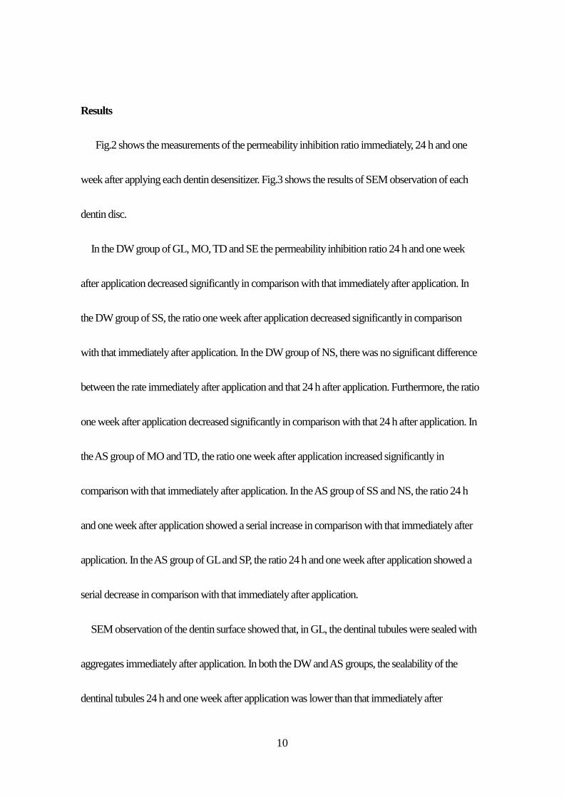
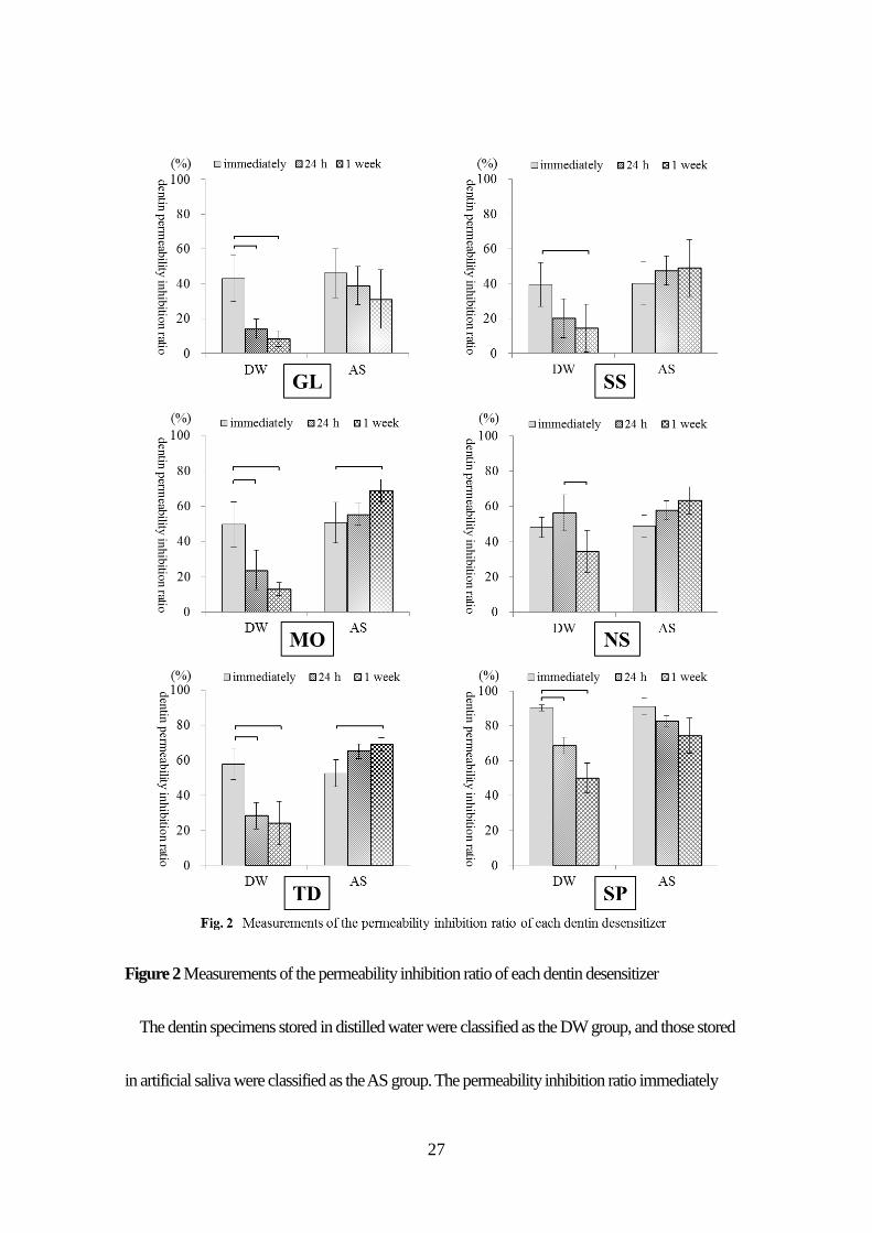
Results
Fig.2 shows the measurements of the permeability inhibition ratio immediately, 24 h and one
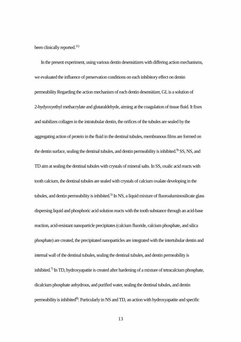
week after applying each dentin desensitizer. Fig.3 shows the results of SEM observation of each
dentin disc.
In the DW group of GL, MO, TD and SE the permeability inhibition ratio 24 h and one week
after application decreased significantly in comparison with that immediately after application. In
the DW group of SS, the ratio one week after application decreased significantly in comparison
with that immediately after application. In the DW group of NS, there was no significant difference
between the rate immediately after application and that 24 h after application. Furthermore, the ratio
one week after application decreased significantly in comparison with that 24 h after application. In
the AS group of MO and TD, the ratio one week after application increased significantly in
comparison with that immediately after application. In the AS group of SS and NS, the ratio 24 h
and one week after application showed a serial increase in comparison with that immediately after
application. In the AS group of GL and SP, the ratio 24 h and one week after application showed a
serial decrease in comparison with that immediately after application.
SEM observation of the dentin surface showed that, in GL, the dentinal tubules were sealed with
aggregates immediately after application. In both the DW and AS groups, the sealability of the
dentinal tubules 24 h and one week after application was lower than that immediately after

11
application. In SS, MO, NS and TD, aggregates sealed the dentinal tubules immediately after
application, covering the dentin surface. In the DW group, the sealability of the dentinal tubules 24
h and one week after application was lower than that immediately after application. In the AS group,
although the aggregates covering the dentin surface 24hand one week after application decreased in
comparison with that immediately after application, a decrease in the sealability of the dentinal
tubules was not noted. In SP, aggregates of polymers sealed the dentinal tubules immediately after
application, covering the dentin surface. In both the DW and AS groups, although exposure of
dentinal tubules 24 h and one week after application was not observed, the aggregates covering the
dentin surface had become rough.

12
Discussion
Dentin hypersensitivity is caused by physical stimulation such as changes in temperature and
chemical stimulation such as changes in pH for exposed dentin. It is recently considered that dentin
hypersensitivity is aggravated by abfraction and enamel microcracks due to bruxism and clenching
caused by stress.24) The mechanism of dentin hypersensitivity has not yet been clearly elucidated.
There are several hypotheses regarding the sensory transmission mechanism of dentin
hypersensitivity. The hydrodynamic theory, in which the movement of the fluid in the dentinal
tubules with external stimulation stimulates the free nerve endings in the dental pulp and causes
pain, is convincing25,26) because opening of the dentinal tubules on the hypersensitivity-contracted
dentin surface is noted.27,28) In dentin hypersensitivity cases based on the hydrodynamic theory, pain
can be reduced by preventing the movement of the tissue fluid in the dentinal tubules, using
appropriate methods to seal the orifices of the dentinal tubules.
Treatment of dentin hypersensitivity consists of preservation therapy such as topical application
of medicament, iontophoresis, laser treatment, coverage using adhesive materials and removal of
the dentin pulp in severe cases, and the optimal treatment is selected in each case. Among the types
of treatment for dentin hypersensitivity, the topical application of medicament is frequently the first
choice due to its simplicity and immediate effect. However, the dislodging of medicaments due to
tooth brushing, and the serial decrease in effect due to the elution of the medicament in saliva have
13
been clinically reported.11)
In the present experiment, using various dentin desensitizers with differing action mechanisms,
we evaluated the influence of preservation conditions on each inhibitory effect on dentin
permeability Regarding the action mechanism of each dentin desensitizer, GL is a solution of
2-hydyoxyethyl methacrylate and glutaraldehyde, aiming at the coagulation of tissue fluid. It fixes
and stabilizes collagen in the intratubular dentin, the orifices of the tubules are sealed by the
aggregating action of protein in the fluid in the dentinal tubules, membranous films are formed on
the dentin surface, sealing the dentinal tubules, and dentin permeability is inhibited.9) SS, NS, and
TD aim at sealing the dentinal tubules with crystals of mineral salts. In SS, oxalic acid reacts with
tooth calcium, the dentinal tubules are sealed with crystals of calcium oxalate developing in the
tubules, and dentin permeability is inhibited.5) In NS, a liquid mixture of fluoroaluminosilicate glass
dispersing liquid and phosphoric acid solution reacts with the tooth substance through an acid-base
reaction, acid-resistant nanoparticle precipitates (calcium fluoride, calcium phosphate, and silica
phosphate) are created, the precipitated nanoparticles are integrated with the intertubular dentin and
internal wall of the dentinal tubules, sealing the dentinal tubules, and dentin permeability is
inhibited.7) In TD, hydroxyapatite is created after hardening of a mixture of tetracalcium phosphate,
dicalcium phosphate anhydrous, and purified water, sealing the dentinal tubules, and dentin
permeability is inhibited8). Particularly in NS and TD, an action with hydroxyapatite and specific

14
reactions are expected in cases of enamel microcracks due to bruxism and clenching aggravating
hypersensitivity Furthermore, a clinical characteristic is that only washing with water or rinsing is
necessary after their application, and air drying is unnecessary MO is a solution of methyl
methacrylate-styrenesulfonic acid copolymer emulsion and oxalic acid, aiming at the sealing of
dentinal tubules by crystals of mineral salts and polymers. Copolymers of methyl methacrylate and
styrenesulfonic acid react with hydroxyapatite in the intertubular dentin, macromolecular stratified
films are formed on the dentin surface, inorganic polymer plugs con. taining calcium oxalate are
deposited in the dentinal tubular orifices, sealing the dentinal tubules, and dentin permeability is
inhibited.6) It is clinically necessary to perform repeated application, and drying with air after
application. SP aims at dentinal tubular sealing with resin. Polymerization of its main components,
which are phosphate monomers, Bis-GMA, TEGDMA, and HEMA, forms resin tags in the
dentinal tubules, macromolecular polymer films are formed on the dentin surface, and the dentinal
tubules are sealed.29) It is considered that since the current composite resin tooth bonding system is
applied, SP is excellent in terms of its sealability and long-term stability.
Regarding differences in the permeability inhibition ratio between preservation methods of the
specimens, in the DW group, all dentin desensitizers (GL, SS, MO, NS, TD and SP) showed a
decrease in the permeability inhibition ratio one week after application in comparison with that
immediately after application. Furthermore, SEM observation in the DW group showed a decrease

15
in the sealability of the dentinal tubules with all dentin desensitizers. Therefore, in the DW group, it
was considered that the elution of the sealing component of each dentin desensitizer resulted in the
decrease in the permeability inhibition ratio. On the other hand, in the AS groups, GL and SP
showed a decrease in the permeability inhibition ratio one week after application in comparison
with that immediately after application. It was considered that this was because actions such as the
recharging of ions in saliva were absent, and bioactive effects did not occur. However, SS, MO, NS
and TD showed an increase in the permeability inhibition ratio one week after application in
comparison with that immediately after application. In SEM observation one week after application
in the AS group, GL and SP showed a decrease in the sealability of the dentinal tubules and rough
aggregates covering the dentin surface, whereas SS, MO, NS and TD showed the favorable
sealability of tubules. It was considered that this was because crystallization of aggregates formed
on the dentin surface was facilitated by the ions in Salivhet Aerosol, sealing the dentinal tubules.
Thanatvarakorn et al.30) reported that, when human dentin after applying dentin desensitizers was
immersed in artificial saliva containing CaCl2, KH2PO4, and NaN3, inorganic crystals occurred on
the dentin surface, and the dentinal tubules were sealed. Salivhet Aerosol used in the present study
is artificial saliva whose main components are NaCl, KCI, CaCl2, MgCl and KH2PO4. Also, in the
actual oral cavity, there is a possibility that a bioactive action occurs due to the crystallization of
aggregates with ions released from CaC12, KH2PO4 and NaN3 contained in saliva, and a reaction

16
similar to that in the AS group can be expected.

17
Conclusions
The following conclusions were obtained from the present experimental results:
When the specimens were stored in distilled water, all dentin desensitizers showed a decrease in
the sealability of the dentinal tubules.
When the specimens were stored in artificial saliva imitating the human intraoral cavity, dentin
desensitizers Super Sea1, MS Coat One, Nanoseal and Teethmate Desensitizer showed a serial
increase in the sealability of the dentinal tubules, suggesting the possibility of a bioactive actlon.
18
References
1. Dowell P, Addy M. Dentine hypersensitivity – A review. Aetiology, symptoms and theories
of pain production. J Clin Periodontol 1983; 10: 341–350.
2. Porto IC, Andrade AK, Motes MA. Diagnosis and treatment of dentinal hypersensitivity. J
Oral Sci 2009; 51: 323-332.
3. Nagata T, Ishida H, Shinohara H, Nishikawa S, Kasahara S, Wakano Y, Daigen S, Troullos
ES. Clinical evaluation of a potassium nitrate dentifrice for the treatment of dentinal
hypersensitivity. J Clin Periodontol 1994; 21: 217-221.
4. Ito K, Ando K, Akahira N, Otogoto J, Murai S, Suido H, Ozawa S, Yamamoto Y. The
sensitivity-reducing effect of a dental rinse containing aluminum lactate on dentinal
hypersensitivity. J Jpn Soc Periodontol 1995; 37: 317-324. (in Japanese)
5. Ito H, Yoshihama T, Nakaya H, Kamoi. Clinical efficacy of 25% potassium oxalate solution
of dentin hypersensitivity after periodontal therapy. J Jpn Soc Periodontol 1993; 35: 162-171.
(in Japanese)
6. Ishihata H, Matsumoto H, Sunakawa M, Maita E, Suda H, Horiuchi H. Clinical evaluation of
desensitizing effect of "pain-free desensitizer". Jpn J Conserv Dent 1998; 41: 1180-1186. (in
Japanese)

19
7. Han L, Ishizaki H, Fukushima M, Okiji T. Effect of a prototype fluoride-containing tooth
surface coating material on enamel and dentin surfaces. Jpn J Conserv Dent 2012; 55: 53-59.
(in Japanese)
8. Endo H, Kawamoto R, Takahashi F, Takenaka H, Yoshida F, Nojiri K, Takamizawa T, Ando
S, Miyazaki M. Evaluation of a calcium phosphate desensitizer using an ultrasonic device.
Dent Mater J 2013; 32: 456-461.
9. Inoue M, Yoshikawa K, Okamoto A, Kota K, Fujii B, Iwaku M. Clinical evaluation of
GLUMA3 primer to dentin hypersensitivity. Jpn J Conserv Dent 1996; 39: 768-776. (in
Japanese)
10. Han L, Fukushima M, Okiji T. Fluoride, Calcium and Phosphorus Uptake by Dentin from a
Resin Modified Glass Ionomer-based Material for Dentin Hypersensitivity Treatment. Jpn J
Conserv Dent 2010; 53: 502-507. (in Japanese)
11. Suge T, Ishikawa K, Matsuo T, Ebisu S. Application of ammonium hexafluorosilicate for the
treatment of dentin hypersensitivity. Jpn J Conserv Dent 2007; 50: 313-320. (in Japanese)
12. Zennyu K, Yoshikawa K, Yamamoto K. Effect of laser lrradiation on dentin permeability
using an in vitro model of hypersensitive dentin. Jpn J Conserv Dent 2008; 51: 48-62. (in
Japanese)

20
13. Pashley DH, Galloway SE. The effects of oxalate treatment on the smear layer of ground
surfaces of human dentin. Arch Oral Biol 1983; 30: 731-737.
14. Pashley DH, Nelson R, Pashley EL. In-vivo fluid movement across dentine in the dog. Arch
Oral Biol 1981; 26: 707-710.
15. Mitchem JC, Terkla LG, Gronas DG. Bonding of resin dentin adhesives under simulated
physiological conditions. Dent Mater 1988; 4: 351-353.
16. Nőr JE, Feigal RJ, Dennison JB, Edwards CA. Dentin bonding : SEM comparison of the
resin-dentin interface in primary and permanent teeth. J Dent Res 1996; 75: 1396-1403.
17. Sauro S, feshley DH, Montanari M, Chersoni S, Carvalho RM, Toledano M, Osorio R, Tay
FR, Prati C. Effect of simulated pulpal pressure on dentin permeability and adhesion of
self-etch adhesives. Dent Mater 2007; 23: 705-713.
18. Haldi J, Wynn W. Protein fractions of the blood plasma and dental-pulp fluid of the dog. J
Dent Res 1963; 42: 1217-1221.
19. Coffy CT, Ingram MJ, Bjorndal AM. Analysis of human dentinal fluid. Oral Surg 1970; 30:
835-837.
20. Pashley DH, Nelson R, Williams EC, Kepler EE. Use of dentin-fluid protein concentrations to
measure pulp capillary reflection coefficients in the dog. Arch Oral Biol 1981; 26: 703-706.

21
21. Semba T, Tabata S, Wada K, Nakama T. Morphological aspects on the fluid flow in the
dentine(II) : On blood capillaries in the rat incisor pulp. Ann Kagoshima Dent 1992; 12: 15-26.
(in Japanese)
22. Sasazaki H, Okuda R. Periodic observation of the exudation from the dentinal tubules with
time. Jpn J Conserv Dent 1994; 37: 1708-1718. (in Japanese)
23. Someya Y, Inaba D, Yonemitsu M. The influence of dentinal fluid flow on remineralization of
cavity bottom by glass ionomer cement. J Dent Hlth 2002; 52: 43-47. (in Japanese)
24. Han L, Sunada M, Okmoto A, Fukushima M, Okiji T. The prevalence and related symptoms
of enamel cracks : a clinical survey. Jpn J Conserv Dent 2008; 51: 614-621. (in Japanese)
25. Pashley DH. Dentin permeability, dentin sensitivity, and treatment through tubule
occlusion. J Endod 1986; 12:465-474.
26. Ahlquist M, Franzén O, Coffey J, Pashley D. Dental pain evoked by hydrostatic pressures
applied to exposed dentin in man: a test of the hydrodynamic theory of dentin sensitivity. J
Endod 1994; 20: 130-134.
27. Yoshiyama M, Masada J, Uchida A, Ishida H. Scanning electron microscopic characterization
of sensitive vs. insensitive human radicular dentin. J Dent Res 1989; 68: 1498-1502.

22
28. Yoshiyama M, Noiri Y, Ozaki K, Uchida A, Ishikawa Y, Ishida H. Transmission electron
microscopic characterization of hypersensitive human radicular dentin. J Dent Res 1990; 69:
1293-1297.
29. Yoshikawa K, Yasuo K, Zennyu K, Minami M, Yamamoto K. Basic performance of
“TOKUYAMA SHIELD FORCE PLUS”. The Quintessence 2012; 31: 676-682. (in
Japanese)
30. Thanatvarakorn O, Nakashima S, Sadr A, Prasansuttiporn T, Ikeda M, Tagami J. In vitro
evaluation of dentinal hydraulic conductance and tubule sealing by a novel calcium-phosphate
desensitizer. J Biomed Mater Res B Appl Biomater 2013; 101: 303-309.

23
各種知覚過敏抑制材の象牙細管封鎖性について
野村 雄司,保尾 謙三,岩田 有弘,吉川 一志,山本 一世
大阪歯科大学 歯科保存学講座
略表題:各種知覚過敏抑制材の象牙細管封鎖性について
責任著者連絡先:保尾謙三
〒573‐1121 大阪府枚方市樟葉花園町8‐1 大阪歯科大学歯科保存学講座
TEL:072‐864‐3077, FAX:072‐864‐3177
E-mail: [email protected]

24
抄録
目的:近年,齲蝕を伴わない一過性の冷痛または擦過痛を主とした象牙質知覚過敏
症を罹患する患者が増加してきている.象牙質知覚過敏症の治療法のうち,薬物塗布に
よる治療法は,簡便性と即効性の点から象牙質知覚過敏症の治療において第一選択とな
ることが多く,その作用機序も多岐にわたり,多数の製品が臨床応用されている.今回,
薬物塗布に用いられる象牙質知覚過敏抑制材の象牙細管封鎖性について,知覚過敏症罹
患モデル象牙質を用いて,象牙質透過抑制率の測定を行うことに加えて,知覚過敏抑制
材塗布後の保管環境が透過抑制率の経時的な変化に与える影響について検討を行った.
材料と方法:象牙質ディスクは抜去したヒト臼歯から作製した.試料はPashleyによ
り報告された装置を模して作製した装置に用いられ,試料を装置に接続して内圧が25
mmHgになるように規定した.各象牙質知覚過敏抑制材を塗布後,試料を蒸留水中また
は人口唾液中に24時間,1週間保管し,象牙質の透過性を測定した
結果:全ての象牙質知覚過敏抑制材のDW群で,塗布直後の象牙質透過抑制率と比べ
て,1週間後の象牙質透過抑制率は有意な低下,または低下傾向が認められた.SS,MO,
NS,TDのAS群では,塗布直後の象牙質透過抑制率と比べて,1週間後の象牙質透過抑
制率は有意な上昇,または経時的な上昇傾向が認められた.GL,SPのAS群では,塗
布直後の象牙質透過抑制率と比べて,1週間後の象牙質透過抑制率は経時的な低下傾向
が認められた.

25
結論:以上より,ヒト口腔内を模倣した人工唾液中に保管した場合,象牙質知覚過敏
抑制材Super Seal,MS Coat One,Nano seal,Teethmate Desensitizerについて経時的な象牙
細管封鎖性の向上が認められ,バイオアクティブな作用がおこる可能性が示唆された.
キーワード:象牙質知覚過敏症,封鎖性,知覚過敏症罹患モデル象牙質

26
Figure 1 Schematic diagram on model of hypersensitive dentin
A specimen stage was put a dentin disc specimen and a gum ring placed on the pulp side
between the upper and lower sides of a stainless steel holder. A specimen stage was connected with
a three-way cock B, a glass capillary tube, a three-way cock A, a glass injection syringe and a
pressure gauge. The model of hypersensitive dentin was filled with dentinal tubular fluid, and the
internal pressure of the model of hypersensitive dentin was adjusted to 25 mmHg.
27
Figure 2 Measurements of the permeability inhibition ratio of each dentin desensitizer
The dentin specimens stored in distilled water were classified as the DW group, and those stored
in artificial saliva were classified as the AS group. The permeability inhibition ratio immediately

28
was measured immediately after the application of dentin desensitizers. After having immersed
dentin specimens with applied dentin desensitizer in DW and AS for 24 h and for one week, the
permeability inhibition ratio at 24 h and at one week was measured. The ratio was tested using
one-way layout analysis of variance and Tukey’s analysls (p < 0.05). Significant differences in
each group were represented by the bar.

29
Figure 3 SEM images of each dentin disc
The dentin specimens stored in distilled water were classified as the DW group, and those
stored in artificial saliva were classified as the AS group. SEM micrographs of the dentin surface

30
were observed immediately after the application of dentin desensitizers. After having immersed
dentin specimens with applied dentin desensitizers in DW and AS for 24 h and for one week, SEM
micrographs of the dentin surface were observed. The precipitates were deposited on the dentin
surface (blank arrows). Partially open dentin tubules were observed (white arrows).

31
Related Documents